Minireview on the Relations between Gut Microflora and ...
15
Review Article Minireview on the Relations between Gut Microflora and Parkinson’s Disease: Further Biochemical (Oxidative Stress), Inflammatory, and Neurological Particularities Ovidiu-Dumitru Ilie, 1 Alin Ciobica , 1 Jack McKenna, 2 Bogdan Doroftei, 3,4 and Ioannis Mavroudis 2,5 1 Department of Research, Faculty of Biology, “Alexandru Ioan Cuza” University, Carol I Avenue, no. 11, 700505 Iasi, Romania 2 Leeds Teaching Hospitals NHS Trust, Great George St., Leeds LS1 3EX, UK 3 Faculty of Medicine, University of Medicine and Pharmacy “Grigore T. Popa”, University Street, no. 16, 700115 Iasi, Romania 4 Origyn Fertility Center, Palace Street, no. 3C, 700032 Iasi, Romania 5 Laboratory of Neuropathology and Electron Microscopy, School of Medicine, Aristotle University of Thessaloniki, 541 24 Thessaloniki, Greece Correspondence should be addressed to Alin Ciobica; [email protected] Received 3 October 2019; Revised 20 December 2019; Accepted 4 January 2020; Published 5 February 2020 Academic Editor: Tuane B. Sampaio Copyright © 2020 Ovidiu-Dumitru Ilie et al. This is an open access article distributed under the Creative Commons Attribution License, which permits unrestricted use, distribution, and reproduction in any medium, provided the original work is properly cited. The aetiology of Parkinson’s disease (PD) is a highly debated topic. Despite the progressive increase in the number of patients diagnosed with PD over the last couple of decades, the causes remain largely unknown. This report is aimed at highlighting the main features of the microbial communities which have been termed “the second brain” that may be a major participant in the etiopathophysiology of PD. It is possible that dysbiosis could be caused by an overactivity of proinflammatory cytokines which act on the gastrointestinal tract as well as infections. The majority of patients who are diagnosed with PD display gastrointestinal symptoms as one of the earliest features. In addition, an unbalanced cycle of oxidative stress caused by dysbacteriosis may have the effect of gradually promoting PD’s specific phenotype. Thus, it seems that bacteria possess the ability to manipulate the brain by initiating specific responses, defining their capability to configure the human body, with oxidative stress playing a pivotal role in preventing infections but also in activating related signalling pathways. 1. Introducing Some Basic Aspects about (Gut) Microflora: The Unseen Companion—Functions and Future Perspectives The Human Genome Project (HGP) identified that the human DNA consists of 3 billion base pairs, respectively, 20,500 genes and nearly double the number of coding pro- teins, and 1.4 million single-nucleotide polymorphisms (SNPs) when it was officially completed in 2003 [1]. The emergence of the Human Microbiome Project (HMP) in 2008 stimulated a significant increase in further research in commensal bacteria, culminating in an increase in the num- ber of studies regarding the relationships between intestinal flora and the etiopathophysiology of neurodegenerative and psychiatric disorders [2]. It has been well established that all microorganisms that populate our body are grouped into four major ecosystems. The greatest number of associations is being gathered at the level of the digestive tract, with a density of 10 14 . This is approximately ten times more entities than the total number of cells involved in the structure of an individual. The human microbiome possesses over one hundred and fifty times Hindawi Oxidative Medicine and Cellular Longevity Volume 2020, Article ID 4518023, 15 pages https://doi.org/10.1155/2020/4518023
Transcript of Minireview on the Relations between Gut Microflora and ...
Review ArticleMinireview on the Relations between Gut Microflora andParkinson’s Disease: Further Biochemical (Oxidative Stress),Inflammatory, and Neurological Particularities
Ovidiu-Dumitru Ilie,1 Alin Ciobica ,1 Jack McKenna,2 Bogdan Doroftei,3,4
and Ioannis Mavroudis2,5
1Department of Research, Faculty of Biology, “Alexandru Ioan Cuza” University, Carol I Avenue, no. 11, 700505 Iasi, Romania2Leeds Teaching Hospitals NHS Trust, Great George St., Leeds LS1 3EX, UK3Faculty of Medicine, University of Medicine and Pharmacy “Grigore T. Popa”, University Street, no. 16, 700115 Iasi, Romania4Origyn Fertility Center, Palace Street, no. 3C, 700032 Iasi, Romania5Laboratory of Neuropathology and Electron Microscopy, School of Medicine, Aristotle University of Thessaloniki,541 24 Thessaloniki, Greece
Correspondence should be addressed to Alin Ciobica; [email protected]
Received 3 October 2019; Revised 20 December 2019; Accepted 4 January 2020; Published 5 February 2020
Academic Editor: Tuane B. Sampaio
Copyright © 2020 Ovidiu-Dumitru Ilie et al. This is an open access article distributed under the Creative Commons AttributionLicense, which permits unrestricted use, distribution, and reproduction in any medium, provided the original work isproperly cited.
The aetiology of Parkinson’s disease (PD) is a highly debated topic. Despite the progressive increase in the number of patientsdiagnosed with PD over the last couple of decades, the causes remain largely unknown. This report is aimed at highlighting themain features of the microbial communities which have been termed “the second brain” that may be a major participant in theetiopathophysiology of PD. It is possible that dysbiosis could be caused by an overactivity of proinflammatory cytokines whichact on the gastrointestinal tract as well as infections. The majority of patients who are diagnosed with PD displaygastrointestinal symptoms as one of the earliest features. In addition, an unbalanced cycle of oxidative stress caused bydysbacteriosis may have the effect of gradually promoting PD’s specific phenotype. Thus, it seems that bacteria possess theability to manipulate the brain by initiating specific responses, defining their capability to configure the human body, withoxidative stress playing a pivotal role in preventing infections but also in activating related signalling pathways.
1. Introducing Some Basic Aspects about (Gut)Microflora: The UnseenCompanion—Functions andFuture Perspectives
The Human Genome Project (HGP) identified that thehuman DNA consists of 3 billion base pairs, respectively,20,500 genes and nearly double the number of coding pro-teins, and 1.4 million single-nucleotide polymorphisms(SNPs) when it was officially completed in 2003 [1]. Theemergence of the Human Microbiome Project (HMP) in
2008 stimulated a significant increase in further research incommensal bacteria, culminating in an increase in the num-ber of studies regarding the relationships between intestinalflora and the etiopathophysiology of neurodegenerative andpsychiatric disorders [2].
It has been well established that all microorganisms thatpopulate our body are grouped into four major ecosystems.The greatest number of associations is being gathered at thelevel of the digestive tract, with a density of 1014. This isapproximately ten times more entities than the total numberof cells involved in the structure of an individual. The humanmicrobiome possesses over one hundred and fifty times
HindawiOxidative Medicine and Cellular LongevityVolume 2020, Article ID 4518023, 15 pageshttps://doi.org/10.1155/2020/4518023
more bacterial genes and a biomass production weighingequivalent to that of the human brain. The average totalnumber of microbes populating a reference male with a nor-mal constitution is close to forty trillion. Increased numbersof pluricellular organisms could be viewed as ideal amphi-trions, alongside our tenants (large collections of archaea,bacteria, fungi, and viruses), ensuring an invisible endo-and exoskeleton thanks to this symbiotic bond [3–6].
The human body harbours between five hundred andone thousand species which are subsequently divided intothree enterotypes: Ruminococcus, Bacteroides, and Prevo-tella. Next-generation sequencing protocols are widelyused to both identify and to characterise these communi-ties [7–9]. The gastrointestinal tract (GI) hosts trillionsof microbes, with each community exerting beneficial orharmful effects upon the normal development of the centralnervous system (CNS). However, dysbiosis is associated withan increased susceptibility to various diseases. The aforemen-tioned echoes the “repair your gut and you will repair yourbrain” [10–12].
Without a shadow of doubt, it is clear that we haveevolved in tandem with this microworld throughout the mil-lennia, with themicroflora becoming an integrated part of anyhuman being. Joshua Lederberg coined the term “micro-biome” in order to describe the collection of commensal, sym-biotic, and pathogenic entities. Antonie van Leeuwenhoek isthe first person who analysed the major differences at the fae-cal and oral level in the 1680s [13]. They saw the light of dayabout four billion years ago, long before the appearance ofthe first man and oxygenation of the earth [14, 15].
Gastrointestinal (GI) microbiota fulfil crucial functionswith the aim of maintaining metabolic homeostasis such asdirect inhibition of pathogen overgrowth, development ofenteric protection, biosynthesis of vitamins, energy modula-tion, and immunological and xenobiotic effects. In addition,they aid drug metabolism by producing essential small bioac-tive molecules like short-chain fatty acids (SCFAs) (butyrate,acetate, and propionate), bile acids, choline, amino acids andphenolic derivatives (AAA), polysaccharide A (PSA), indole,and nicotinic, aminoethylsulfonic, or retinoic acids, precur-sors involved in mediating interactions with the human bodyby keeping the integrity of neurohormonal axes [16–19].
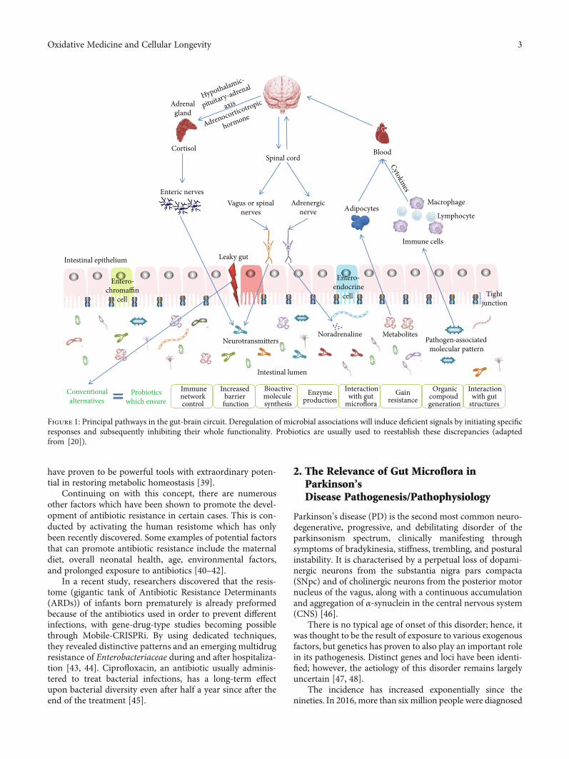
Unfortunately, the relationship between GI flora and thebrain is insufficiently understood. The influence that thegut flora exerts on the local organs in the immediate vicin-ity, as well as on those located distally, is taking placethrough a variety of routes, for example, immune, enteric,and neural pathways. Thus, the gut-brain axis (GBA)could be defined as a dense network formed by cells fromthe enteric, peripheral, and central nervous system in associ-ation with the hypothalamic-pituitary-adrenal (HPA) axis(Figure 1) [20–22].
Historically, there has been a tendency to believe thateach one of us possesses the same gut microflora, a theorythat has been proven to be only partially true. There are inter-individual and intergenerational variations in the micro-biome which is influenced by the changing environment,nutritional factors, and genetic contributions. The literaturesuggests numerous clues that sustain this hypothesis; for
example, not even twins harbour the same composition ofmicroflora [23]. The similarities are even less prevalentamongst siblings, but nonetheless, in very small percentages,there are similarities in flora composition even in unrelatedindividuals [24, 25].
One of the most important factors in shaping the normalneonatal enteric colonization of microflora is the deliverymethod. Although the gut of an unborn baby is theoreticallysterile in the mother’s womb, the development of the neona-tal microbiota is initiated by the neonate transversing thebirth channel, where there is a subsequent exposure to a largeamount of maternal microbial communities which shapesthe microbiota of the infant. By analysing different cohortsvia computational research, a set of specific bacterial geneswere classified in limited habitats (e.g., placenta), evidencethat is sustained after the analysis of meconium sampleswhere it was revealed that colonization may be initiatedin utero [26–29].
On the other hand, a recent publication contradicts thesefindings. The study design was aimed at determining whetherpreeclampsia, small for gestational age (SGA), and spontane-ous preterm birth (PTB) were correlated with the existence ofbacterial signatures in the placenta. Authors concluded thatthe placenta is devoid of such populations but neverthelessprovides favourable conditions for pathogenic species suchas Streptococcus agalactiae; this species is prevalent in almost5% of the total samples collected before the beginning ofprocedures [30].
There are a large amount of species facultatively anaer-obic (enterobacteria and enterococci) that are found in theGI of children, with their whole existence depending onthe dietary supply, thus creating propitious conditions forthe evolution of anaerobic microbes. However, the child-hood microbiota may also be influenced by other environ-mental factors, such as exposure to healthcare facilitiesand other children culminating in complex and dynamicmicrobiota [31, 32].
In this context, natural birth is supported in order tomaintain the balance between beneficial and harmful micro-organisms. However, in the last few decades, the number ofcaesarean sections (C-section) has increased dramaticallywhich is worrying. Women are not adequately informedabout the risk to the baby through a C-section deliverymethod, possibly predisposing the infant to a series of epide-miological illnesses. Obesity, allergies, anaphylactic reactionsto asthma, and autoimmune diseases are a few examples ofconditions which may be influenced by the commensalbacteria and thus the delivery method. It is evident thatC-section indirectly promotes various diseases throughthe effect of the neonatal microbiota [33, 34].
The delivery mode creates a disbalance amongst gram-positive and gram-negative species, which can be beneficialto certain species such as Lactobacillus, Bifidobacterium,Eubacterium, and Bacteroides, to the detriment of thosepathogens like Clostridium, Campylobacter, Staphylococcus,Shigella, Shiga toxin-producing Escherichia coli, Acinetobac-ter, and Escherichia coli [35–38]. Breastfeeding has thepotential to reestablish this balance, alongside conventionalalternatives, for example, syn-, pre-, and probiotics, which
2 Oxidative Medicine and Cellular Longevity
have proven to be powerful tools with extraordinary poten-tial in restoring metabolic homeostasis [39].
Continuing on with this concept, there are numerousother factors which have been shown to promote the devel-opment of antibiotic resistance in certain cases. This is con-ducted by activating the human resistome which has onlybeen recently discovered. Some examples of potential factorsthat can promote antibiotic resistance include the maternaldiet, overall neonatal health, age, environmental factors,and prolonged exposure to antibiotics [40–42].
In a recent study, researchers discovered that the resis-tome (gigantic tank of Antibiotic Resistance Determinants(ARDs)) of infants born prematurely is already preformedbecause of the antibiotics used in order to prevent differentinfections, with gene-drug-type studies becoming possiblethrough Mobile-CRISPRi. By using dedicated techniques,they revealed distinctive patterns and an emerging multidrugresistance of Enterobacteriaceae during and after hospitaliza-tion [43, 44]. Ciprofloxacin, an antibiotic usually adminis-tered to treat bacterial infections, has a long-term effectupon bacterial diversity even after half a year since after theend of the treatment [45].
2. The Relevance of Gut Microflora inParkinson’sDisease Pathogenesis/Pathophysiology
Parkinson’s disease (PD) is the second most common neuro-degenerative, progressive, and debilitating disorder of theparkinsonism spectrum, clinically manifesting throughsymptoms of bradykinesia, stiffness, trembling, and posturalinstability. It is characterised by a perpetual loss of dopami-nergic neurons from the substantia nigra pars compacta(SNpc) and of cholinergic neurons from the posterior motornucleus of the vagus, along with a continuous accumulationand aggregation of α-synuclein in the central nervous system(CNS) [46].
There is no typical age of onset of this disorder; hence, itwas thought to be the result of exposure to various exogenousfactors, but genetics has proven to also play an important rolein its pathogenesis. Distinct genes and loci have been identi-fied; however, the aetiology of this disorder remains largelyuncertain [47, 48].
The incidence has increased exponentially since thenineties. In 2016, more than six million people were diagnosed
Hypothalamic-
pituitary-adrenal
axis
Adrenocorticotropic
hormone
Cytokines
Cortisol
Enteric nerves
Intestinal lumen
Adrenergic nerve
Spinal cordBlood
Adipocytes
Immune cells
Lymphocyte
Macrophage
Entero-chromaffin
cellTight
junction
Leaky gutIntestinal epithelium
Entero-endocrine
cell
NeurotransmittersMetabolites
Adrenal gland
Noradrenaline
Vagus or spinal nerves
Conventional alternatives
Enzyme production
Increased barrier function
Interaction with gut
microflora
Bioactive moleculesynthesis
Immune network control
Gain resistance
Organic compoudgeneration
Interaction with gut
structuresProbiotics
which ensure
Pathogen-associated molecular pattern
Figure 1: Principal pathways in the gut-brain circuit. Deregulation of microbial associations will induce deficient signals by initiating specificresponses and subsequently inhibiting their whole functionality. Probiotics are usually used to reestablish these discrepancies (adaptedfrom [20]).
3Oxidative Medicine and Cellular Longevity
with PD, and it became the second most commonneurodegenerative disorder worldwide [49].
A relationship amongst enteric neurons and gut micro-flora has been reported due to new discoveries around toll-like receptors, proteins with a key role in the innate immunesystem [50, 51], and their modulation potential upon theHPA axis [52], followed by a further production of chemicalsinvolved in the brain’s optimal functioning [53]. There is newevidence concerning the importance of toll-like receptor 4 inmediating neuroinflammatory states, resulting in the disrup-tion of intestinal flora, while rotenone KO-treated mice had areduction of specific symptomatology [54]. In rotenonemodels, chronic stress induces a deregulation of HPA whichmay culminate in dysbacteriosis, characterised by a signifi-cant reduction in the number of species belonging to thegenus Bifidobacterium, to the detriment of Escherichia coli.Prolonged exposure leads to an increased intestinal per-meability which creates a “leaky gut,” dysosmia, and colitisby inducing specific neuroanatomical and neurochemicalchanges [55–59].
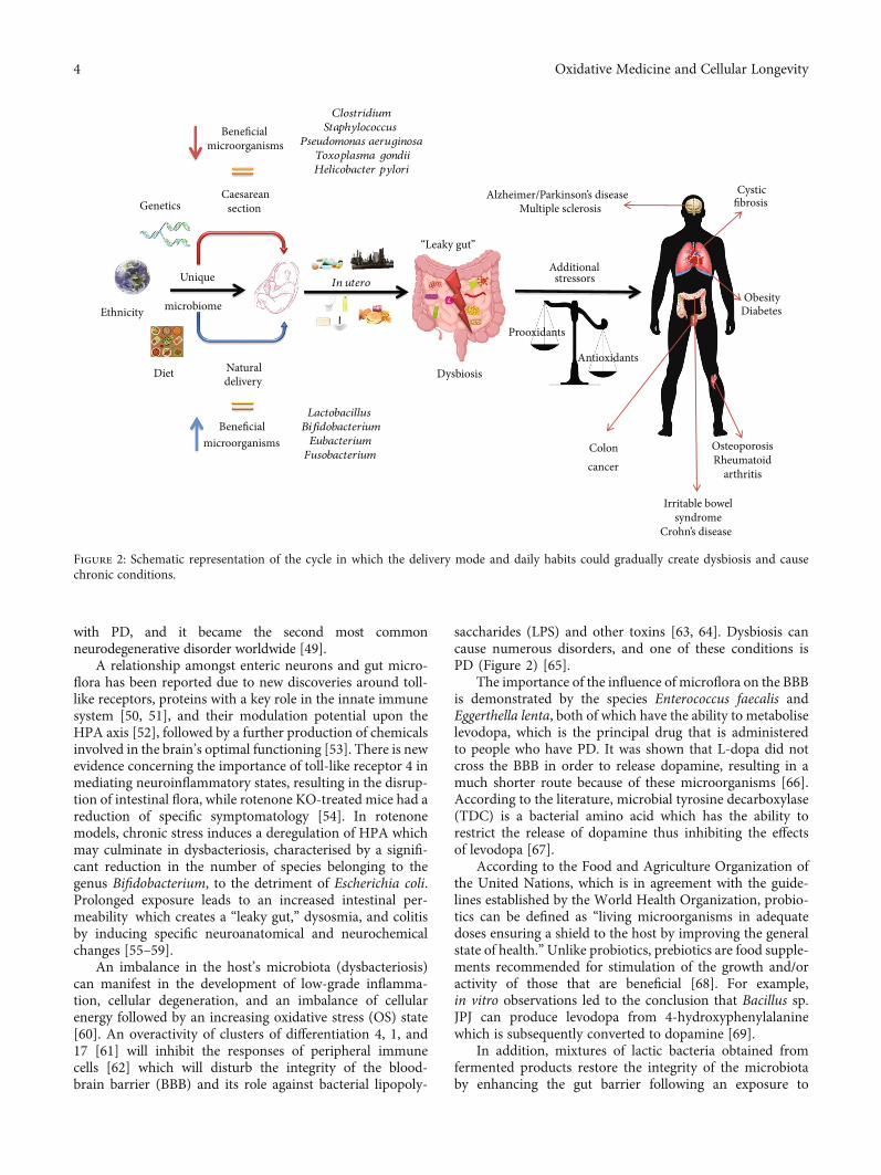
An imbalance in the host’s microbiota (dysbacteriosis)can manifest in the development of low-grade inflamma-tion, cellular degeneration, and an imbalance of cellularenergy followed by an increasing oxidative stress (OS) state[60]. An overactivity of clusters of differentiation 4, 1, and17 [61] will inhibit the responses of peripheral immunecells [62] which will disturb the integrity of the blood-brain barrier (BBB) and its role against bacterial lipopoly-
saccharides (LPS) and other toxins [63, 64]. Dysbiosis cancause numerous disorders, and one of these conditions isPD (Figure 2) [65].
The importance of the influence of microflora on the BBBis demonstrated by the species Enterococcus faecalis andEggerthella lenta, both of which have the ability to metaboliselevodopa, which is the principal drug that is administeredto people who have PD. It was shown that L-dopa did notcross the BBB in order to release dopamine, resulting in amuch shorter route because of these microorganisms [66].According to the literature, microbial tyrosine decarboxylase(TDC) is a bacterial amino acid which has the ability torestrict the release of dopamine thus inhibiting the effectsof levodopa [67].
According to the Food and Agriculture Organization ofthe United Nations, which is in agreement with the guide-lines established by the World Health Organization, probio-tics can be defined as “living microorganisms in adequatedoses ensuring a shield to the host by improving the generalstate of health.”Unlike probiotics, prebiotics are food supple-ments recommended for stimulation of the growth and/oractivity of those that are beneficial [68]. For example,in vitro observations led to the conclusion that Bacillus sp.JPJ can produce levodopa from 4-hydroxyphenylalaninewhich is subsequently converted to dopamine [69].
In addition, mixtures of lactic bacteria obtained fromfermented products restore the integrity of the microbiotaby enhancing the gut barrier following an exposure to
Alzheimer/Parkinson’s diseaseMultiple sclerosis
ObesityDiabetes
OsteoporosisRheumatoid
arthritis
Cystic fibrosis
Colon cancer
Dysbiosis
In uteroUnique
microbiome
Genetics
Ethnicity
Diet
Caesareansection
Beneficial microorganisms
Natural delivery
Beneficial microorganisms
ClostridiumStaphylococcus
Pseudomonas aeruginosaToxoplasma gondiiHelicobacter pylori
LactobacillusBifidobacteriumEubacteriumFusobacterium
Additional stressors
“Leaky gut”
Antioxidants
Prooxidants
Irritable bowelsyndrome
Crohn’s disease
Figure 2: Schematic representation of the cycle in which the delivery mode and daily habits could gradually create dysbiosis and causechronic conditions.
4 Oxidative Medicine and Cellular Longevity
antibiotics in certain intervals [70]. There are novel tech-niques which facilitate the manipulation of the gut microfloraby suppressing pathogens in the epithelium and intestines, inorder to regulate the activity of immune cells [71]. Finally,synbiotics are a mixture of the two categories mentioned ear-lier, with the main aim of increasing the duration of life andsettlement of those already existing in the GI [72].
Faecal Microbiota Transplantation (FMT) is a treatmentthat facilitates the reconstruction of the gut flora wherebyfaecal matter from a healthy donor is donated to a patientthereby changing the underlying microflora. This treatmentis used in the treatment of resistant Clostridium difficile infec-tions. Microbial Transfer Therapy (MTT) is a similar proto-col to FMT, both of them demonstrating their potentials intreating metabolic deficiencies [73, 74].
The differences in the clinical manifestation of PD meanthat the management needs to be individualised. For exam-ple, chronic idiopathic constipation (CIC) is encountered inPD subjects and can be associated with anorectal and colonicdysmotility [75]. FMT intervention caused motor impair-ment in mice and humans, promoting a reduction in Lach-nospiraceae and Ruminococcaceae strains [76]. In progeroidmice, however, FTM reduced both morbidity and mortality.These observations can also be applied in human patients,where a reduction in Proteobacteria in parallel with increasedVerrucomicrobia concentrations was documented [77].
The discovery of bacteriophages with more recent clus-tered regularly interspaced short palindromic repeats(CRISPR) and the associated nuclease 9 has led to an arrayof possibilities to manipulate the human microbiome[78, 79]. This technique started from the discovery of foreignsequences of DNA from viruses that were incorporated intobacteria. Those sequences confer immunity against futureinteractions with viruses and have shown extraordinarypotential in manipulating the human genome. This tech-nique is used to influence the genome of those resistant toantibiotics [80] or metabolise various drugs with the aim ofintegrating this system into conventional products.
Surveys published over the years support the concept ofthe gut-brain network and vice versa; some of them regardinga better understanding of influence exerted by the micro-biome on PD patients are summarised in Table 1.
3. Gut Infections as a Promoter inParkinson’s Disease
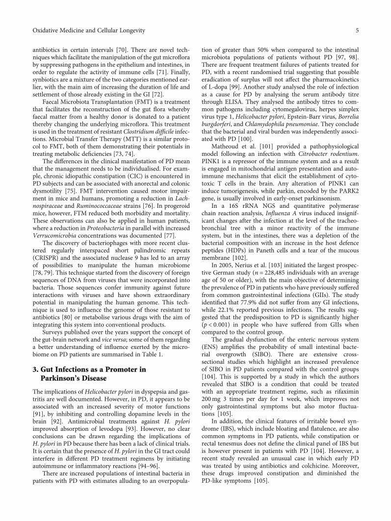
The implications of Helicobacter pylori in dyspepsia and gas-tritis are well documented. However, in PD, it appears to beassociated with an increased severity of motor functions[91], by inhibiting and controlling dopamine levels in thebrain [92]. Antimicrobial treatments against H. pyloriimproved absorption of levodopa [93]. However, no clearconclusions can be drawn regarding the implications ofH. pylori in PD because there has been a lack of clinical trials.It is certain that the presence ofH. pylori in the GI tract couldinterfere in different PD treatment regimens by initiatingautoimmune or inflammatory reactions [94–96].
There are increased populations of intestinal bacteria inpatients with PD with estimates alluding to an overpopula-
tion of greater than 50% when compared to the intestinalmicrobiota populations of patients without PD [97, 98].There are frequent treatment failures of patients treated forPD, with a recent randomised trial suggesting that possibleeradication of surplus will not affect the pharmacokineticsof L-dopa [99]. Another study analysed the role of infectionas a cause for PD by analysing the serum antibody titrethrough ELISA. They analysed the antibody titres to com-mon pathogens including cytomegalovirus, herpes simplexvirus type 1, Helicobacter pylori, Epstein-Barr virus, Borreliaburgdorferi, and Chlamydophila pneumoniae. They concludethat the bacterial and viral burden was independently associ-ated with PD [100].
Matheoud et al. [101] provided a pathophysiologicalmodel following an infection with Citrobacter rodentium.PINK1 is a repressor of the immune system and as a resultis engaged in mitochondrial antigen presentation and auto-immune mechanisms that elicit the establishment of cyto-toxic T cells in the brain. Any alteration of PINK1 caninduce tumorigenesis, while parkin, encoded by the PARK2gene, is usually involved in early-onset parkinsonism.
In a 16S rRNA NGS and quantitative polymerasechain reaction analysis, Influenza A virus induced insignif-icant changes after the infection at the level of the tracheo-bronchial tree with a minor reactivity of the immunesystem, but in the intestines, there was a depletion of thebacterial composition with an increase in the host defencepeptides (HDPs) in Paneth cells and a tear of the mucousmembrane [102].
In 2005, Nerius et al. [103] initiated the largest prospec-tive German study (n = 228,485 individuals with an averageage of 50 or older), with the main objective of determiningthe prevalence of PD in patients who have previously sufferedfrom common gastrointestinal infections (GIIs). The studyidentified that 77.9% did not suffer from any GI infections,while 22.1% reported previous infections. The results sug-gested that the predisposition to PD is significantly higher(p < 0:001) in people who have suffered from GIIs whencompared to the control group.
The gradual dysfunction of the enteric nervous system(ENS) amplifies the probability of small intestinal bacte-rial overgrowth (SIBO). There are extensive cross-sectional studies which highlight an increased prevalenceof SIBO in PD patients compared with the control groups[104]. This is supported by a study in which the authorsrevealed that SIBO is a condition that could be treatedwith an appropriate treatment regime, such as rifaximin200mg 3 times per day for 1 week, which improves notonly gastrointestinal symptoms but also motor fluctua-tions [105].
In addition, the clinical features of irritable bowel syn-drome (IBS), which include bloating and flatulence, are alsocommon symptoms in PD patients, while constipation orrectal tenesmus does not define the clinical panel of IBS butis however present in patients with PD [104]. However, arecent study revealed an unusual case in which early PDwas treated by using antibiotics and colchicine. Moreover,these drugs improved constipation and diminished thePD-like symptoms [105].
5Oxidative Medicine and Cellular Longevity
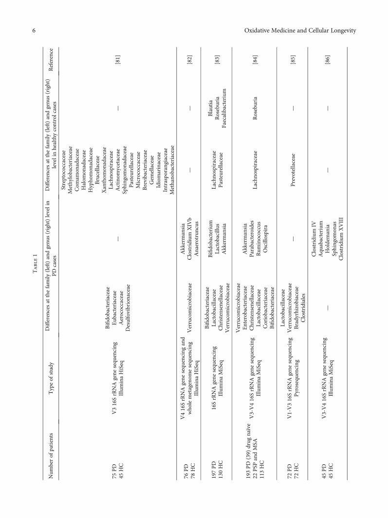
Table1
Num
berof
patients
Typeof
stud
yDifferencesat
thefamily
(left)andgenu
s(right)levelin
PD
cases
Differencesat
thefamily
(left)andgenu
s(right)
levelinhealthycontrolcases
Reference
75PD
45HC
V316SrRNAgene
sequ
encing
IlluminaHiSeq
Bifido
bacteriaceae
Eub
acteriaceae
Aerococcaceae
Desulfovibrionaceae
—
Streptococcaceae
Methylobacteriaceae
Com
amon
adaceae
Halom
onadaceae
Hypho
mon
adaceae
Brucellaceae
Xanthom
onadaceae
Lachno
spiraceae
Actinom
ycetaceae
Sphingom
onadaceae
Pasteurellaceae
Micrococcaceae
Brevibacteriaceae
Gem
ellaceae
Idiomarinaceae
Intraspo
rangiaceae
Methano
bacteriaceae
—[81]
76PD
78HC
V416SrRNAgene
sequ
encing
and
who
lemetagenom
esequ
encing
IlluminaHiSeq
Verrucomicrobiaceae
Akkermansia
Clostridium
XIV
bAnaerotruncus
——
[82]
197PD
130HC
16SrRNAgene
sequ
encing
IlluminaMiSeq
Bifido
bacteriaceae
Lactobacillaceae
Christensenellaceae
Verrucomicrobiaceae
Bifido
bacterium
Lactobacillus
Akkermansia
Lachno
spiraceae
Pasteurellaceae
Blautia
Roseburia
Faecalibacterium
[83]
193PD
(39)
drug
naïve
22PSP
andMSA
113HC
V3-V416SrRNAgene
sequ
encing
IlluminaMiSeq
Verrucomicrobiaceae
Enterobacteriaceae
Christensenellaceae
Lactobacillaceae
Coriobacteriaceae
Bifido
bacteriaceae
Akkermansia
Parabacteroides
Rum
inococcus
Oscillospira
Lachno
spiraceae
Roseburia
[84]
72PD
72HC
V1-V316SrRNAgene
sequ
encing
Pyrosequencing
Lactobacillaceae
Verrucomicrobiaceae
Bradyrhizobiaceae
Clostridiales
—Prevotellaceae
—[85]
45PD
45HC
V3-V416SrRNAgene
sequ
encing
IlluminaMiSeq
—
Clostridium
IVAqu
abacterium
Holdemania
Sphingom
onas
Clostridium
XVIII
——
[86]
6 Oxidative Medicine and Cellular Longevity
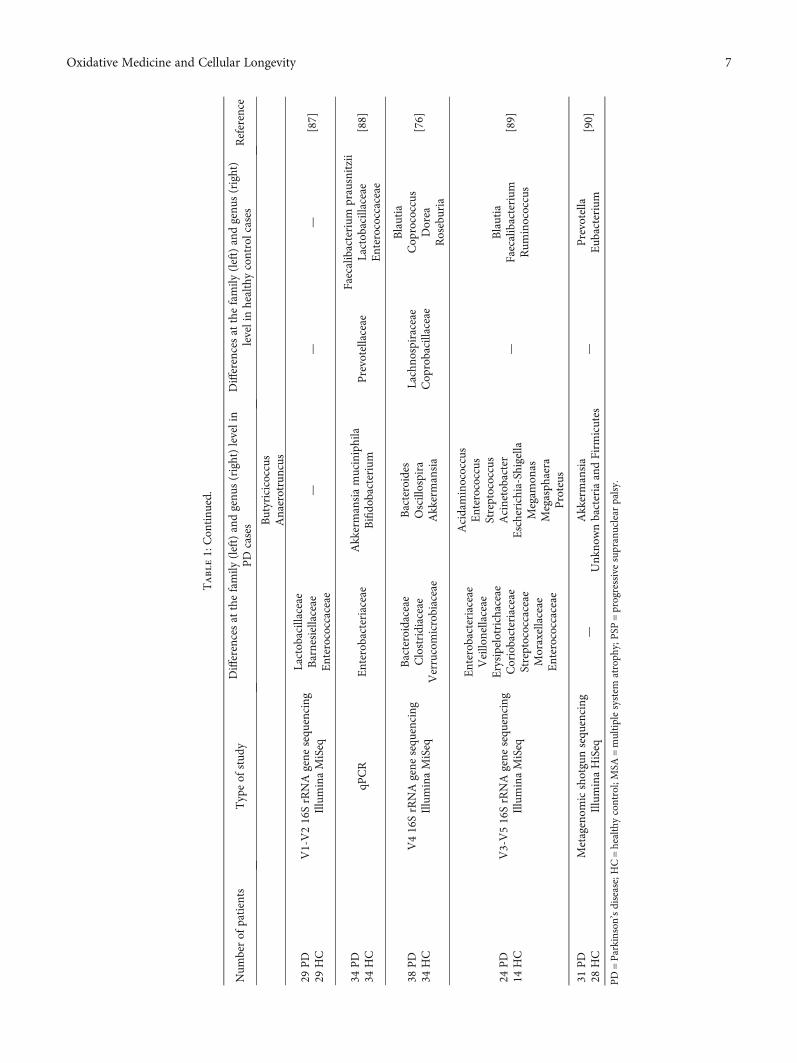
Table1:Con
tinu
ed.
Num
berof
patients
Typeof
stud
yDifferencesat
thefamily
(left)andgenu
s(right)levelin
PD
cases
Differencesat
thefamily
(left)andgenu
s(right)
levelinhealthycontrolcases
Reference
Butyricicoccus
Anaerotruncus
29PD
29HC
V1-V216SrRNAgene
sequ
encing
IlluminaMiSeq
Lactobacillaceae
Barnesiellaceae
Enterococcaceae
——
—[87]
34PD
34HC
qPCR
Enterobacteriaceae
Akkermansiamuciniphila
Bifido
bacterium
Prevotellaceae
Faecalibacterium
prausnitzii
Lactobacillaceae
Enterococcaceae
[88]
38PD
34HC
V416SrRNAgene
sequ
encing
IlluminaMiSeq
Bacteroidaceae
Clostridiaceae
Verrucomicrobiaceae
Bacteroides
Oscillospira
Akkermansia
Lachno
spiraceae
Cop
robacillaceae
Blautia
Cop
rococcus
Dorea
Roseburia
[76]
24PD
14HC
V3-V516SrRNAgene
sequ
encing
IlluminaMiSeq
Enterobacteriaceae
Veillonellaceae
Erysipelotrichaceae
Coriobacteriaceae
Streptococcaceae
Moraxellaceae
Enterococcaceae
Acidaminococcus
Enterococcus
Streptococcus
Acinetobacter
Escherichia-Shigella
Megam
onas
Megasph
aera
Proteus
—Blautia
Faecalibacterium
Rum
inococcus
[89]
31PD
28HC
Metagenom
icshotgunsequ
encing
IlluminaHiSeq
—Akkermansia
Unk
nownbacteriaandFirm
icutes
—Prevotella
Eub
acterium
[90]
PD=Parkinson
’sdisease;HC=healthycontrol;MSA
=multiplesystem
atroph
y;PSP
=progressivesupranuclear
palsy.
7Oxidative Medicine and Cellular Longevity
4. Gastrointestinal Deficiencies? TheLinks between the Gut Communities’Structure and Parkinson’s Disease
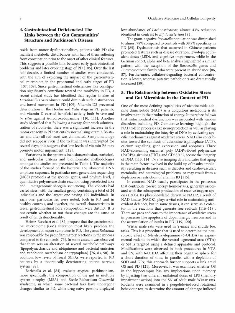
Aside from motor dysfunctionalities, patients with PD alsomanifest metabolic disturbances with half of them sufferingfrom constipation prior to the onset of other clinical features.This suggests a possible link between early gastrointestinalproblems and later evolving stage of PD [106]. Over the lasthalf decade, a limited number of studies were conducted,with the aim of exploring the impact of the gastrointesti-nal microbiota in the prodromal and early stages of PD[107, 108]. Since gastrointestinal deficiencies like constipa-tion significantly contribute toward the morbidity in PD, arecent clinical study has identified that regular intakes ofLactobacillus casei Shirota could diminish such disturbancesand bowel movement in PD [109]. Vitamin D3 preventeddeterioration in the Hoehn and Yahr stage in PD patients,and vitamin D exerted beneficial activity both in vivo andin vitro against 6-hydroxydopamine [110, 111]. Anotherstudy identified that following a twenty-four-week adminis-tration of riboflavin, there was a significant increase in themotor capacity in PD patients by normalising vitamin B6 sta-tus and after all red meat was eliminated. Symptomatologydid not reappear even if the treatment was interrupted forseveral days; this suggests that low levels of vitamin B6 maypromote motor impairment [112].
Variations in the patient’s inclusion/exclusion, statistical,and molecular criteria and bioinformatic methodologiesamongst the studies are presented in Table 1. The majorityof the studies focused on the bacterial 16S ribosomal DNAamplicon sequence, in particular next-generation sequencing(NGS) protocols at the species, genus, and phylum level, 1quantitative polymerase chain reaction using preselected taxaand 1 metagenomic shotgun sequencing. The cohorts hadvaried sizes, with the smallest group containing a total of 24individuals and the largest one having 197 individuals. Ineach one, particularities were noted, both in PD and inhealthy controls, and together, the overall characteristics infaecal gastrointestinal flora composition were distinct. It isnot certain whether or not these changes are the cause orresult of GI dysfunctionality.
Heintz-Buschart et al. [82] propose that the gastrointesti-nal microbiome (GM) alteration most likely precedes thedevelopment of motor symptoms in PD. The genus Ralstoniawas responsible for proinflammatory reactions in the mucosacompared to the controls [76]. In some cases, it was observedthat there was an alteration of several metabolic pathways(lipopolysaccharide and ubiquinone and bacterial emissionand xenobiotic metabolism or tryptophan) [76, 83, 90]. Inaddition, low levels of faecal SCFAs were reported in PDpatients by a theoretically deteriorating enteric nervoussystem [88].
Barichella et al. [84] evaluate atypical parkinsonism,more specifically, the composition of the gut in multiplesystem atrophy (MSA) and Steele-Richardson-Olszewskisyndrome, in which some bacterial taxa have undergonechanges similar to PD, while drug-naïve persons displayed
low abundance of Lachnospiraceae, almost 43% reductionidentified in contrast to Bifidobacterium [81].
The gram-negative Prevotella population was diminishedto almost 78% compared to controls with 38.9% specificity inPD [85]. Dysbacteriosis that occurred in Chinese patientspromoted features such as disease duration, levodopa equiv-alent doses (LED), and cognitive impairment, while in theGerman cohort, alpha and beta analysis highlighted a similarpattern with the exception of the Barnesiella genus andEnterococcaceae family who were present in abundance [86,87]. Furthermore, cellulose-degrading bacterial concentra-tion is lower, whereas putative pathobionts are dramaticallyincreased [89].
5. The Relationship between Oxidative Stressand Gut Microbiota in the Context of PD
One of the most defining capabilities of nicotinamide ade-nine dinucleotide (NAD) as a ubiquitous metabolite is itsinvolvement in the production of energy. It therefore followsthat mitochondrial dysfunction was associated with variousdisorders, including PD. Cumulative learnings highlight theNAD role in processes like neuroprotection as well as playinga role in maintaining the integrity of DNA by activating spe-cific mechanisms against oxidative stress. NAD also contrib-utes toward the synthesis of adenosine triphosphate (ATP),calcium signalling, gene expression, and apoptosis. ThreeNAD-consuming enzymes, poly (ADP-ribose) polymerase(PARPs), sirtuins (SIRT), and CD38/157, secure the integrityof DNA [113, 114]. In vivo imaging data indicates that agingis the main factor involved in the build-up of insults, implic-itly resulting in diseases such as diabetes and cardiovascular,metabolic, and neurological problems, or may result from adepletion or restriction of vitamin B3 [115].
In contrast, NAD usually participates in the processesthat contribute toward energy homeostasis, generally associ-ated with the subsequent production of reactive oxygen spe-cies (ROS). Its phosphorylated derivative NADP, a result ofNAD kinase (NADK), plays a vital role in maintaining anti-oxidant defences, but in some tissues, it can serve as a cofac-tor in the reactions that generate free radicals [116–118].There are pros and cons to the importance of oxidative stressin processes like apoptosis of dopaminergic neurons and inthe accumulation of insults in PD [119, 120].
Wistar male rats were used in Y-maze and shuttle boxtasks. This is a procedure that is used to determine the neu-rotoxic effect of 6-hydroxydopamine (6-OHDA) in experi-mental rodents in which the ventral tegmental area (VTA)or SN is targeted using a defined apparatus and protocol.Modifications were observed in both procedures in VTAand SN, with 6-OHDA affecting their cognitive sphere fora short duration of time, in parallel with a depletion ofSOD and GPx; this approach further supports a link amidOS and PD [121]. Moreover, it was examined whether OSin the hippocampus has any implications upon memoryby injecting two different unilateral doses of LPS (memoryimpairment action) into the SN of adult male Wistar rats.Rodents were examined in a pergolide-induced rotationalbehaviour test to determine the amount of damage inflicted
8 Oxidative Medicine and Cellular Longevity
upon nigrostriatal dopaminergic neurons. In the hippocam-pus of LPS-treated rats, levels of malondialdehyde were sig-nificantly higher compared with those in controls whichwere measured in Y-maze (within was noted correlationsbetween behavioural deficiencies as indexes for OS) andradial arm maze tasks [122]. In a quite similar manner,all of the aforementioned compounds and tests were com-bined into one study, with behavioural deficiencies beingmore pronounced only in LPS- and LPS+6-OHDA-treatedrats [123].
Even though our cells are equipped with mechanismswhich counteract the accumulation of insults, the fact thatwe are strictly aerobic organisms can have severe repercus-sions on the state of health and the reduction of molecularoxygen to O2 and H2O during the cellular respiration processthat leads to the synthesis of adenosine triphosphate (ATP).This promotes the production of free radicals, with 20% ofthe total oxygen supply consumed by the brain being con-verted into ROS. These reactive species generated by nicotin-amide adenine dinucleotide phosphate oxidase (NOX) andnitric oxide synthase (NOS) perform functions like resistanceagainst infections and the activation of various signallingpathways [124, 125].
NADPH is in excess compared to NADP whose ratiois much lower than 1; in this context, apart from theNAD/NADH ratio, cells maintain two opposite redox pairswith NADP/NADPH in a continuous reductive state, andthis redox stability is compatible with the NADPH role inbiosynthesis and detoxification with oxygen. NADPH is akey reducing substrate for transforming oxidised glutathioneinto reduced glutathione as a protective element against tox-icity of ROS. An increased ratio of NADH/NAD is associatedwith a petulant production of reactive oxygen species and theinhibition of α-ketoglutarate dehydrogenase due to a mito-chondrial dysfunction and the inability of antioxidantenzymes such as superoxide dismutase (SOD) and glutathi-one peroxidase (GPx) to maintain balance [126, 127].
Besides ROS, cumulative surveys highlight the impor-tance of reactive nitrogen species (RNS), entities generatedas a result of interactions between superoxide (O2
-) and nitricoxide (NO), resulting in large amounts of peroxynitrite. NO,produced by NOS, commonly exists under three isoforms,known as endothelial NOS (eNOS), neuronal NOS (nNOS),and inducible NOS (iNOS), which can be found in glial cells[128–131]. Peroxynitrite has the ability to induce DNA frag-mentation and lipid peroxidation because of its oxidativestructure and even dose-dependent impairment indepen-dently of dopamine normal cycle and death [129, 130]. In situhybridisation and immunohistochemistry of postmortembrain tissue revealed high expression of iNOS and nNOS inPD patients which further highlights the role of NO. In thesubstantia nigra, the gliosis is linked to an upregulation ofthe iNOS, while it is linked to the inhibition of nNOS againstcytotoxicity of MPTP (neurotoxin). Neuronal death stillremains an enigma, but with the current evidence, it can beconcluded that oxidative stress and mitochondrial dysfunc-tions are interconnected, especially at the level of respiratorychain, highlighted by a petulant production of reactive oxy-gen species which leads finally to apoptosis in PD [132, 133].
Dopamine (DA) (excitatory and inhibitory role of synap-tic transmission) as a construct produced from DA neuronscan in turn be a source of OS due to its unstable nature selec-tivity for SNpc (substantia nigra pars compacta) whichundergoes self-oxidation in order to form dopamine qui-nones and free radicals, reactions catalysed by oxygen,enzymes, or metals [134, 135]. Interestingly, with an exces-sive amount of cytosolic DA outside of the synaptic vesicles,this neurotransmitter is easily metabolised by monoamineoxidase (MAO), a participant in the regulation of DA levelsby monoamine oxidase A (MAO-A), localised in catechol-aminergic neurons [136].
Alternatively, in degeneration that occurs in PD or aging,monoamine oxidase B (MAO-B) becomes the predominantenzyme that metabolises DA and can be found in glial cellsand then taken up by astrocytes [137]. In transgenic mice,the wilful induction of this enzyme in astrocytes had as resulta selective and progressive loss of nigral dopaminergic neu-rons [138]. It has been shown that DA quinones have theability to shape proteins which subsequently may be involvedin PD pathophysiology, for example, α-synuclein, parkin,protein deglycase DJ-1, and ubiquitin carboxy-terminalhydrolase L1, in α-synuclein DA quinone modifying itsmonomer by promoting the conversion to a cytotoxic proto-fibril form [139]. These quinones can be oxidised into amino-chrome, whose redox cycle capacity ultimately causes thedepletion of NADPH and the generation of superoxide. Thisis subsequently transformed into neuromelanin (brain pig-ment that might play a role in neurodegeneration), occurringwithin the SNpc [140, 141]. Taking into account the cir-cuit of PD and that the dorsal motor nucleus of the vagusnerve (DMnX) is the primary hive cluster to α-synuclein,in vivo models provide additional clues regarding the partic-ipation of oxidative stress into the spreading of a “mutated”α-synuclein within and outside the CNS by promoting celland protein interrelations. They show that cholinergic neu-rons are very sensitive to the accumulation of reactive oxy-gen species (ROS) [142]. Accumulation of α-synuclein,encoded by the SNCA gene, is also a risk factor for PD, con-taining inclusions in the enteric nervous system and poste-rior motor nucleus of the vagus [143, 144], determiningoverinflammatory reactions, intestinal permeability, andoxidative stress [145, 146].
Following the analysis of 117 tissue samples and 161 fromcontrols, biopsies revealed an accumulation of α-synuclein atthe level of the various oesophageal tunics and ganglia, withimplications of this protein usually involved in neurotrans-mitter release being much more complex [147, 148]. Also,the 465-residue E3 ubiquitin ligase parkin is covalently mod-ified by dopamine becoming insoluble, leading to ubiquitinE2 ligase inactivation, in the SN, with catechol-“mutated”parkin being observed in patients with PD, but not in otherregions of the brain [149].
The modification of ubiquitin carboxy-terminal hydro-lase L1 (UCH-L1) and protein deglycase DJ-1 by dopaminequinones was observed in dopaminergic cells, but also inmitochondria, and because of the cysteine residue they pos-sess, quinones are responsible for the inactivation of theseenzymes [150]. In a transgenic murine model ((Thy-1)-
9Oxidative Medicine and Cellular Longevity
h[A30P]-α-synuclein) with SOD2 haplodeficiency, at 1 yearand 4 months, exhibiting significant features of synucleino-pathy compared full SOD2 control, the results indicate thatan elevated level of OS could mediate the progression of PD[142]. Kim et al. [151] tested Braak’s theory that α-syn couldspread into the brain from the gut via the vagus nerve. Theyinjected preformed α-syn fibrils in a novel gut-to-brainmouse model and found that α-syn is dispersed first intothe posterior motor nucleus of the vagus and then in caudalportions of the rhombencephalon. Furthermore, specificsymptoms were present temporarily, but truncal vagotomyand deficiency of α-syn prevented its further spreading.
In the model gut bacterium Enterococcus durans (MTCC3031), oxidative stress induced by C6H4(CO)2C2H(CH3) andH2O2 deregulates the redox ratio (55% for menadione and28% for H2O2) by decreasing folate synthesis of these gram-positive bacteria, known to play an important role againstcolorectal cancer [152]. By measuring the amount of hydro-gen production for both gram-negative and gram-positivebacteria, it is speculated that the bacteria participate in theprogression of PD [153].
Pathogen-associated molecular patterns (PAMPs) areconserved motifs that activate pattern recognition receptors(PRRs) found on the surface of diverse pathogens and induceROS. Lipopolysaccharides (LPS) produced by bacteria areusually recognised by these PRRs which generate down-stream signals and activate the NF-κB pathway and induceinflammatory responses [154]. In the case of commensalbacteria, the lipopolysaccharides produce and release for-myl peptides which are recognised by formyl peptidereceptors (FPRs), a class that belongs to the G protein-coupled receptors; many signalling cascades utilise thesereceptors for converting a large variety of external stimuli(agonist neurotransmitters, ions, and hormones) into intra-cellular responses, perceiving and stimulating ROS produc-tion [155]. Due to the fact that our intestines house distinctcell types by initiating specific responses, ROS produced bymucosa-resident cells or by recruiting innate immune cellsare crucial for an optimal antimicrobial activity. An unbal-anced ROS synthesis through activating certain gene variantsand upregulation of oxidases or of a mitochondrial dysfunc-tion is associated with Crohn’s disease or ulcerative colitis. Inthis way, the abnormal profiles of intestinal flora may lead toinflammation of the intestines often seen in people withinflammatory bowel disease (IBD) [156, 157].
6. Conclusions
It can be concluded that there are numerous factors (antibi-otics, diet, birth mode, or stress) which gradually promotethe onset of enteric dysbacteriosis which may trigger disor-ders of the CNS. There are relatively few studies thathighlight the relationship between intestinal flora and PD;researchers argue that these limitations will be overcomedue to the fact that the human microbiome is currently themain barrier to the emergence of personalised medicine. Oxi-dative stress is an integrative component to the function of allorganisms, regardless of the current status (homeostasis ordisease). This paper summarised most of the existing evi-
dence in the literature, and it can be concluded that the widerimplications of the human microbiome are complex andrequires further research to improve the current understand-ing of the mechanisms underlying neurodegenerative disor-ders like Parkinson’s disease.
Conflicts of Interest
All authors declare that they have no conflict of interest todisclose, except for Alin Ciobica which is supported by theresearch grant mentioned below.
Acknowledgments
AC is supported by a research grant for Young Teams offeredby UEFISCDI Romania (no. PN-IIIP1-1.1-TE-2016-1210,contract no. 58 from 02/05/2018).
References
[1] E. S. Lander, L. M. Linton, B. Birren et al., “Initial sequencingand analysis of the human genome,” Nature, vol. 409,no. 6822, pp. 860–921, 2001.
[2] P. J. Turnbaugh, R. E. Ley, M. Hamady, C. M. Fraser-Liggett,R. Knight, and J. I. Gordon, “The human microbiome pro-ject,” Nature, vol. 449, no. 7164, pp. 804–810, 2007.
[3] R. Sender, S. Fuchs, and R. Milo, “Revised estimates for thenumber of human and bacteria cells in the body,” PLoS Biol-ogy, vol. 14, no. 8, article e1002533, 2016.
[4] F. Bäckhed, R. E. Ley, J. L. Sonnenburg, D. A. Peterson, andJ. I. Gordon, “Host-bacterial mutualism in the human intes-tine,” Science, vol. 307, no. 5717, pp. 1915–1920, 2005.
[5] B. Zhu, X.Wang, and L. Li, “Human gut microbiome: the sec-ond genome of human body,” Protein & Cell, vol. 1, no. 8,pp. 718–725, 2010.
[6] P. D. Cani, “Human gut microbiome: hopes, threats andpromises,” Gut, vol. 67, no. 9, pp. 1716–1725, 2018.
[7] J. A. Gilbert, M. J. Blaser, J. G. Caporaso, J. K. Jansson, S. V.Lynch, and R. Knight, “Current understanding of the humanmicrobiome,” Nature Medicine, vol. 24, no. 4, pp. 392–400,2018.
[8] M. Arumugam, J. Raes, E. Pelletier et al., “Enterotypes of thehuman gut microbiome,” Nature, vol. 473, no. 7346, pp. 174–180, 2011.
[9] The Human Microbiome Project Consortium, “Structure,function and diversity of the healthy human microbiome,”Nature, vol. 486, no. 7402, pp. 207–214, 2012.
[10] E. Thursby and N. Juge, “Introduction to the human gutmicrobiota,” Biochemical Journal, vol. 474, no. 11,pp. 1823–1836, 2017.
[11] S. Ghaisas, J. Maher, and A. Kanthasamy, “Gut microbiomein health and disease: linking the microbiome-gut-brain axisand environmental factors in the pathogenesis of systemicand neurodegenerative diseases,” Pharmacology & Therapeu-tics, vol. 158, pp. 52–62, 2016.
[12] I. Sekirov, S. L. Russell, L. C. M. Antunes, and B. B. Finlay,“Gut microbiota in health and disease,” Physiological Reviews,vol. 90, no. 3, pp. 859–904, 2010.
[13] L. K. Ursell, J. L. Metcalf, L. W. Parfrey, and R. Knight,“Defining the human microbiome,” Nutrition Reviews,vol. 70, Supplement 1, pp. S38–S44, 2012.
10 Oxidative Medicine and Cellular Longevity
[14] S. J. Mojzsis, G. Arrhenius, K. D. McKeegan, T. M. Harrison,A. P. Nutman, and C. R. L. Friend, “Evidence for life on Earthbefore 3,800 million years ago,” Nature, vol. 384, no. 6604,pp. 55–59, 1996.
[15] A. Kappler, C. Pasquero, K. O. Konhauser, and D. K.Newman, “Deposition of banded iron formations byanoxygenic phototrophic Fe(II)-oxidizing bacteria,” Geology,vol. 33, no. 11, pp. 865–868, 2005.
[16] S. M. Jandhyala, R. Talukdar, C. Subramanyam, H. Vuyyuru,M. Sasikala, and D. Nageshwar Reddy, “Role of the normalgut microbiota,” World Journal of Gastroenterology, vol. 21,no. 29, pp. 8787–8803, 2015.
[17] J. Chow, S. M. Lee, Y. Shen, A. Khosravi, and S. K.Mazmanian, “Host-bacterial symbiosis in health and dis-ease,” Advances in Immunology, vol. 107, pp. 243–274, 2010.
[18] S. Krishnan, N. Alden, and K. Lee, “Pathways and functionsof gut microbiota metabolism impacting host physiology,”Current Opinion in Biotechnology, vol. 36, pp. 137–145,2015.
[19] M. Levy, E. Blacher, and E. Elinav, “Microbiome, metabolitesand host immunity,” Current Opinion in Microbiology,vol. 35, pp. 8–15, 2017.
[20] A. E. Slingerland and C. K. Stein-Thoeringer, “Microbiomeand diseases: neurological disorders,” in The Gut Microbiomein Health and Disease, D. Haller, Ed., pp. 295–310, SpringerInternational Publishing, 2018.
[21] T. R. Sampson and S. K. Mazmanian, “Control of brain devel-opment, function, and behavior by the microbiome,” CellHost & Microbe, vol. 17, no. 5, pp. 565–576, 2015.
[22] H. J. M. Harmsen and M. C. de Goffau, “The human gutmicrobiota,” inMicrobiota of the Human Body, A. Schwiertz,Ed., pp. 95–108, Springer International Publishing, 2016.
[23] P. J. Turnbaugh, C. Quince, J. J. Faith et al., “Organismal,genetic, and transcriptional variation in the deeply sequencedgut microbiomes of identical twins,” Proceedings of theNational Academy of Sciences of the United States of America,vol. 107, no. 16, pp. 7503–7508, 2010.
[24] E. G. Zoetendal, A. D. L. Akkermans, W. M. Akkermans-vanVliet, J. A. G. M. de Visser, and W. M. de Vos, “The hostgenotype affects the bacterial community in the human gas-tronintestinal tract,”Microbial Ecology in Health and Disease,vol. 13, no. 3, pp. 129–134, 2009.
[25] R. Hansen, K. P. Scott, S. Khan et al., “First-pass meconiumsamples from healthy term vaginally-delivered neonates: ananalysis of the microbiota,” PLoS One, vol. 10, no. 7, articlee0133320, 2015.
[26] M. C. Collado, S. Rautava, J. Aakko, E. Isolauri, andS. Salminen, “Human gut colonisation may be initiated inutero by distinct microbial communities in the placenta andamniotic fluid,” Scientific Reports, vol. 6, no. 1, article23129, 2016.
[27] K. Aagaard, J. Ma, K. M. Antony, R. Ganu, J. Petrosino, andJ. Versalovic, “The placenta harbors a unique microbiome,”Science Translational Medicine, vol. 6, no. 237, article237ra65, 2014.
[28] J. Li, H. Jia, X. Cai et al., “An integrated catalog of referencegenes in the human gut microbiome,” Nature Biotechnology,vol. 32, no. 8, pp. 834–841, 2014.
[29] T. Ding and P. D. Schloss, “Dynamics and associations ofmicrobial community types across the human body,” Nature,vol. 509, no. 7500, pp. 357–360, 2014.
[30] M. C. de Goffau, S. Lager, U. Sovio et al., “Human placentahas no microbiome but can contain potential pathogens,”Nature, vol. 572, no. 7769, pp. 329–334, 2019.
[31] A. P. Chaia and G. Oliver, “Intestinal microflora and meta-bolic activity,” in Gut Flora, Nutrition, Immunity and Health,R. Fuller and G. Perdigón, Eds., pp. 77–98, Blackwell Publish-ing Ltd, 2003.
[32] C. Palmer, E. M. Bik, D. B. DiGiulio, D. A. Relman, and P. O.Brown, “Development of the human infant intestinal micro-biota,” PLoS Biology, vol. 5, no. 7, article e177, 2007.
[33] C. L. Roberts, C. S. Algert, J. B. Ford, A. L. Todd, and J. M.Morris, “Pathways to a rising caesarean section rate: apopulation-based cohort study,” BMJ Open, vol. 2, no. 5,article e001725, 2012.
[34] L. F. Stinson, M. S. Payne, and J. A. Keelan, “A critical reviewof the bacterial baptism hypothesis and the impact ofcesarean delivery on the infant microbiome,” Frontiers inMedicine, vol. 5, article 135, 2018.
[35] M. G. Dominguez-Bello, E. K. Costello, M. Contreras et al.,“Delivery mode shapes the acquisition and structure of theinitial microbiota across multiple body habitats in new-borns,” Proceedings of the National Academy of Sciences ofthe United States of America, vol. 107, no. 26, pp. 11971–11975, 2010.
[36] G. Biasucci, M. Rubini, S. Riboni, L. Morelli, E. Bessi, andC. Retetangos, “Mode of delivery affects the bacterial commu-nity in the newborn gut,” Early Human Development, vol. 86,Supplement 1, pp. 13–S15, 2010.
[37] J. Penders, C. Thijs, C. Vink et al., “Factors influencing thecomposition of the intestinal microbiota in early infancy,”Pediatrics, vol. 118, no. 2, pp. 511–521, 2006.
[38] J. M. Hunt, “Shiga toxin-producing Escherichia coli (STEC),”Clinics in Laboratory Medicine, vol. 30, no. 1, pp. 21–45,2010.
[39] T. M. Marques, J. F. Cryan, F. Shanahan et al., “Gut microbi-ota modulation and implications for host health: Dietarystrategies to influence the gut-brain axis,” Innovative FoodScience & Emerging Technologies, vol. 22, pp. 239–247,2014.
[40] S. Kim, A. Covington, and E. G. Pamer, “The intestinalmicrobiota: antibiotics, colonization resistance, and entericpathogens,” Immunological Reviews, vol. 279, no. 1,pp. 90–105, 2017.
[41] W. van Schaik, “The human gut resistome,” PhilosophicalTransactions of the Royal Society B: Biological Sciences,vol. 370, no. 1670, article 20140087, 2015.
[42] E. L. Sullivan, E. K. Nousen, K. A. Chamlou, and K. L. Grove,“The impact of maternal high-fat diet consumption on neuraldevelopment and behavior of offspring,” International Jour-nal of Obesity Supplements, vol. 2, pp. S7–S13, 2012.
[43] A. J. Gasparrini, B. Wang, X. Sun et al., “Persistent metage-nomic signatures of early-life hospitalization and antibiotictreatment in the infant gut microbiota and resistome,”Nature Microbiology, vol. 4, no. 12, pp. 2285–2297, 2019.
[44] J. M. Peters, B. M. Koo, R. Patino et al., “Enabling geneticanalysis of diverse bacteria with Mobile-CRISPRi,” NatureMicrobiology, vol. 4, no. 2, pp. 244–250, 2019.
[45] L. Dethlefsen, S. Huse, M. L. Sogin, and D. A. Relman, “Thepervasive effects of an antibiotic on the human gut microbi-ota, as revealed by deep 16S rRNA sequencing,” PLoS Biology,vol. 6, no. 11, article e280, 2008.
11Oxidative Medicine and Cellular Longevity
[46] W. Poewe, K. Seppi, C. M. Tanner et al., “Parkinson disease,”Nature Reviews Disease Primers, vol. 3, no. 1, 2017.
[47] N. Ball, W. P. Teo, S. Chandra, and J. Chapman, “Parkinson'sdisease and the environment,” Frontiers in Neurology, vol. 10,article 218, 2019.
[48] P. L. Zhang, Y. Chen, C. H. Zhang, Y. X. Wang, andP. Fernandez-Funez, “Genetics of Parkinson's disease andrelated disorders,” Journal of Medical Genetics, vol. 55,no. 2, pp. 73–80, 2018.
[49] GBD 2016 Parkinson’s Disease Collaborators, E. R. Dorsey,A. Elbaz et al., “Global, regional, and national burden ofParkinson's disease, 1990-2016: a systematic analysis for theGlobal Burden of Disease Study 2016,” The Lancet Neurology,vol. 17, no. 11, pp. 939–953, 2018.
[50] I. Barajon, G. Serrao, F. Arnaboldi et al., “Toll-like receptors3, 4, and 7 are expressed in the enteric nervous system anddorsal root ganglia,” Journal of Histochemistry and Cyto-chemistry, vol. 57, no. 11, pp. 1013–1023, 2009.
[51] P. Brun, M. C. Giron, M. Qesari et al., “Toll-like receptor 2regulates intestinal inflammation by controlling integrity ofthe enteric nervous system,” Gastroenterology, vol. 145,no. 6, pp. 1323–1333, 2013.
[52] N. Sudo, “Role of microbiome in regulating the HPA axis andits relevance to allergy,” Chemical Immunology and Allergy,vol. 98, pp. 163–175, 2012.
[53] F. De Vadder, P. Kovatcheva-Datchary, D. Goncalves et al.,“Microbiota-generated metabolites promote metabolic bene-fits via gut-brain neural circuits,” Cell, vol. 156, no. 1-2,pp. 84–96, 2014.
[54] P. Perez-Pardo, H. B. Dodiya, P. A. Engen et al., “Role ofTLR4 in the gut-brain axis in Parkinson's disease: a transla-tional study from men to mice,” Gut, vol. 68, no. 5,pp. 829–843, 2019.
[55] H. B. Dodiya, C. B. Forsyth, R. M. Voigt et al., “Chronicstress-induced gut dysfunction exacerbates Parkinson's dis-ease phenotype and pathology in a rotenone-induced mousemodel of Parkinson's disease,” Neurobiology of Disease, no. -article 104352, 2018.
[56] L. H. Morais, D. B. Hara, M. A. Bicca, A. Poli, and R. N.Takahashi, “Early signs of colonic inflammation, intestinaldysfunction, and olfactory impairments in the rotenone-induced mouse model of Parkinson's disease,” BehaviouralPharmacology, vol. 29, pp. 199–210, 2018.
[57] F. Pan-Montojo, O. Anichtchik, Y. Dening et al., “Progres-sion of Parkinson's disease pathology is reproduced by intra-gastric administration of rotenone in mice,” PLoS One, vol. 5,no. 1, article e8762, 2010.
[58] P. Perez-Pardo, H. B. Dodiya, P. A. Engen et al., “Gut bacte-rial composition in a mouse model of Parkinson's disease,”Beneficial Microbes, vol. 9, no. 5, pp. 799–814, 2018.
[59] C. B. Forsyth, K. M. Shannon, J. H. Kordower et al.,“Increased intestinal permeability correlates with sigmoidmucosa alpha-synuclein staining and endotoxin exposuremarkers in early Parkinson's disease,” PLoS One, vol. 6,no. 12, article e28032, 2011.
[60] E. E. Noble, T. M. Hsu, and S. E. Kanoski, “Gut to braindysbiosis: mechanisms linking western diet consumption,the microbiome, and cognitive impairment,” Frontiers inBehavioral Neuroscience, vol. 11, p. 9, 2017.
[61] N. Kamada, S. U. Seo, G. Y. Chen, and G. Núñez, “Role of thegut microbiota in immunity and inflammatory disease,”
Nature Reviews Immunology, vol. 13, no. 5, pp. 321–335,2013.
[62] K. Berer and G. Krishnamoorthy, “Commensal gut flora andbrain autoimmunity: a love or hate affair?,” Acta Neuropatho-logica, vol. 123, no. 5, pp. 639–651, 2012.
[63] H. B. Stolp, K. M. Dziegielewska, C. J. Ek, A. M. Potter, andN. R. Saunders, “Long-term changes in blood-brain barrierpermeability and white matter following prolonged systemicinflammation in early development in the rat,” The EuropeanJournal of Neuroscience, vol. 22, no. 11, pp. 2805–2816, 2005.
[64] H. B. Stolp, P. A. Johansson, M. D. Habgood, K. M.Dziegielewska, N. R. Saunders, andC. J. Ek, “Effects of neona-tal systemic inflammation on blood-brain barrier permeabil-ity and behaviour in juvenile and adult rats,” CardiovascularPsychiatry and Neurology, vol. 2011, Article ID 469046,10 pages, 2011.
[65] A. Mulak and B. Bonaz, “Brain-gut-microbiota axis inParkinson's disease,” World Journal of Gastroenterology,vol. 21, no. 37, pp. 10609–10620, 2015.
[66] V. Maini Rekdal, E. N. Bess, J. E. Bisanz, P. J. Turnbaugh, andE. P. Balskus, “Discovery and inhibition of an interspeciesgut bacterial pathway for levodopa metabolism,” Science,vol. 364, no. 6445, article eaau6323, 2019.
[67] S. P. van Kessel, A. K. Frye, A. O. El-Gendy et al., “Gut bacte-rial tyrosine decarboxylases restrict levels of levodopa in thetreatment of Parkinson's disease,” Nature Communications,vol. 10, no. 1, p. 310, 2019.
[68] M. E. Sanders, D. J. Merenstein, G. Reid, G. R. Gibson, andR. A. Rastall, “Probiotics and prebiotics in intestinal healthand disease: from biology to the clinic,” Nature Reviews Gas-troenterology & Hepatology, vol. 16, no. 10, pp. 605–616,2019.
[69] S. N. Surwase and J. P. Jadhav, “Bioconversion of L-tyrosineto L-DOPA by a novel bacterium Bacillus sp. JPJ,” AminoAcids, vol. 41, no. 2, pp. 495–506, 2011.
[70] Y. Shi, X. Zhao, J. Zhao et al., “A mixture of Lactobacillusspecies isolated from traditional fermented foods promoterecovery from antibiotic-induced intestinal disruption inmice,” Journal of Applied Microbiology, vol. 124, no. 3,pp. 842–854, 2018.
[71] S. Doron and S. L. Gorbach, “Probiotics: their role in thetreatment and prevention of disease,” Expert Review ofAnti-Infective Therapy, vol. 4, no. 2, pp. 261–275, 2006.
[72] S. M. Kearney and S. M. Gibbons, “Designing synbiotics forimproved human health,” Microbial Biotechnology, vol. 11,no. 1, pp. 141–144, 2018.
[73] O. C. Aroniadis and L. J. Brandt, “Fecal microbiota transplan-tation: past, present and future,” Current Opinion in Gastro-enterology, vol. 29, no. 1, pp. 79–84, 2013.
[74] N. G. Rossen, J. MacDonald, E. M. de Vries et al., “Fecalmicrobiota transplantation as novel therapy in gastroenterol-ogy: a systematic review,”World Journal of Gastroenterology,vol. 21, no. 17, pp. 5359–5371, 2015.
[75] R. D. Abbott, H. Petrovitch, L. R. White et al., “Frequency ofbowel movements and the future risk of Parkinson's disease,”Neurology, vol. 57, no. 3, pp. 456–462, 2001.
[76] A. Keshavarzian, S. J. Green, P. A. Engen et al., “Colonicbacterial composition in Parkinson's disease,” MovementDisorders, vol. 30, no. 10, pp. 1351–1360, 2015.
[77] C. Bárcena, R. Valdés-Mas, P. Mayoral et al., “Healthspanand lifespan extension by fecal microbiota transplantation
12 Oxidative Medicine and Cellular Longevity
into progeroid mice,” Nature Medicine, vol. 25, no. 8,pp. 1234–1242, 2019.
[78] J. D. Sander and J. K. Joung, “CRISPR-Cas systems for edit-ing, regulating and targeting genomes,” Nature Biotechnol-ogy, vol. 32, no. 4, pp. 347–355, 2014.
[79] P. D. Hsu, E. S. Lander, and F. Zhang, “Development andapplications of CRISPR-Cas9 for genome engineering,” Cell,vol. 157, no. 6, pp. 1262–1278, 2014.
[80] M. A. B. Shabbir, M. Z. Shabbir, Q. Wu et al., “CRISPR-cassystem: biological function in microbes and its use to treatantimicrobial resistant pathogens,” Annals of Clinical Micro-biology and Antimicrobials, vol. 18, no. 1, p. 21, 2019.
[81] A. Lin, W. Zheng, Y. He et al., “Gut microbiota in patientswith Parkinson's disease in southern China,” Parkinsonism& Related Disorders, vol. 53, pp. 82–88, 2018.
[82] A. Heintz-Buschart, U. Pandey, T. Wicke et al., “The nasaland gut microbiome in Parkinson's disease and idiopathicrapid eye movement sleep behavior disorder,” MovementDisorders, vol. 33, no. 1, pp. 88–98, 2018.
[83] E. M. Hill-Burns, J. W. Debelius, J. T. Morton et al., “Parkin-son's disease and Parkinson's disease medications have dis-tinct signatures of the gut microbiome,” MovementDisorders, vol. 32, no. 5, pp. 739–749, 2017.
[84] M. Barichella, M. Severgnini, R. Cilia et al., “Unraveling gutmicrobiota in Parkinson's disease and atypical parkinson-ism,” Movement Disorders, vol. 34, no. 3, pp. 396–405,2019.
[85] F. Scheperjans, V. Aho, P. A. Pereira et al., “Gut microbi-ota are related to Parkinson's disease and clinical pheno-type,” Movement Disorders, vol. 30, no. 3, pp. 350–358,2015.
[86] Y. Qian, X. Yang, S. Xu et al., “Alteration of the fecal micro-biota in Chinese patients with Parkinson's disease,” Brain,Behavior, and Immunity, vol. 70, pp. 194–202, 2018.
[87] F. Hopfner, A. Künstner, S. H. Müller et al., “Gut microbiotain Parkinson disease in a northern German cohort,” BrainResearch, vol. 1667, pp. 41–45, 2017.
[88] M. M. Unger, J. Spiegel, K. U. Dillmann et al., “Short chainfatty acids and gut microbiota differ between patients withParkinson's disease and age-matched controls,” Parkinson-ism & Related Disorders, vol. 32, pp. 66–72, 2016.
[89] W. Li, X. Wu, X. Hu et al., “Structural changes of gut micro-biota in Parkinson's disease and its correlation with clinicalfeatures,” Science China Life Sciences, vol. 60, no. 11,pp. 1223–1233, 2017.
[90] J. R. Bedarf, F. Hildebrand, L. P. Coelho et al., “Functionalimplications of microbial and viral gut metagenome changesin early stage L-DOPA-naïve Parkinson's disease patients,”Genome Medicine, vol. 9, no. 1, p. 39, 2017.
[91] M. Pierantozzi, A. Pietroiusti, G. Sancesario et al., “ReducedL-dopa absorption and increased clinical fluctuations in Heli-cobacter pylori-infected Parkinson's disease patients,”Neuro-logical Sciences, vol. 22, no. 1, pp. 89–91, 2001.
[92] O. A. Senkovich, J. Yin, V. Ekshyyan et al., “Helicobacterpylori AlpA and AlpB bind host laminin and influence gastricinflammation in gerbils,” Infection and Immunity, vol. 79,no. 8, pp. 3106–3116, 2011.
[93] A. H. Tan, S. Mahadeva, C. Marras et al., “Helicobacter pyloriinfection is associated with worse severity of Parkinson's dis-ease,” Parkinsonism & Related Disorders, vol. 21, no. 3,pp. 221–225, 2015.
[94] K. Rees, R. Stowe, S. Patel et al., “Helicobacter pylori eradica-tion for Parkinson's disease,” The Cochrane Database ofSystematic Reviews, no. 11, article CD008453, 2011.
[95] R. A. Barker and A. P. Cahn, “Parkinson's disease: an autoim-mune process,” The International Journal of Neuroscience,vol. 43, no. 1-2, pp. 1–7, 1988.
[96] H. Arai, T. Furuya, Y. Mizuno, and H. Mochizuki, “Inflam-mation and infection in Parkinson's disease,” Histology andHistopathology, vol. 21, no. 6, pp. 673–678, 2006.
[97] M. Gabrielli, P. Bonazzi, E. Scarpellini et al., “Prevalence ofsmall intestinal bacterial overgrowth in Parkinson's disease,”Movement Disorders, vol. 26, no. 5, pp. 889–892, 2011.
[98] A. H. Tan, S. Mahadeva, A. M. Thalha et al., “Small intestinalbacterial overgrowth in Parkinson's disease,” Parkinsonism &Related Disorders, vol. 20, no. 5, pp. 535–540, 2014.
[99] A. Fasano, F. Bove, M. Gabrielli et al., “The role of small intes-tinal bacterial overgrowth in Parkinson's disease,”MovementDisorders, vol. 28, no. 9, pp. 1241–1249, 2013.
[100] X. L. Bu, X. Wang, Y. Xiang et al., “The association betweeninfectious burden and Parkinson's disease: A case- controlstudy,” Parkinsonism & Related Disorders, vol. 21, no. 8,pp. 877–881, 2015.
[101] D. Matheoud, T. Cannon, A. Voisin et al., “Intestinal infec-tion triggers Parkinson's disease-like symptoms in Pink1−/−
mice,” Nature, vol. 571, no. 7766, pp. 565–569, 2019.
[102] S. Yildiz, B. Mazel-Sanchez, M. Kandasamy, B. Manicassamy,and M. Schmolke, “Influenza A virus infection impacts sys-temic microbiota dynamics and causes quantitative entericdysbiosis,” Microbiome, vol. 6, no. 1, p. 9, 2018.
[103] M. Nerius, G. Doblhammer, and G. Tamgüney, “GI infec-tions are associated with an increased risk of Parkinson’sdisease,” Gut, 2019.
[104] S. K. Dutta, S. Verma, V. Jain et al., “Parkinson's disease: theemerging role of gut dysbiosis, antibiotics, probiotics, andfecal microbiota transplantation,” Journal of Neurogastroen-terology and Motility, vol. 25, no. 3, pp. 363–376, 2019.
[105] H. L. Chiang and C. H. Lin, “Altered gut microbiome andintestinal pathology in Parkinson’s disease,” Journal of Move-ment Disorders, vol. 12, no. 2, pp. 67–83, 2019.
[106] J. König, J. P. M. Mall, I. Rangel, H. Edebol, and R. J.Brummer, “The role of the gut microbiota in brain func-tion,” in In Probiotics and Prebiotics: Current Researchand Future Trends, K. Venama and P. A. Carmo, Eds.,pp. 381–389, Caister Academic Press, 2015.
[107] F. Scheperjans, “The prodromal microbiome,” MovementDisorders, vol. 33, no. 1, pp. 5–7, 2018.
[108] T. H. Mertsalmi, V. T. E. Aho, P. A. B. Pereira et al., “Morethan constipation - bowel symptoms in Parkinson's diseaseand their connection to gut microbiota,” European Journalof Neurology, vol. 24, no. 11, pp. 1375–1383, 2017.
[109] E. Cassani, G. Privitera, G. Pezzoli et al., “Use of probiotics forthe treatment of constipation in Parkinson's disease patients,”Minerva Gastroenterologica e Dietologica, vol. 57, no. 2,pp. 117–121, 2011.
[110] M. Suzuki, M. Yoshioka, M. Hashimoto et al., “Randomized,double-blind, placebo-controlled trial of vitamin D supple-mentation in Parkinson disease,” The American Journal ofClinical Nutrition, vol. 97, no. 5, pp. 1004–1013, 2013.
[111] M. P. Smith, A. Fletcher-Turner, D.M. Yurek, andW.A. Cass,“Calcitriol protection against dopamine loss induced by
13Oxidative Medicine and Cellular Longevity
intracerebroventricular administration of 6-hydroxydopa-mine,” Neurochemical Research, vol. 31, no. 4, pp. 533–539,2006.
[112] C. G. Coimbra and V. B. Junqueira, “High doses of riboflavinand the elimination of dietary red meat promote the recoveryof some motor functions in Parkinson's disease patients,”Brazilian Journal of Medical and Biological Research,vol. 36, no. 10, pp. 1409–1417, 2003.
[113] W. Ying, “NAD+/NADH and NADP+/NADPH in cellularfunctions and cell death: regulation and biological conse-quences,” Antioxidants & Redox Signaling, vol. 10, no. 2,pp. 179–206, 2008.
[114] D. Surjana, G. M. Halliday, and D. L. Damian, “Role of nico-tinamide in DNA damage, mutagenesis, and DNA repair,”Journal of Nucleic Acids, vol. 2010, Article ID 157591,13 pages, 2010.
[115] X. H. Zhu, M. Lu, B. Y. Lee, K. Ugurbil, andW. Chen, “In vivoNAD assay reveals the intracellular NAD contents and redoxstate in healthy human brain and their age dependences,”Proceedings of the National Academy of Sciences of the UnitedStates of America, vol. 112, no. 9, pp. 2876–2881, 2015.
[116] G. Sultani, A. F. Samsudeen, B. Osborne, and N. Turner,“NAD+ : a key metabolic regulator with great therapeuticpotential,” Journal of Neuroendocrinology, vol. 29, no. 10,article e12508, 2017.
[117] T. S. Blacker and M. R. Duchen, “Investigating mitochondrialredox state using NADH and NADPH autofluorescence,”Free Radical Biology & Medicine, vol. 100, pp. 53–65, 2016.
[118] T. Sharif, E. Martell, C. Dai et al., “Regulation of cancer andcancer-related GenesviaNAD+,” Antioxidants & RedoxSignaling, vol. 30, no. 6, pp. 906–923, 2019.
[119] O. Hwang, “Role of oxidative stress in Parkinson's disease,”Experimental Neurobiology, vol. 22, no. 1, pp. 11–17, 2013.
[120] P. Jenner, “Oxidative stress in Parkinson's disease,” Annals ofNeurology, vol. 53, Supplement 3, pp. S26–S38, 2003.
[121] L. Hritcu, A. Ciobica, and V. Artenie, “Effects of right-unilateral 6-hydroxydopamine infusion-induced memoryimpairment and oxidative stress: relevance for Parkinson’sdisease,” Central European Journal of Biology, vol. 3, no. 3,pp. 250–257, 2008.
[122] L. Hritcu and A. Ciobica, “Intranigral lipopolysaccharideadministration induced behavioral deficits and oxidativestress damage in laboratory rats: relevance for Parkinson'sdisease,” Behavioural Brain Research, vol. 253, pp. 25–31,2013.
[123] L. Hritcu, A. Ciobica, M. Stefan, M. Mihasan, L. Palamiuc,and T. Nabeshima, “Spatial memory deficits and oxidativestress damage following exposure to lipopolysaccharide in arodent model of Parkinson's disease,” Neuroscience Research,vol. 71, no. 1, pp. 35–43, 2011.
[124] M. E. Armitage, K. Wingler, H. H. H. W. Schmidt, and M. la,“Translating the oxidative stress hypothesis into the clinic:NOX versus NOS,” Journal of Molecular Medicine, vol. 87,no. 11, pp. 1071–1076, 2009.
[125] W. M. Johnson, A. L. Wilson-Delfosse, and J. J. Mieyal, “Dys-regulation of glutathione homeostasis in neurodegenerativediseases,” Nutrients, vol. 4, no. 10, pp. 1399–1440, 2012.
[126] N. Zhang and A. A. Sauve, “Regulatory effects of NAD+metabolic pathways on sirtuin activity,” Progress in MolecularBiology and Translational Science, vol. 154, pp. 71–104,2018.
[127] L. Puspita, S. Y. Chung, and J. W. Shim, “Oxidative stress andcellular pathologies in Parkinson's disease,” Molecular Brain,vol. 10, no. 1, p. 53, 2017.
[128] K. A. Malkus, E. Tsika, and H. Ischiropoulos, “Oxidativemodifications, mitochondrial dysfunction, and impaired pro-tein degradation in Parkinson's disease: how neurons are lostin the Bermuda triangle,” Molecular Neurodegeneration,vol. 4, no. 1, p. 24, 2009.
[129] A. C. Carr, M. R. McCall, and B. Frei, “Oxidation of LDL bymyeloperoxidase and reactive nitrogen Species,” Arterioscle-rosis, Thrombosis, and Vascular Biology, vol. 20, no. 7,pp. 1716–1723, 2000.
[130] C. Szabó, H. Ischiropoulos, and R. Radi, “Peroxynitrite:biochemistry, pathophysiology and development of thera-peutics,” Nature Reviews Drug Discovery, vol. 6, no. 8,pp. 662–680, 2007.
[131] E. C. Hirsch, T. Breidert, E. Rousselet, S. Hunot,A. Hartmann, and P. P. Michel, “The role of glial reactionand inflammation in Parkinson's disease,” Annals of theNew York Academy of Sciences, vol. 991, pp. 214–228, 2003.
[132] D. J. Eve, A. P. Nisbet, A. E. Kingsbury et al., “Basal ganglianeuronal nitric oxide synthase mRNA expression in Parkin-son's disease,” Brain Research Molecular Brain Research,vol. 63, no. 1, pp. 62–71, 1998.
[133] G. T. Liberatore, V. Jackson-Lewis, S. Vukosavic et al.,“Inducible nitric oxide synthase stimulates dopaminergicneurodegeneration in the MPTP model of Parkinson dis-ease,” Nature Medicine, vol. 5, no. 12, pp. 1403–1409, 1999.
[134] J. Segura-Aguilar, I. Paris, P. Muñoz, E. Ferrari, L. Zecca, andF. A. Zucca, “Protective and toxic roles of dopamine inParkinson's disease,” Journal of Neurochemistry, vol. 129,no. 6, pp. 898–915, 2014.
[135] P. Muñoz, S. Huenchuguala, I. Paris, and J. Segura-Aguilar,“Dopamine oxidation and autophagy,” Parkinson’s Disease,vol. 2012, Article ID 920953, 13 pages, 2012.
[136] F. A. Zucca, E. Basso, F. A. Cupaioli et al., “Neuromelanin ofthe human substantia nigra: an update,” NeurotoxicityResearch, vol. 25, no. 1, pp. 13–23, 2014.
[137] M. B. Youdim, D. Edmondson, and K. F. Tipton, “The thera-peutic potential of monoamine oxidase inhibitors,” NatureReviews Neuroscience, vol. 7, no. 4, pp. 295–309, 2006.
[138] J. K. Mallajosyula, D. Kaur, S. J. Chinta et al., “MAO-B eleva-tion in mouse brain astrocytes results in Parkinson's pathol-ogy,” PLoS One, vol. 3, no. 2, article e1616, 2008.
[139] K. A. Conway, J. C. Rochet, R. M. Bieganski, and Lansbury PTJr, “Kinetic stabilization of the α-synuclein protofibril by adopamine-α-synuclein adduct,” Science, vol. 294, no. 5545,pp. 1346–1349, 2001.
[140] D. Sulzer, J. Bogulavsky, K. E. Larsen et al., “Neuromelaninbiosynthesis is driven by excess cytosolic catecholaminesnot accumulated by synaptic vesicles,” Proceedings of theNational Academy of Sciences of the United States of America,vol. 97, no. 22, pp. 11869–11874, 2000.
[141] C. Ohtsuka, M. Sasaki, K. Konno et al., “Differentiation ofearly-stage parkinsonisms using neuromelanin-sensitivemagnetic resonance imaging,” Parkinsonism& Related Disor-ders, vol. 20, no. 7, pp. 755–760, 2014.
[142] O. Scudamore and T. Ciossek, “Increased oxidative stressexacerbates α-synuclein aggregation in vivo,” Journal ofNeuropathology and Experimental Neurology, vol. 77, no. 6,pp. 443–453, 2018.
14 Oxidative Medicine and Cellular Longevity
[143] T. R. Sampson, J. W. Debelius, T. Thron et al., “Gut microbi-ota regulate motor deficits and neuroinflammation in amodel of Parkinson's disease,” Cell, vol. 167, no. 6,pp. 1469–1480.e12, 2016.
[144] M. G. Cersosimo and E. E. Benarroch, “Pathological corre-lates of gastrointestinal dysfunction in Parkinson's disease,”Neurobiology of Disease, vol. 46, no. 3, pp. 559–564, 2012.
[145] A. J. Noyce, J. P. Bestwick, L. Silveira-Moriyama et al., “Meta-analysis of early nonmotor features and risk factors forParkinson disease,” Annals of Neurology, vol. 72, no. 6,pp. 893–901, 2012.
[146] W. Poewe, “Non-motor symptoms in Parkinson's disease,”European Journal of Neurology, vol. 15, Supplement 1,pp. 14–20, 2008.
[147] D. Hilton, M. Stephens, L. Kirk et al., “Accumulation ofα-synuclein in the bowel of patients in the pre-clinicalphase of Parkinson's disease,” Acta Neuropathologica,vol. 127, no. 2, pp. 235–241, 2014.
[148] E. Fitzgerald, S. Murphy, and H. A. Martinson, “Alpha-synuclein pathology and the role of the microbiota inParkinson's disease,” Frontiers in Neuroscience, vol. 13,p. 369, 2019.
[149] M. J. LaVoie, B. L. Ostaszewski, A. Weihofen, M. G.Schlossmacher, and D. J. Selkoe, “Dopamine covalently mod-ifies and functionally inactivates parkin,” Nature Medicine,vol. 11, no. 11, pp. 1214–1221, 2005.
[150] V. S. Van Laar, A. J. Mishizen, M. Cascio, and T. G. Hastings,“Proteomic identification of dopamine-conjugated proteinsfrom isolated rat brain mitochondria and SH-SY5Y cells,”Neurobiology of Disease, vol. 34, no. 3, pp. 487–500, 2009.
[151] S. Kim, S. H. Kwon, T. I. Kam et al., “Transneuronal propaga-tion of pathologic α-synuclein from the gut to the brainmodels Parkinson's disease,” Neuron, vol. 103, no. 4,pp. 627–641.e7, 2019.
[152] S. Jose, P. Bhalla, and G. K. Suraishkumar, “Oxidative stressdecreases the redox ratio and folate content in the gutmicrobe, Enterococcus durans (MTCC 3031),” ScientificReports, vol. 8, no. 1, article 12138, 2018.
[153] A. Suzuki, M. Ito, T. Hamaguchi et al., “Quantification ofhydrogen production by intestinal bacteria that are specifi-cally dysregulated in Parkinson's disease,” PLoS One,vol. 13, no. 12, article 0208313, 2018.
[154] V. Weissig and D. Guzman-Villanueva, “Nanocarrier-basedantioxidant therapy: promise or delusion?,” Expert Opinionon Drug Delivery, vol. 12, no. 11, pp. 1783–1790, 2015.
[155] I. Migeotte, D. Communi, and M. Parmentier, “Formylpeptide receptors: a promiscuous subfamily of G protein-coupled receptors controlling immune responses,” Cytokine& Growth Factor Reviews, vol. 17, no. 6, pp. 501–519, 2006.
[156] R. B. Sartor, “Microbial influences in inflammatory boweldiseases,” Gastroenterology, vol. 134, no. 2, pp. 577–594,2008.
[157] G. Aviello and U. G. Knaus, “ROS in gastrointestinal inflam-mation: rescue or sabotage?,” British Journal of Pharmacol-ogy, vol. 174, no. 12, pp. 1704–1718, 2017.
15Oxidative Medicine and Cellular Longevity