Mining for Biological Control Agents against B. glumae ...
68
University of Arkansas, Fayeeville ScholarWorks@UARK eses and Dissertations 5-2019 Mining for Biological Control Agents against B. glumae, the Causal Agent of Bacterial Panicle Blight of Rice. Katherine Anne Wilkinson University of Arkansas, Fayeeville Follow this and additional works at: hps://scholarworks.uark.edu/etd Part of the Agronomy and Crop Sciences Commons , Plant Biology Commons , and the Plant Pathology Commons is esis is brought to you for free and open access by ScholarWorks@UARK. It has been accepted for inclusion in eses and Dissertations by an authorized administrator of ScholarWorks@UARK. For more information, please contact [email protected]. Recommended Citation Wilkinson, Katherine Anne, "Mining for Biological Control Agents against B. glumae, the Causal Agent of Bacterial Panicle Blight of Rice." (2019). eses and Dissertations. 3226. hps://scholarworks.uark.edu/etd/3226
Transcript of Mining for Biological Control Agents against B. glumae ...

University of Arkansas, FayettevilleScholarWorks@UARK
Theses and Dissertations
5-2019
Mining for Biological Control Agents against B.glumae, the Causal Agent of Bacterial Panicle Blightof Rice.Katherine Anne WilkinsonUniversity of Arkansas, Fayetteville
Follow this and additional works at: https://scholarworks.uark.edu/etd
Part of the Agronomy and Crop Sciences Commons, Plant Biology Commons, and the PlantPathology Commons
This Thesis is brought to you for free and open access by ScholarWorks@UARK. It has been accepted for inclusion in Theses and Dissertations by anauthorized administrator of ScholarWorks@UARK. For more information, please contact [email protected].
Recommended CitationWilkinson, Katherine Anne, "Mining for Biological Control Agents against B. glumae, the Causal Agent of Bacterial Panicle Blight ofRice." (2019). Theses and Dissertations. 3226.https://scholarworks.uark.edu/etd/3226
Mining for Biological Control Agents against B. glumae, the Causal Agent of Bacterial Panicle Blight of Rice.
A thesis submitted in partial fulfillment of the requirements for the degree of Master of Science in Plant Pathology
by
Katherine Wilkinson University of Arkansas
Bachelor of Science in Biology, 2016
May 2019 University of Arkansas
This thesis is approved for recommendation to the Graduate Council.
Clemencia Rojas, Ph.D. Thesis Director
Ken Korth, Ph.D. Committee Member
Ioannis Tzanetakis, Ph.D. Committee Member
Vibha Srivastava, Ph.D. Committee Member

Abstract
Burkholderia glumae is the causal agent of the emerging disease, Bacterial Panicle
Blight of rice, a serious disease that can significantly decrease yield and poses a threat to rice
production worldwide. This thesis is concerned with searching for a biological control agent to
control this disease. Plant associated microbes are a good source of beneficial bacteria which
can be exploited for use as a biological control agent. It is possible that the microbiomes of
cultivars which are known to be more resistant to plant pathogens may contain more microbes
which inhibit those pathogens and therefore could be used as biological control agents in
agriculture. This thesis can be divided into two parts, part one which is concerned with
identifying inhibitory bacteria from the moderately resistant rice cultivar Jupiter and from the
Rojas Lab collection, and part two which is concerned with identifying the mode of control these
inhibitory bacteria use against B. glumae. The hypotheses for part one are that there are
different rice associated bacteria present on the susceptible cultivar Bengal and the moderately
resistant cultivar Jupiter, and that the rice associated bacteria unique to Jupiter add to its
resistance to the pathogen. The hypotheses for part two are that the inhibitory bacteria identified
in part one use either competition or antibiosis to inhibit B. glumae. These hypotheses were
tested using a variety of experiments including microbiome isolation, inhibition assays, plant
inoculations and inhibitory compound isolation experiments to find bacteria which could control
B. glumae, and to discover their mechanisms of inhibition. Though the bacteria isolated from
Bengal and Jupiter were comprised of different bacterial strains, the Jupiter-specific bacteria
were not particularly good at inhibiting the growth of the pathogen. Two strains of bacteria from
the Rojas lab collection, Burkholderia cenocepacia and Pseudomonas fluorsecens, however
were found to be very successful at inhibiting the growth of B. glumae and successfully reduced
symptoms of infection in vivo. Furthermore it was found that these two bacterial strains do in
fact control B. glumae through the production of inhibitory compounds.

©2019 by Katherine Wilkinson All Rights Reserved
Acknowledgements
Funding from the Department of Plant Pathology, University of Arkansas Department of
Agriculture
Committee members: Clemencia M Rojas Ph.D, Vibha Srivastava Ph.D, Ken Korth Ph.D,
Ioannis Tzanetakis Ph.D
Rice and bacterial field isolates: Yeshi Wamishe Ph.D
Rojas Lab post-doctorate fellows: Raksha Singh Ph.D and Ramegowda Vankategowda Ph.D
Rojas lab students: Laura Ortega and Catalina Rodriquez
Statistics: Scott Winters
Pereira lab for the use of the rice de-husker
Table of Contents
Introduction 1
Chapter 1. Literature Review 3
Chapter 1. References 10
Chapter 2. : Identifying rice-associated bacteria specific to a resistant rice cultivar as
potential biological control agents against Burkholderia glumae
13
Chapter 2. References 29
Chapter 2. Appendix 32
Chapter 3. Harnessing Burkholderia cenocepacia and Pseudomonas fluorescens as
sources of antimicrobials against Burkholderia glumae
43
Chapter 3. References 53
Chapter 3. Appendix 55
Conclusion 61

List of Tables and Figures
Table 1. Bacteria isolated from seeds of cultivars Jupiter and Bengal Pg 40
Table 2. Bacteria isolated from leaves of cultivars Jupiter and Bengal Pg 41
Table 3. Bacterial strains used in this study Pg 42
Figure 2.1. Comparison of the bacterial populations in the seeds of rice genotypes Jupiter and Bengal
Pg 32
Figure 2.2. Comparison of the leaf microbiome in rice genotypes Jupiter and Bengal Pg 33
Figure 2.3. Validation of the identity of B. glumae Pg 34
Figure 2.4. . Identification of bacteria with antagonistic activities against B. glumae. Pg 35
Figure 2.5. P. fluorescens and B. cenocepacia cause growth inhibition of B. glumae strains UAPB 10, 11, 13
Pg 36
Figure 2.6. Co-inoculation of B. glumae with B. cenocepacia or P. fluorescens reduce disease symptoms in rice.
Pg 37
Figure 2.7. Co-inoculation of B. glumae with B. cenocepacia reduces disease symptoms on rice panicles
Pg 38
Figure 2.8. B. cenocepacia and P. fluorescens do not have long-term persistence in rice
Pg 38
Figure 3.1. Comparison of bacterial growth of B. cenocepacia, B. glumae and P. fluorescens in vitro.
Pg 55
Figure 3.2. Competition assay between B. glumae and P. fluorescens in vitro Pg 56
Figure 3.3. Competition assay in vivo and competitive index between B. glumae and P. fluorescens.
Pg 57
Figure 3.4. B. glumae grown in the presence of filter sterilized KB broth which was incubated with agar plugs from zone of inhibition plates containing B. glumae and P. fluorescens or B. cenocepacia
Pg 58
Figure 3.5. Lyophilized supernatants from P. fluorescens and B. cenocepacia reduce growth of B. glumae.
Pg 59
Figure 3.6. Lyophilized supernatants from P. fluorescens and B. cenocepacia reduced disease symptoms caused by B. glumae
Pg 60

1
Introduction
Burkholderia glumae is the bacterial pathogen responsible for the disease Bacterial
Panicle Blight of rice. Symptoms of this disease include seedling stunting and chlorosis,
discoloration of the rice spikelets, rice panicles not filling resulting in decreased yield, and
discoloration of the sheath on infected plants (Iiyama 1995; Nandakumar et al., 2009; Wamishe
et al., 2015). This disease is favored by hot humid weather, specifically hot summer nights,
which makes it a concerning emerging disease due to global warming (Wamishe et al., 2015;
Ham et al., 2011; Mizobuchi et al., 2016).
The methods of control for this pathogen in the United States are currently limited. There
are no completely resistant cultivars of rice to the disease (Mizobuchi et al., 2016). Cultural
control methods include early planting to plants flowering during hot summer nights which favor
the disease (Wamishe et al., 2015). Chemical control methods include the seed or foliar spray
treatments of the quinolone antibiotic oxolinic acid which is commonly used in Asia, but is not
approved for use in the United States (Hikichi et al., 1993; Maeda et al., 2004). With few
methods of control available to growers in the United States, this research aims to look at
potential biological control agents against the disease.
Biological control agents isolated from rice associated bacteria have been shown to
have some success in suppressing the pathogen (Shrestha et al., 2016). Though those results
were not reproducible, this research intends to expand upon it and search for more bacterial
strains which suppress the pathogen. In this thesis we explored two sources for potential
biological control agents, firstly the bacteria associated with the moderately resistant rice cultivar
Jupiter, and secondly bacteria from the Rojas Lab collection. Bacterial strains from moderately
resistant rice cultivar Jupiter and susceptible rice cultivar Bengal were isolated and compared
with the hypothesis that rice cultivars with different susceptibilities to B. glumae infection would

2
also have different rice associated bacterial compositions. Bacteria isolated from Jupiter alone
were then tested for their suppressive activity against B. glumae with the hypothesis that
bacteria present on Jupiter and not Bengal were partially responsible for Jupiter’s moderate
resistance to the disease. The second source mined for potential biological control agents was
the Rojas Lab collection.
After bacteria with antagonistic activity against B. glumae were identified their
mechanisms of control were explored. Based on the results of our experiments two main
methods of control were explored, antibiosis, or the production of inhibitory compounds, and
competition, or the ability of one bacterial strain to grow more quickly and overtake the other,
with the hypotheses being that the potential biological control bacteria were using antibiosis or
competition as their main mechanism to control B. glumae.
3
Chapter 1. Literature Review
1.1. Bacterial Panicle blight of rice
Bacterial panicle blight of rice is caused by two Burkholderia species: Burkholderia glumae
and B. gladioli, with B. glumae being more prevalent and aggressive compared to B. gladioli (Fory
et al., 2014; Nandakumar and Rush, 2007; Nandakumar et al., 2009). This disease is distributed
worldwide, and in the United States has caused severe losses in Arkansas, Texas, Mississippi
and Louisiana (Nandakumar et al., 2009). The disease is also known under the name bacterial
seedling rot and bacterial grain rot (Goto and Ohata, 1956). B. glumae and B. gladioli can grow
at temperatures higher than 40oC; warmer than the optimal growth temperature for most plant
pathogenic bacteria (Nandakumar et al., 2009). Hot and humid weather leads to severe B. glumae
infection in the field (Wamishe et al., 2015) and, consequently, the most devastating effects of
the disease were observed in 2010, when temperatures during the growing season were higher
than average especially at night. Consequently, these pathogens have the potential to become
more problematic with the continued rise of global temperature (Ham et al., 2011; Mizobuchi et
al., 2016). The mechanisms by which B. glumae is transmitted and causes infection are not fully
understood. However, the availability of genomic sequences for several strains (Lim et al., 2009;
Knapp et al., 2015; Johnson et al., 2015) and increased interest in investigating these pathogens,
will provide further understanding of the disease to guide efforts to control it or decrease losses
in rice production.
1.2. Symptoms
Although the disease cycle is not fully understood, it is known that transmission of the
pathogen occurs through contaminated seeds. Upon seed germination, bacteria start an epiphytic
lifestyle and further migrate to different parts of the plant (Li et al., 2016) where it causes disease

4
symptoms in sheath and panicles. Symptoms on infected seedlings include stunting and chlorosis
(Iiyama, 1995). In older plants, symptoms in vegetative tissue are characterized by gray lesions
surrounded by brown margins (Nandakumar et al., 2009). During the reproductive stages,
bacteria infect reproductive tissues in the panicle interfering with grain development. Symptoms
in kernels start with a brown margin at their base with the branches of the panicle remaining green
(Nandakumar, 2009; Wamishe et al., 2015). At later stages, the entire panicle turns light brown
with most of the kernels unfilled; as a result, the panicles remain erect, in contrast with healthy
plants where the weight of the grains causes panicle bending (Nandakumar, 2009; Wamishe et
al., 2015).
1.3. Mechanisms of pathogenicity
B. glumae produces several virulence factors that are likely required at different stages of
the disease. The main virulence factor, responsible for the symptoms is the toxin "Toxoflavin".
Toxoflavin toxicity is due to generation of hydrogen peroxide (Latuasan and Berends, 1961) which
causes cell death and subsequent chlorosis and stunting in seedlings, sprouts and roots and
symptoms of blight in maturing panicles (Sato et al., 1989; Iiyama et al., 1995). Disease is almost
always associated with B. glumae strains that produce toxoflavin (Iiyama et al., 1995;
Nandakumar et al., 2009; Karki et al,.2012). Toxoflavin biosynthesis requires the activities of the
biosynthetic genes toxA, toxB, toxC, toxD and toxE that are clustered in an operon (Suzuki et al.,
2004; Kim et al.,2004), whereas toxoflavin transport requires genes toxF, toxG, toxH and toxI
(Kim et al., 2004). Both operons are regulated by the regulatory proteins ToxJ and ToxR, which
in turn are upregulated by TofR (Kim et al, 2004).
Other virulence factors have been identified based on the reduced virulence of mutants
harboring mutations in specific genes, indicating that those genes encode virulence factors.
However, it is not clear how these virulence factors contribute to pathogenesis. Based on analysis
of the sequenced genomes, B. glumae encode type II and type III secretion systems (Lim et al.,

5
2009; Knapp et al., 2016; Johnson et al., 2015; Fory et al., 2014), and using a proteomics
approach 46 proteins were found to be secreted as part of the type three secretion system (Kang
et al., 2009). Additional virulence factors produced by B. glumae and likely secreted in a type II-
dependent manner include a lipase (Devescovi et al., 2007) and two endopolygalaturonases,
PehA and PehB (Degrassi et al., 2008). PehA and PehB were shown to have enzymatic activity
in vitro, but their assumed function in the pathogenicity of B. glumae was not fully demonstrated
(Degrassi et al., 2008).
The type III secretion system is functional as demonstrated by its ability to elicit a
hypersensitive response in tobacco (Kang et al.,2008) and to deliver heterologous proteins to
plants (Sharma et al,. 2013). Only three putative effectors for the B. glumae type III secretion
system have been identified: HrpK1, Eop3 and HrpW, either by proteomic analysis or by
sequence comparisons with effectors of other plant pathogenic bacteria (Kang et al., 2008; Fory
et al., 2014). Because identifying bacterial effectors based on sequence motifs is inherently
difficult due to complex rules associated with their amino acid sequences (Petnicki-Ocwieja et al.,
2002), other possible effectors encoded in the B. glumae genome remain to be identified.
Although more investigations are needed to fully understand the function of the secretion systems
in B. glumae, it is clear that they contribute to virulence as mutants defective in either system
showed reduced ability to grow in planta and reduced virulence in panicles, even though they
were able to produce normal amounts of toxoflavin (Kang et al., 2008).
1.4. Regulation of virulence
All the virulence factors implicated in B. glumae pathogenesis are regulated by quorum
sensing, a mechanism of bacterial communication that activates gene expression in a population-
density dependent manner (Vadakkan et al., 2018). Bacterial communication is mediated by
signaling molecules called autoinducers (Vadakkan et al., 2018). In B. glumae, quorum sensing
is regulated by a LuxI/LuxR- type quorum sensing system, wherein the LuxI ortholog TofI encodes
6
the N-acyl-homoserine lactone synthase that synthesizes two autoinducers: N-octanoyl
homoserine lactone (C8-HSL) and N-hexanoyl homoserine lactone (C6-HSL) (Kim et al, 2004).
The LuxR- ortholog TofR encodes the transcriptional regulator that regulate the expression of
target genes containing lux box-like promoter sequences upon binding C8-HSL (Kim et al, 2004).
The B. glumae quorum sensing system is essential for pathogenicity as, quorum sensing mutants
have significant reduction in virulence (Devescovi et al, 2007).
1.5. Disease Management
1.5.1. Chemical Control
The quinolone antibiotic oxolinic acid, used as seed treatment and as foliar sprays, is the
only available chemical treatment to control bacterial panicle blight was widely used in Asia
(Hikichi et al., 1993, Maeda et al., 2004), but is not approved for use in the United States.
Oxolinic acid enhances DNA supercoiling by targeting the DNA gyrase gene GyrA, which is
responsible for bacterial DNA winding and unwinding. Increase in supercoiling inhibits DNA
synthesis (Franco and Drlica, 1989). However, the identification of several B. glumae strains
resistant to oxolinic acid has limited its use (Maeda et al., 2004).
Alternative methods of chemical control could include using compounds that interfere with
quorum sensing such as those inhibiting the synthesis of the autoinducer (Chung et al., 2011).
However, there is no evidence of any further testing to determine if they are effective reducing
or eliminating the disease.
1.5.2. Cultural Practices
BGRcast is a forecast system used in Korea that uses temperature and humidity to
calculate the likelihood of a bacterial panicle blight epidemic, and makes recommendations for
oxolinic acid application to prevent an epidemic (Lee et al., 2015). This system has an accuracy
rate of 71.4% (Lee et al., 2015). Other cultural practices including planting pathogen-free seed
7
can help reduce the amount of inoculum present in a field as the pathogen can be seed borne
(Wamishe et al., 2015). Early planting can also help reduce the severity of disease as has been
observed in the United States when planting from late March to mid-April versus planting in May
as has been the common practice (Wamishe et al., 2015). This is likely effective because the rice
planted earlier in the season flowers earlier and therefore escapes the hottest part of the summer.
1.5.3. Resistant rice cultivars
There are no rice cultivars that show complete resistance to B. glumae, however, there
are several cultivars with partial resistance (Mizobuchi et al., 2016). Identifying sources of
resistance has been challenging because different methods of inoculation produce different
results (Mizobuchi et al., 2016). One QTL has been located for resistance to bacterial seedling
rot caused by B. glumae (qRBS1) which is located in a 393-kb interval of the short arm of
chromosome 10 in Niponbare (Mizobuchi et al., 2013). There are twelve more QTLs for bacterial
grain rot associated with resistance to B. glumae located across seven different rice
chromosomes (Mizobuchi et al., 2016).
High levels of resistance to B. glumae have been achieved by generating transgenic rice
lines overexpressing BSR1 (Broad-Spectrum Resistance 1), a gene encoding a receptor-like
kinase that confers resistance to two rice diseases: blast (Magnaporthe oryzae) and leaf blight
(Xanthomonas oryzae pv. oryzae) (Debouzet et al., 2011; Maeda et al., 2016).
This demonstrates that different rice genotypes contain resistance genes effective against
B. glumae that could be used to develop completely resistant cultivars in the future.
1.5.4. Biological Control
Several efforts have been initiated to identify sources of biological control against B.
glumae. For example, Paenibacillus polymyxa JH2 is an environmental bacteria that produces a
toxoflavin-degrading enzyme (Jung et al, 2011). However, no follow-up studies have been
8
conducted to determine if this bacteria can be an effective biological control agent against B
glumae. In a more recent study, a metagenomics library was used to isolate E. coli mutants that
were able to grow in the presence of toxoflavin, and identified a toxoflavin-degrading enzyme,
TxeA (Choi et al., 2018). Therefore, this enzyme could potentially be used to generate transgenic
plants that could degrade the bacterial toxin or, to engineer antagonistic bacteria that could
compete with B. glumae.
Another approach aimed at identifying potential biological control agents against B.
glumae consisted of isolating rice-associated bacteria and further evaluating whether they could
inhibit B. glumae growth in vitro (Shrestha at al., 2016). Using this approach, twenty nine bacterial
strains from the genera Bacillus and Lysinibacillus were identified that showed inhibitory activity
against B. glumae in vitro. Five of the Bacillus strains were further used in field experiments
wherein rice plants were pre-treated with such strains before inoculation with B. glumae. Pre-
treatment resulted in reduced disease severity. However, these results were not reproducible in
subsequent years (Shrestha et al., 2016), limiting the value of the results found in vitro.
2. Justification and objectives
Because of the lack of effective methods to control Bacterial Panicle Blight, new research
is needed to develop disease control methods. Although some genes have been identified
conferring resistance against B. glumae, there is still limited information on them to make them
useful for conventional breeding or transgenic approaches. Moreover, the mechanisms of
pathogenicity in B. glumae are not fully understood and therefore, designing targeted approaches
to control the disease are not feasible at the moment. However, because previous work
investigating the rice-associated bacteria identified bacterial strains capable to inhibiting the
growth of B. glumae in vitro (Shrestha at al., 2016), that constitutes evidence that using similar
approaches could yield the identification of potentially biological control agents with activity in vitro
and in planta. Therefore, one of the objectives of this study is to analyze the rice-associated
9
microbial populations to identify biological control agents against B. glumae. Because previous
work has shown that one of the factors that influences the composition of the plant-associated
microbes is the genotype of the plant (Rossman et al., 2017), one of the hypothesis of this study
is that different rice genotypes have different microbial composition associated with them. The
second hypothesis is that resistant rice cultivars harbor microorganisms that can suppress the
growth of B. glumae. Identifying suppressing, culturable, bacteria would enable their use as
biological control agents. Identification of biological control agents against B. glumae will lead to
the characterization of their mechanism of action, a second objective of this work.
10
References
Choi, J. E., Nguyen, C. M., Lee, B., Park, J. H., Oh, J. Y., Choi, J. S., Kim, J. C., Song, J. K.
2018. 'Isolation and characterization of a novel metagenomics enzyme capable of degrading bacterial phytotoxin toxoflavin', PLoS One, 13.
Chung, J., E. Goo, S. Yu, O. Choi, J. Lee, J. Kim, H. Kim, J. Igarashi, H. Suga, J. S. Moon, I. Hwang, and S. Rhee. 2011. 'Small-molecule inhibitor binding to an N-acyl-homoserine lactone synthase', Proceedings of the National Academy of Sciences of the United States of America, 108: 12089-94.
Debouzet, J. G., Maeda, S., Sugano, S., Ohtake, M., Hayashi, N., Ichikawa, T., Kondou, Y., Kuroda, H., Horii, Y., Matsui, M., Oda, K., Hirochika, H., Takatsuji, H., Mori, M. 2010. 'Screening for resistance against Pseudomonas syringae in rice-FOX Arabidopsis lines identified as putative receptor-like cytoplasmic kinase gene that confers resistance to major bacterial and fungal pathogens in Arabidopsis and rice', Plant Biotechnology, 9: 466-85.
Degrassi, G., G. Devescovi, J. Kim, I. Hwang, and V. Venturi. 2008. 'Identification, characterization and regulation of two secreted polygalacturonases of the emerging rice pathogen Burkholderia glumae', Fems Microbiology Ecology, 65: 251-62.
Devescovi, G., J. Bigirimana, G. Degrassi, L. Cabrio, J. J. LiPuma, J. Kim, I. Hwang, and V. Venturi. 2007. 'Involvement of a quorum-sensing-regulated lipase secreted by a clinical isolate of Burkholderia glumae in severe disease symptoms in rice', Applied and Environmental Microbiology, 73: 4950-58.
Fory, P. A., L. Triplett, C. Ballen, J. F. Abello, J. Duitama, M. G. Aricapa, G. A. Prado, F. Correa, J. Hamilton, J. E. Leach, J. Tohme, and G. M. Mosquera. 2014. 'Comparative analysis of two emerging rice seed bacterial pathogens', Phytopathology, 104: 436-44.
Franco, R. J., and K. Drlica. 1989. 'Gyrase Inhibitors Can Increase Gyra Expression and DNA Supercoiling', Journal of Bacteriology, 171: 6573-79.
Goto K, Ohata, K. 1956. 'New Bacterial Diseases of Rice (Bacterial Brown Stripe and Bacterial Grain Rot', Annals of the Phytopathological Society of Japan, 21.
Ham, J. H., R. A. Melanson, and M. C. Rush. 2011. 'Burkholderia glumae: next major pathogen of rice?', Mol Plant Pathol, 12: 329-39.
Hikichi, Y. 1993. 'Antibacterial activity of oxolinic acid on Pseudomonas glumae', Japanese Journal Phytopathology, 59: 369-74.
Iiyama, K., Furuya, N., Takanami, Y., Noraki, M. . 1995. "A role of phytotoxin in virulence of Pseudomonas glumae " In Japanese Journal Phytopathology, 470-76.
Johnson S. L, Bishop-Lilly K. A., Ladner J. T., Daligault H, D., Davenport K. W., Jaissle J, Frey K. G., Koroleva G. I., Bruce D. C., Coyne S., Broomall S. M., Li P. E., Teshima H., Gibbons H. S., Palacios G. F., Rosenzweig C. N., Redden C. L., Xu Y., Minogue T. D., Chain P. S. 2015. 'Complete Genome Sequences for 59 Burkholderia Isolates, Both Pathogenic and Near Neighbor', American Society for Microbiology, 3.

11
Jung, W. S., J. Lee, M. I. Kim, J. Ma, T. Nagamatsu, E. Goo, H. Kim, I. Hwang, J. Han, and S. Rhee. 2011. 'Structural and Functional Analysis of Phytotoxin Toxoflavin-Degrading Enzyme', PLoS One, 6.
Kang, Y., J. Kim, S. Kim, H. Kim, J. Y. Lim, M. Kim, J. Kwak, J. S. Moon, and I. Hwang. 2008. 'Proteomic analysis of the proteins regulated by HrpB from the plant pathogenic bacterium Burkholderia glumae', Proteomics, 8: 106-21.
Karki, H. S., B. K. Shrestha, J. W. Han, D. E. Groth, I. K. Barphagha, M. C. Rush, R. A. Melanson, B. S. Kim, and J. H. Ham. 2012. 'Diversities in Virulence, Antifungal Activity, Pigmentation and DNA Fingerprint among Strains of Burkholderia glumae', PLoS One, 7.
Kim, J., J. G. Kim, Y. Kang, J. Y. Jang, G. J. Jog, J. Y. Lim, S. Kim, H. Suga, T. Nagamatsu, and I. Hwang. 2004. 'Quorum sensing and the LysR-type transcriptional activator ToxR regulate toxoflavin biosynthesis and transport in Burkholderia glumae', Molecular Microbiology, 54: 921-34.
Knapp, A., S. Voget, R. Gao, N. Zaburannyi, D. Krysciak, M. Breuer, B. Hauer, W. R. Streit, R. Muller, R. Daniel, and K. E. Jaeger. 2016. 'Mutations improving production and secretion of extracellular lipase by Burkholderia glumae PG1', Applied Microbiology and Biotechnology, 100: 1265-73.
Latuasan, H. E., and W. Berends. 1961. 'On the origin of the toxicity of toxoflavin', Biochim Biophys Acta, 52: 502-8.
Lee, Y. H., S. J. Ko, K. H. Cha, and E. W. Park. 2015. 'BGRcast: A Disease Forecast Model to Support Decision-making for Chemical Sprays to Control Bacterial Grain Rot of Rice', Plant Pathology Journal, 31: 350-62.
Li, L., L. Wang, L. M. Liu, Y. X. Hou, Q. Q. Li, and S. W. Huang. 2016. 'Infection Process of Burkholderia glumae Before Booting Stage of Rice', Journal of Phytopathology, 164: 825-32.
Lim, J., T. H. Lee, B. H. Nahm, Y. Do Choi, M. Kim, and I. Hwang. 2009. 'Complete Genome Sequence of Burkholderia glumae BGR1', Journal of Bacteriology, 191: 3758-59.
Maeda, S., N. Hayashi, T. Sasaya, and M. Mori. 2016. 'Overexpression of BSR1 confers broad-spectrum resistance against two bacterial diseases and two major fungal diseases in rice', Breeding Science, 66: 396-406.
Maeda, Y., A. Kiba, K. Ohnishi, and Y. Hikichi. 2004. 'Implications of amino acid substitutions in GyrA at position 83 in terms of oxolinic acid resistance in field isolates of Burkholderia glumae, a causal agent of bacterial seedling rot and grain rot of rice', Applied and Environmental Microbiology, 70: 5613-20.
Mizobuchi, R., S. Fukuoka, S. Tsushima, M. Yano, and H. Sato. 2016. 'QTLs for Resistance to Major Rice Diseases Exacerbated by Global Warming: Brown Spot, Bacterial Seedling Rot, and Bacterial Grain Rot', Rice, 9.
Mizobuchi, R., H. Sato, S. Fukuoka, T. Tanabata, S. Tsushima, T. Imbe, and M. Yano. 2013. 'Mapping a quantitative trait locus for resistance to bacterial grain rot in rice', Rice, 6.

12
Nandakumar, R., M. C. Rush, and F. Correa. 2007. 'Association of Burkholderia glumae and B-gladioli with panicle blight symptoms on rice in Panama.', Plant Disease, 91: 767-67.
Nandakumar, R., A. K. M. Shahjahan, X. L. Yuan, E. R. Dickstein, D. E. Groth, C. A. Clark, R. D. Cartwright, and M. C. Rush. 2009. 'Burkholderia glumae and B. gladioli Cause Bacterial Panicle Blight in Rice in the Southern United States', Plant Disease, 93: 896-905.
Petnicki-Ocwieja, T., Schneider, D. J., Tam, V. C., Chancey, S. T., Shan, L., Jamir, Y., Schechter, L. M., Janes, M. D., Buell, C. R., Tang, X., Collmer, A., Alfano, J. R. 2002. 'Genome-wide identification of proteins secreted by the Hrp type III protein secretion system of Pseudomonas syringae pv. tomato DC3000', Proceedings of the National Academy of Sciences of the United States of America, 99: 7652-57.
Rossman, M., Sarango-Flores, S. W., Chiaramonte, J. B., Kmit, M. C. P., Mendes, R. 2017. 'Plant Microbiome: Composition and Functions in Plant Compartments', The Brazilian Microbiome: 7-20.
Sato, Z., Koiso, Y., Iwasaki, S., Matsuda, I., Shirata, A. 1989. 'Toxins produced by Pseudomonas gluma', Japanese Journal Phytopathology, 55: 353-56.
Sharma, S., S. Sharma, A. Hirabuchi, K. Yoshida, K. Fujisaki, A. Ito, A. Uemura, R. Terauchi, S. Kamoun, K. H. Sohn, J. D. G. Jones, and H. Saitoh. 2013. 'Deployment of the Burkholderia glumae type III secretion system as an efficient tool for translocating pathogen effectors to monocot cells', Plant Journal, 74: 701-12.
Shrestha, B. K., H. S. Karki, D. E. Groth, N. Jungkhun, and J. H. Ham. 2016. 'Biological Control Activities of Rice-Associated Bacillus sp Strains against Sheath Blight and Bacterial Panicle Blight of Rice', PLoS One, 11.
Suzuki, F., Sawada, H., Azegami, K., Tsuchiya, K. 2004. 'Molecular characterization of the tox operon involved in the toxoflavin biosynthesis of Burkholderia glumae', Journal of General Plant Pathology, 70: 97-107.
Vadakkan, K., A. A. Choudhury, R. Gunasekaran, J. Hemapriya, and S. Vijayanand. 2018. 'Quorum sensing intervened bacterial signaling: Pursuit of its cognizance and repression', J Genet Eng Biotechnol, 16: 239-52.
Wamishe, Y., C. Kelsey, S. Belmar, T. Gebremariam, and D McCarty. 2015. "Bacterial Panicle Blight of Rice in Arkansas." In University of Arkansas. Division of Agriculture. Research & Extension.

13
Chapter 2: Identifying rice-associated bacteria specific to a resistant rice cultivar as
potential biological control agents against Burkholderia glumae
Introduction
Microorganisms in the environment establish beneficial and pathogenic interactions with
eukaryotes. One of those interactions is commensalism, where one of the organisms benefit,
but there is no benefit or penalty for the other. Commensal microbial populations can affect the
health of the organisms they are associated with. For example, commensal microbes
associated with the human body, have been linked to non-infectious diseases such as obesity,
inflammatory bowel disease, cardiovascular disease, colon cancer, rheumatoid arthritis and
dental diseases, among others (Gilbert et al., 2016).
Similar to the contribution of commensal microorganisms to human health, plant-
associated microorganisms, usually in commensalistic relationships, can contribute to plant
health in different ways: increasing nutrient acquisition, providing hormones for plant growth and
preventing infectious diseases either indirectly, by inducing defense responses or directly, by
interacting with plant pathogens (Berg et al., 2009; Bakker et al., 2013). Root-associated
microorganisms in the rhizosphere are responsible for the occurrence of "suppressive soils", in
which a given pathogen, although present, is unable to cause disease (Baker and Cook, 1974),
like the suppression of "Take-all” (Weller, 2007), "Fusarium wilt" (Alabouvette, 1999) and
"Tobacco black rot" (Kyselkova et al., 2009). The evidence that the soil microbial populations
are responsible for suppressive soils has been provided by experiments showing that soil
pasteurization decreases suppressive properties, whereas addition of organic amendments that
support microbial populations increase disease suppression (Mendes et al., 2011). Whereas
the individual microorganisms that contribute to this phenomenon have not been characterized,
it has been proposed that plant-associated microorganisms suppress pathogens by competing

14
with them for nutrients or by secreting compounds that inhibit pathogen growth (Berendsen et
al., 2012).Thus, identification of plant associated microbes that negatively impact a given
pathogen could provide environmentally conscious alternatives to chemical control (Berg et al.,
2009).
High throughput technologies investigating the microbial composition in plants, have
revealed connections between the composition of the microbial populations, the plant
environment, type of tissue and genotype (Muller et al., 2016). In the case of rice, the microbial
communities associated with roots and leaves have been investigated by next generation
sequencing approaches (Sessitsch et al., 2012; Knief et al., 2012; Edwards et al., 2015), and
similar studies although in smaller scale, have used rice-associated microorganisms to control
fungal rice diseases such as Rice blast (Spence et al., 2014) and Sheath blight
(Kanjanamaneesathian et al.,1998; Kazempour, 2004; Shrestha et al., 2016; Singh et al.,2016).
Moreover, rice-associated bacteria was previously investigated as potential biological control
agents against B. glumae (Shrestha et al., 2016), and that work successfully identified twenty
nine bacterial strains from the genera Bacillus and Lysinibacillus that showed inhibitory activity
against B. glumae in vitro, being the most effective strains of B. subtilis, B. methylotrophicus and
B. amyloliquefaciens. However, these strains were not consistently effective under field
conditions (Shrestha et al., 2016).
The main objective of this study is to characterize the bacterial populations between two
rice genotypes exhibiting differential responses against Burkholderia glumae, one being
moderately resistant (cultivar Jupiter) and the other being susceptible (cultivar Bengal). The
hypothesis of this study is that rice genotypes that are cultivar Jupiter contains a microbial
composition that inhibits B. glumae growth or its pathogenic activities. To test this hypothesis,
bacteria specifically found in the Jupiter will be further tested to identify strains with antagonistic

15
activity against B. glumae. The second objective is to prospect from bacterial strains with
antagonistic activity against B. glumae using the Rojas laboratory culture collection that includes
bacteria previously isolated from the field. Identification of bacteria with antagonistic activity will
further facilitate their use as biological control agents. Identification of biological control agents
will facilitate further studies to test their efficacy in greenhouse conditions and eventually, field
assays.
Materials and Methods
Bacterial strains
Three strains of B. glumae (UAPB10, UAPB11, UAPB12 and UAPB13) were obtained
from Dr. Yeshi Wamishe (University of Arkansas Rice Research and Extension Center, Stuttgart
AR). Other bacterial strains used were from the Rojas lab collection. Bacterial strains and their
origins are listed in Table 3. Bacterial strains were retrieved from glycerol stocks kept at -80oC,
streaked on King's B (KB) agar and incubated at 30oC for 18 h. Individual colonies were then
cultured in 5mL KB broth overnight in a 30oC shaker. These cultures were then centrifuged at
6,000rpm for 10 minutes and washed with sterile water three times. Bacterial concentration was
measured using a spectrophotometer and bacterial inoculum were diluted in water to different
concentrations depending on the experiment.
Rice cultivars
Rice cultivars Jupiter and Bengal are medium grain japonica that are moderately resistant and
susceptible, respectively to B. glumae. Cultivar Wells is a long grain indica cultivar that is highly
susceptible to B. glumae. Seeds from these cultivars were obtained from Dr. Yeshi Wamishe
(Rice Research and Extension, Stuttgart, AR).

16
Isolation of bacteria associated with seeds and leaves
To isolate bacterial populations from seeds, 5g of seeds (with husks on) from each
genotype were ground using mortars and pestles in sterile 1 x phosphate buffered-saline (PBS),
pH 7.4. Ground seeds were added to 50 ml of sterile 1 x PBS, pH 7.4 and incubated for 2h at
4oC. Solution containing ground seeds were serially diluted to 10-1, 10-2, 10-3 and 10-4 in 1ml
sterile water, plated on King’s B (KB) agar and incubated at 30oC for 24 h to retrieve bacterial
populations.
To isolate bacterial populations from leaves, 20 seeds from each genotype were de-
husked and sterilized in 70% bleach solution for five minutes. The seeds were then placed in
sterile LC1 (Sunshine- Sun Gro) soil mix, covered with a thin layer of soil and grown for two
weeks. After two weeks, four seedlings were transplanted to larger pots containing sterile soil.
Plants were sprayed with ferrous sulfate every two days and fertilized every two weeks. At eight
weeks, mature leaves were harvested and weighted, and five grams were cut into one-inch-
long sections, ground in sterile 1 x PBS, pH 7.4 and added to 50 mL of sterile 1x PBS. pH 7.4
and incubated for 2h at 4oC. Solution containing ground leaves were serially diluted to 10 -1, 10-
2, 10-3 and 10-4, plated on KB agar and incubated at 30oC for 24h to retrieve bacterial
populations.
Identification of bacterial strains associated with rice seeds and leaves
To characterize the culturable bacteria associated with seeds and leaves in ‘Jupiter’ and
‘Bengal’, twenty colonies were randomly selected from the dilution plates of 10-3 containing
bacterial colonies clearly separated from others. Individual colonies were grown in 500 µL of
Luria Bertani (LB) broth overnight for genomic DNA extraction. Overnight cultures were
centrifuged for 5 min at 13,000rpm and pellets were resuspended in 500 µL of STE (100
mM NaCl, 10 mM Tris-Cl, pH 8.0, 1 mM EDTA) and 40 µL of 10% SDS. Samples were boiled
for 5 min and then cooled on ice for 5 min. Samples were centrifuged for 5 min at 13,000 rpm
17
and 500µL of the supernatants were transferred to new tubes. DNA was precipitated with 250µL
of isopropanol and incubated for 5 min at room temperature. DNA was collected by
centrifugation for 5 min at 13,000rpm. Pellets were washed with 500µL of 70% ethanol, allowed
to dry and then resuspended in 500µL sterile water.
To identify bacterial strains the 16S rRNA gene was amplified by PCR using bacterial
genomic DNA as template and with the 16S rRNA primers: 27F 5'
AGAGTTTGATCCTGGCTCAG 3' and 1492R 5' GGTTACCTTGTTACGACTT 3'. The PCR was
performed using the following conditions: denaturation at 98oC for 1:00 min, followed by 40
cycles of 98oC for 10 sec, 45oC for 30 sec 60oC for 2:00 min) and final extension at 72oC for
10:00 min. PCR products were run on a 1% agarose gel with Gel Red® at 100V for one hour.
Amplicons were sequenced at Eton Bioscience (San Diego, CA) using as sequencing primers
the 16S rRNA primers 27F and 1492R. Full length 16S rRNA sequences were assembled using
SeqMan (DNAStar) (Madison, WI) and bacteria were identified by comparing the sequence with
bacterial sequences available in the National Center for Biotechnology Information (NCBI)
database, using BLASTN
Screening for antagonistic activity against B. glumae
Bacterial strains isolated from ‘Jupiter’ as well as bacteria available in the Rojas lab
collection were used in a growth inhibition assay. For that purpose, overnight cultures of B.
glumae strains UAPB10, 11 and 13 were washed in sterile water and the bacterial concentration
measured. Prepared bacteria were mixed with molten KB agar maintained at 50oC to a final
concentration of OD600= 0.01 (1x10⁶ CFU/mL) and poured into sterile Petri plates. Five 6mm
diameter sterile filter paper disks were added to solidified plates.
Bacterial strains isolated from ‘Jupiter’ and bacteria from the Rojas lab collection were
prepared as described above and diluted to OD600= 0.01 or OD600= 1 (1X10⁸CFU/mL),
depending on the assay. Five microliters of bacteria were added to four of the filter disks on the
18
KB agar containing B. glumae. The fifth disk was used as negative control with 5 µl sterile water.
Plates were incubated at 30oC for 48h. Growth inhibition of B. glumae, visualized as a halo
around the filter disks containing bacteria, was assessed after two days. The diameter of the
zone of inhibition was measured and used to calculate the area of the zone of inhibition to which
it was subtracted the area of the filter paper disk. Average area of the zone of inhibition was
calculated from the triplicates by determining the area of the zone of inhibition and subtracting
the area of the filter paper disk. The experiment was repeated three times.
Pathogenicity assays
To evaluate the effect of antagonistic bacteria on disease development, B. glumae
alone, or in combination with potentially antagonistic bacteria, was inoculated into eight- week-
old rice plants by sheath injection and three- month- old flowering rice plants by panicle dip
inoculation. For these experiments rice plants from genotype Wells were used as this cultivar is
more susceptible to B. glumae than Bengal and its growth was more even under greenhouse
conditions in comparison with the growth of Bengal. For sheath injection, overnight cultures of
B. glumae and the antagonistic bacteria B. cenocepacia and P. fluorescens were diluted to
OD600= 0.125 (1X10⁸ CFU/mL). Twenty microliters of B. glumae inoculum alone, or in
combination with either B. cenocepacia, P. fluorescens or E. coli at 1:1 ratios were injected into
the sheath of eight-week old plants from susceptible genotype ‘Wells’ using insulin syringes in
triplicates. Plants injected with water were used as negative controls. Plants were transferred to
growth chambers with temperatures of 35oC/28oC (day/night) and 60-65% relative humidity for
eight days. Plants were monitored each day and lesions were measured on day eight.
For panicle spray inoculation, overnight cultures of B. glumae, P. fluorescens and B.
cenocepacia were diluted to OD600= 1 (1X10⁸ CFU/mL) in water containing 1% Tween 20. Three
to four emerging panicles from ‘Wells’ were sprayed thoroughly with 20mL of inoculum using an
airbrush, either B. glumae alone, B. glumae/ B. cenocepacia co-inoculation, or B. glumae/ P.

19
fluorescens co-inoculation. Plants were transferred to a growth chamber with temperatures
35oC/28oC (day/night) and 60-65% relative humidity. Panicles were then covered in autoclave
plastic bags for 48 hours and then left uncovered in the growth chamber for an additional 24
hours. Disease symptoms were evaluated at 3 days post inoculation by counting the number of
discolored spikelets and the total number of spikelets per panicle to calculate percentage of
discoloration.
Persistence Assay
Overnight cultures of B. cenocepacia and P. fluorescens were diluted to OD600= 0.125
(1x10⁸ CFU/mL). Four-week old ‘Wells’ plants were inoculated with 20µL B. cenocepacia or P.
fluorescens by sheath injection. Plants were transferred to a growth chamber with temperatures
35oC/28oC (day/night) and 60-65% relative humidity and grown for 5, 10 and 20 days. At 0, 5,
10 and 20 days post-inoculation, aboveground portions of the plants were harvested, weighed,
and ground in a sterile mortar and pestle with 1mL sterile water. Ground tissues were serially
diluted and plated on KB plates that were incubated at 30oC for 24 h or until individual colonies
developed.
Results
Isolation of culturable bacterial populations from ‘Bengal’ and ‘Jupiter’
Sequence analysis of the 16S rRNA gene of bacterial strains isolated from seeds revealed that
the most abundant culturable bacterial strains in both genotypes are: Pseudomonas sp. NF81
that makes up 33.33% of the Bengal seed culturable bacterial population and 37.5% of the
Jupiter seed culturable bacterial population (Figure 1). Less abundant strains included
Pseudomonas tolaasi strain GD76 at 6.25% in Jupiter and 5.56% in Bengal, Pseudomonas
putida strain PSDM3 at 6.25% in Jupiter and 5.56% in Bengal, and Pseudomonas putida strain
FB15 at 25% in Jupiter and 11.1% in Bengal. Three Jupiter-specific bacteria were found on the
20
seeds, Pseudomonas fulva strain Pful-2 (6.25%), Stenotrophomonas sp. DIV102 (12.5%), and
Pseudomonas gessardii strain 5611 (6.25%). Bengal-specific strains included Pantoea sp. 3030
(5.56%), Pseudomonas sp. GE-52 (5.56%), Stenotrophomonas sp. (5.56%), Stenotrophomonas
sp. 2012A (5.56%), Pseudomonas sp. strain DDM8 (5.56%), Pseudomonas sp. GAO7 (11.1%)
and Pseudomonas sp. G0838 (5.56%) in seed (Figure 1).
Comparison of the leaf culturable bacterial population of Bengal and Jupiter showed that
the most prevalent strain of bacteria in both Bengal and Jupiter was Pseudomonas sp. YSA5,
making up 33% of the Jupiter leaf culturable bacterial population and 40% of the Bengal leaf
culturable bacterial population. Less abundant strains included Pseudomonas sp. GE-52 at
10% in Jupiter and 10% in Bengal, Bacterium KLnb3 at 10% in Jupiter and 5% in Bengal,
Pseudomonas pseudoalcaligenes strain MHF ENV 11 at 5% in Jupiter and 10% in Bengal, and
Pseudomonas sp. ABC at 5% in Jupiter and 5% in Bengal. Five Jupiter- specific strains were
isolated from the leaf culturable bacterial population; Pseudomonas putida strain MR1 (5%),
Pseudomonas japonica strain ICE217 (5%), Pseudomonas sp. strain S2-3 (5%), Pseudomonas
sp. NY10-1 (10%) and Pseudomonas fluorescens strain Bp-15 (5%) (Figure 2). Bengal-specific
strains included Pseudomonas sp. strain DDM8 (25%) and Pseudomonas sp. S28 (5%) (2015)
in leaf (Figure 2).
Overall, sequence analysis of the 16S rRNA gene of bacterial strains isolated revealed that the
genera Pseudomonas is ubiquitous regardless of genotype or tissue. However, it was possible
to identify unique genera present in seeds and leaves from the different genotypes. Some
genera were present only in the seeds or leaves and some were present only in one of the
genotypes.
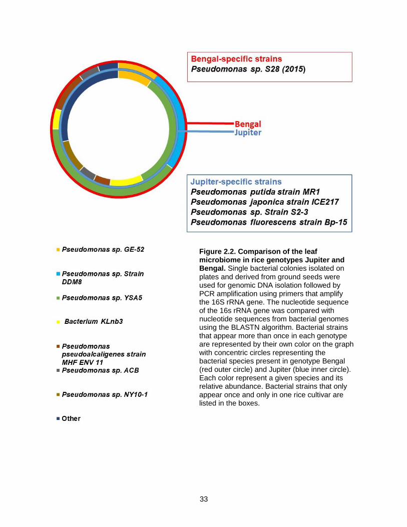
Validation of B. glumae identity
Because the identification of the B. glumae strains was only based on their growth in the
semi-selective media CCNT (Kawardani et al., 2000), which can lead to mis-identification, the

21
identity of those strains was confirmed by PCR using the following published primers: Bg 23S-
ITS1F(TGCTACGAAGAGCACTCTAAG), R (ACATGCACTTGTTCGCTTG), Bg specific F
(ACGTTCAGGGATACTGAGCAG), R (AGTCTGTCTCGCTCTCCCGA )
Bg 23S ITS-2 F (ACACGGAACACCTGGGTA), R (TCGCTCTCCCGAAGAGAT)
and Bg gyrB F (GAAGTGTCGCCGATGGAG), R (CCTTCACCGACAGCACGCAT) (Karki et
al., 2012). All the primers successfully generated PCR product when using UAPB10 and
UAPB11 as templates. Since Bg 23S ITS-1, ITS-2 and gyrB are conserved in bacteria, the lack
of amplification of UAPB12 and 13 with those primers is probably due to poor quality of the
templates. A PCR product was obtained with the Bg specific primer in UAPB13 but the
equivalent band was very weak in UAPB12. Because the Bg specific is supposed to amplify
only B. glumae, this primers pair was considered diagnostic for B. glumae and the positive
amplification indicates that UAPB10, UAPB11 and UAPB13 are all B. glumae. Further
sequence analysis of PCR products after amplification with the 16S rRNA primers confirmed
that UAPB 10, UAPB 11 and UAPB 13 were indeed B. glumae, while UAPB 12 is Klebiella
pneumonia.
Screening of bacterial strains antagonistic activity against B. glumae
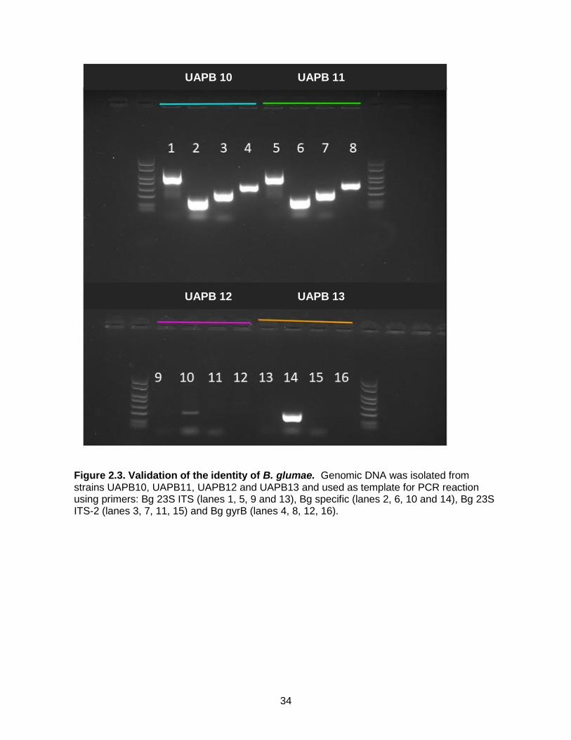
Five bacterial strains specifically associated with seeds or leaves of the genotype Jupiter
were chosen to investigate growth inhibition against B. glumae (UAPB13). Those strains are:
Pseudomonas fulva strain Pful-2, Pseudomonas putida strain MR1, Pseudomonas japonica
strain ICE217, Pseudomonas sp. Strain S2-3 and Pseudomonas fluorescens strain Bp-15. In
parallel, 10 strains of bacteria from the Rojas lab collection were also tested. The five strains
isolated from Jupiter did not show a strong inhibitory effect against B. glumae (Figure 4A).
However, out of the 10 laboratory strains, seven: PBL3, PBL9, PBL15, PBL16, PBL18, PBL19
and PBL20 caused a visible zone of growth inhibition (Figure 4A). Calculations on the areas of
zone inhibition showed that PBL3, PBL18 and PBL 20 had the largest areas of the zones of

22
inhibition: 334.7mm2, 415.6mm2 and 450.5mm2, respectively (Figure 4B). Therefore, these
strains were chosen for further characterization.
Identification of strains with antagonistic activity against B. glumae
To further characterize the inhibitory bacterial strains PBL3, PBL18, and PBL20,
genomic DNA was extracted and the 16s rRNA gene was amplified by PCR using 16s rRNA
primers. The 16s rRNA sequence of PBL3 showed 100% identity at the nucleotide level to the
16s rRNA gene of Pseudomonas fluorescens while the 16s rRNA sequence of PBL18 and
PBL20 showed 100% identity at the nucleotide level to the 16s rRNA gene of Burkholderia
cenocepacia (PBL18). PBL18 was chosen for the remainder of the study.
B. cenocepacia and P. fluorescens inhibit growth of three B. glumae strains
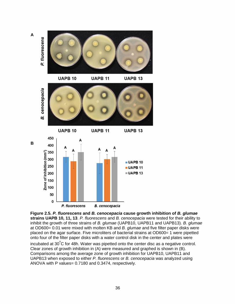
To determine whether B. cenocepacia and P. fluorescens inhibited growth for B. glumae in
general or just B. glumae UAPB13, two other strains of B. glumae, UAPB10 and UAPB11 were
tested in the inhibition experiments. All three strains presented large zones of inhibition when
grown with a filter paper disk containing either PBL3 or PBL18 (Figure 5A). When tested with P.
fluorescens, the zones of inhibition were 318.8mm2 for UAPB10, 287.4mm2 for UAPB 11 and
353.4mm2 for UAPB 13 (Figure 5B). When tested with B. cenocepacia, the zones of inhibition
were 274.1mm2 for UAPB 10, for 302.3mm2 UAPB 11 and 318.8mm2 for UAPB 13 (Figure 5B).
Statistical analysis showed that there was not a statistically significant difference in growth
inhibition of the different strains of B. glumae by B. cenocepacia or P. fluorescens. Therefore,
UAPB13 was used for the remaining of the experiments because the Rojas lab had already
generated a gentamycin resistant derivative of UAPB13, which will be used in subsequent
experiments.
23
P. fluorescens and B. cenocepacia reduce disease symptoms caused by B. glumae by
sheath injection.
To determine if B. cenocepacia and P. fluorescens could be effective biological control
agents against B. glumae, B. cenocepacia and P. fluorescens were co-inoculated with B.
glumae by sheath injection (Figure 6). E.coli was also co-inoculated with B. glumae as control.
Plants inoculated with B. glumae alone showed disease symptoms in the stem characterized by
brown lesions surrounding the area of inoculation (Figure 6A) and had average lesion lengths of
58.3mm (Figure 6B). Similar disease symptoms were obtained when co-inoculating B. glumae
with E. coli and in that case the average lesion lengths was 71.6mm (Figure 6B). However,
plants that were inoculated with the combinations of B. glumae/B. cenocepacia or B. glumae/P.
fluorescens had very small lesions (Figure 6A) that measured 1 mm when plants were
inoculated with B. glumae/B. cenocepacia and 3.6mm when plants were co-inoculated with B
glumae and P. fluorescens (Figure 6B).
Because in the field, the symptoms of B. glumae infection are mainly seen in the panicle,
it was necessary to test the effectiveness of P. fluorescens and B. cenocepacia reducing
disease symptoms caused by B. glumae in panicles. Thus, panicle co-inoculations were carried
out (Figure 7). At three days post inoculation plants inoculated with B. glumae alone had an
average of 25.26% discolored panicles, while plants co-inoculated with B. glumae and B.
cenocepacia had 13.99% discolored panicles (Figure 7b). These results showed that, B.
cenocepacia is effective at reducing symptoms of B. glumae infection (p value = 0.0109).
Unfortunately, results for P. fluorescens panicle co-inoculations were inconsistent with an
average of 16.51% discolored panicles and a large error bar resulting in a non-significant
reduction of symptoms (p value= 0.2431).

24
B. cenocepacia and P. fluorescens do not persist in planta
To investigate the feasibility of using P. fluorescens and B. cenocepacia as true
biological control agents against B. glumae, the long-term persistence of these strains was
evaluated by inoculating them in rice and retrieving after 5, 10 and 20 days. Inoculation of rice
with B. cenocepacia showed that B. cenocepacia growth increases from day 0 to 5, then
decreases from day 10 to 20 resulting in an overall decrease in the amount of bacteria found in
the plant after twenty days. Although it appears that there is a trend toward increase in bacterial
numbers from 0 to 10 dpi, the statistical analysis showed that the numbers of bacteria at those
time points are not significantly different. However, the numbers of bacteria at 20 dpi are
significantly lower than at 10 dpi, but equivalent to the initial numbers at 0dpi (Figure 8).
Inoculation of rice with P. fluorescens showed that there are equivalent numbers of bacteria
between 0 and 5 days indicating that P. fluorescens did not grow. Moreover, after 5 days, there
is a progressive decline in bacterial numbers resulting in a significant reduction in bacterial
numbers (Figure 8). This data suggests that B. cenocepacia and P. fluorescens are unable to
persist or proliferate in the plant and therefore would make poor biological control agents in the
field.
Discussion
This study was a proof-of-concept to evaluate bacterial populations between a rice
genotype that is moderately resistant to B. glumae (Jupiter) and another that is susceptible
(Bengal). The study investigated leaves and seeds as they are relevant to the biology of B.
glumae. Analysis of the bacterial communities associated with rice seeds have not been done
before, but several studies have investigated the bacterial communities associated with rice
leaves and roots. In such studies, the bacterial community in the leaves was found to be
different and less complex than that of the rhizosphere, but comparable to the bacterial
communities in the phyllosphere of other plant species (Knief et al.,2012).
25
Analyses of the bacterial communities in the leaves of several plants have shown that
they are represented by few bacterial genera, the most prevalent being Pseudomonas,
Sphingomonas, Methylobacterium, Bacillus, Massilia, Arthrobacter and Pantoea (Bulgarelli et
al.,2013). In this study, the most common bacterial genera associated with rice seeds and
leaves was Pseudomonas spp., which is not surprising considering the versatility of this bacteria
colonizing different environments (Piex et al., 2009). Specifically, Pseudomonas sp. NF81 was
the most prevalent isolate in seeds and represented 33.33% of the culturable bacteria in Bengal
and 37.5% of the culturable bacteria in Jupiter. Pseudomonas sp. YSA5 was the most
prevalent isolate in leaves and represented 33% of the culturable bacteria in Bengal and 40% of
the culturable bacteria in Jupiter. While Pseudomonas sp. was present in both Jupiter and
Bengal, its frequency and diversity was larger in Jupiter. The leaves in Bengal also have a high
percentage of Pseudomonas sp. strain DDM8 which is not present in the leaves of genotype
Jupiter.
When comparing the microbial diversity between Jupiter and Bengal, the results showed
that the bacterial composition is different from each other in both leaves and seeds, although
those differences do not appear to be too dramatic. The genotype-mediated differences in the
composition of the microbial communities associated with plants has been observed before
(Wagner et al., 2016). Although it is still not fully understood how specific plant genotypes
recruit specific microorganisms, a model for the recruitment of microorganisms to the roots has
been proposed. In this model microorganisms in the soil are recruited to the root and, at later
stages the genotype of the plant fine-tunes the final microbial composition to fit the plant’s
unique environment and needs (Bulgarelli et al., 2013). It is possible that similar mechanisms
operate in leaves and accounts for the differences between Jupiter and Bengal.
Whereas there were specific genera associated with each genotype in each organ,
common bacterial genera were retrieved within each organ. In other words, most of the
differences were found between organs than between genotypes. For example, Pseudomonas
26
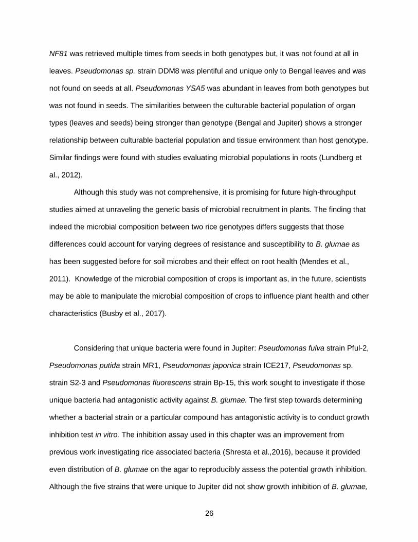
NF81 was retrieved multiple times from seeds in both genotypes but, it was not found at all in
leaves. Pseudomonas sp. strain DDM8 was plentiful and unique only to Bengal leaves and was
not found on seeds at all. Pseudomonas YSA5 was abundant in leaves from both genotypes but
was not found in seeds. The similarities between the culturable bacterial population of organ
types (leaves and seeds) being stronger than genotype (Bengal and Jupiter) shows a stronger
relationship between culturable bacterial population and tissue environment than host genotype.
Similar findings were found with studies evaluating microbial populations in roots (Lundberg et
al., 2012).
Although this study was not comprehensive, it is promising for future high-throughput
studies aimed at unraveling the genetic basis of microbial recruitment in plants. The finding that
indeed the microbial composition between two rice genotypes differs suggests that those
differences could account for varying degrees of resistance and susceptibility to B. glumae as
has been suggested before for soil microbes and their effect on root health (Mendes et al.,
2011). Knowledge of the microbial composition of crops is important as, in the future, scientists
may be able to manipulate the microbial composition of crops to influence plant health and other
characteristics (Busby et al., 2017).
Considering that unique bacteria were found in Jupiter: Pseudomonas fulva strain Pful-2,
Pseudomonas putida strain MR1, Pseudomonas japonica strain ICE217, Pseudomonas sp.
strain S2-3 and Pseudomonas fluorescens strain Bp-15, this work sought to investigate if those
unique bacteria had antagonistic activity against B. glumae. The first step towards determining
whether a bacterial strain or a particular compound has antagonistic activity is to conduct growth
inhibition test in vitro. The inhibition assay used in this chapter was an improvement from
previous work investigating rice associated bacteria (Shresta et al.,2016), because it provided
even distribution of B. glumae on the agar to reproducibly assess the potential growth inhibition.
Although the five strains that were unique to Jupiter did not show growth inhibition of B. glumae,

27
three strains from the laboratory collection showed large zones of growth inhibition and those
strains were identified as P. fluorescens and B. cenocepacia and those strains were further
used for in planta experiments.
In contrast to previous work where rice-associated bacteria showing growth inhibition in
vitro did not have reproducible effects when inoculated in plants (Shresta et al. 2016), in this
work, co-inoculation of P. fluorescens and B. cenocepacia with B. glumae consistently reduced
disease symptoms. This effect can be directly attributed to these bacterial strains as plants
inoculated with the combination B. glumae/E. coli still had disease symptoms that resemble the
symptoms observed by B. glumae alone.
The finding that P. fluorescens had antagonistic activities is not surprising as
Pseudomonas spp. are commonly found in suppressive soils and have been well-studied and
used as biological control agents against soil-borne pathogens in the past (Weller, 2007;
Kyselkova et al., 2009). Another strain of Pseudomonas, Pseudomonas sp. Rh323 was effective
controlling Xanthomonas oryzae pv. oryzae and, reduced disease symptoms associated with
the disease under field conditions (Yasmin et al., 2016). Similar to P. fluorescens, Burkholderia
spp have also been identified as biological control agents for plant diseases including Fusarium
moniliforme on maize (Hebbar et al., 1992), Pythium Damping-off (Bowers and Parke, 1993)
and Rhizoctonia stem rot (Cartwright and Benson, 1995).
While both P. fluorescens and B. cenocepacia were effective in reducing disease under
controlled conditions, more work is needed to investigate conditions to increase its persistence.
Neither one of them showed good persistence in leaves after sheath injection, but as soil-borne
bacteria, it is possible that they have higher persistence in the soil, the preferred habitat for
these bacteria. If that is the case, the antagonistic activities of these bacteria will be relevant to
control bacterial panicle blight at early stages of the disease when the bacteria is associated
with the seed.

28
The use of B. cenocepacia as a biological control agent is unlikely as this bacterium is a
member of the Burkholderia cepacia complex (Bcc) , a group of opportunistic pathogens of
humans and are especially dangerous to individuals with cystic fibrosis (Parke and Gurian-
Sherman, 2001). Therefore, its use could pose a risk to human health. However, the isolation of
compounds produced by these bacteria will pave the way towards controlling bacterial panicle
blight of rice.

29
References
Alabouvette, C. 1999. 'Fusarium wilt suppressive soils: an example of disease-suppressive soils', Australasian Plant Pathology, 28: 57-64.
Bakker, P. A. H. M., R. F. Doornbos, C. Zamioudis, R. L. Berendsen, and C. M. J. Pieterse. 2013. 'Induced Systemic Resistance and the Rhizosphere Microbiome', Plant Pathology Journal, 29: 136-43.
Berendsen, R. L., C. M. J. Pieterse, and P. A. H. M. Bakker. 2012. 'The rhizosphere microbiome and plant health', Trends in Plant Science, 17: 478-86.
Berg, G. 2009. 'Plant-microbe interactions promoting plant growth and health: perspectives for controlled use of microorganisms in agriculture', Applied Microbiology and Biotechnology, 84: 11-18.
Berg, G., M. Grube, M. Schloter, and K. Smalla. 2014. 'Unraveling the plant microbiome: looking back and future perspectives', Front Microbiol, 5: 148.
Bowers, J. H., and J. L. Parke. 1993. 'Epidemiology of Pythium Damping-Off and Aphanomyces Root-Rot of Peas after Seed Treatment with Bacterial Agents for Biological-Control', Phytopathology, 83: 1466-73.
Bulgarelli, D., K. Schlaeppi, S. Spaepen, E. V. L. van Themaat, and P. Schulze-Lefert. 2013. 'Structure and Functions of the Bacterial Microbiota of Plants', Annual Review of Plant Biology, Vol 64, 64: 807-38.
Busby, P. E., Soman, C., Wagner, M. R., Friesen, M. L., Kremer, J., Bennett, A., Morsy, M., Eisen, J. A., Leach, J. E., Dangl, J. L. 2017. 'Research priorities for harnessing plant microbes in sustainable agriculture', PLoS One, 15.
Cartwright, D. K., and D. M. Benson. 1995. 'Comparison of Pseudomonas Species and Application Techniques for Biocontrol of Rhizoctonia Stem Rot of Poinsettia', Plant Disease, 79: 309-13.
Edwards, J., C. Johnson, C. Santos-Medellin, E. Lurie, N. K. Podishetty, S. Bhatnagar, J. A. Eisen, and V. Sundaresan. 2015. 'Structure, variation, and assembly of the root-associated microbiomes of rice', Proceedings of the National Academy of Sciences of the United States of America, 112: E911-E20.
Gilbert, J. A., R. A. Quinn, J. Debelius, Z. J. Z. Xu, J. Morton, N. Garg, J. K. Jansson, P. C. Dorrestein, and R. Knight. 2016. 'Microbiome-wide association studies link dynamic microbial consortia to disease', Nature, 535: 94-103.
Hebbar, K. P., D. Atkinson, W. Tucker, and P. J. Dart. 1992. 'Suppression of Fusarium-Moniliforme by Maize Root-Associated Pseudomonas-Cepacia', Soil Biology & Biochemistry, 24: 1009-20.
Kanjanamaneesathian, M., C. Kusonwiriyawong, A. Pengnoo, and L. Nilratana. 1998. 'Screening of potential bacterial antagonists for control of sheath blight in rice and

30
development of suitable bacterial formulations for effective application', Australasian Plant Pathology, 27: 198-206.
Karki, H. S., B. K. Shrestha, J. W. Han, D. E. Groth, I. K. Barphagha, M. C. Rush, R. A. Melanson, B. S. Kim, and J. H. Ham. 2012. 'Diversities in Virulence, Antifungal Activity, Pigmentation and DNA Fingerprint among Strains of Burkholderia glumae', PLoS One, 7.
Kawardani, M., Okada, K. 2000. 'New selective medium for isolation of Burkholderia glumae from rice seeds', General Plant Pathology, 66: 234-37.
Kazempour, M. N. 2004. 'Biological Control of Rhizoctonia solani, the Causal Agent of Rice Sheath Blight by Antagonistics Bacteria in Greenhouse and Field Conditions', Plant Pathology Journal3: 88-96.
Knief, C., N. Delmotte, S. Chaffron, M. Stark, G. Innerebner, R. Wassmann, C. von Mering, and J. A. Vorholt. 2012. 'Metaproteogenomic analysis of microbial communities in the phyllosphere and rhizosphere of rice', Isme Journal, 6: 1378-90.
Kyselkova, M., J. Kopecky, M. Frapolli, G. Defago, M. Sagova-Mareckova, G. L. Grundmann, and Y. Moenne-Loccoz. 2009. 'Comparison of rhizobacterial community composition in soil suppressive or conducive to tobacco black root rot disease', Isme Journal, 3: 1127-38.
Lundberg, D. S., Lebeis, S. L., Paredes, S. H., Yourstone, S., Gehring, J., Malfatti, S., Tremblay, J., Engelbrekston, A., Kunin, V., del Rio, T. C., Eickhorst, T., Ley, R. E., Hugenholtz, P., Tringe, S. G., Dangl, J. L. 2012. 'Defining the core Arabidopsis thaliana root microbiome', Nature, 488: 86-90.
Mendes, R., M. Kruijt, I. de Bruijn, E. Dekkers, M. van der Voort, J. H. M. Schneider, Y. M. Piceno, T. Z. DeSantis, G. L. Andersen, P. A. H. M. Bakker, and J. M. Raaijmakers. 2011. 'Deciphering the Rhizosphere Microbiome for Disease-Suppressive Bacteria', Science, 332: 1097-100.
Muller, D. B., C. Vogel, Y. Bai, and J. A. Vorholt. 2016. 'The Plant Microbiota: Systems-Level Insights and Perspectives', Annual Review of Genetics, Vol 50, 50: 211-34.
Parke, J. L., and D. Gurian-Sherman. 2001. 'Diversity of the Burkholderia cepacia complex and implications for risk assessment of biological control strains', Annual Review of Phytopathology, 39: 225-58.
Piex, A., Ramirez-Bahena, M. H. 2009. 'Historical evolution and current status of the taxonomy of genus Pseudomonas', Infection Genetics and Evolution, 9: 1132-47.
Sessitsch, A., P. Hardoim, J. Doring, A. Weilharter, A. Krause, T. Woyke, B. Mitter, L. Hauberg-Lotte, F. Friedrich, M. Rahalkar, T. Hurek, A. Sarkar, L. Bodrossy, L. van Overbeek, D. Brar, J. D. van Elsas, and B. Reinhold-Hurek. 2012. 'Functional Characteristics of an Endophyte Community Colonizing Rice Roots as Revealed by Metagenomic Analysis', Molecular Plant-Microbe Interactions, 25: 28-36.

31
Shrestha, B. K., H. S. Karki, D. E. Groth, N. Jungkhun, and J. H. Ham. 2016. 'Biological Control Activities of Rice-Associated Bacillus sp Strains against Sheath Blight and Bacterial Panicle Blight of Rice', PLoS One, 11.
Singh, U. B., D. Malviya, Wasiullah, S. Singh, J. K. Pradhan, B. P. Singh, M. Roy, M. Imram, N. Pathak, B. M. Baisyal, J. P. Rai, B. K. Sarma, R. K. Singh, P. K. Sharma, S. D. Kaur, M. C. Manna, S. K. Sharma, and A. K. Sharma. 2016. 'Bio-protective microbial agents from rhizosphere eco-systems trigger plant defense responses provide protection against sheath blight disease in rice (Oryza sativa L.)', Microbiological Research, 192: 300-12.
Spence, C., E. Alff, C. Johnson, C. Ramos, N. Donofrio, V. Sundaresan, and H. Bais. 2014. 'Natural rice rhizospheric microbes suppress rice blast infections', Bmc Plant Biology, 14.
Wagner, M. R., D. S. Lundberg, T. G. del Rio, S. G. Tringe, J. L. Dangl, and T. Mitchell-Olds. 2016. 'Host genotype and age shape the leaf and root microbiomes of a wild perennial plant', Nature Communications, 7.
Weller, D.M. 2007. 'Pseudomonas biocontrol agents of soilborne pathogens: looking back over 30 years', Phytopathology, 97: 250-56.
Yasmin, S., A. Zaka, A. Imran, M. A. Zahid, S. Yousaf, G. Rasul, M. Arif, and M. S. Mirza. 2016. 'Plant Growth Promotion and Suppression of Bacterial Leaf Blight in Rice by Inoculated Bacteria', PLoS One, 11: e0160688.

32
Appendix
Figure 2.1. Comparison of the bacterial populations in the seeds of rice genotypes Jupiter and Bengal. Single bacterial colonies isolated on plates and derived from ground seeds were used for genomic DNA isolation followed by PCR amplification using primers that amplify the 16S rRNA gene. The nucleotide sequence of the 16s rRNA gene was compared with nucleotide sequences from bacterial genomes using the BLASTN algorithm. Bacterial strains that appear more than once in each genotype are represented by their own color on the graph with concentric circles representing the bacterial species present in genotype Bengal (red outer circle) and Jupiter (blue inner circle). Each color represent a given species and its relative abundance. Bacterial strains that only appear once and only in one rice cultivar are listed in the boxes.
33
Figure 2.2. Comparison of the leaf microbiome in rice genotypes Jupiter and Bengal. Single bacterial colonies isolated on plates and derived from ground seeds were used for genomic DNA isolation followed by PCR amplification using primers that amplify the 16S rRNA gene. The nucleotide sequence of the 16s rRNA gene was compared with nucleotide sequences from bacterial genomes using the BLASTN algorithm. Bacterial strains that appear more than once in each genotype are represented by their own color on the graph with concentric circles representing the bacterial species present in genotype Bengal (red outer circle) and Jupiter (blue inner circle). Each color represent a given species and its relative abundance. Bacterial strains that only appear once and only in one rice cultivar are listed in the boxes.
34
Figure 2.3. Validation of the identity of B. glumae. Genomic DNA was isolated from strains UAPB10, UAPB11, UAPB12 and UAPB13 and used as template for PCR reaction using primers: Bg 23S ITS (lanes 1, 5, 9 and 13), Bg specific (lanes 2, 6, 10 and 14), Bg 23S ITS-2 (lanes 3, 7, 11, 15) and Bg gyrB (lanes 4, 8, 12, 16).
UAPB 10 UAPB 11
UAPB 12 UAPB 13
35
Figure 2.4. Identification of bacteria with antagonistic activities against B. glumae. Five
bacterial strains isolated from leaves and seeds from rice cultivar Jupiter (JL2, JL5 JL13, JL16, JS6) and 10 bacterial strains from the Rojas Lab collection (PBL3, PBL9, PBL15, PBL16, PBL18, PBL19, PBL20, PBL23, PBL24, PBL33) were tested for their ability to inhibit growth of B. glumae (UAPB13). B. glumae at OD600 =0.001 were mixed with molten KB agar and five sterile filter paper disks were placed on the agar surface. Five microliters of bacterial strains at OD600= 1 were pipetted onto four of the filter paper disk. Water was added to the fifth disk (in center) to be
used as control. Plates were incubated at 30oC for 48h. Clear zones of growth inhibition in (A)
were measured (B). The differences among means was calculated using ANOVA statistical analysis with a p value <0.0001.
A
B
36
Figure 2.5. P. fluorescens and B. cenocepacia cause growth inhibition of B. glumae
strains UAPB 10, 11, 13. P. fluorescens and B. cenocepacia were tested for their ability to inhibit the growth of three strains of B. glumae (UAPB10, UAPB11 and UAPB13). B. glumae at OD600= 0.01 were mixed with molten KB and B. glumae and five filter paper disks were placed on the agar surface. Five microliters of bacterial strains at OD600= 1 were pipetted onto four of the filter paper disks with a water control disk in the center and plates were
incubated at 30oC for 48h. Water was pipetted onto the center disc as a negative control.
Clear zones of growth inhibition in (A) were measured and graphed is shown in (B). Comparisons among the average zone of growth inhibition for UAPB10, UAPB11 and UAPB13 when exposed to either P. fluorescens or B. cenocepacia was analyzed using ANOVA with P values= 0.7180 and 0.3474, respectively.
A
B

37
Figure 2.6. Co-inoculation of B. glumae with B. cenocepacia or P. fluorescens reduce disease symptoms in rice. Eight-week-old rice plants from cultivar Wells were inoculated in the sheath with B. glumae alone or in combination with E. coli, B. cenocepacia or P. fluorescens, or mock-treated with water. Disease symptoms were evaluated after eight days (A). Bars represent average lesion lengths for three replicates (B). Treatments were compared using ANOVA statistical analysis with a P- value <0.001.
A
B
A
A
B B B

38
Figure 2.7. Co-inoculation of B. glumae with B. cenocepacia reduces disease symptoms on rice panicles. Emerging panicles from cultivar Wells were sprayed with 20mL of either B. glumae inoculum OD600= 1 or a co-inoculatant of B. glumae OD600= 1 and B. cenocepacia OD600= 1 or a co-inoculation of B. glumae OD600 =1 and P. fluorescens OD600=1. Symptoms were observed after 3 dpi (A) and number of discolored spikelets counted. Bars in (B) represent the percent of discolored spikelets. Data was analyzed using a t test with a p value = 0.0109 for B. glumae vs B. glumae/ B. cenocepacia and p- value = 0.2431 for B. glumae vs B.glumae/P.fluorescens.
A
B

39
Figure 2.8. B. cenocepacia and P. fluorescens do not have long-term persistence in rice. Four-week-old rice plants cultivar Wells were inoculated with 20µL OD
600=0.125 B.
cenocepacia or P. fluorescens. Plants were ground and diluted on KB plates at 0, 5, 10 and 20 days to determine persistence of the bacteria in plant tissues. The comparison of bacterial numbers over time for B. cenocepacia and P. fluorescens was analyzed using ANOVA. P- value= 0.1257 for B. cenocepacia and 0.0589 for P. fluorescens.

40
Table 1. Bacteria isolated from seeds of cultivars Jupiter and Bengal
Isolate number Closest species in GenBank based on 16S rDNA sequence
BS1 Pseudomonas sp. NF81
BS2 Pseudomonas sp. NF81 BS3 Pseudomonas tolaasii strain GD76 BS4 Pseudomonas sp. NF81 BS5 Pantoea sp. B3030 BS6 Pseudomonas sp. NF81 BS7 Pseudomonas sp. GE-52 BS8 Pseudomonas sp. NF81 BS9 Stenotrophomonas sp. BS10 Pseudomonas sp. NF81 BS11 N/A BS12 Pseudomonas putida strain FB15 BS13 Pseudomonas sp. GAO7 BS14 Stenotrophomonas sp. 2012A BS15 Pseudomonas putida strain PSDM3 BS16 Poor quality sequence BS17 Pseudomonas sp. GAO7 BS18 Pseudomonas putida strain FB15 BS19 Pseudomonas sp. G0838 BS20 Pseudomonas sp. Strain DDM8 JS1 Pseudomonas sp. NF81 JS2 Pseudomonas sp. NF81 JS3 Pseudomonas sp. NF81 JS4 Pseudomonas tolaasii strain GD76 JS5 Pseudomonas sp. NF81 JS6 Pseudomonas fulva strain Pful-2 JS7 Stenotrophomonas sp. DIV102 JS8 Pseudomonas sp. NF81 JS9 Pseudomonas sp. NF81 JS10 Stenotrophomonas sp. DIV102 JS11 Pseudomonas putida strain FB15 JS12 Pseudomonas gessardii strain 5611 JS13 Poor quality sequence JS14 Pseudomonas putida strain FB15 JS15 Poor quality sequence JS16 Pseudomonas putida strain FB15 JS17 Poor quality sequence JS18 Poor quality sequence JS19 Pseudomonas putida strain PSDM3 JS20 Pseudomonas putida strain FB15

41
Table 2. Bacteria isolated from leaves of cultivars Jupiter and Bengal
Isolate number
Closest species in GenBank based on 16 s rDNA sequence
BL1 Pseudomonas sp. strain DDM8 BL2 Pseudomonas sp. GE-52 BL3 Pseudomonas pseudoalcaligenes strain MHF ENV 11 BL4 Pseudomonas sp. YSA5 BL5 Bacterium KLnb3 / Pseudomonas sp. strain AC530 BL6 Pseudomonas pseudoalcaligenes strain MHF ENV 11 BL7 Pseudomonas sp. Strain DDM8 BL8 Pseudomonas sp. S28 (2015) BL9 Pseudomonas sp. Strain DDM8 BL10 Pseudomonas sp. YSA5 BL11 Pseudomonas sp. Strain DDM8 BL12 Pseudomonas sp. YSA5 BL13 Pseudomonas sp. YSA5 BL14 Pseudomonas sp. YSA5 BL15 Pseudomonas sp. GE-52 BL16 Pseudomonas sp. ACB BL17 Pseudomonas sp. YSA5 BL18 Pseudomonas sp. Strain DDM8 BL19 Pseudomonas sp. YSA5 BL20 Pseudomonas sp. YSA5 JL1 Pseudomonas sp. NY10-1 JL2 Bacterium KLnb3 / Pseudomonas fluorescens strain Bp-15 JL3 Pseudomonas sp. YSA5 JL4 Pseudomonas sp. YSA5 JL5 Pseudomonas sp. Strain S2-3 JL6 Pseudomonas sp. YSA5 JL7 Pseudomonas sp. GE-52 JL8 Poor quality sequence JL9 Pseudomonas pseudoalcaligenes strain MHF ENV 11 JL10 Pseudomonas sp. YSA5 JL11 Bacterium KLnb3 / Pseudomonas sp. Strain AC530 JL12 Bacterium KLnb3 / Pseudomonas sp. Strain AC530 JL13 Pseudomonas japonica strain ICE217 JL14 Pseudomonas sp. ACB JL15 Pseudomonas sp. YSA5 JL16 Pseudomonas putida strain MR1 JL17 Pseudomonas sp. YSA5 JL18 Pseudomonas sp. YSA5 JL19 Pseudomonas sp. NY10-1 JL20 Pseudomonas sp. GE-52
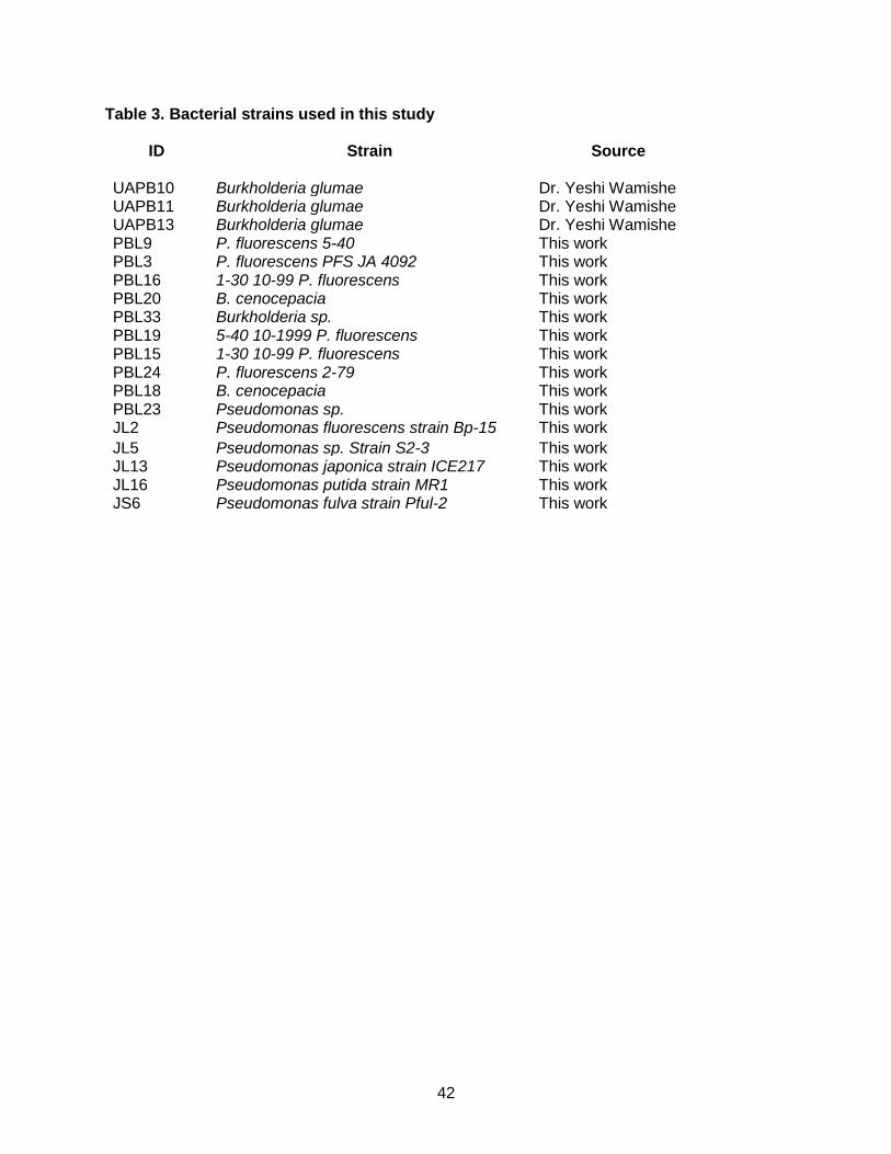
42
Table 3. Bacterial strains used in this study
ID Strain Source
UAPB10 Burkholderia glumae Dr. Yeshi Wamishe UAPB11 Burkholderia glumae Dr. Yeshi Wamishe UAPB13 Burkholderia glumae Dr. Yeshi Wamishe PBL9 P. fluorescens 5-40 This work PBL3 P. fluorescens PFS JA 4092 This work PBL16 1-30 10-99 P. fluorescens This work PBL20 B. cenocepacia This work PBL33 Burkholderia sp. This work PBL19 5-40 10-1999 P. fluorescens This work PBL15 1-30 10-99 P. fluorescens This work PBL24 P. fluorescens 2-79 This work PBL18 B. cenocepacia This work PBL23 Pseudomonas sp. This work JL2 Pseudomonas fluorescens strain Bp-15 This work
JL5 Pseudomonas sp. Strain S2-3 This work JL13 Pseudomonas japonica strain ICE217 This work JL16 Pseudomonas putida strain MR1 This work JS6 Pseudomonas fulva strain Pful-2 This work

43
Chapter 3: Harnessing Burkholderia cenocepacia and Pseudomonas fluorescens as
sources of antimicrobials against Burkholderia glumae
Introduction
Microorganisms associated with plants in the rhizosphere or phyllosphere can have
antagonistic activities against pathogens, and consequently can be used as biological control
agents. Two main mechanisms of biological control are: competition and antibiosis. Through
competition a given biological control agent withdraws essential nutrients for pathogen growth,
or occupies a specific area that prevent the pathogen from gaining access to the host (Pal and
McSpadden Gardener, 2006). Through antibiosis, the biological control agent produces
antibiotics, lytic enzymes or waste products that disrupt or kill the pathogen (Pal and
McSpadden Gardener, 2006). One example of antibiosis include the control of Pythium ultimum
by Trichoderma harzianum, a fungus that produces enzymes, which damage the cell walls of
the pathogen (Benhamou and Chet, 1997). Another classic example of biological control
through antibiosis is the control of Agrobacterium tumefaciens, by a nonpathogenic bacterium,
Agrobacterium radiobacter, which produces the antibiotic Agrocin 84 (Kerr, 1980). Although
biological control approaches are desirable over chemical control of plant diseases when
making ecological considerations, identification of biological control agents is not trivial.
However, with the increasing knowledge on the microbial communities associated with plants, it
is becoming possible to identify individual microorganisms with biological control properties, or
to manipulate those microbial communities to manage diseases and increase plant health (Berg
et al., 2009; Busby et al., 2017).
In previous Chapter 2, two bacterial strains with antagonistic activity against B. glumae were
identified. These antagonistic bacteria are two strains are B. cenocepacia and P. fluorescens,
and their antagonistic activity was demonstrated in vitro, with the reduction of growth of B.
glumae, as well as in planta, by reducing disease symptoms caused by B. glumae, when they

44
were co-inoculated with the pathogen. In spite of this promising results, these antagonistic
bacteria cannot be used as living biological control agents for the following reasons: 1. P.
fluorescens and B. cenocepacia did not show long-term persistence in rice and, 2. B.
cenocepacia is an opportunistic human pathogen and therefore their widespread use can have
an inherent risk to human health. Notwithstanding these challenges, the finding that these
bacteria have antagonistic activity against B. glumae makes them useful, initially for research
purposes to investigate the mechanisms behind this growth inhibition, and in the future, to
isolate molecules responsible for this activity. Thus, this research is significant to advance the
development of effective methods to control bacterial panicle blight of rice. This chapter will
investigate the mechanisms behind growth inhibition of B. glumae by P. fluorescens and B.
cenocepacia and will also identify methods to isolate fractions of P. fluorescens and B.
cenocepacia with antagonistic activity against B. glumae in the absence of bacteria.
Methods
Bacterial strains used in this study
B. glumae strain UAPB 13 was collected from rice fields in Stuttgart, AR by Dr. Yeshi
Wamishe. B. glumae (pGFP-TIR) is a derivative of strain UAPB13, which was transformed by
electroporation with the plasmid (pGFP-TIR) (Miller and Lindow, 1997) that carries GFP and
confers gentamycin resistance.
Evaluation of bacterial growth in vitro
B. glumae, P. fluorescens, B. cenocepacia and E. coli were grown on King's B (KB)
media plates for two days. Single colonies were transferred to 5 ml KB broth and grown
overnight at 30oC with shaking. Overnight cultures were centrifuged at 6,000 rpm for 10 minutes
and washed with sterile water three times. Bacterial concentrations were read using a
spectrophotometer at OD600nm and diluted to OD600= 0.2 (1x10⁸CFU/ml) in 100mL KB broth.
45
Cultures were incubated at 30oC with constant agitation for eight hours. Every 4h, 1mL aliquots
were withdrawn to assess bacterial growth by measuring absorbance at OD600nm. This
experiment was repeated three times.
Competition assay in vitro
A single colony of B. glumae (pGFP-TIR) was grown in 5ml KB broth at 30oC for 18h
with continuous agitation. On the second day 100 µl of this culture were transferred to 100mL of
KB broth and grown for 18-24h until it reached an OD600=0.1. On the second day, a single
colony of P. fluorescens was grown in 5ml of KB broth at 30oC for 18h with continuous agitation
and transferred to 100ml of KB broth and grown until reaching at OD600= 0.01. After both strains
reached the desired OD600, they were mixed in a 10:1 (B. glumae: P. fluorescens) ratio in a total
volume of 50 ml. In a different flask, B. glumae (pGFP-TIR) alone was grown as control. One
hundred microliters aliquots were taken at 0, 4, 8, 24 and 48h and these aliquots were serially
diluted and plated on KB plates containing gentamycin to enumerate B. glumae (pGFP-TIR).
Competition Assay in planta
Four week-old rice plants cultivar Wells were inoculated by sheath injection with B.
glumae (pGFP-TIR) or with inoculum mixtures B. glumae (pGFP-TIR)/ P. fluorescens. Colonies
of B. glumae (pGFP-TIR) and P. fluorescens were grown overnight in liquid LB broth cultures.
Cultures were then diluted to OD600= 0.125 in 10mL tubes. Plants were stem injected with 20µL
of B. glumae (pGFP-TIR) or co-inoculated with B. glumae (pGFP-TIR) OD600 = 0.125 and P.
fluorescens OD600 = 0.125. Aboveground portions of inoculated plants were harvested, weighed,
ground, serially diluted and plated on gentamycin plates at 0, 3 and 5dpi to count B. glumae
(pGFP-TIR) growth alone and in the presence of P. fluorescens. This experiment was repeated
three times.

46
Calculation of competitive Index in planta
From the competition assay, bacteria were plated on KB without antibiotics and KB containing
gentamycin. B. glumae (pGFP-TIR) was enumerated by counting bacterial colonies on
KB+gentamycin plates. P. fluorescens was enumerated by counting bacterial colonies on KB
and subtracting from those the number of colonies found on KB+gentamycin plates. The
competitive index was calculated as:
CI=cfu B. glumae GFP/cfu P. fluorescens (Output)
cfu B. glumae GFP/cfu P. fluorescens (Input) (Macho et al., 2007).
Where output corresponds to the number of both B. glumae (pGFP-TIR) and P. fluorescens
counted at 0, 3 and 5 dpi, while input corresponds to the number of bacteria that were
inoculated at 0 dpi.
Isolation of cell-free secreted fractions with biological activity from P. fluorescens and B.
cenocepacia obtained from zones of growth inhibition
B. glumae at OD600= 0.01 was mixed with molten agar and 5µl of P. fluorescens or B.
cenocepacia at OD600= 1 were placed on filter paper disks placed on the surface of the agar and
incubated for two days to generate the zone of growth inhibition. The portions of the agar
containing the zones of inhibition were used to isolate secreted fractions. For that purpose, 3
agar plugs (5mm each) were cut out from the zone of inhibition, transferred to 3 ml of sterile
water or KB and incubated at 30oC with constant agitation for 18h. After incubation, water and
KB were filter sterilized using a 0.22 µM filter.
To evaluate the biological activity of the cell-free preparations, these preparations were
mixed with KB in a 1:1 ratio and inoculated with a single colony of B. glumae. KB alone was also
inoculated with a single colony of B. glumae as control. B. glumae either in KB or in KB mixed
with cell-free preparations derived from P. fluorescens or B. cenocepacia were grown for 18h at
30oC and bacterial cultures were serially diluted and plated to enumerate bacteria.

47
Isolation of cell-free secreted fractions with biological activity obtained from
liquid cultures of P. fluorescens and B. cenocepacia
P. fluorescens and B. cenocepacia cultures were grown in 100mL Luria Bertani (LB)
broth overnight at 30oC by shaking. Cultures were centrifuged for 10 minutes at 6,000 rpm and
the supernatant was collected. Twenty mL of the supernatants were aliquoted into 50 mL flasks
and the tops were covered with kimwipes secured with tape to allow for airflow. The samples
were then placed in the lyophilizer and dried for 24 hours to remove all water. Pure LB broth
samples were lyophilized to be used as control. Lyophilized LB or supernatants from P.
fluorescens or B. cenocepacia cultures were resuspended in sterile water at 0.01 g/ml.
Reconstituted LB or bacterial supernatants were filter sterilized using a 0.22 µM filter. To
evaluate the biological activity of cell-free preparations, reconstituted fractions were added to
KB broth at 1:1 (1.5mL: 1.5mL) ratio and used to grow a single colony of B. glumae. As control,
a single colony of B. glumae was grown on KB. Cultures of B. glumae ware grown at 30oC
shaker for 24 hours. After 24h, cultures were serially diluted and plated on KB plates to
enumerate bacterial populations.
Inoculation of rice plants with B. glumae containing lyophilized supernatants from P.
fluorescens and B. cenocepacia.
Lyophilized supernatants from B. cenocepacia, P. fluorescens or lyophilized LB broth
were added to prepare inoculums of B. glumae at a concentration of 0.5g/mL. B. glumae at
OD600=0.125 without added lyophilized materials was used as control. Twenty microliters of
these inoculums were injected in triplicates, into the sheath of eight-week old plants from
cultivar Wells using insulin syringes. Plants were transferred to growth chamber under with
temperatures 35oC/28oC (day/night) and 60-65% relative humidity for eight days. Plants were
monitored each day and lesions were measured and photograph after eight days of inoculation.
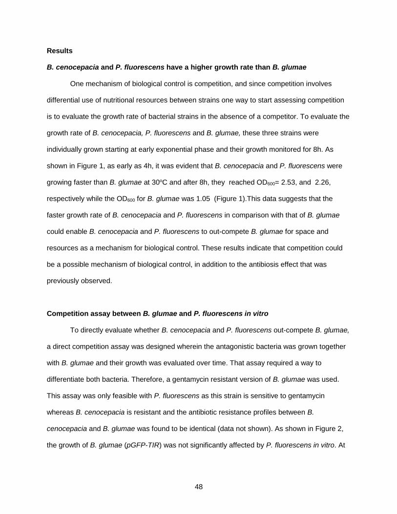
48
Results
B. cenocepacia and P. fluorescens have a higher growth rate than B. glumae
One mechanism of biological control is competition, and since competition involves
differential use of nutritional resources between strains one way to start assessing competition
is to evaluate the growth rate of bacterial strains in the absence of a competitor. To evaluate the
growth rate of B. cenocepacia, P. fluorescens and B. glumae, these three strains were
individually grown starting at early exponential phase and their growth monitored for 8h. As
shown in Figure 1, as early as 4h, it was evident that B. cenocepacia and P. fluorescens were
growing faster than B. glumae at 30oC and after 8h, they reached OD600= 2.53, and 2.26,
respectively while the OD600 for B. glumae was 1.05 (Figure 1).This data suggests that the
faster growth rate of B. cenocepacia and P. fluorescens in comparison with that of B. glumae
could enable B. cenocepacia and P. fluorescens to out-compete B. glumae for space and
resources as a mechanism for biological control. These results indicate that competition could
be a possible mechanism of biological control, in addition to the antibiosis effect that was
previously observed.
Competition assay between B. glumae and P. fluorescens in vitro
To directly evaluate whether B. cenocepacia and P. fluorescens out-compete B. glumae,
a direct competition assay was designed wherein the antagonistic bacteria was grown together
with B. glumae and their growth was evaluated over time. That assay required a way to
differentiate both bacteria. Therefore, a gentamycin resistant version of B. glumae was used.
This assay was only feasible with P. fluorescens as this strain is sensitive to gentamycin
whereas B. cenocepacia is resistant and the antibiotic resistance profiles between B.
cenocepacia and B. glumae was found to be identical (data not shown). As shown in Figure 2,
the growth of B. glumae (pGFP-TIR) was not significantly affected by P. fluorescens in vitro. At
49
each time point, the number of B. glumae (pGFP-TIR) recovered were equivalent to the
numbers of B. glumae (pGFP-TIR) recovered when grown together with P. fluorescens.
Competition assay between B. glumae and P. fluorescens in planta
Previous results (Chapter 2) showed that co-inoculation of B. glumae with P. fluorescens
reduced disease symptoms in rice and one hypothesis is that P. fluorescens competes with B.
glumae. To test that hypothesis, rice plants were co-inoculated with B. glumae (pGFP-TIR) and
P. fluorescens to determine the competitive index (CI). As shown in Figure 3, the CI is 1 at 0
dpi, showing that the initial concentrations of B. glumae (pGFP-TIR) and P. fluorescens were
the same. Because the input corresponds to the number of bacteria at 0 dpi, the value of the
CI is directly related to the ratios of B. glumae (pGFP-TIR)/P. fluorescens. Thus, the CI of 0.25
at 3 dpi indicates that the numbers of B. glumae (pGFP-TIR) are higher than those of P.
fluorescens. At 5 dpi, the competition index is -1.57 suggesting that B. glumae is out-competing
P. fluorescens (pGFP-TIR) since a negative competition index indicates the presence of more
B. glumae than P. fluorescens.
Isolation of cell-free fractions from P. fluorescens and B. cenocepacia with antagonistic
activity against B. glumae
The growth inhibition of B. glumae caused by P. fluorescens and B. cenocepacia in vitro
(Chapter 2), led to the hypothesis that these bacteria release compounds that inhibit the growth
of B. glumae. To test this hypothesis two different methods were used to generate cell-free
preparations with antagonistic activity against B. glumae. The first method used agar plugs
obtained from the zones of inhibition and the second method used secreted and concentrated
fractions of bacterial cultures supernatants.
Amending KB broth with fractions obtained from agar plugs corresponding to zones of
inhibition caused by P. fluorescens and B. cenocepacia, reduced the growth of B. glumae as

50
indicated by reduced turbidity (Figure 4A). In agreement with this results, the numbers of B.
glumae grown on KB containing agar plugs from LB plates, not exposed to either P. fluorescens
or B. cenocepacia reached 107 CFU/ml. However, B. glumae grown in KB broth amended with a
cell-free preparation derived from P. fluorescens reached 104 CFU/ml and B. glumae grown in
KB broth supplemented with a cell-free preparation derived from B. cenocepacia reached 105
CFU/ml (Figure 4B) indicating that the cell-free preparations derived from P. fluorescens and B.
cenocepacia reduced growth of B. glumae by 10000-fold and 1,000-fold, respectively.
Using secreted supernatants that were further lyophilized to concentrate them while
preserving biological activity showed a similar pattern of growth inhibition albeit to a lesser
extent. B. glumae grown in KB broth amended with lyophilized LB reached populations higher
than 107CFU/ml, whereas B. glumae grown in KB broth amended with lyophilized secreted
fractions from P. fluorescens and B. cenocepacia reached numbers higher than 106CFU/ml
(Figure 5).
Secreted fractions from P. fluorescens and B. cenocepacia reduce disease symptoms
caused by B. glumae
To determine if lyophilized secreted fractions from P. fluorescens and B. cenocepacia
have antagonistic activity against B. glumae in planta, lyophilized fractions were added to B.
glumae inoculum and injected into rice plants. All plants inoculated in this experiment
developed lesions, however plants inoculated with B. glumae alone or with lyophilized LB had
larger stem lesions (Figure 6A) that averaged 126.25mm and 115.5 mm, respectively (Figure
6B). In contrast plants inoculated with B. glumae that contained lyophilized secreted fractions
from P. fluorescens or B. cenocepacia had average lesion lengths of 59.25 and 93.25mm,
respectively (Figure 6B), demonstrating that the antagonistic activity against B. glumae was
preserved in the lyophilized secreted fractions.
51
Discussion
B. cenocepacia and P. fluorescens were found to inhibit the growth of B. glumae in vitro
and reduce symptoms of panicle blight when co-inoculated with B. glumae (Chapter 2). To
facilitate the use of this finding to control panicle blight, this study showed that the antagonistic
activity associated was preserved in cell-free preparations derived from B. cenocepacia and P.
fluorescens. Isolating fractions with antagonistic activity from the agar has the advantage that the
inhibitory compound (s) can diffuse into any liquid including water and preparations in water will
facilitate future experiments to chemically characterize the compound (s) associated with that
activity. Isolating culture supernatants for further lyophilization has the advantage that allows
optimization of the amounts needed to achieve the desirable activity. While this work only used
one concentration (0.01 g/ml), future work will determine optimal concentrations to reduce disease
symptoms and B. glumae populations in the plant.
Altogether these findings supported one of the hypotheses that P. fluorescens and B.
cenocepacia use antibiosis as a mechanism to inhibit growth of B. glumae with the concomitant
effects on disease development. Pseudomonas species are known to produce diverse groups of
antibiotics including phloroglucinols, phenazines, pyoluteorin, pyrrolnitrin, HNC (hydrogen
cyanide) and lipopeptides (Haas and Keel, 2003), several of them with demonstrated activity
controlling plant diseases. For example, the phenazine antibiotic phenazine-1-carboxylate
produced by Pseudomonas fluorescens 2-75 (NRRL B-15132) was found to control
Gaeumannomyces graminis var, tritici, the causal agent of take-all of wheat (Thomashow and
Weller, 1988). Pseudomonas sp. also produce siderophores that take up the available iron
required for microbial growth and that effect has been observed with Pseudomonas sp. Rh323
against Xanthomonas oryzae pv. oryzae and Pseudomonas fluorescens strain B10 against
Fusarium oxysporum f. sp. lini and Gaeumannomyces graminis var tritici (Kloepper et al., 1980;
Yasmin et al., 2016). Similarly, some Burkholderia sp. also produce antibiotics. For example, B.
cepacia can also produce pyrrolnitrin which is a class of broad spectrum antibiotic and

52
antifungal compounds (Raaijmakers et al., 2002; El-Banna and Winkelmann, 1998). It is
possible that the growth inhibition of B. glumae is associated with antibiotics or siderophores
previously characterized or with new compounds. Future chemical work isolating and
characterizing those compounds will determine that.
This work also sought to determine whether competition could be another mechanism
associated with the antagonistic activities of P. fluorescens and B. cenocepacia against B.
glumae. The higher growth rate of P. fluorescens and B. cenocepacia in comparison with the
growth rate of B. glumae in vitro, suggests that these bacteria utilize nutrients from the media
faster than B. glumae, therefore limiting B. glumae growth. However, direct competition
experiments in vitro and in planta to evaluate the growth of B. glumae in the presence of P.
fluorescens did not support the mechanism of competition. In vitro experiments showed that the
numbers of B. glumae were the same when grown alone or with P. fluorescens. The competition
experiments in planta that included measuring the competitive index showed that B. glumae out-
competes P. fluorescens. These results are consistent with the results obtained after testing
persistence of P. fluorescens in planta. As a non-pathogen P. fluorescens is unable to use
plants to grow while as a true pathogen, B. glumae is able to multiple and reach higher
numbers.
The identification of two bacterial strains with antagonistic activity against B. glumae
and the discovery that cell- free secreted fractions preserve that activity is significant as it will
pave the way to further analyses of those fractions to identify the compound(s) responsible for
that activity which in turn will facilitate the use of these compounds as a possible solution to
control B. glumae and mitigate the damages caused by Bacterial Panicle Blight.
53
References Benhamou, N., and Chet, I. 1997. 'Cellular and molecular mechanisms involved in the
intersection between Trichoderma harzianum and Pythium ultimum', Applied and Environmental Microbiology, 63: 2095-99.
Berg, G. 2009. 'Plant-microbe interactions promoting plant growth and health: perspectives for controlled use of microorganisms in agriculture', Applied Microbiology and Biotechnology, 84: 11-18.
Busby, P. E., Soman, C., Wagner, M. R., Friesen, M. L., Kremer, J., Bennett, A., Morsy, M., Eisen, J. A., Leach, J. E., Dangl, J. L. 2017. 'Research priorities for harnessing plant microbes in sustainable agriculture', PLoS One, 15.
el-Banna, N., and G. Winkelmann. 1998. 'Pyrrolnitrin from Burkholderia cepacia: antibiotic activity against fungi and novel activities against streptomycetes', J Appl Microbiol, 85: 69-78.
Haas, D., and C. Keel. 2003. 'Regulation of antibiotic production in root-colonizing Pseudomonas spp. and relevance for biological control of plant disease', Annual Review of Phytopathology, 41: 117-53.
Kerr, A. 1980. 'Biological-Control of Crown Gall through Production of Agrocin 84', Plant Disease, 64: 25-&.
Kloepper, J. W., Leong, J., Teintze, M., and Schroth, M. N. . 1980. 'Pseudomonas siderophores: A mechanism explaining disease suppression in soils', Current Microbiology, 4: 317-20.
Macho, A. P., A. Guidot, P. Barberis, C. R. Beuzon, and S. Genin. 2010. 'A Competitive Index Assay Identifies Several Ralstonia solanacearum Type III Effector Mutant Strains with Reduced Fitness in Host Plants', Molecular Plant-Microbe Interactions, 23: 1197-205.
Miller, W. G., and S. E. Lindow. 1997. 'An improved GFP cloning cassette designed for prokaryotic transcriptional fusions', Gene, 191: 149-53.
Pal, K. K. and McSpadden Gardener, B. 2006. 'Biological Control of Plant Pathogens', The Plant Health Instructor.
Raaijmakers, J. M., M. Vlami, and J. T. de Souza. 2002. 'Antibiotic production by bacterial biocontrol agents', Antonie Van Leeuwenhoek, 81: 537-47.
Thomashow, L. S., Weller, D. M. 1988. 'Role of a Phenazine Antibiotic from Pseudomonas fluorescens in Biological Control of Gaeumannomyces graminis var. triciti', Journal of Bacteriology, 170: 3499-508.
Wang, K., L. Kang, A. Anand, G. Lazarovits, and K. S. Mysore. 2007. 'Monitoring in planta bacterial infection at both cellular and whole-plant levels using the green fluorescent protein variant GFPuv', New Phytologist, 174: 212-23.

54
Yasmin, S., A. Zaka, A. Imran, M. A. Zahid, S. Yousaf, G. Rasul, M. Arif, and M. S. Mirza. 2016. 'Plant Growth Promotion and Suppression of Bacterial Leaf Blight in Rice by Inoculated Bacteria', PLoS One, 11: e0160688.
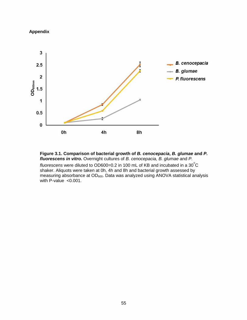
55
Appendix
Figure 3.1. Comparison of bacterial growth of B. cenocepacia, B. glumae and P. fluorescens in vitro. Overnight cultures of B. cenocepacia, B. glumae and P.
fluorescens were diluted to OD600=0.2 in 100 mL of KB and incubated in a 30oC
shaker. Aliquots were taken at 0h, 4h and 8h and bacterial growth assessed by measuring absorbance at OD600. Data was analyzed using ANOVA statistical analysis with P-value <0.001.
OD
600n
m

56
Figure 3.2. Competition assay between B. glumae and P. fluorescens in vitro. B. glumae (pGFP-TIR) was grown in 100 mL of KB broth alone or in combination with P.
fluorescences (ration 10:1). Single and mixed cultures were incubated in 30oC by shaking.
Aliquots from single and mixed cultures were taken at 0h, 4h, 8h, 24h and 48h, serially diluted and plated on KB containing gentamycin to specifically enumerate B. glumae (pGFP-TIR) bacteria. The graph represents bacterial numbers of B. glumae (pGFP-TIR) alone and when growth with P. fluorescens. For each time point, the growth of B. glumae (pGFP-TIR) grown alone was compared with that of B. glumae (pGFP-TIR) grown with P. fluorescens and analyzed using ANOVA statistical analysis with P- value = 0.6310 at 0h, 0.0365 at 4h, 0.514 at 8h, 0.8211 at 24h and 0.3696 at 48h.

57
Figure 3.3. Competition assay in vivo and competitive index between B. glumae and P. fluorescens. Four- week-old rice plants, cultivar Wells were inoculated with B. glumae (pGFP-TIR) alone or mixed with P. fluorescens. Bacterial inoculums were adjusted to OD600= 0.125. Plants were ground, serially diluted and plated at 0, 3 and 5 dpi on KB plates and KB containing gentamycin. The numbers of B. glumae (pGFP-TIR) were counted on KB plates containing gentamycin. The numbers of P. fluorescens were calculated by subtracting the numbers of B. glume (pGFP-TIR) from the total number of colonies obtained in KB alone. Bars represent the competitive index for each time point.

58
Figure 3.4. B. glumae grown in the presence of filter sterilized KB broth which
was incubated with agar plugs from zone of inhibition plates containing B. glumae and P. fluorescens or B. cenocepacia. Agar plugs were taken from LB agar not exposed to P. fluorescens or B. cenocepacia and from the zones of inhibition caused by B. cenocepacia or P. fluorescens on B. glumae plates. Agar plugs were
shaken in 3mL KB broth for 24h at 30oC to allow diffusion of inhibitory compounds into
the KB. After incubation, these preparations were filter-sterilized. One milliliter of each preparation was mixed with 1 ml of KB and used to grow a single colony of B. glumae.
Tubes were shaken at 30oC for 24h to qualitatively evaluate bacterial growth (A). One
hundred microliters of bacterial growth was serially diluted and plated to count populations of B. glumae (B). Bars represent the growth of B. glumae in the presence of the different preparations and the comparison among preparations was analyzed by ANOVA statistical analysis with P- value = 0.0153.
A
B
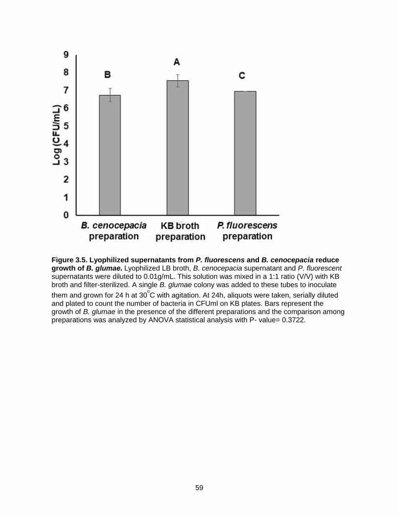
59
Figure 3.5. Lyophilized supernatants from P. fluorescens and B. cenocepacia reduce growth of B. glumae. Lyophilized LB broth, B. cenocepacia supernatant and P. fluorescent supernatants were diluted to 0.01g/mL. This solution was mixed in a 1:1 ratio (V/V) with KB broth and filter-sterilized. A single B. glumae colony was added to these tubes to inoculate
them and grown for 24 h at 30oC with agitation. At 24h, aliquots were taken, serially diluted
and plated to count the number of bacteria in CFUml on KB plates. Bars represent the growth of B. glumae in the presence of the different preparations and the comparison among preparations was analyzed by ANOVA statistical analysis with P- value= 0.3722.

60
Figure 3.6. Lyophilized supernatants from P. fluorescens and B. cenocepacia reduced disease symptoms caused by B. glumae. Eight- week- old rice plants cultivar Wells plants were injected with 20µL B. glumae at OD600= 0.125µL or B. glumae containing 0.1g/mL of lyophilized supernatants from either P. fluorescens or B. cenocepacia cultures or lyophilized LB broth. Disease symptoms were observed at 8 days after inoculation (A). Bars in (B) show the length of the lesions. The differences among lesion sizes were analyzed using ANOVA statistical analysis, P- value= 0.0003.
A
B

61
Conclusion
This research was performed to identify a biological control agent against the emerging
rice disease Bacterial Panicle Blight of rice, caused by the bacteria Burkholderia glumae. Two
potential sources of biological control agents were mined, Jupiter rice associated bacteria, and
the Rojas Lab collection. The hypothesis that the composition of rice associated bacteria from
moderately resistant Jupiter and susceptible Bengal would be different was supported by the
experiments and six bacterial strains unique to Jupiter were tested for their inhibitory activity
against B. glumae. Though some Jupiter rice associated bacteria did show small amounts of
inhibition against the pathogen, the effect was not enough to warrant further exploration as
biological control agents though it is possible that they contribute to the moderate resistance of
the rice cultivar, which was our second hypothesis. Two bacterial strains, Pseudomonas
fluorescens and Burkholderia cenocepacia, from the Rojas Lab collection showed very
promising results and were thus pursued. These bacterial strains were able control B. glumae in
vitro and in planta suggesting they would be successful at controlling the disease in the field.
Unfortunately these bacterial strains were not able to persist well in planta and B. cenocepacia,
being an opportunistic human pathogen, would not be safe to use as a biological control
treatment in the field. However when the mechanisms of control for these bacteria were
explored they gleaned promising results. When the hypothesis that these biological control
bacteria used competition as their main control agent was explored it was not supported by the
data, but when the hypothesis of antibiosis was explored it was found that supernatant
containing secreted fractions from the control agents but no live cells were still able to inhibit the
pathogen in vitro and in planta. This is promising as it shows the potential for a control method
against the disease to be developed without having to use bacterial strains which do not persist
well in the field and are potentially harmful to human health.