
Minimum requirements for ookinete to oocyst transformation in Plasmodium
12
Minimum requirements for ookinete to oocyst transformation in Plasmodium Victoria Carter a,1 , Ade ´la M.L. Nacer a , Ann Underhill a , Robert E. Sinden b , Hilary Hurd a, * a Centre for Applied Entomology and Parasitology, Institute for Science and Technology in Medicine, Huxley Building, Keele University, Staffordshire ST5 5BG, UK b Infection and Immunity Section, Department of Biological Sciences, Sir Alexander Fleming Building, Imperial College of Science, Technology and Medicine, Imperial College Road, London SW7 2AZ, UK Received 30 January 2007; received in revised form 8 March 2007; accepted 21 March 2007 Abstract During their passage through a mosquito vector, malaria parasites undergo several developmental transformations including that from a motile zygote, the ookinete, to a sessile oocyst that develops beneath the basal lamina of the midgut epithelium. This transfor- mation process is poorly understood and the oocyst is the least studied of all the stages in the malaria life cycle. We have used an in vitro culture system to monitor morphological features associated with transformation of Plasmodium berghei ookinetes and the role of basal lamina components in this process. We also describe the minimal requirements for transformation and early oocyst development. A defined sequence of events begins with the break-up of the inner surface membrane, specifically along the convex side of the ookinete, where a protrusion occurs. A distinct form, the transforming ookinete or took, has been identified in vitro and also observed in vivo. Contrary to previous suggestions, we have shown that no basal lamina components are required to trigger ookinete to oocyst transfor- mation in vitro. We have demonstrated that transformation does not occur spontaneously; it is initiated in the presence of bicarbonate added to PBS, but it is not mediated by changes in pH alone. Transformation is a two-step process that is not completed unless a range of nutrients are also present. A minimal medium is defined which supports transformation and oocyst growth from 7.8 to 11.4 lm by day 5 with 84% viability. We conclude that ookinete transformation is mediated by bicarbonate and occurs in a similar manner to the differ- entiation of sporozoite to the hepatic stage. Ó 2007 Australian Society for Parasitology Inc. Published by Elsevier Ltd. All rights reserved. Keywords: Plasmodium berghei; Ookinete; Oocyst; Basal lamina; Laminin; In vitro; Transformation 1. Introduction Malaria remains a major life-threatening disease in trop- ical and sub-tropical parts of the world, causing 300–500 million clinical cases, over one million deaths and severe eco- nomic and social losses (Sachs and Malaney, 2002). The causative agents of the disease, apicomplexans of the genus Plasmodium, are amongst the most studied parasites because of their devastating impact on human health and welfare. Plasmodium parasites cycle between a vertebrate host and mosquito vector, experiencing changing environmental con- ditions as they develop from invasive to intracellular forms in the vertebrate, and invasive, intracellular and extracellular forms in the mosquito. Despite the importance of mosquito stages for malaria transmission, the majority of research on Plasmodium is focused upon the asexual stages that invade vertebrate erythrocytes; studies facilitated by the develop- ment, in 1976 (Trager and Jensen, 1976), of an in vitro culture technique that has rendered these stages more accessible to experimentation (Hurd et al., 2003). 0020-7519/$30.00 Ó 2007 Australian Society for Parasitology Inc. Published by Elsevier Ltd. All rights reserved. doi:10.1016/j.ijpara.2007.03.005 * Corresponding author. Tel.: +44 1782 583034; fax: +44 1782 583516. E-mail address: [email protected] (H. Hurd). 1 Present address: Department of Infectious & Tropical Diseases, Pathology Molecular Biology Unit, London School of Hygiene & Tropical Medicine, Keppel Street, London WC1E 7HT, UK. www.elsevier.com/locate/ijpara International Journal for Parasitology 37 (2007) 1221–1232
-
Upload
victoria-carter -
Category
Documents
-
view
213 -
download
0
Transcript of Minimum requirements for ookinete to oocyst transformation in Plasmodium

www.elsevier.com/locate/ijpara
International Journal for Parasitology 37 (2007) 1221–1232
Minimum requirements for ookinete to oocysttransformation in Plasmodium
Victoria Carter a,1, Adela M.L. Nacer a, Ann Underhill a,Robert E. Sinden b, Hilary Hurd a,*
a Centre for Applied Entomology and Parasitology, Institute for Science and Technology in Medicine, Huxley Building, Keele University,
Staffordshire ST5 5BG, UKb Infection and Immunity Section, Department of Biological Sciences, Sir Alexander Fleming Building, Imperial College of Science,
Technology and Medicine, Imperial College Road, London SW7 2AZ, UK
Received 30 January 2007; received in revised form 8 March 2007; accepted 21 March 2007
Abstract
During their passage through a mosquito vector, malaria parasites undergo several developmental transformations including thatfrom a motile zygote, the ookinete, to a sessile oocyst that develops beneath the basal lamina of the midgut epithelium. This transfor-mation process is poorly understood and the oocyst is the least studied of all the stages in the malaria life cycle. We have used an in vitroculture system to monitor morphological features associated with transformation of Plasmodium berghei ookinetes and the role of basallamina components in this process. We also describe the minimal requirements for transformation and early oocyst development. Adefined sequence of events begins with the break-up of the inner surface membrane, specifically along the convex side of the ookinete,where a protrusion occurs. A distinct form, the transforming ookinete or took, has been identified in vitro and also observed in vivo.Contrary to previous suggestions, we have shown that no basal lamina components are required to trigger ookinete to oocyst transfor-mation in vitro. We have demonstrated that transformation does not occur spontaneously; it is initiated in the presence of bicarbonateadded to PBS, but it is not mediated by changes in pH alone. Transformation is a two-step process that is not completed unless a range ofnutrients are also present. A minimal medium is defined which supports transformation and oocyst growth from 7.8 to 11.4 lm by day 5with 84% viability. We conclude that ookinete transformation is mediated by bicarbonate and occurs in a similar manner to the differ-entiation of sporozoite to the hepatic stage.� 2007 Australian Society for Parasitology Inc. Published by Elsevier Ltd. All rights reserved.
Keywords: Plasmodium berghei; Ookinete; Oocyst; Basal lamina; Laminin; In vitro; Transformation
1. Introduction
Malaria remains a major life-threatening disease in trop-ical and sub-tropical parts of the world, causing 300–500million clinical cases, over one million deaths and severe eco-nomic and social losses (Sachs and Malaney, 2002). Thecausative agents of the disease, apicomplexans of the genus
0020-7519/$30.00 � 2007 Australian Society for Parasitology Inc. Published b
doi:10.1016/j.ijpara.2007.03.005
* Corresponding author. Tel.: +44 1782 583034; fax: +44 1782 583516.E-mail address: [email protected] (H. Hurd).
1 Present address: Department of Infectious & Tropical Diseases,Pathology Molecular Biology Unit, London School of Hygiene & TropicalMedicine, Keppel Street, London WC1E 7HT, UK.
Plasmodium, are amongst the most studied parasites becauseof their devastating impact on human health and welfare.
Plasmodium parasites cycle between a vertebrate host andmosquito vector, experiencing changing environmental con-ditions as they develop from invasive to intracellular forms inthe vertebrate, and invasive, intracellular and extracellularforms in the mosquito. Despite the importance of mosquitostages for malaria transmission, the majority of research onPlasmodium is focused upon the asexual stages that invadevertebrate erythrocytes; studies facilitated by the develop-ment, in 1976 (Trager and Jensen, 1976), of an in vitro culturetechnique that has rendered these stages more accessible toexperimentation (Hurd et al., 2003).
y Elsevier Ltd. All rights reserved.

1222 V. Carter et al. / International Journal for Parasitology 37 (2007) 1221–1232
In the mosquito, gametocytes complete their differentia-tion, fertilisation occurs and the zygote transforms into amotile ookinete within 24 h of having imbibed a bloodmeal. This extracellular ookinete migrates out of the bloodbolus, traverses the midgut epithelium and transforms intoa vegetative oocyst in the basal subepithelial space betweenthe midgut epithelium and the basal lamina (BL). Theyoung oocyst grows and undergoes sporogony, producingsporozoites that migrate to the salivary glands. Experimen-tation with systems that substitute for conditions in thevector for a period of some 50 years finally led to the devel-opment of methods to culture all the mosquito stages ofseveral malaria species. Species that have been successfullycultured from gametocyte to sporozoite include Plasmo-dium falciparum (Warburg and Schneider, 1993), Plasmo-
dium gallinaceum (Warburg and Miller, 1992),Plasmodium berghei (Al-Olayan et al., 2002a) and Plasmo-
dium yoelii (Porter-Kelley et al., 2006). For one of thesespecies, P. berghei, a method for the efficient productionof ookinetes in vitro has been developed (Rosales-Ronqu-illo and Silverman, 1974; Weiss and Vanderberg, 1977)and this has provided material for detailed studies of game-togenesis, fertilisation and zygote development. Severalstudies have demonstrated that initiation of Plasmodium
gametogenesis requires specific triggers. The change inenvironment from a mammalian to insect host providescues such as a drop in temperature, and a mosquito factoridentified as xanthurenic acid (Nijhout and Carter, 1978;Arai et al., 2001) which can regulate gametogenesis. Thesetriggers, or substitutes for them, are present in the in vitrosystems used to produce ookinetes (Weiss and Vanderberg,1977; Carter et al., 2003).
The next critical event in the development of the sporo-gonic stages of Plasmodium is that of the transformationof ookinete to oocyst. Our knowledge of this phase of devel-opment is minimal and the process of rounding up of theookinete to form an oocyst had not been studied in vitroprevious to this work. We have identified components ofthe culture system that are essential requirements to initiateand sustain the formation of young oocysts from ookinetes.
It had been proposed that contact with components ofthe BL may act as a trigger for ookinete transformationin vivo (Weathersby, 1952; Meis and Ponnudurai, 1987;Ramasamy et al., 1997; Adini and Warburg, 1999; Schnei-der and Shahabuddin, 2000; Dessens et al., 2003; Arrighiet al., 2005; Vlachou et al., 2006) and culture systems thatallow the transformation of ookinetes to oocysts, oocystgrowth and sporozoite production have included a BL sub-stitute, Matrigel and a co-cultured cell line such as Drosoph-
ila melanogaster S2, that may also contribute moleculespresent in the mosquito BL (Warburg and Miller, 1992;Warburg and Schneider, 1993; Al-Olayan et al., 2002a).Matrigel, an extracellular matrix derived from Engel-breth–Holm–Swarm mouse sarcoma, is comprised of lami-nin (56%), collagen IV (31%), enactin (8%), heparan sulfateproteoglycans and growth factors (BD Biosciences). Severalookinete proteins, such as the surface proteins P25/P28
(Siden-Kiamos et al., 2000), secreted ookinete adhesive pro-tein (SOAP) (Dessens et al., 2003), circumsporozoite- andTRAP-related protein (CTRP) (Dessens et al., 1999) andvon Willebrand factor A domain-related-protein (WARP)(Yuda et al., 2001), that are reportedly essential for ooki-nete and oocyst development in vivo, have been shown tobind to BL components such as laminin and collagen (Adiniand Warburg, 1999; Arrighi et al., 2005). This binding couldprovide a signal that induces transformation, thus addingweight to the hypothesis that contact with the BL may beessential for parasite development (Dessens et al., 2003).Alternatively, other components of the culture system suchas those present in the insect cell medium used as a substi-tute for haemolymph, may be essential requirements forookinete transformation.
In vivo, ookinete transformation is a transient and unsyn-chronised event but in vitro we can observe the processoccurring in thousands of ookinetes at any one time. Addi-tionally, controlled interventions that enable careful dissec-tion of putative transformation signals can be made andthe minimal nutritional/environmental requirements forsporogonic development can be assessed. Using ookinetesthat had been obtained in vitro (Carter et al., 2003) as a start-ing point, we have been able to describe the process of trans-formation and to identify a novel transient stage that we havecalled a transforming ookinete or took. We have demon-strated that, contrary to previous suggestions, BL compo-nents, and in particular laminin, are not required fortransformation and early oocyst growth and that transfor-mation appears to be a two-step process in which initiationis bicarbonate-dependent and complete transformationrequires a range of nutrients.
2. Materials and methods
All reagents were purchased from Sigma unless other-wise specified.
2.1. Parasites and ookinete harvesting
Experiments were carried out in accordance with theUK Animals (Scientific Procedures) Act 1986 usingapproved protocols. CD mice were treated with phen-ylhydrazine 2 days prior to infection by i.p. inoculationwith P. berghei (clone 259cl2 expressing green fluorescentprotein (Franke-Fayard et al., 2004)) obtained from adonor mouse between the second and sixth passage fromcryopreserved stock. Infected blood was collected frommice with a parasitaemia of 10–20% via cardiac punctureinto a heparinised syringe. Gametocytaemic blood waspassed through a pre-equilibrated, 5 ml sterile column con-taining 1 ml glass wool and 3 ml of Whatman CF11 cellu-lose powder (Beckton & Dickenson) to remove white bloodcells. The blood was collected into T25 culture flasks withRPMI 1640 medium supplemented with 24 mM sodiumbicarbonate, 0.4 mM hypoxanthine, 10,000 U penicillin/10 mg streptomycin and 20% FBS in a 1:10 ratio (blood:

V. Carter et al. / International Journal for Parasitology 37 (2007) 1221–1232 1223
media). Gametocytes developed into ookinetes after 18 h ofculture at 19 �C.
Ookinetes were separated on a MidiMacs (Miltenyi Bio-tec, Germany) separation system as previously described(Carter et al., 2003). Briefly this involved passing the bloodand supplemented RPMI mixture through a pre-separationfilter (to remove cellular debris and coagulated blood) anda MidiMac LS column mounted on a magnet. Ookinetesadhere to magnetised beads packed within the columnand can be eluted by removing the column from the mag-net. The rate of blood flow was reduced by adding a 23Gneedle to the base of the LS column to increase parasitecontact time with the column.
2.2. Culture conditions
Culture conditions were as previously described (Al-Olayan et al., 2002a). Ookinetes were seeded at a densityof 1 · 104 per well into 8-well LabTek chamber slides orinto 48-well plates (IWAKI) and cultured in oocyst culturemedium (OCM: see Table 1) unless otherwise stated. Wellswere either pre-coated with Matrigel diluted 1:1 withSchneider’s Drosophila insect medium or incubation tookplace in non-coated chamber slides. Parasites were eitherco-cultured in a 10:1 ratio with D. melanogaster S2 cells(Matrigel + S2), cultured in non-coated chamber slideswith S2 cells (no-coating + S2), or in non-coated slideswithout co-cultured cells (no-coating � S2). Parasites weremaintained at 19 �C for 5 days, unless otherwise stated.Media was replenished on day 3 (100 ll removed andreplaced). All experiments were performed with six repli-cate wells on two or three separate occasions.
Drosophila melanogaster S2 cells were maintained in T25culture flasks (Griener Bio-One) at 27 �C in supplementedSchneider’s insect cell culture medium (ICM) with 10%heat-inactivated insect cell culture tested FBS, penicillin(50 U/ml) and streptomycin (50 lg/ml).
2.3. Assessment of viability and transformation
Initially, a comparison was made between a Live/Deadviability/cytotoxicity kit (Molecular Probes) used accord-ing to the manufacturers’ instructions, and Erythrosin B(0.1% in PBS diluted 1:1 with the parasite culture medium)exclusion (Krause et al., 1984). No difference was detected
Table 1Components of full-oocyst medium (OCM) per 100 ml
Oocyst medium component Final concentration/100 ml
Sodium bicarbonate (mM) 23.8Hypoxanthine (mM) 3.68Penicillin (U)/streptomycin (mg) 10,000; 10Gentamicin (mg) 20Para-aminobenzoic acid (lM) 44Foetal bovine serum (%) 15Lipid/cholesterol (%) 0.2Schneider’s medium (%) 85
Media were adjusted to pH 7 prior to filtration.
between the results for cell viability between these tests(data not shown) and the latter test was used routinely inthis study. Material was removed from culture wells andcentrifuged at 2000g for 1 min to pellet parasites or insectcells. For parasite transformation rates, sample pellets wereresuspended in 5 ll of PBS and 5 ll of Erythrosin B andmounted onto a glass slide for quantification. One-hundredlive parasites were counted for each condition under oilimmersion, and classified according to their morphologicalappearance of ookinete (banana), transforming ookinete(snail-like) or oocyst (sphere with a minimum diameter of7 lm and highly motile internal contents). Counts wereperformed on triplicate samples. Transformation rateswere determined by counting the number of live (Erythro-sin B-excluding) ookinetes present on the initial day of cul-ture, compared with the number after transformation(disappearance of ookinetes with concomitant increase inyoung oocysts denotes transformation as non-transformingookinetes remain intact for several days).
For visualisation of nuclear material, ookinetes werefixed for 10 min in 1:1 ice cold acetone:methanol, washedand then incubated with anti-P28 monoclonal antibodyfor 1 h at room temperature before further washing andincubation with an anti-mouse IgG antibody diluted1:200 for 1 h. Samples were washed again then mountedin Vectashield containing DAPI (Vector Laboratories)and visualised on an inverted Lieca DM IRB fluorescencemicroscope (Lieca Microsystems, Germany).
2.4. Electron microscopy
Plasmodium berghei samples were pelleted at 500g for4 min 2 days post-culture and fixed in 2.5% glutaraldehydein sodium cacodylate buffer (0.1 M, pH 7.4). Samples werepost-fixed in 1% osmium tetroxide dispersed in 3% agar,dehydrated in graded ethyl alcohols and embedded inSpurr’s resin following standard protocols. Ultrathin sec-tions, cut with a diamond knife on a Reichart ‘‘UltracutE’’, were mounted on grids and stained with uranyl acetate.Grids were examined with a JOEL 1230 TEM and imagesrecorded using ‘‘analySIS’’ software.
2.5. Ookinete rescue
Ookinetes were observed to remain viable when culturedin PBS alone for up to 7 days, as determined by ErythrosinB exclusion, but did not transform. To initiate transforma-tion, PBS (100, 200 or 300 ll) was removed from theculture well and replaced with 100, 200 or 300 ll of full-oocyst medium on day 1, 2 or 3 of culture. Samples werecentrifuged at 2000g for 1 min and parasite viability wasassessed as above.
2.6. Mosquito infections
Anopheles stephensi were starved for 24 h prior to blood-feeding for 20 min on gametocytaemic CD mice infected

1224 V. Carter et al. / International Journal for Parasitology 37 (2007) 1221–1232
with P. berghei (PbGFPCON clone 259cl2). The mosquitoeswere thereafter maintained on a 4% glucose solution anddissected in PBS 26–27 h p.i. Midguts from infected mos-quitoes were cut open and the blood meal vigorouslywashed out with PBS before examination under oil emer-sion on the inverted fluorescence microscope for the pres-ence of tooks on the BL side of the gut.
2.7. ELISA
Drosophila S2 cells have been reported to secrete laminin(Fessler et al., 1987). To confirm secretion of laminin byour cell line, Drosophila S2 cells from a confluent flask werecounted on a haemocytometer and the total volume ofsupernatant from the culture was measured to calculatethe number of cell equivalents per 100 ll of medium. Addi-tionally, conditioned medium was collected from wells inwhich 1 · 105 cells were incubated for 1, 3, 5, 7 or 9 daysat 19 �C in 400 ll of OCM (2.5 · 104 cells per 100 ll) todetermine if laminin secretion could be detected in parasiteculture conditions.
ELISA plates were coated with 100 ll of conditionedmedium, diluted 1:50 in coating buffer (15 mM Na2CO3,35 mM NaHCO3, pH 9.6) and serial dilutions made there-after to 1:2000 and allowed to bind overnight at 4 �C.Plates were washed, blocked and incubated with antibodiesraised against the b- and c-chains of Drosophila laminin(Kumagai et al., 1997) diluted at 1:1000 in milk/PBS/Tween. Plates were then washed, incubated with an alka-line phosphatase (AP)-conjugated secondary antibody(Sigma) diluted 1:1000 in milk/PBS/Tween, and washedagain. The substrate, p-nitrophenylphosphate, was usedfor visualisation. Control wells had primary antibody omit-ted. In addition, Schneider’s medium and FBS were testedfor laminin and its absence confirmed.
2.8. Detection of laminin bound to culture plates
To establish whether laminin could bind to cultureplates, making it unavailable in solution to developing par-asites, culture plates were incubated for 24 h at 19 �C with100 lg of mouse laminin in PBS. The plates were washedwith PBS and assayed for laminin by immunofluorescenceusing a polyclonal anti-laminin antibody directed againstall three chains. Similarly, Drosophila S2 cells were culturedin OCM for 24 h, the cells were then removed, and the cul-ture plates assayed to determine whether laminin secretedby the cells was bound to the plate.
2.9. Minimal media
Using sterile PBS as a base, components of OCM(Schneider’s medium, heat-inactivated FBS, lipoproteinand cholesterol, NaHCO3 and hypoxanthine) were addedsingularly or in combination at appropriate concentrations.During further medium minimisation, Albumax II (Invit-rogen) was used as a replacement for FBS and Schneider’s
medium was replaced by individual components in groupsof (i) amino acids, (ii) inorganic salts and (iii) other compo-nents, as listed by the manufacturer.
2.10. Media pH
Full-media were used for studies on the effects of themedia pH. Media were adjusted by adding 5 M NaOH atroom temperature before filtration (0.22 lm). However,as pH was found to steadily rise during the culture period,25 mM N-2-hydroxyethylpiperazine-N 0-2-ethanesulfonicacid (Hepes) was also used as a buffer to maintain initialpH values of 6, 7, 8 and 9. PBS adjusted to pH 6, 7, 8 or9 was used for comparison.
2.11. Culture vessels
In the absence of either Matrigel or co-cultured cells,ookinetes were observed to adhere to the surface (IWAKICat. No. 3830-048); as determined by no movement upongentle agitation. To eliminate possible cues received fromadhesion, ookinetes were seeded into non-treated IWAKImultiwell plates (Cat. No. 1830-048) at the same density.To remove potential transformation cues received frommutual adhesion, ookinetes were placed one per well intreated and non-treated plates.
2.12. Statistical analyses
Data were analysed using Minitab version 14. TheAnderson-Darling and Bartlett’s test were used to test fornormal distribution and equality of variance, respectively.Data from repeat experiments were compared using a Gen-eral Linear Model and, if not significantly different, pooled,tested for normality and equality of variance and analysedusing the General Linear Model to compare transforma-tion rates, growth and viability across conditions andacross time.
3. Results
3.1. Markers for viability
A major constraint in characterising the process oftransformation has been to demonstrate conclusively thatan ookinete has undergone differentiation to become anoocyst. Using P. berghei (PbGFPCON clone 259cl2) thatconstitutively expresses green fluorescent protein (GFP),we have observed that GFP expression, although becomingfaint, often persists in parasites permeable to Erythrosin B.GFP expression was therefore deemed a less reliable viabil-ity indicator in our culture system.
Viability of ookinetes at the outset typically ranged from80% to 90%. Parasites continued to exclude the vital dyeErythrosin B after rounding up to form oocysts, thus sup-porting the view that the changing morphology that we

V. Carter et al. / International Journal for Parasitology 37 (2007) 1221–1232 1225
observe during transformation is not associated with dyingookinetes.
3.2. Markers for transformation
A P. berghei ookinete transforms from a banana-likeshape, approximately 10 lm in length, to become a spher-ical young oocyst with a diameter of 7–8 lm. In an attemptto find mRNA markers that would help to identify a newlytransformed oocyst and distinguish it from other parasitestages that may be present in the culture, we looked atthe following well-characterised housekeeping or putativeookinete/early oocyst-specific molecules for signs ofchanges in transcription: CTRP, WARP, sporozoite micro-neme protein essential for cell traversal (SPECT), Myosin
Fig. 1. Formation of young Plasmodium berghei oocysts in vitro. (A–C) P28 (suoocysts (C). (D) PbGFPCON tooks during late transformation in culture. (E) Pbpost-blood feeding (composite of three images). (F) Image adapted from Sindevii). Transformation begins with a small hump forming on the outer, convex, e(tooks) then take on a snail-like appearance (v) and the nucleus is observed to mvisible (distinguishing these forms from zygotes; (vi) just prior to the formation12–36 h, depending on nutrient availability.
A, hypoxanthine–guanine phosphorybosyltransferase(HGPRT), scavenger receptor, P28, RNA S and circums-porozoite protein (CSP). However, none of these were suit-able as no change in transcript abundance occurred duringthe transformation process or in the early oocyst (data notshown). We were therefore unable to positively identify thepresence of early oocyst-specific features by PCR analysisand assessment has relied on morphological observationsand the demonstration of growth.
3.3. Morphological and ultra-structural description of
transformation
The in vitro transformation of P. berghei ookinetes toyoung oocysts follows a precise morphological pattern,
rface) and DAPI (nuclear) staining of ookinetes (A), tooks (B) and youngGFPCON early transforming ookinetes in Anopheles stephensi midguts 27 hn et al. (1987) (i–iv) to incorporate ookinete to oocyst transformation (v–dge of the ookinete, often towards the apical end. Transforming ookinetes
ove into the developing oocyst. Remnants of the ookinete tips are clearlyof spherical young oocysts. The entire population of ookinetes transform in

1226 V. Carter et al. / International Journal for Parasitology 37 (2007) 1221–1232
distinct from that of zygote through retort to ookinete.Whereas immature ookinetes form via an extension froma spherical zygote, projecting from just one region (Sindenet al., 1985, 1987), fully mature, banana-shaped ookinetesprogress to oocysts by rounding up via the formation ofa protrusion in the middle of the ookinete that always orig-inates on the outer convex edge. We have identified thisphase as a transforming ookinete (took) (see Fig. 1A–Cand F). The nucleus is in the hump, the two-tail ends ofthe ookinete are still visible in late tooks (Fig. 1B and D)and DAPI-positive material appears to increase in size
Fig. 2. TEM images of an ookinete (A), and tooks (B–F) in vitro. (A) Ookinettook: the AC is still clearly visible as are the microtubules (MT) and the beginMembrane generated during this process differs from the ookinete pellicle (sinnuclear material can be seen to have moved into the newly formed area. Doubarrows indicate the transition areas from double to newly formed single mremaining pellicle. (F) Late took: remnants of the ookinete tips with double m2 lm; D–F, 1 lm.
when in the newly forming oocyst (Fig. 1C). We regardedfully rounded tooks as having completed transformationto an early oocyst. Within each culture, the process oftransformation of mature ookinetes to oocysts is asynchro-nous but all viable ookinetes have become oocysts in24–48 h. Ookinetes that have permeable membranes donot complete transformation.
Plasmodium berghei (PbGFPCON) tooks have also beenobserved in vivo on mosquito midguts 28 h post-feedingon gametocytaemic blood (Fig. 1E). At the ultra-structurallevel, in vitro, the single plasmalemma of the newly forming
e, with nucleus (N), pellicle (P) and apical complex (AC) visible. (B) Earlyning of the early oocyst wall, forming a hump on the outer ookinete edge.gle arrow), and forms the basis of the oocyst wall (OW). (C) Early took:le and single membranes define the new OW and ookinete pellicle. Brokenembranes. (D and E) Mid-stage tooks showing microtubules below theembranes can be seen at either end of the forming oocyst: scale bar: A–C,
V. Carter et al. / International Journal for Parasitology 37 (2007) 1221–1232 1227
oocyst can be distinguished from the triple membrane ofthe ookinete pellicle and microtubules are no longer visiblein this area (Fig. 2B–F). A distinct junction between theookinete pellicle and the single plasmalemma can be seen(Fig. 2D and E) and the nucleus can be observed in thehump of the took (Fig. 2C).
3.4. The role of basal lamina components and co-cultured
cells in ookinete to oocyst transformation
Following incubation of gametocytaemic blood for 18 h,cultures were enriched for ookinetes; however, they stillcontained asexual stage parasites, gametocytes and zygotesin the process of transforming into ookinetes. The propor-tion of ookinetes in the total parasite population decreasedfrom �25% on day 0 to �3% on day 1 and less than 1% onday 2 post-culture. No tooks were present on day 0 butthey represented approximately 10% of the total fluoresc-ing parasites when viewed on days 1 and 2, and a concom-itant increase in diameter of spherical parasites fromapproximately 7 to 8 lm occurred from day 1 to day 2.As development can only proceed in one direction, thesespheres were regarded as newly transformed oocysts. Over90% of the ookinetes cultured with Drosophila S2 cells inthe presence of the basal lamina substitute, Matrigel, trans-formed into young oocysts (termed maximal transforma-tion rate) over a 1 to 2 day period and underwent somegrowth in diameter by day 3.
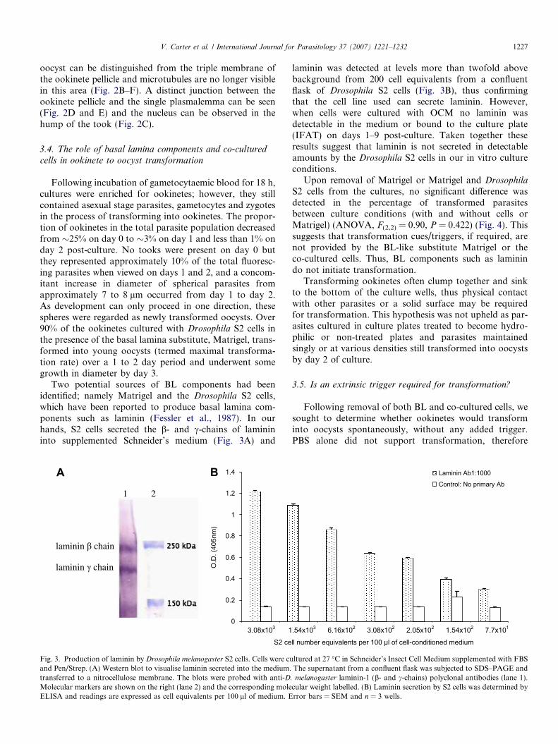
Two potential sources of BL components had beenidentified; namely Matrigel and the Drosophila S2 cells,which have been reported to produce basal lamina com-ponents such as laminin (Fessler et al., 1987). In ourhands, S2 cells secreted the b- and c-chains of laminininto supplemented Schneider’s medium (Fig. 3A) and
A
1 2
0
0.2
0.4
0.6
0.8
1
1.2
1.4
S2 ce
O.D
. (40
5nm
)
B
laminin chain
laminin chain
3.08x10³ 1
Fig. 3. Production of laminin by Drosophila melanogaster S2 cells. Cells were cuand Pen/Strep. (A) Western blot to visualise laminin secreted into the medium.transferred to a nitrocellulose membrane. The blots were probed with anti-DMolecular markers are shown on the right (lane 2) and the corresponding moleELISA and readings are expressed as cell equivalents per 100 ll of medium. E
laminin was detected at levels more than twofold abovebackground from 200 cell equivalents from a confluentflask of Drosophila S2 cells (Fig. 3B), thus confirmingthat the cell line used can secrete laminin. However,when cells were cultured with OCM no laminin wasdetectable in the medium or bound to the culture plate(IFAT) on days 1–9 post-culture. Taken together theseresults suggest that laminin is not secreted in detectableamounts by the Drosophila S2 cells in our in vitro cultureconditions.
Upon removal of Matrigel or Matrigel and Drosophila
S2 cells from the cultures, no significant difference wasdetected in the percentage of transformed parasitesbetween culture conditions (with and without cells orMatrigel) (ANOVA, F(2,2) = 0.90, P = 0.422) (Fig. 4). Thissuggests that transformation cues/triggers, if required, arenot provided by the BL-like substitute Matrigel or theco-cultured cells. Thus, BL components such as laminindo not initiate transformation.
Transforming ookinetes often clump together and sinkto the bottom of the culture wells, thus physical contactwith other parasites or a solid surface may be requiredfor transformation. This hypothesis was not upheld as par-asites cultured in culture plates treated to become hydro-philic or non-treated plates and parasites maintainedsingly or at various densities still transformed into oocystsby day 2 of culture.
3.5. Is an extrinsic trigger required for transformation?
Following removal of both BL and co-cultured cells, wesought to determine whether ookinetes would transforminto oocysts spontaneously, without any added trigger.PBS alone did not support transformation, therefore
ll number equivalents per 100 µl of cell-conditioned medium
Laminin Ab1:1000
Control: No primary Ab
.54x103 6.16x10² 3.08x10² 2.05x10² 1.54x10² 7.7x101
ltured at 27 �C in Schneider’s Insect Cell Medium supplemented with FBSThe supernatant from a confluent flask was subjected to SDS–PAGE and
. melanogaster laminin-1 (b- and c-chains) polyclonal antibodies (lane 1).cular weight labelled. (B) Laminin secretion by S2 cells was determined byrror bars = SEM and n = 3 wells.

0102030405060708090
100
Matrigel +S2 No Coating +S2 No coating -S2
Culture conditions
% T
rans
form
ed o
ocys
ts
Fig. 4. Maximal ookinete to oocyst transformation rates 24-h post-culture. Transformation rates were assessed by counting the number ofparasites out of 100 that had changed from ookinete shape to youngoocysts. No significant difference (ANOVA, F(2,2) = 0.90, P = 0.422) wasobserved between full-culture conditions, culture without Matrigel andculture without Matrigel and Drosophila S2 cells. Bars represent SEM.The experiment was repeated three times and parasites counted in threeseparate wells for each condition.
1228 V. Carter et al. / International Journal for Parasitology 37 (2007) 1221–1232
precluding time-dependent transformation in these mini-mal conditions. Viable ookinetes could be maintained inPBS for up to 7 days (e.g. viability on day 1, 84%; day 3,65% and day 6, 5%). Removal of 100, 200 or 300 ll ofthe PBS and replacement with the same quantity ofOCM stimulated transformation in a time and volumedependent manner (see Table 2). If still viable, ookinetescould be induced to transform up to 3 days post-PBS cul-ture, thus suggesting that the transformation process willnot be supported unless appropriate cues are received bythe parasite, and that these cues are present in OCM in lim-iting amounts.
3.6. Minimal requirements for ookinete to oocyst
transformation
The addition of single OCM components to PBS hadvarying effects on parasite development (Table 3). Notably,sodium bicarbonate (23.8 mM) alone supported transfor-mation of ookinetes to live tooks, but parasites failed tocomplete the entire rounding-up process to form youngoocysts. In contrast, a 0.2% lipid/cholesterol componentwas toxic to ookinetes, killing approximately 25% within4 h of culture and all ookinetes within 24 h, and althougha tenth of the concentration was not toxic, it did not stim-ulate transformation.
The addition of combinations of heat-inactivated FBS(HI FBS) and sodium bicarbonate or Schneider’s mediumand sodium bicarbonate to PBS permitted complete trans-formation at maximal rates. However, the latter combina-tion resulted in the appearance of a white precipitate,obscuring views of ookinetes and leading to the death ofparasites within 2–3 days. Transformation also occurredwhen sodium bicarbonate was replaced with sodium car-bonate, although a precipitate was again formed. Growth
of viable oocysts proceeded for 5 days in PBS containing15% HI FBS and 23.8 mM sodium bicarbonate (minimalmedia) The mean diameter of young oocysts increasedfrom 7 to 8 lm on day 1 post-culture to 11–12 lm onday 5 (Table 4). However, maintaining the parasites in min-imal media did not permit complete sporogonic develop-ment and oocysts were no longer viable after 7 days.
As serum is notoriously difficult to define, we substituted15% FBS in PBS with 15% Albumax, but this failed to sup-port transformation. We therefore tried to identify groupsof components of Schneider’s medium (e.g. amino acids,salts, sugars or yeast) that alone might sustain develop-ment. None of these groups permitted oocyst development,however partial transformation did occur in the inorganicsalts group (data not shown). These results indicate that,whilst bicarbonate alone may initiate transformation, com-plete transformation requires a combination of essentialnutrients and thus transformation may be a two-stageprocess.
3.7. Media pH and transformation
As the pH of bicarbonate buffers is strongly influencedby pCO2 and hence sodium bicarbonate concentration,we adjusted the pH of OCM to establish if this alone wascritical for transformation (Table 5). A high pH (9.0)greatly accelerated the rate of transformation; which beganwithin 4 h of culture. However, at pH 9.0, GFP fluores-cence was less intense and oocyst plasma membrane break-down occurred from day 2 post-culture, after oocysts hadattained diameters of 10–15 lm. Low pH (6.0) severelyimpaired oocyst growth. Transformation was maximal atpH 7 and 8.
4. Discussion
Using our in vitro system, we have been able to observemany thousands of ookinetes undergo transformation toyoung oocysts. Our observations of tooks in vivo havebeen far less frequent, as only a few of an invading popu-lation will be transforming at any one time. However, itis clear that this method of transformation certainly occursin vivo. It would appear that the break-up of the inner sur-face membrane of the ookinete begins specifically along theconvex side of the ookinete and spreads from this point,and there is likely to be an increase in the surface area ofthe single plasmalemma during this process. We wereunable to detect remnants of the inner surface membranein areas where a single plasmalemma was visible and theapical complex did not appear to undergo immediate de-differentiation (Canning and Sinden, 1973) but persisteduntil late into the took phase. The process of transforma-tion thus appears to be similar to that of the sporozoiteto the exoerythrocytic stage in the hepatocyte (Meiset al., 1983).
Ookinete to oocyst transformation represents one of themost severe bottlenecks for the malaria parasite, often

Table 4Growth and viability of Plasmodium berghei oocysts over a 5 day period of culture in minimal media
Time post-culture Day 1 Day 3 Day 5
Viability (n = 100) (%) 92 89 84Mean oocyst size (n = 100) (lm) 7.80 ± 0.1 9.51 ± 0.17 11.36 ± 0.15
Example of oocyst
Parasites were cultured in PBS containing 15% HI FBS and 23.8 mM sodium bicarbonate. One-hundred viable-oocyst diameters were measured each day,observed under phase contrast. Sizes are expressed as means ± SEM.
Table 2Transformation of ookinetes to oocysts maintained in PBS
Oocyst medium added Day 0 Day 1 Day 2 Day 3 Day 4 Day 5
400 ll added day 0 – X XX XX XX XX
100 ll added day 0 – – X XX XX XX
100 ll added day 1 – – – X XX XX
100 ll added day 2 – – – – X XX
200 ll added day 0 – X XX XX XX XX
200 ll added day 1 – – X XX XX XX
200 ll added day 2 – – – X XX XX
300 ll added day 0 – X XX XX XX XX
300 ll added day 1 – – X XX XX XX
300 ll added day 2 – – – X XX XX
Ookinetes were incubated in 400 ll of PBS which was then replaced with oocyst medium as indicated. A dose-dependent response was seen in the amountand speed of transformation. Data from ookinetes cultured in 400 ll of oocyst medium from the day of culture are displayed for comparison. Sampleswere taken from three independent experiments and media was replaced on day 3.–, no transformation; X, partial transformation (tooks); XX, complete transformation of the majority of ookinetes to oocysts.
Table 3Minimal requirements for triggering ookinete to oocyst transformation in PBS
Oocyst medium component Day 0 Day 1 Day 2 Day 3
Full-media – X XX XX
PBS alone – – – –PBS + hypoxanthine – – – –PBS + PABA – – – –PBS + lipids – · · ·PBS + sodium bicarbonate – X X X
PBS + Schneider’s medium – X X XX
PBS + HI FBS – X X XX
PBS + Schneider’s + sodium bicarbonate – X XX XX
PBS + HI FBS + sodium bicarbonate – X XX XX
Stages of transformation (compared with full-media) were assessed on days 0, 1, 2 and 3 post-culture when single and dual components of oocyst mediumwere added to PBS (see Table 1 for concentrations). Samples were observed from three independent experiments.–, no transformation; X, partial transformation (tooks); XX, complete transformation to oocyst; ·, death of parasites.HI FBS, heat-inactivated FBS; PABA, para-aminobenzoic acid.
V. Carter et al. / International Journal for Parasitology 37 (2007) 1221–1232 1229
reducing ookinete numbers to single digits (Dimopouloset al., 2002). Culturing the mosquito stages of the rodentmalaria parasite in vitro, in the absence of the unfavorableconditions generated in the blood meal bolus and by the
insects’ innate defence system, permits transformation ofthe majority of live ookinetes to the oocyst stage andthereby suggests that most parasites have the potential todevelop rather than undergoing apoptosis as they do in

Table 5Effects of pH on transformation rates
pH Day 1 Day 2 Day 3 Day 4 Day 5
Full-media
6 – – X X XX
7 X XX XX XX XX
8 X XX XX XX XX
9 XX XX XX · ·
PBS
6 – – – – –7 – – – – –8 – – – – –9 – – – – –
Hepes (25 mM) was added to full-oocyst medium to retain the appropriatepH. PBS alone, irrespective of pH, does not permit ookinete to oocysttransformation. Full-oocyst media permits transformation at a wide pHrange, but is inefficient or toxic at low and high pH’s, respectively.
–, no transformation; X, partial morphological transformation; XX,complete transformation to oocyst; ·, death of parasites.
1230 V. Carter et al. / International Journal for Parasitology 37 (2007) 1221–1232
the midgut lumen (Al-Olayan et al., 2002b). The investiga-tions reported here suggest that this transformation is notinternally controlled but that specific external compoundsare required to stimulate this process.
Some authors have made observations that suggest thatthe BL provides the necessary cues for oocyst transforma-tion (Adini and Warburg, 1999; Arrighi et al., 2005). Forexample, when gametocytes are injected directly into thehaemocoel, ectopic oocysts develop attached to the BL sur-rounding tissues such as the fat body and Malpighiantubules (Weathersby, 1954; Beaudoin et al., 1974). Forthese reasons, many culture protocols have employed aBL substitute (Matrigel) and it has been proposed thatinteractions between ookinete surface or microneme pro-teins and the mosquito BL play some role in oocyst devel-opment (for review see Siden-Kiamos and Louis, 2004).Here, we demonstrate conclusively that ookinetes willtransform in the absence of any source of BL componentssuch as laminin or collagen. Additionally, adhesion to asolid substrate or other cells is not required to trigger trans-formation. This may indicate that ookinete transformationis driven by soluble factors derived from the haemolymph.
Developing below the BL, in the subepithelial space,ookinetes and oocysts are bathed in haemolymph compo-nents, although direct contact with haemocytes is unlikely.Haemolymph from infected or non-infected A. stephensi
mosquitoes has been recorded to have a pH ranging from6.5 to 6.8 (Mack and Vanderberg, 1978). Whilst ourin vitro investigation indicates that maximal initiation ofookinete to oocyst transformation and subsequent growthoccurs within a pH range of 7–8 we did not examine eventsat pHs between 6 and 7, leaving the possibility that a pH inthis range may be more favourable.
With the addition of sodium bicarbonate alone, ookine-tes proceed to early stages of transformation as indicatedby a change in shape, but do not complete developmentto young oocysts. It would appear that sodium bicarbonateis important for transformation and we suggest it may be a
requirement for the first part of a two-step process. Follow-ing took formation, nutrients such as amino acids and sug-ars are needed for transformation to be completed andsome growth to occur. These findings appear to reflectthose reported in an investigation of triggers for in vitroexflagellation in P. berghei. Exflagellation could be stimu-lated in an extracellular bicarbonate concentration of28 mM, whereas variation of the pH from 7.2 to 8.6 in asolution depleted of bicarbonate did not, alone, providethe stimulus (Claudianos et al., 2002).
The requirement for a range of nutrients for the secondstep of transformation suggests metabolite stores areseverely limited in the ookinete and insufficient to sustainthe immediate morphological changes that occur duringcomplete transformation.
In contrast to the majority of parasites that infect mos-quitoes, both Hepatozoon caiman (Lainson et al., 2003) andPlasmodium spp. develop underneath the BL on the midgutof their respective vectors, namely Culex fatigans andAnopheles spp., and do not enter the haemocoel immedi-ately after exiting the gut lumen. This suggests that theremay be an advantage to be gained by developing in thissite. Recently, using double-stranded RNA (dsRNA) genesilencing technology, Arrighi et al. (2005) demonstratedthat depletion of mosquito laminin led to a substantialdecrease in oocyst numbers and suggested that the presenceand integrity of the BL is vital for parasite development. Sofar, its role has not been elucidated but it may be of impor-tance in vivo but not in vitro. The proposal that interac-tions between mosquito laminin and molecules in theoocyst capsule may result in the masking of the oocystfrom the mosquito’s immune system, thereby protectingthe developing oocyst from being melanised, has beenrecently supported by findings of Warburg et al. (2007).They demonstrated that beads coated in laminin or theookinete surface protein P28 are protected from melanisa-tion when injected into the mosquito haemocoel. It wouldbe useful investigate this hypothesis further using a refrac-tory, melanizing, strain of mosquito.
In conclusion, we believe we present the first descriptionof an intermediary form between ookinete and oocyst,namely the took. Transformation of P. berghei ookinetesto young oocysts in vitro is not a simple time-dependentphenomenon; it requires a precise environmental stimulus,but not one provided by basal lamina components. Thefirst stage of transformation is bicarbonate dependent,yet full transformation also requires nutrients and a suit-able pH. Minimal requirements for transformation havebeen reduced in this culture system to 15% HI FBS and23.8 mM sodium bicarbonate adjusted to pH 7.0.
These findings advance our understanding of the leaststudied phase of malaria parasites, that of the oocyst. Theydemonstrate the benefit that an in vitro system can bring toinvestigations of the biology of the mosquito stages of theparasite. In addition, re-evaluation of the potential forattenuated sporozoite-based malaria vaccines (Good,2005; Waters et al., 2005) has brought to the forefront

V. Carter et al. / International Journal for Parasitology 37 (2007) 1221–1232 1231
the need for competent systems to culture sporogonicstages of the life cycle.
Acknowledgements
We are grateful for the kind gifts of P. berghei clone259cl2 from Andy Waters, for Drosophila anti-lamininantibodies from Yasuo Kitagawa and for polyclonal anti-laminin antibodies from Charles Strueli. We also thankKaren Walker for assistance with electron microscopy.Funding for this project was provided by The WellcomeTrust (069128/Z/02/Z and 069162/Z/02/Z). We thankthem for their support.
References
Adini, A., Warburg, A., 1999. Interaction of Plasmodium gallinaceum
ookinetes and oocysts with extracellular matrix proteins. Parasitology1999, 331–336.
Al-Olayan, E.M., Beetsma, A.L., Butcher, G.A., Sinden, R.E., Hurd, H.,2002a. Complete development of mosquito phases of the malariaparasite in vitro. Science 295, 677–679.
Al-Olayan, E.M., Williams, G.T., Hurd, H., 2002b. Apoptosis in themalaria protozoan, Plasmodium berghei: a possible mechanism forlimiting intensity of infection in the mosquito. Int. J. Parasitol. 32,1133–1143.
Arai, M., Billker, O., Morris, H.R., Panico, M., Delcroix, M., Dixon,D., Ley, S.V., Sinden, R.E., 2001. Both mosquito-derived xanthu-renic acid and a host blood-derived factor regulate gametogenesis ofPlasmodium in the midgut of the mosquito. Mol. Biochem.Parasitol. 116, 17–24.
Arrighi, R.B., Lycett, G., Mahairaki, V., Siden-Kiamos, I., Louis, C.,2005. Laminin and the malaria parasite’s journey through themosquito midgut. J. Exp. Biol. 208, 2497–2502.
Beaudoin, R.L., Strome, C.P., Tubergen, T.A., 1974. Plasmodium berghei
berghei: ectopic development of the ANKA strain in Anopheles
stephensi. Exp. Parasitol. 36, 189–201.Canning, E.U., Sinden, R.E., 1973. The organization of the ookinete and
observations on nuclear division in oocysts of Plasmodium berghei.Parasitology 67, 29–40.
Carter, V., Cable, H.C., Underhill, B.A., Williams, J., Hurd, H., 2003.Isolation of Plasmodium berghei ookinetes in culture using Nycodenzdensity gradient columns and magnetic isolation. Malar J. 2, 35.
Claudianos, C., Dessens, J.T., Trueman, H.E., Arai, M., Mendoza, J.,Butcher, G.A., Crompton, T., Sinden, R.E., 2002. A malaria scavengerreceptor-like protein essential for parasite development. Mol. Micro-biol. 45, 1473–1484.
Dessens, J.T., Beetsma, A.L., Dimopoulos, G., Wengelnik, K., Crisanti,A., Kafatos, F.C., Sinden, R.E., 1999. CTRP is essential for mosquitoinfection by malaria ookinetes. EMBO J. 18, 6221–6227.
Dessens, J.T., Siden-Kiamos, I., Mendoza, J., Mahairaki, V., Khater, E.,Vlachou, D., Xu, X.J., Kafatos, F.C., Louis, C., Dimopoulos, G.,Sinden, R.E., 2003. SOAP, a novel malaria ookinete protein involvedin mosquito midgut invasion and oocyst development. Mol. Microbiol.49, 319–329.
Dimopoulos, G., Christophides, G.K., Meister, S., Schultz, J., White,K.P., Barillas-Mury, C., Kafatos, F.C., 2002. Genome expressionanalysis of Anopheles gambiae: responses to injury, bacterial challenge,and malaria infection. Proc. Natl. Acad. Sci. USA 99, 8814–8819.
Fessler, L.I., Campbell, A.G., Duncan, K.G., Fessler, J.H., 1987.Drosophila laminin: characterization and localization. J. Cell Biol.105, 2383–2391.
Franke-Fayard, B., Trueman, H., Ramesar, J., Mendoza, J., van derKeur, M., van der Linden, R., Sinden, R.E., Waters, A.P., Janse, C.J.,2004. A Plasmodium berghei reference line that constitutively expresses
GFP at a high level throughout the complete life cycle. Mol. Biochem.Parasitol. 137, 23–33.
Good, M.F., 2005. Vaccine-induced immunity to malaria parasites and theneed for novel strategies. Trends Parasitol. 21, 29–34.
Hurd, H., Al-Olayan, E.M., Butcher, G.A., 2003. In vitro methods forculturing vertebrate and mosquito stages of Plasmodium. MicrobesInfect. 5, 321–327.
Krause, A.W., Carley, W.W., Webb, W.W., 1984. Fluorescent erythrosinB is preferable to trypan blue as a vital exclusion dye for mammaliancells in monolayer culture. J. Histochem. Cytochem. 32, 1084–1090.
Kumagai, C., Kadowaki, T., Kitagawa, Y., 1997. Disulfide-bondingbetween Drosophila laminin beta and gamma chains is essential foralpha chain to form alpha betagamma trimer. FEBS Lett. 412, 211–216.
Lainson, R., Paperna, I., Naiff, R.D., 2003. Development of Hepatozoon
caimani (Carini, 1909) Pess a, De Biasi & De Souza, 1972 in theCaiman Caiman C. crocodilus, the frog Rana catesbeiana and themosquito Culex fatigans. Mem. Inst. Oswaldo Cruz 98, 103–113.
Mack, S.R., Vanderberg, J.P., 1978. Hemolymph of Anopheles stephensi
from noninfected and Plasmodium berghei-infected mosquitoes. 1.Collection procedure and physical characteristics. J. Parasitol. 64, 918–923.
Meis, J.F., Ponnudurai, T., 1987. Ultrastructural studies on the interac-tion of Plasmodium falciparum ookinetes with the midgut epithelium ofAnopheles stephensi mosquitoes. Parasitol. Res. 73, 500–506.
Meis, J.F., Verhave, J.P., Jap, P.H., Sinden, R.E., Meuwissen, J.H., 1983.Malaria parasites – discovery of the early liver form. Nature 302, 424–426.
Nijhout, M.M., Carter, R., 1978. Gamete development in malariaparasites: bicarbonate-dependent stimulation by pH in vitro. Parasi-tology 76, 39–53.
Porter-Kelley, J.M., Dinglasan, R.R., Alam, U., Ndeta, G.A., Sacci Jr.,J.B., Azad, A.F., 2006. Plasmodium yoelii: axenic development of theparasite mosquito stages. Exp. Parasitol. 112, 99–108.
Ramasamy, M.S., Kulasekera, R., Wanniarachchi, I.C., Srikrishnaraj,K.A., Ramasamy, R., 1997. Interactions of human malaria parasites,Plasmodium vivax and P. falciparum, with the midgut of Anopheles
mosquitoes. Med. Vet. Entomol. 11, 290–296.Rosales-Ronquillo, M.C., Silverman, P.H., 1974. In vitro ookinete
development of the rodent malarial parasite, Plasmodium berghei. J.Parasitol. 60, 819–824.
Sachs, J., Malaney, P., 2002. The economic and social burden of malaria.Nature 415, 680–685.
Schneider, D., Shahabuddin, M., 2000. Malaria parasite development in aDrosophila model. Science 288, 2376–2379.
Siden-Kiamos, I., Louis, C., 2004. Interactions between malaria parasitesand their mosquito hosts in the midgut. Insect Biochem. Mol. Biol. 34,679–685.
Siden-Kiamos, I., Vlachou, D., Margos, G., Beetsma, A., Waters, A.P.,Sinden, R.E., Louis, C., 2000. Distinct roles for pbs21 and pbs25 in thein vitro ookinete to oocyst transformation of Plasmodium berghei. J.Cell Sci. 113 (Pt. 19), 3419–3426.
Sinden, R.E., Hartley, R.H., Winger, L., 1985. The development ofPlasmodium ookinetes in vitro: an ultrastructural study including adescription of meiotic division. Parasitology 91 (Pt. 2), 227–244.
Sinden, R.E., Winger, L., Carter, E.H., Hartley, R.H., Tirawanchai, N.,Davies, C.S., Moore, J., Sluiters, J.F., 1987. Ookinete antigens ofPlasmodium berghei: a light and electron-microscope immunogoldstudy of expression of the 21 kDa determinant recognized by atransmission-blocking antibody. Proc. Roy. Soc. Lond. B Biol. Sci.230, 443–458.
Trager, W., Jensen, J.B., 1976. Human malaria parasites in continuousculture. Science 193, 673–675.
Vlachou, D., Schlegelmilch, T., Runn, E., Mendes, A., Kafatos, F.C.,2006. The developmental migration of Plasmodium in mosquitoes.Curr. Opin. Genet. Dev. 16, 384–391.
Warburg, A., Miller, L.H., 1992. Sporogonic development of a malariaparasite in vitro. Science 255, 448–450.

1232 V. Carter et al. / International Journal for Parasitology 37 (2007) 1221–1232
Warburg, A., Schneider, I., 1993. In vitro culture of the mosquito stages ofPlasmodium falciparum. Exp. Parasitol. 76, 121–126.
Warburg, A., Shrern, A., Cohen, N., Dahan, N., 2007. Laminin and aPlasmodium ookinete surface protein inhibit melanotic encapsulationof sephadex beads in the hemacoel of mosquitoes. Microbes Infect. 9,192–199.
Waters, A.P., Mota, M.M., van Dijk, M.R., Janse, C.J., 2005.Parasitology. Malaria vaccines: back to the future? Science 307,528–530.
Weathersby, A.B., 1952. The role of the stomach wall in the exogenousdevelopment of Plasmodium gallinaceum as studies by means of
haemocoel injections of susceptible and refractory mosquitoes. J.Infect. Dis. 91, 198–205.
Weathersby, A.B., 1954. The ectopic development of malarial oocysts.Exp. Parasitol. 3, 538–543.
Weiss, M.M., Vanderberg, J.P., 1977. Studies on Plasmodium ookinetes:II. In vitro formation of Plasmodium berghei ookinetes. J. Parasitol.63, 932–934.
Yuda, M., Yano, K., Tsuboi, T., Torii, M., Chinzei, Y., 2001. vonWillebrand Factor A Domain-related Protein, a novel micronemeprotein of the malaria ookinete highly conserved throughout thePlasmodium parasites. Mol. Biochem.l Parasitol. 116, 65–72.


![Life Sciences...76 3 Contribution of Natural Products to Drug Discovery in Tropical Diseases mosquito [2]. Plasmodium falciparum, Plasmodium vivax, Plasmodium ovale, Plasmodium malariae,andPlasmodium](https://static.fdocuments.in/doc/165x107/6049cbda4f3447749747f712/life-sciences-76-3-contribution-of-natural-products-to-drug-discovery-in-tropical.jpg)
