Minimally invasive use of coloured composite resin in ... · cases requires interdisciplinary...
7
CASE REPORT Minimally invasive use of coloured composite resin in aesthetic restoration of periodontially involved teeth: Case report M.A. Wahbi a, * , H.S. Al Sharief b , H. Tayeb b , A. Bokhari b a Conservative Dentistry, Makkah Dental Centre, P.O. Box 3381, Makkah, Saudi Arabia b Makkah Dental Centre, P.O. Box 6152, Makkah 21955, Saudi Arabia Received 1 July 2012; revised 5 January 2013; accepted 5 February 2013 Available online 14 March 2013 KEYWORDS Minimally invasive aesthetics coloured composite resin; Pink and white composite resin veneers; Open gingival embrasure; Gingival coloured composite resin Abstract Gingival recession causes not only aesthetic problems, but problems with oral hygiene, plaque accumulation, speech, and tooth sensitivity. Replacing the missing gingival tissue with com- posite resin, when indicated, can be a time- and cost-effective solution. Here we report the case of a 25-year-old female who presented with generalized gingival recession. Black triangles were present between the maxillary and mandibular anterior teeth due to loss of interdental tissues, caused by recent periodontal surgery. She also had slightly malposed maxillary anterior teeth. The patient elected to replace gingival tissue with pink composite resin and to alter the midline with composite resin veneers. The first treatment phase involved placement of pink gingival composite to restore the appearance of interdental papilla to her upper (16, 15, 14, 13, 12, 11, 21, 22, 23, and 24) and lower (34, 33, 32, 31, 41, 42, 43, and 44) teeth. Phase two was to place direct composite resin bonded veneers on her upper (16, 15, 14, 13, 12, 11, 21, 22, 23, and 24) teeth to alter the midline and achieve desired colour. The third treatment phase was to level the lower incisal edge shape by enameloplasty (31, 32, 41, and 42) to produce a more youthful and attractive smile. This case report and brief review attempt to describe the clinical obstacles and the current treatment options along with a sug- gested protocol. Use of contemporary materials such as gingival coloured composite to restore lost gingival tissue and improve aesthetics can be a simple and cost-effective way to manage patients affected by generalized aggressive periodontitis (AgP). ª 2013 Production and hosting by Elsevier B.V. on behalf of King Saud University. * Corresponding author. Tel./fax: +966 2 5270440. E-mail address: [email protected] (M.A. Wahbi). Peer review under responsibility of King Saud University. Production and hosting by Elsevier The Saudi Dental Journal (2013) 25, 83–89 King Saud University The Saudi Dental Journal www.ksu.edu.sa www.sciencedirect.com 1013-9052 ª 2013 Production and hosting by Elsevier B.V. on behalf of King Saud University. http://dx.doi.org/10.1016/j.sdentj.2013.02.001
Transcript of Minimally invasive use of coloured composite resin in ... · cases requires interdisciplinary...

The Saudi Dental Journal (2013) 25, 83–89
King Saud University
The Saudi Dental Journal
www.ksu.edu.sawww.sciencedirect.com
CASE REPORT
Minimally invasive use of coloured composite resin
in aesthetic restoration of periodontially involved
teeth: Case report
* Corresponding author. Tel./fax: +966 2 5270440.
E-mail address: [email protected] (M.A. Wahbi).
Peer review under responsibility of King Saud University.
Production and hosting by Elsevier
1013-9052 ª 2013 Production and hosting by Elsevier B.V. on behalf of King Saud University.
http://dx.doi.org/10.1016/j.sdentj.2013.02.001
M.A. Wahbia,*, H.S. Al Sharief
b, H. Tayeb
b, A. Bokhari
b
a Conservative Dentistry, Makkah Dental Centre, P.O. Box 3381, Makkah, Saudi Arabiab Makkah Dental Centre, P.O. Box 6152, Makkah 21955, Saudi Arabia
Received 1 July 2012; revised 5 January 2013; accepted 5 February 2013
Available online 14 March 2013
KEYWORDS
Minimally invasive aesthetics
coloured composite resin;
Pink and white composite
resin veneers;
Open gingival embrasure;
Gingival coloured composite
resin
Abstract Gingival recession causes not only aesthetic problems, but problems with oral hygiene,
plaque accumulation, speech, and tooth sensitivity. Replacing the missing gingival tissue with com-
posite resin, when indicated, can be a time- and cost-effective solution. Here we report the case of a
25-year-old female who presented with generalized gingival recession. Black triangles were present
between the maxillary and mandibular anterior teeth due to loss of interdental tissues, caused by
recent periodontal surgery. She also had slightly malposed maxillary anterior teeth. The patient
elected to replace gingival tissue with pink composite resin and to alter the midline with composite
resin veneers. The first treatment phase involved placement of pink gingival composite to restore the
appearance of interdental papilla to her upper (16, 15, 14, 13, 12, 11, 21, 22, 23, and 24) and lower
(34, 33, 32, 31, 41, 42, 43, and 44) teeth. Phase two was to place direct composite resin bonded
veneers on her upper (16, 15, 14, 13, 12, 11, 21, 22, 23, and 24) teeth to alter the midline and achieve
desired colour. The third treatment phase was to level the lower incisal edge shape by enameloplasty
(31, 32, 41, and 42) to produce a more youthful and attractive smile. This case report and brief
review attempt to describe the clinical obstacles and the current treatment options along with a sug-
gested protocol. Use of contemporary materials such as gingival coloured composite to restore lost
gingival tissue and improve aesthetics can be a simple and cost-effective way to manage patients
affected by generalized aggressive periodontitis (AgP).ª 2013 Production and hosting by Elsevier B.V. on behalf of King Saud University.

84 M.A. Wahbi et al.
1. Introduction
Aesthetic dentistry involves harmonious integration of smiledesign, conception, and material selection. This is accom-
plished by a comprehensive knowledge of facial aesthetics,tooth morphology, available restoration techniques, and com-munication skills. An understanding of smile components,
teeth, gingival tissues, and lips, is crucial. The maxillary centralincisors are the visual focal point for the smile, and they shouldbe dominant and symmetrical. The ideal gingival levels aredetermined by establishing the correct width-to-length ratio
of the maxillary anterior teeth (Marus, 2006; Wolfart et al.,2005). Generally, gingival tissue runs parallel to the upper lipand its architecture is bilaterally symmetrical. The zenith of
this tissue on the maxillary central incisors and canines isskewed slightly to the distal side (Feigenbaum, 1991; Magneand Belser, 2004). The soft tissue height of the maxillary lateral
incisors is approximately 1–2 mm incisal in comparison to thetissue height of the maxillary centrals and canines when a lineis drawn from central incisors to canine tissue zeniths. The gin-
gival embrasures should be bilaterally symmetrical and allowfor interdental papillary architecture (Feigenbaum, 1991;Magne and Belser, 2004).
Aggressive periodontitis (AgP) includes both localized and
generalized forms of periodontitis in which there is rapiddestruction of the periodontal ligament and the alveolar bonein younger and otherwise systemically healthy individuals
(Roshna and Nandakumar, 2012). Untreated AgP can resultin flaring, protrusion, diastema, extrusion, rotation, and drift-ing of the teeth. Malocclusion caused by pathologic migration
of teeth can be corrected by orthodontics once AgP has beenstabilized with periodontal therapy (Roshna and Nandaku-mar, 2012).
Regeneration of the periodontal supporting structures lostdue to periodontal disease and restoration of aesthetic appear-ance and function of the periodontium is difficult and in mostcases requires interdisciplinary approaches (Maeda et al., 2005;
Coachman et al., 2010; Roshna and Nandakumar, 2012).Techniques for periodontal regeneration include the use ofgrafts, barrier membranes or guided tissue regeneration, bio-
logic modifiers like growth and differentiation factors, andextracellular matrix proteins such as enamel matrix proteins,or any combination of the above (Roshna and Nandakumar,
2012).Gingival recession with the loss of the interdental papilla
(also known as open gingival embrasures or ‘‘black triangles’’)causes a notable defect in the patient’s smile when it affects the
anterior teeth. This gingival defect also contributes to retentionof food debris, adversely affecting the health of the periodon-tium (Kurth and Kokich, 2001; Donovan, 2009; Roshna and
Nandakumar, 2012). Treatments include surgical or prostheticapproaches. Surgical treatment can be successful in creatingaesthetically pleasing and anatomically correct tissue contours
when small volumes of tissue are being reconstructed. How-ever, long term results are variable due to the complexity ofthe interdental space and its vascularity (Babu et al., 2011).
Surgical costs, healing time, discomfort, and varying long termresults make this choice unpopular (Barzilay and Irene, 2003;Clark, 2008; Jacobson and Frank, 2008; Patil et al., 2011).
Prosthetic approaches to reproduction of artificial gingival
tissue include porcelain, acrylics (denture base material), sili-
cone based soft materials, or co-polyamide and composite re-sin. Each material has advantages, disadvantages, and specificindications (Barzilay and Irene, 2003; Capa, 2007; Coachman
and Calamita, 2010). A removable prosthesis allows a largervolume of tissue to be replaced without disturbing other dentalunits and proper cleaning is still feasible (Barzilay and Irene,
2003; Patil et al., 2011). For patients requesting fixed restora-tion of soft tissue in the aesthetic zone, the use of gingiva-coloured (pink) porcelain can help in recreating natural tooth
proportions and provide realistic alternatives to surgery(Sarver, 2004; Martegani et al., 2007; Alani et al., 2011;Malcmacher, 2005). The use of pink composite resin as anartificial gingival tissue was first described by Zalkind and
Hochman (1997) in the management of a cervical defect. Whenused for soft tissue restoration, the cervical contour must becreated such that plaque retention is prevented. This can be
achieved using appropriate application instruments, adequatemoisture control, and curing. For optimal aesthetics, a pseudogingival sulcus can be created around the margin of the resto-
ration that coincides with the previous free gingival margin.An advantage of using composite resin over pink porcelain isdirect placement.
For proper tooth alignment, a clinician should present onlytreatment options that involve predictable, conservative resto-rations that preserve healthy tooth structure (Jacobson andFrank, 2008). Orthodontic alignment is expensive and time
consuming, but usually successful. Minor alignment discrepan-cies take a few months to correct orthodontically, whereasmoderate-to-severe alignment problems could take 6–
12 months or more to resolve (Jacobson and Frank, 2008).The appearance of tooth alignment can be achieved using por-celain veneer restoration (PVR), but this procedure usually re-
quires aggressive removal of tooth structure (Christensen,2004; Christensen, 2006; Croll, 2003; Heymann and Kokich,2002; Spear, 2004; Jacobson and Frank, 2008) and therefore
is not a conservative measure. More conservative treatmentoptions such as orthodontics, direct bonding composite resin,and enameloplasty should be offered to the patient. Moreover,the inability to restoratively improve gingival relationships
with PVRs may result in achieving less-than-optimalaesthetics.
For patients with aesthetic and functional defects from loss
of gingival tissue, composite resin material can be a timely andcost-effective alternative to surgical options.
2. Case report
This study was approved by the Hospitals Human Ethics Com-mittee. Written informed consent was obtained from the pa-
tient for publication of this case report and accompanyingimages. The authors report no conflicts of interest in the prep-aration of this case report.
A 25-year-old female patient reported to the Department of
Conservative Dentistry, Makkah Dental Center, Makkah,Saudi Arabia with a chief complaint of generalized gingivalrecession with black triangles present between the maxillary
and mandibular anterior teeth (Figs. 1 and 2) caused by gener-alized aggressive periodontitis (GAgP). Once the disease wasstabilized, the patient was closely monitored for 7 months
prior to starting restorative therapy. After restoration, the pa-tient was observed every 2 months for 18 months.
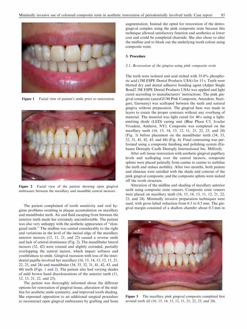
Figure 1 Facial view of patient’s smile prior to restoration.
Figure 2 Facial view of the patient showing open gingival
embrasure between the maxillary and mandible central incisors.
Figure 3 The maxillary pink gingival composite completed first
around teeth ## (16, 15, 14, 13, 12, 11, 21, 22, 23, and 24).
Minimally invasive use of coloured composite resin in aesthetic restoration of periodontially involved teeth: Case report 85
The patient complained of tooth sensitivity and oral hy-giene problems resulting in plaque accumulation on maxillary
and mandibular teeth. Air and fluid escaping from between theanterior teeth made her extremely uncomfortable. The patientwas also very unhappy with the aesthetic appearance of ‘‘elon-
gated teeth.’’ The midline was canted considerably to the rightand variations in the level of the incisal edge of the maxillaryanterior incisors (12, 11, 21, and 22) caused a reverse smileand lack of central dominance (Fig. 2). The mandibular lateral
incisors (32, 42) were rotated and slightly extruded, partiallyoverlapping the central incisor, which impart softness andyouthfulness to smile. Gingival recession with loss of the inter-
dental papilla involved her maxillary (16, 15, 14, 13, 12, 11, 21,22, 23, and 24) and mandibular (34, 33, 32, 31, 41, 42, 43, and44) teeth (Figs. 1 and 2). The patient also had varying shades
of mild brown band discolourations of the anterior teeth (11,12, 13, 21, 22, and 23).
The patient was thoroughly informed about the different
options for restoration of gingival tissue, alteration of the mid-line for aesthetic smile symmetry, and improved tooth shading.She expressed opposition to an additional surgical procedureto reconstruct open gingival embrasures by grafting and bone
augmentation. Instead she opted for restoration of the dento-gingival complex using the pink composite resin because thistechnique allowed satisfactory function and aesthetics at lower
cost and could be completed chairside. She also chose to alterthe midline and to block out the underlying tooth colour usingcomposite resin.
3. Procedure
3.1. Restoration of the gingiva using pink composite resin
The teeth were isolated and acid etched with 35.0% phospho-
ric acid (3M ESPE Dental Products USA) for 15 s. Teeth wereblotted dry and dental adhesive bonding agent (Adper SingleBond2 3M ESPE Dental Products USA) was applied and light
cured according to manufacturers’ instructions. The pink gin-gival composite (anaxGUM Pink Composite, Anaxdent, Stutt-gart, Germany) was scalloped between the teeth and naturalgingiva without preparation. The gingival base was made in
layers to create the proper contours without any overhang ofmaterial. The material was light cured for 40 s using a light-emitting diode (LED) curing unit (Blue Phase C5, Ivoclar
Vivadent, Amherst, NY). Composite was completed on themaxillary teeth (16, 15, 14, 13, 12, 11, 21, 22, 23, and 24)(Fig. 3) before placement on the mandibular teeth (34, 33,
32, 31, 41, 42, 43, and 44) (Fig. 4). Final contouring was per-formed using a composite finishing and polishing system (En-hance Dentsply Caulk Dentsply International Inc. Milford).
After soft tissue restoration with aesthetic gingival papillarylevels and scalloping over the central incisors, compositesplints were placed palatally from canine to canine to stabilizethe teeth and reduce mobility. After two months, both patient
and clinician were satisfied with the shade and contour of thepink gingival composite, and the composite splints were nickedoff the tooth structure.
Alteration of the midline and shading of maxillary anteriorteeth using composite resin veneers. Composite resin veneerswere placed on maxillary teeth (16, 15, 14, 13, 11, 12, 21, 22,
23, and 24). Minimally invasive preparation techniques wereused, with gross labial reduction from 0.3 to 0.5 mm. The gin-gival margin consisted of a shallow chamfer about 0.3 mm in

Figure 4 Facial view of the completed direct pink gingival
composite around maxillary teeth ## (16, 15, 14, 13, 12,11, 21, 22,
23, and 24) and mandibular teeth ## (34, 33, 32, 31, 41, 42, 43, and
44).
Figure 5 Facial view of the completed direct composite resin
veneers for maxillary teeth ## (16, 15, 14, 13, 12, 11, 21, 22, 23,
and 24) before finishing and polishing. Altering the incisal shape
and leveling mandibular (32, 31, 41, and 42) anterior incisal edge
by enameloplasty.
Figure 6 The definitive restorations exhibited a harmonious,
natural form and achieved the aesthetic expectations of the
patient.
86 M.A. Wahbi et al.
depth, placed using a 6844-016 round-end two-grit tapered dia-mond (Brasseler USA; Savannah, GA). The preparations ex-
tended sufficiently interproximally to conceal the margins.The incisal preparation was very minimal.
The prepared surfaces were acid etched with 35.0% phos-
phoric acid (3M ESPE Dental Products USA) for 15 s. Teethwere blotted dry and dental adhesive bonding agent (AdperSingle Bond2 3M ESPE Dental Products USA) was applied
and light cured according to manufacturer’s instructions.The composite resin veneer was placed using an incrementallayering technique to create a polychromatic effect. For this
patient, shades A2B, A2E, and YT (Filtek Supreme Ultra,3M ESPE, St. Paul, MN, USA) were placed along the labialenamel to mimic natural teeth. The composite material wassculpted by free-hand layering using a Greenstein Colour com-
posite instrument (Safident; Gland, Switzerland) and eachlayer was cured for 10 s. Because of the minimal preparationdesign and mild tooth discolouration, no dentin shade (A2D)
was necessary. Placement of each layer was closely monitoredfrom the incisal view to prevent over bulking, which wouldnegatively impact the health of the soft tissue. Cervically the
composite veneer finished at the gingival pink composite, whilethe veneer blended incisally. The material was light cured for40 s using the LED curing unit. Finally, the mandibular incisaledge shape was altered by enameloplasty (31, 32, 41, and 42),
producing a more youthful and aesthetically pleasing smile(Fig. 5). However, Fig. 6 shows harmonious aesthetic expecta-tions for the patient. While Figs. 7 and 8 show pre- and post-
operative aesthetic anticipation of the patient’s restorations inthe new smile design that is satisfactory at 18 months post res-toration (Fig. 9).
4. Discussion
The emerging field of ‘‘cosmetic periodontics’’ has come about
through collaboration between dentists, orthodontists, andperiodontists. Features that define a desirable smile have beenrefined while retaining consideration and respect for individual
variations (Tanaka et al., 2008). Challenging cases involving
the anterior area require a more comprehensive approachand a deeper understanding of the pink component of the
smile, the gingiva. The gingival architecture represents theframe for the teeth. If it is not restored correctly, either surgi-cally or prosthetically, it will impair the final three dimensional
aesthetic (Zalkind and Hochman, 1997; McCoy et al., 1998;Coachman and Calamita, 2010).
Patients with advanced periodontitis are at high risk forgingival recession and open gingival embrasures (‘‘black trian-
gles’’). Surgical treatment for the aesthetic and functionalproblems caused by open gingival embrasures are often insuf-ficient to re-establish ideal aesthetics. Prosthetic gingival resto-
ration with gingival coloured ‘‘pink’’ composite resin materialscan overcome the limitations of grafting (Coachman et al.,2009) and can be a good alternative for reconstructing
tissue lost due to ridge deformities (Coachman et al., 2010).

Figure 7 Pre and postoperative aesthetic appearance of the
completed direct resin veneers. Note the harmony of the patient’s
restorations in the new smile design.
Figure 8 Pre and postoperative aesthetic appearance of the
completed direct resin veneers. Note the harmony of the patient’s
restorations in the new smile design.
Figure 9 Patient’s smile at 18 months post restoration showing
satisfactory function and aesthetics.
Minimally invasive use of coloured composite resin in aesthetic restoration of periodontially involved teeth: Case report 87
Therefore, composite resin should be a consideration in the ini-tial treatment plan.
When using composite resin for gingival restoration, the
buccal and palatal/lingual aspect of the restoration should pro-vide comfort during mastication, ensure optimum phonetics,avoid food entrapment, and promote air sealing between the
patient’s natural gingiva and the prosthetic gingiva(Coachman and Calamita, 2010). The application must becarefully assessed to prevent undue thickness which could
cause plaque retention, compression of the papilla, and inflam-mation, leading to subsequent aesthetic problems (Burke et al.,1994; Tanaka et al., 2008). The greatest challenge in usingrestorations to alter interproximal embrasure form is for the
clinician to carry the restoration subgingivally to gradualizethe contour change. Attempting to leave the margin at gingivallevels will leave ledges of restorative material and will nega-
tively impact the papillary form (Spear, 2007).In the case presented here, the patient had gingival and
ridge recession leading to open embrasures that caused func-
tional and aesthetic problems (Figs. 1 and 2). A major consid-eration in alveolar ridge recession is the vertical aspect of thedefect, including papillae and gingival margin levels. The use
of pink composite resin was indicated in this case to restorethe gingival architecture and to guide the shape of the inter-dental papilla (Figs. 4 and 5) and was a time- and cost-efficientalternative to surgery. After gingival restoration, the patient’s
maxillary teeth were splinted to prevent mobility and improveprognosis, as suggested (Strassler, 2009).
Problems with midline and tooth discolouration can both
be improved with veneers. Newer dental adhesive materialsdo not require as much mechanical retention, allowing mini-mally invasive preparations and conservation of tooth struc-
ture (Murdoch-Kinch and McLean, 2003). Mostmanufacturers of dental adhesives offer both a total-etch adhe-sive and a self-etch adhesive. Research shows the highest mean
bond strengths to enamel were obtained with total-etch adhe-sives (Bagis et al., 2008; Strassler and Sensi 2008). New resinmaterials and better adhesive bonding allow minimally inva-sive preparation, yet still allow predictable bond strength
and longevity. These features satisfy numerous clinical indica-tions with immediate results for even the most cosmetically dis-cerning patients (Terry, 2004; Koczarski and Fligor 2005).
Conservation of healthy tooth structure is especially importantin patients who have only cosmetic concerns (Heymann andSwift, 2001; Christensen, 2006; and Radz, 2008).
The use of composite veneers can be a cost- and time-savingalternative to porcelain when indicated (Fahl, 2007; Dietschiand Devigus, 2011). Long term (5 year) satisfaction with com-posite veneers has been reported (Zorba et al., 2010). In addi-
tion, both pink and white composite materials have beenshown to have similar bond strength (An et al., 2011) Forthe patient presented here, tooth preparation was minimal,
conserving natural tooth structure while providing maximumenamel substrate for a stronger bond. A total-etch adhesivewas used instead of a self-etch to obtain better bonding to en-
amel. The composite resin veneer application achieved a moreaesthetic midline placement and easily blocked out the under-lying mild tooth discolouration. Following this and any other
restoration, proper hygiene and maintenance proceduresshould be carefully discussed with the patient, as they are par-amount for the long-term success of the restoration(Coachman and Calamita, 2010).

88 M.A. Wahbi et al.
This paper describes a clinical situation in which gingivallycoloured composite was effectively used to restore missing gin-gival tissue, solving aesthetic and functional problems. All pro-
cedures were minimally invasive and cost effective. The goal ofboth patient and clinician is to create a comfortable, healthy,and cleansable restoration while maintaining a high aesthetic
level. At 18 months post restoration, the patient reports noproblems (Fig. 9).
5. Conclusions
We report 18 months post restoration patient’s and clinician’ssatisfaction with aesthetics and function of gingival and mid-
line rehabilitation by application of pink and white direct com-posite material using minimally invasive procedures. Use ofpink composite for restoration of the gingiva allows immediate
results at a relatively low cost without potential surgical post-operative recovery complications. With this method, large tis-sue volumes are easily replaced by artificial materials.Composite resin veneers were also successfully placed to alter
midline and achieve better tooth colour.
References
Alani, A., Maglad, A., Nohl, F., 2011. The prosthetic management of
gingival aesthetics. Br. Dent. J. 210 (2), 63–69.
An, H.S., Park, J.M., EJ, P., 2011. Evaluation of shear bond strengths
of gingiva-colored composite resin to porcelain* metal and zirconia
substrates. J. Adv. Prosthodont. 3 (3), 166–171.
Babu, K.B., Madhu Babu, D.S., Dodani, K., Sekhar, Leela, Reddy,
N.R., 2011. Reconstruction of the interdental papillae using an
interdisciplinary approach following orthodontic treatment: a case
report. Int. J. Contemp. Dent. 2 (2).
Bagis, B., Aydoðan, E., Bagis, Y.H., 2008. Direct restorative treatment
of missing maxillary laterals with composite laminate veneer: a case
report. Open Dent. J. 2, 93–95.
Barzilay, I., Irene, T., 2003. Gingival prostheses––a review. J. Can.
Dent. Assoc. 69 (2), 74–78.
Burke, S., Burch, J.G., Tetz, J.A., 1994. Incidence and size of
pretreatment overlap and posttreatment gingival embrasure space
between maxillary central incisors. Am. J. Orthod. Dentofacial
Orthop. 105, 511.
Capa, N., 2007. An alternative treatment approach to gingival
recession: gingiva-colored partial porcelain veneers: a clinical
report. J. Prosthet. Dent. 98 (2), 82–84.
Christensen, G.J., 2004. What is a veneer? Resolving the confusion. J.
Am. Dent. Assoc. 135 (11), 1574–1576.
Christensen, G.J., 2006. Veneer mania. J. Am. Dent. Assoc. 137, 1161–
1163.
Clark, D., 2008. Restoratively driven papilla regeneration: correcting
the dreaded ‘black triangle’. Tex. Dent. J. 125 (11), 1112–1115.
Coachman, C., Calamita, M., 2010. The reconstruction of pink and
white esthetics. Inter. Dent. SA 12 (3), 88–93.
Coachman, C., Salama, M., Garber, D., Calamita, M., Salama, H.,
2009. Prosthetic gingival reconstruction in a fixed partial restora-
tion. Part 1: introduction to artificial gingiva as an alternative
therapy. Int. J. Periodontics Restor. Dent. 29, 471–477.
Coachman, C., Van Dooren, E., Gurel, G., Calamita, M., Calgaro, M.,
de Souza Neto, J., 2010. Minimally invasive reconstruction in
implant therapy: the prosthetic gingival restoration. QDT 33, 61–75.
Croll, T.P., 2003. Dentistry ... we have a problem. J. Esthet. Restor.
Dent. 15 (4), 201–202.
Dietschi, D., Devigus, A., 2011. Prefabricated composite veneers:
historical perspectives, indications and clinical application. Eur. J.
Esthet. Dent. 6, 178–187.
Donovan, T.E., 2009. Clinical management of root caries. J. Indiana
Dent. Assoc. 88 (1), 23–24.
Fahl, N.J., 2007. A polychromatic composite layering approach for
solving a complex class IV/direct veneer/diastema combination:
part II. Pract. Proced. Aesthet. Dent. 19, 17–22.
Feigenbaum, N.L., 1991. Aspects of aesthetic smile design. Pract.
Periodontics Aesthet. Dent. 3 (3), 9–13.
Heymann, H.O., Kokich, V.G., 2002. Instant orthodontics: viable
treatment option or ‘‘quick fix’’ cop-out? J. Esthet. Restor. Dent.
14 (5), 263–264.
Heymann, H.O., Swift Jr., E.J., 2001. Is tooth structure not sacred
anymore? J. Esthet. Restor. Dent. 13 (5), 283.
Jacobson, N., Frank, C.A., 2008. The myth of instant orthodontics: an
ethical quandary. J. Am. Dent. Assoc. 139 (4), 424–434.
Koczarski, M.J., Fligor, 2005. Posterior aesthetics using a single-
shaded composite resin: a case presentation. Pract. Proced. Aesthet.
Dent. 17 (2), 145–148.
Kurth, J.R., Kokich, V.G., 2001. Open gingival embrasures after
orthodontic treatment in adults: prevalence and etiology. Am. J.
Orthod. Dentofacial Orthop. 120 (2), 116–123.
Maeda, S., Maeda, Y., Ono, Y., Nakamura, K., Sasaki, T., 2005.
Interdisciplinary treatment of a patient with severe pathologic
tooth migration caused by localized aggressive periodontitis. Am. J.
Orthod. Dentofacial Orthop. 127 (3), 374–384.
Magne, P., Belser, U.C., 2004. Novel porcelain laminate preparation
approach driven by a diagnostic mock-up. J. Esthet. Restor. Dent.
16 (1), 7–16 [discussion 17–8].
Malcmacher, L., 2005. No-preparation porcelain veneers–back to the
future! Dent. Today 24 (3), 86, 88, 90–1.
Martegani, P., Silvestri, M., Mascarello, F., Scipioni, T., Ghezzi, C.,
Rota, C., et al, 2007. Morphometric study of the interproximal
unit in the esthetic region to correlate anatomic variables affecting
the aspect of soft tissue embrasure space. J. Periodontol. 78 (12),
2260–2265.
Marus, R., 2006. Treatment planning and smile design using composite
resin. Pract. Proced. Aesthet. Dent. 18 (4), 235–241.
McCoy, R.B., Anderson, M.H., Lepe, X., Johnson, G.H., 1998.
Clinical success of class V composite resin restorations without
mechanical retention. J. Am. Dent. Assoc. 129 (5), 593–599.
Murdoch-Kinch, C.A., McLean, 2003. Minimally invasive dentistry. J.
Am. Dent. Assoc. 134, 87–95.
Patil, S., Prabhu, V., Danane, N.R., 2011. Gingival veneer: mask the
unesthetic. J. Indian Soc. Periodontol. 15 (3), 284–287.
Radz, G., 2008. Elective veneers: meeting patients’ expectations while
addressing current ethical questions of elective care. Inside Dent. 4
(3), 48–52.
Roshna, T., Nandakumar, K., 2012. Generalized aggressive periodon-
titis and its treatment options: case reports and review of the
literature. Case reports in medicine 10.1155//535321, 1–17.
Sarver, D.M., 2004. Principles of cosmetic dentistry in orthodontics:
part 1. Shape and proportionality of anterior teeth. Am. J. Orthod.
Dentofacial Orthop. 126, 749–753.
Spear, F.M., 2004. The esthetic correction of anterior dental mal-
alignment conventional vs. instant (restorative) orthodontics. J.
Calif. Dent. Assoc. 32 (2), 133–141.
Spear, F.M., 2007. Interdisciplinary esthetic management of anterior
gingival embrasures. Inside Dent. 3 (3).
Strassler, H.E., 2009. Tooth stabilization improves periodontal prog-
nosis: a case report. Dent. Today 28 (9), 88–93.
Strassler, H., Sensi, 2008. Applications of etch-and-rinse adhesive
bonding for esthetic restorative dentistry. Funct. Esthet. Restor.
Dent. 2 (1), 26–33.
Tanaka, O.M., Furquim, B.D.’.A., Pascotto, R.C., Ribeiro, G.L.U.,
Bosio, J.A., Maruo, H., 2008. The dilemma of the open gingival
embrasure between maxillary central incisors. J. Contemp. Dent.
Pract. 6 (9), 092–98.
Terry, D.A., 2004. Restoring the dento-gingival complex, Part 2. Dent.
Today 23 (6), 96–100.

Minimally invasive use of coloured composite resin in aesthetic restoration of periodontially involved teeth: Case report 89
Wolfart, S., Thormann, H., Freitag, S., Kern, M., 2005. Assessment of
dental appearance following changes in incisor proportions. Eur. J.
Oral Sci. 113 (2), 159–165.
Zalkind, M., Hochman, N., 1997. Alternative method of conservative
esthetic treatment for gingival recession. J. Prosthet. Dent. 77 (6),
561–563.
Zorba, Y.O., Bayindir, Y.Z., Barutcugil, C., 2010. Cagatay barutcugil.
Direct laminate veneers with resin composites: two case reports
with five-year follow-ups. J. Contemp. Dent. Pract. 11 (4), 1–7.