Minimally Invasive Glaucoma Surgery (MIGS) for individuals ...
161
20 21 HEALTH TECHNOLOGY ASSESSMENT: Minimally Invasive Glaucoma Surgery (MIGS) for individuals with glaucoma REPORT
Transcript of Minimally Invasive Glaucoma Surgery (MIGS) for individuals ...

2021HEALTH TECHNOLOGY ASSESSMENT: Minimally Invasive Glaucoma Surgery (MIGS) for individuals with glaucoma
REPORT

2 Table of contents
Institution
Title
Norwegian Institute of Public Health Division for Health Services Minimally Invasive Glaucoma Surgery (MIGS) for individuals with glaucoma. A health technology assessment
Norwegian title Minimal-invasiv glaukomkirurgi (MIGS) for individer med glaukom. En metodevurdering
Responsible Camilla Stoltenberg, Director-General Authors
Lund, Ulrikke Højslev, Norwegian Institute of Public Health Bidonde, Julia, Norwegian Institute of Public Health Kornør, Hege, Norwegian Institute of Public Health Reinar, Liv Merete, Norwegian Institute of Public Health Fagerlund, Beate Charlotte, Norwegian Institute of Public Health Nguyen, Lien, Norwegian Institute of Public Health Ursin, Lars Øystein, Norwegian University of Science and Technology Lerner, Martin, Norwegian Institute of Public Health Robberstad, Bjarne, Norwegian Institute of Public Health
ISBN 978-82-8406-144-3
Project number ID2018_072 Type of report Health technology assessment (HTA)
No. of pages 124 (159 including attachments) Commissioner Commissioning Forum for the Regional Health Authorities (RHA Forum) in
the National System for Managed Introduction of New Health Technologies Subject headings
(MeSH) Health Technology Assessment; Systematic Review; Health Economic Eval-uation; Minimally Invasive Glaucoma Surgery; MIGS; Surgery; Eye Disease
Citation
Lund UH, Bidonde J, Kornør H, Reinar LM, Fagerlund BC, Nguyen L, Ursin LØ, Lerner M, Robberstad B. Minimally Invasive Glaucoma Surgery (MIGS) for individuals glaucoma. A health technology assessment. Oslo: Norwegian Institute of Public Health, 2021.

3 Table of contents
Table of contents
TABLE OF CONTENTS 3
KEY MESSAGES 6
EXECUTIVE SUMMARY 7
HOVEDBUDSKAP 13
SAMMENDRAG 14
PREFACE 20
ABBREVIATIONS 22
OBJECTIVES 23
BACKGROUND 24 Glaucoma 24 Glaucoma management 27 Existing research syntheses 29
CLINICAL EFFECTIVENESS AND SAFETY 32 Methods 32 Inclusion criteria 32 Literature search 34 Study selection 34 Assessment of risk of bias in included studies 35 Data extraction 35 Data analysis 35 Assessment of certainty of the evidence 36 Results 37 Search results and selection of studies 37 Characteristics of included studies 38 Risk of bias in included studies 41 Effectiveness of MIGS (CADTH HTA) 42 Effectiveness of MIGS (NIPH supplementary review) 55 Safety of MIGS (CADTH HTA) 59 Safety of MIGS (NIPH supplementary review) 59
ORGANIZATIONAL ASPECTS 64 Background 64

4 Table of contents
Method 64 Patient selection 64 Capacity and number of treatments 65
HEALTH ECONOMIC EVALUATION 66 Background 66 Introduction to Economic Evaluation of Health Care Programmes 66 Priority setting criteria 67 Literature review of previous health economic evaluations of MIGS 68 Method 71 General 71 Model structure 72 Patient populations and interventions 73 Model parameters 76 Budget impact analysis 87 Severity considerations – absolute shortfall (AS) 89 Results 90 Severity considerations – absolute shortfall 90 Incremental cost–effectiveness estimates in the analysis 90 Sensitivity analyses 103 Budget impact analysis 107
ETHICS 111 Method 111 Brief description of the situation, alternatives and stakeholders 111 Analysis of the ethical challenges 112 Summary of the analysis 113
DISCUSSION 114 Key findings summary 114 Evidence quality 115 Strengths and weaknesses 116 Generalisability of findings 120 Consistency with other reviews 121 Implication of results on practice 122 Considerations of the prioritisation criteria in light of available evidence 124 Need for further research 124
CONCLUSION 125
REFERENCES 126
APPENDICES 133 Appendix 1: Progress log 133 Appendix 2: Inconsistencies between the protocol and the final report 135 Appendix 3: Search strategy 136 Appendix 4: Eligible studies from top-up search 144 Appendix 5: GRADE evidence profile 145 Appendix 6: Studies excluded from NIPH supplementary review, with reasons 146

5 Table of contents
Appendix 7: Characteristics of studies included in NIPH supplementary review 154 Appendix 8: Organizational aspects: questionnaire 159

6 Key messages
Key messages
This health technology assessment (HTA) summarises and supplements a 2019 Canadian HTA on the effectiveness and safety of micro-invasive glaucoma surgery (MIGS) versus other treatment options. Further, it contains cost-effectiveness analysis based on the Canadian HTA, in ad-dition to patient partners’ considerations, organizational and ethical considerations relevant to discussions of MIGS’ role in Norwegian rou-tine care. The Canadian evidence, which included 32 studies and 24 comparisons, was inconclusive due to very low to low certainty. Our supplementary findings show that: • MIGS with Hydrus Microstent combined with cataract surgery
reduces intraocular pressure (IOP) at 24 months, compared with cataract surgery alone (high-certainty evidence)
• MIGS with iStent inject combined with cataract surgery probably reduces IOP at 24 months, compared with cataract surgery alone (moderate-certainty evidence)
• For other techniques there is either no or little difference between the MIGS and control interventions, or it is uncertain whether there is a difference in effectiveness
• Neither MIGS procedures, nor alternative surgical strategies appear to be at high risk of adverse events
• Lifetime total cost for glaucoma treatment ranged from NOK 30 000 to NOK 83 000 per patient, depending on treatment strategy and baseline disease stage. The incremental Quality adjusted life years (QALYs) for MIGS between comparators ranged between – 0.080 and 0.057
• MIGS is suitable as a outpatient surgery without hospital admission. Clinicians need training. Clear criteria for patient selection shuld be developed. Experts predict that the number of MIGS procedures may increase to twice as many in 2024 than today
• The clinical evidence on MIGS is limited. The main reason for this is the lack of comparative studies. Our health economic evaluation shows some scenarios where MIGS may be cost-effective, depending on comparator and disease stage. Our analysis puts individuals with glaucoma in severity class 1.
Title: Minimally Invasive Glaucoma Surgery (MIGS) for individuals with glaucoma. A health technology assessment -----------------------------------------
Type of publication: Health technology assess-ment -----------------------------------------
Doesn’t answer everything: We did not address legal aspects re-lated to use of MIGS in Norway -----------------------------------------
Publisher: Norwegian Institute of Public Health -----------------------------------------
Updated: Last search for studies: November 2020 -----------------------------------------
Internal peer reviewers: Kjetil Gundro Brurberg, Department Di-rector, NIPH -----------------------------------------
External peer reviewers: Alexander Thrane, MD, PhD, Haukeland University Hospital Eline Aas, Associate Professor, University of Oslo Turid Skei Tønset, MD, Oslo University Hospital

7 Executive summary
Executive summary
Background
Glaucoma is a substantial public health problem, with a large negative impact on qual-ity of life and the utilization of health care resources. Glaucoma refers to a group of eye conditions with a characteristic pattern of progressive damage to the optic nerve. Raised intraocular pressure (IOP) is the best characterized risk-factor, but IOP can also be normal. Globally, glaucoma is considered the leading cause of irreversible vision loss and one of the leading causes of blindness overall. There are approximately 77,000 Norwegian individuals with a glaucoma diagnosis. The incidence is expected to in-crease because of demographic changes and because the disease can now be diagnosed at earlier stages than before. Minimally invasive glaucoma surgery (MIGS) represents a class of new surgical procedures and devices to address this issue. Experts suggest MIGS may result in shorter procedure and recovery times than traditional surgical pro-cedures, and that MIGS make it possible to offer treatment at an earlier stage of glau-coma. The indications for each MIGS procedure depend on its mechanism of action and the individual patient’s target IOP and concomitant eye diseases. MIGS procedures and devices can be used alone or in conjunction with cataract surgery. The procedure is al-ready offered in many public hospitals in Norway. Objective
The objectives of this health technology assessment (HTA) were to: 1) supplement the evidence of (effectiveness and safety ) of MIGS in an HTA published by the Canadian Agency for Drugs and Technologies in Health (CADTH) in January, 2019, 2) conduct a health economic evaluation of MIGS from a Norwegian health care perspective, and 3) assess organizational and ethical aspects of MIGS in a Norwegian setting. Method
Clinical effectiveness and safety We have summarized CADTH’s HTA evidence of effectiveness and safety, and adapted CADTH’s methods in the conduct of our supplementary review of more recent studies. CADTH carried out systematic literature searches in August and November 2017, while our updated searches were carried out in August 2019 and November 2020. Searches were run in electronic medical databases, such as MEDLINE, EMBASE, CINAHL and Cochrane Central Register of Controlled Trials, using peer-reviewed search strategies. Two reviewers independently selected studies meeting our inclusion criteria. Likewise,

8 Executive summary
two reviewers independently judged the included studies’ risk of bias. One reviewer ex-tracted predefined data, and another reviewer checked the data extraction. The pri-mary outcome in CADTH’s HTA was quality of life, and intraocular pressure in the Nor-wegian Institute of Public Health (NIPH) HTA supplementary review. When possible, mean differences with 95 % confidence intervals were calculated, and effect estimates were pooled for similar comparisons. When pooling was not possible, findings were re-ported narratively. One reviewer assessed the certainty of the evidence with the GRADE approach, and a second reviewer checked the assessments. Health economic evaluation We based our health economic analysis on the previous HTA carried out by CADTH. We developed six different decision analytic cost-effectiveness models in TreeAge Pro, to estimate incremental cost utility rate (ICER). The models provide insight into costs, health effects, survival, and disease stage. The analysis was carried out from a modified Norwegian health care perspective. We have estimated absolute shortfall for patients with glaucoma. We handled uncertainties in model parameters by assigning probability distributions to the parameters and performing probabilistic sensitivity analysis (PSA), designed as a Monte Carlo simulation. We also performed one-way sensitivity analyses for each of the six models to explore potential impact of uncertainty in single parame-ters. In addition, we estimated the budget impact of introducing MIGS as a routine treatment option in Norway for patients with glaucoma. Organizational aspects In order to evaluate the organizational consequences related to the implementation of MIGS and a potential increase in volume of MIGS performed in Norwegian hospitals, we asked clinical experts from five of the state-run hospitals that perform MIGS in Norway, to answer a questionnaire regarding their present capacity and procedure used: patient selection, procedures and ongoing trials. Ethical perspectives We analysed central ethical implications of MIGS implementation, and the analysis was proceeded in three major steps: First, brief description of the situation, alternative ac-tions and solutions, and the involved stakeholders. Second, analysis of the ethical chal-lenges and possible consequences in terms of the four principles: benefit, harm, auton-omy and justice.
Results
Clinical effectiveness and safety CADTH HTA CADTH’s HTA included 35 publications from 32 studies; 10 randomized trials and 22 non-randomised studies. The evidence included 24 specific comparisons: one compari-son of a MIGS versus another MIGS, six comparisons of a MIGS combined with cataract surgery versus cataract surgery alone, nine comparisons of a MIGS combined with cata-ract surgery versus filtration surgery combined with cataract surgery, six comparisons

9 Executive summary
of a MIGS combined with filtration surgery versus filtration surgery alone, two compar-isons of a MIGS versus pharmacotherapy, and one comparison of a MIGS versus laser therapy. CADTH’s authors considered all the included studies to have some risk of bias. As shown in the table below with summary of findings by comparison and outcomes (reproduced with kind permission from CADTH), there was largely no statistically sig-nificant difference between intervention and comparator and this can be explained be-cause the heterogeneity in interventions and comparisons did not allowed pooling. . The evidence was considered very low or low certainty across comparisons and out-comes.
NIPH supplementary review We identified and included eight studies (seven randomised trials and one non-ran-domised study) that compared a MIGS procedure alone or in combination with cataract surgery, with another MIGS procedure or non-MIGS procedures. None of the additional found studies could be pooled with study results in the CADTH HTA. As showed in the summary of findings table below, there was high-certainty evidence of lower IOP with Hydrus in combination with cataract surgery than with cataract sur-gery alone, and moderate-certainty evidence of lower unmedicated IOP with iStent in-ject in combination with cataract surgery than with cataract surgery alone. Otherwise, there was no or little difference between the MIGS and interventions in control groups, or the certainty of the evidence was too low to make a judgement. The evidence for the safety of MIGS was inconclusive across the comparisons, but there appeared to be little or no risk of complications associated with any of the treatment options. There also appears to be no standarized method to measure safety.

10 Executive summary
Summary of findings: Intraocular pressure (IOP)
Effectiveness of MIGS for open-angle glaucoma in adults Patient or population: adults with open-angle glaucoma Intervention: MIGS alone or in combination with other procedure Comparison: other MIGS or other procedure
Comparison Anticipated absolute effects*
MD (95% CI) № of participants (studies)
Certainty of the evidence (GRADE)
Comments
Intraocular pressure [mm Hg]
One MIGS procedure versus another MIGS procedure
Hydrus vs 2x iStent MD 1.9 lower
(2.91 lower to 0.89 lower) 148
(1 RCT) ⨁⨁◯◯
LOW a,b Follow-up: 12 months
One MIGS procedure versus another procedure (non-MIGS)
iStent+phaco vs phaco alone
MD 1.9 lower
(3.32 lower to 0.48 lower) 48
(1 observational study) ⨁◯◯◯ VERY LOW b,c
Follow-up: 12 months
iStent inject+phaco vs phaco alone
MD 0.7 lower
(1.27 lower to 0.13 lower) 570
(2 RCTs) ⨁⨁⨁◯ MODERATE a
Follow-up: 24 months
Hydrus+phaco vs phaco alone
MD 1.8 lower
(2.73 lower to 0.87 lower) 331
(1 RCT) ⨁⨁⨁⨁
HIGH Follow-up: 24 months
Trab360+phaco vs phaco alone
MD 2.8 lower
(5.49 lower to 0.11 lower) 18
(1 RCT) ⨁◯◯◯ VERY LOW b,d
Follow-up: 24 months
Gold MicroShunt vs Ahmed glaucoma
valve
MD 0.7 lower (2.71 lower to 1.31 higher) 12
(1 RCT) ⨁◯◯◯ VERY LOW b,e
Follow-up: 5 years
iTrack+phaco vs fil-tration sur-gery+phaco
MD 1.7 lower
(3.29 lower to 0.11 lower) 59 (1 RCT)
⨁◯◯◯ VERY LOW b,f
Follow-up: 12 months
*The risk in the intervention group (and its 95% confidence interval) is based on the assumed risk in the comparison group and the rela-tive effect of the intervention (and its 95% CI). CI: Confidence interval; MD: Mean difference; phaco: phacoemulsification (cataract surgery)
GRADE Working Group grades of evidence High certainty: We are very confident that the true effect lies close to that of the estimate of the effect Moderate certainty: We are moderately confident in the effect estimate: The true effect is likely to be close to the estimate of the effect, but there is a possibility that it is substantially different Low certainty: Our confidence in the effect estimate is limited: The true effect may be substantially different from the estimate of the effect Very low certainty: We have very little confidence in the effect estimate: The true effect is likely to be substantially different from the esti-mate of effect
Explanations a. Possible risk of performance bias in personnel b. Wide confidence interval and/or sub-optimal information size c. Serious bias due to confounding d. High risk of performance and detection bias.

11 Executive summary
e. High risk of attrition bias; unclear risk of bias in most other domains. f. High risk of selection, performance and attrition bias; unclear risk of detection and reporting bias.
Health economic evaluation The lifetime total cost per patient for glaucoma treatment ranged between NOK 30 000 and NOK 84 000 depending on the treatment strategy and patient’s baseline disease stage. The incremental quality adjusted life years (QALYs) for MIGS between compara-tors ranged between – 0.080 and 0.057. Organizational aspects MIGS surgery is done at many public hospitals in Norway in addition to a few private clinics. There is a lack of formalised indications for the use of MIGS in Norway. MIGS is suitable for outpatient surgery without hospital admission. Ophthalmologists need spe-cific MIGS training to perform the surgery (certification). The MIGS-procedures have a shorter learning curve than traditional glaucoma surgery, and has in other countries also been implemented by non-glaucoma specialists / general cataract surgeons for this reason (i.e. some benefit in terms of regional accessibility in Norway can be expected). MIGS procedures require less follow-up than traditional surgery, so there may be fewer post-intervention controls per patient in the hospital. Finally, a better treatment option may also lead to more patients being operated. The need for glaucoma surgery may in-crease due to increased population growth in the relevant age group. Experts predict that the demand for MIGS procedures will increase annually from today to twice as many by 2024. Ethical perspectives There are potential benefits of MIGS. The central ethical concerns of MIGS implementa-tion are, in terms of justice, to guarantee patients equal access to treatment, regardless of ability to pay, place of residence, social status, or cultural background. The chal-lenges for patient autonomy seem manageable, if patients can be thoroughly informed about risks of and alternatives to a given MIGS procedure.
Discussion
The strengths of CADTH’s and our health technology assessments include updated sys-tematic searches in electronic databases, pre-specified inclusion and exclusion criteria, double independent screening of identified records, independent assessments of risk of bias in included studies, systematic data extraction and reporting, and assessments of the certainty of the evidence. We believe we have identified most eligiblie studies that were not included in CADTH’s HTA, thus providing a useful supplement. We had to make several assumptions to develop the six health economic models, lead-ing to some uncertainty surrounding our economic results. MIGS is a heterogeneous group of devices with potentially different costs, risk profiles and relative treatment ef-fects. We replicated the model from CADTH with respect to model structure and effi-cacy data. The input is adjusted according to Norwegian conditions.

12 Executive summary
It is important to ask whether the introduction of MIGS will lead to a change in Norwe-gian practice. According to our clinical experts, the impact on practice will be limited. Our health economic evaluation is consistent with other evaluations on health econom-ics, among others the CADTH report, regarding uncertainty around estimates due to lack of data. Cost components may be different in different countries. To obtain high quality documentation for the myriad of possible MIGS comparisons, there is a need for more well-designed randomised trials, with larger numbers of par-ticipants and longer follow-up. Further, detailed micro-costing of MIGS procedures may allow for greater certainty in the true absolute and incremental costs of MIGS to better inform the potential economic value of MIGS. For patients and clinicians, it would be helpful if evidence-based guidelines were developed or applied to a Norwegian setting in addition to local and/or national registrers. Studies on the most used MIGS procedures in Norway (as iStent, Xen, Preserflo) are in-cluded, but might not be well documented in this report, as some relevant clinical com-parisons have not been conducted yet (to our knowledge). The evaluation of effect and cost-effectiveness might therefore have some limits. Conclusion
MIGS with Hydrus Microstents combined with cataract surgery reduces intraocular pressure (IOP) at 24 months, compared to cataract surgery alone. MIGS with Hydrus Microstents probably reduces IOP at 12 months, compared to MIGS with 2x iStents. For other comparisons and outcomes, it is uncertain whether there is a difference in IOP re-duction. Neither MIGS procedures nor alternative surgical strategies appear to be at high risk of adverse events, and it is uncertain whether complications occur more or less frequently in either category. Definitive conclusions on the cost-effectiveness of MIGS are uncertain, given the uncertainty in the analysis. The economic evaluation pro-vided some scenarios where MIGS might be cost-effective, depending on comparator and disease stage. The clinical evidence on MIGS is limited. The main reason for this is the lack of compar-ative studies. Our health economic evaluation shows some scenarios where MIGS may be cost-effective, depending on comparator and disease stage. Our analysis puts indi-viduals with glaucoma in severity class 1.

13 Hovedbudskap
Hovedbudskap
Denne metodevurderingen (HTA) oppsummerer og sup-plerer en kanadisk HTA fra 2019 om effekt og sikkerhet ved minimal-invasiv glaukomkirurgi (MIGS). Videre gjorde vi kost-nytteanalyser basert på den kanadiske HTAen, i til-legg til brukerperspektiv, organisatoriske og etiske vurde-ringer som er relevante i en diskusjon om hvorvidt MIGS bør være et rutinetilbud i norsk praksis. Det kanadiske kunnskapsgrunnlaget, som omfattet 32 stu-dier og 24 sammenlikninger, var usikkert på grunn av svært lav til lav tillit til resultatene. Våre supplerende funn viser at: • MIGS med Hydrus Microstent kombinert med
kataraktkirurgi reduserer intraokulært trykk (IOP) etter 24 måneder, sammenliknet med kataraktkirurgi alene (høy tillit til resultatet)
• MIGS med iStent inject og kataraktkirurgi reduserer trolig IOP etter 24 måneder, sammenliknet med kataraktkirurgi alene (middels tillit til resultatet)
• Det er usikkert hvorvidt det er noen forskjell i effekt mellom MIGS og kontrollgruppene for andre sammenligninger
• Det ser ikke ut til å være noen betydelig forskjell mellom MIGS og kontrollgruppene i risiko for uønskede hendelser/skader
• Total livstidskostnad per pasient for glaukombehandling ble estimert mellom 30 000 norske kroner og 83 000 norske kroner avhengig av behandlingsstrategi og sykdomsstadie ved start. Inkrementell QALY for MIGS sammenlignet med komparatorer var mellom – 0.080 og 0.057
• MIGS egner seg for poliklinisk kirurgi. Øyeleger må ha opplæring for å utføre MIGS. Det bør utvikles klare kriterier for pasientseleksjon. Eksperter predikerer en dobling av antall MIGS prosedyrer i 2024 enn antallet i dag
• Kunnskapsgrunnlaget for effekt og sikkerhet om MIGS er begrenset. Hovedgrunnen er mangel på sammenliknende studier. Vår helseøkonomiske vurdering viser at MIGS kan være kostnadseffektive, avhengig av sammenliknng og sykdomsutvikling. Vår analyse setter individer med glaukom i gruppe for alvorlighetsgrad 1.
Tittel: Minimal-invasiv glaukomkirurgi (MIGS) for individer med glaukom. En metodevurdering ------------------------------------
Publikasjonstype: Metodevurdering ------------------------------------
Svarer ikke på alt: Vi har ikke sett på juridiske aspekter ved bruk av MIGS i Norge ------------------------------------
Hvem står bak denne publikasjo-nen? Folkehelseinstituttet har gjennomført oppdraget etter forespørsel fra Bestillerforum RHF ------------------------------------
Når ble litteratursøket utført? Søk etter studier ble avsluttet: November 2020 Intern fagfelle: Kjetil Gundro Brurberg, Avde-lingsdirektør, FHI -------------------------------------- Eksterne fagfeller: Alexander Thrane, Overlege PhD, Haukeland Universitetssykehus Eline Aas, Førsteamanuensis, Universitetet i Oslo Turid Skei Tønset, overlege, Oslo Universitetssykehus

14 Sammendrag
Sammendrag
Bakgrunn
Glaukom er et betydelig folkehelseproblem med stor påvirkning på livskvalitet for pasi-enter og pårørende. Pasienter med glaukom har behov for betydelig hjelp fra helsetje-nestene. Glaukom refererer til en sykdomsgruppe som kjennetegnes av en progressiv ødeleggelse av synsnerven i et karakteristisk mønster. Økt intraokulært trykk (IOP) er den best beskrevne risikofaktoren, men IOP kan også være normalt. Globalt betraktes glaukom som den vanligste årsaken til irreversibelt synstap og en av de vanligste årsa-kene til blindhet. I Norge er om lag 77 000 individer diagnostisert med glaukom. Insi-densen er forventet å øke på grunn av demografiske endringer og fordi tilstanden nå kan diagnostiseres tidligere enn før. Minimal-invasiv glaukomkirusrgi (MIGS) er en ny gruppe av kirurgiske prosedyrer og utstyr. Ifølge eksperter kan MIGS gi kortere prose-dyre- og restitusjonstider sammenlignet med tradisjonelle kirurgiske prosedyrer, noe som gjør det mulig å utføre MIGS på et tidligere sykdomsstadium. Indikasjonene for hver enkelt MIGS-prosedyre varierer med virkningsmekanisme og pasientens individu-elle mål for intraokulært trykk (IOP). MIGS-prosedyrer og -utstyr kan utføres alene el-ler i kombinasjon med katarakt kirurgi. Inngrepet tilbys allerede på mange offentlige sykehus i Norge. Mål
Hensikten med denne metodevurderingen var å 1) supplere kunnskapsgrunnlaget for effekt og sikkerhet av MIGS sammenliknet med andre behandlingsstrategier i en meto-devurdering (HTA) publisert av the Canadian Agency for Drugs and Technologies in Health (CADTH) i januar 2019, 2) undersøke kostnadseffektiviteten ved MIGS opp mot prioriteringskriteriene gjeldende i norsk helsetjeneste: nytte-, ressursbruk- og alvorlig-hetskriteriet, og 3) vurdere organisatoriske konsekvenser og etiske aspekter relatert til bruk av MIGS i Norge. Metode
Klinisk effekt og sikkerhet Vi har oppsummert kunnskapsgrunnlaget i CADTH-HTAen og tilpasset CADTHs meto-der i utføringen av vår supplerende gjennomgang av nyere studier. CADTH søkte syste-matisk etter litteratur i august og november 2017, mens våre oppdateringssøk ble gjen-nomført i august 2019 og november 2020. Søkene ble kjørt ved hjelp av fagfellevur-derte søkestrategier i elektroniske medisinske databaser som MEDLINE, EMBASE, CINAHL og Cochrane Central Register of Controlled Trials.

15 Sammendrag
To forskere valgte, uavhengig av hverandre, ut studier som møtte inklusjonskriteriene. Videre vurderte to forskere uavhengig av hverandre også risiko for skjevheter i de ink-luderte studiene. Én forsker hentet ut forhåndsbestemte data, og en annen forsker sjek-ket datauthentingen. Hovedutfallet i CADTH-HTAen var livskvalitet, mens det var intra-okkulært trykk i FHIs supplerende gjennomgang. Der det var mulig ble gjennomsnitts-forskjeller med 95% konfidensintervall beregnet, og effektestimater for like sammen-ligninger slått sammen. Til slutt vurderte en forsker tilliten til resultatene ved hjelp av GRADE-tilnærmingen, og en annen forsker sjekket vurderingene. Helseøkonomisk evaluering Vi baserte vår helseøkonomiske analyse på en tidligere HTA utført av CADTH. Vi utvik-let seks ulike beslutnings-analytiske kostnadseffektivitets modeller i TreeAge Pro for å estimere ICER. Modellene gir innsikt i kostnader, helseeffekter, overlevelse og syk-domsstadie. Analysen ble utført ut ifra et modifisert norsk helsetjenesteperspektiv. Vi har estimert absolutt prognosetap for pasienter med glaukom. Vi håndterte usikkerhet i modell-parametere ved å tildele sannsynlighetsfordelinger til parameterene og ut-førte «probabilistic sensitivity analysis» (PSA), utformet som Monte Carlo simuleringer. Vi utførte også enveis sensitivitetsanalyser for hver av de seks modellene for å under-søke potensiell påvirkning til usikkerheten på enslige parametere. I tillegg, estimerte vi budsjett konsekvens ved å introdusere MIGS som en rutine behandlingsalternativ i Norge for pasienter med glaukom. Organisatoriske aspekter For å evaluere organisatoriske konsekvenser relatert til implementering av MIGS og en potensiell økning i volum av MIGS utført i norske sykehus, ba vi kliniske eksperter ved fem offentlige sykehus som utfører MIGS svare på et spørreskjema knyttet til deres ka-pasitet og prosedyrer. Etiske perspektiver Vi analyserte sentrale etiske implikasjoner av MIGS implementering. Analysen ble ut-ført ved å kort beskrive situasjonen, alternative strategier, løsninger og involverte in-teressenter. Etiske utfordringer og mulige konsekvenser ble deretter analysert gjen-nom de fire prinsippene: velferd, ikke skade, autonomi, og rettferdighet.
Resultat
Klinisk effekt og sikkerhet CADTH-HTAen CADTH-HTAen inkluderte 35 publikasjoner fra 32 studier; 10 randomiserte forsøk og 22 ikke-randomiserte studier. Kunnskapsgrunnlaget omfattet 24 spesifikke sammen-likninger: én sammenlikning av én type MIGS versus en annen MIGS, seks sammenlik-ninger av MIGS kombinert med kataraktkirurgi versus kataraktkirurgi alene, ni sam-menlikninger av MIGS kombinert med kataraktkirurgi versus filtrasjonskirurgi kombi-nert med kataraktkirurgi, seks sammenlikninger av MIGS kombinert med filtrasjons-

16 Sammendrag
kirurgi versus filtrasjonskirurgi alene, to sammenlikninger av MIGS versus medikamen-tell behandling og en sammenlikning av MIGS versus laserbehandling. CADTH-forfat-terne vurderte at det var risiko for skjevheter i samtlige studier. Som vist i oppsummeringstabellen nedenfor (gjengitt med vennlig tillatelse fra CADTH), viste funnene at det i all hovedsak ikke var noen statistisk signifikant forskjell mellom intervensjon- og kontrollgruppe, eller en statistisk test av forskjell manglet. CADTH-forfatterne hadde svært lav til lav tillit til resultatene.
FHIs supplerende systematiske oversikt Vi inkluderte åtte studier (sju randomiserte og én ikke-randomisert studie) som sam-menliknet MIGS-prosedyren alene eller i kombinasjon med kataraktkirurgi versus et annet behandlingsalternativ. Ingen av resultatene i den supplerende oversikten kunne slås sammen med resultatene i CADTH-HTAen. Som vist i tabellen med oppsummering av resultater nedenfor, var det intraokulære trykket lavere med Hydrus Microstent + kataraktkirurgi enn med kataraktkirurgi alene, og med iStent inject + kataraktkirurgi sammenliknet med kataraktkirurgi alene. Det var ellers ingen eller liten forskjell mellom MIGS og kontrollgruppene, eller det var usikkert om det var noen forskjell i effekt. Kunnskapsgrunnlaget for sikkerhet var usikkert på tvers av sammenlikningene, men det så ikke ut til at noen av behandlingsalternativene innebar høy risiko for komplika-sjoner.

17 Sammendrag
Oppsummering av resultater: Intraokulært trykk (IOP)
Effekt av MIGS for åpenvinklet glaukom hos voksne Pasient eller populasjon: voksne med åpenvinklet glaukom Intervensjon: MIGS alene eller i kombinasjon med annen prosedyre Komparator: annen MIGS eller annen prosedyre
Komparator
Forventet absolutte effekter* MD (95% KI) MIGS № av deltakere
(studier)
Sikkerhet av evi-densen
(GRADE) Kommentarer
IOP (mm Hg)
En type MIGS versus en annen type MIGS
Hydrus vs 2x iStent MD 1,9 lavere
(2,91 lavere til 0,89 la-vere)
148 (1 RCT)
⨁⨁◯◯ LAV a,b
Oppfølging: 12 måneder
MIGS versus en an-nen prosedyre
iStent+phaco vs phaco alene
MD 1,9 lavere
(3,32 lavere til 0,48 la-vere)
48 (1 observasjons
studie)
⨁◯◯◯ VELDIG LAV b,c
Oppfølging: 12 måneder
iStent inject+phaco vs phaco alene
MD 0,7 lavere
(1,27 lavere til 0,13 la-vere)
570 (2 RCTer)
⨁⨁⨁◯ MIDDELS a
Oppfølging: 24 måneder
Hydrus+phaco vs phaco alene
MD 1,8 lavere
(2,73 lavere til 0,87 la-vere)
331 (1 RCT)
⨁⨁⨁⨁ HØY
Oppfølging: 24 måneder
Trab360+phaco vs phaco alene
MD 2,8 lavere
(5,49 lavere til 0,11 la-vere)
18 (1 RCT)
⨁◯◯◯ VELDIG LAV b,d
Oppfølging: 24 måneder
Gold MicroShunt vs Ahmed glaucoma
valve
MD 0,7 lavere (2,71 lavere til 1,31 høy-
ere)
12 (1 RCT)
⨁◯◯◯ VELDIG LAV b,e
Oppfølging: 5 år
iTrack+phaco vs fil-tration sur-gery+phaco
MD 1,7 lavere
(3,29 lavere til 0,11 la-vere)
59 (1 RCT)
⨁◯◯◯ VELDIG LAV b,f
Oppfølging: 12 måneder
*Risikoen i intervensjonsgruppen (og dens 95% konfidens intervall) er basert på antatt risiko i komparator gruppen og den relative ef-fekten hos intervensjonen (og dens 95% KI). KI: Konfidens intervall; MD: Gjennomsnittlig forskjell; phaco: phacoemulsification (katarakt kirurgi)
GRADE Arbeids Gruppes grader av sikkerhet Høy sikkerhet: Vi er veldig sikre på at sann effekt ligger nært effekt estimatene Moderat sikkerhet: Vi er moderat sikker på effekt estimatene: Den sanne effekten er sannsynlig nært effekt estimatene, men det er en mulighet for at den er betydelig forskjellig Lav sikkerhet: Vår tillit til effekt estimatene er begrenset: Den sanne effekten kan være betydelig forskjellig fra effekt estimatene Veldig lav sikkerhet: Vi har veldig liten tillit til effekt estimatene: Den sanne effekten er sannsynligvis betydelig forskjellig fra effekt estima-tene
Forklaringer a. Mulig risiko for utførelsesskjevhet (personnell). b. Bredt konfidensintervall og/eller subpotimal utvalgsstørrelse. c. Alvorlig skjevhet på grunn av konfundering. d. Høy risiko for utførelses- og oppdagelsesskjevhet . e. Høy risiko for frafallsskjevhet; uklar risiko for skjevheter i de fleste av domenene i en av studiene. f. Høy risiko for seleksjons-, utførelses- og frafallsskjevhet; uklar risiko for oppdagelses- og rapporteringsskjevhet..

18 Sammendrag
Helseøkonomisk evaluering I et livstidsperspektiv er total-kostnaden for glaukombehandling estimert til mellom 30 000 og 83 000 norske kroner per pasient, avhengig av behandlingsstrategi og pasien-tens sykdomsstadie ved start. Inkrementell QALY for MIGS sammenlignet med kompa-ratorer lå mellom – 0.080 og 0.057. Organisatoriske aspekter I Norge er MIGS kirurgi organisert på et regionalt nivå, og utføres på mange offentlige sykehus og i noe grad ved iFocus og Volvat-Orbita sine privatklinikker. Spesifikke krite-rier for seleksjon av hvilke pasienter som kan eller ikke kan dra nytte av en spesifikk MIGS er ikke utviklet, etter hva Folkehelseinstituttet kjenner til. MIGS foretas som poli-klinisk kirurgi uten sykehusinnleggelse. Øyeleger trenger opplæring for å utføre MIGS. MIGS prosedyre krever mindre oppfølging enn tradisjonell kirurgi, noe som kan føre til færre kontroller per pasient ved sykehus. På en annen side kan nye og bedre behand-lingsalternativ lede til at flere pasienter tilbys operasjon. Behovet for glaukomkirurgi kan øke på grunn av økt populasjonsvekst i den relevante aldersgruppen eller utvidet pasientgrunnlag. Eksperter anslår at antall MIGS prosedyrer vil øke årlig til dobbelt så mange i 2024 enn i dag. Etiske perspektiver Hvis Beslutningsforum RHF sier ja til å innføre MIGS blir det viktig å garantere pasien-ter lik tilgang til behandling, uavhengig av betalingsevne, bosted, sosial status, eller kul-turell bakgrunn. Det blir også viktig å sikre at alternativer forblir tilgjengelig for pasien-ter som ikke kan dra nytte av tilgjengelige MIGS prosedyrer. Diskusjon
Denne metodevurderingen er basert på oppdaterte systematisk søk i elektroniske data-baser, forhåndsbestemte inklusjons- og eksklusjonskriterier, uavhengig screening av identifiserte artikler, uavhengige vurderinger av risiko for skjevheter i inkluderte stu-dier, systematisk datauttak og -rapportering, og vurderinger av tilliten på dokumenta-sjonen. Vi tror at vi har identifisert de mest relevante studiene som ikke var inkludert i CADTH sin metodevurdering, og at disse utgjør et godt supplement. Vi gjorde flere antakelser for å kunne utføre seks helseøkonomiske modeller, noe som førte til usikkerhet rundt de økonomiske resultatene. MIGS er en heterogen gruppe med utstyr med potensiellt forskjellige kostnader og varierende behandlingseffekt. Vi repliserte modellene fra CADTH med tanke på modellstruktur og effektdata, og kan ha oversett viktige variabler. Data er tilpasset etter norske forhold. Hvis MIGS blir introdu-sert i Norge, er et viktig spørsmål på hvilken måte nåværende praksis blir endret. Ifølge våre kliniske eksperter vil det ikke ha en stor påvirkning på organiseringen. Vår helse-økonomiske vurdering er konsistent med andre evalueringer av helseøkonomi, blant annet CADTH rapporten, med tanke på usikkerhet rundt estimatene på grunn av mangel på data. Kostnadskomponenter kan være forskjellige i ulike land.

19 Sammendrag
For å oppnå høy tillit til dokumentasjonen for de utallige mulige MIGS sammenlig-ningene, er det behov for mer spesifikke designete randomiserte studier. Videre kan en detaljert mikrokostnadstilnærming til MIGS-prosedyrene gi høyere sikkerhet i sann ab-solutt og inkrementell kostnad for MIGS til å bedre informere om potensiell økonomisk verdi av MIGS. Studier vedrørende de mest brukte MIGS prosedyrer i Norge (som iStent, Xen, Preser-flo) er inkludert, men ikke nødvendigvis godt dokumentert i denne rapporten. Det mangler, så vidt vi vet, relevante, sammenliknende kliniske studier. Evalueringen av ef-fekt og kostnadseffektivitet vil derfor ikke være helt representativ eller dekkende. Konklusjon
Hydrus Microstent i kombinasjon med kataraktkirurgi reduserer intraokulært trykk (IOP) ved 24 måneder, sammenlignet med kataraktkirurgi alene. Hydrus Microstent re-duserer trolig intraokulært trykk (IOP) ved 12 måneder, sammenlignet med 2x iStent. For andre sammenligninger er det usikkert om det er en forskjell i IOP reduksjon. Hver-ken MIGS-prosedyrene eller andre kirurgiske strategier ser ut til å innebære høy risiko for uønskede hendelser, og det er usikkert hvorvidt MIGS-prosedyrene færre eller flere komplikasjoner sammenliknet med andre prosedyrer. Kostnadseffektiviteten av MIGS er usikker. Den økonomiske evalueringen ga noen scenarioer hvor MIGS kan være kost-nadseffektiv, men dette avhenger av komparator og sykdomsstadie. Kunnskapsgrunnlaget for effekt og sikkerhet om MIGS er begrenset. Hovedgrunnen er mangel på sammenliknende studier. Vår helseøkonomiske vurdering viser at MIGS kan være kostnadseffektivte, avhengig av sammenliknng og sykdomsutvikling. Vår analyse setter individer med glaukom i gruppe for alvorlighetsgrad 1.

20 Preface
Preface
The Division of Health Services at the Norwegian Institute of Public Health (NIPH) re-ceived a commission from The Commissioning Forum for The Regional Health Authori-ties (RHA Forum), in the National System for Managed Introduction of New Health Technologies within the Specialist Health Service in Norway, to undertake a Health Technology Assessment (HTA) on Minimally Invasive Glaucoma Surgery (MIGS) for pa-tients with glaucoma. On June 21st, 2018, Glaukos Corporation submitted a proposal for a new national HTA regarding the use of a trabecular bypass MIGS device implantation with iStent Inject in patients with primary open-angle glaucoma, pseudoexfoliative glaucoma or pigmentary glaucoma (1). The RHA Ordering Forum assessed the proposal, together with a horizon scanning report (2), on September 24th 2018, and commissioned NIPH to conduct a single HTA (i.e., the assessment of a single MIGS device). Because there were several suppliers of MIGS devices, a single HTA was deemed not appropriate, and on October 22th 2018 the RHA Forum instead commissioned NIPH to conduct a multiple HTA to assess relative effect, safety, cost-effectiveness of all MIGS devices for treatment of indi-viduals with glaucoma in Norway (3;4). In this HTA, NIPH have collaborated with clinical experts and patient partners, with specialist knowledge of competence within glaucoma.
Project management and participants*
Project leader: Ulrikke Højslev Lund, Health Economist
Responsible for the project: Liv Merete Reinar, Deputy Department Director (from Oct 2020) Øyvind Melien, Former Department Director (until Oct. 2020)
Internal project participants: Julia Bidonde, Researcher (on leave) Hege Kornør, Department Director Beate Charlotte Fagerlund, Health Economist Lien Nguyen, Information Specialist Elisabeth Hafstad, Information Specialist Martin Lerner, Senior Advisor Bjarne Robberstad, Health Economist
External clinical experts: External patient partners:
Jon Henrik Tveit, MD, Oslo University Hospital Marit Fagerli, MD, St. Olav Hospital University Hospital Hildegunn Halvorsen, MD, Haukeland University Hospital Are Lindland, MD, Sorlandet Hospital Asle Haukaas, Board Member, Norwegian Glaucoma Association
21 Preface
* The project group, that consisted of both internal and external participants, started its activities at the end of May 2019. The NIPH had an introductory meeting with the project group 14th of June 2019. The internal project group at NIPH would like to extend a large thank you to our clinical experts, patient partners, external ethicist, external reviewers and internal reviewers who all provided valuable insights and comments to the draft report. Further, we want to thank Blerta Avdiu for participating as an internship student in health economics in this project during spring 2019. We also want to thank Vigdis Underland for checking our data extraction in Appendix 7, characteristics of studies included in NIPH supple-mentary review. NIPH have based this HTA on a published MIGS HTA conducted by Canadian Agency for Drugs and Technologies in Health (CADTH) published in January 2019. We contacted CADTH and agreed on collaborating in this process, and we would like to thank for the opportunity to re-use and adapt parts of their HTA (5). We have attached a progress log for this HTA in appendix 1. Declaration of interest: None of the authors, contributors or peer reviewers state any conflicts of interest. NIPH assumes final responsibility for the content of this report.
Kåre Birger Hagen Director of Reviews and Health Technology Assess-ments
Liv Merete Reinar Deputy Department Direc-tor, Reviews and Health Technology Assessments
Ulrikke Højslev Lund Project lead and Senior Ad-viser health economics
External ethicist:
Mette Mellem, Low Vision Teacher, Norwegian Association of the Blind and Partially Sighted Arne Tømta, Low Vision Teacher, Norwegian Association of the Blind and Partially Sighted Lars Øystein Ursin, Associate Professor, Department of Public Health and Nursing, Norwegian University of Science and Tech-nology
Internal reviewers: Kjetil Gundro Brurberg, Department Director, NIPH
External reviewers: Alexander Thrane, MD, PhD, Consultant, Haukeland University Hospital Turid Skei Tønset, MD Consultant, «Seksjon for Fremre seg-ment, Øyeavdelingen Ullevål», Oslo University Hospital Eline Aas, Associate Professor, University of Oslo

22 Abbreviations
Abbreviations
Abbreviation Term
ACG Angle closure glaucoma
AS Absolute shortfall
CADTH Canadian Agency for Drugs and Technologies in Health
CEA Cost-effectiveness analysis
CEAC Cost-effectiveness acceptability curve
CI Confidence interval
CUA Cost-utility analysis
dB Decibels
ECP Endoscopic Cyclophotcoagulation
GRADE Grading of Recommendations Assessment, Development and Evaluation
HRQoL Health-related quality of life
HTA Health Technology Assessment
ICER Incremental cost effectiveness ratio
ICUR Incremental cost utility ratio
IOP Intraocular pressure
MD Mean difference
MIGS Minimally Invasive Glaucoma Surgery
NICE National Institute for Clinical Excellence
NIPH Norwegian Institute of Public Health
NOK Norwegian kroner
OAG/POAG Open Angle Glaucoma/Primary Open Angle Glaucoma
OR Odds ratio
PICO Population Intervention Comparator Outcome
PSA Probabilistic sensitivity analysis
QALY Quality-adjusted life-year
QoL Quality of life
RCT Randomized controlled trial
RHA Regional Health Authority
RR Relative risk / risk ratio
SR Systematic review
VF Visual field
WTP Willingness-to-pay

23 Objectives
Objectives
The purpose of this HTA was to support well-informed decisions in health care that will lead to improved quality of services. This HTA responds to the question of what is the optimal use of MIGS devices and procedures for adults with open-angle glaucoma? In this HTA, we aimed to evaluate the clinical effectiveness, safety, cost-effectiveness, or-ganizational consequences and ethical perspectives of MIGS in patients with glaucoma. We compared MIGS with current treatment options, against the prioritization criteria applicable in the Norwegian health care: the benefit, the resource use, and the severity criterion (6). The specific objectives of this HTA were to:
1) Supplement the evidence of clinical effectiveness and safety from a recent HTA by the Canadian Agency for Drugs and Techologies in Health (CADTH) (5), with regard to selected MIGS procedures versus each other or another comparator (i.e., pharmacotherapy, laser therapy, filtration surgery, cataract surgery), both as a stand-alone procedure or performed in combination with cataract surgery, in the treatment of open-angle glaucoma.
2) Conduct a health economic evaluation, ascertaining cost-effectiveness of MIGS
compared with conventional treatment options in patients with glaucoma, in a Norwegian health care perspective. We quantify the severity criterion by calculating absolute shortfall for individuals with glaucoma that receive conventional care. We also assess the impact of introduction of MIGS as routine treatment for patients with glaucoma on the Norwegian health care budget.
3) Assess organisational challenges and consequences linked to establishing MIGS
as a treatment option in Norway.
4) Assess potential ethical issues raised by the use of MIGS in treatment of glaucoma in Norway.
We include patient partners’ in the external advisory team in order to understand their own perspectives and experiences regarding glaucoma treatment and healthcare ser-vices, as well as the perspectives of their caregivers.

24 Background
Background
Glaucoma is a disease with great impact on quality of life for patients and relatives; it represents a substantial public health problem and reports an increasing prevalence. As a result of this, patients with glaucoma utilize more health care resources (7).
Glaucoma
Glaucoma refers to a group of eye conditions, in which there is a characteristic pattern of progressive damage to the optic nerve (the nerve for vision), which can lead to irre-versible visual loss and potentially blindness (8). Since there are many other non-glau-comatous diseases that also can affect the optic nerve, it is often the pattern in which it affects the appearance of the nerve clinically (excavation), on scans and on visual field testing that establishes the diagnosis. Optic nerve damage can develop over time if there is an imbalance between the production and drainage of eye fluid (aqueous hu-mor). Ocular drainage principally ocurrs in the area between iris and cornea where various disease processes can impair fluid outflow and the eye pressure consequently increases (9). In a healthy eye the outflow of aqueous humor occurs by two main routes: The first is the trabecular outflow tract, which is located at the angle of the ante-rior chamber of eye, where the trabeculum is a “filter-like” structure providing the pri-mary resistance for pressure-dependent egress of fluid from the eye into a modified cir-cular lymphatic vessel called Schlemm’s canal, and from there to the venous circulation. The second is the uveoscleral tract, where aqueous humor passes in a pressure-inde-pendent manner across the iris root and cilliary muscle into the supracilliary and su-prachoroidal spaces, and then drain into the venous circulation. Glaucoma is a slowly progressing disease, sometimes called the “silent thief of sight”. Glaucoma remains the leading cause of blindness worldwide, after cataracts (7) Be-cause central vision often remains intact as the disease progresses, irreversible harm can result before the patient notices “tunnel vision” or other types of visual impair-ment. Up to 50% of people in the industrialised world are unaware of their glaucoma condition and are therefore not receiving appropriate treatment (10). Early diagnosis and treatment could help prevent permanent visual defects and blindness. (8;11). (7)(7). Status of the visual field is often used as the most important reference when dis-cussing disease stages in glaucoma. A discrete-levels staging system modified from the Hodapp-Parrish classification has been used for several years. Glaucoma usually affects both eyes, but often manifests in an asymmetrical fashion. One eye may have advanced

25 Background
glaucomatous damage, while the other eye has very little or none (12). According to Pe-ters et al., there is a 26.5% risk of blindness in one eye after 10 years and a 5.5% for bi-lateral blindness. After 20 years the risks are 38.1% and 13.5% respectively (13).
The causes of glaucoma remain unknown. However, some factors have been identified to possibly increase the risk of developing and the progression of the disease. Examples of such risk factors include: high intraocular pressure (IOP; i.e., pressure inside the eye), increasing age, family history of glaucoma, ethnicity, eye injuries, long-term corti-sone treatment for other reasons, diabetes, hypertension, hypothyroidism and cardio-vascular disease. IOP is the most important and modifiable risk factor (12;14). Accord-ing to The European Glaucoma Society, these factors associated with the progression of established glaucoma have been analyzed in several trials: Early Manifest Glaucoma Trial (EMGT), Collaborative Initial Glaucoma Treatment Study (CIGTS), Collaborative Normal Tension Glaucoma Study (CNTGS), and Advanced Glaucoma Intervention Study (AGIS) (12). Intraocular pressure (IOP)
IOP in the general population is normally distributed with a right skew, and the average in adult populations has been estimated at 15-16 mmHg (measured in millimeters of mercury), with a standard deviation of about 3.0 mmHg. Normal IOP has been defined as two standard deviations above normality (i.e., 21 mmHg), and an IOP above this level is considered to be elevated (12). Twenty to 30% of people with glaucoma do not have elevated eye pressures. IOP can become elevated when there is an imbalance be-tween production and drainage of the fluid that nourishes the lens and cornea, known as aqueous humour. Clinical trials and epidemiological studies have shown that ele-vated IOP is a major risk factor for developing glaucoma, and that optimal control of IOP slows glaucoma progression and thus reduces the risk of optic nerve damage (7;15;16). The risk of developing glaucoma for those with IOP measurements of 26 mmHg or greater is estimated to be 12 times higher than for those with IOP within nor-mal level (12). Further, for every 1 mm Hg increase in IOP, there is a 10% increased risk of both development of glaucoma and disease progression (17). This is also the case for patients with normal IOP at diagnosis. Often, clinicians set a target IOP as a long-term treatment goal when they are working with patients suffering from glaucoma. A target IOP could be defined as the IOP level at which clinicians believe that further glaucomatous optic neuropathy is unlikely to oc-cur. The determination of a target IOP differs between patients, and the target IOPs change constantly depending on whether the glaucoma shows signs of progression. Thus, the target IOP is where the rate of disease progression is acceptable, as there is almost always some progression (e.g. 0.1-0.3 dB/year on visual field). The determina-tion of target IOP is based upon several factors, such as the stage of disease, the base-line untreated IOP level, and the presence of risk factors for the development of glau-coma or its progression (12).
26 Background
Incidence and prevalence
Glaucoma affects approximately 66.8 million people worldwide, and in Norway, ap-proximately 77,000 individuals are treated for glaucoma annually (1.5 % of the Norwe-gian population) (18). Glaucoma is most common among the elderly population and the incidence increases with age. It is estimated that 2.2% of the population aged 40 and over have glaucoma; the estimated prevalence of the whole population is 0.9% (19). The incidence of glaucoma is expected to increase in the coming years because of de-mographic changes that result in an ageing population and an increase in life expec-tancy, as well as better awareness of the disease and better diagnostic procedures (20). Types of glaucoma
There are several types of glaucoma; the two main types are open-angle glaucoma (OAG) and angle-closure glaucoma (ACG), which are both defined by an IOP greater than 21 mmHg (21). Although elevated IOP is often associated with the disease, ele-vated IOP is not necessary for the diagnosis, and a signficiant proportion of patients have normal tension glaucoma (NTG). OAG or ACG depend on whether the drainage channels for aqueous humour in the front of the eye appear open or closed. OAG is a chronic and progressive condition and occurs when the system responsible for drain-ing fluid from the eye (i.e., Schlemm’s canal and the trabecular meshwork (TM)) is ana-tomically open but functioning sub-optimally. On the other hand, ACG occurs when the fluid draining system is anatomically blocked. ACG can both be acute and chronic. In acute ACG, the disease may be painful and emergency care can be necessary. More of-ten the ACG is chronic, progressive, and without symptoms. Globally, OAG and ACG ac-count for about half of all glaucoma cases, where OAG represents the more common form of glaucoma with approximately 80% of the patient proportion. However, NTG is less easy to detect and might be underreported. The main reason to distinguish the two types from each other is the initial therapeutic approach (i.e., iridotomy or iridectomy), differences in optimal timing and amenability to other interventions (e.g. cataract sur-gery), and the possible late complications or the complications occuring when these in-idviduals with glaucoma undergo filtration surgery (5;7). Cataract
Cataract is an opacification of the lens inside the eye, which leads to impaired vision. Symptoms may include blurry or double vision, faded colors, halos around light, and difficulties seeing at night. Cataracts are often due to aging but may also occur due to trauma or radiation exposure, be present from birth, or occur following eye surgery for other problems. Risk factors include diabetes, smoking, alcohol, corticosteroid use and prolonged exposure to sunlight. Cataract normally develops slowly and can affect one or both eyes, and cause half of all cases of blindness and 33% of visual impairment worldwide (22). Annually, about 38,000 age-related cataract surgeries are carried out in Norway (23). Cataract may naturally coexist with glaucoma, have a causative effect on glaucoma, or may be a result of glaucoma surgery. Patients with both cataracts and glaucoma require special considerations, and several recent studies indicate that cataract surgery can also play a role in the management of glaucoma (24).

27 Background
Glaucoma management
There is no curative treatment for glaucoma and vision loss from glaucoma is irreversi-ble. The goal of current treatment is to address the only known reversible risk factor for glaucoma, IOP, and thereby to minimize disease progression and prevent further nerve damage and loss of vision (ref). The most common treatment (e.g. eye drops or MIGS) seeks to lower IOP by either reducing the production of aqueous humour or en-hancing its drainage (5;25). By achieving a significant and sustained decrease in IOP the subsequent risk of disease progression is reduced and the patient’s health-related qual-ity of life (HRQoL) is preserved (8). Quality of life is closely linked with visual function, but can also be influenced by choice of treatment (e.g. eye drops, laser, filtering sur-gery). Choice of treatment is often dependent on the stage of the disease (26). Patients with early and moderate glaucoma often have good visual function and modest reduc-tion in quality of life. On the other hand, quality of life is considerably reduced if both eyes have advanced visual function loss (12).(26)(26) Major risk factors for glaucoma blindness are age and the stage of the disease at diagnosis. As such, treatment should be individualized to the rate of progression (RoP) and needs of each patient (12). Current diagnostic and treatment pathways in Norway
The European Glaucoma Society (EGS) updates its guidelines for glaucoma regularly, including patient examination, and treatment principles and options, and the Norwe-gian glaucoma society follow these guidelines (12). All patients receiving eye care should undergo a clinical examination and be examined for glaucoma risk factors to rule out the disease. Consideration of risk factors (risk as-sessment) may be important, it may help to identify individuals who can be targeted for early detection and to guide management decisions about the initiation and escalation of treatment in patients with established glaucoma. Methods used to diagnose glau-coma in Norway? are based on patient examination including among other things to-nometry (methods of measurement for IOP), ophtalmoscopy (examining/assessing the optic nerve), imaging of the optic neve (optical coherence tomography, OCT), perimetry (to examine the visual field (VF)) and gonioscopy (to inspect the anterior chamber an-gle). Patients that are diagnosed with glaucoma should have follow-up consultations with an ophthalmologist where the regularity of tests is dependent on the glaucoma stage, rate of progression and other factors. Disease progression in glaucoma differs greatly between patients. Determining the rate of VF progression is the standard for monitoring disease progression and the EGS guidelines recommend three VF tests per year during the first two years after diagnosis to determine the disease progression rate. The frequency of testing may be reduced after two years for patients that have no progression. Progression rate might be influenced by the type of glaucoma. It is also im-portant to ensure that patients are able to follow up with therapy (12).

28 Background
The initial treatment (first line treatment) for most forms of glaucoma upon diagnosis is topical medication (antiglaucoma drugs) with an IOP-lowering eye drop as mono-therapy or a combination with eye drops with different mechanisms of action (pharma-cotherapy). Selective laser trabeculoplasty (SLT) might also be an option (27). How-ever, laser therapy is not recommended for some individuals with glaucoma because of contraindications. With more severe cases of glaucoma, and when pharmacotherapy and laser treatment have failed to result in adequate IOP, a final step is to offer glau-coma surgery. The most common glaucoma surgery, stated as the gold standard, is tra-beculectomy, followed by tube/shunt implantation. Both providing an alternate drain-age for the eye fluid into the subconjunctival space, and thus lowering IOP. The down-side of these surgeries is that they are more complex interventions with a considerable risk of serious complications, longer recovery time and potentially lifelong discomfort to the patient. The success rate for these surgical procedures decreases with repeated surgery. Continued pharmacotherapy is usually required after both laser therapy and surgery (12;28;29). To NIPHs knowledge, glaucoma surgery typically is performed at public hospitals, and not at all or in very few private clinics in Norway today (28). Minimally Invasive Glaucoma Surgery (MIGS)
MIGS (Minimally Invasive Glaucoma Surgery- I am sure MIGS acronym has been intro-duced already) is a potential surgical alternative to current treatment of glaucoma that seeks to reduce IOP. MIGS represents a class of various new surgical procedures and devices developed since the early 2000s to provide a minimally invasive surgical ap-proach to glaucoma treatment that limits damage to the conjunctiva (28;30;31). Ex-perts suggest that, in addition to causing minimal or no damage to the conjunctiva, MIGS may also result in shorter procedure times and patient recovery times than tradi-tional surgical procedures. The approach of a MIGS procedure is either ab-interno (from inside of the eye) or ab-externo (from outside of the eye) (5). As of October 2019, NIPH was aware of 15 MIGS devices and procedures in use. The in-dications for each specific MIGS-procedure can vary depending on its mechanism of ac-tion and the individual patient’s target IOP. MIGS can be used as a stand-alone proce-dure or in conjunction with cataract surgery, possibly with a higher success rate than traditional glaucoma surgery in combination with cataract surgery (28). MIGS can be categorized by recipient reservoir, as Schlemm’s canal/trabecular meshwork (TM), su-prachoroidal space or subconjunctival space, according to where fluid is redirected during the procedure (30): Schlemm’s canal / Trabecular meshwork (TM) Increasing trabecular outflow by bypassing the TM using a device • iStent • iStent inject • Hydrus Increasing trabecular outflow by bypassing the TM using tissue ablation/removal • Trabectome

29 Background
• Kahook Dual Blade Increasing trabecular outflow by bypassing the TM via 360º suture • GATT (Gonioscopy Assisted Transluminal Trabeculotomy) • iTrack • Visco360 • Trab360 Suprachoroidal space • Solx Gold Shunt • iStent Supra • Aquashunt Aqueous humor reduction • Endoscopic cyclophotocoagulation (ECP) Recently, there has been a growing demand for use of MIGS both in Norway and glob-ally. To the best of our knowledge, several hospitals (Oslo University Hospital, Sør-landet Hospital, Haukeland University Hospital, Stavanger, St.Olav Hospital- University Hospital Trondheim, Ålesund Hospital, Molde Hospital, University Hospital of North Norway, Drammen hospital, Elverum Hospital and probably more) in Norway currently offer some type of MIGS to individuals with glaucoma. In addition to these hospitals, the private iFocus and Volvat-Orbita private clinics offer some MIGS surgeries (28). According to the clinical experts the choice of type of MIGS is often dependent on the stage of the disease, target pressure, tolerance/intolerance/allergy to drops. iStent and Hydrus is typically recommended for the mild and moderate stages of glaucoma and Xen (MIGS+ or hybrid MIGS) for more severe cases. Cataract surgery in combination with glaucoma treatment or alone
When there is a visually significant cataract and glaucoma surgery is indicated, cataract surgery and glaucoma surgery procedures can be performed combined or sequentially. Cataract surgery may be combined with one of several glaucoma surgeries including trabeculectomy, glaucoma drainage devices, canaloplasty, and other MIGS. The MIGS procedures are especially suited for combining with cataract surgery since they can usually be performed by using the same incision through which the cataract is re-moved. However, they rely on the eye’s natural drainage system and may not get the eye pressure to a low enough level for some patients (24).
Existing research syntheses
In January 2019, CADTH published a health technology assessment of MIGS. The con-clusion from the CADTH report on optimal use of MIGS is that there is insufficient evi-dence on clinical effectiveness and safety of MIGS versus comparators and there is no definitive evidence on which specific MIGS might be preferable. Although pointing at limitations in the evidence base, MIGS is suggested to have a potential role in the treat-ment of adult patients with glaucoma if some factors are considered and disclosed to

30 Background
patients. Factors which include, among others, the diversity of MIGS options, surgeon’s experience and health care system related issues such as geographical location and fi-nancial considerations (5).
In addition there are four relevant systematic reviews published in the past 5 years. They are all published Cochrane systematic reviews (15;32-34).
The aim of Zhang and colleagues’ systematic review from 2015 (15) was to compare the effectiveness and safety of combined glaucoma and cataract surgery with cataract surgery alone. The review included nine RCTs, with a total of 655 participants (657 eyes), and follow-up periods ranging from 12 to 30 months. Glaucoma surgery type varied among the studies: four studies used trabeculectomy, three studies used iStent® implants, and two studies used trabecular aspiration. All these studies found greater decrease in mean IOP postoperatively in the combined surgery group compared with cataract surgery alone. Complications were reported at 12 months (two studies), 12 to 18 months (one study), and two years (four studies) after surgery. Due to the small number of events reported across studies and treatment groups, the difference be-tween groups was uncertain for all reported adverse events. Authors graded the over-all quality of the evidence as low due to observed inconsistency in study results, impre-cision in effect estimates, and risks of bias in the included studies.
The aim of Le and colleagues’ systematic review from 2019 (33)was to assess the effec-tiveness and safety of ab interno trabecular bypass surgery with iStent (or iStent inject) for open-angle glaucoma in comparison to conventional medical, laser, or surgical treatment. The review included seven RCTs (765 eyes of 764 participants). Four RCTs compared iStent in combination with phacoemulsification to phacoemulsification alone; summary estimates suggest that participants in the iStent in combination with phacoemulsification group were more likely to be topical medicine-free be-tween 6 and 18 months than those in the phacoemulsification alone group. Data from two RCTs also suggested that iStent in combination with phacoemulsification com-pared to phacoemulsification alone may have offered a small reduction in number of IOP-lowering drops. It is uncertain whether there was any difference in terms of mean reduction in IOP from baseline (no meta-analysis). Two RCTs compared treatment with iStent to medical therapy; one of the two trials used the iStent inject. Both trials reported that over 90% of participants in the treatment groups were drop-free com-pared to no participants in the medical therapy groups at six to 18 months. One RCT compared treatment with one versus two versus three iStents. There was no difference in terms of participants who were drop-free at 36 months or less; however, at longer follow-up (i.e. at 42 months) participants in the one iStent treatment were less likely to be drop-free than those in the two iStent or three iStent. The type and timing of com-plications reported varied by RCTs. Similar proportions of participants who underwent treatment with iStent in combination with phacoemulsification and who underwent phacoemulsification alone needed secondary glaucoma surgery. None of RCTs reported findings related to quality of life. Authors assessed most trials at unclear or high risk of bias due to flaws on methods (i.e. random sequence or concealing allocation; blinding and detection bias). Authors graded the certainty of evidence as very low.
A third Cochrane systematic review, Otarola 2020 (34), evaluated the efficacy and safety of ab interno trabecular bypass surgery with the Hydrus microstent in treating

31 Background
people with open angle glaucoma (OAG). They included three international multicenter randomised trials with 808 people. Two studies compared the Hydrus microstent com-bined with cataract surgery to cataract surgery alone, in participants with visually sig-nificant cataracts and OAG. There was moderate-certainty evidence that adding the Hy-drus microstent to cataract surgery increased the proportion of participants who were medication-free from about half to more than three quarters at 12-months. The Hydrus microstent combined with cataract surgery reduced the medium-term mean change in unmedicated IOP (after washout) by 2 mmHg more compared to cataract surgery alone. Few adverse events were reported in either group.
The fourth systematic review in the Cochrane Library, King 2018 (32), aimed to evalu-ate the efficacy and safety of subconjunctival draining minimally-invasive glaucoma de-vices in treating people with open angle glaucoma and ocular hypertension whose con-dition is inadequately controlled with drops. The authors searched for trials in July 2018. However, they found no studies that met their inclusion criteria.

32 Clinical effectiveness and safety
Clinical effectiveness and safety
METHODS
In this health technology assessment (HTA), we have based the effectiveness and safety section of on the HTA by the Canadian Agency for Drugs and Health Technology (CADTH) published in January 2019 (5). We have supplemented the CADTH HTA with a review of more recent studies that were not included by CADTH. We have based our methodology on the methods used by CADTH, with some adaptations which are speci-fied where they apply. Further, we used NIPH’s methods handbook «Slik oppsummerer vi forskning» (35) as described in the published protocol (36). Any discrepancies between the protocol and the final report are accounted for in Appendix 2.
Inclusion criteria
We adapted the inclusion and exclusion criteria from CADTH HTA following a consen-sus process involving the NIPH technical team, Norwegian clinical experts and patient partners (5;28;37). Our adapted inclusion criteria narrowed down the population to open-angle glaucoma only, as well as some minor alterations in eligible interventions, outcomes and languages (Table 1).

33 Clinical effectiveness and safety
Table 1: Inclusion criteria NIPH criteria NIPH adaptations
to CADTH HTA cri-teria
Population Adults (i.e., age of ≥ 18 years) with open-angle glaucoma (primary and secondary, e.g. pigmentary, pseudoexfolia-tive)
Limited to open-an-gle glaucoma
Interventions Any of the following MIGS, as stand-alone procedure or in conjunction with cataract surgery: iStent, iStent inject, Hydrus, Trabectome, Kahook Dual Blade, GATT (Gonioscopy Assisted Transluminal Tra-beculotomy), iTrack, Visco360, Trab360, Solx Gold, Shunt, iStent Supra, Aquashunt, Xen Gel Stent, InnFocus Microshunt, Endoscopic cyclophotocoagulation (ECP)
• Added: iTrack, Visco 360, Trab 360, Solx Gold Shunt, iStent Su-pra, Aquashunt, InnFocus and Mi-croshunt
• Removed: CyPass Micro-Stent
Comparators • A different MIGS device or procedure by itself or per-formed in conjunction with cataract surgery
• Pharmacotherapy alone • Laser therapy (e.g., excimer laser trabeculotomy or se-
lective laser trabeculoplasty) • Filtration surgery – trabeculectomy, including non-
penetrating surgery (e.g. viscocanalostomy, deep sclerectomy)
• Filtration surgery – aqueous shunt implantation (e.g. Ahmed glaucoma valve, Baerveldt glaucoma implant)
• Filtration surgery performed in combination with cat-aract surgery (i.e., phacotrabeculectomy)
• Cataract surgery (i.e., phacoemulsification) alone
No adaptations
Outcomes Primary outcomes: IOP*, IOP fluctuation* Secondary outcomes: Quality of Life (QoL), number of glaucoma medication use*, vision related QoL*, visual field loss*, visual impairment, visual acuity, retinal Nerve Fibre Layer (RNFL) thickness* Safety: adverse events and complications (e.g., transient IOP fluctuation, infection, hyphema, hypotony, device occlusion or malposition, need for additional proce-dure(s), or cataract formation, suprachoroidal haemor-rhage, visual loss, endothelial cell loss)
• Added: RNFL thick-ness
• IOP and IOP fluctu-ation primary out-comes
• QoL secondary out-come
• Added: Adverse ef-fects of pharma-cotherapy
Study designs • Randomised trials • Prospective non-randomised controlled clinical tri-
als such as cohort studies we included only studies with a comparator
No adaptations
Languages No limitations Limit to English and French
IOP: intraocular pressure; MIGS: minimally invasive glaucoma surgery. *These outcomes were identified as being of par-ticular importance to patients in the input received from patient partners.

34 Clinical effectiveness and safety
Exclusion criteria
• Studies with any of the following populations: o Adults with juvenile-onset/congenital glaucoma o Adults with closed-angle glaucoma o Adults with ocular hypertension but no evidence of optic nerve damage or
formal diagnosis of glaucoma o Animal or ex vivo populations
• Studies with triple surgery (MIGS + two other non-MIGS procedures) • Retrospective studies • Case series and case reports • Review articles • Editorials, letters, and commentaries • Studies of any design published as trial registry records only, conference abstracts,
presentations, or thesis documents
Literature search
We have adapted and updated CADTH’s search strategy for primary studies to address clinical effectiveness and safety of MIGS for adults with open-angle glaucoma. We opened the search to other languages in order to capture literature applicable or from Scandinavian countries. We carried out the searches on 3rd August 2019, and updated the search in November 2020, for studies published in year 2000 or later. A research librarian searched the following electronic databases, using a peer-reviewed search strategy (Appendix 3): • Ovid MEDLINE(R) and Epub Ahead of Print, In-Process & Other Non-Indexed
Citations and Daily [1946-] • OVID EMBASE [1974-] • Cochrane Database of Systematic Review (Wileys & Sons) • Cochrane Central Register of Controlled Trials (Wileys & Sons) • DARE (Centre for Reviews and Dissemination) • CINAHL (EBSCO) • Clinicaltrials.gov (U.S. National Library of Medicine) • International Clinical Trials Registry Platform (World Health Organization)
On 13th November 2020, we carried out a top-up search, re-running the search strate-gies in MEDLINE, EMBASE, and Cochrane Library for studies published in August 2019 or later.
Study selection
We selected studies found in the main (2019) NIPH search in a two-step selection strat-egy:
1. Title and abstract screening: two researchers independently screened titles and abstracts using Covidence software (38), selecting those that appeared eligible for full-text review.

35 Clinical effectiveness and safety
2. Full-text screening: two researchers independently screened the full-text articles meeting our inclusion criteria.
Disagreements in either of the two steps were resolved through a consensus meeting, and a third researcher was involved when needed. For the top-up (2020) search, we repeated the two-step selection strategy, but with one researcher screening each record.
Assessment of risk of bias in included studies
Two independent researchers assessed the risk of bias in the included studies using the Cochrane Risk of Bias tool v1.0 for randomized control trials and the Risk of Bias in non-RCTs (a modified/simplified version of the ROBINS-I tool) (39;40). Any disagreements were resolved through consensus between the researchers, or by consultation with a third party if needed. Due to time constraints, we did not assess risk of bias in included studies from the NIPH 2020 top-up search.
Data extraction
We extracted the following data from the studies; Study author/year, country, study de-sign, inclusion and exclusion criteria, number of participants, age, gender, type of glau-coma, severity/stage of glaucoma, type of MIGS, control group intervention, follow up time, study duration, conflict of interest and funding of study, as well as outcomes of in-terest to this review. We entered the data into electronic files created and piloted for this project to facilitate independent data extraction. One researcher extracted data and a second one checked for accuracy. Any potential disagreements were resolved through consensus, or by con-sultation with a third researcher. Due to time contraints, we did not extract data from eligible studies from the 2020 search, but listed them in Appendix 4.
Data analysis
If more than one study was included and data was sufficiently homogeneous in clinical, methodological and statistical aspects, we pooled the results using random effects meta-analysis. We calculated mean differences (MD) and 95% confidence intervals (CIs) for all included outcomes. We conducted separate analyses for randomised and non-randomised studies. We used the RevMan software (41) to generate forest plots for individual effect estimates.

36 Clinical effectiveness and safety
We assumed a minimal clinically important difference (MCID) in intraocular pressure of 1 mmHg as being clinically relevant, dependent on condition. In the absence of litera-ture in this area, we based this MCID on expert opinion (28).
Assessment of certainty of the evidence
We assessed the certainty of evidence for intraocular pressure using the Grading for Recommendations Assessment, Development, and Evaluation (GRADE) approach (42;43) (Table 2). The assessments were performed by one reviewer and verified by a second reviewer, and are presented in GRADE summary of findings tables (Appendix 5). Table 2: GRADE classification
Grade Definition Symbols High We are very confident that the true effect lies close to that of the
estimate of the effect
Moderate We are moderately confident in the effect estimate: The true effect is likely to be close to the estimate of the effect, but there is a possi-bility that it is substantially different
Low Our confidence in the effect estimate is limited: The true effect may be substantially different from the estimate of the effect
Very low We have very little confidence in the effect estimate: The true ef-fect is likely to be substantially different from the estimate of effect
Optimal information size for intraocular pressure
We calculated the optimal information size for intraocular pressure assuming a mini-mal clinically important difference of 1 mmHg, a population standard deviation of 2.5, α of 0.05 and β of 0.80. The required number of patients in each group was 99.

37 Clinical effectiveness and safety
RESULTS
Search results and selection of studies
CADTH HTA Based on a literature search carried out in November 2017, the authors of CADTH HTA identified 2,349 citations. In addition, they retrieved nine potentially relevant reports from other sources. Their title and abstract screening identified 87 potentially relevant citations, which were retrieved for full-text screening. Of these, 52 publications did not meet the eligibility criteria and were excluded. Thirty-two studies in 35 publications met the inclusion criteria and were included. Please see the full CADTH HTA report for details (5). NIPH supplementary search
A total of 1,433 citations were identified in the literature search. After de-duplication and screening 1381 titles and abstracts, we obtained and assessed 158 records for eli-gibility in full text. Of these, 150 were excluded for the following reasons: wrong study design (45 records), trial registry record (51 records), wrong comparator (18 records), duplicate (19 records), wrong intervention (9 records), wrong publication type (6 rec-ords), wrong outcomes (1 record), and unable to translate (1 record) (Appendix 6). Eight trials met the inclusion criteria (44-51) (Figure 1).
Figure 1: Flow chart of study selection process (NIPH search).
1,433 Records identified through
database searching
1,381 Titles and abstracts screened
after de-duplication 1,223
Records excluded
158 Full-text records assessed
for eligibility
150 Full-text articles ex-cluded, with reasons
8 Studies included

38 Clinical effectiveness and safety
The top-up search identified 520 additional records after de-duplication. Eight were el-igible, of which three were new trials and five were companion papers to trials already identified by the main NIPH search or in CADTH HTA (Appendix 5). Due to time re-strictions we have not included the new trials in our analyses.
Characteristics of included studies
Studies and comparisons included in CADTH HTA
The 32 studies (35 publications) in CADTH HTA examined effectiveness and safety across a total of 24 comparisons (Table 3). There was one comparison of a MIGS versus another MIGS, six comparisons of a MIGS combined with cataract surgery versus cata-ract surgery alone, nine comparisons of a MIGS combined with cataract surgery versus cataract surgery combined with filtration surgery, six comparisons of a MIGS combined with filtration surgery versus filtration surgery alone, two comparisons of a MIGS ver-sus pharmacotherapy, and one comparison of a MIGS versus laser therapy. Six of the studies could be pooled in meta-analyses. The authors narratively synthe-sized findings from all other studies. Please see the full CADTH HTA report for detailed study characteristics (5). Table 3: Studies and comparisons included in CADTH HTA
Studies MIGS Comparator
Comparison: MIGS vs. another MIGS
Katz 2018 (52); 2015 (53) iStent 2x iStent vs. 3x iStent
Comparison: MIGS + cataract surgery vs. cataract surgery alone Fea 2015 (54); Fea 2010 (55); Craven 2012 (56); Samuelson 2011 (57); El Wardani 2015 (58) iStent + Phaco Phaco alone Kang 2017 (59); Perez Bar-tolome 2017 (60); Sheybani 2015 (61); Siegel 2015 (62); Francis 2014 (63) ECP + Phaco Phaco alone El Wardani 2015 (58); Fer-nandez-Barrientos 2010 (64) 2x iStent + Phaco Phaco alone
Vold 2016 (65) CyPass Micro-Stent + Phaco Phaco alone Pfeiffer 2015 (66); Samuelson 2018 (67) Hydrus Microstent + Phaco Phaco alone
Comparison: MIGS + cataract surgery vs. filtration surgery + cata-ract surgery
Dorairaj 2018 (68) KDB + Phaco iStent + Phaco Kurji 2017 (69); Khan 2015 (70) Trabectome + Phaco 2x iStent + Phaco Gonnerman 2017 (71) Trabectome + MICS 2x iStent Inject + MICS Vlasov 2017 (72); Belovay 2012 (73) iStent + Phaco
2x iStent+Phaco vs. 3x iStent + Phaco
Ferguson 2017 (74) ECP + iStent + Phaco iStent + Phaco Moghimi 2018 (75) ECP + Phaco Trabectome + Phaco

39 Clinical effectiveness and safety
Studies MIGS Comparator Ting 2018 (76) Trabectome + Phaco Trabeculectomy with MMC + Phaco
Kinoshita-Nakano 2018 (77) Trabectome + Phaco Trabeculectomy + Phaco
Marco 2017 (78) ECP + Phaco Trabeculectomy with MMC + Phaco
Comparison: MIGS vs. filtration surgery Murakami 2017 (79) ECP Second GDD (BGI) Lima 2004 (80) ECP AGI
Pahlitzsch 2017 (81); Jea 2012 (82) Trabectome Trabeculectomy with MMC
Pahlitzsch 2017 (81) 2x iStent Inject Trabeculectomy with MMC
Pahlitzsch 2017 (81) Trabectome or 2x iStent In-ject (grouped together) Trabeculectomy with MMC
Schlenker 2017 (83) XEN 45 microstent with MMC Trabeculectomy with MMC Comparison: MIGS vs. pharmacotherapy
Vold 2016 (65) 2x iStent Travoprost (prostaglandin F analog)
Fea 2014 (84) 2x iStent Inject
Combination Latanoprost/timolol (prostaglandin F analog and beta-blocker)
Comparison: MIGS vs. laser therapy
Fea 2017 (85) Hydrus Microstent SLT 2x = two devices; 3x = three devices; Phaco = phacoemulsification (cataract surgery); ECP = en-doscopic cyclophotocoagulation; KDB = Kahook Dual Blade; MICS = micro-incision cataract surgery; MMC = mitomycin C; BGI = Baerveldt glaucoma implant 250 or 350; AGI = Ahmed glaucoma implant; SLT = selective laser trabeculoplasty. Studies and comparisons included in NIPH supplementary review
There were seven randomised trials (44;46-51) and one non-randomised trial (45) among the eight included studies. Three studies were published in 2019 (47;49;86), three in 2018 (45;46;50), one in 2016 (51) and one in 2015 (48). The studies’ first au-thors were based in Germany (45;46), the USA (47;49), Poland (44) Canada (86), Israel (51) and Japan (50), respectively. The COMPARE study (Ahmed 2019) was a mu-litcenter trial with 12 centers across 9 countries. The included studies examined effectiveness and safety in a total of seven comparisons (Table 4). There was one comparison of a MIGS versus another MIGS, four comparisons of a MIGS combined with cataract surgery versus cataract surgery alone, and two com-parisons of a MIGS combined with cataract surgery versus cataract surgery combined with filtration surgery. None of the included studies examined effectiveness and safety comparing a MIGS combined with filtration surgery versus filtration surgery alone, pharmacotherapy, or laser therapy. One of the studies, Jones 2019 (47), was a follow-up of Samuelson 2018 (67), which was included in CADTH HTA. Jones 2019 compared Hydrus + phacoemulsification with phacoemulsification alone. Only one of the studies examined a comparison that was

40 Clinical effectiveness and safety
also examined in studies included in CADTH HTA: Alnawaiseh 2018 (45) compared iStent + phacoemulsification with phacoemulsification alone. Being a non-randomised trial, however, results from this study could not be pooled with the randomised trials included in CADTH HTA to update the effect estimate. Thus, none of the results from any of the studies in the NIPH supplementary review could be pooled with study re-sults in the CADTH HTA (5). Table 4: Studies and comparisons included in NIPH supplementary review
Studies MIGS Comparator
Comparison: MIGS vs. another MIGS Ahmed 2019 (86) 2x iStent Hydrus
Comparison: MIGS + cataract surgery vs. cataract surgery alone
Alnawaiseh 2018 (45) iStent + Phaco Phaco alone Best 2019 (46); Samuel-son 2019 (49) iStent Inject + Phaco Phaco alone
Sato 2018 (50) Trab360 + Phaco Phaco alone
Jones 2019 (47) Hydrus + Phaco Phaco alone
Comparison: MIGS + cataract surgery vs. filtration surgery + cataract surgery
Rekas 2015 (48) iTrack + Phaco Filtration Surgery (NPDS) + Phaco
Skaat 2016 (51) Gold MicroShunt 24um or 48um Ahmed glaucoma valve Phaco = phacoemulsification (cataract surgery); The recipient reservoir for MIGS in all comparisons but one was Schlemm’s canal/tra-becular meshwork. The exception was the comparison MIGS vs filtration surgery, in which the recipient reservoir for MIGS was suprachoroidal space.
Mean patient age ranged from approximately 69 years to 74 years (range 45 to 84 years) across studies, and men and women were overall equally represented. Most pa-tients were diagnosed with primary open angle glaucoma (moderate or severe) when recruited in the studies. A few studies included patients with other conditions, the most common diagnosis being pseudoexfoliation. All studies included intraocular pressure (IOP) as outcome, primarily measured by Goldmann applanation tonometry. The num-ber of medications was an outcome in all studies except for the two in which pharma-cotherapy was the comparator. The method of measuring number of glaucoma medica-tions was unclear ornot reported. Other outcomes measured were visual field loss, and visual acuity. Safety was reported in most studies as intraoperative or post-operative AEs and complications. Safety was measured and reported in a non-systematic way, so it is unclear if the information in the studies corresponds to the only AEs in the studies. Including in the safety/AEs was also information about the need for a secondary proce-dure, most often being need for cataract surgery. For more details on the characteristics of the included studies, please see the Risk of bias table 5, the effectiveness section below where we describe the studies under their respective comparisons, and also Appendix 7.

41 Clinical effectiveness and safety
Risk of bias in included studies
Randomised trials in CADTH HTA
Ten of the 32 studies in CADTH HTA were randomised trials. The authors considered all of them to be at possible risk of selection bias, while only one trial was considered to be at risk of performance bias. One trial was judged to be at risk of detection bias for the outcome intraocular pressure. Two trials were at risk of attrition bias, and five tri-als were at risk of reporting bias. One trial had an additional source of bias. Please see the full CADTH HTA report for details (5). Randomised trials in NIPH supplementary review
We judged the risk of selection bias as low in four of the randomised trials and unclear in three trials, based on available descriptions of sequence generation procedures (Ta-ble 5). Risk of selection bias was judged as low in two studies, unclear in four studies and high in one study, based on study allocation concealment procedures. In our judge-ment, risk of performance bias was low in one study only, unclear in two studies and high in four studies, due to lack of blinding of personnel delivering the interventions. We judged risk of detection bias as low in three studies, unclear in four studies and high in one study. The risk of attrition bias was judged as low in three studies, unclear in one study and high in three studies. We judged the risk of reporting bias as low in one study only, unclear in two studies and, due to indications of selective reporting, high in four studies. Table 5: Risk of bias summary, randomised trials Selection
Bias - Se-quence Generation
Selection Bias Allocation Conceal-ment
Perfor-mance Bias
Detection Bias Objective Measure
Detection Bias Sub-jective Measure
Attrition Bias
Reporting Bias
Ahmed 2019 (86) Low Low High Low Unclear Low High Best 2019 (46) Unclear Unclear Unclear Unclear Unclear High Unclear Jones 2019 (47) Low Unclear Low Low Unclear Low Low Rekas 2015 (48) Low High High Unclear Low High Unclear Samuelson 2019 (49) Low Unclear High Low Low Unclear High Sato 2018 (50) Low Unclear High High High Low High Skaat 2016 (51) Unclear Low Unclear Unclear Unclear High High
Non-randomised studies in CADTH HTA
Twenty-two of the 32 studies in CADTH HTA were non-randomised studies. The au-thors considered 16 of them to be at serious risk of bias due to confounding, and one study was at critical risk. Bias in the selection of participants was considered serious in one study and critical in two studies, while bias in the classification of intervention did not seem to be a problem in any of the studies. One study was judged to be at critical

42 Clinical effectiveness and safety
risk of bias due to deviations from intended interventions, and 13 studies were at seri-ous risk. Bias due to missing data was serious in ten studies and critical in one study. Bias in the measurement of intraocular pressures was considered serious in all but one study. Two studies were considered to be at critical risk of bias in selection of the re-ported result, and six studies were at serious risk. Please see the full CADTH HTA re-port for details (5). Non-randomised trial in NIPH supplementary review
Overall, risk of bias did not seem to be a large concern in the non-randomised trial in-cluded in the NIPH supplementary review (table 6) (45). Table 6: Risk of bias summary, non-randomised trial
Bias
due
to c
onfo
undi
ng
Bias
in s
elec
tion
of p
arti
cipa
nts
Bias
in C
lass
ifica
tion
of I
nter
-ve
ntio
n
Bias
due
to d
evia
tion
s fr
om in
-te
nded
inte
rven
tion
s
Bias
due
to m
issi
ng d
ata
Bias
in th
e m
easu
rem
ent o
ut-
com
e
Bias
in s
elec
tion
of t
he r
epor
ted
resu
lt
Alnawaiseh 2018 (45) Serious Low Low Low Low Moderate Moderate
Effectiveness of MIGS (CADTH HTA)
CADTH’s primary outcome of interest, quality of life, was only assessed in three of the included studies (Figure 2 below), while intraocular pressure and number of medica-tions used were measured in all studies and comparisons. Visual field and visual acuity were measured in four and fifteen comparisons, respectively. For most comparisons and outcomes, there was no statistically significant difference between intervention and comparator, or there was no statistical comparison (Figure 2). The authors of the CADTH HTA carried out a total of six meta-analyses across three comparisons: iStent + phacoemulsification (phaco) versus phaco alone, Hydrus Mi-crostent + phaco versus phaco elone, and Trabectome + phaco versus 2x iStents + phaco alone. For all these meta-analyses, intraocular pressure and number of medica-tions used were the outcomes of interest. All other results were reported narratively. In this section, we present the comparisons with meta-analyses first, followed by sum-maries of the narrative results. For details, please see the full CADTH HTA report (5).

43 Clinical effectiveness and safety
Figure 2: High-level summary of findings by comparison and outcome in CADTH HTA (5) Reproduced with kind permission from CADTH.

44 Clinical effectiveness and safety
iStent + phacoemulsification vs phacoemulsification alone Two randomised trials compared iStent + phacoemulsification (phaco) (n=129 eyes) with phaco alone (n=147 eyes). Intraocular pressure At 12 months, the pooled mean difference in intraocular pressure was –0.42 mm Hg (95% CI – 1.30 to 0.46; Figure 3x). CADTH HTA authors’ certainty rating of the evidence for this outcome was low.
Figure 3: Mean difference (95% confidence interval) in intraocular pressure between the iStent + phaco and phaco alone groups at 12-month follow-up in CADTH HTA (5). Reproduced with kind permission from CADTH.
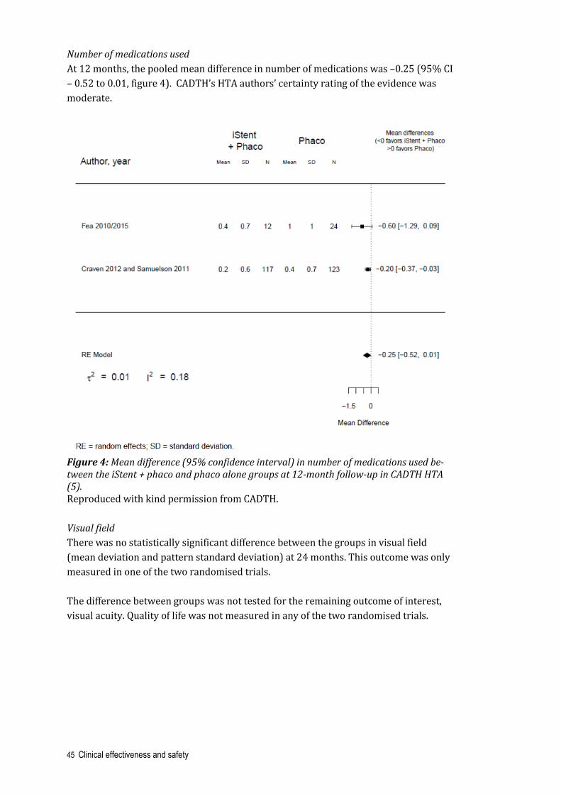
45 Clinical effectiveness and safety
Number of medications used At 12 months, the pooled mean difference in number of medications was –0.25 (95% CI – 0.52 to 0.01, figure 4). CADTH’s HTA authors’ certainty rating of the evidence was moderate.
Figure 4: Mean difference (95% confidence interval) in number of medications used be-tween the iStent + phaco and phaco alone groups at 12-month follow-up in CADTH HTA (5). Reproduced with kind permission from CADTH. Visual field There was no statistically significant difference between the groups in visual field (mean deviation and pattern standard deviation) at 24 months. This outcome was only measured in one of the two randomised trials. The difference between groups was not tested for the remaining outcome of interest, visual acuity. Quality of life was not measured in any of the two randomised trials.

46 Clinical effectiveness and safety
Hydrus Microstent + phacoemulsification vs phacoemulsification alone Two randomised trials compared Hydrus Microstent + phacoemulsification (phaco) (n=419 eyes) with phaco alone (n=237 eyes). Intraocular pressure At 24 months, the pooled mean difference in intraocular pressure was –1.87 mm Hg (95% CI - 2.49 to - 1.26; Figure 5). CADTH HTA authors’ certainty rating of the evidence was high.
Figure 5: Mean difference (95% confidence interval) in intraocular pressure between the Hydrus + phaco and phaco alone groups at 24-month follow-up in CADTH HTA (5). Reproduced with kind permission from CADTH.

47 Clinical effectiveness and safety
Numbers of medications used At 24 months, the pooled mean difference in number of medications was –0.41 (95% CI – 0.52 to 0.01; Figure 6). The CADTH HTA authors’ certainty rating of the evidence was low. The remaining outcomes of interest, quality of life, visual field and visual acuity, were not measured in any of the two randomised trials.
Figure 6: Mean difference (95% confidence interval) in number of medications used be-tween the Hydrus + phaco and phaco alone groups at 24-month follow-up in CADTH HTA (5). Reproduced with kind permission from CADTH.

48 Clinical effectiveness and safety
Trabectome + phacoemulsification vs 2x iStent + phacoemulsification Two retrospective cohort studies compared Trabectome + phacoemulsification (phaco) (n=88) with 2x iStent + phaco (n=83). Intraocular pressure At six months, the pooled mean difference in intraocular pressure was 2.55 mm Hg (95% CI 1.44 to 4.26; Figure 7) in favour of 2x iStent + Phaco. The CADTH HTA authors’ certainty rating of the evidence was very low.
Figure 7: Mean difference (95% confidence interval) in intraocular pressure between the Trabectome + phaco and 2x iStent + phaco alone groups at six-month follow-up in CADTH HTA (5). Reproduced with kind permission from CADTH.

49 Clinical effectiveness and safety
Numbers of medications used At 12 months, the pooled mean difference in number of medications was 0.41 (95% CI – 0.65 to 1.46, figure 8) in favour of 2x iStent + phaco. The CADTH HTA authors’ cer-tainty rating of the evidence was very low. There was no statistically significant difference between groups in visual acuity at 12 months. The remaining outcomes of interest, quality of life and visual field, were not measured.
Figure 8: Mean difference (95% confidence interval) in number of medications used be-tween the Trabectome + phaco and 2x iStent + phaco alone groups at 12-month follow-up in CADTH HTA (5). Reproduced with kind permission from CADTH.

50 Clinical effectiveness and safety
MIGS vs another MIGS
1x iStent vs 2x iStent vs 3x iStent One randomized trial compared one iStent (n=38 eyes) with two (n=41 eyes) or three (n=40 eyes) iStents. At 18 months, intraocular pressure was significantly reduced from baseline in eyes with one, two, or three iStents, and the reduction was incrementally greater with in-creasing numbers of iStents (reductions of approximately 4 mm Hg, 6 mm Hg, and 8 mm Hg after medication washout for one, two, and three iStents respectively). There were no statistically significant differences between groups in visual field. Differ-ences between groups in number of medications and visual acuity were not tested sta-tistically. MIGS vs pharmacotherapy
2x iStent vs travoprost, or 2x iStent Inject vs latanoprost + timolol One randomised trial compared two iStent (n=54 eyes) with travoprost (n=47 eyes), and one randomised trial compared two iStent Inject (n=94 eyes) with latanoprost and timolol (n=98 eyes). There was no statistically significant difference between groups in number of medica-tions used. Differences between groups were not tested statistically for the remaining outcomes of interest (intraocular pressure, visual field and visual acuity). MIGS vs laser therapy
Hydrus Microstent vs selective laser trabeculoplasty One prospective cohort study compared Hydrus Microstent (n=31 eyes) with selective laser trabeculoplasty (SLT) (n=25 eyes). There was no statistically significant difference between groups in intraocular pres-sure. At 12 months, the reduction from baseline in number of medications used was greater in the Hydrus group (approximately 1.4 medications) than in the SLT group (approximately 0.5 medications). Differences between groups were not tested statisti-cally for the remaining outcomes of interest (visual field and visual acuity). MIGS vs filtration surgery
Endoscopic cyclophotocoagulation vs glaucoma drainage device One retrospective cohort study and one non-randomised clinical trial compared Endo-scopic cyclophotocoagulation (ECP) (n=59 eyes) with Baerveldt glaucoma implant (BGI) (n=48 eyes) and Ahmed glaucoma implant (AGI) (n=34 eyes), respectively. There were no statistically significant differences between groups for any of the out-comes of interest (intraocular pressure, number of medications used, visual field and visual acuity).

51 Clinical effectiveness and safety
Trabectome vs trabeculectomy with mitomycin C One prospective and one retrospective cohort study compared Trabectome (n=158 eyes) with trabeculectomy with mitomycin C (MMC) (n=127 eyes). At 30 months, intraocular pressure was significantly higher in the Trabectome group (16.6 mm Hg) than in the trabeculectomy with MMC group (10.0 mm Hg) in the retro-spective study. At 30 months, the absolute number of medications used was signifi-cantly greater in the Trabectome group (2.3 medications) than in the trabeculectomy with MMC group (0.4), also in the retrospective study. At six months, one out of 12 quality of life measures was significantly greater (mean difference of approximately 13 points on a 100-point scale) in the Trabectome than in the trabeculectomy with MMC group. The difference between groups was not tested statistically for the remaining outcome of interest (visual acuity). 2x iStent Inject vs trabeculectomy with mitomycin C One prospective cohort study compared 2x iStent (n=20 eyes) with trabeculectomy with mitomycin C (MMC) (n=25 eyes). There was no statistically significant difference between groups in quality of life. Differ-ences between groups were not tested statistically for the remaining outcomes of inter-est (intraocular pressure, number of medications used and visual acuity). Trabectome or 2x iStent Inject vs trabeculectomy with mitomycin C One prospective cohort study compared Trabectome or 2x iStent (grouped together) (n=63 eyes) with trabeculectomy with mitomycin C (MMC) (n=25 eyes). The number of medications used was significantly higher in the Trabectome or 2x iStent group than in the trabeculectomy with MMC group at all follow-ups. There were no statistically significant differences between groups in intraocular pressure or visual acuity. The difference between groups was not tested for the remaining outcome of in-terest, number of medications used. Xen45 vs trabeculectomy with mitomycin C One retrospective cohort study compared Xen45 (n=185 eyes) with trabeculectomy with MMC (n=169 eyes). There were no statistically significant differences between groups for any of the out-comes of interest (intraocular pressure, number of medications used, visual field and visual acuity).

52 Clinical effectiveness and safety
MIGS + cataract surgery vs cataract surgery alone
Endoscopic cyclophotocoagulation + phacoemulsification vs phacoemulsification alone One prospective and four restrospective cohort studies compared endoscopic cyclo-photocoagulation (ECP) and phacoemulsification (phaco) (n=555 eyes) with phaco alone (n=282). Intraocular pressure In the prospective cohort study, there was a statistically significant difference in intra-ocular pressure at 36 months between the ECP+ phaco group and the phaco alone group (~15 mm Hg vs. 17 mm Hg). In the retrospective cohort studies, there were no statistically significant difference between groups in reduced intraocular pressure. Number of medications used The number of medications used at baseline differed between the intervention and comparator groups in four of the five cohort studies, impeding an interpretation of fol-low-up results. Visual acuity In two retrospective cohort studies, there was no statistically significant difference in visual acuity or best-corrected visual acuity at 36 months. Quality of life and visual field were not measured in any of the studies. 2x iStent + phacoemulsification vs phacoemulsification alone One randomised trial compared 2x iStent + phacoemulsification (phaco) (n=17 eyes) with phaco alone (n=16 eyes). At 12 months, the intraocular pressure and the number of medications used were sig-nificantly lower in the 2x iStent + phaco group than in the phaco alone group. The re-maining outcomes of interest, quality of life, visual field and visual acuity, were not measured. CyPass Micro-Stent + phacoemulsification vs phacoemulsification alone One randomised trial compared CyPass Micro-Stent + phacoemulsification (phaco) (n=374 eyes) with phaco alone (n=131 eyes). At 12 and 24 months, the intraocular pressure and the number of medications used were significantly lower in the CyPass Micro-Stent + phaco group than in the phaco alone group. The remaining outcomes of interest, quality of life, visual field and visual acuity, were not measured.

53 Clinical effectiveness and safety
MIGS + cataract surgery vs a different MIGS + cataract surgery
Goniotomy with Kahook Dual Blade + phacoemulsification vs iStent + phacoemulsi-fication One retrospective cohort study compared goniotomy with Kahook Dual Blade (KDB) + phacoemulsification (phaco) (n=237) versus iStent + phaco (n=198). At 6 months, the reduction from baseline in intraocular pressure and in number of medications used was significantly greater in the KDB + phaco group than in the iStent + phaco group. There was no statistically significant difference between the groups in visual acuity. The remaining outcomes of interest, quality of life and visual field, were not measured. Trabectome + micro-incision cataract surgery vs 2x iStent + micro-incision cata-ract surgery One retrospective cohort study compared Trabectome + micro-incision cataract sur-gery (MICS) (n=25) with 2x iStent + MICS (n=25). There were no statistically significant differences between groups for any of the meas-ured outcomes of interest (intraocular pressure, number of medications used and vis-ual acuity). The remaining outcomes of interest, quality of life and visual field, were not measured. Different numbers of iStent + phacoemulsification One non-randomised clinical trial and one retrospective cohort study compared differ-ent numbers of iStent + phacoemulsification (phaco): 1x iStent + phaco (n=39), 2x iStent + phaco (n=58) and 3x iStent + phaco (n=25). At 12 months, there was no statistically significant difference between any of the groups in intraocular pressure. There was no statistically significant difference be-tween 1x iStent + phaco and 2x iStent + phaco groups in numbers of medications used. The number of medications used was higher in the 2x iStent + phaco group than in the 3x iStent + phaco group. The difference between groups in visual acuity was not statis-tically tested. The remaining outcomes of interest, quality of life and visual field, were not measured. Endoscopic cyclophotocoagulation + iStent + phacoemulsification vs iStent + phacoemulsification One retrospective cohort study compared endoscopic cyclophotocoagulation (ECP) + iStent + phacoemulsification (phaco) (n=51) with iStent + phaco (n=50). At 12 months, reductions in intraocular pressure and the number of medications used were significantly greater in the ECP + iStent + phaco group than in the iStent + phaco group. The remaining outcomes of interest, quality of life, visual field and visual acuity, were not measured.

54 Clinical effectiveness and safety
Endoscopic cyclophotocoagulation + phacoemulsification vs Trabectome + phacoemulsification One retrospective cohort study compared endoscopic cyclophotocoagulation (ECP) + phacoemulsification (phaco) (n=35) with Trabectome + phaco (n=26). There were no statistically significant differences between groups for any of the out-comes of interest (intraocular pressure, number of medications used and visual field). The remaining outcomes of interest, quality of life and visual acuity, were not meas-ured. MIGS + cataract surgery vs filtration surgery + cataract surgery
Trabectome + phacoemulsification vs trabeculectomy with mitomycin C + phacoemulsification One randomized trial compared Trabectome + phacoemulsification (phaco) (n=10) with trabeculectomy with mitomycin C (MMC) + phaco (n=9). There were no statistically significant differences between groups in intraocular pres-sure or number of medications used. The remaining outcomes of interest, quality of life, visual field and visual acuity, were not measured. Trabectome + phacoemulsification vs trabeculectomy + phacoemulsification One prospective and one retrospective cohort study compared Trabectome + phacoemulsification (phaco) (n=47) with trabeculectomy + phaco (n=29). There were no statistically significant differences between groups in intraocular pres-sure or number of medications used. The remaining outcomes of interest, quality of life, visual field and visual acuity, were not measured. Endoscopic cyclophotocoagulation + phacoemulsification vs trabeculectomy with mitomycin C + phacoemulsification One retrospective cohort study compared endoscopic cyclophotocoagulation (ECP) + phacoemulsification (phaco) (n=24) with trabeculectomy with mitomycin C (MMC) + phaco (n=29). There was no statistically significant difference between groups in inocular pressure or visual acuity. At six months, the number of medications used was significantly higher in the ECP + phaco group than in the trabulectomy with MMC + phaco group. The remain-ing outcomes of interest, quality of life and visual field, were not measured.

55 Clinical effectiveness and safety
Effectiveness of MIGS (NIPH supplementary review)
Across studies and comparisons, the intraocular pressure (IOP) measured at 12 to 24 months’ follow-up was slightly lower in the MIGS arms than in the control arms (Figure 9). We present effect estimates for each individual comparison below.
Figure 9: Intraocular pressure across studies/comparisons MIGS vs another MIGS
Hydrus vs 2x iStent Ahmed 2019 (44) compared Hydrus with 2x iStent in 152 eyes/patients. This was a multicentre study (12 sites and 9 countries), including individuals with open angle, pseudoexfoliative, or pigmentary mild to moderate glaucoma. Mean age was 66 years, and participants were from European, Latin America, Asian and African American an-cestry. There were more female participants than men (ratio 84:68). Outcomes of inter-est in Ahmed 2019 were IOP at 12 months and number of glaucoma medications used. The remaining outcomes of interest (ie IOP fluctuation, quality of life, vision related quality of life, visual field loss, visual impairment, visual acuity and retinal nerve fibre layer thickness) were not evaluated in this study. IOP At 12 months the mean unmedicated IOP was 17.3 mmHg (SD 3.7) in the Hydrus arm and 19.2 mmHg (SD 2.4) 2x iStent arm (MD -1.9 mmHg; 95 % CI -2.91 to -0.89) (Com-parison 1.1.1; Fig 9). The difference was clinically important. Certainty of the evidence: low (Appendix 5). Number of medications used At 12 months the mean reduction from baseline in medication use was 1.0 medications (SD 1.2) in the Hydrus arm and 1.6 medications (SD 1.2) in the 2x iStent arm (MD 0.6 medications; 95% CI 0.21, 0.99).

56 Clinical effectiveness and safety
MIGS + cataract surgery vs cataract surgery alone
iStent + phacoemulsification vs phacoemulsification alone The non-randomised trial Alnawaiseh 2018 (45) compared iStent + phacoemulsifica-tion with phacoemulsification alone in 48 eyes (one eye per patient). The study was conducted in Germany. Patients’ mean age was 73.5 years in the iStent group and 72.8 years in the cataract group. Twenty-eight patients (58%) were women. The only out-come of interest in this study was IOP. IOP At 12 months the mean IOP was 13.2 mmHg (SD 2.3) and 15.1 mmHg (SD 2.7) in the iStent + phacoemulsification and the phacoemulsification alone arms, respectively (MD -1.90; 95% CI -3.32, -0.48) (Comparison 1.1.2; Fig 9). The difference was clinically im-portant. Certainty of the evidence: very low (Appendix 5). iStent inject + phacoemulsification vs phacoemulsification alone Best 2019 (46) and Samuelson 2019 (49) investigated the effects of iStent inject phacoemulsification versus phacoemulsification alone in two randomized trials con-ducted in Germany and in 41 US sites, respectively. A total of 570 eyes of 561 patients were randomized to one of the treatment arms. Mean patient age was approximately 66 to 81 years. Patients were described as ‘white’ (73%) in one study; we have no in-formation about the other. There were more males than females (ratio 289:216; data from Samuelson 2019 only). Samuelson 2019 reported IOP and number of medications used at 24 months. In Best 2019, patients were followed up with varying time intervals up to 38 months, with mean 14 months. The remaining outcomes of interest (ie IOP fluctuation, quality of life, vision related quality of life, visual field loss, visual impair-ment, visual acuity and retinal nerve fibre layer thickness) were not evaluated in these trials. IOP The pooled mean difference in IOP between the iStent inject + phacoemulsification and phacoemulsification alone arms was -0.70 mmHg (95% CI -1.27, -0.13) (single study ef-fect estimates in comparison 1.1.3; Fig 9). The difference was not clinically important. Certainty of the evidence: moderate (Appendix 5). Number of medications used The mean reduction of medications used at 24 months in Samuelson 2019 was 1.2 (SD 1.0) and 0.8 (SD1.0) in the iStent inject + phacoemulsification arm and the phacoemul-sification alone arm, respectively (MD -0.40; 95% CI -0.61, -0.19). Best 2019 did not re-port standard deviations, but the mean number of medications used at 4 months was 1.5 and 2.1 in the iStent inject + phacoemulsification arm and in the phacoemulsifica-tion alone arm, respectively.

57 Clinical effectiveness and safety
Trab360 + phacoemulsification vs phacoemulsification alone Sato 2018 compared Trab360 + phacoemulsification with phacoemulsification alone in 18 eyes/patients in Japan (50). This randomised trial included patients diagnosed with coexisting mild to moderate cataract and open angle glaucoma. Patients’ mean age was 74.2±9.5 years in the Trab360 + phacoemulsification arm and 74.4±3.6 in the phacoemulsification alone arm. There were more female participants than male (ratio 8:10). Sato 2018 reported IOP at 24 months, and number of medications used. The re-maining outcomes of interest (ie IOP fluctuation, quality of life, vision related quality of life, visual field loss, visual impairment, visual acuity and retinal nerve fibre layer thick-ness) were not evaluated in these trials. IOP At 24 months the mean IOP was 11.8 mmHg (SD 2.3) and 15.1 mmHg (SD 2.7) in the Trab360 + phacoemulsification and the phacoemulsification alone arms, respectively (MD -2.80; 95% CI -5.49, -0.11) (Comparison 1.1.5; Fig 9). The difference was clinically important. Certainty of the evidence: very low (Appendix 5). Number of medications used The mean number of medications used at 24 months was 1.0 (SD 1.7) and 1.2 (SD 1.4) in the Trab360 + phacoemulsification and the phacoemulsification alone arms, respec-tively (MD -0.90; 95% CI -1.46, -0.34). Hydrus + phacoemulsification vs phacoemulsification alone Jones 2019 (47), a 24 month follow-up of Samuelson 2018 (87), which was included in the CADTH HTA, compared Hydrus + phacoemulsification with phacoemulsification alone. The trial was a multicentre study conducted in the United States including 331 eyes/patients with mild to moderate open angle glaucoma, pigmentary or pseudoexfo-liative glaucoma. Patients’ mean age was 70.3 (SD 7.1) and 71.1 years (SD 7.2) in the two arms, respectively. There were slightly more female than male patients (ratio 174:157). Regarding ethnicity, the majority of patients were “white and a small propor-tion of Asian, black or African American or other origin”. IOP At 24 months the mean unmedicated IOP was 17.5 mmHg (SD 3.9) and 19.3 mmHg (SD 4.2) in the Hydrus + phacoemulsification and the phacoemulsification alone arms, re-spectively (MD -1.80; 95% CI -2.73, -0.87) (Comparison 1.1.4; Fig 9). The difference was not clinically important. Certainty of the evidence: high (Appendix 5). Number of medications used The mean number of medications used at 24 months was 0.4 (SD 0.8) and 0.8 (SD 0.9) in the Hydrus + phacoemulsification and the phacoemulsification alone arms, respec-tively (MD -0.40; 95% CI -0.60, -0.20).

58 Clinical effectiveness and safety
MIGS vs filtration surgery
Gold MicroShunt (GMS) 24um or 48um vs Ahmed glaucoma valve (AGV) Skaat 2016 (51) was conducted in Israel in patients 21 years and older diagnosed with open angle, pseudoexfoliative or pigmentary glaucoma in one or both eyes, with at least one failed trabeculectomy within the last 60 days. The trial included 29 eyes/patients. Patients’ mean age was 71 (SD 4.1), 72.6 (SD 4.5) and 72.1 (SD 4.9) years in the AGV, GMS 24um and GMS 48um arms, respectively. Fifteen patients were female and 14 were male. Ethnicity was not specified. Outcomes of interest included in Skaat 2016 were IOP and medication use. The remaining outcomes of interest were not evalu-ated for clinical effectiveness. IOP At 5 years, the mean IOP was 15.6 mmHg (SD 1.4) and 16.3 mmHg (SD 1.8) in the com-bined GMS (24um + 48um) and AGV arms, respectively (MD -0.70 mmHg; 95% CI -2.71, 1.31) (Comparison 1.1.6; Fig 9). The difference was not clinically important. Certainty of the evidence: very low (Appendix 5). Number of medications used The mean number of medications used at 5 years was 3.0 (SD 0.7) and 3.2 (SD 0.5) in the combined GMS (24um + 48um) and AGV arms, respectively (MD 0.20; 95% CI -0.57, 0.97). MIGS + cataract surgery vs filtration surgery + cataract surgery
iTrack + phacoemulsification vs filtration surgery + phacoemulsification Rekas 2015 (48) was conducted at the Military Institute in Poland. It included individu-als diagnosed with open angle glaucoma, pseudoexfoliative and pigmentary glaucoma with daily fluctuations in IOP and no-compliance with medication or allergy to topical medications. The trial included 59 eyes in patients. Patients’ mean age was 74.6 years (SD 8.9) and 73.3 years (SD 5.8) in the two arms, respectively. Thirty-three of the pa-tients were male and 28 were females (ethnicity not specified). Included outcomes of interest were IOP, medication use and visual acuity. No other outcomes of interest were measured. IOP At 12 months, the mean IOP was 12.6 mmHg (SD 2.7) and 14.3 mmHg (SD 3.5) in the iTrack + phacoemulsification and filtration surgery + phacoemulsification alone arms, respectively (MD -1.70 mmHg; 95% CI -3.29, -0.11) (Comparison 1.1.7; Fig 9). The dif-ference was clinically important. Certainty of the evidence: very low (Appendix 5). Number of medications used The mean number of medications used at 12 months was 0.27 (SD 0.67) and 0.55 (SD 0.94) in the iTrack + phacoemulsification and the filtration surgery + phacoemulsifica-tion arms, respectively (MD -0.28; 95% CI -0.70, 0.14). Visual acuity

59 Clinical effectiveness and safety
There was no difference in mean corrected distance visual acuity measure (CDVA) at 12 months. The logarithm (base 10) of the minimal angle of resolution (LogMAR) was 0.11 (SD 0.17) and 0.11 (SD 0.16) in the two arms, respectively (MD 0.00; 95% CI -0.08, 0.08).
Safety of MIGS (CADTH HTA)
The CADTH HTA authors characterised the evidence regarding the safety of MIGS as “limited”. None of the included studies reported methods of measuring adverse events or any restrictions on what was considered an adverse event. Statistical comparisons between groups occurred infrequently. Due to these limitations, the CADTH HTA au-thors rated the evidence regarding safety as “very low” certainty for all comparisons and all adverse events. That is, it is uncertain whether there is any difference regarding safety between different types of MIGS, between MIGS and other methods, or between MIGS in combination with cataract surgery, and other methods. Please see the full CADTH HTA report for details (5).
Safety of MIGS (NIPH supplementary review)
Safety (i.e., adverse events) was measured and reported differently by study authors. Some authors included pre-and-post-operative events, others classified the events as mild or moderate, others described events by follow up time (i.e., year 1 or 2), and some provided a brief description. Aiming not to lose any information, we report in the tables below adverse events at any point as reported by authors with number of partic-ipants and percentages (n, %), and risk ratios with 95% confidence intervals. Our over-all judgement of the certainty of the evidence was very low. MIGS vs another MIGS
Hydrus vs 2x iStent Ahmed 2019 (44) recorded the following adverse events and complications associated with the comparison Hydrus vs 2x iStent: new cataracts, best-corrected visual acuity (BVCA) loss >2 lines, IOP elevation >10 mmHg over baseline, device obstructions, hy-potony, device migration, dislocation and secondary surgical intervention (Table 7). The certainty of the evidence was very low. Table 7: Adverse events. Comparison: Hydrus vs 2x iStent
Adverse event Hydrus n (%)
2x iStent n (%)
Risk ratio [95% CI]
N=74 N=76 New cataracts 2 (2.7) 1 (1.3) 2.05 [0.19, 22.17] BVCA loss >2 lines 3 (4.0) 1 (1.3) 3.08 [0.33, 28.95] IOP elevation >10 mmHg 3 (4.0) 4 (5.2) 0.77 [0.18, 3.32] Device obstructions 10 (13.5) 9 (11.8) 1.14 [0.49, 2.65] Secondary surgical intervention 0 (0) 3 (3.9) 0.15 [0.01, 2.79] Hypotony, device migration, dislocation 0 (0) 0 (0) --
CI: confidence interval; BVCA: best-corrected visual acuity; IOP: intraocular pressure.

60 Clinical effectiveness and safety
MIGS + cataract surgery vs cataract surgery alone
iStent inject + phacoemulsification vs phacoemulsification alone Best 2019 (46) reported one case of bleeding during interventions, which was quickly resolved, in the iStent inject + phacoemulsification arm. In the phacoemulsification alone arm, there were two cases of very high postoperative IOP and pain. Samuelson 2019 (49) recorded and reported postoperative ocular adverse events oc-curring in ≥2% of the study eyes (Table 8). Compared to phacoemulsification alone, the combination of iStent inject and phacoemulsification seemed to increase the risk of for-eign body sensation, IOP increase > 10 mmhg at >1 month, perioperative ocular pain, blurred vision/visual disturbance, and secondary surgical intervention. Further, cor-neal abrasion, corneal opacity, hyperemia, non-proliferative diabetic retinopathy and IOP increase requiring medications or surgical intervention where among adverse events that seemed to occur more frequently in the phacoemulsification only arm. The certainty of the evidence was very low. Table 8: Adverse events. Comparison: iStent inject + phacoemulsification vs phacoemulsi-fication alone
Adverse event
iStent inject + phaco
n (%)
Phaco alone n (%)
Risk ratio [95% CI]
N=387 N=118 Foreign body sensation 9 (2.3) 0 (0.0) 5.83 [0.34, 99.38] IOP increase > 10mmhg at month >1 8 (2.1) 1 (0.8) 2.44 [0.31, 19.30] Perioperative ocular pain 8 (2.1) 1 (0.8) 2.44 [0.31, 19.30] Blurred vision/visual disturbance 9 (2.3) 2 (1.7) 1.37 [0.30, 6.26] Extraocular inflammation 9 (2.3) 2 (1.7) 1.37 [0.30, 6.26] Any intraocular inflammation 22 (5.7) 5 (4.2) 1.34 [0.52, 3.47] Secondary surgical intervention 22 (5.4) 6 (5.0) 1.12 [0.46, 2.69] Ocular surface disease 62 (16.1) 20 (16.8) 0.95 [0.60, 1.50] Epiretinal membrane 9 (2.3) 3 (2.5) 0.91 [0.25, 3.32] Ocular allergies 11 (2.8) 4 (3.4) 0.84 [0.27, 2.58] Vitreous floaters 8 (2.1) 3 (2.5) 0.81 [0.22, 3.02] Loss of BSCVA ≥2 lines 10 (2.6) 5 (4.2) 0.61 [0.21, 1.75] Posterior vitreous detachment 10 (2.6) 5 (4.2) 0.61 [0.21, 1.75] Corneal abrasion 8 (2.1) 4 (3.4) 0.61 [0.19, 1.99] Corneal opacity 4 (1.0) 3 (2.5) 0.41 [0.09, 1.79] Non-proliferative diabetic retinopathy 2 (0.5) 3 (2.5) 0.20 [0.03, 1.20] Hyperemia 3 (0.8) 7 (5.9) 0.13 [0.03, 0.50] IOP increase requiring medications or surgical intervention
1 (0.3) 3 (2.5) 0.10 [0.01, 0.97]
Stent obstruction 24 (6.2) NA - CI: confidence interval; BSVCA: best spectacle-corrected visual acuity; IOP: intraocular pressure

61 Clinical effectiveness and safety
Trab360 + phacoemulsification vs phacoemulsification alone Sato 2018 (50) recorded and reported adverse events associated with Trab360 + phacoemulsification vs phacoemulsification alone (Table 9). Compared to phacoemul-sification alone, the combination of Trab360 and phacoemulsification seemed to in-crease the risk of intraoperative reflux bleeding and hyphema, and decrease the risk of additional glaucoma surgery. There appeared to be no difference between the two con-ditions with regard to IOP elevation and rate of endothelial cell loss. The certainty of the evidence was very low. Table 9: Adverse events. Comparison: Trab360 + phacoemulsification vs phacoemulsifica-tion alone
Adverse event Trab360 + phaco n (%)
Phaco alone n/N
Risk ratio [95% CI]
N=9 N=9 Intraoperative reflux bleeding 9 (100) 0 (0) 19.00 [1.27, 284.24] Hyphema 2 (22) 0 (0) 5.00 [0.27, 91.52] IOP elevation 3 (33) 3 (33) 1.00 [0.27, 3.69] Additional glaucoma surgery 0 (0) 1 (11) 0.33 [0.02, 7.24] Mean rate of endothelial cell loss 5.7% (SD 3.4) 6.1% (SD 5.9) -0.4%* [-4.85, 4.05]
CI: confidence interval; IOP: intraocular pressure; *Mean difference Hydrus + phacoemulsification vs phacoemulsification alone Jones 2019 (47) reported cumulative adverse events associated with Hydrus + phacoemulsification versus phacoemulsification alone, through 24 months (Table 10). Synechiae, uveititis/irititis, hyphema, corneal abrasion, subconjunctival bleeding and vitreous complications appeared to occur more frequently in the Hydrus + phacoemul-sification arm. Conjunctivitis, IOP elevation and secondary surgical interventions ap-peared to occur more frequently in the phacoemulsification only arm. The certainty of the evidence was very low. Table 10: Cumulative adverse events through 24 months. Comparison: Hydrus + phacoemulsification vs phacoemulsification alone Adverse event Hydrus+phaco
n (%) Phaco alone
n (%) Risk ratio [95% CI]
N=219 N=112 Synechiae 15 (6.8) 1 (0.9) 7.67 [1.03, 57.33] Uveitis/iritis 10 (4.6) 0 (0.0) 10.79 [0.64, 182.41] Conjunctivitis 8 (3.7) 10 (8.9) 0.41 [0.17, 1.01] Elevated IOP >10mmHg over baseline 2 (0.9) 3 (2.7) 0.34 [0.06, 2.01] Hyphema 3 (1.4) 1 (0.9) 1.53 [0.16, 14.58] Corneal abrasion 4 (1.8) 0 (0.0) 4.62 [0.25, 85.11] Subconjunctival hemorrhage 4 (1.8) 0 (0.0) 4.62 [0.25, 85.11] Device obstruction 4 (1.8) -- -- Vitreous complications 3 (1.4) 1 (0.9) 1.53 [0.16, 14.58] Secondary surgical interventions 1 (0.5) 3 (2.7) 0.17 [0.02, 1.62]
CI: confidence interval; IOP: intraocular pressure

62 Clinical effectiveness and safety
iStent + phacoemulsification vs phacoemulsification alone The non-randomised trial Alnawaiseh 2018 (45) did not report any adverse events as-sociated with the comparison iStent + phacoemulsification vs phacoemulsification alone. MIGS vs filtration surgery
Gold MicroShunt (GMS) 24um or 48um vs Ahmed glaucoma valve (AGV) Skaat 2016 (51) reported that there were no major complications in any of the treat-ment arms, but did register some minor complications (Table 11). All over, there seemed to be more postoperative minor complications in the Ahmed glaucoma valve arm, but the study authors noted that all complications resolved within weeks. The cer-tainty of the evidence was very low. Table 11: Adverse events. Comparison: Gold MicroShunt 24um/48um vs Ahmed glau-coma valve
Adverse event Gold MicroShunt 24um/48um
n (%)
Ahmed glaucoma valve n (%)
Risk ratio [95% CI]
N=20 N=9 Injection 11 (55) 9 (100) 0.58 [0.38, 0.87] Hyphema 3 (15) 2 (22) 0.68 [0.14, 3.37] Choroidals 1 (5) 2 (22) 0.23 [0.02, 2.17] Shallow anterior chamber 2 (10) 2 (22) 0.45 [0.07, 2.71] Hypotony 0 (0) 1 (11) 0.16 [0.01, 3.56]
CI: confidence interval
MIGS + cataract surgery vs filtration surgery + cataract surgery
iTrack + phacoemulsification vs filtration surgery + phacoemulsification Rekas 2015 (48) recorded and reported a number of perioperative complications (Ta-ble 12). Adverse events seemed to occur more frequently in the iTrack + phacoemulsifi-cation arm, except for bleb fibrosis, which occurred more often in the filtration surgery + phacoemulsification arm. There was no difference observed between the two arms in intraoperative TDM rupture and iris carceration. The certainty of the evidence was very low.

63 Clinical effectiveness and safety
Table 12: Adverse events. Comparison: iTrack + phacoemulsification vs filtration surgery + phacoemulsification
Adverse event iTrack + phaco n (%)
Filtration sur-gery + phaco
n (%)
Risk ratio [95% CI]
N=29 N=30 Intraoperative TDM rupture 1 (3.4) 1 (3.3) 1.03 [0.07, 15.77] Hyphema 17 (58.6) 0 (0.0) 36.17 [2.28, 574.84] Descemet membrane detachment 1 (3.4) 0 (0.0) 3.10 [0.13, 73.14] Choroid detachment 2 (6.9) 0 (0.0) 5.17 [0.26, 103.21] Hypotony until 30 days 5 (19.0) 3 (10.0) 1.72 [0.45, 6.57] Bleb fibrosis 0 (0.0) 8 (26.7) 0.06 [0.00, 1.01] Iris incarceration 1 (3.4) 1 (3.3) 1.03 [0.07, 15.77]
TDM: trabeculo Descemet’s membrane

64 Organizational aspects
Organizational aspects
This chapter about organizational aspects related to using MIGS in Norway is not adapted from CADTH.
Background
MIGS surgery in Norway is generally done at public hospitals with eye units in Norway (i.e. Oslo University Hospital, Sørlandet Hospital, Haukeland University Hospital, Sta-vanger University Hospital , St.Olav Hospial – University Hospital Trondheim, Ålesund Hospital, Molde Hospital, Drammen Hospital, Elverum Hospital, probably more) in ad-dition to a few private clinics (to our knowledge iFocus and Volvat-Orbita). This chap-ter is based on input from our clinical experts, representing Oslo University Hospital, Hospital of Sothern Norway, Arendal, Haukeland University Hospital, St. Olav’s Univer-sity Hospital, Trondheim and University of North Norway (28;88).
Method
In order to evaluate the organizational consequences related to the implementation of MIGS and a potential increase in volume of MIGS performed in Norwegian hospitals, we asked clinical experts from the five respective state-run hospitals, to answer a ques-tionnaire regarding their present capacity and procedure used: patient selection, pro-cedures and ongoing trials. We received answers from all five hospitals. The questions used in the questionnaire are listed in Appendix 8.
Patient selection
There exists one professional guideline within ophthalmology with a specific chapter for glaucoma, but there is no national register for glaucoma surgery (28;88). These guidelines provide broad advice, they point out that there are many factors that play a role in choice of procedure, and that individual considerations are needed. Medical knowledge is constantly evolving, and Norwegian ophthalmology follows the European Glaucoma Society (EGS) Guidelines (12).

65 Organizational aspects
Clinicians need to identify which specific patients may or may not benefit from a partic-ular MIGS procedure. To our knowledge, criteria for patient selection are not specifi-cally developed in Norway. The patients requiring MIGS treatment are referred by an ophthalmologist to a specialised eye unit offering MIGS surgery. What determines which MIGS is chosen is one/more of the following: the type of glaucoma, what stage the glaucoma is in, what the estimated target pressure is, the patient’s age and compli-ance regarding treatment (28). There are three types of recipient reservoars for MIGS procedures: 1.) Subconjunctival space, 2.) Suprachoridal space, and 3.) Trabecular meshwork/schlemm canal. Different MIGS procedures are used in different settings due to different mechanisms of function. Procedures that use subconjunctival space and trabecular meshwork as a recipient res-ervoir are the most used MIGS procedures in Norway today (28).
The subconjunctival space procedure is usually provided to patients with low target pressure. A trabecular meshwork procedure can be chosen when there is a need to combine glaucoma with cataract surgery and in cases of mild/moderate glaucoma where there the target pressure doesn’t have to be as low. Also, the use of this proce-dure is relevant amongst others when the patient has intolerance to topic medication (28). To our knowledge iStent inject, ABiC (both trabecular meshwork procedures) and XEN Gel Implant (subconjunctival space procedure) dominate the Norwegian marked.
Capacity and number of treatments
MIGS procedures require less follow-up than traditional surgery in early phase after surgery, so the benefit may be fewer controls per patient in the hospital. However, bet-ter treatment options may lead to more patients getting operated, thus resulting in a similar overall demand of follow-up consultations. MIGS is suitable for outpatient sur-gery without hospital admission. This usually requires access to outpatient follow-up on day 1, (3) and 7. Ophthalmologists need to be trained and course certification is re-quired to perform MIGS (28). The need for glaucoma surgery may increase due to increased population growth in the relevant age group, in addition to increased experience with MIGS among clinicians. Mainly based on extrapolation of considered needs estimated in 2012 and up to 2030 as presented in the Konus report (28) conducted by The Norwegian Ophthalmological Association, clinical experts predict the number of MIGS procedures will increase annu-ally to twice as many in 2024 compared to today (2020).

66 Health economic evaluation
Health economic evaluation
BACKGROUND
Introduction to Economic Evaluation of Health Care Programmes
The basic aim of an economic evaluation is to identify, measure and compare costs and consequences (effectiveness) of different strategies in an incremental analysis, one in which the differences in costs between strategies are compared with differences in their consequences (89). Results of an economic evaluation between two strategies can be expressed as an incremental cost-effectiveness ratio (ICER), defined by this equa-tion:
The health care sector, similarly to society in general, is constrained by scarce re-sources. Therefore, economic evaluations are useful tools for decision makers facing questions of how to prioritize treatments when the objective is to maximize health ben-efits with scarce resources. For an economic evaluation to be meaningful in a decision making process, the ICER is usually compared to a ceiling ratio that reflects the decision maker’s maximum willingness to pay (WTP) for a health gain (89). Therefore, the deci-sion rule for an economic evaluation can be expressed as:
where λ equals WTP, and means that if the ICER of a strategy is equal to or below the ceiling ratio, introduction of the strategy represents good value for money (89). This decision rule does not take uncertainty into account. Economic evaluations are often based on decision models, such as decision trees and Markov models, calculatinge results based on various input parameters. There are al-ways uncertainties related to the values of these parameters, which is why sensitivity

67 Health economic evaluation
analyses are important features of economic evaluations. In short, sensitivity analyses illustrate how robust the results of the model are to variations in model parameters (89). Probabilistic sensitivity analysis (PSA) is a kind of sensitivity analysis where probabil-ity distributions are assigned to the model parameters. The advantage of PSA is that un-certainties of many parameters in the model can be considered simultaneously, making it very realistic for real world considerations. The basic approach in PSA is to replace the deterministic mean value with a distribution (based on published standard errors), where values are generated by random draws from the assigned distribution. By doing this repeatedly, with a specified number of iterations, the model produces estimates of the probabilities of alternative strategies to be cost-effective for a range of ceiling val-ues of WTP. Results from PSAs are often presented as scatter plots showing point esti-mates of the ICER for all iterations in a cost-effectiveness plane, and cost-effectiveness acceptability curves (CEACs) showing the probabilities of the alternatives being cost-effective subject to changing values of WTP (89). In short, making a model probabilistic means that it is possible to estimate the overall parameter uncertainty associated with a decision to implement alternative strategies (89).
Priority setting criteria
There are three general criteria for priority setting in the Norwegian health care sector: the benefit criterion, the resource criterion, and the severity criterion (6). The benefit criterion The benefit criterion primarily refers to a technology’s expected health gains, i.e. in-creased longevity and/or improved health-related quality of life. By combining these two dimensions of health gains into a single outcome measure, referred to as quality-adjusted life-year (QALY), it is possible to compare outcomes across different diseases, patient groups and types of treatments. According to the benefit criterion, priority in-creases with the size of the expected health benefit of the strategy (6). The resource criterion The resource criterion focuses on how the healthcare sector allocates its limited re-sources. Introducing a new technology may create demands for personnel, equipment, facilities, etc., that could alternatively be used to provide treatments for other patients. This reality is referred to as the “opportunity cost” of a new technology. The larger quantity of resources that are allocated to a technology for one patient group, the fewer resources are available for treating others. In addition to resource use within the health care sector, a technology may also generate costs for other sectors. In Norway, the re-source criterion is taken into account by weighing resource use (measured as monetary costs) against effectiveness (measured as health outcomes) in a cost-effectiveness anal-ysis of the technology of interest. In addition, a budget impact analysis is expected from

68 Health economic evaluation
the bodies making decisions about health care funding. According to the resource crite-rion, priority increases the fewer resources that are needed for the strategy if intro-duced (6). The severity criterion Severity is measured as “absolute shortfall”, defined as the expected loss of future health (QALYs) associated with a specified diagnosis. For treatment of a disease, sever-ity in Norway is calculated as the average expected QALY loss for the relevant patient population given the current treatment option. The greater the absolute shortfall asso-ciated with a disease, the higher costs per QALY-gained would the government be pre-pared to pay. According to the severity criterion, priority increases with expected fu-ture health loss resulting from the disease (6;90). The Norwegian White paper on priority setting The Norwegian White paper on priority setting (Meld. St. 34 (2015–2016)) indicates that weighting of resource use against utility should be based on the opportunity cost principle, and that priority should be further increased according to severity (absolute shortfall) (6). The Norheim commission and the Magnussen group suggested that an absolute shortfall of less than 2.0 QALYs should indicate diseases with the lowest level of severity, while an absolute shortfall above 20 QALYs would indicate diseases in the highest severity class. There is no official societal willingness to pay threshold for health interventions in Norway. However, the Norheim commission and Magnussen group suggested a step-wise weighting scale for societal willingness to pay for different severity classes, that ranges from 1 if absolute shortfall were less than two QALYs, up to 3 if absolute shortfall were 20 or more QALYs (90;91).
Literature review of previous health economic evaluations of MIGS
A review of the literature was conducted to identify published health economic evalua-tions on patients with glaucoma treated with MIGS that may be applicable to a Norwe-gian setting. The review identified no study that had been conducted in a Norwegian setting, and there is generally limited evidence available regarding cost-effectiveness of MIGS for glaucoma. Most of the identified economic evaluations of glaucoma treatment focused on non-MIGS comparisons, such as pharmacotherapy, laser therapy, and surgi-cal treatments, like filtration surgery (92-95). We identified some studies that consid-ered separately either the costs or HRQoL associated with MIGS, but these did not in-corporate the efficacy of treatments with costs together and, as such, did not assess the cost-effectiveness of MIGS (81;96). While HRQoL studies have some value, given com-parable quality of the procedures between countries, the value of costs analyses from other countries do not reflect Norwegian costs levels. Further, the identified studies were either retrospective case studies or industry - sponsored RCTs with short follow-up times. The reported results were associated with high levels of uncertainty (5;31)

69 Health economic evaluation
CADTHs health economic evaluation
The report from the CADTH examined the cost-effectiveness of MIGS, with or without cataract surgery, compared with alternative strategies (i.e., pharmacotherapy, laser therapy, filtration surgery, cataract surgery, or filtration surgery combined with cata-ract surgery) over a patient’s lifetime from a Canadian health care perspective. The pri-mary outcome was ICER, i.e., incremental cost per QALYs gained in 2018 Canadian dol-lars (CAD). Their findings suggest that there are some cases where MIGS may be cost-effective, and in other cases, MIGS is unlikely to be economically beneficial. We have presented the results from CADTHs base case economic models in table 13. The incre-mental difference in QALYs and costs were relatively small (i.e., incremental QALYs ranged from –0.07 to 0.039, and incremental costs ranged from CAD –3,267 (NOK -20 484) to CAD 1,726 (NOK 10 822)), among all models, suggesting that the alternative procedures were not dramatically different from each other neither in terms of costs or benefits. Incremental costs between comparators occurred early of the time horizon in the model and were mostly driven by initial surgery-related costs. Except for the com-parison of MIGS (Hydrus Microstent) combined with cataract surgery compared with cataract surgery alone, which was based on “high” quality evidence, the rest of the com-parisons were informed by clinical studies with evidence of lower quality. CADTH con-cluded that adequately powered studies using clinically important outcome measures with longer follow-up periods would be useful to confirm and validate the findings of their health economic assessment (5).

70 Health economic evaluation
Table 13: CADTHs* results of the probabilistic base case cost-utility analysis (lifetime horizon, discounted).
Strategy Total costs (CAD)
Effects (QALYs)
Incremen-tal Costs (CAD)
Incremental Ef-fect (QALYs)
ICER (CAD/QALY)
Model 1: Pharmacotherapy 2 x iStent Inject (MIGS)
11,900 12,641
12.85 12.89
741
0.039
18,808
Model 2: Laser therapy Hydrus Microstent (MIGS)
9,013 10,739
10.36 10.34
1,726
-0.023
MIGS dominated (highest cost, less effective)
Model 3A: Filtration surgery Trabectome (MIGS)
13,375 12,672
12.49 12.42
-703
-0.070
10,093
Model 3B: Filtration surgery ECP (MIGS)
14,621 11,354
10.85 10.83
-3,267
-0.027
121,959
Model 4: Cataract surgery alone Hydrus Microstent (MIGS) + cataract surgery
8,431 10,072
9.04 9.06
1,641
0.026
63,626
Model 5: Filtration surgery + cataract surgery Trabectome (MIGS) + cataract surgery
11,309 10,836
7.92 7.89
-473
-0.032
14,968
ICER = incremental cost-effectiveness ratio; MIGS = minimally invasive glaucoma surgery; ECP = Endoscopic Cyclophotcoag-ulation; QALYs = quality adjusted life-years; $ = Canadian dollars. *Reference: CADTH (5).
71 Health economic evaluation
METHOD
General
Our overall objective was to examine the cost-effectiveness of MIGS, as stand-alone procedure and in combination with cataract surgery, compared with current treatment options, for the treatment of glaucoma. This health economic evaluation was conducted according to a protocol developed a priori (36). The choice of sets of interventions and comparators was based on the com-missioning from The National System for Managed Introduction of New Health Tech-nologies within the Specialist Health Service in Norway, and refined in cooperation with a panel of Norwegian clinical experts and patient partners (3;28;37). We based our health economic analysis on a previous HTA carried out by CADTH, which corre-sponded well to the views reflected by our Norwegian clinical experts and patient part-ners. Our analytical approach is therefore based on an adaptation of the decision ana-lytical model underlying their HTA, and on considerations of data identified in the clini-cal review documented previously in this report (5;28;37). We developed six different decision analytic cost-effectiveness models in TreeAge Pro ® 2020, to estimate ICER, which is appropriate in the context of a Norwegian health care priority setting. The approach is also able to capture benefits related to morbidity consequence related to the progression of glaucoma on visual field. The clinical condi-tion and its potential treatments have no mortality effects. The models provide insight into costs, health effects, survival, and disease stage. Relevant costs were expressed in 2019 Norwegian kroner (NOK), and unit costs collected from previous years were ad-justed in accordance with the consumer price index (98). In accordance with the Gov-ernment White Paper about priority setting, (Meld. St. 34 (2015-2016)) the analysis was carried out from a modified Norwegian health care perspective (direct medical costs plus travel costs for patients) and both costs and effects were discounted using an annual discount rate of 4% (6;99). The health care perspective is relevant for prioriti-sation of interventions within a fixed budget (no expansion of the budget is assumed). As glaucoma and its treatment have long-term consequences, the analysis is carried out using a lifetime horizon. Following the recommendations from the Norwegian White paper on priority setting and the severity criterion, we have estimated absolute shortfall for patients with glau-coma (6). The cost-effectiveness can be considered for each of the six models for the complete range of willingness to pay (WTP) threshold suggested by the Norheim and Magnussen commissions, i.e., from 275 000 (reflecting severity weight = 1) up to 825 000 NOK (severity weight = 3) per QALY (90;91). We handled uncertainties in model parameters by assigning probability distributions to the parameters and performing PSA, designed as a Monte Carlo simulation, with 10 000 iterations. Results from PSAs are presented as scatter plots in a cost-effectiveness plane, and as CEACs. We also performed one-way sensitivity analyses for each of the six

72 Health economic evaluation
models to explore potential impact of uncertainty in single parameters. For each of the six models, we present the results of the one-way sensitivity analyses as tornado dia-grams. In addition, we estimated the budget impact of introducing MIGS as a routine treatment option in Norway for patients with glaucoma.
Model structure
We made several adaptations to the model which the HTA from CADTH was based on, including both adaptations of model structure and input data. The adaptations were in agreement with a panel of Norwegian clinical experts and representatives of Norwe-gian patient organizations (28;37). All six models were based on a Markov model struc-ture. Each model was designed to compare one intervention and one comparator for the six different treatment scenarios. As mentioned above, we chose to incorporate clinical efficacy and safety data from the HTA carried out by CADTH. All the included clinical comparisons and pathways mod-elled in this previous HTA were assumed to be of value in a Norwegian context. The Ca-nadian clinical review concluded that it was not possible to determine significant differ-ences in effectiveness and safety between alternative MIGS devices. The clinical review reported 24 unique comparisons but found great clinical heterogeneity between stud-ies. Indirect treatment comparison to determine relative effectiveness between MIGS devices was therefore considered inappropriate (e.g., network meta-analysis). As such, only pairwise comparisons were done in the economic evaluation (5). Each model incorporated four live Markov health states, in addition to an absorbing health state “death”. The health states are mutually exclusive, meaning that patients can be in only one of them at any time, and complete, meaning that they cover all possible health situations of the patient group. Each health state is associated with state-specific utility weights and costs, and costs- and utilities are aggregated depending on the time spent in different health states. In the base-case analysis, we followed a hypothetical cohort of glaucoma patients up to 100 years, and patients can move between health states between each annual cycle, depending on transition probabilities. The live Mar-kov health states in the model were defined according to the Hodapp-Parrish-Anderson grading scale for disease stage, where visual field (VF) is measured by mean deviation (MD) in decibels (dB), based on a previous model by NICE (100);
- Mild stage (VF MD of 0 to -6 dB); - Moderate stage (VF MD of -6.01 to -12 dB); - Advanced stage (VF MD of -12.01 to -20 dB); and - Severe visual impairment/blindness (VF MD of < -20 dB).
This grading scale was used to trace the progression of patients between the model health states and allowed consideration of patients entering the model at different se-
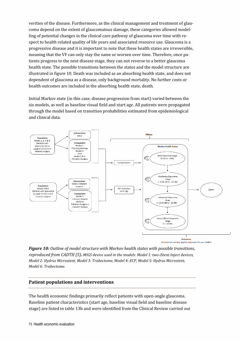
73 Health economic evaluation
verities of the disease. Furthermore, as the clinical management and treatment of glau-coma depend on the extent of glaucomatous damage, these categories allowed model-ling of potential changes in the clinical care pathway of glaucoma over time with re-spect to health related quality of life years and associated resource use. Glaucoma is a progressive disease and it is important to note that these health states are irreversible, meaning that the VF can only stay the same or worsen over time. Therefore, once pa-tients progress to the next disease stage, they can not reverse to a better glaucoma health state. The possible transitions between the states and the model structure are illustrated in figure 10. Death was included as an absorbing health state, and does not dependent of glaucoma as a disease, only background mortality. No further costs or health outcomes are included in the absorbing health state, death. Initial Markov state (in this case, disease progression from start) varied between the six models, as well as baseline visual field and start age. All patients were propagated through the model based on transition probabilities estimated from epidemiological and clinical data.
Figure 10: Outline of model structure with Markov health states with possible transitions, reproduced from CADTH (5). MIGS device used in the models: Model 1: two iStent Inject devices, Model 2: Hydrus Microstent, Model 3: Trabectome, Model 4: ECP, Model 5: Hydrus Microstent, Model 6: Trabectome.
Patient populations and interventions
The health economic findings primarily reflect patients with open-angle glaucoma. Baseline patient characteristics (start age, baseline visual field and baseline disease stage) are listed in table 13b and were identified from the Clinical Review carried out

74 Health economic evaluation
by CADTH (5). Our clinical experts considered model 1, 2 and 5 to be most relevant in Norwegian conditions. Although, the baseline age in model 1 might be somewhat low. The patient populations were adults diagnosed with glaucoma (models 1 - 4) and glau-coma + cataract combined (models 5 - 6). The starting age was calculated based on weighted average from included clinical studies, ranged from 64 to 72 years old. As described in the background chapter, the patient population with glaucoma consists of elderly, and the prevalence increases with age. It is estimated that more than 10% among people above 80 years have the disease (101). The Hodapp-Parrish-Anderson grading scale was used to assume the initial progression stage in each model (table 14). As the potential use of MIGS within the treatment pathway for glaucoma is unclear, it is important to define disease stage at baseline to allow modelling patients’ disease pro-gression within each model (5). Table 13b: Baseline characteristics of patients in each model based on CADTH (5).
Model and Strategies
Population Baseline Age Baseline VF (dB) Glaucoma Stage*
Model 1: 2x iStent Inject (MIGS) vs. pharmacotherapy
Glaucoma
64
Average used for both strategies:
-6.65 dB
Moderate
Model 2: Hydrus Microstent (MIGS) vs. laser therapy
Glaucoma
70
-8.43 dB*
Moderate
Model 3: Trabectome (MIGS) vs. filtra-tion surgery
Glaucoma
65
Average used for both strategies:
-6.45 dB
Moderate
Model 4: ECP (MIGS) vs. fil-tration surgery
Glaucoma
65
-13.94 dB*
Advanced
Model 5: Hydrus Mi-crostent (MIGS) + cataract surgery vs. cataract sur-gery alone
Glaucoma + cataract
72
Average used for both strategies:
-3.61 dB
Mild
Model 6: Trabectome (MIGS) + cataract surgery vs. filtra-tion surgery + cataract surgery
Glaucoma + cataract
71
-11.6 dB*
Advanced
CADTH = Canadian Agency for Drugs and Technologies in Health; VF = visual field; dB = decibels; 2x = two devices; MIGS = Minimally Invasive Glaucoma Surgery; vs = versus; ECP = Endoscopic Cyclophotcoagulation; *According to the Hodapp-Par-ish-Anderson grading scale; *MIGS Baseline VF assumed. Reference: CADTH (5). As described in the chapter about model structure, different MIGS devices are not com-pared with each other, only with non-MIGS procedures. It is important to note that the clinical evidence used in the model is based on various types of MIGS devices, and that

75 Health economic evaluation
the underlying MIGS device may vary between the different models. Efficacy, safety and costs may vary between various MIGS devices. This is in accordance with the fact men-tioned above that it was not possible to make indirect pairwise comparisons between MIGS devices, and the implicit assumption that MIGS procedures therefore are compa-rable in terms of efficacy and safety. Detailed information about which MIGS is used in which model is listed in table 14, based on the individual efficacy and safety studies (5). We have listed interventions and comparators, used in the models, to treat patients with glaucoma only and patients with both glaucoma and cataract. Type of MIGS is in-cluded without accounting for status on CE-mark. Medication use and reduction for managing IOP was included in both arms of the models, except from model 1 (5). Table 14: Interventions, comparators and studies in the health economic evaluation based in CADTH (5).
Model Type of MIGS Supplier of MIGS Type of com-parator
Studies used in CADTH (5)
Model 1: MIGS vs. pharmacotherapy
2x iStent Inject
Glaukos Corporation /
Pharmerit
Latanoprost +
Timolol
Fea et al. 2014 (84)
Model 2: MIGS vs. Laser therapy
Hydrus
Microstent
Ivantis Inc
Selective laser trabeculoplasty
(SLT)
Fea et al. 2017 (85)
Model 3: MIGS vs. filtration surgery
Trabectome
NeoMedix Inc
Trabeculectomy with Mitomycin
C (MMC)
Jea et al. 2012 (82)
Model 4:
MIGS vs. filtration surgery
Endoscopic Cyclophotcoag-ulation (ECP)
BVI / Endo Optiks
Glaucoma Drainage Device (Baeveldt Glau-coma Implant
(BGI) or Ahmed Glaucoma
Implant (AGI))
Murakami et al. 2017 (79); Lima et al. 2004 (80)
Model 5: MIGS + cataract surgery vs. cataract surgery alone
Hydrus
Microstent
Ivantis Inc
Phacoemulsifica-
tion
Pfeiffer et al. 2015 (66); Samuelson et al. 2018 (67)
Model 6: MIGS + cataract surgery vs. filtration surgery + cataract surgery
Trabectome
NeoMedix Inc
Trabeculotomy + Phacoemulsifica-
tion
Kinoshita-Nakano et al. 2018 (102)
CADTH = Canadian Agency for Drugs and Technologies in Health; 2x = two devices; MIGS = Minimally Invasive Glaucoma Surgery; vs = versus. In the model, patients who experience worsening of the visual field (VF) from “moder-ate” to “advanced” stage are assumed to receive subsequent lines of treatments. Specifi-cally, when patients reached advanced-stage glaucoma (<-12dB), trabeculectomy was performed. The rate of moving through the health states after such secondary treat-ments were based on reduction in IOP when receiving this secondary intervention. Model validation

76 Health economic evaluation
The model structure and data inputs were presented to the clinical experts and patient partners to ensure that the model, its parameters, and its assumption reflect clinical practice in Norway as well as the available body of literature (i.e., face validity). Inter-nal validity was assessed through a peer review process to ensure the mathematical calculations were performed correctly and were consistent with the model specifica-tion.
Model parameters
Below we have described what values we have used as data inputs for our model parameters.
Epidemiology
Treatment effect was measured as reduction in IOP. The speed of deterioration (VF re-duction) was based on the natural rate of glaucoma progression (dB) in untreated pa-tients and how effectively the alternative treatments reduce IOP. We assumed that the relative efficacy of treatment would be similar regardless of disease stage (defined by VF). Differences in QALYs between strategies reflect differences in disease progression and side-effects between treatment strategies.
Transition Probabilities
We adopted the strategy used by CADTH to estimate the rate of glaucoma progression defined by visual field (VF) from change in IOP (5). Here, modelling was used to derive the relationship between rate of glaucoma progression (db) and change in IOP. IOP is easy to observe, but an indirect health measure, while dB is a direct but less observable health measure that expresses visual field. For natural history of disease, we assumed that untreated patients progressed (natural progression; NP) annually at a rate of -0.6 dB, in accordance with the Early Manifest Glaucoma Trial (103). NP was assumed to be constant and applied to all patients in each treatment arm, regardless of glaucoma stage. In the Early Manifest Glaucoma Trial treatment with laser therapy and medication re-sulted in an IOP reduction of 5.1 mm Hg and was associated with a reduction in the rate of VF progression from -0.05 dB at baseline to -0.03 dB per month. This change in IOP corresponded to a reduction factor of 0.6 dB for VF progression (i.e., -0.03 dB and -0.05 dB). The standardized reduction (SR) per unit of IOP reduction was then calculated as: SR = Reduction factor (1/IOP reduction) = 0.6 dB (1/5.1 mmHg) = 0.905 dB By using this equation, the IOP reduction of alternative treatments reported from clinical studies was used to estimate changes in disease progression. The CADTH report provides an example where MIGS (two iStent Injects, 2nd generation) yielded an annual IOP reduction of 12.2 mm Hg, while pharmacotherapy (i.e., Latano-prost + Timolol) gave an annual IOP reduction of 11.6 mm Hg (used in Model 1) (5;84). The annual rate of disease progression with treatment (PT) can then be calculated as:

77 Health economic evaluation
Annual baseline progression in untreated patients (NP) * standardized reduction (SR) (annual IOP reduction) = - 0.6 dB * 0.905 dB 12.2mmHg = - 0.177 dB (MIGS, Two iStent Injects) = - 0.6 dB * 0.905 dB 11.6mmHg = - 0.188 dB (Pharmacotherapy) While the CADTH report use this information to estimate annual probabilities of transi-tioning between health states (mild – moderate – advanced – severe/blind), we use log-ical expressions to more directly model when patients deteriorate from one health state to another. For example, when the dB fall below - 6 dB the patients deteriorate from mild to moderate stage of glaucoma, while they further deteriorate to advanced when the es-timated dB falls below – 12 dB. In the model, treatments slow down the process of dete-rioration so that patients remain in the better health states for longer. We assume that there is no condition- or treatment-related mortality. However, to capture background mortality we used age-adjusted life tables from Statistics Norway (104). Clinical Efficacy
Relative treatment efficacy in the economic model was based on the most commonly reported outcomes from the identified studies of the clinical review: IOP reduction. Ac-cording to CADTH, the selection of clinical studies to be used as the base case was based on the following criteria: 1) when meta-analysis was available, the pooled clinical measure was used (model 5); 2) when a statistically significant difference (least con-servative estimate) was observed in IOP reduction (Model 3) or 3) when 12-month data were reported (models 4 and 6). For Model 1, Fea et al. 2014 was selected as the base case with the medication strategy assumed to entail two medication (i.e., average costs of one and three medication therapy) (84). Only one study was available for Model 2 (85). Treatment effects, in terms of reduced IOP, were measured at 12 months rates to inform the model inputs. We assumed a 10% decline per year in the IOP reduc-tion after the trial follow-up period for all interventions (5). For subsequent treatment IOP reduction was assumed constant over time, and independent of the primary inter-vention. Note that non-adherence to drugs was not considered, and we assumed it to be 100% in all models (table 16).
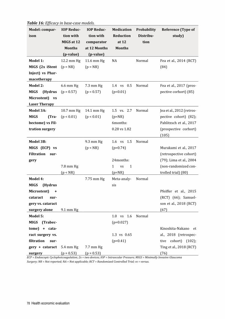
78 Health economic evaluation
Table 16: Efficacy in base-case models. Model: compar-ison
IOP Reduc-tion with
MIGS at 12 Months
(p-value)
IOP Reduc-tion with
comparator at 12 Months
(p-value)
Medication Reduction
at 12 Months
Probability Distribu-
tion
Reference (Type of study)
Model 1: MIGS (2x iStent Inject) vs Phar-macotherapy
12.2 mm Hg (p = NR)
11.6 mm Hg (p = NR)
NA Normal Fea et al., 2014 (RCT) (84)
Model 2: MIGS (Hydrus Microstent) vs Laser Therapy
6.6 mm Hg (p = 0.57)
7.3 mm Hg (p = 0.57)
1.4 vs 0.5 (p<0.01)
Normal Fea et al., 2017 (pros-pective corhort) (85)
Model 3A: MIGS (Tra-bectome) vs Fil-tration surgery
10.7 mm Hg (p < 0.01)
14.1 mm Hg (p < 0.01)
1.5 vs. 2.7 (p=NR) 6months: 0.28 vs 1.82
Normal Jea et al., 2012 (retros-pective cohort) (82); Pahlitzsch et al., 2017 (prospective corhort) (105)
Model 3B: MIGS (ECP) vs Filtration sur-gery 7.8 mm Hg
(p < NR)
9.3 mm Hg (p < NR)
1.6 vs 1.5 (p=0.74) 24months: 1 vs 1 (p=NR)
Normal Murakami et al., 2017 (retrospective cohort) (79); Lima et al., 2004 (non-randomized con-trolled trial) (80)
Model 4: MIGS (Hydrus Microstent) + cataract sur-gery vs. cataract surgery alone 9.1 mm Hg
7.75 mm Hg Meta-analy-sis
Normal
Pfeiffer et al., 2015 (RCT) (66); Samuel-son et al., 2018 (RCT) (67)
Model 5: MIGS (Trabec-tome) + cata-ract surgery vs. filtration sur-gery + cataract surgery
5.4 mm Hg (p = 0.53)
7.7 mm Hg (p = 0.53)
1.0 vs 1.6 (p=0.027) 1.3 vs 0.65 (p=0.41)
Normal
Kinoshita-Nakano et al., 2018 (retrospec-tive cohort) (102); Ting et al., 2018 (RCT) (76)
ECP = Endoscopic Cyclophotcoagulation; 2x = two devices; IOP = Intraocular Pressure; MIGS = Minimally Invasive Glaucoma Surgery; NR = Not reported; NA = Not applicable; RCT = Randomized Controlled Trial; vs = versus.

79 Health economic evaluation
Health-related Quality of Life
CADTH reported quality of life outcomes measured with the preference-based instru-ment, EQ-5D, considered appropriate for cost-utility analyses (5). The utility values were taken from a Dutch study with 531 patients with ocular hypertension or POAG (106). In order to ensure that we had the best available utility data compatible with Norwegian guidelines, we searched for published articles with HRQoL values. We found one British study which was also based on EQ-5D (107). The British utility data did not deviate much from the Dutch utility data reported by CADTH. Therefore, we considered it reasonable to use the utilities presented in CADTH. For patients with glaucoma, util-ity values were derived from the discrete event simulation model developed by Van Gestel et al. (106): Health Related Quality of Life weight = 0.88 – 0.101 * pharmacotherapy + 0.011 * VF – 0.065 * Cataract For each health state defined by a range of VF, we used the midpoint VF in this calcula-tion (mild: - 3 dB, moderate: - 9 dB, advanced: - 16 dB, and severe/blind: - 26 dB). For patients with cataract in addition to glaucoma, we subtracted a constant of - 0.065 from the utility values in patients with glaucoma only (5) (table 17).

80 Health economic evaluation
Table 17: State utilities in the health economic model based on EQ-5D* Utilities associ-ated with health states, side effects and surgery.
Variable Descrip-
tion/state of health
Health related quality of life weight
Source/ comment
Patients with glaucoma only
Mild stage 0.847 Van Gestel 2012* (106)
Moderate stage 0.781 CADTH 2019 (5) Advanced stage 0.704 Severe/blindness Death
0.594 0.000
Patients with glaucoma and cataract
Mild stage 0.782
Van Gestel 2012* (106) Moderate stage 0.716 CADTH 2019 (5) Advanced stage 0.639 Severe/blindness Death
0.529 0.000
Disutilities (per event) Filtration or ma-jor complica-tions requiring surgeries
0.008
Van Gestel 2012* (106)
CADTH 2019 (5)
Minor complica-tions
0.000
*The state utilities presented in this table are derived from EQ-5D utilities in Van Gestel 2012 (106) and reported by CADTH (5).
We used safety data from CADTHs HTA. As mentioned, we pooled possible complica-tions into three categories: minor complications, major complications and secondary surgery complications based on incidence probabilities in Table 18. All the possible complications would appear in the “acute phase” after treatment, and were assumed to be one-time events at the time of the surgical procedure. We applied a disutility of -0.008 to filtration and major complications requiring secondary surgery, while we as-sumed the disutility associated with minor complications to be negligible.

81 Health economic evaluation
Table 18: Incidence probabilities for adverse events in the base-case model
Intervention
No compli-cations
Minor com-plications
Major com-plications
Secondary Surgical In-
terven-tions
Reference
Model 1
iStent Inject (MIGS)*
# 22% 0% NA Vold et al. 2016 (65)
Pharmacotherapy # 17% 0% NA
Model 2
Hydrus Microstent (MIGS) # 16%
10%
NA
Fea et al. 2017 (85) Laser therapy # 40% 0% NA
Model 3
Trabectome (MIGS) # 4%
0%
NA
Jea et al. 2012 (82) Filtration Surgery # 36% 13% NA
Model 4
ECP (MIGS) # 32% 12% NA
Lima et al. 2004 (80) Filtration Surgery # 76%
26% NA
Model 5
Hydrus Microstent (MIGS) + cataract surgery # 18% - 35 %
19%
2% Pfeiffer et al. 2015,
Samuelson et al. 2018 (66;67) Cataract sugery # 18% 2% 5%
Model 6
Trabectome (MIGS) # 100%
NA 0%
Ting et al. 2018, Marco et al. 2017 (76;78)
Filtration surgery +
cataract surgery # 99% 44% 7%
ECP = Endoscopic Cyclophotocoagulation; MIGS = Minimally Invasive Glaucoma Surgery; NA = not applicable; # = we have
assumed that the proportion that was not reported with any complications had no complications in the model; HRQoL =
Health Related Quality of Life. *Assumed rate of complications for iStent Inject would be similar to iStent (5).
Costs
For costing, we apply the perspective of the public health services. We captured all medical costs associated with the alternative treatments, including device or drug costs, procedure costs, complication and rehabilitation costs, as well as the cost of stay-ing in a specific health state (ophthalmologists visits/consultations, and tests) (Table 19). All costs are reported as 2021 Norwegian Kroner (NOK), and cost information originated from other years were inflated to 2021 costs using the Consumer Price In-dex for all items in Norway (98).

82 Health economic evaluation
Cost of procedures We obtained information about procedure costs associated with treatment of glaucoma and cataract from the Norwegian Directorate of Health, Norwegian diagnosis related groups (DRGs) prices from 2021 (108). The estimates were based on average costs per patient for the entire hospital stay including surgery, medicines, materials, stay at the intensive care-unit and regular ward. The cost did not include the value added tax (109). In general, a DRG payment covers all charges associated with an inpatient stay from the time of admission to discharge. Based on DRG information, we estimated a unit cost per-patient for the different treatments. We calculated this by multiplying the weight associated with each DRG with NOK 46 719, which is the 2021 unit price per DRG (108). For MIGS we added the cost of the devices in addition to the cost of the procedure. As there is no DRG available for MIGS yet, often the case for new interventions, we utilized the DRG of cataract surgery (phacoemulisification). More specifically, we assumed that the DRG for MIGS is equal to half the DRG for cataract surgery, based on estimates of time required to perform the MIGS procedure relative to the time required to perform cataract surgery. According to CADTH a MIGS procedure lasts for about 10 minutes (range from 4 – 18 minutes), while a cataract procedure lasts for about 20 minutes, which subsequently affects also personal costs (5). Personal costs are often a great part of procedure costs and therefore DRG costs. For comparators, we assumed that devices used in the procedure was included in the DRG cost (108). Therefore, it may be double counting for MIGS intervention if we add the whole DRG cost, as we already accounted for device cost here. Norwegian clinical experts and representatives from the relevant companies provided cost-information about the MIGS-devices that are currently used in Norwegian hospi-tals. As far as we know there are no national tender or agreements on MIGS device prices today, and there are therefore some differences in devices and prices between hospitals and regional health authorities (RHAs) (28). According to the panel of experts the main difference in costs between MIGS devices and the comparators would be the implant itself. Further, start-up or investment cost associated with MIGS treatment are generally minimal or are covered by manufactures (i.e., possible costs of training staff performing MIGS treatment). We assumed start-up costs to be negligible and did not account for this (28). For pharmacotherapy (medication) costs in model 1, no DRGs were included as pa-tients do not need to receive this treatment option at the hospital. We obtained prices on pharmacotherapy from the Norwegian prescription database (18). Patients’ were assumed to be on two medications at baseline until secondary treatment occurred (49). We calculated per-bottle cost of each medication, and a wastage adjustment factor was added to the cost:

83 Health economic evaluation
1 medication = NOK 1 860 per year 2 medication = NOK 3 719 per year 3 medication = NOK 5 579 per year Annual cost for medication was used as unit cost to calculate cost of relative medication reduction for model two to six. We based relative medication reduction on numbers re-ported in CADTH (5). The difference in medication use after 12-month follow-up was assumed to decline 10% per year. For example, in model 2, where the relative medica-tion reduction at 12 months for Hydrus Microstent versus laser therapy was reported to be 1.4 versus 0.5 (i.e., 0.9 less for laser therapy), an incremental medication cost of 0.9 units was added to the laser therapy group (85).
Table 19: Device, drug, and procedure (DRGs) costs used in the analyses (gamma distri-bution used for PSA).
Strategy Device / drug cost (NOK)
DRG cost (NOK)
DRG code and weight
Reference
Model 1
MIGS (2x iStent Inject)
----
7 195
DRG 42O
(50%), weight 0.308
Expert opinion & The Norwegian Direc-torate of Health
(28;108)
Pharmaco- therapy
3 719
NA
NA
The Norwegian pre-scription database &
The Norwegian Direc-torate of Health
(18;108) Model 2
MIGS (Hydrus Microstent)
---- 7 195 DRG 42O (50%),
weight 0.308
Assumption & The Norwegian Direc-
torate of Health (108) Laser therapy NA 1 682 DRG 36R,
weight 0.036 The Norwegian Direc-torate of Health (108)
Model 3
MIGS (Trabectome)
----
7 195
DRG 42O (50%),
weight 0.308
Assumption & The Norwegian Direc-
torate of Health (108) Filtration Surgery
NA 16 912 DRG36S, weight 0.362
The Norwegian Direc-torate of Health (108)
Model 4
MIGS (ECP)
----
7 195
DRG 42O (50%),
weight 0.308
Assumption & The Norwegian Direc-
torate of Health (108) Filtration Surgery
NA 16 912 DRG 36S, weight 0.362
The Norwegian Direc-torate of Health (108)
Model 5
MIGS (Hydrus Microstent) + cataract surgery
----
7 195 + 14 389
DRG 42O (50%),
Assumption & The Norwegian Direc-
torate of Health (108)

84 Health economic evaluation
the same x 1,25. **MIGS devices we did not retrieve any prices on, we used ----NOK :----NOK Assumed same as 2x iStent:*cost associated with 2x iStent;. MIGS = Minimally Invasive Glaucoma Surgery; ECP = Endoscopic Cyclophotocoagulation; NA = not applicable; NOK = Norwegian kroner; DRG = Diagnose Related Groups; 2x = two devices.
Cost of complications The costs of treating complications were based on prevalence rates of adverse events, reported in the clinical studies used by CADTH (table 20) (5). In terms of complications, it was assumed that complications relating to surgery would be a one-time event that happened at the time of the original procedure. Thus, complications from treatment were included in the model as a one-time cost. We divided complications into three categories, minor complication, major complication and secondary surgical intervention, and allocated cost on weighted averages according to frequencies of occurence. In addition, we accounted for no complication in the model. We assumed that a patient with a complication requires some additional resource use. Patients with minor and major complication would require two additional ophthalmol-ogist consultations, but 10% of those with major complications would in addition require minor eye interventions. Also, patients undergoing secondary surgical interventions were assumed to require two additional ophthalmologist consultations after minor eye intervention. A minor eye intervention was assumed to be trabeculectomy surgery and therefore the same cost.
weight 0.308 + DRG 42O,
weight 0.308 Cataract surgery NA 14 389 DRG 42O,
weight 0.308 The Norwegian Direc-torate of Health (108)
Model 6
MIGS (Trabectome) + cataract surgery
----
7 195 + 14 389
DRG 42O (50%),
weight 0.308 + DRG 42O,
weight 0.308
Assumption & The Norwegian Direc-
torate of Health (108)
Filtration surgery + cataract surgery
NA
8 456 + 7 195
Average of DRG 36S, weight
0.362, and DRG 42O, weight
0.308
The Norwegian Direc-torate of Health (108)

85 Health economic evaluation
Table 20: Annual Cost of complications used in the analysis (Gamma distribution).
NOK = Norwegian kroner; DRG = Diagnosis related groups.
Cost health states Health state costs and associated resource utilization in Norway was determined by quantity of ophthalmologist consultations and set of tests including vision field defect test, optic disc imaging, and IOP measurement required in a specific health state. Oph-thalmologists consultations are recommended at least every four to 12 months, depend-ing on the stage of glaucoma (table 21). We used European guidelines, Norwegian sources, and consulted our clinical experts for this (12;28;108;110). We used the follow-ing unit costs for consultations and test (the unit costs were found in the unit cost data-base published by the Norwegian Medicines Agency (12;28;108;110)): Consultation = NOK 725 VF defect test = NOK 179 Optic disc imaging = NOK 179 We considered transport cost to be the same in all of the different strategies, and there-fore it would most likely not affect the results.
Complication Unit cost in NOK
Description Reference
No complication 0 No cost CADTH (5)
Minor complication 2 336 2 consultations (follow-up visits) Cost based on 2x
DRG 902O
CADTH (5), assumption & The Norwegian Di-
rectorate of Health (108)
Major complication 4 027 All major complica-tions lead to 2 con-
sultations (follow-up visits, 2x DRG 902O), and 10% would re-quire surgical inter-
vention
CADTH (5), assumption & The Norwegian Di-rectorate of Health (108)
Secondary Surgical in-tervention
16 912 Cost based on DRG 36S (same as tra-
beculectomy)
CADTH (5) & The Nor-wegian Directorate of
Health (108)

86 Health economic evaluation
Table 21: Annual Health State Cost estimates used in the analyses (Gamma distribution)
VF = visual field; NOK = Norwegian kroner. Probabilistic sensitivity analysis
The reference case reflects the probabilistic results based on 10 000 Monte Carlo simu-lations. The probabilistic results characterize the extent to which uncertainty in many parameter values combined affects the cost-effectiveness estimates in the model. Standard distributional forms were taken to describe the probability distribution func-tions relating to input parameters: relative efficacy (relative IOP reduction and relative medication reduction) were characterized by normal distributions, utility and compli-cation rates were characterized by beta distribution, and costs were characterized by gamma distributions. The inference information for each distribution were based on the reference case or meta-analysis (5). Cost-effectiveness acceptability curves demon-strating the probability that a modality would be considered optimal for a range of WTP threshold were also presented. The uncertainty surrounding cost parameters were assessed by using gamma distribu-tion in the health economic model. Beta distributions were used for utility values.
Health state Unit cost in NOK Description Reference
Mild Health State
1 949
2 consultations, 1.4 VF test,
1.4 optic disc imaging
Expert opinion (28) & unit cost database pub-lished by the
Norwegien Medi-cines Agency
(110) Moderate Health State
2 709
3 consultations, 1.5 VF test,
1.5 optic disc imaging
Expert opinion (28) & unit cost database pub-lished by the
Norwegien Medi-cines Agency
(110) Advanced Health State
2 780
3 consultations, 1.7 VF test,
1.7 optic disc imaging
Expert opinion (28) & unit cost database pub-lished by the
Norwegien Medi-cines Agency
(110) Severe/Blind Health State
1 189
1 consultations, 1.3 VF test,
1.3 optic disc imaging
Expert opinion (28) & unit cost database pub-lished by the
Norwegien Medi-cines Agency
(110)

87 Health economic evaluation
Sensitivity analyses
In addition to performing probabilistic sensitivity analysis, we carried out a series of one-way sensitivity analyses in order to investigate how uncertainty around single parameters could affect our cost-effectiveness results. We present results of this analysis as a tornado diagram in the results chapter. Confidence ranges (value interval) for sensitivity analyses were calculated as base case value +/- 25%, while the standard errors for estimation of gamma distributions were based on the formula: (Value interval/2) * 1,96.
Budget impact analysis
Budget impact analysis can be defined as an assessment of the financial consequences of adopting a new intervention at a population level. In other words, budget impact is the total incremental cost (additional costs) of introduction of an intervention versus non-introduction (i.e., the total expenditure of inserting the new method minus the to-tal costs of not doing so) (109). The allocation of number of patients receiving different treatment options related to in-troduction of MIGS in Norway are unknown, and we have therefore not estimated the total budget impact for the specialist health services of the intervention in a national perspective. Further, due to these uncertainties we have not extracted total costs calcu-lated by the Markov model. However, we have made a simplified calculation of MIGS device cost and procedure cost only (both as a stand-alone procedure and in combina-tion with cataract surgery). We did not include cost inputs as health state costs and cost of treating procedure-related complications that we used in the cost-effectiveness model. We have not calculated total cost of treatment comparators. We used undis-counted costs, in line with recommendations from the International Society for Phar-macoeconomics and Outcomes Research (ISPOR) for budget impact analyses, and five years time horizon (111). To NIPH’s knowledge, in 2018, there were about 200 MIGS procedures performed in Norway. Based on expert opinions we have assumed that there are 255 patients that will receive MIGS in Norway in 2019, 50% as stand-alone procedure and 50% in combi-nation with cataract surgery. According to our clinical experts, there will be a doubling of the current number MIGS procedures five years ahead by implementing MIGS as a public funded treatment in Norway (28). By taking our clinical experts opinions into consideration, we conservatively assume that this growth will be a doubling in five years by introducing MIGS to Norwegian hospitals. Based on the above assumptions we calculated the numbers of patients eligible for MIGS as prognosis for the next four years. Tables 22 and 23 shows unit cost input used in our calculations. We have used average cost of MIGS devices used in our models. For treating glaucoma costs for iStent inject

88 Health economic evaluation
x2, Hydrus Microstent, Trabectome, and ECP are included. Our estimates for MIGS com-bined with cataract surgery includes costs for Hydrus Microstent and Trabectome.
Table 22: Input data to budget impact analysis for MIGS procedures (no cataract) MIGS device Unit cost device*
(NOK) DRG cost MIGS (NOK)
Unit cost device + DRG cost (NOK)
Reference
iStent Inject (x2)
---- 7 195 ---- Expert opinion (28) and as-sumption
Hydrus microstent
---- 7 195 ---- Expert opinion (28) and as-sumption
Trabectome ---- 7 195 ---- Expert opinion (28) and as-sumption
ECP ---- 7 195 ---- Expert opinion (28) and as-sumption
Average cost ---- 7 195 ---- Expert opinion (28) and as-sumption
*Unit cost device is included WAT (25%). x2 = 2 devices; NOK = Norwegian kroner.
Table 23: Input data to budget impact analysis for MIGS with cataract procedures MIGS device Unit cost
device (NOK)
DRG cost MIGS (NOK)
DRG cost cataract surgery (NOK)
Unit cost de-vice + DRG cost MIGS + DRG cost cata-ract surgery (NOK)
Reference
Hydrus microstent
---- 7 195 14 389 ---- Expert opinion (28) and as-sumption
Trabectome ---- 7 195 14 389 ---- Expert opinion (28) and as-sumption
Average cost ---- 7 195 14 389 ---- Expert opinion (28) and as-sumption
NOK = Norwegian kroner.

89 Health economic evaluation
Severity considerations – absolute shortfall (AS)
We calculated absolute shortfall (AS) based on projections about life expectancies from the health economic model. Calculation of AS has been described in more detail in the submission guideline for pharmaceutical reimbursements of the Norwegian Medicines Agency, which is based on the white paper on priority setting, and a Norwegian life ta-ble and age adjusted health related quality of life information from a general Swedish population (6;104;109;112). Absolute shortfall is defined as the difference in quality adjusted life expectancies at age (A) without the disease (QALYsA), and prognosis with the disease with current standard of care (PA): AS = QALYsA – PA In the calculations, undiscounted numbers for QALYsA and PA are used.

90 Health economic evaluation
RESULTS
Severity considerations – absolute shortfall
In accordance with the economic models, we have listed patients age when entering the model in table 24. Further, the table describes the expected quality adjusted life expec-tancy at this age, and what the prognosis with disease is expected to be in QALYs for standard treatment, based on simulations from the health economic model with life-time (up to 100 years) horizon. The absolute shortfall in each model with these as-sumptions is:
Table 24: Absolute shortfall in each sub-model Model Age QALYs at age with-
outh disease (QAL-YsA)
Prognosis with disease (PA)
Absolute shortfall (AS)*
Model 1 64 17.0 16.7 0.3
Model 2 70 12.9 12.6 0.3
Model 3 65 16.3 16.1 0.2
Model 4 65 16.3 14.1 2.2
Model 5 72 11.6 11.4 0.2
Model 6 71 12.3 10.4 1.9 QALY: quality-adjusted life year; *AS = QALYs at age withouth the disease – Prognosis with the disease.
This puts patients with glaucoma in the least severe of the six severity classes sug-gested by the Magnussen group, irrespective of model. These classes range from: AS < 4 QALYs lost (severity class 1), 4-7,9; 8-11,9; 12- 15,9; 16-19,9, and AS ≥ 20 QALYs (se-verity class 6) (90). Accordingly, severity is not an argument for giving extra priority to glaucoma patients.
Incremental cost–effectiveness estimates in the analysis
The baseline cost-effectiveness results of all models are based on the probabilistic anal-yses and presented in table 25; demonstrating lifetime expected costs and QALYs, in-cremental costs and QALYs, as well as the ICER. Each model considered a different set of comparators, and therefore represent different patient populations. It is therefore inappropriate to calculate incremental values across the different models, but still highly relevant to compare the ICERs if a stepwise scale up of the procedure to different population group is considered. The lifetime total cost per patient for glaucoma treat-ment ranged between NOK 52 000 and NOK 84 000 depending on the treatment strat-egy and patient’s baseline disease stage. For all models, strategies involving MIGS were between 3 and 21 thousand more costly over the lifetime. The absolute effects (dis-counted quality adjusted life expectancies) varied between 14 and 17 QALYs in the

91 Health economic evaluation
models, with average age when receiving treatment as the most important factor. In-cremental QALYs between MIGS and comparators ranged between -0.080 and 0.052. The detailed results are subsequently presented.
Table 25: Results of the base-case cost-effectiveness analyses (lifetime horizon, dis-counted). Intervention Total costs
(NOK) Effects (QALYs)
Incremental Costs (NOK)
Incremental Effect (QALYs)
ICER (NOK/QALY)
Model 1: Pharma-cotherapy 2 x iStent In-ject (MIGS)
64 269 ----
16.695 16.711
----
0.016
---
Model 2: Laser therapy Hydrus Mi-crostent (MIGS)
29 799 ----
12.648 12. 567
----
-0.080
Dominated
Model 3: Filtration surgery Trabectome (MIGS)
75 834 ----
16.043 16.068
----
0.024
---
Model 4: Filtration surgery ECP (MIGS)
72 253 ----
14.108 14.141
----
0.033
---
Model 5: Cataract sur-gery Hydrus Mi-crostent (MIGS) + cat-aract surgery
51 568 ----
11.399 11.438
----
0.038
---
Model 6: Filtration surgery + cat-aract surgery Trabectome (MIGS) + cat-aract surgery
62 722 ----
10.483 10.540
----
0.057
---

92 Health economic evaluation
Incremental cost-effectiveness estimates in model 1
As the clinical review only found clinical studies comparing iStent Inject (2nd genera-tion) to pharmacotherapy, the findings below specifically address the cost-effectiveness of iStent compared with pharmacotherapy. The potential cost-effectiveness of other MIGS devices to a pharmacotherapy strategy remains unclear and could not be ex-plored in this economic evaluation. The results of the probabilistic sensitivity analysis with a lifetime horizon are illus-trated in figure 11. The blue dots in the scatterplot represent results for patients fol-lowing MIGS (iStent Inject) and the red ones represent pharmacological treatment. The blue “cloud” is, on average, situated to the right and higher than the red “cloud”, indi-cating that MIGS is likely to be more costly, and a bit more effective than pharmacologi-cal treatment for patients at a moderate glaucoma stage. The truncated right side of the scatter plot expresses the maximum achievable benefit in the model. ----------------------------------------------------------------------------------------------------------------------------------------------------------------------------------------------------------------------------------------------------------------------------------------------------------------------------------------------------------------------------------------------------------------------------------------------------------------------------------------------------------------------------------------------------------------------------------------- ----------------------------------------------------------------------------------------------------------------------------------------------------------------------------------------------------------------------------------------------------------------------------------------------------------------------------------------------------------------------------------------------------------------------------------------------------------------------------------------------------------------------------------------------------------------------------------------------------------------------------------------------------------------------------------------------------------------------------------------------------------------------------------------------------------------------------------------------------------------------------------------------------------------------------------------------------------------------------------------------------------------------------------------------------------------------------------------------------------------------------------------------------------------------------------- Figure 11: Cost-effectiveness scatterplot for model 1. The expected results of the Monte Carlo simulation in base-case analysis are presented in Table 25, and show that the total expected intervention-related costs per patient (in moderate glaucoma stage) in a lifetime horizon are about 64 0000 NOK for patients who are treated with pharmacotherapy and ---- NOK for patients who undergo MIGS (2 x iStent Inject). These costs include procedures, complication, rehabilitation and health state costs. The incremental cost for MIGS patients is thus about ---- NOK. During a life-time perspective MIGS patients accumulate slightly more QALYs, with a different of 0.016 QALYs. The modest difference in health effect is the main driver of the result that MIGS costs ---- per QALY (ICER).

93 Health economic evaluation
The cost-effectiveness acceptability curves in figure 12 demonstrate that pharma-cotherapy has a higher probability of being cost-effective than MIGS for the entire range of willingness-to-pay (WTP) thresholds. suggested by the Norheim commission and Magnussen group (90;91). When simultaneously taking into account all parameter uncertainties, the probability of MIGS being cost-effective does not exceed about 1/3. ----------------------------------------------------------------------------------------------------------------------------------------------------------------------------------------------------------------------------------------------------------------------------------------------------------------------------------------------------------------------------------------------------------------------------------------------------------------------------------------------------------------------------------------------------------------------------------------- ----------------------------------------------------------------------------------------------------------------------------------------------------------------------------------------------------------------------------------------------------------------------------------------------------------------------------------------------------------------------------------------------------------------------------------------------------------------------------------------------------------------------------------------------------------------------------------------- ----------------------------------------------------------------------------------------------------------------------------------------------------------------------------------------------------------------------------------------------------------------------------------------------------------------------------------------------------------------------------------------------------------------------------------------------------------------------------------------------------------------------------------------------------------------------------------------------------------------------------------------------------------------------------------------------------------------------------------------------------------------------------------------------------------------------------------------------------------------------------------------------------------------------------------------------------------------------------------------------------------------------------------------------------------------------------------------------------------------------------------------------------------------------------------- Figure 12: Cost-effectiveness acceptability curves indicating the probability that either intervention is cost-effective for a WTP range from zero to 1 000 000 NOK per QALY. Incremental cost-effectiveness estimates in model 2
The systematic review only identified clinical studies that compared Hydrus Microstent to laser therapy, and thus these are the alternatives compared. The potential cost-effec-tiveness of other MIGS devices to a laser therapy strategy remains unclear. The results of the probabilistic sensitivity analysis with a lifetime horizon are illus-trated in figure 13. The blue dots in the scatterplot represent results for patients that received laser therapy and the red ones MIGS (Hydrus Microstent). The red “cloud” is, on average, situated less to the right than the blue “cloud” and above the blue “cloud”, indicating that MIGS is likely to be more costly, and less effective than laser therapy for patients at a mild glaucoma stage. The truncated right side of the scatter plot expresses the maximum achievable benefit in the model.

94 Health economic evaluation
------------------------------------------------------------------------------------------------------------------------------------------------------------------------------------------------------------------------------------------------------------------------------------------------------------------------------------------------------------------------------------------------------------------------------------------------------------------------------------------------------------------------------------------------------------------------------------------------------------------------------------------------------------------------------------------------------------------------------------------------------------------------------------------------------------------------------------------------------------------------------------------------------------------------------------------------------------------------------------------------------------------------------------------------------------------------------------------------------------------------------------------------------------------------------------ Figure 13: Cost-effectiveness scatterplot for model 2. The expected results of the Monte Carlo simulation in base-case analysis are presented in Table 25, and show that the total expected intervention-related costs per patient (in moderate glaucoma stage) in a lifetime horizon are about 30 000 NOK for patients who are treated with laser therapy and ---- NOK for patients who undergo MIGS (Hydrus Mi-crostent). These costs include procedures, complication, rehabilitation, and health state costs. The incremental cost for MIGS patients is thus about ---- NOK. During a lifetime perspective MIGS patients accumulate less health, with a difference of – 0.080 QALYs. Consequently, MIGS is dominated by laser therapy for this patient population. The cost-effectiveness acceptability curves (figure 14) show that MIGS’s propability of being the cost-effective alternative will not exceed about 1/3 irrespective of willingness to pay for health.

95 Health economic evaluation
----------------------------------------------------------------------------------------------------------------------------------------------------------------------------------------------------------------------------------------------------------------------------------------------------------------------------------------------------------------------------------------------------------------------------------------------------------------------------------------------------------------------------------------------------------------------------------------- ----------------------------------------------------------------------------------------------------------------------------------------------------------------------------------------------------------------------------------------------------------------------------------------------------------------------------------------------------------------------------------------------------------------------------------------------------------------------------------------------------------------------------------------------------------------------------------------------------------------------------------------------------------------------------------------------------------------------------------------------------------------------------------------------------------------------------------------------------------------------------------------------------------------------------------------------------------------------------------------------------------------------------------------------------------------------------------------------------------------------------------------------------------------------------------- Figure 14: Cost-effectiveness acceptability curves indicating the probability that either intervention is cost-effective for a WTP range from zero to 1 000 000 NOK per QALY. Incremental cost-effectiveness estimates in model 3
As the clinical review only found clinical studies that have compared Trabectome to fil-tration surgery, the findings below specifically address the cost-effectiveness of Tra-bectome compated with filtration surgery. The potential cost-effectiveness of other MIGS devices to a laser therapy strategy remains unclear and could not be explored in this Economic Evaluation. The results of the probabilistic sensitivity analysis with a lifetime horizon are illus-trated in figure 15. The blue dots in the scatterplot represent results for patients that received filtration surgery and the red ones MIGS (Trabectome). The red “cloud” is, on average, situated more to the right than the blue “cloud” and above the blue “cloud”, in-dicating that MIGS is likely to be more costly, and slightly more effective than filtration surgery for patients at a moderate glaucoma stage. The truncated right side of the scat-ter plot expresses the maximum achievable benefit in the model.

96 Health economic evaluation
------------------------------------------------------------------------------------------------------------------------------------------------------------------------------------------------------------------------------------------------------------------------------------------------------------------------------------------------------------------------------------------------------------------------------------------------------------------------------------------------------------------------------------------------------------------------------------------------------------------------------------------------------------------------------------------------------------------------------------------------------------------------------------------------------------------------------------------------------------------------------------------------------------------------------------------------------------------------------------------------------------------------------------------------------------------------------------------------------------------------------------------------------------------------------------------------------------------------------------------------------------------------------------------------------------------------------------------------------------------------------------------------------------------------------------------------------------------------------------------------------------------------------------------------------------------------------------------------------------------------------------------------------------------------------------------------------------------ Figure 15: Cost-effectiveness scatterplot for model 3. ICER The expected results of the Monte Carlo simulation in base-case analysis are presented in Table 25, and show that the total expected intervention-related costs per patient (in moderate glaucoma stage) in a lifetime horizon are about 76 000 NOK for patients who are treated with filtration surgery and about ---- NOK for patients who undergo MIGS (Trombectome). These costs include procedures, complication, rehabilitation and health state costs. The incremental cost for MIGS patients is thus about ---- NOK. Over a lifetime perspective MIGS patients accumulate slightly more QALYs, with a different of 0.024 QALYs. ICER is estimated to be about ---- NOK. Below, we present cost-effectiveness acceptability curves at willingness-to-pay (WTP) thresholds for one additional QALY between zero and 1 000 000 NOK (figure 16). The figure demonstrates that filtration surgery has a higher probability of being cost-effec-tive up to a WTP threshold of about 282 000 NOK. The observed non-linearities are ar-tefacts of the assumed average baseline visual field (Table 14), the assumed IOP reduc-tions with both treatments (Table 16) and subsequent duration before transitioning a milder to a more severe glaucoma stage (Figure 10).

97 Health economic evaluation
---------------------------------------------------------------------------------------------------------------------------------------------------------------------------------------------------------------------------------------------------------------------------------------------------------------------------------------------------------------------------------------------------------------------------------------------------------------------------------------------------------------------------------------------------------------------------------------------------------------------------------------------------------------------------------------------------------------------------------------------------------------------------------------------------------------------------------------------------------------------------------------------------------------------------------------------------------------------------------------------------------------------------------------------------------------------------------------------------------------------------------------------------------------------------------------------------------------------------------------------------------------------------------------------------------------------------------------------------------------------------------------------------------------------------------------------------------------------------------------------------------------------------------------------------------------------------------------------------------------------------------------------------------------------------------------------------------------------------------------------------------------------------- Figure 16: Cost-effectiveness acceptability curves indicating the probability that either intervention is cost-effective for a WTP range from zero to 1 000 000 NOK per QALY. Incremental cost-effectiveness estimates in model 4
As the clinical review only found clinical studies comparing ECP to filtration surgery, the findings below specifically address the cost-effectiveness of ECP compated with fil-tration surgery. The potential cost-effectiveness of other MIGS devices to a laser ther-apy strategy remains unclear and could not be explored in this economic evaluation. The results of the probabilistic sensitivity analysis with a lifetime horizon are illus-trated in figure 17. The blue dots in the scatterplot represent results for patients that received filtration surgery and the red ones MIGS (ECP). The colored dots are mixed, which indicates that it is not obvious which strategy that is more cost effective. The truncated right side of the scatter plot expresses the maximum achievable benefit in the model.

98 Health economic evaluation
-------------------------------------------------------------------------------------------------------------------------------------------------------------------------------------------------------------------------------------------------------------------------------------------------------------------------------------------------------------------------------------------------------------------------------------------------------------------------------------------------------------------------------------------------------------------------------------------------------------------------------------------------------------------------------------------------------------------------------------------------------------------------------------------------------------------------------------------------------------------------------------------------------------------------------------------------------------------------------------------------------------------------------------------------------------------------------------------------------------------------------------------- Figure 17: Cost-effectiveness scatterplot for model 4. ICER The expected results of the Monte Carlo simulation in base-case analysis are presented in Table 25, and show that the total expected intervention-related costs per patient (in advanced glaucoma stage) in a lifetime horizon are about 72 000 NOK for patients who are treated with filtration surgery and about ---- NOK for patients who undergo MIGS (ECP). These costs include procedures, complication, rehabilitation, and health state costs. The incremental cost for MIGS patients is thus about ---- NOK. During a lifetime perspective MIGS patients accumulate more QALYs, with a different of 0.033 QALYs. MIGS costs about ---- NOK per QALY (ICER). Below, we present cost-effectiveness acceptability curves at willingness-to-pay (WTP) thresholds for one additional QALY between zero and 1 000 000 NOK (figure 18). The figure demonstrates that filtration surgery has a higher probability of being cost-effec-tive than MIGS up to a WTP threshold of about 94 000 NOK, when simultaneously con-sidering all parameter uncertainties.

99 Health economic evaluation
----------------------------------------------------------------------------------------------------------------------------------------------------------------------------------------------------------------------------------------------------------------------------------------------------------------------------------------------------------------------------------------------------------------------------------------------------------------------------------------------------------------------------------------------------------------------------------------------------------------------------------------------------------------------------------------------------------------------------------------------------------------------------------------------------------------------------------------------------------------------------------------------------------------------------------------------------------------------------------------------------------------------------------------------------------------------------------------------------------------------------------------------------------------------------------- Figure 18: Cost-effectiveness acceptability curves indicating the probability that either intervention is cost-effective for a WTP range from zero to 1 000 000 NOK per QALY. Incremental cost-effectiveness estimates in model 5
As the clinical review only found clinical studies that have compared Hydrus Microstent + cataract surgery to cataract surgery, the findings below specifically address the cost-effectiveness of Hydrus Microstent + cataract surgery compated with cataract surgery. The potential cost-effectiveness of other MIGS devices to a laser therapy strategy re-mains unclear and could not be explored in this Economic Evaluation. The results of the probabilistic sensitivity analysis with a lifetime horizon are illus-trated in figure 19. The blue dots in the scatterplot represent results for patients that received cataract surgery alone and the red ones MIGS (Hydrus Microstent) combined with cataract surgery. The red “cloud” is, on average, situated slightly more to the right than the blue “cloud” and above the blue “cloud”, indicating that MIGS is likely to be more costly, and a bit more effective than cataract surgery for patients at a mild glau-coma stage. The truncated right side of the scatter plot expresses the maximum achiev-able benefit in the model.

100 Health economic evaluation
-------------------------------------------------------------------------------------------------------------------------------------------------------------------------------------------------------------------------------------------------------------------------------------------------------------------------------------------------------------------------------------------------------------------------------------------------------------------------------------------------------------------------------------------------------------------------------------------------------------------------------------------------------------------------------------------------------------------------------------------------------------------------------------------------------------------------------------------------------------------------------------------------------------------------------------------------------------------------------------------------------------------------------------------------------------------------------------------------------------------------------------------- Figure 19: Cost-effectiveness scatterplot for model 5. ICER The expected results of the Monte Carlo simulation in base-case analysis are presented in Table 25, and show that the total expected intervention-related costs per patient (in mild glaucoma stage) in a lifetime horizon are about 51 000 NOK for patients who are treated with cataract surgery alone and about ---- NOK for patients who undergo MIGS (Hydrus Microstent) in combination with cataract surgery. These costs include proce-dures, complication, rehabilitation, and health state costs. The incremental cost for MIGS in combination with cataract surgery patients is thus about ---- NOK. During a lifetime perspective, patients who receive MIGS in combination with cataract surgery accumulate more QALYs, with a different of 0.038 QALYs. MIGS combined with cataract surgery costs about ---- NOK per QALY (ICER). Below, we present cost-effectiveness acceptability curves at willingness-to-pay (WTP) thresholds for one additional QALY between zero and 1 000 000 NOK (figure 20). The figure demonstrates that cataract surgery alone has a higher probability of being cost-effective than MIGS combined with cataract surgery, when simultaneously taking into account all parameter uncertainties.
101 Health economic evaluation
----------------------------------------------------------------------------------------------------------------------------------------------------------------------------------------------------------------------------------------------------------------------------------------------------------------------------------------------------------------------------------------------------------------------------------------------------------------------------------------------------------------------------------------------------------------------------------------------------------------------------------------------------------------------------------------------------------------------------------------------------------------------------------------------------------------------------------------------------------------------------------------------------------------------------------------------------------------------------------------------------------------------------------------------------------------------------------------------------------------------------------------------------------------------------------- Figure 20: Cost-effectiveness acceptability curves indicating the probability that either intervention is cost-effective for a WTP range from zero to 1 000 000 NOK per QALY. Incremental cost-effectiveness estimates in model 6
As the clinical review only found clinical studies that have compared Trabectome + cat-aract surgery to filtration surgery + cataract surgery, the findings below specifically ad-dress the cost-effectiveness of Trabectome + cataract surgery compated with filtration surgery + cataract surgery. The potential cost-effectiveness of other MIGS devices to a laser therapy strategy remains unclear and could not be explored in this Economic Evaluation. The results of the probabilistic sensitivity analysis with a lifetime horizon are illus-trated in figure 21. The blue dots in the scatterplot represent results for patients that received filtration surgery combined with cataract surgery and the red ones MIGS (Trombectome) combined with cataract surgery. The red “cloud” is, on average, situ-ated more to the right than the blue “cloud” and slightly above the blue “cloud”, indicat-ing that MIGS is likely to be more costly, and more effective than filtration surgery com-bined with cataract surgery for patients at an advanced glaucoma stage. The truncated right side of the scatter plot expresses the maximum achievable benefit in the model.

102 Health economic evaluation
----------------------------------------------------------------------------------------------------------------------------------------------------------------------------------------------------------------------------------------------------------------------------------------------------------------------------------------------------------------------------------------------------------------------------------------------------------------------------------------------------------------------------------------------------------------------------------------------------------------------------------------------------------------------------------------------------------------------------------------------------------------------------------------------------------------------------------------------------------------------------------------------------------------------------------------------------------------------------------------------------------------------------------------------------------------------------------------------------------------------------------------------------------------------------------- Figure 21: Cost-effectiveness scatterplot for analysis in model 6. ICER The expected results of the Monte Carlo simulation in base-case analysis are presented in Table 25, and show that the total expected intervention-related costs per patient (in advanced glaucoma stage) in a lifetime horizon are about 63 000 NOK for patients who are treated with filtration surgery + cataract surgery and ---- NOK for patients who un-dergo MIGS (Trombectome) in combination with cataract surgery. These costs include procedures, complication, rehabilitation, and health state costs. The incremental cost for MIGS in combination with cataract surgery patients is thus about ---- NOK. During a lifetime perspective, patients who receive MIGS in combination with cataract surgery accumulate more QALYs, with a different of 0.057 QALYs. MIGS combined with cataract surgery costs about ---- NOK per QALY (ICER). Below, we present cost-effectiveness acceptability curves at willingness-to-pay (WTP) thresholds for one additional QALY between zero and 1 000 000 NOK (figure 22). The figure demonstrates that filtration surgery combined with cataract surgery has a higher probability of being cost-effective than MIGS combined with cataract surgery up to a WTP threshold of 360 000 NOK, when simultaneously taking into account all parame-ter uncertainties.

103 Health economic evaluation
----------------------------------------------------------------------------------------------------------------------------------------------------------------------------------------------------------------------------------------------------------------------------------------------------------------------------------------------------------------------------------------------------------------------------------------------------------------------------------------------------------------------------------------------------------------------------------------------------------------------------------------------------------------------------------------------------------------------------------------------------------------------------------------------------------------------------------------------------------------------------------------------------------------------------------------------------------------------------------------------------------------------------------------------------------------------------------------------------------------------------------------------------------------------------------- Figure 22: Cost-effectiveness acceptability curves indicating the probability that either intervention is cost-effective for a WTP range from zero to 1 000 000 NOK per QALY.
Sensitivity analyses
We have presented our sensitivity analyses in tornado diagrams for each model. A tornado diagram is a graphical method for presenting a series of one-way sensitivity analyses and shows how cost-utility results (ICER) are influenced by variation in individual model parameters.The blue bar represents low parameter estimate and the red one represents high values of the parameter. Sensitivity analyses in model 1
Figure 23 presents parameters with greatest impact on results in model 1. We can observe that the results are most affected by variation in baseline visual field, cost intervention MIGS and progression visual field natural data. We can see that the if we had another value for baseline visual field within the interval it would not have been below our WTP threshold anyhow. On the other hand, natural progression visual field for untreated patients may be below this threshold if the rate changes to slower rate of progression than -0.6 per year. CADTH used -0.92 dB rater than -0.6 dB per year in a scenario analysis, based on the reported decline in VF in untreated patients in some studies. However, this is the reverse of what could lead to an ICER below our threshold.
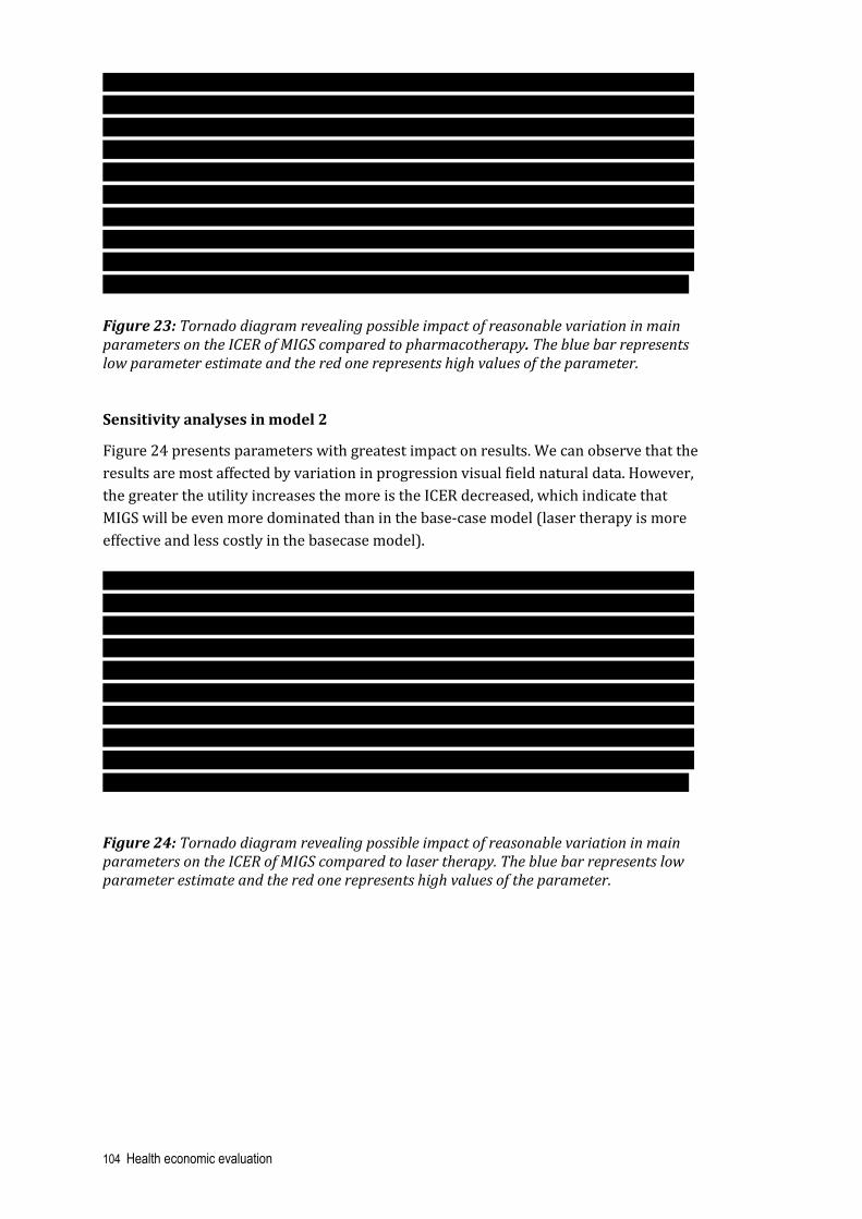
104 Health economic evaluation
----------------------------------------------------------------------------------------------------------------------------------------------------------------------------------------------------------------------------------------------------------------------------------------------------------------------------------------------------------------------------------------------------------------------------------------------------------------------------------------------------------------------------------------------------------------------------------------------------------------------------------------------------------------------------------------------------------------------------------------------------------------------------------------------------------------------------------------------------------------------------------------------------------------------------------------------------------------------------------------------------------------------------------------------------------------------------------------------------------------------------------------------------------------------------------- Figure 23: Tornado diagram revealing possible impact of reasonable variation in main parameters on the ICER of MIGS compared to pharmacotherapy. The blue bar represents low parameter estimate and the red one represents high values of the parameter. Sensitivity analyses in model 2
Figure 24 presents parameters with greatest impact on results. We can observe that the results are most affected by variation in progression visual field natural data. However, the greater the utility increases the more is the ICER decreased, which indicate that MIGS will be even more dominated than in the base-case model (laser therapy is more effective and less costly in the basecase model). ----------------------------------------------------------------------------------------------------------------------------------------------------------------------------------------------------------------------------------------------------------------------------------------------------------------------------------------------------------------------------------------------------------------------------------------------------------------------------------------------------------------------------------------------------------------------------------------------------------------------------------------------------------------------------------------------------------------------------------------------------------------------------------------------------------------------------------------------------------------------------------------------------------------------------------------------------------------------------------------------------------------------------------------------------------------------------------------------------------------------------------------------------------------------------------- Figure 24: Tornado diagram revealing possible impact of reasonable variation in main parameters on the ICER of MIGS compared to laser therapy. The blue bar represents low parameter estimate and the red one represents high values of the parameter.

105 Health economic evaluation
Sensitivity analyses in model 3
Figure 25 presents parameters with greatest impact on results. We can observe that the results are most affected by variation in cost intervention MIGS, followed by cost intervention filtration data. Here, the description about undertainty around the cost intervention parameters for model 1 is applicable. ----------------------------------------------------------------------------------------------------------------------------------------------------------------------------------------------------------------------------------------------------------------------------------------------------------------------------------------------------------------------------------------------------------------------------------------------------------------------------------------------------------------------------------------------------------------------------------------------------------------------------------------------------------------------------------------------------------------------------------------------------------------------------------------------------------------------------------------------------------------------------------------------------------------------------------------------------------------------------------------------------------------------------------------------------------------------------------------------------------------------------------------------------------------------------------- Figure 25: Tornado diagram revealing possible impact of reasonable variation in main parameters on the ICER of MIGS compared to filtration surgery. The blue bar represents low parameter estimate and the red one represents high values of the parameter. Sensitivity analyses in model 4
Figure 26 presents parameters with greatest impact on results. We can observe that the results are most affected by variation in intervention costs. ----------------------------------------------------------------------------------------------------------------------------------------------------------------------------------------------------------------------------------------------------------------------------------------------------------------------------------------------------------------------------------------------------------------------------------------------------------------------------------------------------------------------------------------------------------------------------------------------------------------------------------------------------------------------------------------------------------------------------------------------------------------------------------------------------------------------------------------------------------------------------------------------------------------------------------------------------------------------------------------------------------------------------------------------------------------------------------------------------------------------------------------------------------------------------------- Figure 26: Tornado diagram revealing possible impact of reasonable variation in main parameters on the ICER of MIGS compared to filtration surgery. The blue bar represents low parameter estimate and the red one represents high values of the parameter.

106 Health economic evaluation
Sensitivity analyses in model 5
Figure 27 presents parameters with greatest impact on results. We can observe that the results are most affected by variation in cost intervention MIGS combined with cataract surgery followed by baseline visual field and cost intervention cataract surgery data. ----------------------------------------------------------------------------------------------------------------------------------------------------------------------------------------------------------------------------------------------------------------------------------------------------------------------------------------------------------------------------------------------------------------------------------------------------------------------------------------------------------------------------------------------------------------------------------------------------------------------------------------------------------------------------------------------------------------------------------------------------------------------------------------------------------------------------------------------------------------------------------------------------------------------------------------------------------------------------------------------------------------------------------------------------------------------------------------------------------------------------------------------------------------------------------- Figure 27: Tornado diagram revealing possible impact of reasonable variation in main parameters on the ICER of MIGS combined with cataract surgery compared to cataract surgery alone. The blue bar represents low parameter estimate and the red one represents high values of the parameter. Sensitivity analyses in model 6
Figure 28 presents parameters with greatest impact on results. We can observe that the results are most affected by variation in costs related to the interventions, followed by the annual IOP reduction for the comparator strategy, filtration + cataract.

107 Health economic evaluation
----------------------------------------------------------------------------------------------------------------------------------------------------------------------------------------------------------------------------------------------------------------------------------------------------------------------------------------------------------------------------------------------------------------------------------------------------------------------------------------------------------------------------------------------------------------------------------------------------------------------------------------------------------------------------------------------------------------------------------------------------------------------------------------------------------------------------------------------------------------------------------------------------------------------------------------------------------------------------------------------------------------------------------------------------------------------------------------------------------------------------------------------------------------------------------- Figure 28: Tornado diagram revealing possible impact of reasonable variation in main parameters on the ICER of MIGS combined with cataract surgery compared to filtration surgery combined with cataract surgery. The blue bar represents low parameter estimate and the red one represents high values of the parameter.
Budget impact analysis
We calculated the budgetary impact of introducing MIGS to Norwegian patients with glaucoma and patients with glaucoma and cataract. In the budget impact analysis, we tried to explore how the potential expansion of MIGS would influence the total number of MIGS performed in the next five years. The costs used in the next tables are pre-sented in table 19, 22 and 23 (Cost section). The costs are not discounted. The prediction depends on several factors, including any change in clinical practice from current practice, the relative changes in procedure costs and the number of pa-tients eligible for different treatment alternatives. The results of the predicted cost im-pact of MIGS as a stand-alone procedure and MIGS in combination with cataract sur-gery are shown in Table 26.

108 Health economic evaluation
Table 26 Predicted impact of MIGS on the number of patients and results of the budget impact; estimated costs based on future practice compared to estimated costs based on current practice Year 2020 2021 2022 2023 2024
Number of MIGS procedures (50% as stand-alone procedure and 50% in combination with cataract surgery)
255 319 383 446 510
Cost of MIGS alone 2 479 236 3 099 047 3 718 856 4 338 666 4 958 475
Cost of MIGS in com-bination with cata-ract surgery
4 823 835 6 029 794 7 235 753 8 441 711 9 647 670
Sum 7 303 073 9 128 841 10 954 609 12 780 377 14 606 145 Costs are in Norwegian kroner.
Further, we tried to examine the budgetary consequences of introducing MIGS as a stand alone procedure compared with pharmaeuticals alone, laser therapy alone and filtration surgery alone, and budgetary consequences of introducing MIGS in combina-tion with cataract surgery compared to cataract surgery alone and filtration in combi-nation with cataract surgery. The potential additional costs are presented in the tables 27 to 31 below. Table 27 presenting the costs associated to MIGS as a stand-alone procedure compared with costs related to pharmaceuticals, and the potential additional costs of introducing MIGS alone for glaucoma patients receiving pharamaceuticals today. Table 27: Predicted budget impact of MIGS alone compared with pharmaceuticals alone Year 2020 2021 2022 2023 2024
Cost of MIGS alone 2 479 238 3 099 047 3 718 856 4 338 666 4 958 475
Cost of pharma 474 173 592 716 711 259 829 802 948 345
Sum 2 005 065 2 506 331 3 007 598 3 508 864 4 010 130 Costs are in Norwegian kroner.
Table 28 presenting the costs associated with MIGS alone compared with costs related to laser therapy, and the potential additional costs of introducing MIGS alone for glau-coma patients receiving laser therapy today.

109 Health economic evaluation
Table 28: Predicted budget impact of MIGS alone compared with laser therapy
Year 2020 2021 2022 2023 2024
Cost of MIGS alone 2 479 238 3 099 047 3 718 856 4 338 666 4 958 475
Cost of laser therapy 214 455 268 068 321 683 375 297 428 910
Sum 2 264 783 2 830 978 3 397 174 3 963 369 4 429 565 Costs are in Norwegian kroner.
Table 29 presenting the costs associated with MIGS alone compared with costs related to filtration surgery, and the potential additional costs of introducing MIGS alone for glaucoma patients receiving filtration surgery today. Table 29: Predicted budget impact of MIGS alone compared with filtration surgery Year 2020 2021 2022 2023 2024
Cost of MIGS alone 2 479 238 3 099 047 3 718 856 4 338 666 4 958 475
Cost of filtration sur-gery
2 156 280 2 695 350 3 234 420 3 773 490 4 312 560
Sum 322 958 403 697 484 436 3565 176 645 915 Costs are in Norwegian kroner.
Table 30 presenting the costs associated with MIGS in combination with cataract sur-gery compared with costs related to cataract surgery alone, and the potential additional costs of introducing MIGS in combination o cataract with surgery for glaucoma patients receiving cataract surgery as a stand-alone procedure today. Table 30: Predicted budget impact of MIGS in combination with cataract surgery com-pared with cataract surgery alone Year 2020 2021 2022 2023 2024
Cost of MIGS in com-bination with cata-ract surgery
4 823 835 6 029 794 7 235 753 8 441 711 9 647 670
Cost of cataract sur-gery
1 834 598 2 293 247 2 751 896 3 219 545 3 669 195
Sum 2 989 238 3 736 547 4 483 856 5 231 166 5 978 475 Costs are in Norwegian kroner.
Table 31 presenting the costs associated with MIGS in combination with cataract sur-gery compared with costs related to filtration surgery in combination with cataract sur-

110 Health economic evaluation
gery, and the potential additional costs of introducing MIGS in combination with cata-ract surgery for glaucoma patients receiving filtration surgery in combination with cat-aract surgery today. Table 31: Predicted budget impact of MIGS in combination with cataract surgery com-pared with filtration surgery in combination with cataract surgery Year 2020 2021 2022 2023 2024
Cost of MIGS in com-bination with cata-ract surgery
4 823 835 6 029 794 7 235 753 8 441 711 9 647 670
Cost of filtration sur-gery in combination with cataract surgery
1 995 503 2 494 378 2 993 254 3 492 129 3 991 005
Sum 2 828 333 3 535 416 4 242 498 4 949 582 5 656 665 Costs are in Norwegian kroner.

111 Ethics
Ethics
This chapter about ethical aspects of using MIGS in Norway is not adapted from CADTH.
Method
In this section we have analysed central ethical implications of MIGS implementation. The purpose was to identify key ethical concerns raised by MIGS, to assess the main challenges, and offer recommendations on how to deal with these challenges. The anal-ysis proceeded in three major steps:
1. Brief description of the situation, alternative actions and solutions, and the involved stakeholders 2. Analysis of the ethical challenges and possible consequences in terms of the four principles: benefit, harm, autonomy and justice 3. Summary of the analysis
These steps were chosen to fit the central ethical issues raised by using MIGS devices and procedures, based on the methodologies used or discussed in relevant HTA ethics guidelines and reports (113-115).
Brief description of the situation, alternatives and stakeholders
MIGS comprise of several surgical procedures and devices that present an alternative to or complement current treatment of glaucoma. The potential advantages of MIGS are shorter procedure times, earlier treatment, shorter recovery times, and probably higher success rates both as a standalone treatment and for subsequent surgical treat-ments.
MIGS have been developed and used since the turn of the millennium. MIGS procedures are already offered at Norwegian hospitals, and there is a growing demand. As MIGS represent a group of relatively new treatment options, our knowledge of optimal use and long-term effects is still incomplete.
Current alternatives to MIGS are IOP lowering eye drops, laser treatment, and tradi-tional surgical procedures. The compliance rates for eye drops is low, and laser therapy is not recommended for all patients. Traditional surgical procedures are often post-poned in patient treatment, because of the risk of complications, demanding follow-up procedures, and potential lifelong discomfort for the patient.

112 Ethics
MIGS have the potential to offer better alternatives for glaucoma patients. The use of MIGS procedures may, however, lower the development of and competence in tradi-tional surgical procedures. This might reduce the quality of treatment for some pa-tients, especially patients that cannot benefit from the available MIGS procedures.
Involved stakeholders are glaucoma patients and their relatives, medical doctors and other health personnel, health authorities, public hospitals, private medical centres, and therapy developers including pharmaceutical industry.
Analysis of the ethical challenges
We structure the ethical analysis in terms of the four principles: benefit, harm, auton-omy and justice (116).
Benefit
The principle of benefit denotes the production of benefits for others, and the positive balance of good minus bad effects for individuals and society.
Glaucoma is a chronic disease often leading to blindness. As there is currently no cura-tive treatment for glaucoma, and the progressive vision loss experienced by glaucoma patients is irreversible, current treatment options focus on reduction of IOP, nerve damage, and further loss of vision. Being a chronic disease, a comprehensive life-cycle perspective on benefit is the most suitable.
In a life-cycle perspective, the potential benefits of MIGS compared to alternative treat-ment options are significant, both for individual patients and society at large. Avoiding the potential loss of life quality for patients in adhering to, and often failure of adhering to, a strict pharmaco-therapeutical regime, is such a significant benefit. The possibility of earlier surgical intervention and resulting prevention of deteriorating eyesight is an-other.
Because of the reduced risk of complications, MIGS treatments can be offered to the pa-tient at an earlier stage of treatment. This might reduce irreversible damage from pro-longed non-treatment. Compared to traditional surgical options, MIGS might reduce risks of serious post-surgical medical complications, patient discomfort, and reduced quality of life.
Harm
The principle of harm denotes not inflicting avoidable harmful effects on others. Imple-mentation of MIGS procedures without a sound knowledge base of short- and long-term risks and benefits might induce harm to patients (117). To avoid such harm, MIGS implementation and development should follow standard approval procedures and in-ternational guidelines (12).

113 Ethics
Autonomy
The principle of autonomy denotes the opportunity for genuine self-determination and freedom of choice for individuals.
Patients must be thoroughly informed about risks of and alternatives to the MIGS pro-cedure in question, as well as the knowledge status of the information provided.
Justice
The principle of justice denotes the equal distribution of risks and benefits between in-dividuals and groups.
From the principle perspective of justice, health authorities should be careful not to in-troduce incentive structures that could lead hospitals to prefer/avoid the use of certain MIGS procedures for economical rather than medical reasons, as this might result in unjust treatment choices between patient groups. Outsourcing MIGS to private medical centres should also be set up to avoid incentive structures producing injustice between patient groups, especially if centres have close ties with pharmaceutical industry.
Access to MIGS treatment should not depend on the ethnic or cultural background, geo-graphical location, or personal economic resources of the patient.
Summary of the analysis
The are potential benefits of MIGS, but the potential harms must be avoided or mini-mized. The challenges for patient autonomy seem manageable, if patients can be thor-oughly informed about risks of and alternatives to a given MIGS procedure. In terms of justice, it is important to guarantee patients equal access to treatment, regardless of ability to pay, place of residence, social status, or cultural background.
–

114 Discussion
Discussion
Key findings summary
Key findings on effectiveness and safety
CADTH HTA CADTH HTA included 32 studies from 35 publications; 10 randomized trials and 22 non-randomised studies. The evidence included 24 specific comparisons: one compari-son of a MIGS versus another MIGS, six comparisons of a MIGS combined with cataract surgery versus cataract surgery alone, nine comparisons of a MIGS combined with cata-ract surgery versus cataract surgery combined with filtration surgery, six comparisons of a MIGS combined with filtration surgery versus filtration surgery aone, two compari-sons of a MIGS versus pharmacotherapy, and one comparison of a MIGS versus laser therapy. The CADTH authors considered all the included studies to be at risk of bias. To summarise, the findings showed that there were essentially no statistically signifi-cant differences between interventions and comparators, or it was not statistical tested. NIPH review We identified and included eight studies (seven randomised trials and one non-ran-domised study) that compared a MIGS procedure alone or in combination with cataract surgery, with another MIGS procedure or non-MIGS procedures. None of the results from any of these studies could be pooled with study results in the CADTH HTA. There was moderate-certainty evidence of lower unmedicated intraocular pressure (IOP) at 24 months with the iStent inject combination with cataract surgery than with cataract surgery alone, and high-certainty evidence of lower IOP at 24 months with Hy-drus in combination with cataract surgery than with cataract surgery alone. Otherwise, there was no or little difference between the MIGS and interventions in control groups, or the certainty of the evidence was too low to make a judgement. The evidence for the safety of MIGS was inconclusive across the comparisons.
Key findings of health economic evaluation
The results of the clinical review, based on the HTA from CADTH, impacted the ap-proach taken in our health economic evaluation. The clinical evidence was generally of poor quality as noted in the clinical review. There was substantial heterogeneity be-tween studies comparing MIGS with other treatment strategies, and it was considered

115 Discussion
clinically inappropriate to conduct a network meta-analysis to pool all possible com-parisons. The MIGS group as a whole is so broad that it encompasses therapies that ad-dress different parts of the glaucoma treatment pathway, and have different control comparators. As such, the health economic analysis consisted of six models with pair-wise comparisons, and each model considered MIGS with a different set of compara-tors, patients with varying glaucoma stages, and different types of MIGS devices. Com-parisons of results between models are therefore not appropriate. The health economic models demonstrated lifetime expected costs and QALYs, incre-mental costs and QALYs, and ICERs. Our probabilistic analysis suggested that the cost-effectiveness of MIGS varied depending on the patient population’s baseline disease stage and the comparator. A lifetime total cost per patient for glaucoma treatment ranged between 30 000 NOK and 83 000 NOK. The incremental differences in costs ranged between 3 000 NOK and 24 000 NOK per patient. The incremental QALYs for MIGS between comparators ranged between – 0.080 and 0.057 QALYs. MIGS (2x iStent inject) seemed to gain more QALYs at a higher cost when compared with pharmacotherapy in moderate glaucoma stage (model 1), or when MIGS (ECP) was compared with filtration surgery in advanced glaucoma stage (model 4). The same result applied when MIGS (Hydrus Microstent) was performed in combination with cat-aract surgery compared with cataract surgery alone in mild glaucoma stage (model 5). Results were sensitive to various parameters in the different models. The incremental differences in costs and QALYs over a lifetime horizon were relatively small. Unlike QALYs, the incremental costs tended to occur relatively early, largely due to the initial costs of the device and procedure that occur within the first year. The attractiveness of MIGS may differ depending on the cost of the device, and the procedure, from a cost perspective. From a health care perspective, these findings illustrate that MIGS may be cost-effective in specific situations, but if used indiscriminately, MIGS may not always be the cost-effective treatment strategy for certain glaucoma patients. Results on the cost-effectiveness of MIGS should be interpreted with caution given the uncertainty in relative efficacy and costs. Scenarios where there may be multiple treatment options for a MIGS patient, may require further evidence prior to recommendations for the op-timal use of treatments in patients with glaucoma. Our budget impact analysis estimated a cost of about 17 million after five years if intro-ducing MIGS in the Norwegian specialist health care. The patient volume in need of MIGS is assumed to increase from 255 patients to 510 patients in five years.
Evidence quality
Certainty of the evidence of effectiveness and safety
CADTH HTA The CADTH HTA authors judged the certainty of the evidence as very low to low across the 24 comparisons and outcomes. Very low certainty-evidence cannot be interpreted,

116 Discussion
and it is highly likely that future research findings may be very different from the cur-rent evidence. Low certainty-evidence gives a weak indication of effectiveness and safety, and it is likely that future research findings may differ from the current evi-dence. NIPH supplementary review We assessed the certainty of the evidence for the primary outcome, intraocular pres-sure, as high for one comparison (Hydrus + cataract surgery vs cataract surgery alone), moderate for one comparison (Hydrus vs 2x iStent) , low for one comparison (iStent in-ject + cataract surgery vs cataract surgery alone) and very low for the four remaining comparisons. A major reason for downgrading the certainty of the evidence was seri-ous to very serious concerns for risk of bias in the included studies. Another important reason for downgrading the certainty was that the evidence largely consisted of results from small single studies that could only be pooled in two cases. Further, imprecision was a concern for two of the comparisons. The quality of the economic model used
We made several assumptions to conduct the six health economic models leading to un-certainty around our economic results. For example, we chose to use a lifetime horizon. Most clinical studies, however, considered outcomes at only one year, and the long-term relative effects of various treatment strategies are unknown. We extrapolated the incremental QALYs over a lifetime, and thus estimated differences in QALYs should be interpreted with caution. Health states in the models were defined according to the Hodapp-Parrish-Anderson grading scale. Further, there is uncertainty in the relation-ship between IOP changes and its direct impact on VF status and vision related QoL in a lifetime perspective. Treatment efficacy were commonly described as a change in IOP in the clinical studies and in the economic models we chose to utilize a predictive equa-tion to describe the relationship between changes in IOP and its impact on visual field. We do not know the predictive ability of this equation that was used in the models. Finally, MIGS is a heterogeneous group of devices with potentially different costs and different relative treatment effects, which can have an impact on our result. There re-mains uncertainty with respect to decision making on what patients are suited to what type of treatment strategy, for example, there may be subgroups of patients with more aggressive glaucoma who may not be appropriate for all treatment strategies.
Strengths and weaknesses
Strengths and weaknesses of the systematic reviews of effectiveness and safety
The strengths of CADTH’s and our systematic reviews of the evidence for effectiveness and safety include updated systematic searches in electronic databases, pre-specified inclusion and exclusion criteria, independent screening of identified records, independ-ent assessments of risk of bias in included studies, systematic data extraction and re-porting, and assessments of the certainty of the evidence.

117 Discussion
Due to time constraints, the NIPH supplementary review did not include retrospective non-randomised studies, ongoing studies and unpublished studies. Including these study categories would probably not add anything to the evidence, but still be informa-tive. Strengths and weaknesses of health economic evaluation
Firstly, we replicated the model from CADTH with respect to model structure and effi-cacy data, and we do not know if we have missed some important variables. The input is adjusted according to Norwegian conditions. A second limitation concerns how the clinical review transferred over to the health eco-nomic evaluation. The comparative cost-effectiveness of different MIGS devices could not be addressed. Another limitation was the area with impact on our health economic modelling was the natural history of glaucoma. To model the lifetime economic consequences of treatment strategies, the model had to incorporate the expected natural history of the disease. We used the Hodapp-Parrish-Anderson grading scale to examine this, and incorporated visual field changes over time in patients with glaucoma, based on the CADTH report (5;103). According to CADTH several studies evaluating the natural history of glaucoma are expected in the coming years, and face validity of the health economic model should be reassessed when such studies become available (5). Further, we had to do extrapola-tions to model a lifetime horizon, with long-term costs and consequences, as the clinical studies used report on surrogate outcomes over short time periods. Furthermore, costs were challenging to assess because of limited availability of data on all MIGS devices. We used DRGs in our cost estimates which may influence the uncer-tainty in our results. DRG costs are based on various ranges in costs and represent av-erage estimates on different procedures. This approach may double count some cost items, for example, costs of MIGS devices. There is no DRG for MIGS, which is often the case for new strategies. Further, assessment of actuel costs may be valuable and allow greater certainty in the incremental- and total cost estimates, like detailed Norwegian micro-costing studies of MIGS and comparator strategies (i.e., real world evidence). Sensitivity analyses strengthen this economic evaluation and highlights that the cost of MIGS plays an important role in determining its cost-effectiveness. In addition, we included costs that are not incorporated in the DRG costs, i.e., costs in-fluenced by glaucoma stage in patients, inpatients or outpatients, stand-alone treat-ment or combination treatment etc. As glaucoma stage has been identified as a predic-tor of higher health state costs, devices aimed at advanced or high IOP glaucoma pa-tients may have higher cost-savings potential (118;119). We included extra time used on combining MIGS procedure with cataract surgery com-pared to cataract surgery alone. On the other hand, according to our clinical experts, the difference in costs in this case are only the price of MIGS. We received prices on MIGS-devices from our clinical experts. As such, we only got prices on the MIGS-devices

118 Discussion
that are in use in Norwegian hospitals today. Device prices vary between hospitals to-day because of no national tender (28). We used the average of the prices we retrieved. As a conservative assumption, on MIGS-devices that are not in use in Norway, we esti-mated prices as the average cost of the price on the devices we received prices on. In addition, we did not account for possible discounts on prices.
First, one strength in our economic analysis is that we developed decision analytic models for POAG, which commonly has been approached with Markov state-transition models using different glaucoma staging systems. This approach has been justified from a clinical point of view as it correctly represents the stepwise changes in costs and utilities as the disease progresses. We did the health economic analysis from a broad health care perspective, and there-fore, we did not include societal costs. In the glaucoma disease area, societal costs may be important aspects as this is a progressive disease. For example, in our analysis it is less costly with a blind patient than with a patient with moderate glaucoma, as there are no costs occurring for the specialist health care sector in the blind stage. However, there may be high societal costs to take care of a blind patient, as well as costs occur-ring when patients are absent from work etc. Our clinical experts and patient partners mentioned these aspects as especially important to include in an evaluation in the area of glaucoma (28;37). However, a societal perspective was omitted in the final report given it being outside the scope of our commission. Other aspects that may have an impact on the conclusion of cost-effectiveness is medi-cation adherence on relative effectiveness and disease progression and medication-specific disutilities. Our analysis assumed no adherence rates for medication and no medication-specific disutilities. However, CADTH reported that, if other assumptions were selected for the pharmacotherapy strategy (e.g., lower drug adherence or disutility for medication use), MIGS may be a beneficial treatment strategy in populations where adherence to medication is expected to be low or if there are side effects experienced on medication. In the case of MIGS combined with cataract surgery, which is included in model 5 and 6, and applies to Trabectome and Hydrus Microstent (types of MIGS). According to our clinical experts, in principle all MIGS could be combined with cataract surgery. How-ever, it is most applicable to combine iStent with cataract surgery. In some cases, XEN is also combined with cataract surgery, but mainly used as a stand-alone procedure. Usu-ally, when patients also have cataract it will be combined with that kind of procedure. We assume that in the models where cataract surgery is included, patients have both glaucoma and cataract. According to our clinical experts, this relates to 10 to 15 % of the cases. There are several weaknesses to compare MIGS combined with cataract sur-gery with other treatment strategies to glaucoma treatment. According to our clinical experts MIGS combined with cataract surgery is most relevant to compare with cata-ract surgery combined with other type of MIGS device (but we do not compare MIGS with MIGS), cataract surgery combined with other type of glaucoma surgery, or cata-ract surgery alone. According to our clinical experts 70% of patients above 70 years de-velop cataract (some with limited cataract that do not need surgery). Most patients

119 Discussion
with glaucoma are above 70 years, and therefore there is a strong coexistence of these eye disorders (28).
Further, there may be some uncertainty in our calculation of absolute shortfall (AS). This applies for example to uncertainty in the estimates of age or prognosis. According to our clinical experts there are also some differences with respect to what MIGS is used or not. We are unsure if all MIGS and procedures are comparable with all treatment options and comparators in terms of when to use them. In addition, the pro-cedure group (subconjunctival space) of XEN and Innfocus, the last one more invasive, are by many considered not to be a MIGS. The subconjunctival approach is difficult when a patient has received several operations and often a MIGS using another ap-proach has been used. According to our clinical experts there has been a viewpoint of dividing MIGS into groups: MIGS and MIGS+. The clinical experts categorised iStent in-ject as a MIGS and XEN as a MIGS+. In most cases, they have used MIGS as an alternative to eyedrops or laser and MIGS+ as an alternative to trabeculectomy. I.e., these are dif-ferent patient groups which are not directly comparable (28). With respect to MIGS as first-line treatment, there is a risk of complications with all surgery and therefore few will choose MIGS before eye drops. Most clinicians will also choose laser treatment before MIGS, but laser can be poorly suited for some patients, (eg, unable to sit at the slit-lamp mounted laser, past history of uveitis, inadequate view of trabecular meshwork or symptomatic cataract) (27). In terms of mild, moderate and advanced health states, these might not mean the same for first, second line treatment and so on (i.e., you may not only receive first line treatment in mild, second line treat-ment in moderate and so on. i.e., you can have second line treatment in mild). Also, we have assumed that every patient only receives MIGS once in a lifetime horizon. We do not know if this is realistic in “real life”, and if a patient may receive MIGS sev-eral times.
We have no information on earlier treatment of the patients included in the studies (and assume that patients included are in their first treatment scheme. Also, we do not know how the efficacy of treatment is in different disease stages. One example here is, efficacy of filtration surgery in moderate glaucoma health state (model 3) versus effi-cacy of filtration surgery on advanced health state (model 4) (different MIGS in the dif-ferent models). Complications can have an impact on time and outpatient resources/costs. There may be different complications for different treatment strategies, for example, surgery ver-sus pharmacotherapy. We chose to replicate CADTH and pool our complications into groups as minor and major, as well as secondary surgical interventions, and thereby utilising an identical model structure in the different models, but here, different vari-bles are put into the three complication groups. We have not accounted for if a MIGS in-

120 Discussion
tervention can fail, and if this is a complication or not. Does the patient need a re-inter-vention? If yes, how often is this happening? Does the patient get any disutility? Accord-ing to our clinical experts this could be difficult to categorise, as the same complication could be less severe or more severe dependent on the severity of the glaucoma. Infec-tion could be a lot of things, for example, if it is an intraocular infection, also called en-dophthalmitis, blebitis, this is a major complication. On the other hand, minor compli-cations are other extraocular infections. Further, there are some of the minor complica-tions that could be considered major, for example, endothelial cell loss could be both minor and major, dependent on the extent of cell loss. A patient can start different treatments with respect to which disease stage they are in. They also have a different baseline. It is important to define glaucoma stage at baseline to allow modelling the patients disease progression within each model. As such, we have compared MIGS with different comparators depending on glaucoma stage and di-vided stages dependent on decibels. Number of visits and follow up were not included in health economic evaluation. Finally, many of the assumptions in the health economic evaluation rely on clinical expert opinions, and would benefit from real world evidence.
Generalisability of findings
Overall completeness and applicability of evidence from systematic review
We have summarised the CADTH HTA evidence ansupplemented it with more recent evidence. The evidence largely consists of several small, single studies with results that cannot be pooled with each other due to differences in comparisons. So, for the vast majority of comparisons, the evidence is not conclusive. Further, our pre-specified sec-ondary outcomes were rarely reported in the included studies. We assume that applicability of the evidence is acceptable to a Norwegian setting with regard to populations and interventions. However not all interventions or comapari-sons we have reported are relevant to clinical practice in Norway. Generalizability of findings from health economic evaluation
The health economic findings primarily reflects patients with open-angle glaucoma, which is in accordance with what our clinical experts suggested, as MIGS is mainly meant for this patient population (28). The economic evaluation therefore has limita-tions regarding possibilities to address whether cost-effectiveness of MIGS would differ by other types of glaucoma. However, it may be important to not exclude other glau-coma patients in receiving MIGS-treatment from a patient partner perspective (37). The main findings from the health economic evaluation reflect the potential cost-effec-tiveness of MIGS in patients who are receiving treatment at an early stage of glaucoma. However, as disease stage can impact costs, utilities, and options for subsequent treat-ment, the economic analysis was divided into baseline stage when clinical data existed.

121 Discussion
The results from this set of comparisons highlight the fact that the cost-effectiveness of MIGS may depend on the stage of glaucoma. Therefore, population characteristics may influence the cost-effectiveness of MIGS. As mentioned above, to inform relative treatment effects and thereby incorporate natu-ral history of glaucoma for our economic evaluation, we included clinical studies used in the CADTH report, taken from a large Canadian study (5;103). We assume those pa-tient populations reflect characteristics of a Norwegian population. Furthermore, costs and resource utilisation were from Norwegian sources and the findings are expected to be generalisable to the Norwegian context. Although MIGS are categorised as a particular class of strategies, each MIGS may have different clinical effectiveness and safety profiles. There exist some various definitions and opinions on what MIGS is and is not. In Norway, the infrastructure results in long distances to hospital for many patients within geographical areas and the distance to specialised glaucoma centres may be long. Therefore, a question may be if all hospitals should offer MIGS. According to our project group, consisting of clinical experts and patient partners many hospitals offer MIGS today, but the treatment availability might differ between patients in the country.
Consistency with other reviews
Consistency of systematic review with other reviews
Our review is consistent with the CADTH HTA and four Cochrane reviews in that we identified small, single studies with different comparisons. Overall, in our and others’ reviews, it is unclear whether MIGS reduces intraocular pressures, compared to other interventions, in patients with open-angle glaucoma. Adverse events seemed infre-quent in all interventions. None of the results in the included studies in our supplementary review could have been pooled with any of the results from studies in the CADTH HTA because of different comparisons. While all evidence in the CADTH HTA was graded as low or very low cer-tainty, we assessed the certainty as high and moderate, respectively, in two cases. Our assessment of the certainty of the evidence was in line with four recent Cochrane re-views (15;32-34). Consistency of health economic evaluation with other studies
Our health economic evaluation is consistent with other evaluations on health econom-ics, among others the CADTH report, with regards to uncertainty around estimates due to lack of data. Cost components may be different in different countries.

122 Discussion
Implication of results on practice
With respect to implication of results on practice one of the greatest challenges to the organization of MIGS in Norway may be access at system and patient level. At a system level, access varies for patients living in different geographical regions, and Norway’s considerable geographical size can create difficulties in caring for patients with glau-coma that live rurally or remotely. Today, however, many hospitals offer MIGS treat-ment. There might be that difficulty can be present due to long distances, fewer special-ised ophthalmologists, problems attracting trained and experienced ophthalmologists to the area etc. Therefore, patients may receive MIGS treatment later in the glaucoma pathway, and at that stage, ophthalmologists may opt to refer patients for more inva-sive procedures. Our patient partners’ and clinical experts described the systemic bur-dens of having to travel to access MIGS and follow-ups as important aspects in this as-sessment (28;37). Today, there may be some issues at a patient level, as access to MIGS can vary due to the surgery itself being offered by a few private clinics, necessitating the procedure or device to be paid for by the patient. As the specialist health care has a restricted budget that is often shared with other specialties, this can be a barrier to implementing MIGS. In facilities that provide MIGS, these facilities may only fund some potential types of MIGS devices or procedures available for patients. Today different hospitals use differ-ent types of MIGS, and there is no national agreement on what to use and in which cases. According to some experts Xen is mostly used for advanced glaucoma and iStent or ABiC for light/moderate glaucoma. Additionally, as the number of MIGS surgeries in-creases, the time in the OR required for these surgeries also increases, which may lengthen waiting lists for patients who require the intervention. In such instances, pa-tients may choose to get it done privately, but to our knowledge this is not a big issue at the moment. For patients who lack the financial means to pay privately for a MIGS de-vice, or for those whose glaucoma could in principle be treated through medication but are unable to use eye drops as prescribed, the unclear status of MIGS as an “optional upgrade” versus a “medical need” may itself be a barrier to access. One specialist might consider a patient’s circumstances to clearly present medical need for MIGS treatment, whilst another would not. Clearer guidelines on when and for whom MIGS should be considered a medical need will reduce barriers currently created by the discretionary interpretation of these concepts by professionals and health systems (5). Although these are relevant considerations that may impact access to MIGS, this HTA focused on the evidence that would be useful and specifically relevant to decision mak-ers in Norway’s publicly funded specialist health care system. For example, costs in-cluded in the analysis were specific to those paid by RHAs and may not reflect indirect costs from a broader societal perspective (e.g., costs borne by patients and relatives). According to our clinical experts and patient partners there are different use of MIGS in hospitals in Norway today. Implementation of a technology is often dependent on the diffusion of the technology into the professional community. Many physicians are “early adopters” of newer technology, including MIGS devices and procedures, although
123 Discussion
many physicians take a more cautious approach before integrating newer interventions into practice. As MIGS are relatively new technologies, this diffusion may not yet be complete. Nonetheless, new graduates of ophthalmology are frequently trained in MIGS devices and procedures, and often expect this training. MIGS have been noted to have a fairly short learning compared to other surgery for glaucoma. Additionally, MIGS are not included in many clinical practice guidelines for ophtalmol-ogy and this lack of guidance can create difficulties for ophtalmologists when deciding on patient selection, type of MIGS and funding. Further, there is a lack of formalised in-dications for the use of MIGS, that might lead to differences in clinical practice. As such, it is possible that providers’ perceptions of patients’ “compliance” may influence pa-tient selection for MIGS. This could have the effect rewarding “compliant” patients with access to MIGS, while those struggling with other treatment regimens are overlooked as “noncompliant” (5). Moreover, it is probable that clinical discretion is influenced by the available evidence regarding effectiveness and safety, the state of which is in its infancy. Authors of the majority of the studies in the clinical review reported several disclosures, including fi-nancial or non-financial support from industry, other involvement with industry (e.g., consulting for, or employee of, industry), or having other interests in manufacturer companies (e.g., shareholder, stock holder or patent holders). Therefore, MIGS devices and procedures with greater manufacturer support are likely to be better represented in the literature and therefore have more available evidence regarding clinical effec-tiveness and safety and subsequently greater uptake. In the current landscape of MIGS use in Norway, the potential for conflict of interest arises from incentives that institu-tions and professionals may have to recommend the use of specific MIGS devices to pa-tients for reasons extraneous to patients’ individual circumstances and needs. Reme-dies to prevent and mitigate conflicts include transparency by surgeons and institu-tions in acknowledging all potential conflicts, institutional oversight in granting privi-leges for innovative surgeries, and candid discussion with patients. In order to advance more personalised selection of MIGS for patients, it may be essential for health systems, facilities, and professionals to assign and carry out the responsibility of tracking and re-porting outcomes of MIGS usage. As MIGS is a quickly evolving research area, implementation issues involving MIGS may also change rapidly; some implementation issues that were described may no longer be of relevance or there may be novel issues that were not captured. However, the direct and indirect costs of MIGS can be considerable. Therefore, there is need to clarify current policy on access and reimbursement related to MIGS devices and procedures (120). The evidence should be interpreted with caution, given the uncertainty in data about clinical efficacy, safety and cost. Glaucoma management is a rapidly changing field and as substantial new evidence of MIGS devices and procedures emerge, reassessment may be needed. Both patient groups and clinicians have raised positive attitudes and expectations to offer more use of MIGS to patients with claucoma, although also being

124 Discussion
aware of the limitations in the evidence on effect and safety clearly shown in this re-port.
Considerations of the prioritisation criteria in light of available evidence
Lack of high-certainty evidence of MIGS comparisons in the CADTH HTA restrict the possibilities to consider the established prioritisation criteria in Norway; i.e., benefit, resources and severity in the following way:
- Benefit: The clinical evidence on MIGS is limited. The main reason for this is the lack of comparative studies of MIGS.
- Resources: Definitive conclusions on the cost-effectiveness of MIGS is uncertain, given the uncertainty in the analysis. Our health economic evaluation shows some scenarios where MIGS may be cost-effective, depending on comparator and disease stage.
- Severity: Our analysis puts patients with glaucoma in severity class 1 irrespective of model.
Need for further research
To obtain high-certainty evidence for the myriad of possible MIGS comparisons, there is a need for more well-designed randomised trials. Relevant comparisons should also be considered (e.g. XEN should be compared to trab, whilst trabecular bypass surgeries might more reasonably be compared to laser). It is worth noting that the comparator here is often cataract surgery alone, which is not really broadly implemented as a glau-coma therapy in Norway. Further, detailed micro-costing of MIGS procedures may al-low for greater certainty in the true absolute and incremental costs of MIGS to better inform the potential economic value of MIGS.

125 Conclusion
Conclusion
Efficacy and safety MIGS with Hydrus Microstents combined with cataract surgery reduces intraocular pressure (IOP) at 24 months, compared to cataract surgery alone. MIGS with Hydrus Microstents probably reduces IOP at 12 months, compared to MIGS with 2x iStents. For other comparisons and outcomes, it is uncertain whether there is a difference in IOP re-duction. Neither MIGS procedures nor alternative surgical strategies appear to be at high risk of adverse events, and it is uncertain whether complications occur more or less frequently in either category. Organisational aspects MIGS is suitable as a outpatient surgery without hospital admission. Ophthalmologists need to be trained to perform MIGS. Experts predict that the number of annual MIGS procedures might increase annually to twice as many in 2024 than today. Health economics Cost-effectiveness analysis of MIGS are uncertain, given the uncertainty in available data. The economic evaluation provided some scenarios where MIGS may be cost-effec-tive, depending on comparator and disease stage. Ethical aspects The are potential benefits of MIGS, but the potential harms must be avoided or mini-mized. The challenges for patient autonomy seem manageable, if patients can be thor-oughly informed about risks of and alternatives to a given MIGS procedure. It is im-portant to guarantee patients equal access to treatment, regardless of ability to pay, place of residence, social status, or cultural background.
Consideration of the priority criteria Lack of high-certainty evidence of MIGS comparisons in the CADTH HTA restrict the possibilities to consider the established prioritisation criteria in Norway. Regarding benefit the clinical evidence on MIGS is limited. The main reason for this is the lack of comparative studies of MIGS. Regarding resources definitive conclusions on the cost-effectiveness of MIGS is uncertain, given the uncertainty in the analysis. Our health economic evaluation shows some scenarios where MIGS may be cost-effective, depending on comparator and disease stage. Finally, on the aspect of severity: Our anal-ysis puts patients with glaucoma in severity class 1 irrespective of model.

126
References
1. Falvey H, Sandhu H. Trabecular bypass micro-invasive glaucoma surgery (MIGS) device implantation with iStent injectin patients with primary open-angle glaucoma, pseudoexfoliative glaucoma or pigmentary glaucoma. London: Nye Metoder; 2018.
2. Folkehelseinstituttet. Trabekulær stent bypass mikrokirurgi (iStent) for pasienter med grønn stær. Oslo: MedNytt; 2018.
3. Sekretariatet Nye Metoder. Protokoll fra møte i Bestillerforum RHF Mandag 22. oktober. Gardermoen: 2018.
4. Sekretariatet Nye Metoder. Protokoll fra møte i Bestillerforum RHF Mandag 24. september. Oslo: 2018.
5. CADTH. Optimal use of minimally invasive glaucoma surgery: a health technology assessment. Ottawa: CADTH; 2019. CADTH optimal use report. 8. 1b.
6. Helse- og omsorgsdepartementet. Meld. St. 34. Verdier i pasientens helsetjeneste. Melding om prioritering. . 2016.
7. International Council of Ophthalmology. ICO Guidelines for Glausoma Eye Care. 2016. Available from: http://www.icoph.org/downloads/ICOGlaucomaGuidelines.pdf
8. Gupta N, Aung T, Congdon N, Dada T, Lerner F, Olawoye S, et al. International Council of Ophthalmology Guidelines for Glaucoma Eye Care. California: 2015.
9. Norges Blindeforbund. En orientering om grønn stær (glaukom). Oslo2017. 10. Weinreb RN, Aung T, Medeiros FA. The Pathophysiology and Treatment of
Glaucoma. A Review. JAMA 2015;311(18):1901-11. 11. Pillunat LE, Erb C, Jünemann AGM, Kimmich F. Micro-invasive glaucoma surgery
(MiGS): a review of surgical procedures using stents. Clinical Ophthalmology 2017;11:1583-600.
12. European Glaucoma Society. Terminology and guidelines for glaucoma. 4th ed. Italy: 2017. Available from: https://www.eugs.org/eng/guidelines.asp
13. Peters D, Bengtsson B, Heijl A. Lifetime risk of blindness in open-angle glaucoma. American Journal of Ophthalmology 2013;156(4):727-30.
14. BMJ Best Practice. Glaucoma (open angle). Patient information from BMJ: BMJ Publishing Group Limited [cited]. Available from: https://bestpractice.bmj.com/patient-leaflets/en-gb/pdf/1581709449852.pdf
15. Zhang ML, Hirunyachote P, Jampel H. Combined surgery versus cataract surgery alone for eyes with cataract and glaucoma. Cochrane Database of Systematic Reviews 2015;7.
16. BrightFocus© Foundation. Glaucoma: facts & figuresClarksburg (MD)[cited December]. Available from: http://www.brightfocus.org/glaucoma/article/glaucoma-facts-figures
17. Miglior S, Bertuzzi F. Relationship between intraocular pressure and glaucoma onset and progression. Curr Opin Pharmacol 2013;13(1):32-5.
18. Reseptregisteret. Statistikk[cited]. Available from: http://www.reseptregisteret.no/Prevalens.aspx

127
19. Goldschmidt E, Fuchs J, Raitta C. Glaucoma prevalence in the Nordic countries. Acta Ophthalmologica 2009;67(2):204-10.
20. King A, Azuara-Blanco A, Tuulonen A. Glaucoma. British Medical Journal 2013;346.
21. Norsk Glaukomforening. GlaukomtyperHaslum[cited]. Available from: https://glaukomforeningen.no/om-glaukom/primaere-glaukomer/
22. World Health Organization. Blindness and vision impairment prevention,. 2010. Available from: https://www.who.int/blindness/GLOBALDATAFINALforweb.pdf?ua=1
23. Balteskard L, Førde OH, Magnus T, Olsen F, Uleberg B. Dagkirurgi i Norge 2011-2013. 2015.
24. Glaucoma research foundation. When You Have Cataracts and Glaucoma[cited]. Available from: https://www.glaucoma.org/treatment/when-you-have-cataracts-and-glaucoma.php
25. Richter GM, Coleman AL. Minimally Invasive glaucoma surgery: current status and future prospects. Clinical Ophthalmology 2016;10:189-206.
26. Helse Norge. Grønn stær - glaukom: Direktoratet for e-helse [cited]. Available from: https://helsenorge.no/sykdom/oye/glaukom
27. Gazzard G, Konstantakopoulou E, Garway-Heath D, Garg A, Vickerstaff V, Hunter R, et al. Selective laser trabeculoplasty versus drops for newly diagnosed ocular hypertension and glaucoma: the LiGHT RCT. 2019;23:31.
28. Expert opinion, Personal communication with clinical experts working in Health Authorities in Norway. August.
29. Jenssen F, Krefting E, Ulvik SO. Glaukom - Åpenvinklet glaukomOslo: Helsebiblioteket.no [cited]. Available from: https://www.helsebiblioteket.no/retningslinjer/oftalmologi/glaukom/apenvinklet-glaukom
30. Wong SH, Panarelli JF. Update on Microinvasive Glaucoma Surgery. International Ophthalmology Clinics 2018;58(3):101-15.
31. Agrawal P, Bradshaw SE. Systematic Literature Review of Clinical and Economic Outcomes of Micro-Invasive Glaucoma Surgery (MIGS) in Primary Open-Angle Glaucoma. Ophthalmology and Therapy 2018;7:49-73.
32. King AJ, Shah A, Nikita E, Hu K, Mulvaney CA, Stead R, et al. Subconjunctival draining minimally-invasive glaucoma devices for medically uncontrolled glaucoma. Cochrane Database of Systematic Reviews 2018;12.
33. Le JT, Bicket AK, Wang L, Li T. Ab interno trabecular bypass surgery with iStent for open-angle glaucoma. Cochrane Database of Systematic Reviews 2019;3.
34. Otarola F, Virgili G, Shah A, Hu K, Bunce C, Gazzard G. Ab interno trabecular bypass surgery with Schlemm´s canal microstent (Hydrus) for open angle glauco-ma. Cochrane Database of Systematic Reviews 2020;(3).
35. Kunnskapssenteret. Slik oppsummerer vi forskning. Håndbok for Nasjonalt kunnskapssenter for helsetjenesten 2015. Available from: https://www.fhi.no/globalassets/dokumenterfiler/skjema/brukererfaring/2015_handbok_slik_oppsummerer_vi_forskning.pdf
36. Lund UH, Bidonde J, Fagerlund BC, Lerner M, Nguyen L, Robberstad B. Protocol: Minimally Invasive Glaucoma Surgery (MIGS) for individuals with open-angle glaucoma. A health technology assessment. Oslo: Norwegian Institute of Public Health; 2019.
37. Patient partners. Personal communication with patient partners representing Norwegian Glaucoma Association and Norwegian Association of the Blind and Partially Sighted in Norway. 2019-2020.
38. Covidence. Covidence. A Cochrane technology platform. 2019. 39. Higgins JPT, Sterne JAC, Savović J, Page MJ, Hróbjartsson A, Boutron I, et al. A
revised tool for assessing risk of bias in randomized trials Cochrane Methods

128
[cited]. Available from: https://sites.google.com/site/riskofbiastool/welcome/rob-2-0-tool
40. Sterne JAC, Hernán MA, Reeves BC, Savović J, Berkman ND, Viswanathan M, et al. ROBINS-I: a tool for assessing risk of bias in non-randomized studies of interventions: BMJ [cited]. Available from: https://sites.google.com/site/riskofbiastool/welcome/home
41. RevMan. The NCC. Review Manager (RevMan) (Computer program). . Copenhaugen: The Cochrane Collaboration; 2014.
42. BMJ Publishing Group Limited. What is GRADE?[cited]. Available from: https://bestpractice.bmj.com/info/toolkit/learn-ebm/what-is-grade/
43. Schünemann HJ, Oxman AD, Vist GE, Higgins JPT, Deeks JJ, Glasziou P, et al. Chapter 12: Interpreting results and drawing conclusions. 5.2.0 ed.: Cochrane; 2017. Available from: https://training.cochrane.org/handbook
44. Ahmed IIK, Fea A, Au L, Ang RE, Harasymowycz P, Jampel H, et al. A prospective randomized trial comparing Hydrus and iStent micro-invasive glaucoma glaucoma surgery implants for standalone treatment of open-angle glaucoma: the COMPARE Study. Ophthalmology 2020;127(1):52-61.
45. Alnawaiseh M, Muller V, Lahme L, Merte RL, Eter N. Changes in Flow Density Measured Using Optical Coherence Tomography Angiography after iStent Insertion in Combination with Phacoemulsification in Patients with Open-Angle Glaucoma. Journal of ophthalmology 2018.
46. Best UP, Domack H, Schmidt V, Khalifa M. Microinvasive glaucoma surgery-Efficacy of trabecular stents in combined interventions : A clinical study on 65 eyes Ophthalmologe 2019;17:17.
47. Jones J, Koch DD, Vold S, McCabe C, Rhee D, Lewis R, et al. Results from the United States cohort of the HORIZON trial of a Schlemm canal microstent to reduce intraocular pressure in primary open-angle glaucoma. J Cataract Refract Surg 2019;45(9):1305-15.
48. Rękas M, Byszewska A, Petz K, Wierzbowska J, Jünemann A. Canaloplasty versus non-penetrating deep sclerectomy - a prospective, randomised study of the safety and efficacy of combined cataract and glaucoma surgery; 12-month follow-up. Graefe's archive for clinical and experimental ophthalmology 2015;253(4):591-9.
49. Samuelson TW, Sarkisian SR, Jr. L, D. M., Stiles MC, Duh YJ, Romo EA, et al. Prospective, Randomized, Controlled Pivotal Trial of an Ab Interno Implanted Trabecular Micro-Bypass in Primary Open-Angle Glaucoma and Cataract: Two-Year Results. Ophthalmology 2019;126(6):811-21.
50. Sato T, Kawaji T, Hirata A, Mizoguchi T. 360-degree suture trabeculotomy ab interno with phacoemulsification in open-angle glaucoma and coexisting cataract: a pilot study. BMJ Open Ophthalmology 2018;3(1).
51. Skaat A, Sagiv O, Kinori M, Ben Simon GJ, Goldenfeld M, Melamed S. Gold Micro-Shunt Implants Versus Ahmed Glaucoma Valve: long-term Outcomes of a Prospective Randomized Clinical Trial. Journal of Glaucoma 2016;25(2):155-61.
52. Katz LJ, Erb C, Carceller Guillamet A, Fea AM, Voskanyan L, Giamporcaro JE, et al. Long-term titrated IOP control with one, two, or three trabecular micro-bypass stents in open-angle glaucoma subjects on topical hypotensive medication: 42-month outcomes. Clin Ophthalmol 2018;12:255-62.
53. Katz LJ, Erb C, Carceller GA, Fea AM, Voskanyan L, Wells JM, et al. Prospective, randomized study of one, two, or three trabecular bypass stents in open-angle glaucoma subjects on topical hypotensive medication. Clin Ophthalmol 2015;9:2313-20.
54. Fea AM, Consolandi G, Zola M, Pignata G, Cannizzo P, Lavia C, et al. Micro-Bypass Implantation for Primary Open-Angle Glaucoma Combined with Phacoemulsification: 4-Year Follow-Up. Journal of ophthalmology 2015:1-4.

129
55. Fea AM. Phacoemulsification versus phacoemulsification with micro-bypass stent implantation in primary open-angle glaucoma: randomized double-masked clinical trial. Journal of Cataract and Refractive Surgery 2010;36(3):407-12.
56. Craven ER, Katz LJ, Wells JM, Giamporcaro JE. Cataract surgery with trabecular micro-bypass stent implantation in patients with mild-to-moderate open-angle glaucoma and cataract: two-year follow-up. Journal of Cataract and Refractive Surgery 2012;38(8):1339-45.
57. Samuelson TW, Katz LJ, Wells JM, Duh YJ, Giamporcaro JE. Randomized evaluation of the trabecular micro-bypass stent with phacoemulsification in patients with glaucoma and cataract. Ophthalmology 2011;118(3):459-67.
58. El Wardani M, Bergin C, Achache F, Sharkawi E. Evaluating the trabecular micro-bypass stent combined with phacoemulsification compared to phacoemulsification alone. Klin Monbl Augenheilkd 2015;232(4):442-5.
59. Kang S, Luk S, Han H, Cordeiro MF, Ahmed F, Bloom P, et al. Refractive outcome of combined phacoemulsification and endoscopic cyclophotocoagulation. Int Ophthalmol 2017;37(6):1311-7.
60. Pérez Bartolomé F, Rodrigues IA, Goyal S, Bloch E, Lim WS, Alaghband P, et al. Phacoemulsification plus endoscopic cyclophotocoagulation versus phacoemulsification alone in primary open-angle glaucoma. Eur J Ophthalmol 2018;28(2):168-74.
61. Sheybani A, Saboori M, Kim JM, Gammon H, Lee AY, Bhorade AM. Effect of endoscopic cyclophotocoagulation on refractive outcomes when combined with cataract surgery. Can J Ophthalmol 2015;50(3):197-201.
62. Siegel MJ, Boling WS, Faridi OS, Gupta CK, Kim C, Boling RC, et al. Combined endoscopic cyclophotocoagulation and phacoemulsification versus phacoemulsification alone in the treatment of mild to moderate glaucoma. Clin Exp Ophthalmol 2015;43(6):531-9.
63. Francis BA, Berke SJ, Dustin L, Noecker R. Endoscopic cyclophotocoagulation combined with phacoemulsification versus phacoemulsification alone in medically controlled glaucoma. Journal of Cataract and Refractive Surgery. 2014;40(8):1313-21.
64. Fernández-Barrientos Y, García-Feijoó J, Martínez-de-la-Casa JM, Pablo LE, Fernández-Pérez C, García Sánchez J. Fluorophotometric study of the effect of the glaukos trabecular microbypass stent on aqueous humor dynamics. Investigative ophthalmology & visual science. 2010;51(7):3327-32.
65. Vold SD, Voskanyan L, Tetz M, Auffarth G, Masood I, Au L, et al. Newly Diagnosed Primary Open-Angle Glaucoma Randomized to 2 Trabecular Bypass Stents or Prostaglandin: outcomes Through 36 Months. Ophthalmology and Therapy 2016;5(2):161-72.
66. Pfeiffer N, Garcia-Feijoo J, Martinez-de-la-Casa JM, Larrosa JM, Fea A, Lemij H, et al. A Randomized Trial of a Schlemm's Canal Microstent with Phacoemulsification for Reducing Intraocular Pressure in Open-Angle Glaucoma. Ophthalmology 2015;122(7):1283-93.
67. Samuelson TW, Chang DF, Ahmed IIK, Jampel HD, Aung T, Crandall AS, et al. A Schlemm Canal Microstent for Intraocular Pressure Reduction in Primary Open-Angle Glaucoma and Cataract: the HORIZON Study. Ophthalmology 2018.
68. Dorairaj SK, Kahook MY, Williamson BK, Seibold LK, ElMallah MK, Singh IP. A multicenter retrospective comparison of goniotomy versus trabecular bypass device implantation in glaucoma patients undergoing cataract extraction. Clin Ophthalmol 2018;12:791-7.
69. Kurji K, Rudnisky CJ, Rayat JS, Arora S, Sandhu S, Damji KF, et al. Phaco-trabectome versus phaco-iStent in patients with open-angle glaucoma. Can J Ophthalmol 2017;52(1):99-106.
70. Khan M, Saheb H, Neelakantan A, Fellman R, Vest Z, Harasymowycz P, et al. Efficacy and safety of combined cataract surgery with 2 trabecular microbypass

130
stents versus ab interno trabeculotomy. J Cataract Refract Surg 2015;41(8):1716-24.
71. Gonnermann J, Bertelmann E, Pahlitzsch M, Maier-Wenzel AB, Torun N, Klamann MK. Contralateral eye comparison study in MICS & MIGS: Trabectome® vs. iStent inject®. Graefes Arch Clin Exp Ophthalmol 2017;255(2):359-65.
72. Vlasov A, Kim WI. The Efficacy of Two Trabecular Bypass Stents Compared to One in the Management of Open-Angle Glaucoma. Mil Med 2017;182(S1):222-5.
73. Belovay GW, Naqi A, Chan BJ, Rateb M, Ahmed IIK. Using multiple trabecular micro-bypass stents in cataract patients to treat open-angle glaucoma. Journal of Cataract & Refractive Surgery 2012;38(11):1911-7.
74. Ferguson TJ, Swan R, Sudhagoni R, Berdahl JP. Microbypass stent implantation with cataract extraction and endocyclophotocoagulation versus microbypass stent with cataract extraction for glaucoma. J Cataract Refract Surg 2017;43(3):377-82.
75. Moghimi S, Hamzeh N, Mohammadi M, Khatibi N, Bowd C, Weinreb RN. Combined glaucoma and cataract surgery: Comparison of viscocanalostomy, endocyclophotocoagulation, and ab interno trabeculectomy. J Cataract Refract Surg 2018;44(5):557-65.
76. Ting JLM, Rudnisky CJ, Damji KF. Prospective randomized controlled trial of phaco-trabectome versus phaco-trabeculectomy in patients with open angle glaucoma. Canadian Journal of Ophthalmology 2018;53(6):588-94.
77. Kinoshita-Nakano E, Nakanishi H, Ohashi-Ikeda H, Morooka S, Akagi T. Comparative outcomes of trabeculotomy ab externo versus trabecular ablation ab interno for open angle glaucoma. Jpn J Ophthalmol 2018;62(2):201-8.
78. Marco S, Damji KF, Nazarali S, Rudnisky CJ. Cataract and Glaucoma Surgery: Endoscopic Cyclophotocoagulation versus Trabeculectomy. Middle East Afr J Ophthalmol 2017 24(4):177-82.
79. Murakami Y, Akil H, Chaha J, Dustin L, Tan J, Chopra V, et al. Endoscopic cyclophotocoagulation versus second glaucoma drainage device after prior aqueous tube shunt surgery. Clin Exp Ophthalmol 2017;45(3):241-6.
80. Lima FE, Magacho L, Carvalho DM, Susanna R, Avila MP. A prospective, comparative study between endoscopic cyclophotocoagulation and the Ahmed drainage implant in refractory glaucoma. Journal of Glaucoma 2004;13(3):233-7.
81. Pahlitzsch M, Klamann MK, Pahlitzsch ML, Gonnermann J, Torun N, Bertelmann E. Is there a change in the quality of life comparing the micro-invasive glaucoma surgery (MIGS) and the filtration technique trabeculectomy in glaucoma patients? Graefes Arch Clin Exp Ophthalmol 2017;255(2):351-7.
82. Jea SY, Francis BA, Vakili G, Filippopoulos T, Rhee DJ. Ab interno trabeculectomy versus trabeculectomy for open-angle glaucoma. Ophthalmology 2012;119(1):36-42.
83. Schlenker MB, Gulamhusein H, Conrad-Hengerer I, Somers A, Lenzhofer M, Stalmans I, et al. Efficacy, Safety, and Risk Factors for Failure of Standalone Ab Interno Gelatin Microstent Implantation versus Standalone Trabeculectomy. Ophthalmology 2017;124(11):1579-88.
84. Fea AM, Belda JI, Rekas M, Junemann A, Chang L, Pablo L, et al. Prospective unmasked randomized evaluation of the iStent inject® versus two ocular hypotensive agents in patients with primary open-angle glaucoma. Clinical ophthalmology 2014;8:875-82.
85. Fea AM, Ahmed II, Lavia C, Mittica P, Consolandi G, Motolese I, et al. Hydrus microstent compared to selective laser trabeculoplasty in primary open angle glaucoma: one year results. Clin Exp Ophthalmol 2017;45(2):120-7.
86. Ahmed IIK, Podbielski DW, Patel V, Falvey H, Murray J, Botteman M, et al. A Canadian Cost-Utility Analysis of 2 Trabecular Microbypass Stents at Time of

131
Cataract Surgery in Patients with Mild to Moderate Open-Angle Glaucoma. American academy of ophthalmology 2019.
87. Samuelson TW, Chang DF, Ahmed IIK, Jampel HD, Aung T, Crandall AS, et al. A Schlemm Canal Microstent for Intraocular Pressure Reduction in Primary Open-Angle Glaucoma and Cataract: the HORIZON Study. Ophthalmology 2018;(no pagination).
88. Helsebiblioteket. Nasjonal kvalitetshåndbok for oftalmologi. Glaukom[cited]. Available from: https://www.helsebiblioteket.no/retningslinjer/oftalmologi/forord
89. Drummond MF, Sculpher MJ, Claxton K, Stoddart GL, Torrance GW. Methods for the Economic Evaluation of Health Care Programmes. Fourth Edition ed.: Oxford University Press; 2015.
90. Magnussen J, Aaserud M, Granaas T, Magelssen M, Syse A, Celius EG, et al. På ramme alvor. Alvorlighet og prioritering. Rapport fra arbeidsgruppe nedsatt av Helse- og omsorgsdepartementet. 2015.
91. Norges offentlige utredninger (NOU). Åpent og rettferdig – prioriteringer i helsetjenesten. Oslo2014.
92. Guedes RA, Guedes VM, Chaoubah A. Cost-effectiveness comparison between non-penetrating deep sclerectomy and maximum-tolerated medical therapy for glaucoma within the Brazilian National Health System (SUS). Arq Bras Oftalmol 2012;75(1):11-5.
93. Chan PP, Li EY, Tsoi KKF, Kwong YY, Tham CC. Cost-effectiveness of Phacoemulsification Versus Combined Phacotrabeculectomy for Treating Primary Angle Closure Glaucoma. J Glaucoma 2017;26(10):911-22.
94. van Gestel A, Schouten JS, Beckers HJ, Severens JL, Hendrikse F, Webers CA. The long term effectiveness and cost-effectiveness of initiating treatment for ocular hypertension. Acta Ophthalmol 2014;92(6):513-23.
95. El-Khamery AA, Mohamed AI, Swify HE, Mohammed AI. Cost-effectiveness of glaucoma management with monotherapy medications in Egypt. J Adv Pharm Technol Res 2017;8(7):25-8.
96. Tan SZ, Au L. Manchester iStent study: 3-year results and cost analysis. Eye (Lond) 2016;30(10):1365-70.
97. Bartelt-Hofer J, Ben-Debba L, Flessa S. Systematic Review of Economic Evaluations in Primary Open-Angle Glaucoma: Decision Analytic Modeling Insights. PharmacoEconomics 2019;4(1).
98. Statistisk sentralbyrå. Consumer price index[cited]. Available from: https://www.ssb.no/en/priser-og-prisindekser/statistikker/kpi
99. Finansdepartementet. Veileder i samfunnsøkonomiske analyser. 2005. 100. National Collaborating Centre for Acute Care. Glaucoma. Diagnosis and
management of chronic open angle glaucoma and ocular hypertension. National Institute for Health and Clinical Excellence London 2009.
101. Kolko M, al. e. The Prevalence and Incidence of Glaucoma in Denmark in a Fifteen Year Period: A Nationwide Study. PLoS One 2015;10(7).
102. Kinoshita-Nakano E, Nakanishi H, Ohashi-Ikeda H, Morooka S, Akagi T. Comparative outcomes of trabeculotomy ab externo versus trabecular ablation ab interno for open angle glaucoma. Jpn J Ophthalmol 2018:1-8.
103. Heijl A, Leske MC, Bengtsson B, Hyman L, Bengtsson B, Hussein M. Reduction of intraocular pressure and glaucoma progression: results from the Early Manifest Glaucoma Trial. Arch Ophthalmol 2002;120(10):1268-79.
104. Statistisk sentralbyrå. Dødelighetstabeller 2019[cited]. Available from: https://www.ssb.no/dode
105. Pahlitzsch M, Gonnermann J, Maier AB, Bertelmann E, Klamann MK, Erb C. Modified goniotomy as an alternative to trabectome in primary open angle glaucoma and pseudoexfoliation glaucoma: 1 year results. Canadian Journal of Ophthalmology 2017;52(1):92-8.

132
106. van Gestel A. Glaucoma management. Economic evaluations based on a patient level simulation model. 2012.
107. Hernández R, al. e. Monitoring ocular hypertension, how much and how often? A cost-effectiveness perspective. British Journal of Ophthalmology 2016;100(9):1263-8.
108. Helsedirektoratet. Innsatsstyrt finansiering (ISF) – regelverk[cited]. Available from: https://www.helsedirektoratet.no/tema/finansiering/innsatsstyrt-finansiering-og-drg-systemet/innsatsstyrt-finansiering-isf#regelverk-1
109. Statens legemiddelverk. Retningslinjer for dokumentasjonsgrunnlag for hurtig metodevurdering av legemidler. Oslo: Statens legemiddelverk; 2017.
110. Statens legemiddelverk. Enhetskostnadsdatabase[cited]. Available from: https://legemiddelverket.no/offentlig-finansiering/dokumentasjon-for-metodevurdering/enhetskostnadsdatabase
111. Mauskopf JA, Sullivan SD, Annemans L, al. e. Principles of good practice for budget impact analysis: report of the ISPOR Task Force on Good Research Practices— Budget Impact Analysis. Value Health 2007;10(5):336-47.
112. Burström K, Johannesson M, Diderichsen F. Swedish population health-related quality of life results using the EQ-5D. Qual Life Res 2001;10(7):621-35.
113. European Network for Health Technology Assessment (EUnetHTA). HTA Core Model Version 3.0. . 2016. Available from: https://www.eunethta.eu/wp-content/uploads/2018/03/HTACoreModel3.0-1.pdf
114. Kunnskapssenteret. Etikk i vurdering av helsetiltak. 2008. Available from: https://www.fhi.no/publ/eldre/etikk-i-vurdering-av-helsetiltak.-utvikling-av-en-metode-for-a-synliggjore-/
115. Miljeteig I. Ethical concerns of accepting off-label use of rituximab for multiple sclerosis. 2019. Available from: https://www.fhi.no/globalassets/dokumenterfiler/rapporter/2019/ethical-concerns-off-label-use-att.pdf
116. Beauchamp TL, Childress JF. Principles of biomedical ethics. Oxford University Press, USA 2001.
117. Broekman ML, Carriere ME, Bredenoord AL. Surgical innovation: the ethical agenda: A systematic review. Medicine 2016;95(25).
118. Lee P, Walt J, Doyle J, Kotak SV, Evans SJ, Budenz DL, et al. A multicenter, retrospective pilot study of resource use and costs associated with severity of disease in glaucoma. Arch Ophthalmol 2006;124:12-9.
119. Traverso CE, Walt JG, Kelly SP, Hommer AH, Bron AM, Denis P, et al. Direct costs of glaucoma and severity of the disease: a multinational long term study of resource utilisation in Europe. Br J Ophthalmol 2005;89:1245-9.
120. Varma R, Lee PP, Goldberg I, Kotak S. An Assessment of the Health and Economic Burdens of Glaucoma. American Journal of Ophthalmology 2011;152(4):512-22.
121. Norges Blindeforbund. Grønn stær (glaukom)Oslo[cited]. Available from: https://www.blindeforbundet.no/oyehelse-og-synshemninger/gronn-staer-glaukom
ttps://www.fhi.no/globalassets/dokumenterfiler/rapporter/2019/ethical-concerns-off-label-use-att.pdf

133
Appendices
Appendix 1: Progress log
Date Correspondence
24.06.2018
Proposal for HTA submitted to New Methods by manufacturer Glau-kos Corpeation
24.09.2018
- The RHA Forum commissioned NIPH a single HTA on MIGS (iStent Inject)
- Several manufacturers of MIGS contacted NIPH regarding the conduct of an HTA
22.10.2018
The RHA Forum revised the commission, from a single HTA to a mul-tiple HTA because of several manufacturers of MIGS
October/December 2018 NIPH was in contact with Pharmerit (Glaukos Corperation) and Aller-gan about questions regarding the HTA
09.01.2019 NIPH start-up meeting for health economists
February 2019
- NIPH contacted clinical experts first time - NIPH contacted patient partners first time - NIPH contacted Sykehusinnkjøp HF first time - NIPH sent tuestionnaire on organizational consequences to
RHF coordinators - NIPH received documentation on MIGS-device and glaucoma
from Allergan and Phaemerit (Glaukos Corperation) - NIPH in contact with consulting firm working for
manufacturer Santen - Internship student started working on the project
04.03.2019 Uncertainty about clinical experts, NIPH sent another request about this to the secretariat of Nye Metoder
07.03.2019 - Internal project participants on the efficacy and safety parts on board in the project
- NIPH first internal status meeting
18.03.2019
Meeting with project participant on the HTA conducted by CADTH, with focus on patient involvement
27.03.2019
- First meeting with research librarian - All internal project participants in place in the project
01.04.2019 Contacted the secretariat for another request on clinical experts
30.04.2019 Internal project meeting regarding re-use of the HTA from CADTH, and contacted CADTH about search strategy
02.05.2019 All clinical experts in place on the HTA

134
16.05.2019 Patient partner from Norwegian Glaucoma Association in place on the HTA
20.05.2019 Patient partners from Norwegian Association of the Blind and Par-tially Sighted in place on the HTA
14.06.2019 – official start up date
Start-up meeting with internal project team, clinical experts and pa-tient partner at NIPH
August 2019 Search done
18.09.2019 First meeting with ethicist
October 2019 Protocol prepared
06.02.2020 Meeting with Sykehusinnkjøp HF
20.11.2020 Report sent to external project group, internal and external reviewers
06.02.2021 Report sent to internal reviewer and co-authors
18.03.2021 Sent to final external reviewer
06.04.2021 Final approval with director Kåre Birger Hagen
14.04.2021 Sent to secretariat Nye metoder
21.04.2021 Published
*Note that there have been delays due to internal and external priorities, and availability in the time of Covid-19. HTA = Health Technology Assessment; RHA Forum = Commissioning Forum for the Regional Health Authorities; NIPH = Norwegian Institute of Public Health; MIGS = Minimally invasive glaucoma surgery; CADTH = Canadian Agency for Drugs and Technologies in Health

135
Appendix 2: Inconsistencies between the protocol and the final report
Inclusion and exclusion criteria
Due to the lack of studies comparing the effects of MIGS with pharmacotherapies, we did not include adverse effects of pharmacotherapy. As we already had identified the CADTH HTA, which addressed research questions that were identical to ours, was up-to-date, and methodologically sound, we did not include systematic reviews in our update. Also, we narrowed down the inclusion criteria to study designs that provide the best evidence of effectiveness, i.e. randomised trials and prospective non-randomised, controlled studies. Search We chose not to carry out literature searches in other sources than electronic data-bases. The database searches provided several relevant studies, and we did not con-sider it worthwhile to spend additional time hand searching for potentially supple-menting literature. We did not review regular alerts to update the database searches. Instead we updated our literature search, running the search strategy again on 13th November 2020, limited to studies published in August 2019 or later. Missing data We did not contact authors due to time constraints, except for one case, where we con-tacted a study author for a confirmation that the NCT registry number was wrong. Data analysis As there were no dichotomous data reported in the included studies, we did not calcu-late relative risks. For the same reason, we did not use adjusted effects measures in the analyses. Due to study heterogeneity, the planned assessment of statistical heterogeneity was not possible to carry out. Due to the small number of included studies for each comparison, assessment of publi-cation bias was not possible to carry out as planned. For the same reason we could not conduct any subgroup, sensititvity or meta-regression analyses.

136
Appendix 3: Search strategy
Før dublett: 1422 Database: Ovid MEDLINE(R) and Epub Ahead of Print, In-Process & Other Non-In-
dexed Citations and Daily 1946 to August 02, 2019 Date: 03.08.2019 Hits: 553 # Searches Results 1 exp Glaucoma, Open-Angle/ 13430 2 exp Glaucoma Drainage Implants/ 1618 3 Filtering Surgery/ 1191 4 Sclerostomy/ 870 5 Trabeculectomy/ 5263 6 Filtering Surgery/ 1191 7 ((glaucoma* adj2 (open angle or wide angle or simple*)) or antiglaucoma* or anti-
glaucoma*).ti,ab,kf. 14560 8 (open adj5 angle* adj5 (eye or eyes or ocular*)).ti,ab,kf. 2935 9 (glaucoma* or opthalmol*).jw. 13782 10 or/1-9 35976 11 exp Microsurgery/ 32526 12 exp Minimally Invasive Surgical Procedures/ 490285 13 exp Stents/ 73273 14 ((Minimal* or minimiz* or minimis* or micro*) adj5 (incision* or invasive* or
penetrat* or surgery or surgeries)).ti,ab,kf. 92082 15 (Microinvasive or microincision* or microbypass* or small incision* or micro-
surg* or MicroPulse or micro pulse or non penetrat* or nonpenetrat* or less in-vasive or mini device* or minidevice*).ti,ab,kf. 48567
16 MIGS.ti,ab,kf. 235 17 (stent* or microstent* or shunt* or microshunt* or dual blade or dualblade or duo
blade or duoblade or micro blade or microblade or scaffold* or microscaf-fold*).ti,ab,kf. 242805
18 (Trabectome or Ab interno or XGEN or Xen* or Hydrus or Aquashunt or STARflo or Esnoper-Clip or Innfocus or SOLX or Kahook or iStent or iTrack or VISCO360 or TRAB360).ti,ab,kf. 151698
19 (Gonioscopy adj5 Trabeculotomy).ti,ab,kf. 21 20 (excimer adj5 laser adj5 trabeculo*).ti,ab,kf. 20 21 (Endocyclophotocoagulation* or (Cyclophotocoagulation adj5 en-
doscop*)).ti,ab,kf. 128 22 or/11-21 948850 23 10 and 22 2171 24 exp animals/ not humans.sh. 4605113 25 (news or editorial or comment).pt. 1320221

137
26 23 not (24 or 25) 2019 27 limit 26 to yr="2000-current" 1693 28 remove duplicates from 27 1689 29 exp Glaucoma/ or exp Glaucoma Drainage Implants/ or exp Sclerostomy/ or exp
Trabeculectomy/ 51636 30 (glaucoma* or antiglaucoma*).ti,ab,kf. 58582 31 ((open or close or closed or OAG or CAG or POAG or COAG) adj5 angle* adj5 (eye
or eyes or ocular*)).ti,ab,kf. 3083 32 (glaucoma* or opthalmol*).jw. 13782 33 or/29-32 78102 34 exp Microsurgery/ or exp Minimally Invasive Surgical Procedures/ or stents/
548146 35 ((Minimal* or Minimiz* or minimis* or micro*) adj5 (incision* or invasive* or
penetrat* or surgery or surgeries)).ti,ab,kf. 92082 36 (Microinvasive or micro-invasive or microincision* or micro-incision* or micro
bypass* or microbypass* or small incision* or micro-surg* or microsurg* or Mi-croPulse or micro pulse or non penetrat* or nonpenetrat* or less invasive or mini device* or minidevice*).ti,ab,kf. 49191
37 (stent* or microstent* or microshunt* or shunt* or dual blade or dualblade or duo blade or duoblade or micro blade or microblade or scaffold* or microscaf-fold*).ti,ab,kf. 242805
38 MIGS.ti,ab,kf. 235 39 (Trabectome or Ab interno or XGEN or Xen* or iStent or I stent or hydrus or Aq-
uashunt or STARflo or Esnoper-Clip or Cypass or infocus or SOLX or gel stent* or gelatin stent* or canalicular scaffolding or Kahook).ti,ab,kf. 151726
40 ((Gonioscopy adj5 Trabeculotomy) or GATT).ti,ab,kf. 151 41 (Excimer adj5 laser adj5 trabeculotom*).ti,ab,kf. 18 42 (Endocyclophotocoagulation* or (Cyclophotocoagulation adj5 en-
doscop*)).ti,ab,kf. 128 43 Endoscope-assisted goniosynechialysis.ti,ab,kf. 2 44 or/34-43 948679 45 33 and 44 3195 46 (english or french).lg. 26027738 47 45 and 46 2789 48 limit 47 to yr="2000-2017" 1745 49 28 not 48 489 50 47 and 2017 dec.dp. 13 51 28 and (201712* or 2018* or 2019*).ed,ez,ep,dt. 418 52 49 or 50 or 51 553 53 52 not (24 or 25) 553 54 remove duplicates from 53 553 Database: Embase 1974 to 2019 August 02 Date: 03.08.2019 Hits: 556

138
# Searches Results 1 open angle glaucoma/ 16531 2 exp glaucoma surgery/ 14223 3 exp glaucoma drainage implant/ 2419 4 exp glaucoma device/ 2428 5 sclerostomy/ 65 6 trabeculectomy/ 7965 7 filtering operation/ 2430 8 ((glaucoma* adj2 (open angle or wide angle or simple*)) or antiglaucoma* or anti-
glaucoma*).ti,ab,kw. 17579 9 (open adj5 angle* adj5 (eye or eyes or ocular*)).ti,ab,kw. 3556 10 (glaucoma* or opthalmol*).jw. 4269 11 or/1-10 34806 12 exp microsurgery/ 35703 13 minimally invasive surgery/ 39064 14 minimally invasive procedure/ 14392 15 exp stent/ 165772 16 ((Minimal* or minimiz* or minimis* or micro*) adj5 (incision* or invasive* or
penetrat* or surgery or surgeries)).ti,ab,kw. 136113 17 (Microinvasive or microincision* or microbypass* or small incision* or micro-
surg* or MicroPulse or micro pulse or non penetrat* or nonpenetrat* or less in-vasive or mini device* or minidevice*).ti,ab,kw. 65805
18 MIGS.ti,ab,kw,dv. 418 19 (stent* or microstent* or shunt* or microshunt* or dual blade or dualblade or duo
blade or duoblade or micro blade or microblade or scaffold* or microscaf-fold*).ti,ab,kw,dv. 345622
20 (Trabectome or Ab interno or XGEN or Xen* or Hydrus or Aquashunt or STARflo or Esnoper-Clip or Innfocus or SOLX or Kahook or iStent or iTrack or VISCO360 or TRAB360).ti,ab,kw,dv. 200902
21 (Gonioscopy adj5 Trabeculotomy).ti,ab,kw,dv. 24 22 (excimer adj5 laser adj5 trabeculo*).ti,ab,kw,dv. 25 23 (Endocyclophotocoagulation* or (Cyclophotocoagulation adj5 en-
doscop*)).ti,ab,kw,dv. 174 24 or/12-23 785403 25 11 and 24 2686 26 (exp animals/ or exp invertebrate/ or animal experiment/ or animal model/ or
animal tissue/ or animal cell/ or nonhuman/) not (human/ or normal human/ or human cell/) 6269946
27 (news or editorial or comment or conference abstract).pt. 4134335 28 25 not (26 or 27) 2162 29 limit 28 to embase 1858 30 limit 29 to yr="2000-current" 1642 31 remove duplicates from 30 1599 32 exp glaucoma drainage implant/ or exp glaucoma/ or exp glaucoma surgery/ or
exp sclerostomy/ or exp trabeculectomy/ 81997

139
33 (glaucoma* or antiglaucoma*).ti,ab,kw. 68673 34 ((open or close or closed or OAG or CAG or POAG or COAG) adj5 angle* adj5 (eye
or eyes or ocular*)).ti,ab,kw. 3767 35 (glaucoma* or opthalmol*).jw. 4269 36 or/32-35 92417 37 exp microsurgery/ or exp minimally invasive surgery/ or exp minimally invasive
procedure/ or exp stent/ 251008 38 ((Minimal* or Minimiz* or minimis* or micro*) adj5 (incision* or invasive* or
penetrat* or surgery or surgeries)).ti,ab,kw. 136113 39 (Microinvasive or micro-invasive or microincision* or micro-incision* or micro
bypass* or microbypass* or small incision* or micro-surg* or microsurg* or Mi-croPulse or micro pulse or non penetrat* or nonpenetrat* or less invasive or mini device* or minidevice*).ti,ab,kw. 66816
40 (stent* or microstent* or microshunt* or shunt* or dual blade or dualblade or duo blade or duoblade or micro blade or microblade or scaffold* or microscaf-fold*).ti,ab,kw,dv. 345622
41 MIGS.ti,ab,kw,dv. 418 42 (Trabectome or Ab interno or XGEN or Xen* or iStent or I stent or hydrus or Aq-
uashunt or STARflo or Esnoper-Clip or Cypass or infocus or SOLX or gel stent* or gelatin stent* or canalicular scaffolding or Kahook).ti,ab,kw,dv. 200948
43 ((Gonioscopy adj5 Trabeculotomy) or GATT).ti,ab,kw,dv. 188 44 (Excimer adj5 laser adj5 trabeculotom*).ti,ab,kw,dv. 20 45 (Endocyclophotocoagulation* or (Cyclophotocoagulation adj5 en-
doscop*)).ti,ab,kw,dv. 174 46 Endoscope-assisted goniosynechialysis.ti,ab,kw,dv. 1 47 or/37-46 785677 48 36 and 47 3829 49 (exp animals/ or exp invertebrate/ or animal experiment/ or animal model/ or
animal tissue/ or animal cell/ or nonhuman/) not (human/ or normal human/ or human cell/) 6269946
50 (news or editorial or comment or conference abstract).pt. 4134335 51 48 not (49 or 50) 3056 52 (english or french).lg. 28714796 53 51 and 52 2515 54 limit 53 to yr=2000-2017 1692 55 31 not 54 521 56 53 and ("0* dec 2017" or "1* dec 2017" or "2* dec 2017" or "3* dec 2017").dp. 15 57 31 and (201712* or 2018* or 2019*).dd,dc. 379 58 55 or 56 or 57 558 59 limit 58 to embase 558 60 59 not (26 or 27) 558 61 remove duplicates from 60 556

140
Database: Cochrane Library [CDSR & CENTRAL] (Wiley) Date: 03.08.2019 Hits: 220 ID Search Hits #1 [mh "Glaucoma, Open-Angle"] 1716 #2 [mh "Glaucoma Drainage Implants"] 83 #3 [mh ^Sclerostomy] 53 #4 [mh ^Trabeculectomy] 556 #5 [mh ^"Filtering Surgery"] 45 #6 ((glaucoma* NEAR/2 (open-angle or wide-angle or simple*)) or antiglaucoma* or
anti-glaucoma*):ti,ab,kw 3703 #7 (open NEAR/5 angle* NEAR/5 (eye or eyes or ocular*)):ti,ab,kw 1446 #8 (121-#7) 3964 #9 [mh Microsurgery] 624 #10 [mh "Minimally Invasive Surgical Procedures"] 26499 #11 [mh Stents] 3975 #12 ((Minimal* or minimiz* or minimis* or micro*) NEAR/5 (incision* or invasive* or
penetrat* or surgery or surgeries)):ti,ab,kw 8598 #13 (Microinvasive or microincision* or microbypass* or small-incision* or micro-
surg* or MicroPulse or micro-pulse or non-penetrat* or nonpenetrat* or less-invasive or mini-device* or minidevice*):ti,ab,kw 3038
#14 MIGS:ti,ab,kw 16 #15 (stent* or microstent* or shunt* or microshunt* or dual-blade or dualblade or
duo-blade or duoblade or micro-blade or microblade or scaffold* or microscaf-fold*):ti,ab,kw 17641
#16 (Trabectome or Ab interno or XGEN or Xen* or Hydrus or Aquashunt or STARflo or Esnoper-Clip or Innfocus or SOLX or Kahook or iStent or iTrack or VISCO360 or TRAB360):ti,ab,kw 2027
#17 (Gonioscopy NEAR/5 Trabeculotomy):ti,ab,kw 1 #18 (excimer NEAR/5 laser NEAR/5 trabeculo*):ti,ab,kw 8 #19 (Endocyclophotocoagulation* or (Cyclophotocoagulation NEAR/5 en-
doscop*)):ti,ab,kw 15 #20 (8-#19) 51714 #21 #8 and #20 with Cochrane Library publication date Between Jan 2000 and Aug
2019, in Cochrane Reviews 10 #22 #8 and #20 with Publication Year from 2000 to 2019, in Trials 210 #23 #21 or #22 220 Database: CINAHL (EBSCO) Date: 03.08.2019 Hits: 93 # Query Results

141
S1 (MH "Glaucoma+") 7,089 S2 (MH "Filtering Surgery") 53 S3 (MH "Sclerostomy") 29 S4 (MH "Trabeculectomy") 270 S5 TI ( (glaucoma* N1 (open-angle or wide-angle or simple*)) or antiglaucoma* or
anti-glaucoma*) ) OR AB ( (glaucoma* N1 (open-angle or wide-angle or sim-ple*)) or antiglaucoma* or anti-glaucoma*) ) OR SU ( (glaucoma* N1 (open-an-gle or wide-angle or simple*)) or antiglaucoma* or anti-glaucoma*) ) 1,239
S6 TI ( (open N4 angle* N4 (eye or eyes or ocular*)) ) OR AB ( (open N4 angle* N4 (eye or eyes or ocular*)) ) OR SU ( (open N4 angle* N4 (eye or eyes or ocular*)) ) 265
S7 S1 OR S2 OR S3 OR S4 OR S5 OR S6 7,388 S8 (MH "Microsurgery+") 2,791 S9 (MH "Minimally Invasive Procedures+") 11,192 S10 (MH "Stents+") 18,949 S11 TI ( ((Minimal* or minimiz* or minimis* or micro*) N4 (incision* or invasive* or
penetrat* or surgery or surgeries)) ) OR AB ( ((Minimal* or minimiz* or mini-mis* or micro*) N4 (incision* or invasive* or penetrat* or surgery or surger-ies)) ) OR SU ( ((Minimal* or minimiz* or minimis* or micro*) N4 (incision* or invasive* or penetrat* or surgery or surgeries)) ) 22,130
S12 TI ( (Microinvasive or microincision* or microbypass* or small-incision* or mi-crosurg* or MicroPulse or micro-pulse or non-penetrat* or nonpenetrat* or less-invasive or mini-device* or minidevice*) ) OR AB ( (Microinvasive or mi-croincision* or microbypass* or small-incision* or microsurg* or MicroPulse or micro-pulse or non-penetrat* or nonpenetrat* or less-invasive or mini-device* or minidevice*) ) OR SU ( (Microinvasive or microincision* or microbypass* or small-incision* or microsurg* or MicroPulse or micro-pulse or non-penetrat* or nonpenetrat* or less-invasive or mini-device* or minidevice*) ) 11,793
S13 TI MIGS OR AB MIGS OR SU MIGS 222 S14 TI ( (stent* or microstent* or shunt* or microshunt* or dual-blade or dualblade or
duo-blade or duoblade or micro-blade or microblade or scaffold* or microscaf-fold*) ) OR AB ( (stent* or microstent* or shunt* or microshunt* or dual-blade or dualblade or duo-blade or duoblade or micro-blade or microblade or scaf-fold* or microscaffold*) ) OR SU ( (stent* or microstent* or shunt* or microsh-unt* or dual-blade or dualblade or duo-blade or duoblade or micro-blade or mi-croblade or scaffold* or microscaffold*) ) 37,900
S15 TI ( (Trabectome or Ab-interno or XGEN or Xen* or Hydrus or Aquashunt or STARflo or Esnoper-Clip or Innfocus or SOLX or Kahook or iStent or iTrack or VISCO360 or TRAB360) ) OR AB ( (Trabectome or Ab-interno or XGEN or Xen* or Hydrus or Aquashunt or STARflo or Esnoper-Clip or Innfocus or SOLX or Ka-hook or iStent or iTrack or VISCO360 or TRAB360) ) OR SU ( (Trabectome or Ab-interno or XGEN or Xen* or Hydrus or Aquashunt or STARflo or Esnoper-Clip or Innfocus or SOLX or Kahook or iStent or iTrack or VISCO360 or TRAB360) ) 7,940
S16 TI (Gonioscopy N4 Trabeculotomy) OR AB (Gonioscopy N4 Trabeculotomy) OR SU (Gonioscopy N4 Trabeculotomy) 8
142
S17 TI (excimer N4 laser N4 trabeculo*) OR AB (excimer N4 laser N4 trabeculo*) OR SU (excimer N4 laser N4 trabeculo*) 5
S18 TI ( (Endocyclophotocoagulation* or (Cyclophotocoagulation N4 endoscop*)) ) OR AB ( (Endocyclophotocoagulation* or (Cyclophotocoagulation N4 en-doscop*)) ) OR SU ( (Endocyclophotocoagulation* or (Cyclophotocoagulation N4 endoscop*)) ) 34
S19 S8 OR S9 OR S10 OR S11 OR S12 OR S13 OR S14 OR S15 OR S16 OR S17 OR S18 78,022
S20 S7 AND S19 592 S21 S7 AND S19 [Limiters - Exclude MEDLINE records; Published Date: 20000101-
20190831] 424 S22 S7 AND S19 [Limiters - Exclude MEDLINE records; Published Date: 20000101-
20190831; Limiters – Journals] 93 Database: ClinicalTrials.gov (U.S National Library of Medicine) Date: 11.09.2019 Hits: 93 Search 1 [Condition or disease:] Glaucoma, Open-Angle [Other terms:] glaucoma [Study type:] Interventional Studies (Clinical Trials) [Study results:] All studies [Intervention/treatment:] micro OR minimal OR minimally OR minimise OR minimize OR minimising OR minimizing OR migs OR microinvasive OR microincision OR microin-cisions OR microbypass OR microbypasses OR microsurgy OR microsurgeries OR Mi-croPulse OR “micro pulse” Search 2 [Condition or disease:] Glaucoma, Open-Angle [Other terms:] glaucoma [Study type:] Interventional Studies (Clinical Trials) [Study results:] All studies [Intervention/treatment:] non-penetrating OR non-penetration OR non-penetrations OR nonpenetrating OR nonpenetration OR nonpenetrations OR “less invasive” OR “mini device” OR “mini devices” OR minidevice OR minidevices OR stent OR stents OR mi-crostent OR microstents OR shunt Search 3 [Condition or disease:] Glaucoma, Open-Angle [Other terms:] glaucoma [Study type:] Interventional Studies (Clinical Trials) [Study results:] All studies

143
[Intervention/treatment:] shunts OR microshunt OR microshunts OR “dual blade” OR dualblade OR “duo blade” OR duoblade OR “micro blade” OR microblade OR scaffold OR scaffolding OR microscaffold OR microscaffolding OR Trabectome OR “Ab interno” OR XGEN OR Xen* OR Hydrus Search 4 [Condition or disease:] Glaucoma, Open-Angle [Other terms:] glaucoma [Study type:] Interventional Studies (Clinical Trials) [Study results:] All studies [Intervention/treatment:] Aquashunt OR STARflo OR Esnoper-Clip OR Innfocus OR SOLX OR Kahook OR iStent OR iTrack OR VISCO360 OR TRAB360 Database: International Clinical Trials Registry Platform (WHO) Date: 11.09.2019 Hits: 172 micro OR minimal OR minimally OR minimise OR minimize OR minimising OR minimiz-ing OR migs OR microinvasive OR microincision OR microincisions OR microbypass OR microbypasses OR microsurgy OR microsurgeries OR MicroPulse OR “micro pulse” OR non-penetrating OR non-penetration OR non-penetrations OR nonpenetrating OR non-penetration OR nonpenetrations OR “less invasive” OR “mini device” OR “mini devices” OR minidevice OR minidevices OR stent OR stents OR microstent OR microstents OR shunt OR shunts OR microshunt OR microshunts OR “dual blade” OR dualblade OR “duo blade” OR duoblade OR “micro blade” OR microblade OR scaffold OR scaffolding OR mi-croscaffold OR microscaffolding OR Trabectome OR “Ab interno” OR XGEN OR Xen* OR Hydrus OR Aquashunt OR STARflo OR Esnoper-Clip OR Innfocus OR SOLX OR Kahook OR iStent OR iTrack OR VISCO360 OR TRAB360

144
Appendix 4: Eligible studies from top-up search
1. Ahmed IIK, Fea A, Au L, Ang RE, Harasymowycz P, Jampel HD, et al. A Prospective Randomized Trial Comparing Hydrus and iStent Microinvasive Glaucoma Surgery Implants for Standalone Treatment of Open-Angle Glaucoma: The COMPARE Study. Ophthalmology 2020;127(1):52-61.
2. Ahmed IIK, Rhee DJ, Jones J, Singh IP, Radcliffe N, Gazzard G, et al. Three-year findings of the HORIZON trial: a Schlemm canal microstent for pressure reduction in primary open angle glaucoma and cataract. Ophthalmology 2020;06:06.
3. Falkenberry S, Singh IP, Crane CJ, Haider MA, Morgan MG, Grenier CP, et al. Excisional goniotomy vs trabecular microbypass stent implantation: a prospective randomized clinical trial in eyes with mild to moderate open-angle glaucoma. J Cataract Refract Surg 2020;46(8):1165-71.
4. Fechtner RD, Voskanyan L, Vold SD, Tetz M, Auffarth G, Masood I, et al. Five-Year, Prospective, Randomized, Multi-Surgeon Trial of Two Trabecular Bypass Stents versus Prostaglandin for Newly Diagnosed Open-Angle Glaucoma. Ophthalmology glaucoma 2019;2(3):156-66.
5. Kozera M, Konopinska J, Mariak Z, Rekas M. Effectiveness of iStent trabecular micro-bypass sys-tem combined with phacoemulsification vs. phacoemulsification alone in patients with glau-coma and cataract depending on the initial intraocular pressure. Ophthalmic Res 2020;09:09.
6. Laspas P, Garcia-Feijoo J, Martinez-de-la-Casa JM, Larrosa JM, Fea A, Lemij H, et al. Three-Year Results of Hydrus Microstent with Phacoemulsification. Ophthalmol Glaucoma 2019;2(6):440-2.
7. Lenzhofer M, Strohmaier C, Hohensinn M, Hitzl W, Steiner V, Baca B, et al. Change in visual acuity 12 and 24 months after transscleral ab interno glaucoma gel stent implantation with adjunc-tive Mitomycin C. Graefes Arch Clin Exp Ophthalmol 2019;257(12):2707-15.
8. Zebardast N, Zheng C, Jampel HD. Effect of a Schlemm's Canal Microstent on Early Postoperative Intraocular Pressure after Cataract Surgery: An Analysis of the HORIZON Randomized Con-trolled Trial. Ophthalmology 2020;127(10):1303-10.

145
Appendix 5: GRADE evidence profile
Certainty assessment № of patients Effect
Certainty № of
stu-dies
Study design
Risk of bias
Incon-sis-
tency In-
directness Impre-cision
Other con-side-ra-
tions
Effecti-veness
of MIGS
pla-cebo
Absolute (95% CI)
IOP - Hydrus vs 2x iStent
1 randomi-sed trial
serious a
not se-rious
not serious serious b
none 73 75 MD 1.9 lower (2.91 lower to 0.89
lower)
⨁⨁◯◯ LOW
IOP - iStent+phaco vs phaco alone
1 observa-tional study
serious c
serious not serious serious b
none 24 24 MD 1.9 lower (3.32 lower to 0.48
lower)
⨁◯◯◯ VERY LOW
IOP - iStent inject+phaco vs phaco alone
2 randomi-sed trials
very serious
a
not se-rious
not serious not se-rious
none 418 152 MD 0.7 lower (1.27 lower to 0.13
lower)
⨁⨁⨁◯ MODERATE
IOP - Hydrus+phaco vs phaco alone
1 randomi-sed trial
not se-rious
not se-rious
not serious serious none 219 112 MD 1.8 lower (2.73 lower to 0.87
lower)
⨁⨁⨁⨁ HIGH
IOP - Trab360+phaco vs phaco alone
1 random-ised trial
very serious
d
serious not serious very serious
b
none 9 9 MD 2.8 lower (5.49 lower to 0.11
lower)
⨁◯◯◯ VERY LOW
IOP - Gold MicroShunt vs Ahmed glaucoma valve
1 randomi-sed trial
very serious
g
serious d
not serious very serious
b
none 8 4 MD 0.7 lower (2.71 lower to 1.31
higher)
⨁◯◯◯ VERY LOW
IOP - iTrack+phaco vs flitration surgery+phaco
1 randomi-sed trial
very serious
f
serious not serious serious b
none 29 30 MD 1.7 lower (3.29 lower to 0.11
lower)
⨁◯◯◯ VERY LOW
CI: Confidence interval; MD: Mean difference a. High risk of performance and reporting bias; selection and detection bias unclear; b. Wide confidence interval; c. Serious bias due to confounding, d. Small study size; e. High risk of performance, attrition and reporting bias; unclear risk of bias in majority of domains in one of the studies; f. High risk of performance, detection and reporting bias; g. High risk of attrition and reporting bias; unclear risk of bias in most other domains; h. High risk of selection, performance and attrition bias; unclear risk of detection ans reporting bias.

146
Appendix 6: Studies excluded from NIPH supplementary review, with reasons
Editorial, letter or commentary 1. Kalenak JW. Performance and Safety of a New Ab Interno Gelatin Stent in Refractory Glau-
coma at 12 Months. American Journal of Ophthalmology 2018;188:185-6. 2. Voykov B, Blumenstock G, Leitritz MA, Dimopoulos S, Alnahrawy O. Treatment efficacy and
safety of canaloplasty for open-angle glaucoma after 5 years. Clinical and Experimental Oph-thalmology 2015;43(8):768-71.
3. Buys YM. Re: Samuelson et al.: A Schlemm canal microstent for intraocular pressure reduction in primary open-angle glaucoma and cataract: The HORIZON study (Ophthalmology. 2019;126:29-37). Ophthalmology 2019;126(1):e5-e6.
Included in CADTH HTA 1. Glaukos C. Evaluation of the iStent Versus Two Ocular Hypotensive Agents in Patients With
Primary Open-angle Glaucoma (POAG). https://ClinicalTrials.gov/show/NCT00913029; 2009. 2. Fdez-Barrientos Y, Martinez-de-la-Casa JM, Garcia-Feijoo J, Olea-Zorita G, Mendez-Hernandez
C, Fdez-Vidal A, et al. Fluorophotometric Study of the Effect of Cataract Surgery and Trabecu-lar Micro-Bypass on Aqueous Humor Dynamics, Preliminary Results. IOVS 2007;48:ARVO E-Abstract 823.
3. Nct, University of Turin I. A Clinical Trial of Phacoemulsification Versus Phacoemulsification & the iStent Implantation in POAG Patients. 2009.
4. Glaukos C. A Study of the iStent in Combo With Cataract Surgery in Newly Diagnosed Open Angle Glaucoma or OH Patients. https://ClinicalTrials.gov/show/NCT00326066; 2005.
5. Nct. Subjects With Open-angle Glaucoma, Pseudoexfoliative Glaucoma, or Ocular Hyperten-sion Naïve to Medical and Surgical Therapy, Treated With Two Trabecular Micro-bypass Stents (iStent)or Travoprost. Https://clinicaltrialsgov/show/nct01443988 2011.
6. Nct, Glaukos C. A Study of the Trabecular Micro-bypass Stent in Combination With Cataract Surgery in Subjects With Open Angle Glaucoma. 2006.
Can not translate 1. Jo S. Glaucoma Schlemm's canal stent insertion: A systematic review. [Korean]. Journal of the
Korean Medical Association 2016;59(8):637-43. Duplicate 1. Buffault J, Baudouin C, Labbe A. XEN<sup></sup> Gel Stent for management of chronic open
angle glaucoma: A review of the literature. Journal Francais d Opthalmologie 2019;42(2):e37-e46.
2. Gao LD, Ge YR, Cheng JW, Shen Y, Wang H, Wei RL. Efficacy of nonpenetrating glaucoma sur-gery in the treatment of open angle glaucoma: a systematic review. Academic Journal of Sec-ond Military Medical University 2014;35(2):129-35.
3. Samuelson TW, Chang DF, Marquis R, Flowers B, Lim KS, Ahmed IIK, et al. A Schlemm Canal Microstent for Intraocular Pressure Reduction in Primary Open-Angle Glaucoma and Cataract: The HORIZON Study. Ophthalmology 2019;126(1):29-37.
4. Samuelson TW, Sarkisian SR, Lubeck DM, Stiles MC, Duh YJ, Romo EA, et al. Prospective, Ran-domized, Controlled Pivotal Trial of iStent inject Trabecular Micro-Bypass in Primary Open-Angle Glaucoma and Cataract: two-Year Results. Ophthalmology 2019.
5. Jprn U, Hiroshima U. A Randomized Study of Efficacy of trabecular micro bypass stent with cataract surgery for open angle glaucoma <Acronym />. 2017.
6. University of Turin I. A Clinical Trial of Phacoemulsification Versus Phacoemulsification & the iStent Implantation in POAG Patients. https://ClinicalTrials.gov/show/NCT00847158; 2006.
7. Jprn U, Sato E, Internal Medicine C. Outcomes of 360-degree suture trabeculotomy ab interno with cataract surgery in patient with open-angle glaucoma coexisting cataract: a 2-year pro-spective, randomized, comparative study <Acronym />. 2016.
8. Nct. Safety and Efficacy of Three Variants of Canaloplasty With Phacoemulsification to Treat Glaucoma and Cataract. Https://clinicaltrialsgov/show/nct02908633 2016.

147
9. New World Medical I. A Comparison of Two Ab Interno Procedures in the Treatment of Glau-coma in Patients Undergoing Cataract Extraction. https://ClinicalTri-als.gov/show/NCT02784249; 2016.
10. Nct, Solx I. SOLX Gold Shunt Versus Control Implant: Randomized Trial for Refractory Glau-coma. 2006.
11. Nct, Ivantis I. Safety & Effectiveness Study of the Hydrus Microstent for Lowering IOP in Glaucoma Patients Undergoing Cataract Surgery (HORIZON). 2012.
12. Nct, Ivantis I. Safety and Efficacy Trial to Evaluate the Hydrus(TM) Implant in Subjects Under-going Cataract Surgery. 2012.
13. Nct, Ivantis I. Comparing Effectiveness of the Hydrus Microstent (TM) to Two iStents to Lower IOP in Phakic Eyes. 2013.
14. Nct, Ivantis I. Comparing Hydrus Microstent(TM) to the iStent for Lowering IOP in Glaucoma Patients Undergoing Cataract Surgery. 2013.
15. Nct, Glaukos C. Multicenter Investigation of Trabecular Micro-Bypass Stents vs. Laser Trabecu-loplasty <Acronym />. 2014.
16. Nct, Sight Sciences I. Randomized Study of the Safety and Effectiveness of the Sight Sciences VISCO™360 Viscosurgical System Versus SLT in Primary Open Angle Glaucoma (VISCO360 Study). 2016.
17. Nct, Center for Eye Research A, Glaukos C. A Comparison of Cataract Surgery Alone and Cata-ract Surgery With iStent. 2017.
18. Nct, Johns Hopkins U. Trabeculectomy Versus 2-iStent and Prostaglandin Study. 2017. 19. Nct, Military Institute of Medicine P. Safety and Efficacy of Canaloplasty and Non-penetrating
Deep Sclerectomy With Phacoemulsification to Treat Glaucoma and Cataract. 2012. Poster 1. Fea A, Lacroix G, Ghennadian S, Brogliatti B, Grignolo F. A Prospective, Randomized, Double
Blind 15-Month Pilot Study on the Efficacy of Trabecular Stent (iStent) Implantation With Combined Phacoemulsification. American academy of ophthalmology 2008:214.
2. Harasymowycz P. Results from the horizon trial: a randomized study of a schlemm'scanal mi-crostent for reduction ofiop in primary open angle glaucoma. Clinical & Experimental Oph-thalmology 2018;46:66-.
Retrospective study 1. Kostanyan T, Shazly T, Kaplowitz KB, Wang SZ, Kola S, Brown EN, et al. Longer-term Baerveldt
to Trabectome glaucoma surgery comparison using propensity score matching. Graefes Ar-chive for Clinical & Experimental Ophthalmology 2017;255(12):2423-8.
2. Le C, Kazaryan S, Hubbell M, Zurakowski D, Ayyala RS. Surgical Outcomes of Phacoemulsifica-tion Followed by iStent Implantation Versus Goniotomy With the Kahook Dual Blade in Pa-tients With Mild Primary Open-angle Glaucoma With a Minimum of 12-Month Follow-up. Journal of Glaucoma 2019;28(5):411-4.
3. Marcos Parra MT, Salinas Lopez JA, Lopez Grau NS, Ceausescu AM, Perez Santonja JJ. XEN im-plant device versus trabeculectomy, either alone or in combination with phacoemulsification, in open-angle glaucoma patients. Graefes Archive for Clinical & Experimental Ophthalmology 2019;257(8):1741-50.
4. Chansangpetch S, Lau K, Perez CI, Nguyen N, Porco TC, Lin SC. Efficacy of Cataract Surgery With Trabecular Microbypass Stent Implantation in Combined-Mechanism Angle Closure Glaucoma Patients. American Journal of Ophthalmology 2018;195:191-8.
5. Esfandiari H, Shazly TA, Waxman SA, Kola S, Kaplowitz KB, Brown EN, et al. Similar Perfor-mance of Trabectome and Ahmed Glaucoma Devices in a Propensity Score-matched Compari-son. Journal of Glaucoma 2018;27(6):490-5.
6. Lau CSL, Chan JCW, So SF, Chan OCC, Li KKW. Combined Phacoendoscopic Cyclophotocoagu-lation versus Combined Phacotrabeculectomy in the Management of Coexisting Cataract and Glaucoma: A Comparative Study. Journal of ophthalmology 2018;2018:5149154.
7. Gallardo MJ, Supnet RA, Ahmed IIK. Circumferential viscodilation of Schlemm's canal for open-angle glaucoma: ab-interno vs ab-externo canaloplasty with tensioning suture. Clinical Oph-thalmology 2018;12:2493-8.

148
8. Gandolfi SA, Ungaro N, Ghirardini S, Tardini MG, Mora P. Comparison of Surgical Outcomes between Canaloplasty and Schlemm’s Canal Scaffold at 24 Months’ Follow-Up. Journal of oph-thalmology 2016:1-5.
9. Guedes RAP, Gravina DM, Lake JC, Guedes VMP, Chaoubah A. Intermediate Results of iStent or iStent inject Implantation Combined with Cataract Surgery in a Real-World Setting: A Longitu-dinal Retrospective Study. Ophthalmology and Therapy 2019;8(1):87-100.
10. Thederan L, Grehn F, Klink T. Comparison of canaloplasty with trabeculectomy. [German]. Klinische Monatsblatter fur Augenheilkunde 2014;231(3):256-61.
11. Taruttis T, Chankiewitz E, Hammer T. [Comparison of trabeculectomy and canaloplasty : Pres-sure reducing effect and postoperative interventions/complications]. Ophthalmologe 2018;115(2):137-44.
Systematic review 1. Agrawal P, Bradshaw SE. Systematic Literature Review of Clinical and Economic Outcomes of Mi-
cro-Invasive Glaucoma Surgery (MIGS) in Primary Open-Angle Glaucoma. Ophthalmology and Therapy 2018;7(1):49-73.
2. Hu K, Gazzard G, Bunce C, Wormald R. Ab interno trabecular bypass surgery with Trabectome for open angle glaucoma. Cochrane Database of Systematic Reviews 2016;(8).
3. King AJ, Shah A, Nikita E, Hu K, Mulvaney CA, Stead R, et al. Subconjunctival draining mini-mally-invasive glaucoma devices for medically uncontrolled glaucoma. Cochrane Database of Systematic Reviews 2018;12:CD012742.
4. Le JT, Bicket AK, Wang L, Li T. Ab interno trabecular bypass surgery with iStent for open-angle glaucoma. Cochrane Database of Systematic Reviews 2019;3:CD012743.
5. Popovic M, Campos-Moller X, Saheb H, Ahmed IIK. Efficacy and Adverse Event Profile of the iStent and iStent Inject Trabecular Micro-bypass for Open-angle Glaucoma: A Meta-analysis. Journal of Current Glaucoma Practice 2018;12(2):67-84.
6. Zhang ML, Hirunyachote P, Jampel H. Combined surgery versus cataract surgery alone for eyes with cataract and glaucoma. Cochrane Database of Systematic Reviews 2015;2015 (7) (no pag-ination)(CD008671).
7. Toth M, Shah A, Hu K, Bunce C, Gazzard G. Endoscopic cyclophotocoagulation (ECP) for open angle glaucoma and primary angle closure. Cochrane Database of Systematic Reviews 2019;2:CD012741.
Trial registry record 1. Glaukos C. Open-angle Glaucoma Subjects on One Ocular Hypotensive Medication Random-
ized to Treatment With Two Trabecular Micro-bypass Stents or Selective Laser Trabeculo-plasty. https://ClinicalTrials.gov/show/NCT01444105; 2011.
2. Glaukos C. Multicenter Investigation of Trabecular Micro-Bypass Stents vs. Laser Trabeculo-plasty. https://ClinicalTrials.gov/show/NCT02327312; 2014.
3. Ivantis I. Comparing Hydrus Microstent(TM) to the iStent for Lowering IOP in Glaucoma Pa-tients Undergoing Cataract Surgery. https://ClinicalTrials.gov/show/NCT02024464; 2011.
4. Nct. Evaluation of the iStent Versus Two Ocular Hypotensive Agents in Patients With Primary Open-angle Glaucoma (POAG). Https://clinicaltrialsgov/show/nct00913029 2009.
5. Glaukos C. Subjects With Open-angle Glaucoma, Pseudoexfoliative Glaucoma, or Ocular Hy-pertension Naïve to Medical and Surgical Therapy, Treated With Two Trabecular Micro-bypass Stents (iStent Inject) or Travoprost. https://ClinicalTrials.gov/show/NCT01444040; 2011.
6. Glaukos C. Multicenter Study Using Glaukos® Trabecular Micro-Bypass Stent Model GTS400 Using the G2-M-IS Injector System in Conjunction With Cataract Surgery. https://ClinicalTri-als.gov/show/NCT01461291; 2011.
7. Glaukos C. Multicenter Investigation of the Glaukos® Suprachoroidal Stent Model G3 In Con-junction With Cataract Surgery. https://ClinicalTrials.gov/show/NCT01461278; 2011.
8. Glaukos C. GTS400 Stent Implantation in Conjunction With Cataract Surgery in Subjects With Open-angle Glaucoma. https://ClinicalTrials.gov/show/NCT01052558; 2010.
9. Ivantis I. Safety & Effectiveness Study of the Hydrus Microstent for Lowering IOP in Glaucoma Patients Undergoing Cataract Surgery (HORIZON). https://ClinicalTri-als.gov/show/NCT01539239; 2012.
10. Ivantis I. Safety and Efficacy Trial to Evaluate the Hydrus(TM) Implant in Subjects Undergoing Cataract Surgery. https://ClinicalTrials.gov/show/NCT01818115; 2011.

149
11. InnFocus I. InnFocus MicroShunt Versus Trabeculectomy Study. https://ClinicalTri-als.gov/show/NCT01881425; 2013.
12. Nct. Trabeculectomy Versus 2-iStent and Prostaglandin Study. Https://clinicaltri-alsgov/show/nct03274323 2017.
13. ChiCtr. Comparison of the safety and efficacy between Canaloplasty ab interno and Gonios-copy-Assisted Transluminal Trabeculotomy on open angle glaucoma: a prospective random-ized controlled study. Http://wwwwhoint/trialsearch/trial2aspx? Trialid=chictr1800016933 2018.
14. Jprn U. Outcomes of 360-degree suture trabeculotomy ab interno with cataract surgery in pa-tient with open-angle glaucoma coexisting cataract: a 2-year prospective, randomized, com-parative study. Http://wwwwhoint/trialsearch/trial2aspx? Trialid=jprn-umin000021170 2016.
15. Military Institute of Medicine P. Safety and Efficacy of Three Variants of Canaloplasty With Phacoemulsification to Treat Glaucoma and Cataract. https://ClinicalTri-als.gov/show/NCT02908633; 2016.
16. Nct. Safety & Effectiveness Study of the Hydrus Microstent for Lowering IOP in Glaucoma Pa-tients Undergoing Cataract Surgery (HORIZON). Https://clinicaltrialsgov/show/nct01539239 2012.
17. University Eye Hospital W. Trabeculectomy Versus Canaloplasty. https://ClinicalTri-als.gov/show/NCT01228799; 2010.
18. University of A. Trabectome Versus Trabeculectomy With Mitomycin C in Patients With Open Angle Glaucoma. https://ClinicalTrials.gov/show/NCT00901108; 2009.
19. Allergan. XEN-45 Gel Stent Versus Trabeculectomy in Glaucoma: Gold-Standard Pathway Study (GPS). https://ClinicalTrials.gov/show/NCT03654885; 2018.
20. Center for Eye Research A, Glaukos C. A Comparison of Cataract Surgery Alone and Cataract Surgery With iStent. https://ClinicalTrials.gov/show/NCT03106181; 2018.
21. Glaukos C. Subjects With Open-angle Glaucoma, Pseudoexfoliative Glaucoma, or Ocular Hy-pertension Naïve to Medical and Surgical Therapy, Treated With Two Trabecular Micro-bypass Stents (iStent)or Travoprost. https://ClinicalTrials.gov/show/NCT01443988; 2011.
22. Glaukos C. Safety and Efficacy of the GTS400 Stent in Conjunction With Cataract. https://Clini-calTrials.gov/show/NCT00721968; 2007.
23. Glaukos C. Multicenter Post-Approval Study Of The Glaukos® iStent® Trabecular Micro-By-pass Stent System In Conjunction With Cataract Surgery. https://ClinicalTri-als.gov/show/NCT01841450; 2013.
24. Glaukos C. A Study of the Trabecular Micro-bypass Stent in Combination With Cataract Sur-gery in Subjects With Open Angle Glaucoma. https://ClinicalTrials.gov/show/NCT00323284; 2005.
25. Ivantis I. Comparing Effectiveness of the Hydrus Microstent (TM) to Two iStents to Lower IOP in Phakic Eyes. https://ClinicalTrials.gov/show/NCT02023242; 2012.
26. Jprn U. A Randomized Study of Efficacy of trabecular micro bypass stent with cataract surgery for open angle glaucoma. Http://wwwwhoint/trialsearch/trial2aspx? Trialid=jprn-umin000027734 2017.
27. Nct. Trabectome Versus Trabeculectomy With Mitomycin C in Patients With Open Angle Glau-coma. Https://clinicaltrialsgov/show/nct00901108 2009.
28. Nct. Subjects With Open-angle Glaucoma, Pseudoexfoliative Glaucoma, or Ocular Hyperten-sion Naïve to Medical and Surgical Therapy, Treated With Two Trabecular Micro-bypass Stents (iStent Inject) or Travoprost. Https://clinicaltrialsgov/show/nct01444040 2011.
29. Nct. SOLX Gold Shunt Versus Control Implant: randomized Trial for Refractory Glaucoma. Https://clinicaltrialsgov/show/nct00382395 2006.
30. Nct. Safety and Efficacy Trial to Evaluate the Hydrus(TM) Implant in Subjects Undergoing Cata-ract Surgery. Https://clinicaltrialsgov/show/nct01818115 2013.
31. Nct. Safety and Efficacy of the GTS400 Stent in Conjunction With Cataract. Https://clinicaltri-alsgov/show/nct00721968 2008.
32. Nct. Randomized Study of the Safety and Effectiveness of the Sight Sciences VISCO™360 Vis-cosurgical System Versus SLT in Primary Open Angle Glaucoma (VISCO360 Study). Https://clinicaltrialsgov/show/nct02928289 2016.
33. Nct. Open-angle Glaucoma Subjects on One Ocular Hypotensive Medication Randomized to Treatment With Two Trabecular Micro-bypass Stents or Selective Laser Trabeculoplasty. Https://clinicaltrialsgov/show/nct01444105 2011.

150
34. Nct. Multicenter Study Using Glaukos® Trabecular Micro-Bypass Stent Model GTS400 Using the G2-M-IS Injector System in Conjunction With Cataract Surgery. Https://clinicaltri-alsgov/show/nct01461291 2011.
35. Nct. Multicenter Post-Approval Study Of The Glaukos® iStent® Trabecular Micro-Bypass Stent System In Conjunction With Cataract Surgery. Https://clinicaltri-alsgov/show/nct01841450 2013.
36. Nct. Multicenter Investigation of Trabecular Micro-Bypass Stents vs. Laser Trabeculoplasty. Https://clinicaltrialsgov/show/nct02327312 2014.
37. Nct. Multicenter Investigation of the Glaukos® Suprachoroidal Stent Model G3 In Conjunction With Cataract Surgery. Https://clinicaltrialsgov/show/nct01461278 2011.
38. Nct. InnFocus MicroShunt Versus Trabeculectomy Study. Https://clinicaltri-alsgov/show/nct01881425 2013.
39. Nct. GTS400 Stent Implantation in Conjunction With Cataract Surgery in Subjects With Open-angle Glaucoma. Https://clinicaltrialsgov/show/nct01052558 2010.
40. Nct. Comparing Hydrus Microstent(TM) to the iStent for Lowering IOP in Glaucoma Patients Undergoing Cataract Surgery. Https://clinicaltrialsgov/show/nct02024464 2013.
41. Nct. Comparing Effectiveness of the Hydrus Microstent (TM) to Two iStents to Lower IOP in Phakic Eyes. Https://clinicaltrialsgov/show/nct02023242 2013.
42. Nct. A Comparison of Two Ab Interno Procedures in the Treatment of Glaucoma in Patients Undergoing Cataract Extraction. Https://clinicaltrialsgov/show/nct02784249 2016.
43. Nct. A Comparison of Cataract Surgery Alone and Cataract Surgery With iStent. Https://clini-caltrialsgov/show/nct03106181 2017.
44. Sight Sciences I. Randomized Study of the Safety and Effectiveness of the Sight Sciences VISCO™360 Viscosurgical System Versus SLT in Primary Open Angle Glaucoma (VISCO360 Study). https://ClinicalTrials.gov/show/NCT02928289; 2017.
45. Solx I. SOLX Gold Shunt Versus Control Implant: Randomized Trial for Refractory Glaucoma. https://ClinicalTrials.gov/show/NCT00382395; 2005.
46. Johns Hopkins U. Trabeculectomy Versus 2-iStent and Prostaglandin Study. https://ClinicalTri-als.gov/show/NCT03274323; 2018.
47. Nct. A Study of the Glaukos Trabecular Micro-bypass Stent in Open Angle Glaucoma Subjects 1 Stent Versus 2. Https://clinicaltrialsgov/show/nct00326079 2006.
48. Nct. Purpose of This Study is to Evaluate the Safety and Efficacy of One, Two, or Three iStents for the Reduction of Intraocular Pressure in Open-angle Glaucoma Subjects. Https://clinicaltri-alsgov/show/nct01252849 2010.
49. Nct. Open-angle Glaucoma Subjects on One Topical Hypotensive Medication Randomized to Treatment With One or Two Trabecular Micro-bypass Stents in Conjunction With Cataract Sur-gery. Https://clinicaltrialsgov/show/nct01455467 2011.
50. Nct. One, Two, or Three iStents for the Reduction of Intraocular Pressure in Open-angle Glau-coma Subjects. Https://clinicaltrialsgov/show/nct01517477 2012.
Wrong comparator 1. Reitsamer H, Sng C, Vera V, Lenzhofer M, Barton K, Stalmans I, et al. Two-year results of a mul-
ticenter study of the ab interno gelatin implant in medically uncontrolled primary open-angle glaucoma. Graefes Archive for Clinical & Experimental Ophthalmology 2019;257(5):983-96.
2. Mizoguchi T, Nishigaki S, Sato T, Wakiyama H, Ogino N. Clinical results of trabectome surgery for open-angle glaucoma. Clinical ophthalmology (auckland, NZ) 2015;9:1889-94.
3. Nct, AqueSys I. Evaluation of the XEN Implant in Moderate POAG Subjects. 2013. 4. Mansouri K, Guidotti J, Rao HL, Ouabas A, D'Alessandro E, Roy S, et al. Prospective Evaluation
of Standalone XEN Gel Implant and Combined Phacoemulsification-XEN Gel Implant Surgery: 1-Year Results. Journal of Glaucoma 2018;27(2):140-7.
5. Werth JP, Gesser C, Klemm M. Diverse effectiveness of the trabectome for different types of glaucoma. [German]. Klinische Monatsblatter fur Augenheilkunde 2015;232(1):72-8.
6. Gao LD, Ge YR, Cheng JW, Shen Y, Wang H, Wei RL. Efficacy of nonpenetrating glaucoma sur-gery in the treatment of open angle glaucoma: A systematic review. [Chinese]. Academic Jour-nal of Second Military Medical University 2014;35(2):129-35.
7. Gillmann K, Bravetti GE, Mermoud A, Rao HL, Mansouri K. XEN Gel Stent in Pseudoexfoliative Glaucoma: Two-Year Results of a Prospective Evaluation. Journal of Glaucoma 2019;31:31.

151
8. Glaukos C. Purpose of This Study is to Evaluate the Safety and Efficacy of One, Two, or Three iStents for the Reduction of Intraocular Pressure in Open-angle Glaucoma Subjects. https://ClinicalTrials.gov/show/NCT01252849; 2010.
9. Glaukos C. Open-angle Glaucoma Subjects on One Topical Hypotensive Medication Random-ized to Treatment With One or Two Trabecular Micro-bypass Stents in Conjunction With Cata-ract Surgery. https://ClinicalTrials.gov/show/NCT01455467; 2011.
10. Glaukos C. One, Two, or Three iStents for the Reduction of Intraocular Pressure in Open-angle Glaucoma Subjects. https://ClinicalTrials.gov/show/NCT01517477; 2012.
11. Glaukos C. A Study of the Glaukos Trabecular Micro-bypass Stent in Open Angle Glaucoma Subjects 1 Stent Versus 2. https://ClinicalTrials.gov/show/NCT00326079; 2004.
12. Ivantis I. Safety and Efficacy Study of the Hydrus Aqueous Implant in Subjects With POAG. https://ClinicalTrials.gov/show/NCT03065036; 2010.
13. Mansouri K, Gillmann K, Rao HL, Guidotti J, Mermoud A. Prospective Evaluation of XEN Gel Im-plant in Eyes With Pseudoexfoliative Glaucoma. Journal of Glaucoma 2018;27(10):869-73.
14. Sieck EG, Epstein RS, Kennedy JB, SooHoo JR, Pantcheva MB, Patnaik JL, et al. Outcomes of Ka-hook Dual Blade Goniotomy with and without Phacoemulsification Cataract Extraction. Oph-thalmology Glaucoma 2018;1(1):75-81.
Wrong intervention 1. Matlach J, Dhillon C, Hain J, Schlunck G, Grehn F, Klink T. Trabeculectomy versus canaloplasty
(TVC study) in the treatment of patients with open-angle glaucoma: A prospective randomized clinical trial. Acta Ophthalmologica 2015;93(8):753-61.
2. Aquino MC, Barton K, Tan AM, Sng C, Li X, Loon SC, et al. Micropulse versus continuous wave transscleral diode cyclophotocoagulation in refractory glaucoma: a randomized exploratory study. Clinical & Experimental Ophthalmology 2015;43(1):40-6.
3. Gabai A, Cimarosti R, Isola M, Battistella C, Lanzetta P. Efficacy and Safety of Trabeculectomy versus Non-penetrating Surgeries in Open-angle Glaucoma: A Meta-analysis. Journal of Glau-coma 2019;18:18.
4. Guo J, Hayes, Inc. Non-penetrating trabecular surgery versus trabeculectomy for the treatment of open angle glaucoma – meta analysis iStent trabecular micro-bypass stent (Glaukos Corp.) in combination with cataract surgery for treatment of open-angle glaucoma. International Eye Science 2014;14(3):425-9.
5. Military Institute of Medicine P. Safety and Efficacy of Canaloplasty and Non-penetrating Deep Sclerectomy With Phacoemulsification to Treat Glaucoma and Cataract. https://ClinicalTri-als.gov/show/NCT01726543; 2011.
6. Sii S, Barton K, Pasquale LR, Yamamoto T, King AJ, Azuara-Blanco A. Reporting Harm in Glau-coma Surgical Trials: Systematic Review and a Consensus-Derived New Classification System. American Journal of Ophthalmology 2018;194:153-62.
7. Valimaki J. Cataract surgery in patients with glaucoma drainage implants: the hooked tube technique. International Ophthalmology 2019;39(3):605-10.
8. Luo RJ, Zhuo YH, Liu SR, Lin MK, Tian Z. [Long-term effects of non-penetrating trabecular sur-gery versus trabeculectomy for treating glaucoma]. Chung-Hua Yen Ko Tsa Chih [Chinese Journal of Ophthalmology] 2010;46(6):499-502.
9. Lin JH. Clinical observation of glaucoma drainage device implantation in the treatment of open angle glaucoma. [Chinese]. International Eye Science 2016;16(9):1743-5.
Wrong outcomes 1. Wang SY, Singh K, Stein JD, Chang RT. Ocular Antihypertensive Medication Use after iStent
Implantation Concurrent with Cataract Surgery vs Cataract Surgery Alone in a Large US Health Care Claims Database. JAMA Ophthalmology 2019;137(1):21-7.
Wrong study design 1. Junemann AGM, Rejdak R, Hohberger B. [Trabecular micro-invasive glaucoma surgery : Proce-
dures and clinical results]. Ophthalmologe 2018;115(5):363-9. 2. Khaimi MA. Canaloplasty: A Minimally Invasive and Maximally Effective Glaucoma Treatment.
Journal of ophthalmology 2015;2015:1-5.

152
3. Mathew DJ, McKay BR, Basilious A, Belkin A, Trope GE, Buys YM. Adherence to World Glau-coma Association Guidelines for Surgical Trials in the Era of Microinvasive Glaucoma Surgeries. Ophthalmology Glaucoma 2019;2(2):78-85.
4. Michaelov E, Armstrong JJ, Nguyen M, Instrum B, Lam T, Denstedt J, et al. Assessing the Meth-odological Quality of Glaucoma Clinical Practice Guidelines and Their Recommendations on Microinvasive Glaucoma Surgery: A Systematic Review. Journal of Glaucoma 2018;27(2):e44-e9.
5. Resende AF, Patel NS, Waisbourd M, Katz LJ. iStent® Trabecular Microbypass Stent: An Up-date. Journal of ophthalmology 2016:1-9.
6. Vinod K. Suprachoroidal shunts. Current Opinion in Ophthalmology 2018;29(2):155-61. 7. Fingeret M, Dickerson JE, Jr. The Role of Minimally Invasive Glaucoma Surgery Devices in the
Management of Glaucoma. Optometry and Vision Science 2018;95(2):155-62. 8. Chow JTY, Hutnik CML, Solo K, Malvankar-Mehta MS. When Is Evidence Enough Evidence? A
Systematic Review and Meta-Analysis of the Trabectome as a Solo Procedure in Patients with Primary Open-Angle Glaucoma. Journal of ophthalmology 2017:1-8.
9. Drks, Glaukos C. A Prospective Evaluation of Mild to Moderate Open-Angle Glaucoma Sub-jects Treated with Second GenerationTrabecular Micro-bypass Stents and One Suprachoroidal Stent A multicenter study according to MPG §23.b. 2017.
10. ElMallah MK, Seibold LK, Kahook MY, Williamson BK, Singh IP, Dorairaj SK. 12-Month Retro-spective Comparison of Kahook Dual Blade Excisional Goniotomy with Istent Trabecular By-pass Device Implantation in Glaucomatous Eyes at the Time of Cataract Surgery. Advances in Therapy 2019.
11. Grover DS, Kornmann HL, Fellman RL. New Developments in the Surgical Treatment of Glau-coma. Current Ophthalmology Reports 2018;6(3):165-72.
12. Jprn U. A multicenter, randomized, trial to test non-inferiority of microhook ab interno tra-beculotomy against Trabectome for an intraocular pressure lowering effect and safety. Http://wwwwhoint/trialsearch/trial2aspx? Trialid=jprn-umin000030167 2018.
13. Matlach J, Klink T. [Trabeculectomy versus canaloplasty]. Ophthalmologe 2015;112(4):325-31. 14. Anonymous. Japanese Guidelines for Use of iStent Trabecular Micro Bypass in Combination
with Cataract Surgery. Nippon Ganka Gakkai Zasshi - Acta Societatis Ophthalmologicae Japon-icae 2016;120(7):494-7.
15. Ahmed SF, Bhatt A, Schmutz M, Mosaed S. Trabectome outcomes across the spectrum of glaucoma disease severity. Graefes Archive for Clinical & Experimental Ophthalmology 2018;256(9):1703-10.
16. AqueSys I. Evaluation of the XEN Implant in Moderate Primary Open Angle Glaucoma (POAG) Participants. https://ClinicalTrials.gov/show/NCT02006693; 2013.
17. Chatzara A, Chronopoulou I, Theodossiadis G, Theodossiadis P, Chatziralli I. XEN Implant for Glaucoma Treatment: A Review of the Literature. Seminars in Ophthalmology 2019;34(2):93-7.
18. Cheng JW, Cheng SW, Cai JP, Li Y, Wei RL. Systematic overview of the efficacy of nonpenetrat-ing glaucoma surgery in the treatment of open angle glaucoma. Medical Science Monitor 2011;17(7):RA155-RA63.
19. Dang MM, Johnson DK, Dang H. iStent Trabecular Microbypass Stent and Ex-PRESS Glaucoma Implant for the Control of Intraocular Pressure. University of Toronto Medical Journal 2015;92(2):20-3.
20. Chen DZ, Sng CCA. Safety and Efficacy of Microinvasive Glaucoma Surgery. Journal of ophthal-mology 2017:1-13.
21. Green W, Lind JT, Sheybani A. Review of the Xen Gel Stent and InnFocus MicroShunt. Current Opinion in Ophthalmology 2018;29(2):162-70.
22. Mahdavi Fard A, Patel SP, Pourafkari L, Nader ND. Comparing iStent versus CyPass with or without phacoemulsification in patients with glaucoma: a meta-analysis. Therapeutic Advances in Chronic Disease 2019;10:2040622318820850.
23. Rulli E, Biagioli E, Riva I, Gambirasio G, De Simone I, Floriani I, et al. Efficacy and safety of tra-beculectomy vs nonpenetrating surgical procedures: a systematic review and meta-analysis 2013. Available from: http://www.crd.york.ac.uk/CRDWeb/ShowRecord.asp?ID=32013000324
24. Schehlein EM, Kaleem MA, Swamy R, Saeedi OJ. Microinvasive glaucoma surgery: an evidence-based assessment. Expert Review of Ophthalmology 2017;12(4):331-43.
25. Yook E, Vinod K, Panarelli JF. Complications of micro-invasive glaucoma surgery. Current Opinion in Ophthalmology 2018;29(2):147-54.

153
26. Orbach A, Lopez G, Berkowitz E, Tiosano B. [Minimally Invasive Glaucoma Surgeries]. Harefuah 2019;158(1):60-4.
27. Buffault J, Baudouin C, Labbe A. [XEN<sup></sup> Gel Stent for management of chronic open angle glaucoma: A review of the literature (French translation of the article)]. Journal Francais d Opthalmologie 2019;42(4):391-403.

154
Appendix 7: Characteristics of studies included in NIPH supplementary review
Study characteristics Study: Ahmed 2019, Country: 12 centers across 9 countries Publication title: A prospective randomized trial comparing hydrus and iStent microinvasive glaucoma surgery implants for standalone treatment of open-angle glaucoma. The COMPARE study.
Methods Study design: Multicenter randomised clinical trial Inclusion criteria: Open –angle glaucoma Exclusion criteria: Secondary glaucoma, angle closure, previous inci-sional glaucoma surgery, significant ocular pathology, agerelated macular degeneration, diabetic retinopathy
Study population Total: 152 patients, 152 eyes, age 45 to 84 years, 55% female, Intervention group: Hydrus N= 75 Control group: 2iStents N=77 96% POAG* in Hydrus group, 92,2% in 2iStents group. 65,3% Phakic in Hydrus group, 62,3% in 2iStents group
Follow up time Study duration
One year follow up postoperative March 2013 to May 2015
Conflicts of interest Funding
Funding: Glaukos, Ivantis (?) “The study data were 100% source document verified by independ-ent clinical monitors with funding provided by study sponsor.” Finan-cial disclosures authors are given at the end of paper.
Study characteristics Study: Best 2019 Country: Germany Publication title: “Mikroinvasive Glaukomchirurgie– Wirksamkeit von trabekulären Stents bei kombinierten Eingriffen. Eine kli-nische Studie an 65 Augen”
Methods Study design: RCT Inclusion criteria: chronic open-angle glaucoma with at least 2 dif-ferent pressure-lowering drugs Exclusion criteria: low-tension glaucoma, secondary glaucoma and ocular hypertension
Study population Total: 56 patients, 65 eyes. Aged between 66 and 81 years Intervention group: Phako+iStent group (31 eyes, 27 individuals) Control group: Phako (34 eyes, 29 individuals) Type of glaucoma: primary chronic open-angle glaucoma. Glaucoma severity/stage : The phako+iStent group had preopera-tive mean IOP 25,1 mmHg, the Phako group 22,0 mmHg
Follow up time Study duration
Mean follow-up time 14 months (up to 38 months) Inclusion and intervention October 2014 to March 2017 Follow up October 2014 to December 2017
Conflicts of interest Funding
The authors declare no conflicts of interest Not funded by or connected to the industry.

155
Study characteristics Study: Jones 2019 Country: USA, 26 investigational sites Publication title: “Results from the United States cohort of the HORI-ZON trial of a Schlemm canal microstent to reduce intraocular pres-sure in primary open-angle glaucoma”
Methods Study design: RCT Inclusion criteria: age related cataract or moderate primary open-an-gle glaucoma Exclusion criteria: angle-closure glaucoma, any secondary glaucoma, and other diagnoses of various diseases and complications related to the eye.
Study population Total: 331 eyes (US population) (Total in HORIZON (N=556)) Intervention group: Hydrus Microstent implantation (n=219 eyes) mean age 70, 52% female. Control group: phacoemulsification (n=112 eyes), mean age 71, 55% female. Type of glaucoma: age-related cataract and mild to moderate pri-mary open angle glaucoma Glaucoma severity/stage: mild to moderate visual field loss
Follow up time Study duration
Two years follow up
Conflicts of interest Funding
Disclosures at the end of paper. Ivantis Inc., Glaukos Corp., Alcon laboratory Inc., and Allergan Inc. The dataset was audited by the sponsor.

156
Study characteristics Study: Rekas 2015 Country: Poland Publication title: “Canaloplasty versus non-pnetrating deep sclerectomy – a prospective, randomised study of the safety and effi-cacy of combined cataract and glaucoma surgery; 12-month follow-up”
Methods Study design: RCT Inclusion criteria: uncontrolled primary open-angle glaucoma and a cataract; primary open-angle glaucoma (POAG), pseudo-exfoliation glaucoma (PEX), and pigmentary glaucoma (PG), with unsatisfac-tory intraocular pressure (IOP) control despite maximally tolerated topical and systemic medication. Exclusion criteria: any previous surgical procedure within the eye, closed or narrow angle glaucoma, neovascular glaucoma, poorly controlled diabetes mellitus with diabetic retinopathy, advanced macular degeneration, and active inflammatory disease.
Study population Total: 59 patients Intervention group: 29 eyes phaco-canaloplasty (mean age 74,5 years, female/male 12/17) Control group: 30 eyes phaco-non-penetrating deep sclerectomy (mean age 73 years, female/male 14/16) Type of glaucoma: majority POAG Glaucoma severity/stage: LOCS III scale 24/35
Follow up time Study duration
One year
Conflicts of interest “The authors have no proprietary interest in any of the materials, products, or methods mentioned in this article”.

157
Funding
“No financial support was received for this submission”.
Study characteristics Study: Samuelson 2019 Country: USA Publication title: “Prospective, randomized, controlled pivotal trial of Ab Interno Implanted Trabecular Micro-bypass in primary open-angle glaucoma and cataract”
Methods Study design: RCT Inclusion criteria: primary mild to moderate open angle glaucoma Ex-clusion criteria: a.o. angle-closure glaucoma, glaucoma associated with vascular disease, visual field MD worse than -12dB, div ocular or systemic conditions
Study population Total: 505 eyes, 41 sites, multicentre trial, USA population Intervention group: cataract surgery with stent implantation (n=387 eyes, mean age 69 years, 58% women) Control group: cataract surgery (n=118 eyes, mean age 70 years, 54% women) Type of glaucoma: POAG, pre op IOP ≤ 24mmHg, on 1 to 3 medica-tions or unmedicated diurnal IOP 21 to 36 mmHg Glaucoma severity/stage:BSCVA 0.234 at baseline
Follow up time Study duration
Two years Investigation initiated in September 2011
Conflicts of interest
Funding
Financial disclosures at the end of paper (mainly Glaukos and Ivantis) Glaukos Corporation
Study characteristics Study: Sato 2018 Country: Japan Publication title: “360-degree suture trabeculotomy ab interno with phacoemulsification in open-angle glaucoma and coexisting cataract: a pilot study”
Methods Study design: RCT Inclusion criteria: open-angle glaucoma and coexisting cataract Exclusion criteria: neovascular, uveitic or angle recession glau-coma; had previous glaucoma, vitrectomy, buckling surgery or re-fractive surgery; were known to be corticosteroid responders; had severely uncontrolled IOP or severe glaucomatous field de-fects or had ocular disease that would affect safety or interfere with the tests
Study population Total: 18 patients/eyes (total 24 patients, six excluded at study start before randomisation) Mean age 74 years Intervention group: n=9, 360-degree suture trabeculotomy ab in-terno with phacoemulsification Control group: n=9, phacoemulsification alone Type of glaucoma: primary open-angle glaucoma, exfoliation glau-coma Glaucoma severity/stage: Best-corrected visual acuity (logMAR) 0.24±0.25 (intervention) 0.88±0.69 (control)
Follow up time Two years

158
Study duration October 2014 to April 2015 Conflicts of interest
Funding None declared. “The authors have not declared a specific grant for this research from any funding agency in the public, commercial or not-for-profit sectors.”
Study characteristics Study: Skaat 2016 Country: Israel Publication title: “Gold micro-shunt implants versus Ahmed Glau-koma Valve: long-term outcomes of a prospective randomized clini-cal trial”
Methods Study design: 3-armed RCT Inclusion criteria: primary open-angle glaucoma, pseudoexfolia-tion glaucoma, or pigmentary dispersion glaucoma in 1 or both eyes; average baseline IOP of ≤22 mm Hg while on maximally tol-erated medical treatment Exclusion criteria: a.o. best-corrected visual acuity (BCVA) worse than finger counting in 1 or both eyes; the presence of uveitic glaucoma, iridocorneal endothelial syndrome, traumatic glau-coma, or neovascular glaucoma
Study population Total: 29 patients, 29 eyes, mean age 72 years, equal men/women Group 1: an Ahmed glaucoma valve (AGV) n=9 Group 3: a 24mm Gold Micro-Shunt (GMS) n=9 Group 3: a 48mm Gold Micro-Shunt (GMS) n=11 Type of glaucoma: primary open angle (n=21) or pseudoexfoliative (n=8) Glaucoma severity/stage: Phakic (n=10), Pseudophakic (n=19)
Follow up time Study duration
Five years January 2006 to July 2007
Conflicts of interest Funding
“The authors declare no conflicts of interest” None declared

159
Appendix 8: Organizational aspects: questionnaire
1. What type of MIGS is used in your Regional Health Authority today (at the specific hospital)? Which ,ethod is most appropriate to use in Norway?
2. We have received a list (attached below) presetning the type of MIGS, name and provider. Which of these do you think may be relevant to our assessment regards to the questions above?
TYPE OF SURGERY BRAND NAME COMPANY
Subconjunctival space
Xen Gel Stent Allergen
Express Alcon
Microshunt Santen
Suprachoridal space
Gold Solx
iStent Supra Glaukos
Cypass Alcon
Trabecular meshwork/ schlemm canal
Abic (iTrack) Ellex
Kahook Dual Blade New World Medical
Visco360 Sight Sciences
Hydrus Ivantis
iStent Glaukos
Trabectome Neomedix
3. What type of MIGS is appropriate to use in Norway? Is every MIGS device equally relevant? (see table for three types of MIGS).
4. Which suppliers are on the Norwegian market?
5. Are “Sykehusinnkjøp HF” involved in procurement processes related to this area?
6. How many patients received MIGS surgery at hospitals in your region in 2016, 2017 and 2018?
7. How do you select patients?
8. Describe the use of cataract surgery and trabeculectomy.

160
9. Do you use operating room when performing MIGS?
10. What about the capacity?
11. Will an extension of the indication for MIGS lead to organizational consequences for their hospitals and if so, what kind of consequences (investements, equipments, buildings (establishing of special rooms), need for staff with specialized expertise, training, number of hospitals performing MIGS, changed need for follow-up in hospitals and primary health care)?
12. Are there any results from, is it ongoing, or are there relevant research projects planned in the region/hospital?

Published by the Norwegian Institute of Public Health April 2021P.O.B 4404 NydalenNO-0403 OsloPhone: + 47-21 07 70 00The report can be downloaded as pdf at www.fhi.no/en/publ/