
Mineralization of the central system ... · Mineralization ofthe central nervous system in...
6
J. Neurol. Neurosurg. Psychiat., 1970, 33, 147-152 Mineralization of the central nervous system in pseudopseudohypoparathyroidism (PPH) ALDEN W. DUDLEY, JR.1 AND HAL HAWKINS From the Department of Pathology, Duke University School of Medicine, Durham, North Carolina, U.S.A. The parathyroid glands were first recognized anato- mically by Remak in 1855, fully described by Sandstrom in 1880, and their relation to tetany described by Gley in 1891 on the basis of extirpation experiments (see Houssay, 1955; Harrison, 1962). It was not until 1908 that McCallum and Voegtlin associated hypoparathyroid tetany with lowered plasma calcium concentrations and demonstrated the reversibility of the tetany (see Harrison, 1962). Collip isolated parathormone in 1925 and described its ability to induce elevated serum calcium levels. Albright observed the concomitant effect of para- thormone on serum phosphorus levels and suggested that parathormone mediates its effect by (1) in- creasing phosphorus excretion in urine, (2) de- creasing phosphate in the serum, (3) promoting resorption of phosphorus and calcium from bones as a reaction to low serum phosphorus and by direct stimulation of osteoclasts, (4) elevating serum calcium, (5) increasing urinary output of calcium, and (6) further resorption of bone. Hypoparathy- roidism thus became defined as an entity in which there was low serum calcium, high serum calciunm, high serum phosphorus, and low parathormone levels. It was and is most commonly encountered as a surgical complication of thyroidectomy. Pseudohypoparathyroidism (PH) was described in 1942 by Albright, Burnett, Smith, and Parson as a disease state in which there is a low serum calcium and high serum phosphorus but no response to para- thormone administration. Because there is a latent or frank tetany that responds to intravenous calcium, it is postulated that PH represents an end-organ defect. A second constellation of signs has come to be identified with PH. These are: significant family history, short stature, round face, obesity, short metacarpal and/or metatarsal bones, mental deficiency, subcutaneous ossification, occasional calcification of the basal "Present address: Department of Pathology, University of Wisconsin, Madison, Wisconsin, U.S.A. 53706. (Supported by graduate training grant in neuropathology, N.I.N.D.B., N.I.H. 2 Ti NB 5212). ganglia, and convulsions in some patients (Cusmano, Baker, and Finby, 1956; Nichols, Holdsworth, and Reinfrank, 1961; Hertzog, 1968). As might be expected, there have been patients with the skeletal, nervous, and serum alterations of PH that have pro- duced a phosphate diuresis when challenged by parathyroid extracts (Smulyan and Raisz, 1959). Ten years later Albright, Forbes, and Henneman (1952) described pseudopseudohypoparathyroidism (PPH) in which one sees most of the physical and mental changes alluded to above but normal serum calcium and phosphorus concentrations. Subsequent reports have shown the major differences between PH and PPH, respectively, to be: abnormal versus normal serum calcium and phosphorus levels, diagnosis made in childrenas opposed to young adults, infrequent instead of common exostoses, and cal- cification in the basal ganglia in one-third rather than one-fifth of the patients (Palubinskas and Davies, 1959: Gershberg and Weseley, 1960; Tanz, 1960; Todd, Hill, Nickerson, and Tingely, 1961; Cruz and Barnett, 1962; Mann, Altermann, and Hills, 1962). The purro:e of this paper is to describe a case of mineralization of the basal ganglia and dentate nucleus in a patient with PPH and to support the concept of the ability of some PH individuals to convert to the PPH syndrome as they reach the adult years. A deceased brother is also described because of the similarity in clinical history, although a diagnosis was not made during life and serum calcium and phosphorus levels were not obtained. It is significant that the parents were first cousins. CASE 1 This 39-year-old Caucasian female (E.P.) was born in 1929. She was noted to be slow to learn and had enuresis to the age of 19. Menstruation began when she was 11 and was regular. She left school in the 11th grade to marry and worked as an attendant at a state psychiatric hospital for 11 years. She had one male child in 1952. In 147 Protected by copyright. on March 23, 2020 by guest. http://jnnp.bmj.com/ J Neurol Neurosurg Psychiatry: first published as 10.1136/jnnp.33.2.147 on 1 April 1970. Downloaded from
Transcript of Mineralization of the central system ... · Mineralization ofthe central nervous system in...
J. Neurol. Neurosurg. Psychiat., 1970, 33, 147-152
Mineralization of the central nervous system inpseudopseudohypoparathyroidism (PPH)
ALDEN W. DUDLEY, JR.1 AND HAL HAWKINS
From the Department ofPathology, Duke University School of Medicine,Durham, North Carolina, U.S.A.
The parathyroid glands were first recognized anato-mically by Remak in 1855, fully described bySandstrom in 1880, and their relation to tetanydescribed by Gley in 1891 on the basis of extirpationexperiments (see Houssay, 1955; Harrison, 1962).It was not until 1908 that McCallum and Voegtlinassociated hypoparathyroid tetany with loweredplasma calcium concentrations and demonstratedthe reversibility of the tetany (see Harrison, 1962).Collip isolated parathormone in 1925 and describedits ability to induce elevated serum calcium levels.Albright observed the concomitant effect of para-thormone on serum phosphorus levels and suggestedthat parathormone mediates its effect by (1) in-creasing phosphorus excretion in urine, (2) de-creasing phosphate in the serum, (3) promotingresorption of phosphorus and calcium from bones asa reaction to low serum phosphorus and by directstimulation of osteoclasts, (4) elevating serumcalcium, (5) increasing urinary output of calcium,and (6) further resorption of bone. Hypoparathy-roidism thus became defined as an entity in whichthere was low serum calcium, high serum calciunm,high serum phosphorus, and low parathormonelevels. It was and is most commonly encountered as asurgical complication of thyroidectomy.
Pseudohypoparathyroidism (PH) was described in1942 by Albright, Burnett, Smith, and Parson as adisease state in which there is a low serum calciumand high serum phosphorus but no response to para-thormone administration. Because there is a latent orfrank tetany that responds to intravenous calcium, it ispostulated that PH represents an end-organ defect. Asecond constellation of signs has come to be identifiedwith PH. These are: significant family history, shortstature, round face, obesity, short metacarpal and/ormetatarsal bones, mental deficiency, subcutaneousossification, occasional calcification of the basal
"Present address: Department of Pathology, University of Wisconsin,Madison, Wisconsin, U.S.A. 53706. (Supported by graduate traininggrant in neuropathology, N.I.N.D.B., N.I.H. 2 Ti NB 5212).
ganglia, and convulsions in some patients (Cusmano,Baker, and Finby, 1956; Nichols, Holdsworth, andReinfrank, 1961; Hertzog, 1968). As might beexpected, there have been patients with the skeletal,nervous, and serum alterations of PH that have pro-duced a phosphate diuresis when challenged byparathyroid extracts (Smulyan and Raisz, 1959).Ten years later Albright, Forbes, and Henneman
(1952) described pseudopseudohypoparathyroidism(PPH) in which one sees most of the physical andmental changes alluded to above but normal serumcalcium and phosphorus concentrations. Subsequentreports have shown the major differences betweenPH and PPH, respectively, to be: abnormal versusnormal serum calcium and phosphorus levels,diagnosis made in childrenas opposed to young adults,infrequent instead of common exostoses, and cal-cification in the basal ganglia in one-third rather thanone-fifth of the patients (Palubinskas and Davies,1959: Gershberg and Weseley, 1960; Tanz, 1960;Todd, Hill, Nickerson, and Tingely, 1961; Cruz andBarnett, 1962; Mann, Altermann, and Hills, 1962).The purro:e of this paper is to describe a case of
mineralization of the basal ganglia and dentatenucleus in a patient with PPH and to support theconcept of the ability of some PH individuals toconvert to the PPH syndrome as they reach theadult years. A deceased brother is also describedbecause of the similarity in clinical history, althougha diagnosis was not made during life and serumcalcium and phosphorus levels were not obtained. Itis significant that the parents were first cousins.
CASE 1
This 39-year-old Caucasian female (E.P.) was born in1929. She was noted to be slow to learn and had enuresisto the age of 19. Menstruation began when she was 11and was regular. She left school in the 11th grade tomarry and worked as an attendant at a state psychiatrichospital for 11 years. She had one male child in 1952. In
147
Protected by copyright.
on March 23, 2020 by guest.
http://jnnp.bmj.com
/J N
eurol Neurosurg P
sychiatry: first published as 10.1136/jnnp.33.2.147 on 1 April 1970. D
ownloaded from

Alden W. Dudley, Jr. and Hal Hawkins
1960 sheshowed such profound confusion, inattentiveness,and depression that she was released from her job andwas committed by her family as a patient at a second statepsychiatric hospital at 30 years of age. The workingdiagnoses were schizophrenia and depression. She wasshort (5 ft 11 in.; 1-5 m), obese (217 lb; 98-4 kg), andedentulous. The neurological examination and routinelaboratory tests showed no abnormalities. After her firsttreatment with electro-shock therapy, she developedspontaneous convulsions intermittently for several hours.Occasional later attempts to give electro-shock therapyagain provoked prolonged convulsive reactions. Urinaryincontinence developed in the hospital.In 1962 an episode of right ankle pain and swelling
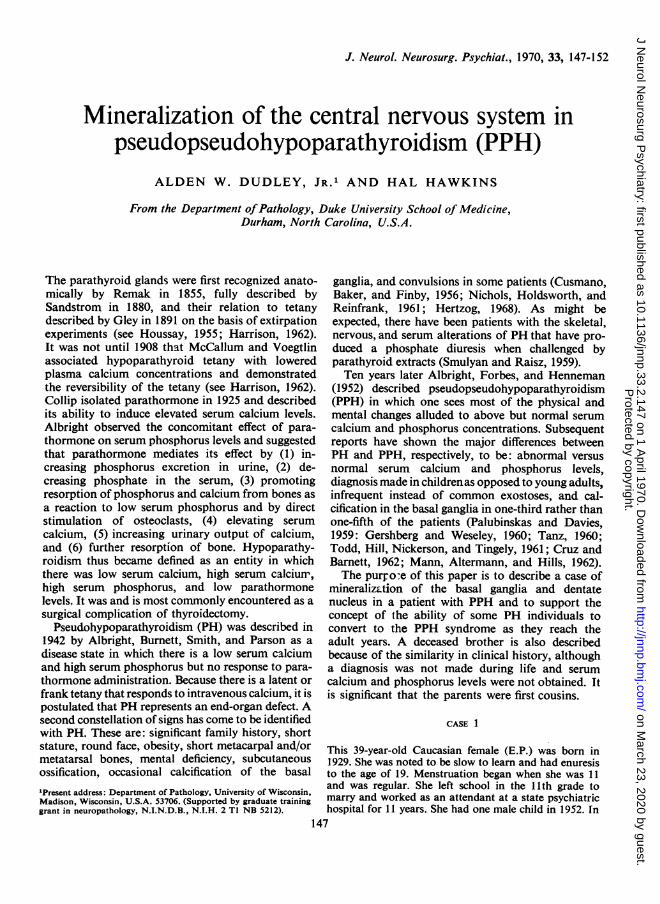
was diagnosed as palindromic arthritis. Radiographs ofthe hands and feet showed normal metacarpal andmetatarsal bones, except for bilateral congenital bonecysts of the talus which were treated surgically. Multipleflecks of subcutaneous calcification were seen on radio-graphs of the right lower leg. In 1965 the patient deve-loped difficulty in walking and a wide, shuffling gait. Athorough neurological evaluation was done. Radiographsof the skull showed calcifications in the pineal, choroidplexuses, and bilaterally in the region of the basal ganglia(Fig. 1). Pneumoencephalograms revealed diffuse cor-tical atrophy and slightly dilated lateral ventricles. Theradiology consultant suggested a diagnosis of pseudo-pseudohypoparathyroidism. An electroencephalogramwas within normal limits. Spinal fluid protein was 39mg/100 ml., and later in the year was 81 mg/100 ml.Eight separate serum calcium determinations from 1965to 1968 (age 36 to 39 years) varied from 9 4 to 10-2mg/100 ml., serum phosphorus between 2 9 and 4-2mg/100 ml.
In 1967 a firm, subcutaneous mass (6 x 10 cm) wasnoted over the patient's anterior right lower leg which wasnon-tender and later regressed, diagnosis unknown.Radiographs showed bilateral subcutaneous flecks ofcalcification in both legs. Cystic changes were noted in theantral area of the mastoid on radiography. The patientbecame more and more disorientated, totally incontinent,and practically bedfast. A repeat electroencephalogramshowed diffuse slowing, and bilateral Babinski reflexesappeared. Faecal impaction, decubitus ulcers, and inter-mittent fever developed as the patient became obtunded.In May 1967, and on several occasions thereafter, spon-taneous grand mal convulsions occurred. Bronchitis andbronchopneumonia developed in 1968 and, in spite ofantibiotic therapy, the patient died in respiratory arrestin June 1968 at 39 years of age.
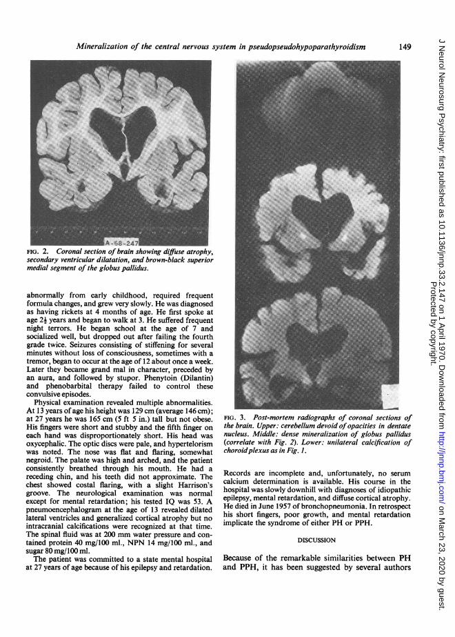
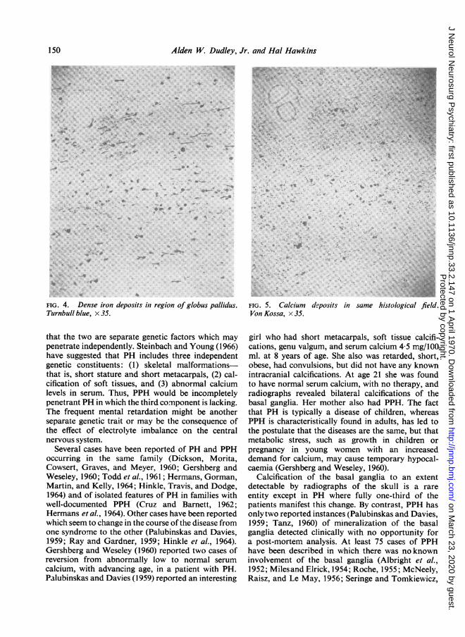
Post-mortem examination of the brain revealed aweight of only 850 g due to diffuse cortical atrophy withhydrocephalus ex vacuo. The medial superior portion ofthe globus pallidus, measuring 1 0 x 0 8 x 0 5 cm andabutting the internal capsule, was discoloured brown-black on fresh coronal sections of the formalin-fixed brain(Fig. 2). This same area was the sole site (other than thepineal gland and choroid plexus) of radiopacity in thefixed brain (Fig. 3). Microscopy revealed scatteredmetallic concretions, averaging 20 Z in diameter, presentthroughout the medial superior portion of the globuspallidus and, to a lesser extent, the dentate nuclei. They
FIG. 1. Plain'skullfilmshowingcalcificationofpinealglaind,choroid plexus (right only) and globus pallidus (bilateral).
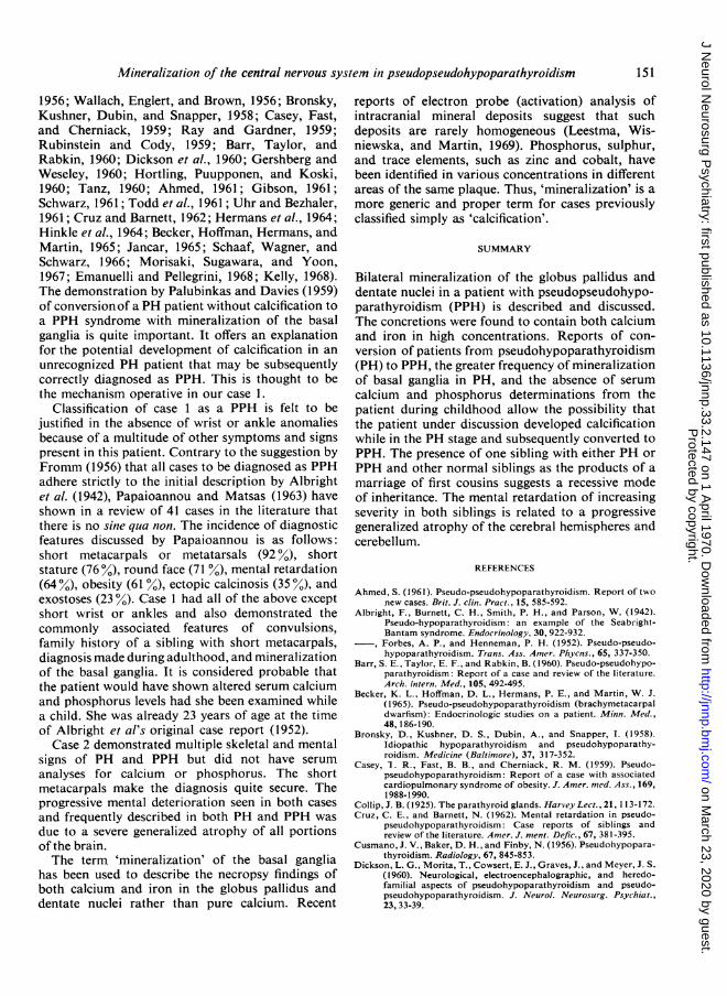
were not associated with vascular structures. Speciastains for iron (Fig. 4) and calcium (Fig. 5), with controls,gave a strong reaction for both metals in the granules.Techniques for the analysis for trace metals were notavailable. The choroid plexus showed normal calcificationwhich stained strongly for calcium but not for iron. Thethoracic spinal cord demonstrated partial demyelinationof the medial posterior columns. The brain did not showany foci of demyelination.The remainder of the examination revealed confluent
bronchopneumonia bilaterally as the cause of death.Submucosal haemorrhages were present throughout thegastrointestinal tract, and the entire colon was markedlydistended with impacted faeces. All other organs werenormal grossly and microscopically. Two normal-appearing parathyroid glands were identified. The pan-creas, adrenals, thyroid, and pituitary showed noabnormalities.
CASE 2
This 33-year-old Caucasian male (J.R.), the brother ofcase 1, was born in 1924. H-e was noted to develop
148
Protected by copyright.
on March 23, 2020 by guest.
http://jnnp.bmj.com
/J N
eurol Neurosurg P
sychiatry: first published as 10.1136/jnnp.33.2.147 on 1 April 1970. D
ownloaded from
Mineralization of the central nervous system in pseudopseudohypoparathyroidism
V-4o8-4 IFIG. 2. Coronal section of brain showing diffuse atrophy,secondary ventricular dilatation, and brown-black superiormedial segment of the globus pallidus.
abnormally from early childhood, required frequentformula changes, and grew very slowly. He was diagnosedas having rickets at 4 months of age. He first spoke atage 24 years and began to walk at 3. He suffered frequentnight terrors. He began school at the age of 7 andsocialized well, but dropped out after failing the fourthgrade twice. Seizures consisting of stiffening for severalminutes without loss of consciousness, sometimes with atremor, began to occur at the age of 12 about once a week.Later they became grand mal in character, preceded byan aura, and followed by stupor. Phenytoin (Dilantin)and phenobarbital therapy failed to control theseconvulsive episodes.
Physical examination revealed multiple abnormalities.At 13 years of age his height was 129 cm (average 146 cm);at 27 years he was 165 cm (5 ft 5 in.) tall but not obese.His fingers were short and stubby and the fifth finger oneach hand was disproportionately short. His head wasoxycephalic. The optic discs were pale, and hypertelorismwas noted. The nose was flat and flaring, somewhatnegroid. The palate was high and arched, and the patientconsistently breathed through his mouth. He had areceding chin, and his teeth did not approximate. Thechest showed costal flaring, with a slight Harrison'sgroove. The neurological examination was normalexcept for mental retardation; his tested IQ was 53. Apneumoencephalogram at the age of 13 revealed dilatedlateral ventricles and generalized cortical atrophy but nointracranial calcifications were recognized at that time.The spinal fluid was at 200 mm water pressure and con-tained protein 40 mg/100 ml., NPN 14 mg/100 ml., andsugar 80 mg/100 ml.The patient was committed to a state mental hospital
at 27 years of age because of his epilepsy and retardation.
FIG. 3. Post-mortem radiographs of coronal sections ofthe brain. Upper: cerebellum devoid ofopacities in dentatenucleus. Middle: dense mineralization of globus pallidus(correlate with Fig. 2). Lower: unilateral calcification ofchoroidplexus as in Fig. 1.
Records are incomplete and, unfortunately, no serumcalcium determination is available. His course in thehospital was slowly downhill with diagnoses of idiopathicepilepsy, mental retardation, and diffuse cortical atrophy.He died in June 1957 of bronchopneumonia. In retrospecthis short fingers, poor growth, and mental retardationimplicate the syndrome of either PH or PPH.
DISCUSSION
Because of the remarkable similarities between PHand PPH, it has been suggested by several authors
149
Protected by copyright.
on March 23, 2020 by guest.
http://jnnp.bmj.com
/J N
eurol Neurosurg P
sychiatry: first published as 10.1136/jnnp.33.2.147 on 1 April 1970. D
ownloaded from
Alden W. Dudley, Jr. and Hal Hawkins
j.I
I
v :
<I4 4r
0
V 4
'VV
'-4,
.. 4,
:--
4*,,
FIG. 4. Dense iron deposits in region of globus pallidus.Turnbull blue, x 35.
that the two are separate genetic factors which maypenetrate independently. Steinbach and Young (1966)have suggested that PH includes three independentgenetic constituents: (1) skeletal malformations-that is, short stature and short metacarpals, (2) cal-cification of soft tissues, and (3) abnormal calciumlevels in serum. Thus, PPH would be incompletelypenetrant PH in which the third component is lacking.The frequent mental retardation might be anotherseparate genetic trait or may be the consequence ofthe effect of electrolyte imbalance on the centralnervous system.
Several cases have been reported of PH and PPHoccurring in the same family (Dickson, Morita,Cowsert, Graves, and Meyer, 1960; Gershberg andWeseley, 1960; Todd et al., 1961; Hermans, Gorman,Martin, and Kelly, 1964; Hinkle, Travis, and Dodge,1964) and of isolated features of PH in families withwell-documented PPH (Cruz and Barnett, 1962;Hermans et al., 1964). Other cases have been reportedwhich seem to change in the course of the disease fromone syndrome to the other (Palubinskas and Davies,1959; Ray and Gardner, 1959; Hinkle et al., 1964).Gershberg and Weseley (1960) reported two cases ofreversion from abnormally low to normal serumcalcium, with advancing age, in a patient with PH.Palubinskas and Davies (1959) reported an interesting
B~ ~~~~~~~~~~~~~~~~
FIG. 5. Calcium deposits in same histological field.Von Kossa, x 35.
girl who had short metacarpals, soft tissue calcifi-cations, genu valgum, and serum calcium 4 5 mg/100ml. at 8 years of age. She also was retarded, short,obese, had convulsions, but did not have any knownintracranial calcifications. At age 21 she was foundto have normal serum calcium, with no therapy, andradiographs revealed bilateral calcifications of thebasal ganglia. Her mother also had PPH. The factthat PH is typically a disease of children, whereasPPH is characteristically found in adults, has led tothe postulate that the diseases are the same, but thatmetabolic stress, such as growth in children orpregnancy in young women with an increaseddemand for calcium, may cause temporary hypocal-caemia (Gershberg and Weseley, 1960).
Calcification of the basal ganglia to an extentdetectable by radiographs of the skull is a rare
entity except in PH where fully one-third of thepatients manifest this change. By contrast, PPH hasonly two reported instances (Palubinskas and Davies,1959; Tanz, 1960) of mineralization of the basalganglia detected clinically with no opportunity fora post-mortem analysis. At least 75 cases of PPHhave been described in which there was noknowninvolvement of the basal ganglia (Albright et al.,1952; Milesand Elrick, 1954; Roche, 1955; McNeely,Raisz, and Le May, 1956; Seringe and Tomkiewicz,
.. .r w w4; 4 xX.. .. # x.
+ +> , .e y'., e :.=
. .,. A 4,: N., iS N.zlF ... e.::>^Si:. # ,:. ", 4 * #'* ,Zp * > 1
e . O .9ie X:
< > > - a g *S #@ee .... dy o?;.0 ... ^ . , . *, a
# 31* . . t * *,h, s 4||E w <*.. t
150
0 . l?, ..
I
Protected by copyright.
on March 23, 2020 by guest.
http://jnnp.bmj.com
/J N
eurol Neurosurg P
sychiatry: first published as 10.1136/jnnp.33.2.147 on 1 April 1970. D
ownloaded from
Mineralization of the central nervous system in pseudopseudohypoparathyroidism
1956; Wallach, Englert, and Brown, 1956; Bronsky,Kushner, Dubin, and Snapper, 1958; Casey, Fast,and Cherniack, 1959; Ray and Gardner, 1959;Rubinstein and Cody, 1959; Barr, Taylor, andRabkin, 1960; Dickson et al., 1960; Gershberg andWeseley, 1960; Hortling, Puupponen, and Koski,1960; Tanz, 1960; Ahmed, 1961; Gibson, 1961;Schwarz, 1961; Todd et al., 1961; Uhr and Bezhaler,1961; Cruz and Barnett, 1962; Hermans et al., 1964;Hinkle et al., 1964; Becker, Hoffman, Hermans, andMartin, 1965; Jancar, 1965; Schaaf, Wagner, andSchwarz, 1966; Morisaki, Sugawara, and Yoon,1967; Emanuelli and Pellegrini, 1968; Kelly, 1968).The demonstration by Palubinkas and Davies (1959)of conversion of a PH patient without calcification toa PPH syndrome with mineralization of the basalganglia is quite important. It offers an explanationfor the potential development of calcification in anunrecognized PH patient that may be subsequentlycorrectly diagnosed as PPH. This is thought to bethe mechanism operative in our case 1.
Classification of case 1 as a PPH is felt to bejustified in the absence of wrist or ankle anomaliesbecause of a multitude of other symptoms and signspresent in this patient. Contrary to the suggestion byFromm (1956) that all cases to be diagnosed as PPHadhere strictly to the initial description by Albrightet al. (1942), Papaioannou and Matsas (1963) haveshown in a review of 41 cases in the literature thatthere is no sine qua non. The incidence of diagnosticfeatures discussed by Papaioannou is as follows:short metacarpals or metatarsals (92 %), shortstature (76 %), round face (71 %), mental retardation(64 %), obesity (61 %), ectopic calcinosis (35 %), andexostoses (23 %). Case 1 had all of the above exceptshort wrist or ankles and also demonstrated thecommonly associated features of convulsions,family history of a sibling with short metacarpals,diagnosis made during adulthood, and mineralizationof the basal ganglia. It is considered probable thatthe patient would have shown altered serum calciumand phosphorus levels had she been examined whilea child. She was already 23 years of age at the timeof Albright et al's original case report (1952).Case 2 demonstrated multiple skeletal and mental
signs of PH and PPH but did not have serumanalyses for calcium or phosphorus. The shortmetacarpals make the diagnosis quite secure. Theprogressive mental deterioration seen in both casesand frequently described in both PH and PPH wasdue to a severe generalized atrophy of all portionsof the brain.The term 'mineralization' of the basal ganglia
has been used to describe the necropsy findings ofboth calcium and iron in the globus pallidus anddentate nuclei rather than pure calcium. Recent
reports of electron probe (activation) analysis ofintracranial mineral deposits suggest that suchdeposits are rarely homogeneous (Leestma, Wis-niewska, and Martin, 1969). Phosphorus, sulphur,and trace elements, such as zinc and cobalt, havebeen identified in various concentrations in differentareas of the same plaque. Thus, 'mineralization' is amore generic and proper term for cases previouslyclassified simply as 'calcification'.
SUMMARY
Bilateral mineralization of the globus pallidus anddentate nuclei in a patient with pseudopseudohypo-parathyroidism (PPH) is described and discussed.The concretions were found to contain both calciumand iron in high concentrations. Reports of con-version of patients from pseudohypoparathyroidism(PH) to PPH, the greater frequency of mineralizationof basal ganglia in PH, and the absence of serumcalcium and phosphorus determinations from thepatient during childhood allow the possibility thatthe patient under discussion developed calcificationwhile in the PH stage and subsequently converted toPPH. The presence of one sibling with either PH orPPH and other normal siblings as the products of amarriage of first cousins suggests a recessive modeof inheritance. The mental retardation of increasingseverity in both siblings is related to a progressivegeneralized atrophy of the cerebral hemispheres andcerebellum.
REFERENCES
Ahmed, S. (1961). Pseudo-pseudohypoparathyroidism. Report of twonew cases. Brit. J. clin. Pract., 15, 585-592.
Albright, F., Burnett, C. H., Smith, P. H., and Parson, W. (1942).Pseudo-hypoparathyroidism: an example of the Seabright-Bantam syndrome. Endocrinology, 30, 922-932.Forbes, A. P., and Henneman, P. H. (1952). Pseudo-pseudo-hypoparathyroidism. Trans. Ass. Amer. Phycns., 65, 337-350.
Barr, S. E., Taylor, E. F., and Rabkin, B. (1960). Pseudo-pseudohypo-parathyroidism: Report of a case and review of the literature.Arch. intern. Med., 105, 492-495.
Becker, K. L., Hoffman, D. L., Hermans, P. E., and Martin, W. J.(1965). Pseudo-pseudohypoparathyroidism (brachymetacarpaldwarfism): Endocrinologic studies on a patient. Minn. Med.,48, 186-190.
Bronsky, D., Kushner, D. S., Dubin, A., and Snapper, 1. (1958).Idiopathic hypoparathyroidism and pseudohypoparathy-roidism. Medicine (Baltimore), 37, 317-352.
Casey, I. R., Fast, B. B., and Cherniack, R. M. (1959). Pseudo-pseudohypoparathyroidism: Report of a case with associatedcardiopulmonary syndrome of obesity. J. Amer. nmed. Ass., 169,1988-1990.
Collip, J. B. (1925). The parathyroid glands. Harvey Lect., 21, 113-172.Cruz, C. E., and Barnett, N. (1962). Mental retardation in pseudo-
pseudohypoparathyroidism: Case reports of siblings andreview of the literature. Anmer. J. ment. Defic., 67, 381-395.
Cusmano, J. V., Baker, D. H., and Finby, N. (1956). Pseudohypopara-thyroidism. Radiology, 67, 845-853.
Dickson, L. G., Morita, T., Cowsert, E. J., Graves, J., and Meyer, J. S.(1960). Neurological, electroencephalographic, and heredo-familial aspects of pseudohypoparathyroidism and pseudo-pseudohypoparathyroidism. J. Neurol. Neurosurg. Psychiat.,23, 33-39.
151
Protected by copyright.
on March 23, 2020 by guest.
http://jnnp.bmj.com
/J N
eurol Neurosurg P
sychiatry: first published as 10.1136/jnnp.33.2.147 on 1 April 1970. D
ownloaded from
Alden W. Dudley, Jr. and Hal Hawkins
Emanuelli, G., and Pellegrini, A. (1968). Su un caso di pseudo-pseudoipoparatirodismo con osteite fibrosa diabete mellito e
diabete insipido. Minerva med., 59, 681-689.Fromm, G. A. (1956). Concerning the term 'pseudo-pseudohypopara-
thyroidism.' J. clin. Endocr., 16, 293-295.Gershberg, H., and Weseley, A. C. (1960). Pseudohypoparathyroidism
and pregnancy: Is pseudo-pseudohypoparathyroidism a mildform of pseudohypoparathyroidism? J. Pediat., 56, 383-386.
Gibson, R. (1961). Brachymetacarpal dwarfism of pseudo-pseudo-hypoparathyroidism with mental defect in siblings. Canad.med. Ass. J., 85, 70-72.
Harrison, T. R. (ed.) (1962). For references to Sandstrom (1880),Gley (1891) and McCallum and Voegtlin see D. S. Bernstein,A. Goldfien and A. W. Thorn, Diseases of the parathyroidglands in Principles of Internal Medicine, p. 599, 4th edition.McGraw Hill: New York.
Hermans, P. E., Gorman, C. A., Martin, W. J., and Kelly, P. J. (1964).Pseudo-pseudohypoparathyroidism (Albright's hereditary osteo-dystrophy): a family study. Mayo Clin. Proc., 39, 81-91.
Hertzog, K. P. (1968). Brachydactyly and pseudo-pseudohypopara-thyroidism. Acta Genet. med. (Roma), 17, 428-438.
Hinkle, D. O., Travis, L. B., and Dodge, W. F. (1964). Albright'shereditary osteodystrophy in a mother and daughter. Tex. Rep.Biol. Med., 23, 463-473.
Hortling, H., Puupponen, E., and Koski, K. (1960). Short metacarpalor metatarsal bones: pseudo-pseudohypoparathyroidism. J.clin. Endocr., 20, 466472.
Houssay, B. A. (ed.) (1955). For references to Remak (1855), Sand-strom (1880), and Gley (1891) see Human Physiology, p. 627,2nd edition. McGraw Hill: New York.
Jancar, J. (1965). Cerebro-metacarpo-metatarsal dystrophy (pseudo-pseudohypoparathyroidism) with chromosomal anomaly. J.med. Genet., 2, 32-37.
Kelly, J. J. (1968). Albright's hereditary osteodystrophy associatedwith disc calcification and bilateral dislocation of the hips.Brit. J. clin. Pract., 22, 399-403.
Leestma, J., Wisniewska, K., and Martin, E. (1969). Electron probeanalysis of mineral deposits in the central nervous system. J.Neuropath. exp. Neurol., 28,157.
Mann, J. B., Altermann, S., and Hills, A. G. (1962). Albright's here-ditary osteodystrophy comprising pseudohypoparathyroidismand pseudopseudohypoparathyroidism: with a report of twocases representing complete syndrome occurring in suc-cessive generations. Ann. intern. Med., 56, 315-342.
McNeely, W. F., Raisz, L. G., and Le May, M. (1956). Dyschondro-plasia with soft tissue calcification and ossification and normal
parathyroid function (Pseudo-pseudohypoparathyroidism).Amer. J. Med., 21, 649-656.
Miles, J., and Elrick, H. (1954). Pseudo-pseudohypoparathyroidism:report ofnew case. J. clin. Endocr., 15, 576-584.
Morisaki, N., Sugawara, S., and Yoon, Y. (1967). A case of pseudo-pseudohypoparathyroidism (Albright). Endocr.jap.,14,327-332.
Nichols, F. L., Holdsworth, D. E., and Reinfrank, R. F. (1961).Familial hypocalcemia, latent tetany, and calcification of thebasal ganglia. Report of a kindred. Amer. J. Med., 30, 518.
Palubinkas, A. J., and Davies, H. (1959). Calcification of the basalganglia of the brain. Amer. J. Roentgenol., 82, 806-822.
Papaionnou, A. C., and Matsas, B. E. (1963). Albright's hereditaryosteodystrophy (without hypocalcemia). Pediatrics, 31, 599-607.
Ray, E. W., and Gardner, L. I. (1959). Pseudo-pseudohypopara-thyroidism in a child. Ibid., 23, 520-529.
Roche, M. (1955). Case of pseudo-pseudohypoparathyroidism. J. clin.Endocr., 15, 964-969.
Rubinstein, E., and Cody, R. (1959). Pseudo-pseudohypoparathyroi-dism with dense calcification of the aorta. Report of a case.
Stanf. med. Bull., 17, 171-174.Schaaf, J., Wagner, A., and Schwarz, G. (1966). Rontgenuntersuch-
ungen bei Patienten mit Pseudohypoparathyreoidismus undPseudo-Pseudohypoparathyreoidismus. I. Rontgensymptome.Fortschr. Rontgenstr., 105, 877-886.
Schwarz, G. (1961). Zur Pathogenese des Pseudo-Pseudohypopara-thyreoidismus. Dtsch. med. Wschr., 86, 257-259.
Seringe, PH., and Tomkiewicz, S. (1956). Pseudo-pseudo-hypopara-thyroidisme familial. Ann. Endocr., 17,655-664.
Smulyan, H., and Raisz, L. G. (1959). Pseudo-pseudohypoparathy-roidism with unusual features. J. clin. Endocr., 19, 478-484.
Steinbach, H. L., and Young, D. A. (1966). The roentgen appearanceof pseudohypoparathyroidism (PH) and pseudo-pseudo-hypoparathyroidism (PPH). Amer. J. Roentgenol., 97, 49-66.
Tanz, S. S. (1960). Pseudo-pseudo-hypoparathyroidism. Three cases
in one family. Amer. J. med. Sci., 239, 453-461.Todd, J. N., III, Hill, S. R., Jr., Nickerson, J. F., and Tingely, J. 0.
(1961). Hereditary multiple exostoses, pseudo-pseudohypo-parathyroidism and other genetic defects of bone, calcium, andphosphorus metabolism. Amer. J. Med., 30,289-298.
Uhr, N., and Bezhaler, H. B. (1961). Pseudo-pseudohypoparathy-roidism: report of three cases in one family. Ann. intern. Med.,54, 443-451.
Wallach, S., Englert, E., Jr., and Brown, H. (1956). The syndrome ofpseudo-pseudohypoparathyroidism. Arch. intern. Med., 98,517-524.
152
Protected by copyright.
on March 23, 2020 by guest.
http://jnnp.bmj.com
/J N
eurol Neurosurg P
sychiatry: first published as 10.1136/jnnp.33.2.147 on 1 April 1970. D
ownloaded from


![[ Ru (H)(H 2 )(PPh 2 CH 2 CH 2 PPh 2 ) 2 ] +](https://static.fdocuments.in/doc/165x107/5681503b550346895dbe37ad/-ru-hh-2-pph-2-ch-2-ch-2-pph-2-2-.jpg)


