MIME robotic device for upper-limb neurorehabilitation in
12
631 JRRD JRRD Volume 43, Number 5, Pages 631–642 August/September 2006 Journal of Rehabilitation Research & Development MIME robotic device for upper-limb neurorehabilitation in subacute stroke subjects: A follow-up study Peter S. Lum, PhD; 1–2* Charles G. Burgar, MD; 3 Machiel Van der Loos, PhD; 4 Peggy C. Shor, OTR; 4 Matra Majmundar, OTR; 4 Ruth Yap, MS 4 1 Hunter Holmes McGuire Department of Veterans Affairs (VA) Medical Center, Richmond, VA; 2 Biomedical Engineering, The Catholic University of America, Washington, DC; 3 Central Texas Veterans Health Care System, Temple, TX; 4 VA Palo Alto Health Care System, Palo Alto, CA Abstract—This study presents results from a randomized con- trolled clinical trial of the Mirror Image Movement Enabler (MIME) robotic device for shoulder and elbow neurorehabilita- tion in subacute stroke patients, including data on the use of its bilateral training mode. MIME incorporates a PUMA 560 robot (Staubli Unimation Inc, Duncan, South Carolina) that applies forces to the paretic limb during unilateral and bilateral move- ments in three dimensions. Robot-assisted treatment (bilateral, unilateral, and combined bilateral and unilateral) was compared with conventional therapy. Similar to a previous study in chronic stroke, combined unilateral and bilateral robotic training had advantages compared with conventional therapy, producing larger improvements on a motor impairment scale and a measure of abnormal synergies. However, gains in all treatment groups were equivalent at the 6-month follow-up. Combined unilateral and bilateral training yielded functional gains that were similar to the gains from equivalent doses of unilateral-only robotic training, although the combined group had more hypertonia and less movement out of synergy at baseline. Robot-assisted treat- ment gains exceeded those expected from spontaneous recovery. These results are discussed in light of the need for further device development and continued clinical trials. Key words: arm, bilateral, hemiparesis, movement, neuroreha- bilitation, rehabilitation, robotics, stroke, subacute, therapy, training. INTRODUCTION The development of robotic treatments is motivated by the need to improve clinical outcomes, the increase of public health burden associated with stroke-related dis- ability [1], and the current emphasis on cost-reduction in healthcare [2]. Most stroke survivors receive one-on-one physical and occupational therapy for the resulting sen- sorimotor impairments. Neurorehabilitation of the upper limb is often abandoned early in favor of compensatory strategies. This decision is motivated by decreasing reim- bursable patient-therapist contact time and the fact that the remaining intact limb with proper training and adaptive aids can perform most activities of daily living (ADL) involving the upper limbs. However, performing ADL one-handed is often cumbersome, increasing the time required and difficulty of the task, compared with per- forming ADL two-handed. These factors suggest that Abbreviations: ADL = activities of daily living, ARM = Assisted Rehabilitation and Measurement (Guide), CI = con- straint-induced, CVA = cerebrovascular accident, FIM = Func- tional Independence Measure, FM = Fugl-Meyer, max = maximum, MCID = minimally clinically important difference, MIME = Mirror Image Movement Enabler, MSS = Motor Sta- tus Score, NDT = NeuroDevelopmental Therapy, ROM = range of motion, RR&D = Rehabilitation Research and Development, VA = Department of Veterans Affairs. * Address all correspondence to Peter S. Lum, PhD; Bio- medical Engineering, The Catholic University of America, Pangborn Hall, Room 131, 620 Michigan Avenue NE; Washington, DC 20064; 202-319-5657; fax: 202-319-4287. Email: [email protected] DOI: 10.1682/JRRD.2005.02.0044
Transcript of MIME robotic device for upper-limb neurorehabilitation in
JRRDJRRD Volume 43, Number 5, Pages 631–642
August/September 2006
Journal of Rehabil itation Research & Development
MIME robotic device for upper-limb neurorehabilitation in subacute stroke subjects: A follow-up study
Peter S. Lum, PhD;1–2* Charles G. Burgar, MD;3 Machiel Van der Loos, PhD;4 Peggy C. Shor, OTR;4 Matra Majmundar, OTR;4 Ruth Yap, MS41Hunter Holmes McGuire Department of Veterans Affairs (VA) Medical Center, Richmond, VA; 2Biomedical Engineering, The Catholic University of America, Washington, DC; 3Central Texas Veterans Health Care System, Temple, TX; 4VA Palo Alto Health Care System, Palo Alto, CA
Abstract—This study presents results from a randomized con-trolled clinical trial of the Mirror Image Movement Enabler (MIME) robotic device for shoulder and elbow neurorehabilita-tion in subacute stroke patients, including data on the use of its bilateral training mode. MIME incorporates a PUMA 560 robot (Staubli Unimation Inc, Duncan, South Carolina) that applies forces to the paretic limb during unilateral and bilateral move-ments in three dimensions. Robot-assisted treatment (bilateral, unilateral, and combined bilateral and unilateral) was compared with conventional therapy. Similar to a previous study in chronic stroke, combined unilateral and bilateral robotic training had advantages compared with conventional therapy, producing larger improvements on a motor impairment scale and a measure of abnormal synergies. However, gains in all treatment groups were equivalent at the 6-month follow-up. Combined unilateral and bilateral training yielded functional gains that were similar to the gains from equivalent doses of unilateral-only robotic training, although the combined group had more hypertonia and less movement out of synergy at baseline. Robot-assisted treat-ment gains exceeded those expected from spontaneous recovery. These results are discussed in light of the need for further device development and continued clinical trials.
Key words: arm, bilateral, hemiparesis, movement, neuroreha-bilitation, rehabilitation, robotics, stroke, subacute, therapy, training.
INTRODUCTION
The development of robotic treatments is motivated by the need to improve clinical outcomes, the increase of
public health burden associated with stroke-related dis-ability [1], and the current emphasis on cost-reduction in healthcare [2]. Most stroke survivors receive one-on-one physical and occupational therapy for the resulting sen-sorimotor impairments. Neurorehabilitation of the upper limb is often abandoned early in favor of compensatory strategies. This decision is motivated by decreasing reim-bursable patient-therapist contact time and the fact that the remaining intact limb with proper training and adaptive aids can perform most activities of daily living (ADL) involving the upper limbs. However, performing ADL one-handed is often cumbersome, increasing the time required and difficulty of the task, compared with per-forming ADL two-handed. These factors suggest that
Abbreviations: ADL = activities of daily living, ARM = Assisted Rehabilitation and Measurement (Guide), CI = con-straint-induced, CVA = cerebrovascular accident, FIM = Func-tional Independence Measure, FM = Fugl-Meyer, max = maximum, MCID = minimally clinically important difference, MIME = Mirror Image Movement Enabler, MSS = Motor Sta-tus Score, NDT = NeuroDevelopmental Therapy, ROM = range of motion, RR&D = Rehabilitation Research and Development, VA = Department of Veterans Affairs.*Address all correspondence to Peter S. Lum, PhD; Bio-medical Engineering, The Catholic University of America, Pangborn Hall, Room 131, 620 Michigan Avenue NE; Washington, DC 20064; 202-319-5657; fax: 202-319-4287. Email: [email protected]: 10.1682/JRRD.2005.02.0044
631
632
JRRD, Volume 43, Number 5, 2006
robotic devices can provide effective training for neurore-habilitation without increasing the burden on the clinicians or increasing the costs of healthcare. If commercially via-ble versions of these robotic devices can be developed, integration of robotic therapy into current practice could alleviate the labor-intensive aspects of neurorehabilitation and thereby increase the efficiency and effectiveness of therapists.
Another motivation for developing robotic treat-ments relates to the growing evidence that recovery from brain injury is heavily influenced by the sensorimotor experience following the injury [3]. An earlier seminal study showed that the highly repetitive task training of constraint-induced (CI) movement therapy, also known as CI therapy, can lead to gains in motor function [4]. More recent studies continue to confirm the positive effects of repetitive movement training on motor recov-ery after stroke [5]. While these studies focused on non-robotic movement training, some evidence exists that robotic repetitive movement training might be even more effective [6], especially in moderately to severely impaired subjects who have difficulty performing unas-sisted repetitive movements. The rationale is that robots might enrich the sensorimotor experience by providing novel patient-environment interactions during active repetitive training. These novel training modes might eventually prove to be more effective than nonrobotic repetitive training. However, to date, we are aware of no study that has demonstrated this evidence.
The robotic device that has received the most clinical testing is the Massachusetts Institute of Technology (MIT)-Manus (Interactive Motion Technologies Inc, Cambridge, Massachusetts). A technical description was first presented in 1998 [7]. MIT-Manus is a 2 degrees-of-freedom robot manipulator that assists shoulder and elbow movement by moving the patients hand in the hori-zontal plane. A novel impedance control mode allows MIT-Manus to be highly compliant when interacting with the patient’s arm, thereby closely matching the human therapist-patient interaction. A series of clinical trials has shown that MIT-Manus provides effective treatment. Acute stroke subjects who received 25 h of MIT-Manus treatment had greater gains in motor function than control subjects who only received a placebo treatment [8–9]. Recent trials with chronic stroke subjects have demon-strated significant clinical gains with MIT-Manus training as well [10–12]. If this robotic treatment can be delivered
cost-effectively, these results would justify use of MIT-Manus as an adjunct to conventional treatment.
Another highly influential robotic device is the Assisted Rehabilitation and Measurement (ARM) Guide (Rehabilitation Institute of Chicago, University of Cali-fornia–Irvine) [13]. A motorized linear constraint pro-vides active-assisted reaching movements in different directions. After 8 weeks of training in the ARM Guide, chronic stroke subjects had functional gains and improve-ments in reaching kinematics [14]. The most important result from this initial study was that the control group who received a matched amount of unassisted reaching movements had statistically identical gains. This empha-sized the fact that highly repetitive active movements have therapeutic value and that the added value of assis-tance from a robot during active movements remained to be demonstrated. Motivated by these pioneering studies with MIT-Manus and ARM Guide, several less-tested approaches are under development [15–16].
Our Past Work with MIME DeviceOver the last 10 years, with funding from the Depart-
ment of Veterans Affairs (VA) Rehabilitation Research and Development (RR&D) Service, we have investigated the use of robotics to facilitate upper-limb neurorehabili-tation following stroke. The long-term goal is to provide patients with movement therapy that has been demon-strated to be equal or superior to what is currently avail-able to patients. Notice that this framework differs from that of the MIT-Manus studies, where robotic training is compared with placebo controls. The motivation for our framework centers on the fact that robotic training is very different from conventional treatment, and it is unlikely that both types of treatment equally treat the range of impairments caused by stroke (hypertonia, weakness, abnormal synergies, etc). While the end goal is functional gain, direct comparison between robotic and conven-tional treatment allows one to identify which impair-ments are best treated by robotic therapy and which are best treated with conventional methods. This information would be a first step toward rational prescription of robotic therapy for treatment of specific impairments. The underlying premise is that robotic treatment may be more effective for some patients and less effective for others. Identifying these rules would lead to more effec-tive integration of robotic training into conventional treatment.
633
LUM et al. Clinical trial of MIME in subacute stroke
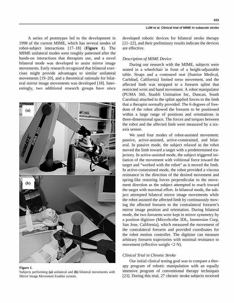
A series of prototypes led to the development in 1998 of the current MIME, which has several modes of robot-subject interactions [17–18] (Figure 1). The MIME unilateral modes were roughly patterned after the hands-on interactions that therapists use, and a novel bilateral mode was developed to assist mirror image movements. Early research recognized that bilateral exer-cises might provide advantages to similar unilateral movements [19–20], and a theoretical rationale for bilat-eral mirror image movements was developed [18]. Inter-estingly, two additional research groups have since
developed robotic devices for bilateral stroke therapy [21–22], and their preliminary results indicate the devices are effective.
Description of MIME DeviceDuring our research with the MIME, subjects were
seated in a wheelchair in front of a height-adjustable table. Straps and a contoured seat (Sunrise Medical, Carlsbad, California) limited torso movement, and the affected limb was strapped to a forearm splint that restricted wrist and hand movement. A robot manipulator (PUMA 560, Staubli Unimation Inc, Duncan, South Carolina) attached to the splint applied forces to the limb that a therapist normally provided. The 6 degrees of free-dom of the robot allowed the forearm to be positioned within a large range of positions and orientations in three-dimensional space. The forces and torques between the robot and the affected limb were measured by a six-axis sensor.
We used four modes of robot-assisted movement: passive, active-assisted, active-constrained, and bilat-eral. In passive mode, the subject relaxed as the robot moved the limb toward a target with a predetermined tra-jectory. In active-assisted mode, the subject triggered ini-tiation of the movement with volitional force toward the target and “worked with the robot” as it moved the limb. In active-constrained mode, the robot provided a viscous resistance in the direction of the desired movement and spring-like restoring forces perpendicular to the move-ment direction as the subject attempted to reach toward the target with maximal effort. In bilateral mode, the sub-ject attempted bilateral mirror image movements while the robot assisted the affected limb by continuously mov-ing the affected forearm to the contralateral forearm’s mirror image position and orientation. During bilateral mode, the two forearms were kept in mirror symmetry by a position digitizer (MicroScribe 3DL, Immersion Corp, San Jose, California), which measured the movement of the contralateral forearm and provided coordinates for the robot motion controller. The digitizer can measure arbitrary forearm trajectories with minimal resistance to movement (effective weight <2 N).
Clinical Trial in Chronic StrokeOur initial clinical testing goal was to compare a ther-
apy program of robotic manipulation with an equally intensive program of conventional therapy techniques [23]. During this trial, 27 chronic stroke subjects received
Figure 1.Subjects performing (a) unilateral and (b) bilateral movements with Mirror Image Movement Enabler system.

634
JRRD, Volume 43, Number 5, 2006
24 one-hour sessions over 2 months. Subjects in the robot group practiced shoulder and elbow movements while assisted by MIME. Subjects in the control group received NeuroDevelopmental Therapy (NDT) [24] targeting proximal upper-limb function. We used the last 5 min of each session to expose the subject to the robot.
In the robot group, targeted reaching movements that started near the body and ended farther away were emphasized. Four point-to-point reaching directions were trained: forward-medial (shoulder flexion, adduction), directly forward (shoulder flexion), forward-lateral (shoulder flexion, abduction, external rotation), and directly lateral (abduction, external rotation). For each of these directions, targets could be located at tabletop, shoulder, or eye level. These 12 targeted reaching motions formed a core set of movements. Subjects prac-ticed some or all of these movements in each session. Movements were kept within each subject’s passive range of motion (ROM). During active-constrained movements, feedback was used to track and motivate performance. This feedback was either the fraction of the movement completed or the time to complete three repetitions. Time permitting, subjects practiced isolated elbow extension movements while assisted by MIME.
A typical control group session involved approxi-mately 10 min of establishing a physical postural base of support and assessing and facilitating the alignment of the shoulder. Approximately 35 min was devoted to graded application of arm use via functional leisure and self-care tasks. Therapists emphasized the reeducation of muscles using a sensorimotor approach to control motor output. Subjects needed to show ability to independently perform basic mass functional movements before progressing to more isolated advanced functional patterns. They pro-gressed within each movement by increasing the number of repetitions, weight of item being handled, height at which tasks were done, etc. In the last 10 min, subjects practiced the highest level task that was completed, with additional review and assessment of the shoulder. Sub-jects received exposure to the robot for 5 min within each session, but the robot provided no active assistance. The robot provided a moving target, and subjects attempted to track the target with their hand or stack cones on top of the robot end effector as it moved. An NDT-certified thera-pist with 13 years experience in treating neurologically injured patients provided therapy. The therapist’s feed-back was consistent with a “conventional therapy” approach. The therapist provided continual comments on the quality of movement to ensure continued interest and
facilitate success of the task. If the subject could not achieve a task, the therapist would suggest other ways of doing it, or if the therapist felt that further attempts at the task would lead to frustration, the level of difficulty would be decreased.
When compared with conventional treatment, robotic therapy had advantages in terms of clinical and biome-chanical measures. The robot group had statistically larger improvements on the proximal Fugl-Meyer (FM) [25] after both 1 month and 2 months of treatment. No between-group differences were found in the distal FM. After 2 months of treatment, the robot group had signifi-cantly greater strength improvements in joint actions that received focused training (shoulder flexion, abduction, adduction, and elbow extension). We measured the kine-matics of free reaching movements by attaching a light-weight, counterbalanced digitizer (Microscribe 3DL, Immersion Corp, San Jose, California) to the forearm. Analysis of these data found that the robot group had larger increases in reach extent in six of the eight move-ments tested. At the 6-month follow-up, the groups no longer differed in terms of the proximal FM scale. These results of this clinical trial are evidence that this regi-mented program of robot-assisted movement had advan-tages compared with an equally intensive program of conventional treatment, primarily in the rate of impair-ment reduction and in quantitative measures of move-ment and strength.
Further examination of the data collected during the robotic training provided evidence of improved muscle activation patterns [26]. Analysis was performed on the interaction forces, kinematics, and electromyograms recorded during training of eight different movement pat-terns in active-constrained mode. These movements were chosen because they were performed consistently in each session and performance feedback was provided to moti-vate maximal performance. Work output during the train-ing was significantly increased by week 3 in five of the eight patterns; by week 5, significant work gains were seen in all eight patterns. These gains were too early to have been because of muscle hypertrophy and were more likely because of increased neural activation of paretic muscles. Electromyographic data provided additional evidence: improved muscle activation patterns were observed in the four movement patterns that started at tabletop level and ended at shoulder level. In contrast, no evidence of improved muscle activation patterns was found in any of the tabletop movements, with increased activation of antagonists in two movement patterns. This

635
LUM et al. Clinical trial of MIME in subacute stroke
dichotomy may have been because of compensation with shoulder girdle movement, which limited the effectiveness of the tabletop movements in promotingneurorehabilitation.
Current Subacute Clinical TrialThe goals of this study were to confirm the previous
results in the clinical trial of chronic subjects and to identify the essential therapeutic features of MIME robotic therapy. In particular, the bilateral mode is unique to MIME, and the study was designed to evaluate the potential unique benefits of this mode. Our working hypothesis was that when the two modes are combined, the bilateral mode enhances the effects of the more con-ventional unilateral mode.
Why the bilateral mode would enhance the effective-ness of the unilateral mode relates to the hypothesis that the potential mechanisms that underlie the two modes are different. The unilateral mode targets corticospinal path-ways from the contralateral damaged cortical hemisphere, while the bilateral mode involves the undamaged hemi-sphere. The bilateral mode may facilitate corticospinal ipsilateral pathways, cortical projections to brain stem pathways, or the damaged hemisphere through the corpus callosum. All or some of these pathways might contribute to motor recovery after stroke.
The most beneficial time to introduce robotic thera-pies needs to be evaluated. Therefore, studies in chronic, subacute, and acute populations are all important to per-form. We chose to perform this study in subacute subjects instead of chronic subjects primarily to avoid secondary maladaptive changes related to increased passive tissue stiffness and contractures, which are likely to be more severe in the chronic stages after stroke. Furthermore, in the subacute phase, the central nervous system may be more amenable to neural plasticity than in the chronic phase. We elected to use subacute subjects instead of acute subjects for the following reasons:1. Although testing in the acute phase compared with the
subacute phase has theoretical advantages, subjects are often too ill to tolerate additional therapy.
2. In the subacute phase, subjects are typically receiving outpatient therapy a few times a week and may tolerate additional therapy but cannot get it because of the costs involved.
3. Pragmatically, robotic therapy might have its largest effect during subacute treatment.
METHODS
Subacute subjects were included in the current study if they had a diagnosis of a single cerebrovascular acci-dent (CVA) and were 1 to 5 months post-CVA. Subjects were allowed to continue any outpatient therapies in which they were enrolled at the time of study acceptance. Subjects were excluded from the study if they exhibited any upper-limb joint pain or ROM limitations that would limit their ability to complete the protocols. Subjects with any unstable cardiovascular, orthopedic, or neurological conditions were also excluded. Cognitive impairments were screened with the Folstein Mini-Mental State Examination, and subjects were excluded if they scored <21 on the Examination.
Subjects were stratified by the FM score and the cerebral hemisphere in which the stroke occurred and then randomly assigned to one of four treatment groups (one control and three robot). Thirty subjects completed the training and posttreatment evaluations. Over 4 weeks, all groups received 15 one-hour treatment sessions held in the same treatment area and supervised by a single occupational therapist. Thus, all subjects received the same treatment time per session, number of sessions per week, and total number of sessions. Subjects in the three robot groups received 50 min of robot-assisted move-ment each session while subjects in the control group received 50 min of conventional treatment. All subjects received 5 min of tone normalization and limb position-ing at the beginning and end of each session. We did not inform subjects of the explicit goals of the clinical trial, only that the effectiveness of several treatments was being tested. The local institutional review committee approved all protocols, and we obtained informed con-sent from all subjects.
The robotic treatment was similar to that used in the earlier chronic study. A core set of 12 targeted reaching movements was used that was identical to the patterns used in the chronic study. Subjects practiced some or all of these movements in each session. The four treatment groups were as follows:1. The robot-unilateral group (n = 9) performed exercises
that progressed from the easiest exercise modes (pas-sive) to the most challenging (active-constrained). No bilateral exercise was performed.
2. The robot-bilateral group (n = 5) practiced the same 12 reaching movements, but only in bilateral mode. Rhythmic circular movements were also performed.

636
JRRD, Volume 43, Number 5, 2006
3. The robot-combined group (n = 10) spent approxi-mately half the treatment time in the unilateral mode and the other half in the bilateral mode. This group received essentially the same robotic treatment as the earlier chronic subjects.
4. The control group (n = 6) received an equivalent inten-sity and duration of conventional therapy targeting proximal upper-limb function based on NDT. The pro-cedures used in the control group were identical to those used in the chronic study.
An occupational therapist blinded to group assign-ments tested all subjects with a battery of clinical evalu-ations immediately before treatment started, immediately posttreatment, and 6 months after treatment ended. Motor impairment was assessed with the upper-limb portion of the FM [25] and the Motor Status Score (MSS) [27]. We used the Functional Independence Measure (FIM) [28] to measure improvements in basic ADL, and the Motor Power examination to assess strength in the upper limb [29]. This motor power examination scores several joints of the proximal upper limb on a 5-point scale. We used the modified Ashworth scale [30] to test for hypertonia in several upper-limb joints. A higher score on the modi-fied Ashworth scale indicates higher tone, so a lower score indicates lower abnormal tone. In all the other scales, higher scores indicate improvement and reduced impairment.
Baseline differences between groups were evaluated with t-tests (continuous and ordinal data) and χ 2 tests (cate-gorical data). The motor FM was separated into proximal (shoulder and elbow: 42 points) and distal (hand and wrist: 24 points) portions for statistical analysis. The proximal MSS (shoulder and elbow parts) was separated into a movement scale (46 points) and a synergy scale (20 points) that assesses the ability to suppress abnormal synergies dur-ing the attempted movements. A higher score on the MSS synergy scale indicates more isolated movement. Because the FM and the MSS movement scales overlap consider-ably, the latter was not included in the analysis. The modi-fied Ashworth scale scores for individual joints were grouped into a proximal score (maximum [max] = 15: shoulder internal rotators, elbow extensors, elbow flexors), and a distal score (max = 30: pronators, supinators, wrist flexors, wrist extensors, digit flexors, digit extensors). For the FIM, only the self-care and transfer sections were con-sidered (max = 63).
Initially, the randomization procedure provided a uni-form number of subjects in each group. However, an
interim analysis at the midpoint of the study indicated that statistical difference between the robot-combined and control groups had already been achieved. At that point, a trend indicated the robot-combined and the robot-unilateral group gains were different. The study investi-gators decided to use the remaining resources to further investigate this trend; these two groups were continued while the control and robot-bilateral groups were discon-tinued. To evaluate our initial hypotheses, we performed two preplanned comparisons using t-tests. The first com-parison was between gains in the robot-combined and robot-unilateral groups. The assumption of homogeneity of variance was tested with the F-test. In cases where this assumption was violated, the Robust Rank-Order test [31] was used. The second comparison was between the robot-combined and the control groups. Because of the small number of control subjects (n = 16), the nonpara-metric Robust Rank-Order test was used.
The data collected in this study were compared with the spontaneous recovery patterns reported by Duncan et al. in a study of 459 stroke subjects [32]. Duncan et al. separated subjects into mild, moderate, and severe stroke categories and charted their recovery patterns on the upper-limb FM. They found that the recovery pattern var-ied depending on the stroke severity. Our subject pool fell between the moderate and severe stroke profiles reported in their study. Therefore, to estimate the spontaneous recovery profile of our subject pool, we calculated least-squares exponential fits for the moderate and severe pro-files. The parameters of these two equations were linearly interpolated until the recovery profile that intersected the baseline data point of our subject pool was found.
RESULTS
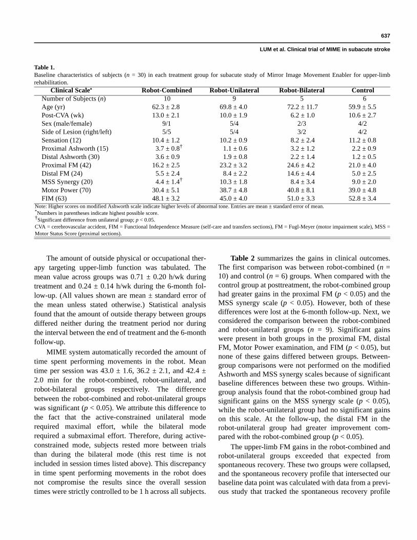
Thirty subjects completed the treatments and the post-treatment evaluations. Six-month follow-up data were available from 23 subjects. One subject dropped out of the study for reasons unrelated to the study. Table 1 summa-rizes the baseline characteristics of the subjects. Compared with the robot-unilateral group (n = 9), the robot-combined group (n = 10) had significantly higher tone in proximal joints (proximal modified Ashworth scale, p < 0.05) and more abnormal synergies (MSS synergy, p < 0.05) at base-line. No significant baseline differences were found between groups in age, weeks post-CVA, sex, side of lesion, or any other clinical evaluations.
637
LUM et al. Clinical trial of MIME in subacute stroke
The amount of outside physical or occupational ther-apy targeting upper-limb function was tabulated. The mean value across groups was 0.71 ± 0.20 h/wk during treatment and 0.24 ± 0.14 h/wk during the 6-month fol-low-up. (All values shown are mean ± standard error of the mean unless stated otherwise.) Statistical analysis found that the amount of outside therapy between groups differed neither during the treatment period nor during the interval between the end of treatment and the 6-month follow-up.
MIME system automatically recorded the amount of time spent performing movements in the robot. Mean time per session was 43.0 ± 1.6, 36.2 ± 2.1, and 42.4 ± 2.0 min for the robot-combined, robot-unilateral, and robot-bilateral groups respectively. The difference between the robot-combined and robot-unilateral groups was significant (p < 0.05). We attribute this difference to the fact that the active-constrained unilateral mode required maximal effort, while the bilateral mode required a submaximal effort. Therefore, during active-constrained mode, subjects rested more between trials than during the bilateral mode (this rest time is not included in session times listed above). This discrepancy in time spent performing movements in the robot does not compromise the results since the overall session times were strictly controlled to be 1 h across all subjects.
Table 2 summarizes the gains in clinical outcomes. The first comparison was between robot-combined (n = 10) and control (n = 6) groups. When compared with the control group at posttreatment, the robot-combined group had greater gains in the proximal FM (p < 0.05) and the MSS synergy scale (p < 0.05). However, both of these differences were lost at the 6-month follow-up. Next, we considered the comparison between the robot-combined and robot-unilateral groups (n = 9). Significant gains were present in both groups in the proximal FM, distal FM, Motor Power examination, and FIM (p < 0.05), but none of these gains differed between groups. Between-group comparisons were not performed on the modified Ashworth and MSS synergy scales because of significant baseline differences between these two groups. Within-group analysis found that the robot-combined group had significant gains on the MSS synergy scale (p < 0.05), while the robot-unilateral group had no significant gains on this scale. At the follow-up, the distal FM in the robot-unilateral group had greater improvement com-pared with the robot-combined group (p < 0.05).
The upper-limb FM gains in the robot-combined and robot-unilateral groups exceeded that expected from spontaneous recovery. These two groups were collapsed, and the spontaneous recovery profile that intersected our baseline data point was calculated with data from a previ-ous study that tracked the spontaneous recovery profile
Table 1.Baseline characteristics of subjects (n = 30) in each treatment group for subacute study of Mirror Image Movement Enabler for upper-limb rehabilitation.
Clinical Scale* Robot-Combined Robot-Unilateral Robot-Bilateral ControlNumber of Subjects (n) 10 9 5 6Age (yr) 62.3 ± 2.8 69.8 ± 4.0 72.2 ± 11.7 59.9 ± 5.5Post-CVA (wk) 13.0 ± 2.1 10.0 ± 1.9 6.2 ± 1.0 10.6 ± 2.7Sex (male/female) 9/1 5/4 2/3 4/2Side of Lesion (right/left) 5/5 5/4 3/2 4/2Sensation (12) 10.4 ± 1.2 10.2 ± 0.9 8.2 ± 2.4 11.2 ± 0.8Proximal Ashworth (15) 3.7 ± 0.8† 1.1 ± 0.6 3.2 ± 1.2 2.2 ± 0.9Distal Ashworth (30) 3.6 ± 0.9 1.9 ± 0.8 2.2 ± 1.4 1.2 ± 0.5Proximal FM (42) 16.2 ± 2.5 23.2 ± 3.2 24.6 ± 4.2 21.0 ± 4.0Distal FM (24) 5.5 ± 2.4 8.4 ± 2.2 14.6 ± 4.4 5.0 ± 2.5MSS Synergy (20) 4.4 ± 1.4† 10.3 ± 1.8 8.4 ± 3.4 9.0 ± 2.0Motor Power (70) 30.4 ± 5.1 38.7 ± 4.8 40.8 ± 8.1 39.0 ± 4.8FIM (63) 48.1 ± 3.2 45.0 ± 4.0 51.0 ± 3.3 52.8 ± 3.4
Note: Higher scores on modified Ashworth scale indicate higher levels of abnormal tone. Entries are mean ± standard error of mean.*Numbers in parentheses indicate highest possible score.†Significant difference from unilateral group; p < 0.05.CVA = cerebrovascular accident, FIM = Functional Independence Measure (self-care and transfers sections), FM = Fugl-Meyer (motor impairment scale), MSS = Motor Status Score (proximal sections).

638
JRRD, Volume 43, Number 5, 2006
of 459 stroke subjects [32] (see “Methods” section). As depicted in Figure 2, mean gains in the robot-combined and robot-unilateral groups exceeded that expected from spontaneous recovery. Statistical testing compared the FM scores in the robot groups with the corresponding FM levels on the spontaneous recovery curve. Immedi-ately after treatment, a trend (p < 0.1) of greater gains was present in the robot groups compared with spontane-ous recovery. By the 6-month follow-up, FM scores in the robot groups exceeded those expected from spontane-ous recovery (p < 0.05).
DISCUSSION
These results are consistent with the previous study on chronic stroke subjects. In both this and the previous chronic stroke study, proximal FM scores indicated that at posttreatment, robot-combined training group had sig-nificantly greater gains than the control group. However, in both studies, gains in robot and control groups were equivalent at the 6-month follow-up. A similar pattern
was observed in the MSS synergy score. Thus, the robot-combined treatment is equivalent to conventional treat-ment in terms of long-term clinical outcomes, but it may accelerate the rate of recovery on some clinical scales.
No significant differences were found between the robot-combined and robot-unilateral treatment on the proximal FM, distal FM, Motor Power examination, and FIM despite that the robot-unilateral subjects spent more time training only the paretic limb. The results also sug-gest less benefit from bilateral therapy alone, because this group had the smallest gains in the proximal FM, distal FM, Motor Power examination, and FIM. These results have many interpretations. One might argue that because the bilateral mode adds complexity and cost to the robotic device, unilateral modes should be used exclu-sively without bilateral therapy. Another interpretation is based on the fact that the active unilateral modes require more focused effort and are more fatiguing than the bilat-eral mode. Combining unilateral and bilateral training might allow extended treatment sessions since the overall effort level is less than in unilateral-only robotic training. Furthermore, the data suggest the bilateral mode may have unique benefits in reducing abnormal synergies.
Table 2.Average gains in clinical scores of subjects (n = 30) in each treatment group from subacute study of Mirror Image Movement Enabler for upper-limb rehabilitation.
Clinical Scale* Robot-Combined Robot-Unilateral Robot-Bilateral ControlPosttreatment
Number of Subjects 10 9 5 6Proximal Ashworth (15) –0.7 ± 0.7 0.9 ± 0.6 –0.4 ± 0.4 –1.3 ± 0.7Distal Ashworth (30) –0.4 ± 0.2 0.0 ± 0.8 –1.0 ± 0.6 0.7 ± 0.6Proximal FM (42) 5.3 ± 1.2† 4.3 ± 1.4 2.4 ± 1.5 2.5 ± 0.6Distal FM (24) 2.3 ± 0.4 3.6 ± 1.3 1.4 ± 0.7 3.3 ± 1.9MSS synergy (20) 4.0 ± 1.0† 0.8 ± 0.9 2.0 ± 2.6 0.7 ± 1.1Motor Power (70) 8.2 ± 1.0 10.1 ± 2.4 3.2 ± 1.0 9.3 ± 1.3FIM (63) 3.1 ± 1.7 3.7 ± 1.0 0.8 ± 0.6 3.2 ± 1.4
6-Month Follow-UpNumber of Subjects 6 7 5 5Proximal Ashworth (15) –0.2 ± 0.5 0.3 ± 1.1 –2.0 ± 0.8 0.2 ± 0.8Distal Ashworth (30) –0.8 ± 0.6 –0.6 ± 0.6 –1.2 ± 0.8 0.8 ± 0.7Proximal FM (42) 6.0 ± 1.4 7.3 ± 2.0 4.4 ± 1.3 7.6 ± 1.2Distal FM (24) 3.0 ± 1.0‡ 8.9 ± 2.1 3.0 ± 1.5 6.2 ± 2.5MSS Synergy (20) 5.8 ± 1.8 4.1 ± 0.9 4.6 ± 2.5 4.6 ± 1.1Motor Power (70) 17.2 ± 2.1 17.9 ± 3.4 11.2 ± 3.2 14.2 ± 2.3FIM (63) 2.8 ± 2.4 4.3 ± 2.7 5.0 ± 1.4 5.2 ± 1.7
Note: Negative score changes on Ashworth scale indicate reduced tone. Entries are mean ± standard error of the mean.*Numbers in parentheses indicate highest possible score.†Significant difference from control; p < 0.05.‡Significant difference from unilateral group; p < 0.05.FIM = Functional Independence Measure (self-care and transfers sections), FM = Fugl-Meyer (motor impairment scale), MSS = Motor Status Score (proximal sections).

639
LUM et al. Clinical trial of MIME in subacute stroke
The robot-combined group showed significant improve-ment in synergy scores, while the robot-unilateral group showed no improvement. However, this result needs to be reproduced in subject groups that are balanced at base-line in terms of abnormal synergies.
Comparing gains from a treatment with a minimal clinically important difference (MCID) is important. For example, gains might be significant, but the treatment still might not be justified if gains are less than the MCID. Although no standard exists for defining MCID for stroke patients, studies have suggested that the MCID is approximately 10 percent of full scale [33–34]. Based on this assumption, mean gains at posttreatment in the robot-unilateral and robot-combined groups were greater than the MCID in the proximal FM, MSS movement, and Motor Power examination. In the FIM, however, gains were less than the MCID. However, the FIM is a very coarse scale and high scores can be achieved by using only the nonparetic arm. Therefore, the FIM may not be
an adequate measure of improved functional use of the paretic upper limb. Thus, the 10 percent assumption might be suspect, and any significant increase in the FIM could be argued to be clinically significant.
Examination of gains in individual subjects suggests the robotic treatment is most effective for subjects in a middle range of motor impairment. Four subjects had gains of greater than 10 points on the proximal FM (two received robot-unilateral treatment and two received robot-combined treatment). These four subjects fell between 15 and 23 inclusive on the proximal FM, while all 30 subjects spanned the range from 7 to 37. Analysis of the data from the previous chronic study also supports this hypothesis. The top three performers in the robot group had pretreatment proximal FM scores between 15 and 23. Thus, in the subacute-chronic phases of recovery, robotic training is apparently most effective in subjects with moderate levels of motor impairment.
One of the most compelling rationales for investigation of robotic therapy is the potential to provide additional effective therapy to patients without increasing the costs of healthcare. If commercially viable robotic devices can be developed that patients can use independently in the home or clinic, our data indicate that training with these robots can be as effective as conventional one-on-one treatment from a therapist. Robotic therapy also increased the rate of recovery compared with conventional treatment, and gains exceeded that which was expected because of spontaneous recovery. These results, coupled with the evidence that higher intensities of conventional therapy produces greater reductions in impairment and disability [35–42], support the use of robots to increase the amount of effective therapy for stroke survivors. One should note that all the chronic subjects and most of the subacute subjects had stopped all formal one-on-one therapy from physical or occupational therapists. Therefore, the additional treatment provided was effective and would not typically have been available to these subjects because of the current structure of the health-care system. An alternative, equally effective method for delivery of this additional therapy is robotic devices.
CONCLUSIONS
The precise role of the robot in robotic training has generated considerable debate recently. More specifically, does the presence of the robot provide added benefits compared with equal intensities of nonrobotic training? This is an important question given that robotic devices
Figure 2.Gains in upper-limb Fugl-Meyer scores compared with spontaneous recovery patterns. Spontaneous recovery data have been replotted (♦ and ) along with exponential fits. Spontaneous recovery curve that intersects baseline point of our subject pool was calculated by interpolating between moderate and severe profiles. *Source: Duncan PW, Lai SM, Keighley J. Defining post-stroke recovery: implications for design and interpretation of drug trials. Neuropharmacology. 2000; 39(5):835–41. [PMID: 10699448]

640
JRRD, Volume 43, Number 5, 2006
are potentially unsafe, complex, and expensive. This sub-ject was highlighted by the study using the ARM Guide; whereby, subjects who practiced on the robot had gains that were comparable with controls that performed an equal amount of unassisted reaching movements [14]. Determining if robotic assistance improves the effective-ness of movement practice is interesting from a scientific viewpoint, and answering this question would guide refinements in the current robotic devices and also poten-tially improve conventional hands-on therapy.
However, from a pragmatic viewpoint, it is doubtful that moderately to severely impaired subjects can be motivated to perform highly repetitive, unassisted move-ments without direct supervision and encouragement from a therapist. In the context of CI therapy, even mildly impaired subjects require physical restraint of the less-impaired limb and direct therapist supervision to achieve the required amounts of task practice. In moderately to severely impaired populations that require more “hands-on” treatment, low-cost versions of robotic therapy devices that can be used independently might be the least expensive means of facilitating repetitive movement training. Because of the expense, additional therapy from a human therapist is unavailable to many patients. Thus, if robotic therapy devices can be made safe and cost-effective (and we believe that they can), then their com-mercial development should proceed. We have demon-strated that MIME is at least as effective as an equivalent dose of hands-on therapy in subacute and chronic stroke populations. Until a major breakthrough occurs in the cure of stroke and its sequelae, robot-assisted therapy appears to have an appropriate role in rehabilitation.
In the next 10 years, several lines of work should proceed in parallel:1. Emphasis should be placed on developing low-cost
versions of the robots that have been thoroughly tested with controlled clinical trials.
2. Both this subacute study and the previous chronic study indicated that between the end of treatment and the 6-month follow-up, control subjects had greater gains than robot group subjects. This finding motivates the development of portable devices that can be used as part of a home-based treatment plan following in-clinic treatment.
3. Clearly, in the upper limb, the potential benefits of robotic therapy cannot be fully evaluated with devices that address shoulder and elbow movement without
treating more distal joints. Additional work is needed to develop devices that integrate wrist and finger function.
4. Other nonrobotic approaches should continue to be investigated and compared with robotic training.
5. Future clinical trials should measure the effects of unsupervised or partially supervised robot therapy. All the studies to date have used a therapist or attendant to supervise the robot treatment. Robots will have the largest impact if patients can be motivated to use them independently. Recent studies have shown that with appropriate technology, the role of the therapist during highly repetitive movement training protocols such as CI therapy can be reduced without loss of treatment effectiveness [43–45].
6. A meta-analysis should be undertaken to establish if the gains from robotic devices are clinically relevant.
ACKNOWLEDGMENTS
To all subjects who participated in this study, we express our deepest appreciation.
This material was based on work supported by the VA RR&D merit review grants B2056RA, B2695I, and B2156T.
The authors have declared that no competing inter-ests exist.
REFERENCES
1. Post-Stroke Rehabilitation Guideline Panel. Post-stroke rehabilitation: Clinical practice guidelines. No. 16. Publica-tion AHCPR 95-0662. Rockville (MD): United States Department of Health and Human Services, Public Health Service, Agency for Health Care Policy and Research; 1995.
2. Dobkin BH. The economic impact of stroke. Neurology. 1995;45(2 Suppl 1):S6–9. [PMID: 7885589]
3. Reinkensmeyer DJ, Emken JL, Cramer SC. Robotics, motor learning, and neurologic recovery. Annu Rev Biomed Eng. 2004;6:497–525. [PMID: 15255778]
4. Taub E, Miller NE, Novack TA, Cook EW 3rd, Fleming WC, Nepomuceno CS, Connell JS, Crago JE. Technique to improve chronic motor deficit after stroke. Arch Phys Med Rehabil. 1993;74(4):347–54. [PMID: 8466415]
5. Mark VW, Taub E. Constraint-induced movement therapy for chronic stroke hemiparesis and other disabilities. Restor Neurol Neurosci. 2004;22(3–5):317–36. [PMID: 15502259]
6. Patton JL, Mussa-Ivaldi FA. Robot-assisted adaptive train-ing: custom force fields for teaching movement patterns.
http://www.ncbi.nlm.nih.gov/entrez/query.fcgi?db=pubmed&cmd=Retrieve&dopt=Abstract&list_uids=7885589

641
LUM et al. Clinical trial of MIME in subacute stroke
IEEE Trans Biomed Eng. 2004;51(4):636–46. [PMID: 15072218]
7. Krebs HI, Hogan N, Aisen ML, Volpe BT. Robot-aided neurorehabilitation. IEEE Trans Rehabil Eng. 1998;6(1): 75–87. [PMID: 9535526]
8. Volpe BT, Ferraro M, Lynch D, Christos P, Krol J, Trudell C, Krebs HI, Hogan N. Robotics and other devices in the treatment of patients recovering from stroke. Curr Athero-scler Rep. 2004;6(4):314–19. [PMID: 15191707]
9. Volpe BT, Krebs HI, Hogan N. Robot-aided sensorimotor training in stroke rehabilitation. Adv Neurol. 2003;92:429–33. [PMID: 12760210]
10. Ferraro M, Palazzolo JJ, Krol J, Krebs HI, Hogan N, Volpe BT. Robot-aided sensorimotor arm training improves out-come in patients with chronic stroke. Neurology. 2003; 61(11):1604–7. [PMID: 14663051]
11. Stein J, Krebs HI, Frontera WR, Fasoli SE, Hughes R, Hogan N. Comparison of two techniques of robot-aided upper limb exercise training after stroke. Am J Phys Med Rehabil. 2004;83(9):720–28. [PMID: 15314537]
12. Fasoli SE, Krebs HI, Stein J, Frontera WR, Hogan N. Effects of robotic therapy on motor impairment and recov-ery in chronic stroke. Arch Phys Med Rehabil. 2003;84(4): 477–82. [PMID: 12690583]
13. Reinkensmeyer DJ, Dewald JP, Rymer WZ. Guidance-based quantification of arm impairment following brain injury: a pilot study. IEEE Trans Rehabil Eng. 1999;7(1): 1–11. [PMID: 10188602]
14. Kahn LE, Lum PS, Reinkensmeyer DJ. Selection of robotic therapy algorithms for the upper extremity in chronic stroke: Insights from MIME and ARM guide results. In: Proceedings of the 8th International Conference on Reha-bilitation Robotics; 2003 Apr 23–25; Daejeon, Korea. Dae-jeon (Korea): Human-Friendly Welfare Robot System Engineering Research Center; 2003. p. 208–10.
15. Lum PS, Reinkensmeyer DJ, Mahoney R, Rymer WZ, Bur-gar CG. Robotic devices for movement therapy after stroke: current status and challenges to clinical acceptance. Top Stroke Rehabil. 2002;8(4):40–53. [PMID: 14523729]
16. Hesse S, Schmidt H, Werner C, Bardeleben A. Upper and lower extremity robotic devices for rehabilitation and for studying motor control. Curr Opin Neurol. 2003;16(6): 705–10. [PMID: 14624080]
17. Lum PS, Burgar C G , Kenney DE, Van der Loos HF. Quan-tification of force abnormalities during passive and active-assisted upper-limb reaching movements in post-stroke hemiparesis. IEEE Trans Biomed Eng. 1999;46(6):652–62. [PMID: 10356872]
18. Burgar C G , Lum PS, Shor PC, Machiel Van der Loos HF. Development of robots for rehabilitation therapy: The Palo Alto VA/Stanford experience. J Rehabil Res Dev. 2000; 37(6):663–73. [PMID: 11321002]
19. Lum PS, Lehman SL, Reinkensmeyer DJ. The bimanual lifting rehabilitator: an adaptive machine for therapy of stroke patients. IEEE Trans Rehabil Eng. 1995;3(2):166–74.
20. Lum PS, Reinkensmeyer DJ, Lehman SL. Robotic assist devices for bimanual physical therapy: preliminary experi-ments. IEEE Trans Rehabil Eng. 1993;1(3):185–91.
21. Luft AR, McCombe-Waller S, Whitall J, Forrester LW, Macko R, Sorkin JD, Schulz JB, Goldberg AP, Hanley DF. Repetitive bilateral arm training and motor cortex activa-tion in chronic stroke: a randomized controlled trial. JAMA. 2004;292(15):1853–61. [PMID: 15494583] Erra-tum in: JAMA. 2004;292(20):2470.
22. Hesse S, Schulte-Tigges G , Konrad M, Bardeleben A, Werner C. Robot-assisted arm trainer for the passive and active practice of bilateral forearm and wrist movements in hemiparetic subjects. Arch Phys Med Rehabil. 2003;84(6): 915–20. [PMID: 12808550]
23. Lum PS, Burgar C G , Shor PC, Majmundar M, Van der Loos M. Robot-assisted movement training compared with conventional therapy techniques for the rehabilitation of upper-limb motor function after stroke. Arch Phys Med Rehabil. 2002;83(7):952–59. [PMID: 12098155]
24. Bobath B. Adult hemiplegia: Evaluation and treatment. 2nd ed. London (England): William Heinemann Medical Books Ltd; 1978.
25. Fugl-Meyer AR, Jaasko L, Leyman I, Olsson S, Steglind S. The post-stroke hemiplegic patient. 1. A method for evalu-ation of physical performance. Scand J Rehabil Med. 1975; 7(1):13–31. [PMID: 1135616]
26. Lum PS, Burgar CG , Shor PC. Evidence for improved muscle activation patterns after retraining of reaching movements with the MIME robotic system in subjects with post-stroke hemiparesis. IEEE Trans Neural Syst Rehabil Eng. 2004;12(2):186–94.
27. Ferraro M, Demaio JH, Krol J, Trudell C, Rannekleiv K, Edelstein L, Christos P, Aisen ML, England J, Fasoli SE, Krebs HI, Hogan N, Volpe BT. Assessing the motor status score: A scale for the evaluation of upper limb motor out-comes in patients after stroke. Neurorehabil Neural Repair. 2002;16(3):283–89. [PMID: 12234090]
28. Hamilton BB, Laughlin JA, Fiedler RC, Granger CV. Inter-rater reliability of the 7-level functional independence mea-sure (FIM). Scand J Rehabil Med. 1994;26(3):115–19. [PMID: 7801060]
29. Gregson JM, Leathley MJ, Moore AP, Smith TL, Sharma AK, Watkins CL. Reliability of measurements of muscle tone and muscle power in stroke patients. Age Ageing. 2000; 29(3):223–28. [PMID: 10855904]
30. Bohannon RW, Smith MB. Interrater reliability of a modi-fied Ashworth scale of muscle spasticity. Phys Ther. 1987; 67(2):206–7. [PMID: 3809245]
http://www.ncbi.nlm.nih.gov/entrez/query.fcgi?db=pubmed&cmd=Retrieve&dopt=Abstract&list_uids=9535526
http://www.ncbi.nlm.nih.gov/entrez/query.fcgi?db=pubmed&cmd=Retrieve&dopt=Abstract&list_uids=1135616

642
JRRD, Volume 43, Number 5, 2006
31. Siegel S, Castellan NJ Jr. Nonparametric statistics for the behavioral sciences. 2nd ed. Boston (MA): McGraw-Hill; 1988. p. 137–44.
32. Duncan PW, Lai SM, Keighley J. Defining post-stroke recovery: implications for design and interpretation of drug trials. Neuropharmacology. 2000;39(5):835–41. [PMID: 10699448]
33. Van der Lee JH, Beckerman H, Knol DL, De Vet HC, Bouter LM. Clinimetric properties of the motor activity log for the assessment of arm use in hemiparetic patients. Stroke. 2004;35(6):1410–14. [PMID: 15087552]
34. Van der Lee JH, Wagenaar RC, Lankhorst GJ, Vogelaar TW, Deville WL, Bouter LM. Forced use of the upper extremity in chronic stroke patients: results from a single-blind ran-domized clinical trial. Stroke. 1999;30(11):2369–75. [PMID: 10548673] Comment in: Stroke. 2000;31(4):986–88.
35. Chen CC, Heinemann AW, Granger CV, Linn RT. Func-tional gains and therapy intensity during subacute rehabili-tation: a study of 20 facilities. Arch Phys Med Rehabil. 2002;83(11):1514–23. [PMID: 12422318]
36. Kwakkel G , Wagenaar RC, Twisk JW, Lankhorst GJ, Koet-sier JC. Intensity of leg and arm training after primary mid-dle-cerebral-artery stroke: a randomised trial. Lancet. 1999;354(9174):191–96. [PMID: 10421300]
37. Parry RH, Lincoln NB, Vass CD. Effect of severity of arm impairment on response to additional physiotherapy early after stroke. Clin Rehabil. 1999;13(3):187–98. [PMID: 10392645]
38. Bode RK, Heinemann AW, Semik P, Mallinson T. Relative importance of rehabilitation therapy characteristics on functional outcomes for persons with stroke. Stroke. 2004; 35(11):2537–42. [PMID: 15472085]
39. Van Peppen RP, Kwakkel G , Wood-Dauphinee S, Hen-driks HJ, Van der Wees PJ, Dekker J. The impact of physi-cal therapy on functional outcomes after stroke: what’s the evidence? Clin Rehabil. 2004;18(8):833–62. [PMID: 15609840]
40. Kwakkel G , Wagenaar RC, Koelman TW, Lankhorst GJ, Koetsier JC. Effects of intensity of rehabilitation after stroke. a research synthesis. Stroke. 1997;28(8):1550–56. [PMID: 9259747]
41. Ottenbacher KJ, Jannell S. The results of clinical trials in stroke rehabilitation research. Arch Neurol. 1993;50(1):37–44. [PMID: 8418798]
42. Langhorne P, Wagenaar RC, Partridge C. Physiotherapy after stroke: more is better? Physiother Res Int. 1996;1(2): 75–88. [PMID: 9238725]
43. Lum PS, Taub E, Schwandt D, Postman M, Hardin P, Uswatte G. Automated Constraint-Induced Therapy Exten-sion (AutoCITE) for movement deficits after stroke. J Reha-bil Res Dev. 2004;41(3A):249–58. [PMID: 15543442]
44. Taub E, Lum PS, Hardin P, Mark VW, Uswatte G . AutoC-ITE: automated delivery of CI therapy with reduced effort by therapists. Stroke. 2005;36(6):1301–4. [PMID: 15879335]
45. Lum PS, Uswatte G, Taub E, Hardin P, Mark VW. A telere-habilitation approach to delivery of constraint-induced movement therapy. J Rehabil Res Dev. 2006;43(3):391–400. [PMID: 17041824]
Submitted for publication February 14, 2005. Accepted in revised form September 6, 2005.
http://www.ncbi.nlm.nih.gov/entrez/query.fcgi?db=pubmed&cmd=Retrieve&dopt=Abstract&list_uids=9259747
http://www.ncbi.nlm.nih.gov/entrez/query.fcgi?db=pubmed&cmd=Retrieve&dopt=Abstract&list_uids=8418798