Milli-electronvolt monochromatization of hard X-rays...
9
research papers 802 doi:10.1107/S090904951102485X J. Synchrotron Rad. (2011). 18, 802–810 Journal of Synchrotron Radiation ISSN 0909-0495 Received 29 July 2010 Accepted 24 June 2011 Milli-electronvolt monochromatization of hard X-rays with a sapphire backscattering monochromator I. Sergueev, a * H.-C. Wille, b R. P. Hermann, c,d D. Bessas, c,d Yu. V. Shvyd’ko, e M. Zaja ˛c a,f and R. Ru ¨ffer a a European Synchrotron Radiation Facility, F-38043 Grenoble, France, b Deutsches Elektronen- Synchrotron, D-22607 Hamburg, Germany, c Ju ¨ lich Center for Neutron Science JCNS and Peter Gru ¨ nberg Institut PGI, JARA-FIT, Forschungszentrum Ju ¨ lich GmbH, D-52425 Ju ¨ lich, Germany, d Faculte ´ des Sciences, Universite ´ de Lie `ge, B-4000 Lie `ge, Belgium, e Advanced Photon Source, Argonne National Laboratory, Argonne, IL 60439, USA, and f Faculty of Physics and Applied Computer Science, AGH University of Science and Technology, 30-059 Krako ´ w, Poland. E-mail: [email protected] A sapphire backscattering monochromator with 1.1 (1) meV bandwidth for hard X-rays (20–40 keV) is reported. The optical quality of several sapphire crystals has been studied and the best crystal was chosen to work as the monochromator. The small energy bandwidth has been obtained by decreasing the crystal volume impinged upon by the beam and by choosing the crystal part with the best quality. The monochromator was tested at the energies of the nuclear resonances of 121 Sb at 37.13 keV, 125 Te at 35.49keV, 119 Sn at 23.88 keV, 149 Sm at 22.50 keV and 151 Eu at 21.54keV. For each energy, specific reflections with sapphire temperatures in the 150–300 K region were chosen. Applications to nuclear inelastic scattering with these isotopes are demonstrated. Keywords: X-ray optics; monochromator; energy resolution; sapphire; backscattering; inelastic scattering. 1. Introduction Several methods used for studying structure and dynamics in condensed matter require high-energy-resolution mono- chromatization of the X-rays. In particular, inelastic X-ray scattering (Burkel, 2000), nuclear resonant scattering (Gerdau & de Waard, 1999/2000) and precise measurements of the crystalline lattice parameters (Shvyd’ko et al. , 2000) demand monochromators with a relative energy resolution (E/E) of 10 7 –10 8 and with maximum throughput. Most high-energy-resolution measurements are nowadays carried out using monochromatization of synchrotron radia- tion based on diffraction by silicon crystals for which large ingots of extremely good quality are available. Depending on the application, two different optical schemes are employed. Experiments where the energy of the X-rays is not deter- mined by the application are normally carried out using a single reflection in backscattering geometry, i.e. with a Bragg angle around /2 (Graeff & Materlik, 1982; Dorner et al., 1986). The energy of the reflected X-rays is defined by the interplanar distance of the chosen reflection and can be tuned by variation of the crystal temperature. The angular accep- tance of backscattering reflections is larger than the diver- gence of the incident synchrotron radiation that provides a high throughput of the monochromator. However, in silicon the number of back-reflections with different interplanar distances is small and only a discrete set of X-ray energies can be explored by this type of backscattering monochromator. Another approach is used for applications where a specific photon energy is required, particularly for nuclear resonance scattering experiments, where the energy is defined by the nuclear transition of a particular isotope. Here, mono- chromatization is achieved in two steps (for reviews see, for example, Toellner, 2000; Shvyd’ko, 2004; Ishikawa et al., 2005b). First, the incoming X-ray beam is collimated by one or more asymmetric low-order reflections in order to match the angular acceptance of the high-order reflection, which, in a second step, reflects X-rays in a narrow energy range. Such monochromators allow tunability of the reflected energy in a wide range by variation of the incident angle of reflection and provide monochromatization around or below 1 meV in the 20–30 keV range. However, the throughput of these multiple- crystal Si high-resolution monochromators (HRMs) is limited by the number of reflections with a typical spectral efficiency around 10% (Baron et al., 2001; Toellner et al., 2006b). The throughput can be improved by lowering the temperature of the crystals which increases the Debye–Waller factor and, consequently, increases the reflectivity and the angular acceptance of the high-order reflections as shown by Toellner et al. (2006a), where a cryogenically cooled Si HRM for
-
Upload
truongtram -
Category
Documents
-
view
222 -
download
0
Transcript of Milli-electronvolt monochromatization of hard X-rays...

research papers
802 doi:10.1107/S090904951102485X J. Synchrotron Rad. (2011). 18, 802–810
Journal of
SynchrotronRadiation
ISSN 0909-0495
Received 29 July 2010
Accepted 24 June 2011
Milli-electronvolt monochromatization ofhard X-rays with a sapphire backscatteringmonochromator
I. Sergueev,a* H.-C. Wille,b R. P. Hermann,c,d D. Bessas,c,d Yu. V. Shvyd’ko,e
M. Zajaca,f and R. Ruffera
aEuropean Synchrotron Radiation Facility, F-38043 Grenoble, France, bDeutsches Elektronen-
Synchrotron, D-22607 Hamburg, Germany, cJulich Center for Neutron Science JCNS and Peter
Grunberg Institut PGI, JARA-FIT, Forschungszentrum Julich GmbH, D-52425 Julich, Germany,dFaculte des Sciences, Universite de Liege, B-4000 Liege, Belgium, eAdvanced Photon Source,
Argonne National Laboratory, Argonne, IL 60439, USA, and fFaculty of Physics and Applied
Computer Science, AGH University of Science and Technology, 30-059 Krakow, Poland.
E-mail: [email protected]
A sapphire backscattering monochromator with 1.1 (1) meV bandwidth for hard
X-rays (20–40 keV) is reported. The optical quality of several sapphire crystals
has been studied and the best crystal was chosen to work as the monochromator.
The small energy bandwidth has been obtained by decreasing the crystal volume
impinged upon by the beam and by choosing the crystal part with the best
quality. The monochromator was tested at the energies of the nuclear
resonances of 121Sb at 37.13 keV, 125Te at 35.49 keV, 119Sn at 23.88 keV, 149Sm
at 22.50 keV and 151Eu at 21.54 keV. For each energy, specific reflections with
sapphire temperatures in the 150–300 K region were chosen. Applications to
nuclear inelastic scattering with these isotopes are demonstrated.
Keywords: X-ray optics; monochromator; energy resolution; sapphire; backscattering;inelastic scattering.
1. Introduction
Several methods used for studying structure and dynamics
in condensed matter require high-energy-resolution mono-
chromatization of the X-rays. In particular, inelastic X-ray
scattering (Burkel, 2000), nuclear resonant scattering (Gerdau
& de Waard, 1999/2000) and precise measurements of the
crystalline lattice parameters (Shvyd’ko et al., 2000) demand
monochromators with a relative energy resolution (�E/E) of
10�7–10�8 and with maximum throughput.
Most high-energy-resolution measurements are nowadays
carried out using monochromatization of synchrotron radia-
tion based on diffraction by silicon crystals for which large
ingots of extremely good quality are available. Depending on
the application, two different optical schemes are employed.
Experiments where the energy of the X-rays is not deter-
mined by the application are normally carried out using a
single reflection in backscattering geometry, i.e. with a Bragg
angle around �/2 (Graeff & Materlik, 1982; Dorner et al.,
1986). The energy of the reflected X-rays is defined by the
interplanar distance of the chosen reflection and can be tuned
by variation of the crystal temperature. The angular accep-
tance of backscattering reflections is larger than the diver-
gence of the incident synchrotron radiation that provides a
high throughput of the monochromator. However, in silicon
the number of back-reflections with different interplanar
distances is small and only a discrete set of X-ray energies can
be explored by this type of backscattering monochromator.
Another approach is used for applications where a specific
photon energy is required, particularly for nuclear resonance
scattering experiments, where the energy is defined by the
nuclear transition of a particular isotope. Here, mono-
chromatization is achieved in two steps (for reviews see, for
example, Toellner, 2000; Shvyd’ko, 2004; Ishikawa et al.,
2005b). First, the incoming X-ray beam is collimated by one or
more asymmetric low-order reflections in order to match the
angular acceptance of the high-order reflection, which, in a
second step, reflects X-rays in a narrow energy range. Such
monochromators allow tunability of the reflected energy in a
wide range by variation of the incident angle of reflection and
provide monochromatization around or below 1 meV in the
20–30 keV range. However, the throughput of these multiple-
crystal Si high-resolution monochromators (HRMs) is limited
by the number of reflections with a typical spectral efficiency
around 10% (Baron et al., 2001; Toellner et al., 2006b). The
throughput can be improved by lowering the temperature of
the crystals which increases the Debye–Waller factor and,
consequently, increases the reflectivity and the angular
acceptance of the high-order reflections as shown by Toellner
et al. (2006a), where a cryogenically cooled Si HRM for

23.88 keV with a bandwidth of 1.3 meV and a spectral effi-
ciency of 37% was presented.
However, for X-ray energies above 30 keV, cooling of the Si
crystal down to nitrogen temperature leads to an angular
acceptance of the high-order reflections of about 0.1–0.2 mrad,
which is two orders of magnitude smaller than the typical
divergence of synchrotron radiation at third-generation
sources. Such a large mismatch between the divergence of the
X-ray beam and the acceptance of the high-order reflections is
difficult to overcome without significant diminution of the
spectral efficiency. A monochromator for 37.1 keV with a
bandwidth of 1.7 meV has been presented (Tsutsui et al.,
2007), for which the Si(4 4 32) reflection at �66 K was used.
The spectral efficiency is not reported in this work; assuming,
however, that no collimating optics was used, the efficiency
can be roughly estimated as the ratio of the angular accep-
tance of the reflection (�0.2 mrad) and the divergence of the
synchrotron beam (�10 mrad), i.e. 2%.
Monochromatization, which combines both high efficiency
using backscattering by a single-crystal and free choice of the
X-ray energy, can be achieved by backscattering from a
sapphire crystal (Shvyd’ko & Gerdau, 1999; Shvyd’ko, 2004).
The lower symmetry of the sapphire compared with silicon
leads to a larger number of different interplanar spacings. As a
consequence, several back-reflections with high efficiency and
small bandwidth can be found for specific energies above
20 keV by adjusting the temperature of the crystal between
100 and 400 K. A sapphire backscattering monochromator
(BSM) has already been applied to observe nuclear reso-
nances of 161Dy at 25.61 keV (Shvyd’ko et al., 2001), 119Sn at
23.88 keV, 151Eu at 21.54 keV (Shvyd’ko et al., 2002), 121Sb at
37.13 keV (Wille et al., 2006) and 125Te at 35.49 keV (Imai et
al., 2007; Wille et al., 2010) with bandwidths of more than
4 meV compared with the 1 meV or sub-meV bandwidths
expected in theory. Unfortunately, insufficient quality of
available sapphire crystals restricts the application of this
method for lattice dynamics studies, where an energy resolu-
tion of 1 meV or less is highly demanded.
Here we present an approach which allows us to cope with
the insufficient quality of available sapphire crystals and to
obtain a monochromator for the 20–40 keV energy range with
a bandwidth of about 1 meV and reasonably high throughput.
Our approach is to decrease the crystal volume impinged upon
by the X-ray beam down to the characteristic size of an ideal
sub-grain of the sapphire crystal. This was done by using a thin
crystal and by focusing and/or limiting the transverse beam
size. Ideal spots for monochromatization were found by
scanning over the crystal.
The efficiency of the monochromator has been tested using
the nuclear resonances of 121Sb, 125Te, 119Sn, 149Sm and 151Eu
with nuclear transition energies between 20 and 40 keV. The
experimental resolution for all energies was 1–1.2 meV, with
spectral efficiencies between 10 and 65%. The mono-
chromator was used for nuclear inelastic scattering by
elemental Sb, Te and �-Sn. The measurements show an effi-
ciency of the monochromator to study the lattice dynamics in
compounds containing those elements. For the 20–30 keV
energy range the spectral bandwidth of the sapphire BSM is
similar to that of the conventional (room-temperature)
multiple-crystal Si HRMs; however, the spectral efficiency is
higher. For all measurements at different energies the same
monochromator and the same crystal has been used. A
particular back-reflection for the desired X-ray energy was
reached by rotation of the monochromator housing and by
temperature variation.
2. Choice of the proper sapphire crystal
The X-ray topography characterization (Chen et al., 2001) of
the sapphire crystals grown by different methods shows that
dislocations, which spoil the reflectivity, exist in most of the
crystals. The orientation of the dislocations and their density
depends on the growth method. On the other hand, even
within one crystal the density of the dislocations varies
significantly, so that regions without dislocations can some-
times be found. As a result, a small defect-free volume can
have a backscattering performance beyond the average of the
crystal.
Limiting the crystal volume impinged upon by the X-ray
beam can be easily performed in the transverse plane by
limiting the beam size by slits or by focusing. At the same time
the longitudinal size of the crystal part which is involved in the
scattering is limited to a few extinction lengths. For the 20–
40 keV energy range this size can vary between 1 and 10 mm,
which is larger than the defect-free parts of the available
crystals. Therefore, thin crystals have to be chosen to restrict
the longitudinal component of the scattering volume, so as to
reduce mosaicity broadening. However, the reduction of the
crystal thickness below a few extinction lengths results in a
broadening of the reflection according to the dynamical theory
of X-ray scattering. For non-perfect crystals this means a
trade-off in the crystal thickness to achieve the best energy
resolution, as the presence of defects has been shown to
significantly increase the energy width of the reflection (Wille
et al., 2006, 2010; Imai et al., 2007).
In the first step of this work we have characterized the
quality of several sapphire crystals by measuring the energy
dependence of the Bragg reflectivity. The experiment was
performed at the nuclear resonance beamline ID18 of the
European Synchrotron Radiation Facility (Ruffer &
Chumakov, 1996). The experimental set-up is shown in Fig. 1
(top). The reflection (0 1 �11 50) (in the hexagonal notation) has
been used at an energy of 23.906 keV in almost exact back-
scattering geometry, with a Bragg angle of 89.81�. The
extinction length of this reflection is 145 mm. The energy of the
incident radiation was monochromated to 0.65 meV band-
width and varied in a �20 meV range around the reflection
using a multiple-crystal Si HRM (Chumakov et al., 19981). The
reflected intensity was measured by an avalanche photodiode
(Baron et al., 2006). The incident beam size was reduced to
research papers
J. Synchrotron Rad. (2011). 18, 802–810 I. Sergueev et al. � Milli-electronvolt monochromatization of hard X-rays 803
1 An energy resolution of 0.97 meV (FWHM) is reported by Chumakov et al.(1998). Using beam collimation with compound refractive lenses it waspossible to obtain a resolution of 0.65 meV, which is close to the theoreticalexpectation for this multiple-crystal Si HRM.

0.1 mm� 0.2 mm (vertical� horizontal) by slits inserted after
the Si HRM. The sapphire crystals were installed onto trans-
lation stages and the energy dependence of the reflectivity was
studied over the entire crystal with 0.5 mm increments in the
horizontal and vertical directions.
Several sapphire crystals produced by different methods
and with different thicknesses have been investigated.
Reflectivity curves with almost theoretical widths of 1.5 meV
were observed for a crystal of thickness 1 mm cut parallel to
the (0 0 0 1) plane grown (at the Institute for Single Crystals,
Kharkov, Ukraine) by the heat-exchange method (Schmid et
al., 1994). The same crystal also shows the smallest dislocation
density in the topography measurements (Shvyd’ko, 2004). A
map of the energy width of the crystal reflectivity taken as the
full width at half-maximum (FWHM) is shown in Fig. 1
together with typical reflectivity curves. Note that the reflec-
tivity curves with large FWHM consist of several peaks. The
width of each peak is comparable with the theoretical width
and the separation between them is of the order of 1–4 meV.
This indicates that the crystal consists of a mosaic of separate
sub-grains of almost perfect quality and rather macroscopic
size (in the sense that only a few grains are seen in the scat-
tering volume of 0.1� 0.2� 1 mm). At the same time the tails
of the reflectivity are enhanced compared with theory, prob-
ably owing to micro defects which lead to diffuse scattering.
This crystal was chosen to be used for the sapphire BSM. In
order to restrict the crystal thickness along the beam, reflec-
tions which are close to the (0 0 0 1) direction were chosen for
each energy.
3. Experimental set-up for nuclearinelastic scattering measurements
The experimental set-up of the mono-
chromator and its application to
observe nuclear inelastic scattering was
carried out at the nuclear resonance
station ID22N of the European
Synchrotron Radiation Facility in the
16-bunch timing mode. The experi-
mental set-up is shown in Fig. 2. The
undulator beam was vertically focused
using Be compound refractive lenses
(Snigirev et al., 1996) to 7 � 3 mrad
angular spread and monochromated
using a Si(1 1 1) high-heat-load mono-
chromator (HHLM) to a bandwidth of
3–7 eV, depending on the incident
energy. (i) For energies below 25 keV,
a sagittally focusing monochromator
consisting of a pair of flat and bent
Si(1 1 1) crystals is installed downstream
of the HHLM (Freund et al., 1998). The
throughput was 40%, i.e. two times
smaller than theoretically calculated,
owing to non-optimal mounting of the
crystals. The beam size at the sapphire
crystal position was �0.2 mm �
0.2 mm. (ii) At energies above 30 keV the horizontal beam
size was defined by slits and the beam size at the sapphire
crystal was �0.4 mm � 1 mm (vertical � horizontal).
Consequently, the flux is reduced to �65% of the original
beam with 1.5 mm horizontal size (FWHM).
The sapphire crystal, which acts as a BSM, scattered the
beam up in the vertical plane with an angular offset of 0.10�
between the incident and reflected beams. The crystal was
located in a nitrogen gas flow cryostat (van der Linden et
al., 2007) with a typical flow rate of 0.2–0.6 l min�1. The
temperature of the gas was controlled by a heater with 1 mK
precision. The temperature of the sapphire crystal was
measured by a calibrated PT100 temperature sensor installed
in the gas close to the crystal (at 1 mm distance). The nuclear
inelastic scattering experiments were carried out by scanning
the temperature of the sapphire crystal �1 K around the
research papers
804 I. Sergueev et al. � Milli-electronvolt monochromatization of hard X-rays J. Synchrotron Rad. (2011). 18, 802–810
Figure 2Experimental set-up for nuclear inelastic scattering measurements atID22N with undulator (U), compound refractive lenses (CRL), high-heat-load monochromator (HHLM), focusing monochromator (FM),sample in the cryostat (S), sapphire crystal in the nitrogen gas cryostat(BSM), avalanche photodiodes detectors in the forward direction (APD2)and close to the sample (APD1). The bottom scale shows the distances inmeters from the source to the optical elements.
Figure 1Experimental set-up and results of the sapphire crystal quality characterization. Top: experimentalset-up at ID18 with the high-heat-load monochromator (HHLM), high-resolution monochromator(HRM) and Si avalanche photodiode (APD). Bottom right: map of the energy width for thereflection (0 1 �11 50) at different points on the sapphire crystal. The dashed ellipses denote the partsof the crystal which were used for monochromatization. Bottom left: energy dependence of thereflectivity for the spots on the crystal indicated by the arrows; the red line is the theoreticalreflectivity calculated for an ideal HRM and sapphire crystal.

temperature corresponding to the nuclear resonance energy,
called here the resonance temperature, with a typical rate of
1 K h�1. The cryostat was installed on a two-circle diffract-
ometer and translation stages in order to adjust particular
reflections and to optimize the position of the beam on the
crystal. The photon flux was measured by three ionization
chambers installed after the high-heat-load, focusing and
backscattering monochromators, respectively.
The samples were installed in a close-cycle cryostat around
3 m from the sapphire crystal, resulting in about the same
beam size on the sample and on the crystal. Avalanche
photodiode detectors (Baron et al., 2006) allowed the delayed
nuclear resonant scattered signal to be separated from the
electronic scattering. A detector close to the sample measured
the products of nuclear inelastic absorption. A detector
installed in the forward direction measures the nuclear
forward scattering (NFS). Owing to the elastic nature of NFS
and the small energy width of the nuclear excited states
(�1 meV), the detection of the NFS signal enables a direct
measurement of the sapphire BSM instrumental function.
4. Nuclear inelastic scattering with different isotopes
The performance of the sapphire BSM was tested by
measuring nuclear resonance scattering on several Mossbauer
isotopes with resonance energies between 20 and 40 keV. The
absolute energy of the nuclear resonance is determined via the
sapphire lattice parameters at the resonance temperature. The
energy scale of the nuclear inelastic scattering is proportional
to the temperature variation around the resonance tempera-
ture with the lattice thermal expansion coefficient as propor-
tionality coefficient. The sapphire thermal expansion
coefficients have been measured by different methods (White
& Roberts, 1983; Lucht et al., 2003) and deviate by up to 5%.
In this work, for the energy calibration, we have used the
dilatometry data on the thermal expansivity obtained by
White & Roberts (1983).
The absolute energy of the nuclear resonances was calcu-
lated from resonance temperature using the absolute value
of the sapphire lattice parameters at room temperature
(Shvyd’ko et al., 2002) and the variation of the lattice with
temperature (White & Roberts, 1983; Burghartz & Schulz,
1994; Lucht et al., 2003). From a comparison of these results
the absolute error on the energy is estimated as 0.5 eV.
4.1. Nuclear resonance with 121Sb at 37.13 keV
Backscattering reflections in sapphire crystal with a spectral
efficiency of more than 50% which match the 37.13 keV
energy of the nuclear transition in 121Sb for a sapphire
temperature between 150 and 300 K are presented in Table 1.
We have chosen the (8 16 24 40) reflection which is inclined by
59.1� to the (0 0 0 1) direction leading to �2 mm crystal
thickness along the beam. For this crystal thickness the
expected theoretical resolution is 0.4 meV and the spectral
efficiency of the reflection is about 60%, as shown in Table 2.
In principle, the scattering vector of the (8 13 21 52) reflection
is even closer to (0 0 0 1); however, the larger extinction length
leads to an efficiency three times smaller.
The nuclear resonance of 121Sb was found at the sapphire
temperature of 236.8 (1) K which corresponds to an energy of
37.1292 (5) keV. This value is consistent with the previously
reported energy of 37.1298 (2) keV (Wille et al., 2006).
Fig. 3(a) shows the instrumental function measured by the
elemental Sb NFS upon temperature variation of the sapphire
crystal. The instrumental function has an energy bandwidth
(FWHM) of 1.2 meV, a maximum spectral efficiency of about
10% and a total flux after the BSM of about 4.5� 107 photons
s�1. The spectral efficiency is derived as the ratio of the flux
per meV before and after the BSM. The incoming flux per
research papers
J. Synchrotron Rad. (2011). 18, 802–810 I. Sergueev et al. � Milli-electronvolt monochromatization of hard X-rays 805
Table 1Sapphire backscattering reflections that match the nuclear transitionenergy in 121Sb and 125Te.
All reflections with spectral efficiency above 50% are shown. For eachreflection the expected crystal temperature T, energy bandwidth �E, spectralefficiency R, extinction length dext and angle ’ between the diffraction vectorof the chosen reflection and (0 0 0 1) are shown. The calculations wereperformed using the dynamical theory of X-ray diffraction with the sapphirecrystal data (Shvyd’ko, 2004) in the thick crystal approximation.
Reflection (hkil) T (K) �E (meV) R (%) dext (mm) ’ (�)
121Sb, E = 37.1292 keV(8 13 21 52) 258 0.14 73 1.48 48.1(7 20 27 14) 146 0.33 87 0.63 79.6(15 13 28 14) 146 0.42 90 0.50 79.6(21 4 25 26) 237 0.22 81 0.99 70.5(8 16 24 40) 237 0.25 84 0.85 59.1
125Te, E = 35.4920 keV(15 1 16 56) 263 0.08 51 2.70 41.1(20 6 26 2) 211 0.24 80 0.90 88.5(5 6 11 68) 219 0.36 86 0.60 23.9(9 1 10 68) 219 0.44 89 0.48 23.9
Figure 3Instrumental functions of the sapphire backscattering monochromatormeasured with NFS of several Mossbauer isotopes at the indicatedenergies. The corresponding reflections are presented in Table 2. The thinred line in (c) shows the theoretical simulation.

meV is calculated as the ratio of the total flux to the energy
bandwidth of the HHLM. The flux per meV after the BSM is
calculated from the total flux and the exact shape of the
instrumental function. The measured energy bandwidth is
three times larger and the spectral efficiency is six times
smaller than the theoretically expected values. This is prob-
ably related to the large scattering volume of the crystal.
Although the energy bandwidth is three times larger than
expected, it is smaller than the previously reported values of
4.5 meV (Wille et al., 2007) and 1.7 meV (Tsutsui et al., 2007).
The nuclear inelastic scattering spectrum for elemental Sb
with natural isotopic abundance is shown in Fig. 4(a). The
measurements were performed by repetitively scanning up
and down the sapphire temperature for about 2 h. The phonon
density of states derived from this spectrum is shown in
Fig. 4(a). It is consistent with the results obtained by inelastic
neutron scattering (Salgado, 1974; see also Schober &
Dederichs, 1981).
4.2. Nuclear resonance with 125Te at 35.49 keV
Several Bragg reflections that match the 125Te resonance
energy (E = 35.49 keV) are presented in Table 1. Among
them we have chosen the (9 1 10 68) reflection which has
the smallest angle relative to the (0 0 0 1) direction and
the smallest (�1 mm) crystal thickness along the beam.
The nuclear resonance signal of 125Te has been observed
at 219.5 (1) K, which corresponds to an energy of
35.4920 (5) keV. Previous measurements with the same
reflection report a resonance temperature of 218 K (Imai et al.,
2007) which is 1.5 K below our result. This discrepancy could
be due to the heat load effect, which will be discussed later.
The measured instrumental function shown in Fig. 3(b) has
an energy bandwidth (FWHM) of 1.1 meV and a spectral
efficiency of 16%. The total flux after the BSM was about 7.5
� 107 photons s�1. This energy resolution is still twice as worse
as the theoretical one, but it is the best
experimentally observed resolution in
the 35–40 keV energy range. We have
measured the nuclear inelastic scattering
on elemental Te with 93% 125Te enrich-
ment. The spectrum was obtained within
2 h and the derived phonon density of
states are shown in Fig. 3(b).
4.3. Nuclear resonances at 21–24 keV
The efficiency of our HRM has also
been tested with nuclear resonances
located in the 21–24 keV energy range:119Sn at 23.88 keV, 149Sm at 22.50 keV
and 151Eu at 21.54 keV. The reflections
for each energy (see Table 2) have been
chosen to have the smallest thickness
of the crystal along the beam. The
temperatures (energies) where reso-
nance signals have been observed agree
well with the values previously reported
(Kikuta, 1994; Koyama et al., 1996; Leupold et al., 1996;
Shvyd’ko et al., 2002).
For the 149Sm nuclear resonance we obtained an energy of
22.5015 (5) keV. This energy is close to the statistical average
of the previously reported values of 22.494 (11) keV (Antman
research papers
806 I. Sergueev et al. � Milli-electronvolt monochromatization of hard X-rays J. Synchrotron Rad. (2011). 18, 802–810
Figure 4Nuclear inelastic scattering measured with the 121Sb, 125Te and 119Snnuclear resonances. The red lines show the inelastic spectra withsubtracted elastic lines. Insets show the phonon densities of statesderived from these data. The blue open diamonds in the inset of (c)denote the phonon density of states of �-Sn obtained by Si HRM at 100 K(Barla et al., 2000).
Table 2Properties of the sapphire backscattering monochromator used in this work to match the nucleartransition energy in 121Sb, 125Te, 119Sn, 149Sm and 151Eu.
For each reflection the table provides the measured resonance temperature T, the correspondingresonance energy E, the extinction length z, the variation of the reflection energy with the crystaltemperature �E/�T, the thickness of the crystal along the beam d, the theoretical �Eth and measured�Eexp energy bandwidth (FWHM), the theoretical Rth and measured Rexp spectral efficiency, and themeasured incident Ninc and reflected Nref flux of the sapphire monochromator at 70 mA storage-ringcurrent.
Isotope
121Sb 125Te 119Sn 149Sm 151Eu
Reflection (8 16 24 40) (9 1 10 68) (4 4 �88 45) (5 10 15 22) (3 2 �55 43)T (K) 236.8 (1) 219.5 (1) 192.8 (1) 251.5 (1) 289.1 (1)E (keV) 37.1292 (5) 35.4920 (5) 23.8793 (5) 22.5015 (5) 21.5412 (5)z (mm) 0.85 0.48 0.28 0.46 0.37�E/�T
(meV mK�1)�0.156 �0.149 �0.084 �0.100 �0.122
d (mm) 1.9 (1) 1.1 (1) 1.1 (1) 2.1 (1) 1.1 (1)�Eth (meV) 0.39 0.70 0.95 0.52 0.77�Eexp (meV) 1.2 (1) 1.1 (1) 1.1 (1) 1.0 (1) 1.1 (1)Rth (%) 59 60 75 59 55Rexp (%) 10 16 65 20 43Ninc 0.8 � 1012 1.5 � 1012 2.2 � 1012 2.7 � 1012 3.4 � 1012
Nref 3.9 � 107 7.7 � 107 7.7 � 108 4.4 � 108 7.4 � 108
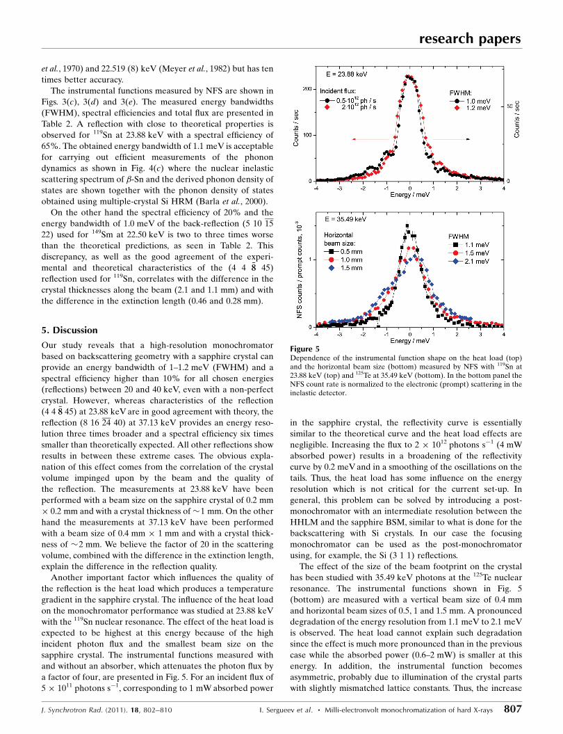
et al., 1970) and 22.519 (8) keV (Meyer et al., 1982) but has ten
times better accuracy.
The instrumental functions measured by NFS are shown in
Figs. 3(c), 3(d) and 3(e). The measured energy bandwidths
(FWHM), spectral efficiencies and total flux are presented in
Table 2. A reflection with close to theoretical properties is
observed for 119Sn at 23.88 keV with a spectral efficiency of
65%. The obtained energy bandwidth of 1.1 meV is acceptable
for carrying out efficient measurements of the phonon
dynamics as shown in Fig. 4(c) where the nuclear inelastic
scattering spectrum of �-Sn and the derived phonon density of
states are shown together with the phonon density of states
obtained using multiple-crystal Si HRM (Barla et al., 2000).
On the other hand the spectral efficiency of 20% and the
energy bandwidth of 1.0 meV of the back-reflection (5 10 15
22) used for 149Sm at 22.50 keV is two to three times worse
than the theoretical predictions, as seen in Table 2. This
discrepancy, as well as the good agreement of the experi-
mental and theoretical characteristics of the (4 4 �88 45)
reflection used for 119Sn, correlates with the difference in the
crystal thicknesses along the beam (2.1 and 1.1 mm) and with
the difference in the extinction length (0.46 and 0.28 mm).
5. Discussion
Our study reveals that a high-resolution monochromator
based on backscattering geometry with a sapphire crystal can
provide an energy bandwidth of 1–1.2 meV (FWHM) and a
spectral efficiency higher than 10% for all chosen energies
(reflections) between 20 and 40 keV, even with a non-perfect
crystal. However, whereas characteristics of the reflection
(4 4 �88 45) at 23.88 keV are in good agreement with theory, the
reflection (8 16 24 40) at 37.13 keV provides an energy reso-
lution three times broader and a spectral efficiency six times
smaller than theoretically expected. All other reflections show
results in between these extreme cases. The obvious expla-
nation of this effect comes from the correlation of the crystal
volume impinged upon by the beam and the quality of
the reflection. The measurements at 23.88 keV have been
performed with a beam size on the sapphire crystal of 0.2 mm
� 0.2 mm and with a crystal thickness of�1 mm. On the other
hand the measurements at 37.13 keV have been performed
with a beam size of 0.4 mm � 1 mm and with a crystal thick-
ness of �2 mm. We believe the factor of 20 in the scattering
volume, combined with the difference in the extinction length,
explain the difference in the reflection quality.
Another important factor which influences the quality of
the reflection is the heat load which produces a temperature
gradient in the sapphire crystal. The influence of the heat load
on the monochromator performance was studied at 23.88 keV
with the 119Sn nuclear resonance. The effect of the heat load is
expected to be highest at this energy because of the high
incident photon flux and the smallest beam size on the
sapphire crystal. The instrumental functions measured with
and without an absorber, which attenuates the photon flux by
a factor of four, are presented in Fig. 5. For an incident flux of
5 � 1011 photons s�1, corresponding to 1 mW absorbed power
in the sapphire crystal, the reflectivity curve is essentially
similar to the theoretical curve and the heat load effects are
negligible. Increasing the flux to 2 � 1012 photons s�1 (4 mW
absorbed power) results in a broadening of the reflectivity
curve by 0.2 meV and in a smoothing of the oscillations on the
tails. Thus, the heat load has some influence on the energy
resolution which is not critical for the current set-up. In
general, this problem can be solved by introducing a post-
monochromator with an intermediate resolution between the
HHLM and the sapphire BSM, similar to what is done for the
backscattering with Si crystals. In our case the focusing
monochromator can be used as the post-monochromator
using, for example, the Si (3 1 1) reflections.
The effect of the size of the beam footprint on the crystal
has been studied with 35.49 keV photons at the 125Te nuclear
resonance. The instrumental functions shown in Fig. 5
(bottom) are measured with a vertical beam size of 0.4 mm
and horizontal beam sizes of 0.5, 1 and 1.5 mm. A pronounced
degradation of the energy resolution from 1.1 meV to 2.1 meV
is observed. The heat load cannot explain such degradation
since the effect is much more pronounced than in the previous
case while the absorbed power (0.6–2 mW) is smaller at this
energy. In addition, the instrumental function becomes
asymmetric, probably due to illumination of the crystal parts
with slightly mismatched lattice constants. Thus, the increase
research papers
J. Synchrotron Rad. (2011). 18, 802–810 I. Sergueev et al. � Milli-electronvolt monochromatization of hard X-rays 807
Figure 5Dependence of the instrumental function shape on the heat load (top)and the horizontal beam size (bottom) measured by NFS with 119Sn at23.88 keV (top) and 125Te at 35.49 keV (bottom). In the bottom panel theNFS count rate is normalized to the electronic (prompt) scattering in theinelastic detector.

of the beam spot size significantly influences the performance
of the monochromator with non-ideal sapphire crystals.
For nuclear inelastic scattering experiments with the
sapphire BSM the measurement of the sapphire temperature
with mK precision is a crucial point. In the current set-up
the temperature sensor was installed in the nitrogen gas
measuring the temperature of the sapphire via convection
heat exchange between the gas and the crystal. Scanning the
temperature up and down with a typical rate of 1 K h�1, we
did not find any difference in the shape of the nuclear inelastic
spectra and the resolution functions, which shows that such a
rate is slow enough to achieve thermal stability in the cryostat.
On the other hand we found that the temperature where
the elastic nuclear resonance scattering has been observed
depends linearly on the incoming flux. The proportionality
coefficient was obtained for each reflection and was taken into
account in the energy calibration. The temperature difference
between the sensor and the scattering part of the sapphire
crystal was about 50 mK per 1 mW of absorbed power for all
energies.
The measured nuclear inelastic spectra from the samples
with isotopes of 119Sn, 149Sm and 151Eu were compared with
the spectra measured using multiple-crystal Si HRMs
(Chumakov et al., 1998; Barla et al., 2004) and showed coin-
cidences in the limit of 5% in the energy calibration. The
discrepancy is probably related to uncertainty in the thermal
expansion coefficients.
Besides the energy bandwidth (FWHM) of the resolution
function, the tails are important for the practical use of the
monochromator. Fig. 6 shows the instrumental functions
measured by NFS (on a logarithmic scale) for the tested
nuclear resonances as compared with the theoretical simula-
tion of the reflectivity. The tails follow the E�2 energy
dependence similar to the theoretical curves. However, they
are enhanced in magnitude, probably because of diffuse
scattering on defects in the sapphire. The main factor affecting
the magnitude of this enhancement in the tails is the crystal
thickness along the beam for the chosen reflections. A clear
difference is seen for the tail enhancement for �1 mm and
�2 mm crystal thicknesses in Fig. 6. This observation
emphasizes the importance of limiting the thickness of the
sapphire crystal used as monochromator.
The enhanced tails of the instrumental function may
complicate subtraction of the elastic peak from the inelastic
spectrum and, as a consequence, the correct information about
the low-energy region of the phonon density of states.
However, this problem is also related to the relative strength
of the scattering in the elastic and inelastic channels of the
nuclear resonant scattering which depends on the properties
(internal conversion coefficient, energy of emitted photons
and characteristic Lamb–Mossbauer factor) of the specific
isotope. In particular, for 119Sn, 125Te and 121Sb the signal in
the elastic channel is rather weak. The influence of the elastic
peak on the inelastic spectrum becomes negligible above 2–
3 meV and the phonon density of states is reliable starting
from this energy, as seen from Fig. 4. On the other hand, for149Sm the signal in the elastic channel is typically much higher
than in the inelastic one and the reliable region of the phonon
density of states starts above 5 meV.
As compared with the multiple-crystal Si HRM, the tails of
the sapphire BSM are enhanced even in the ideal theoretical
case, which is a consequence of the single versus multiple
Bragg reflections. As a result the features in the phonon DOS
are typically broadened. This problem can be solved by the
partial deconvolution of the phonon DOS with the instru-
mental function (Kohn & Chumakov, 2000).
6. Conclusion
In summary, we have demonstrated a versatile high-resolution
monochromator based on backscattering from sapphire crys-
tals working in the energy range 20–40 keV with an energy
bandwidth of �1 meV and with high throughput. The concept
of such a monochromator has been suggested by Shvyd’ko &
Gerdau (1999) but the implementation suffered from the
quality of available sapphire crystals (Shvyd’ko et al., 2001;
Wille et al., 2006, 2010; Imai et al., 2007). Improvement in the
monochromator performance was obtained by decreasing the
crystal volume impinged upon by the beam to a size
comparable with a characteristic concentration of defects in
the crystal. This allows one to choose a part of the crystal with
a minimal number of defects. The efficiency of the mono-
chromator is not the same for all energies and depends on the
chosen reflection. The feasibility of nuclear inelastic scattering
measurements with 121Sb, 125Te, 119Sn, 149Sm and 151Eu was
demonstrated. The partial phonon density of states of these
elements can thus be measured for compounds of current
scientific interest with high resolution and flux. For 125Te at
research papers
808 I. Sergueev et al. � Milli-electronvolt monochromatization of hard X-rays J. Synchrotron Rad. (2011). 18, 802–810
Figure 6Instrumental functions of the sapphire backscattering monochromatormeasured with NFS presented on a logarithmic scale. The data are scaledto have maximum spectral efficiency according to Table 2. (a)–(e) denotethe same isotopes as in Fig. 3. The thin grey lines (red online) showsimulation according to theory with the crystal thicknesses indicated inTable 2.
35.49 keVan energy resolution �E/E = 3� 10�8 was obtained
for the first time.
As compared with multiple-crystal Si HRMs the back-
scattering monochromator has some advantages. The
universality of this monochromator allows easy transforma-
tion from one energy to another which becomes important
with the increase of the number of Mossbauer isotopes studied
by nuclear inelastic scattering. Example applications include
phase-change materials (Wuttig & Yamada, 2007) composed
of Sb and Te or thermoelectric materials such as filled skut-
terudites (Snyder & Toberer, 2008) composed of rare earths
and Sb where phonon dynamics of each particular element is
of importance. Another advantage of such a monochromator
is the spectral efficiency, which is higher than the efficiency of
conventional (room-temperature) multiple-crystal Si HRMs.
The increased efficiency allows one to measure compounds
with a natural isotope abundance which are easily available.
The drawbacks of backscattering monochromators and, in
particular, this sapphire monochromator are related to the
enhanced tail of the instrumental function which complicates
the extraction of the tiny features of the phonon dynamics; in
particular, the precise estimation of the Debye energy for
some elements may be difficult. Another drawback compared
with the inline multiple-crystal Si HRM is the backscattering
geometry which puts some restrictions on the sample envir-
onment; herein, a distance of 5–7 mm between the direct and
backscattered beam was used.
Major improvement of the monochromator efficiency
would be achieved by improving the quality of sapphire
crystals. This would allow for a significant increase in the
efficiency and a decrease of the energy bandwidth. It also
would extend the applicability of this monochromator above
40 keV. On the other hand, even with the existing sapphire
crystals, improving the monochromator properties can be
achieved by improving the efficiency of the transversal
focusing and implementing this focusing above 30 keV.
Besides the Mossbauer isotopes studied in this work, one can
apply the monochromator to carry out nuclear inelastic scat-
tering measurements with 161Dy at 25.66 keV (Chumakov et
al., 2001), 201Hg at 26.27 keV (Ishikawa et al., 2005a), 189Os at
36.18 keV and 129Xe at 39.58 keV.
The authors are very grateful to A. I. Chumakov for support
during the experiment and for helpful discussions. RPH and
DB acknowledge support from the Helmholtz Gemeinschaft
Deutscher Forschungzentren for the Helmholtz University
Young Investigator Group Lattice Dynamic in Emerging
Functional Materials and from the DFG priority program
SPP1386 ‘Nanostructured Thermoelectrics’.
References
Antman, S., Pettersson, H., Zehlev, Z. & Adam, I. (1970). Z. Phys. A,237, 285–291.
Barla, A., Ruffer, R., Chumakov, A. I., Metge, J., Plessel, J. & Abd-Elmeguid, M. M. (2000). Phys. Rev. B, 61, 14881–14884.
Barla, A., Sanchez, J. P., Haga, Y., Lapertot, G., Doyle, B. P., Leupold,O., Ruffer, R., Abd-Elmeguid, M. M., Lengsdorf, R. & Flouquet, J.(2004). Phys. Rev. Lett. 92, 066401.
Baron, A. Q. R., Kishimoto, S., Morse, J. & Rigal, J.-M. (2006). J.Synchrotron Rad. 13, 131–142.
Baron, A. Q. R., Tanaka, Y., Ishikawa, D., Miwa, D., Yabashi, M. &Ishikawa, T. (2001). J. Synchrotron Rad. 8, 1127–1130.
Burghartz, S. & Schulz, B. (1994). J. Nucl. Mater. 212–215, 1065–1068.Burkel, E. (2000). Rep. Prog. Phys. 63, 171–232.Chen, W. M., McNally, P. J., Shvydko, Y. V., Tuomi, T., Lerche, M.,
Danilewsky, A. N., Kanatharana, J., Lowney, D., O’Hare, M.,Knuuttila, L., Riikonen, J. & Rantamaki, R. (2001). Phys. StatusSolidi A, 186, 365–371.
Chumakov, A. I., Barla, A., Ruffer, R., Metge, J., Grunsteudel, H. F.,Grunsteudel, H., Plessel, J., Winkelmann, H. & Abd-Elmeguid,M. M. (1998). Phys. Rev. B, 58, 254–257.
Chumakov, A. I., Ruffer, R., Leupold, O., Barla, A., Thiess, H., Gil,J. M., Alberto, H. V., Vilao, R. C., Ayres de Campos, N., Kohn, V. G.,Gerken, M. & Lucht, M. (2001). Phys. Rev. B, 63, 172301.
Dorner, B., Burkel, E. & Peisl, J. (1986). Nucl. Instrum. Methods Phys.Res. A, 246, 450–451.
Freund, A. K., Comin, F., Hazemann, J. L., Hustache, R., Jenninger,B., Lieb, K. & Pierre, M. (1998). Crystal and Multilayer Optics,edited by A. T. Macrander, A. K. Freund, T. Ishikawa and D. M.Mills, Vol. 3448, pp. 144–155.
Gerdau, E. & de Waard, H. (1999/2000). Nuclear Resonant Scatteringof Synchrotron Radiation. Baltzer Science Publishers.
Graeff, W. & Materlik, G. (1982). Nucl. Instrum. Methods Phys. Res.195, 97–103.
Imai, Y., Yoda, Y., Kitao, S., Masuda, R., Higashitaniguchi, S., Inaba,C. & Seto, M. (2007). Proc. SPIE, 6705, 670512.
Ishikawa, D., Baron, A. Q. R. & Ishikawa, T. (2005a). Phys. Rev. B,72, 140301.
Ishikawa, T., Tamasaku, K. & Yabashi, M. (2005b). Nucl. Instrum.Methods Phys. Res. A, 547, 42–49.
Kikuta, S. (1994). Hyperfine Interact. 90, 335–349.Kohn, V. & Chumakov, A. (2000). Hyperfine Interact. 125, 205–221.Koyama, I., Yoda, Y., Zhang, X., Ando, M. & Kikuta, S. (1996). Jpn. J.
Appl. Phys. 35, 6297–6300.Leupold, O., Pollmann, J., Gerdau, E., Ruter, H. D., Faigel, G., Tegze,
M., Bortel, G., Ruffer, R., Chumakov, A. I. & Baron, A. Q. R.(1996). Europhys. Lett. 35, 671–675.
Linden, P. van der, Wille, H.-C. & Shvyd’ko, Y. V. (2007). AIP Conf.Proc. 879, 915–917.
Lucht, M., Lerche, M., Wille, H.-C., Shvyd’ko, Y. V., Ruter, H. D.,Gerdau, E. & Becker, P. (2003). J. Appl. Cryst. 36, 1075–1081.
Meyer, R. A., Meadows, J. W. T. & Macias, E. S. (1982). J. Phys. G, 8,1413–1429.
Ruffer, R. & Chumakov, A. I. (1996). Hyperfine Interact. 97–98, 589–604.
Salgado, J. (1974). Technical Report Kernforschungsanlage KarlsruheKFK 1954. Karlsruhe, Germany.
Schmid, F., Khattak, C. P. & Felt, D. M. (1994). Am. Ceram. Soc. Bull.73, 39–44.
Schober, H. R. & Dederichs, P. H. (1981). Landolt-Bornstein, Vol. III/13a, pp. 131–132. Berlin: Springer.
Shvyd’ko, Y. (2004). X-ray Optics: High-Energy-Resolution Applica-tions, Vol. 98, Springer Series in Optical Sciences. Berlin: Springer.
Shvyd’ko, Y. V. & Gerdau, E. (1999). Hyperfine Interact. 123, 741–776.
Shvyd’ko, Y. V., Gerken, M., Franz, H., Lucht, M. & Gerdau, E.(2001). Europhys. Lett. 56, 309–315.
Shvyd’ko, Y. V., Lerche, M., Jaschke, J., Lucht, M., Gerdau, E.,Gerken, M., Ruter, H. D., Wille, H.-C., Becker, P., Alp, E. E.,Sturhahn, W., Sutter, J. & Toellner, T. S. (2000). Phys. Rev. Lett. 85,495–498.
Shvyd’ko, Y. V., Lucht, M., Gerdau, E., Lerche, M., Alp, E. E.,Sturhahn, W., Sutter, J. & Toellner, T. S. (2002). J. Synchrotron Rad.9, 17–23.
Snigirev, A., Kohn, V., Snigireva, I. & Lengeler, B. (1996). Nature(London), 384, 49–51.
research papers
J. Synchrotron Rad. (2011). 18, 802–810 I. Sergueev et al. � Milli-electronvolt monochromatization of hard X-rays 809

Snyder, G. J. & Toberer, E. S. (2008). Nat. Mater. 7, 105.Toellner, T. S. (2000). Hyperfine Interact. 125, 3–28.Toellner, T. S., Alatas, A., Said, A., Shu, D., Sturhahn, W. & Zhao, J.
(2006a). J. Synchrotron Rad. 13, 211–215.Toellner, T. S., Hu, M. Y., Bortel, G., Sturhahn, W. & Shu, D. (2006b).
Nucl. Instrum. Methods Phys. Res. A, 557, 670–675.Tsutsui, S., Yoda, Y. & Kobayashi, H. (2007). J. Phys. Soc. Jpn, 76,
065003.White, G. K. & Roberts, R. B. (1983). High Temp. High Pres., 15, 321–
328.
Wille, H.-C., Hermann, R. P., Sergueev, I., Leupold, O., van derLinden, P., Sales, B. C., Grandjean, F., Long, G. J., Ruffer, R. &Shvyd’ko, Y. V. (2007). Phys. Rev. B, 76, 140301.
Wille, H.-C., Hermann, R. P., Sergueev, I., Pelzer, U., Mochel, A.,Claudio, T., Persson, J., Ruffer, R., Said, A. & Shvyd’ko, Y. V.(2010). Europhys. Lett. 91, 62001.
Wille, H.-C., Shvyd’ko, Y. V., Alp, E. E., Ruter, H. D., Leupold, O.,Sergueev, I., Ruffer, R., Barla, A. & Sanchez, J. P. (2006). Europhys.Lett. 74, 170–176.
Wuttig, M. & Yamada, N. (2007). Nat. Mater. 6, 824–832.
research papers
810 I. Sergueev et al. � Milli-electronvolt monochromatization of hard X-rays J. Synchrotron Rad. (2011). 18, 802–810