Midline versus Pfannenstiel Incision scars in repeat ...
30
1. INTRODUCTION 1.1. Types of incision It is a commonly held belief that repeat caesarean section through a low vertical scar provides easier access and fewer complications than an operation through a previous Pfannenstiel incision. The two most frequently used abdominal skin incisions for caesarean section are a subumbilical midline (up and down/vertical, Figure 1.1) and Pfannenstiel (low transverse/suprapubic, Figure 1.2). The description of these techniques is well described in most good surgical texts such as Te Linde’s Operative Gynaecology. 1 and well described in a recent Cochrane systematic review by Mathai and Hofmeyr. 2 The use of either incision is surgeon dependent and motivated by a variety of indications, situations and often the surgeon’s level of expertise. Traditionally caesarean sections were performed by a midline vertical skin incision from pubic symphysis to umbilicus. The rectus muscle and peritoneum are then incised in the relatively avascular midline. Advantages are suggested to be speed, ability to extend the incision upwards and it being the incision of choice if local anaesthesia is to be used. In 1900 Pfannenstiel described his traditional method of low transverse incision for caesarean section. The skin incision is a low transverse type, gently upward curving, following the natural skin fold and located two finger breadths above the pubic symphysis. The incision is then continued with sharp scalpel dissection through subcutaneous tissue to the rectus sheath, which is then incised either side of the midline. After exposing the rectus sheath, the incision is extended transversely with heavy curved Mayo scissors. The upper and lower sheath margins are then grasped with a Kocher toothed clamp, to elevate and provide tension on the tissue. 1 1
Transcript of Midline versus Pfannenstiel Incision scars in repeat ...

1. INTRODUCTION
1.1. Types of incision
It is a commonly held belief that repeat caesarean section through a low vertical scar provides
easier access and fewer complications than an operation through a previous Pfannenstiel
incision. The two most frequently used abdominal skin incisions for caesarean section are a
subumbilical midline (up and down/vertical, Figure 1.1) and Pfannenstiel (low
transverse/suprapubic, Figure 1.2). The description of these techniques is well described in
most good surgical texts such as Te Linde’s Operative Gynaecology.1 and well described in a
recent Cochrane systematic review by Mathai and Hofmeyr. 2 The use of either incision is
surgeon dependent and motivated by a variety of indications, situations and often the surgeon’s
level of expertise. Traditionally caesarean sections were performed by a midline vertical skin
incision from pubic symphysis to umbilicus. The rectus muscle and peritoneum are then
incised in the relatively avascular midline. Advantages are suggested to be speed, ability to
extend the incision upwards and it being the incision of choice if local anaesthesia is to be
used.
In 1900 Pfannenstiel described his traditional method of low transverse incision for caesarean
section. The skin incision is a low transverse type, gently upward curving, following the
natural skin fold and located two finger breadths above the pubic symphysis. The incision is
then continued with sharp scalpel dissection through subcutaneous tissue to the rectus sheath,
which is then incised either side of the midline. After exposing the rectus sheath, the incision is
extended transversely with heavy curved Mayo scissors. The upper and lower sheath margins
are then grasped with a Kocher toothed clamp, to elevate and provide tension on the tissue.
1
1

The underlying muscle is then separated by blunt or sharp dissection from the fascia, ensuring
haemostasis of the perforating vessels. The rectus muscles are separated bluntly, before the
peritoneum is entered sharply in the midline exposing the uterus. In the event of limited
exposure or greater access being required, the Maylard or Cherney muscle releasing
modifications may be used. The Maylard technique is the dividing of one or both rectus
abdominus muscle bodies, taking care for the underlying artery. The Cherney procedure is the
release of the tendinous attachment of the muscle as low as possible on the os pubis.
Another incision type has been found to be superior to both of these2. The Misgav-Ladach
method for caesarean section, as described by Holmgren, Sjöholm and Stark,3 is based on the
Joel-Cohen incision originally introduced for hysterectomy. This is a straight transverse
incision somewhat higher than the Pfannenstiel incision. This method is restrictive in the use
of sharp instruments, preferring manual manipulation. There is a quicker recovery period, and
less use of post-operative antibiotics and analgesics. There is also a shorter anaesthetic and
working time for the operative team. As this incision type has not been accepted into daily
practice and little data on repeated caesarean section is available, it will not be included in this
discussion.
2
2

Figure 1 SUMI: Subumbilical Midline IncisionRock JA, Jones HW. Te Lindes’s Operative Gynaecology 9th Ed. Philadelphia: Lippincott Williams and Wilkins,2003. 1
3
3

Figure 2 Pfannenstiel Incision Rock JA, Jones HW. Te Lindes’s Operative Gynaecology 9th Ed. Philadelphia: Lippincott Williams and Wilkins,2003. 1
4
4

1.2. Current guidelines and preferences
In a study by Tully et al 4 in the United Kingdom, a postal questionnaire was sent to all
Members and Fellows of the Royal College of Obstetricians and Gynaecologists (RCOG)
resident in the United Kingdom. Information was requested about surgical techniques for
elective and emergency caesarean sections. About 78.7% of the members and fellows
responded to the questionnaire. A range of incisional techniques was used. There was wide
variation in surgical techniques, but Pfannenstiel abdominal entry was used by more than 80%
of obstetricians for elective caesarean section.
In the United Kingdom, the National Institute for Clinical Excellence (NICE).5 have noted that
vertical incisions for caesarean section are uncommon, occurring in less than 1% of operations
and have largely been replaced by transverse incisions. The guidelines suggest that caesarean
section should be performed using a transverse incision because this is associated with less
postoperative pain and an improved cosmetic effect compared with a midline incision. They
suggest that the transverse incision of choice should use the advantages mentioned of the Joel-
Cohen incision (straight incision, 3 cm above the symphysis pubis, bluntly opening subsequent
tissue layers and if necessary extended with scissors and not a knife). This is associated with
shorter operating times and reduced postoperative febrile morbidity. Concern is only
mentioned about research being needed to assess the effect of the various surgical techniques
on future surgery such as repeat caesarean section.
While the global trend is towards transverse incisions, vertical incisions are still commonly
performed in developing countries. A popular South African manual for district hospitals,
authored by Breen,6 recommends vertical incision, ‘especially for repeat caesarean section’.
5
5

From a South African point of view, with a large number of deliveries performed in public
sector hospitals, a large motivator for an initial midline incision is that it may be easier to
perform and teach. Also, faster entry is gained to the abdominal cavity and better access is
available, thus making this technique more acceptable for trainees, junior staff and rural
practitioners. In the National Guidelines for Maternity Care in South Africa7, a handbook
published by the Department of Health to assist precisely this group of doctors, several
indications for midline incisions are given. These include increased risk of intraoperative
haemorrhage such as in a case of antepartum haemorrhage, severe pre-eclampsia or expected
difficult delivery, and prolonged labour or rupture of membranes which could lead to an
increased risk of postoperative infection.
South African patient and clinician preferences were recently evaluated by Rwakyendela and
Buchmann.8 In a descriptive study undertaken in three state hospitals in Johannesburg, 400
patients were interviewed in a cohort in which 38% of women had vertical cesarean section
incisions. The authors found that a majority of women would have chosen transverse incisions
if given the choice, thus favouring the cosmetically more acceptable Pfannenstiel type.
Clinicians who were interviewed followed the trend towards the Pfannenstiel incision for
elective cesarean section delivery (96%) and marginally preferred a midline approach for
emergency cesarean section (51%).
1.3. Complications associated with incision types
Local discussion about abdominal skin incisions for caesarean section and their complications
is frequent among obstetricians in the Johannesburg public and academic hospitals.
6
6

Pfannenstiel incisions are sometimes avoided as they supposedly produce more fibrosis, more
adhesions and a higher bladder perforation risk in subsequent caesarean sections. The corollary
to this is the perception that the vertical incision may be weaker or prone to dehiscence. The
comparison of either type was last evaluated by Haeri 9 in Cape Town in 1976, in a
randomized study of 100 transverse and 100 vertical incisions. The author reported an 8
minute longer duration in operating time in transverse incisions, with an increase in midline
wound breakdown and sepsis if the cases were performed as emergencies. This may be
confounded by the lack of prophylactic antibiotic administration intra-operatively in that era of
surgery. Patients were not evaluated on previous caesarean section scarring or followed into
their subsequent pregnancies.
Gross 10 reviewed the benefits of a low transverse incision in obese women, finding that
Pfannenstiel incisions were associated with less post operative pain and earlier ambulation.
This not only improved short-term well-being, but avoids atelectasis and thromboembolism. A
more secure closure, less adipose tissue to incise and a better cosmetic effect were also noted.
The adverse effects included more wound infection and restriction of access to the infant and
the upper abdomen.
Wall et al 11 in New York, USA, reviewed a large series of hospital records (n=239) of obese
women with a body mass index (BMI) of greater than 35, having had a primary caesarean
delivery. Their findings confirm the commonly held notions and conventional teaching that
severely obese parturients have a high incidence of wound complications. The data indicated
that a vertical incision was associated with a higher rate of wound complications than a
7
7

transverse incision. These complications were defined as seroma, haematoma, cellulitis and
purulent wound discharge that led to wound opening.
These findings are explained by a study evaluating the risk factors in abdominal wall sepsis.
Vermillion et al 12 in South Carolina, USA prospectively measured the maximum vertical
depths of subcutaneous incisions of 140 women who had deliveries by caesarean section. The
surgical technique for closure was standardized and drains were not used. Thickness of
subcutaneous tissue appeared to be the only significant risk factor associated with abdominal
wound infection after caesarean delivery.
A Nigerian surgical department study on hernia formation by Adesunkanmi and Faleyimu 13
showed that the incidence of incisional hernia was influenced by the type of incision. Of the
701 patients who had a caesarean section in the study period, 22 (3.1%) developed an
incisional hernia, all of whom had midline incisions. Those who developed incisional hernias
also suffered other postoperative complications and had longer hospital stays. The age and
parity of the patients, and the indications for caesarean section, did not influence the incidence
of incisional hernia in that study.
Hendrix et al 14 attempted to determine whether there was a difference in the frequency of
fascial dehiscence between midline vertical lower abdominal and Pfannenstiel incisions among
women undergoing obstetric and gynaecologic operations. In Detroit, USA, a case control
study on 48 cases of fascial dehiscence in 6 years that complicated 17995 major operations
(8950 cesarean section deliveries and 9405 gynaecological procedures) was undertaken. The
8
8

authors could not provide data to support the belief that Pfannenstiel incisions are stronger
than lower abdominal vertical incisions and thereby reduce the risk for fascial dehiscence.
Wound infection was the most important risk factor for fascial dehiscence among women who
underwent major obstetric and gynaecological operations.
The NICE guidelines 5 provide a different view on the problem of midline weakness, relating it
more to closure technique than type. They quote a meta-analysis of randomised control trials
(RCTs) comparing mass versus layered closure of midline incisions in general surgical
patients. The mass closure technique is where all the layers of the abdominal wall excluding
skin are treated as one and sutured in a running continuous fashion as opposed to suturing each
layer separately, specifically the peritoneum and musculoaponeurotic layers in the layered
closure (a practice currently rarely performed). This meta-analysis found that incisional
hernias and dehiscence were less common with mass closures.
Suture material and techniques in closure at caesarean section are poorly understood and the
current Cochrane review by Alderdice et al15 states that “There is currently no conclusive
evidence about how the skin should be closed after caesarean section. Questions regarding the
best techniques and materials for closure of caesarean section and the associated incidence of
infection, local reaction, analgesia requirement and long term cosmetic appearance remain
unanswered.”
Abdominal wall closure was evaluated in a Cochrane review by Anderson and Gates16. They
were unable to state if any abdominal wall or rectus sheath closure was superior to another.
9
9

The subcutaneous fat may be left to heal, but closing the layer may reduce the risk of
haematoma and seroma formation.
The aspect of complications relating to the delivery being an emergency or an elective
caesarean section, was investigated by Allen et al17 in a Canadian population based study of
over eighteen thousand patients. They showed that maternal morbidity was increased in
caesarean sections in labouring women (emergency) as opposed to elective caesarean sections
in non-labouring patients. The results concur with a five year Finnish birth and hospital
discharge registry study of 110 717 patients by Pallasmaa et al18. They found an increased
incidence in severe maternal morbidity (deep venous thromboembolism, amniotic fluid
embolism, major puerperal infection, severe haemorrhage, uterine rupture and inversion and
events requiring operative intervention after delivery) in emergency compared to elective
caesarean section.
1.4. Intra-abdominal adhesions after caesarean section
Adhesion formation with either incision type or peritoneal closure remains a contentious issue.
Current consensus is towards non-closure of the visceral and parietal peritoneum as seen in
various review articles, 19-21 of RCTs pertaining to facets of caesarean section surgical
technique. The NICE guidelines5 recommend the same. A small Austrian retrospective study
by Nather et al 20 reviewed 30 patients who had their primary and repeat caesarean sections in
the same institution. They reviewed the difference between initially closed and unclosed
peritoneum evaluated at repeat caesarean section. Closure or non-closure of the peritoneum at
primary caesarean section did not affect the development of intra-abdominal adhesions at
10
10

repeat operation. To confound these broader reviews and guidelines, a recent record review by
Hamel21, of 403 repeat caesarean sections analysed the initial and repeat caesarean section
notes of 62 patients that were available and found that closing the peritoneum at primary
caesarean section significantly decreased extensive adhesions at repeat caesarean section.
There was also a significant difference in the amount of adhesions between closing the rectus
muscle and non-closure. Speculating that adhesions result from an open peritoneal cavity
exposed to the subfascial space, the authors surmise that the decrease in formation of
adhesions following closing the rectus muscle may be due to the parietal peritoneal apposition,
because of its proximity to the rectus muscle. Although only a small study, it goes to show that
all is not clear in repeat surgery and more research needs to be done.
With increased adhesion formation comes an increased risk of hollow viscus perforation at
subsequent surgery, as described by Phipps et al22 in a case control study undertaken in
Washington, USA, which analysed the occurrence of bladder perforations as the primary end
point. In 14757 caesarean section deliveries they had 42 perforations and showed that patients
with a previous scar had an almost fourfold increased risk for bladder perforation.
A very recent article by Salim et al 23 in Haifa, Israel has brought a new perspective on
abdominal scarring and adhesion formation. Although not comparing incision types and
scarring, they attempted to use external scar characteristics to predict the incidence and
severity of intra-abdominal adhesion formation. The authors enrolled 107 women with repeat
caesarean sections into this trial, dividing them into raised, flat and depressed scars. Of these
57% were found to have had no adhesions, 16% had filmy adhesions, and 27% had dense
11
11
adhesions. A depressed abdominal scar from a previous caesarean delivery thus correlated well
with the incidence and severity of intra-abdominal adhesions when comparing external
caesarean section scar appearance.
1.5. Incision type and repeat caesarean section
A thorough search of the medical literature, and consultation with colleagues, did not reveal
any research that has addressed repeat caesarean section in terms of incision type. Do the
advantages of either approach at primary caesarean section apply equally at repeat operation?
For example, if studies could show that a vertical primary incision was followed by easier and
less complicated surgery at repeat operation than transverse incision, this would provide some
evidence for considering primary vertical incision in certain circumstances. A major obstetric
reference work - ‘High Risk Pregnancy’24- granted this problem a single line saying that
evidence is lacking at present. One local text book simply advises that a repeat operation
should be done through the same incision as the previous operation.25
2. MATERIALS AND METHODS
2.1 Problem Statement and Objectives
There is a need for research to determine if, in a patient with a previous caesarean section, the
vertical or Pfannenstiel incision is associated with more complications. This would help
12
12

surgeons to either provide patients with a cosmetically and structurally sound wound or real
evidence as to why they need to tolerate a more unsightly scar and risk of herniation.
The objectives of this study were, therefore:
- To establish if there is a difference in time taken from incision to delivery of the baby
between previous subumbilical midline and previous Pfannenstiel incisions.
-To establish if there is a difference in overall operation time between previous subumbilical
midline and previous Pfannenstiel incisions.
-To establish if there is a difference in adhesions found on entry between previous
subumbilical midline and previous Pfannenstiel incisions.
- To establish if intra-operative complications are affected by incision type between previous
subumbilical midline and previous Pfannenstiel incisions.
2.2. Methods
This was a cross-sectional analytic study comparing surgical findings in women at repeat
caesarean section. The study population was all women having repeat caesarean sections at
Chris Hani Baragwanath Maternity Hospital and Johannesburg Hospital, both teaching and
13
13

tertiary referral institutions attached to the University of the Witwatersrand. These hospitals
provide registrar training. Chris Hani Baragwanath Hospital delivers approximately 20 000
women annually, with a caesarean section rate of 27%, and Johannesburg Hospital delivers
about 6 000 women annually, with a 40% caesarean section rate.
Data collection was by retrospective review of patients’ clinical notes taken during routine
repeat caesarean section. It was hoped that the operation notes in the case-files would provide
adequate data to provide the necessary information to answer the research questions as stated
above. Note-keeping was however erratic, with large inconsistencies in level of detail
provided. This has been noted in the literature as a problem by Nicopoullos et al,26 whose audit
showed important omissions in a high percentage of operative delivery notes, with less than
80% of case notes documenting incision time and type, surgical findings of note, type of
uterine incision, the presenting part and complete sutures used. There was also considerable
confusion about the use of the terminology for level of urgency of non-elective caesarean
sections.
It was therefore decided that only the author's own operations would be used for this study, as
he was in the habit of making detailed and consistent notes on surgical findings, especially the
presence and severity of adhesions. The sample was thus the retrospective review of a
consecutive series of the author’s own caesarean sections for women with previous operations.
Inclusion criteria were any patients with a previous subumbilical midline or Pfannenstiel
incision presenting for an emergency or elective caesarean section at the two hospitals.
14
14

In personal communication with Dr David Wiseman of the International Adhesions Society, it
was clarified that there is no true grading for adhesions. He did however give a coarse grading
scheme, Grade 1 being filmy adhesions easily removed with blunt dissection, Grade 2 being
firmer adhesions readily responding to scissor dissection, and Grade 3 requiring aggressive
sharp dissection. With this grading in mind the author interpreted the notes, with coding of
severe adhesions corresponding with a grade 3 adhesion. Comparing SUMI and Pfannenstiel
incisions, there seemed to be no obvious difference in these types of adhesions encountered
inside the abdomen.
All patients in the study had their repeat incision performed in the same way as the initial scar.
There were no specific exclusion criteria as it was a retrospective record review. Data entered
from the files included age, parity, indication for previous caesarean section, indication for
current caesarean section, clinical and surgical findings, including adhesions, and
complications. Start time, baby delivery time, and operation end time were reliably recorded
both in theatre registers and anaesthetic charts. Where there was disagreement in time entries,
the anaesthetic chart was accepted as the correct time. The study was undertaken during
registrar rotations between the hospitals during 2003 and 2004.
2.3. Data Analysis
Data captured from these records was entered on a data sheet (Appendix 1) to allow data
collection from records at irregular times, and the variables given numerical values, to aid
computer capturing. No patient identifying data was included in the capturing. Data was
logged and processed with the statistical program Epi Info 6, comparing means and
15
15
frequencies. Comparisons of continuous variables by Student’s T-test, or the Mann-Whitney
test where applicable, were done. Comparisons of frequencies by Chi-squared test or Fischer’s
exact test were done were applicable. A P-value of less than 0.05 was accepted as indicating
statistical significance.
2.4. Ethics
An application for ethical clearance for this study was made to the Committee for Research on
Human Subjects (Medical) of the University of the Witwatersrand and was granted 17/11/03
no R14/49 Haacke (Appendix 2).The protocol was also presented to the post graduate
committee for use as a research report for the M.Med (O+G) and accepted. Permission to use
patient records was granted by the hospital clinical executives and the obstetrics clinical heads
of department, of Chris Hani Baragwanath and Johannesburg General Hospital.
2.5. Funding
The cost of the research was borne by the author. No external funding was offered or available.
3. RESULTS
The records of 121 patients were reviewed. Comparison was made of variables between the
subumbilical midline incision (SUMI) and Pfannenstiel group. The distribution between the
two groups was similar, with 64 subumbilical (53%) and 57 Pfannenstiel (47%) incisions. Of
the cases reviewed, 64 were operated on at Chris Hani Baragwanath Maternity Hospital (53%)
and 57 at Johannesburg Hospital (47%).
16
16

Table 3.1 Obstetric and demographic data of women undergoing repeat caesarean section comparing Subumbilical midline to Pfannenstiel incision
*Medians and ranges indicated
As seen in Table 3.1, the difference in age of the patients was statistically significant, with the
SUMI group having a mean age of 30.6 (± 5.1), compared to 28.6 (± 5.6) in the Pfannenstiel
group, with a students T test giving a P value of 0.039. Comparing the time of day of the
surgery, 83 (69%) of patients had their operations between 07h00 and 19h00 and 38 (31%)
between 19h00 and 07h00, with no statistical significance as regards incision type. Parity
ranged from one to five in both groups.
Variable
Subumbilical Midline Incision
Pfannenstiel Incision
Significance
N = 64 N = 57 P Value
Age (Mean±SD) 30.6 years (±5.1) 28.6 years (±5.7) 0.039
Parity* 1 (1-5) 1 (1-5) 0.70
One previous caesarean
49 (77%) 47 (82%) 0.42
Gestational age* 39 (27-42) 39 (30-42) 0.96
Emergencycaesarean section
53 (83%) 51 (89%) 0.29
Sterilisation done 20 (31%) 18 (32%) 0.97
17
17

The number of women having one, as opposed to two or more, previous caesareans, did not
differ significantly between the previous SUMI and Pfannenstiel groups (77% v. 82%).
Gestational age, compared by incision type, showed a very similar spread with a median
gestation of 39 weeks for SUMI (range 27-42) and 39 weeks for Pfannenstiel (range 30-42).
Emergency caesarean section rates were similar in the SUMI and Pfannenstiel groups (83% v.
85%).Bilateral tubal ligation was performed similarly evenly between the groups (31% for
SUMI and 32% for Pfannenstiel incisions)
Table 3.2 Intraoperative findings at repeat caesarean section, comparing Subumbilical midline to Pfannenstiel incisions.
VariableSubumbilical
Midline IncisionPfannenstiel
IncisionSignificance
N = 64 N = 57 P Value
Time from incision to delivery (Minutes)*
4.0 (2-16) 5.5 (3-9) <0.0001
Time from incision to end of surgery (Minutes)*
32.4 (27-150) 34.5 (23-105) 0.13
Old scar excised 55 (86%) 17 (30%) <0.0001
18
18

Severe adhesions in anterior abdominal wall
23 (36%) 29 (51%) 0.10
Severe adhesions to bladder
9 (14%) 9 (16%) 0.79
Severe adhesions to Corpus Uteri
5 (8%) 9 (16%) 0.17
*Medians and ranges indicated
The median time taken from start of operation to delivery of the infant showed a significant
advantage in the SUMI group as seen in Table 3.2, SUMI taking a median time of four
minutes (Range 2-16) compared to five and a half minutes (Range 3-9) for the Pfannenstiel
group (P<0.0001).When total operating time was taken into account, comparing both groups,
the median times were: SUMI 32.4 minutes (range 27-150) and Pfannenstiel 34.6 minutes
(23-105), with a P value of 0.13.
Excision of the previous skin scar was performed in 86% of previous SUMI and in 30% of
previous Pfannenstiel incisions (P<0.0001). Scar excision was associated with a shorter
incision to delivery time (median 4.4, range 2.7-11.1) than incision through the scar (median
5.4, range 2.0-16.0; P=0.013 [Mann-Whitney test]).
Severe adhesions on the anterior abdominal wall were less frequent with SUMIs (36%) than
with Pfannenstiel incisions (51%; P=0.10). Severe bladder adhesions occurred with similar
frequency in the two groups (14% and 16% with SUMI and Pfannenstiel incisions
respectively). Severe adhesions to the corpus uteri were less common in SUMI (8%) than in
previous Pfannenstiel incisions (16%; P = 0.17). All cases in both groups had a previous
transverse lower segment uterine incision.
19
19

Table 3.3 shows the breakdown of indications for the present repeat caesarean section
compared by incision type and Table 3.4 shows indications for the previous caesarean
sections. These are grouped and compared by incision type.
Table 3.3 Indications for caesarean section according to incision type.
Indication
SubumbilicalMidline Incision
Pfannenstiel
N=64 N=57
1: No Progress 19 21
2: Fetal Distress 17 12
3: Two Previous Caesarean sections
11 9
4: Breech 3 3
5: PIH 3 5
20
20
6: Other 11 7
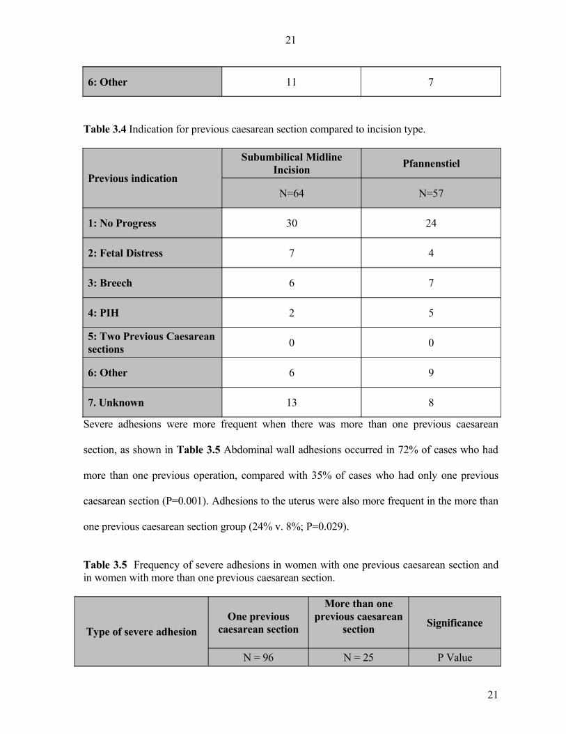
Table 3.4 Indication for previous caesarean section compared to incision type.
Previous indication
Subumbilical Midline Incision
Pfannenstiel
N=64 N=57
1: No Progress 30 24
2: Fetal Distress 7 4
3: Breech 6 7
4: PIH 2 5
5: Two Previous Caesarean sections
0 0
6: Other 6 9
7. Unknown 13 8
Severe adhesions were more frequent when there was more than one previous caesarean
section, as shown in Table 3.5 Abdominal wall adhesions occurred in 72% of cases who had
more than one previous operation, compared with 35% of cases who had only one previous
caesarean section (P=0.001). Adhesions to the uterus were also more frequent in the more than
one previous caesarean section group (24% v. 8%; P=0.029).
Table 3.5 Frequency of severe adhesions in women with one previous caesarean section and in women with more than one previous caesarean section.
Type of severe adhesionOne previous
caesarean section
More than one previous caesarean
sectionSignificance
N = 96 N = 25 P Value
21
21

Abdominal wall 34 (35%) 18 (72%) 0.001
Bladder 12 (13%) 6 (24%) 0.15
Uterus 8 (8%) 6 (24%) 0.029
Some serious surgical complications were encountered. In a woman with a previous
Pfannenstiel incision, the bladder was found to be morbidly adherent to the uterus and was torn
open during delivery of the fetal head. Another patient with a Pfannenstiel incision had severe
omental adhesions to the uterine scar and uterus. Rather more complications were noted in
women with previous subumbilical incisions. In one woman, the bladder was found to be
encased in anterior abdominal wall scar tissue and was accidentally opened during dissection.
In another patient the full anterior surface of the uterus was found to be fused to the entire
length of the abdominal wall scar. Another woman with a SUMI had the uterus encased in
bowel, with ileocolic adhesions covering the right fundus, and the sigmoid colon adherent
from the posterior uterine surface up to the left fundus and down the left broad ligament. One
patient with a SUMI also had the uterus rotated 90 degrees due to severe pillar adhesions
(Thick columnar scar tissue bridges between uterus and anterior abdominal wall). There were
also three patients with severe omental adhesions to the uterus and anterior abdominal wall.
22
22

4. DISCUSSION
The results presented give a good set of data that goes some way to answer the research
questions. The two groups seem evenly matched in potentially confounding variables such as
the place of surgery, time of surgery, parity, number of previous caesarean sections,
emergency or not, and sterilization done. The age difference of the patients in the two groups,
with older patients more likely to have had a SUMI, may possibly be attributed to the fact that
there has been a steady change in practice, with younger patients having more Pfannenstiel
incisions. This may suggest that presently a patient is more likely to have a Pfannenstiel than
in earlier years. Delivery time taken was shown to be significantly shorter in the SUMI group.
This, combined with a higher incidence of scar excision in the subumbilical midline incision,
23
23

should go to prove that the delivery time may actually be even faster if an unsightly scar were
not removed in an emergency. This is based on the fact that although unrecorded so far, it
technically takes longer to excise a large scar than to make a single incision through it. On
reviewing the notes it was shown that there were no fixed criteria for excision of old scars, but
these were usually noted as large or unsightly.
Total operating time seemed to be no different, which is difficult to explain as in the SUMI
group the access is faster and visualization supposedly better. It is possible that closure in the
Pfannenstiel operation is easier and more rapid, in view of less tension on the rectus sheath and
abdominal wall. The trend to greater frequency of severe adhesions in the anterior abdominal
wall in the Pfannenstiel group supports the assumption that the scar is more fibrotic and
difficult to dissect. This also might help to explain the longer entry or delivery time in the
same group. An interesting finding is that scar excision was associated with a reduced incision
to delivery time of a minute. This may be due to improved surgical exposure.
These indications for the current caesarean section and for the previous operations, as shown in
Tables 3.3 and 3.4, indicate that there was an even spread between the two groups. These also
represent a similar profile to that found in daily practice and thus prove to be a representative
population. Not surprisingly, severe adhesions were more frequent in women with more than
one previous operation.
Controlling the variables was not possible as the data was gleaned from records and the data
was only as good as the clinicians’ entries. One can only rely on the staff integrity at operation
24
24

and the knowledge that they would be as accurate as possible for medicolegal reasons. As this
was not a randomized control trial, bias will be present and cannot be avoided. It is recognized
that the type of initial caesarean incision scar made is not random. Certain indications, clinical
or logistical, may have made a certain incision a better option and also alter scar adhesions
found at subsequent operation. An example of this might be a patient having had a caesarean
section for chorioamnionitis in a case of preterm rupture of membranes. The surgeon may
more likely have performed a midline incision, knowing that the patient may have a poorly
formed lower segment and may need a classical uterine incision. She would have a higher risk
of post partum sepsis and would likely have worse adhesions and scar formation, independent
of the type of incision, at the next operation.
Severe complications found were dramatically more in the SUMI group (seven to two). These
can unfortunately not be solely ascribed to the incision type, but may possibly have been
modified by factors such as mentioned in the scenario above. These complications noted
usually required skilled dissection and in some cases general surgical experience, which would
negate the advantage mentioned that repeat SUMI is easier in less skilled or junior hands. The
results of this study, being based on a series of caesarean sections performed by a single
experienced surgeon, who has done more than 2000 caesarean sections, may not be
generalisable to other settings, especially where very inexperienced practitioners are forced to
perform repeat caesarean sections.
25
25
5. CONCLUSIONS
This study, although not having the power of a prospective trial, goes some way to address a
topic about which very little is written in the literature.
Our incision type is often guided by empirical thinking or personal opinion. In these days of
evidence based medicine, studies on these vague areas must be encouraged and pursued.
Although this local study is too small to make sweeping recommendations it certainly has
helped to bring some facts to the fore that may influence practitioners’ actions or ways of
thinking. The facts presented here unfortunately would still have little bearing on the initial
incision type, as the initial practitioner is rarely thinking of the problems of the repeat
caesarean section. The primary incision type would depend on the presenting clinical scenario,
level of skill, support and local practice.
What it does show is that with someone adequately trained in both techniques there is a faster
entry in the previous SUMI group, but not a shortened total operation time. Very little
difference is noted in adhesions found inside the abdomen, but there is probably a tougher
abdominal wall scar to cut through in a Pfannenstiel type entry. The advantages in the previous
SUMI group should be weighed up against a potential body of severe complications
encountered in this series. Thus the initial decision on incision type is still in the hands of the
first surgeon who must balance possible intra operative functionality with the patient’s need
for a cosmetically acceptable scar.
26
26

REFERENCES
1. Rock, JA. and Jones, HW. Te Lindes’s Operative Gynaecology, 9th Ed, Philadelphia,
Lipincot Williams and Wilkins, 2003, pp. 264-269.
2. Mathai, M. and Hofmeyr, GJ. Abdominal surgical incisions for caesarean section, Cochrane
Database of Systematic Reviews, Art. No, CD004453, Issue 1, 2007.
3. Holmgren, G. Sjöholm, and L. Stark, M. The Misgav-Ladach method for cesarean
section: method description, Acta Obstet Gynecol Scand, 78(7), 1999, pp. 615-21.
4. Tully, L. Gates, S. Brocklehurst, P. McKenzie-McHarg, K. and Ayers, S. Surgical
techniques used during caesarean section operations: results of a national survey of practice in
the UK, Eur J Obstet Gynecol Reprod Biol, 102(2), 2002, pp. 120-6.
5. (www.nice.org.uk/CG013NICEguideline)
6. Breen, M.: Essential O&G guidelines for district hospitals. Empangeni, South Africa:
Academy of Family Practice, 1999, pp. 51.
7. National Guidelines for Maternity care in South Africa, 2nd Edition, Department of Health,
Pretoria, 2002, pp. 45.
27
27

8. Rwakyendela, O. and Buchmann, E. Which skin incision for caesarean section? Experiences
of patients and doctors at three South African public hospitals, South African Journal of
Obstetrics and Gynaecology, 12(2), 2006, pp. 77-80.
9. Haeri, AD. Comparison of transverse and vertical skin incisions for Caesarean Section,
South African Medical Journal, 52, 1976, pp. 33-34
10. Gross, TL. Operative considerations in the obese pregnant patient, Clinics in Perinatology,
10, 1983, pp. 411-421
11. Wall, PD. Deucy, EE. Glantz, JC. and Pressman, EK. Vertical skin incisions and wound
complications in the obese parturient, Obstet Gynecol, 102(5 Pt 1), 2003, pp. 952-956.
12. Vermillion, ST. Lamoutte, C. Soper, DE. and Verdeja, A. Wound infection after cesarean:
effect of subcutaneous tissue thickness, Obstet Gynecol, 95(6 Pt 1), 2000, pp. 923-926.
13. Adesunkanmi, AR. and Faleyimu, B. Incidence and aetiological factors of incisional hernia
in post-caesarean operations in a Nigerian hospital, J Obstet Gynaecol, 23(3), 2003, pp.
258-260.
14. Hendrix, SL. Schimp, V. Martin, J. Singh, A. Kruger, M and McNeeley, SG. The
legendary superior strength of the Pfannenstiel incision: a myth? Am J Obstet Gynecol, 182(6),
2000, pp. 1446-1451.
28
28

15. Alderdice, F. McKenna, D. Dornan, J. Techniques and materials for skin closure in
caesarean section. Cochrane Database of Systematic Reviews 2003, Issue 2. Art. No.:
CD003577. DOI: 10.1002/14651858.CD003577
16. Anderson, ER. Gates, S. Techniques and materials for closure of the abdominal wall in
caesarean section. Cochrane Database of Systematic Reviews 2004, Issue 4. Art. No.:
CD004663. DOI: 10.1002/14651858.CD004663.pub2
17. Allen, VM. O'Connell, CM. Liston, RM. Baskett, TF. Maternal morbidity associated with
cesarean delivery without labor compared with spontaneous onset of labor at term. Obstet
Gynecol. 102(3), 2003, pp. 477-82.
18. Pallasmaa, N. Ekblad, U. Gissler, M. Severe maternal morbidity and the mode of delivery.
Acta Obstet Gynecol Scand. 87(6), 2008, pp. 662-8.
19. Hema, KR. and Johanson, R. Techniques for performing caesarean section, Best Pract Res
Clin Obstet Gynaecol, 15(1), 2001, pp. 17-47.
20. Nather, A. Zeisler, H. Sam, CE. Husslein, P and Joura, EA. Non-closure of peritoneum at
cesarean section. Results from repeat cesarean sections, Wien Klin Wochenschr, 113(11-12),
2001, pp. 451-453.
29
29

21. Hamel, K. Incidence of adhesions at repeat cesarean delivery, American Journal of
Obstetrics and Gynecology, Volume 196, Issue 5, 2007, pp. 31-32.
22. Phipps, MG. Watabe, B. Clemons, JL. Weitzen, S. and Myers, DL, Risk factors for bladder
injury during cesarean delivery, Obstet Gynecol, 105, 2005, pp. 156-160.
23. Salim, R. Kadan, Y. Nachum, Z. Edelstein, S. and Shalev, E. Abdominal scar
characteristics as a predictor of intra-abdominal adhesions at repeat cesarean delivery, Fertility
and Sterility , Volume 90 , Issue 6 , 2008, pp 2324 – 2327,
24. Dickinson, J. Cesarean section. In: James, DK. Steer, PJ. Weiner, CP., editors, High Risk
Pregnancy, 2nd Ed, New York: WB Saunders, 1999, pp. 1218.
25. Anthony, J. Caesarean delivery. In: Van der Spuy Z.M., Anthony J., editors, Handbook of
Obstetrics and Gynaecology, Oxford: Oxford University Press, 2002, pp. 119.
26. Nicopoullos, JD. Karrar, S. Gour, A. and Panter, K. Significant improvement in quality of
caesarean section documentation with dedicated operative proforma--completion of the audit
cycle, J Obstet Gynaecol, 23(4), 2003, pp. 381-386.
30
30


![[DDD] Microservice scars](https://static.fdocuments.in/doc/165x107/587756241a28ab84388b74a9/ddd-microservice-scars.jpg)