MICROSURGICAL RECONSTRUCTION OF THE HAND VASCULARIZED … · vascularized bone transfer using the...
84
2/9/2015 1 VASCULARIZED BONE TRANSFER USING THE FIBULA L. SCOTT LEVIN MD FACS MICROSURGICAL RECONSTRUCTION OF THE HAND Vascularized Fibula Graft I. Taylor Cutaneous Reliability Yoshimura, Beppu, Wei
Transcript of MICROSURGICAL RECONSTRUCTION OF THE HAND VASCULARIZED … · vascularized bone transfer using the...

2/9/2015
1
VASCULARIZED BONE TRANSFER USING THE
FIBULAL. SCOTT LEVIN MD FACS
MICROSURGICAL RECONSTRUCTION OF THE HAND
Vascularized Fibula
Graft
I. Taylor
Cutaneous Reliability
Yoshimura, Beppu, Wei

2/9/2015
2
Bone
Location
Fixation
Planning
Vessels
Planning Recipient :
• Fixation
• Vascular Access
• Biomechanics
• Limb Alignment
• Osteotomy
Planning Transplant :
- Bone Length
- Pedicle Length
- Paddle Use
- Paddle Location

2/9/2015
3
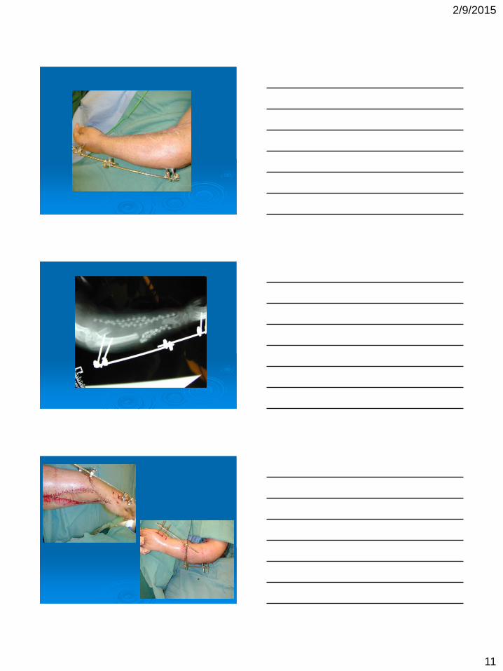
Fixation Options :
• Plates
• Screws
• External Fixation
• K- wires
• Ilizarov
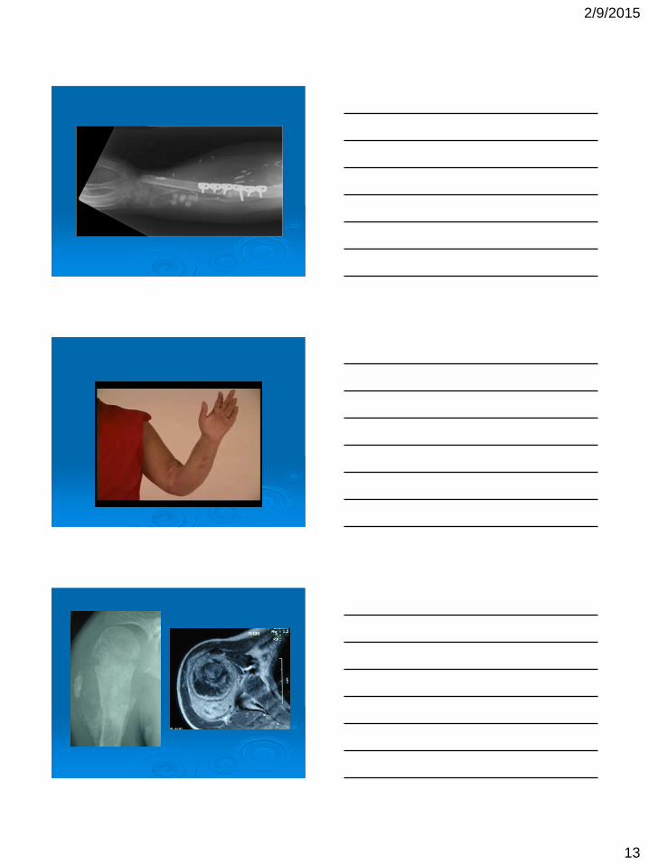
INDICATIONS

2/9/2015
4

2/9/2015
5
2/9/2015
6

2/9/2015
7

2/9/2015
8

2/9/2015
9

2/9/2015
10
2/9/2015
11

2/9/2015
12
2/9/2015
13

2/9/2015
14

2/9/2015
15

2/9/2015
16

2/9/2015
17

2/9/2015
18

2/9/2015
19
2/9/2015
20
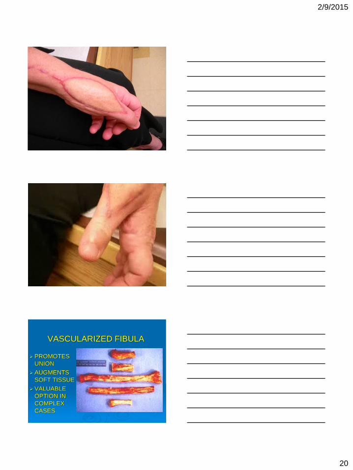
VASCULARIZED FIBULA
PROMOTES
UNION
AUGMENTS
SOFT TISSUE
VALUABLE
OPTION IN
COMPLEX
CASES

1
Free Functional Muscle Transfer
for Upper Extremity Reconstruction
Milan Stevanovic, MD
Professor of Orthopaedic Surgery
University of Southern California
Keck School of Medicine
Disclosure
• There are no conflicts of interest for this presentation and nothing to disclose
Functional Free Muscle
Indications
• Deficiency of critical motor function with no suitable tendon transfer options
• No suitable rotational muscle transfer
• Soft tissue defect requiring coverage in combination with functional loss

2
Free Functional Muscle
Contraindications
• Relative Contraindications–Age > 45 years
–Obesity (weight of limb to be moved by transfer)
• Absolute Contraindications–Medical comorbidities: DM, cardiac
disease, vascular disease, auto-immune disease
Functional Free Muscle
Goals (Manktelow)
• Supply a useful range of motion
• Provide adequate strength for functional activities
Functional Free Muscle
Pre-requisites
• Motivated patient
• Supple passive range of motion
• Suitable recipient site motor nerve and vessels
• Good soft tissue coverage and underlying tissue bed for tendon gliding
3
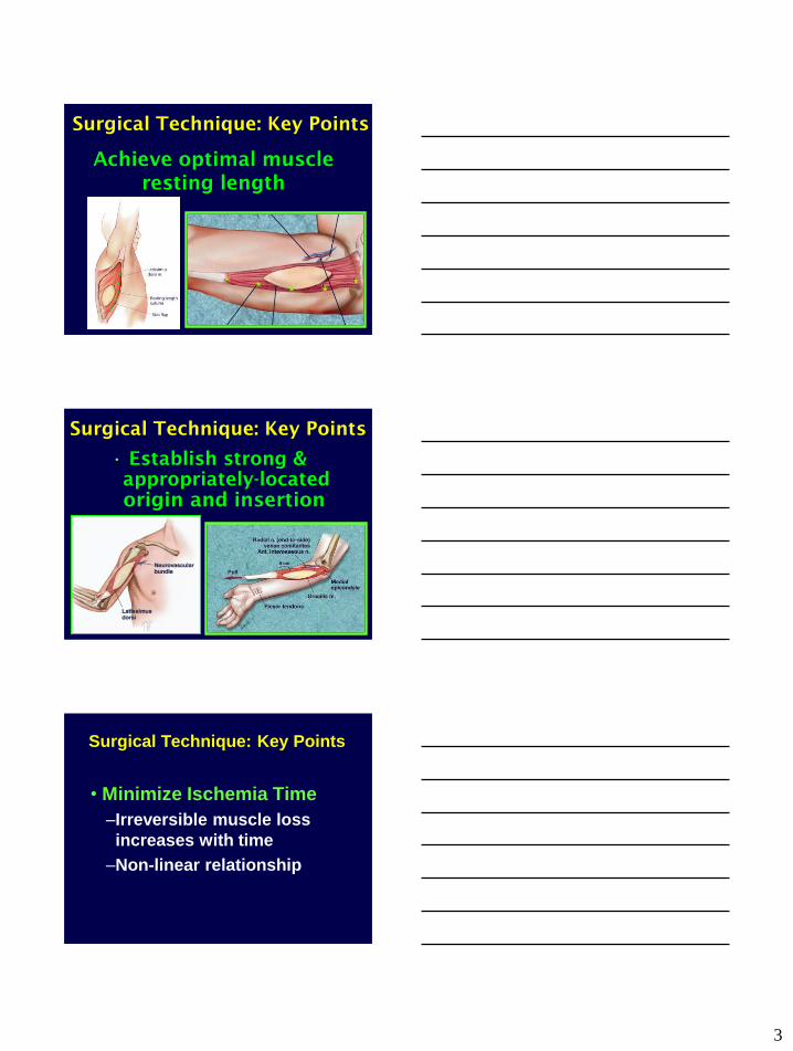
Achieve optimal muscle
resting length
Surgical Technique: Key Points
Surgical Technique: Key Points
• Establish strong &
appropriately-located
origin and insertion
Surgical Technique: Key Points
• Minimize Ischemia Time
–Irreversible muscle loss
increases with time
–Non-linear relationship

4
Surgical Technique: Key Points
• Nerve Considerations
–Recipient site nerve should
be motor fibers
–Neurorraphy should be
done as close as possible
to transplanted muscle
Donor Muscle
Considerations
• Muscle Type
–Pennate(stronger)
• Rectus femoris
Rectus
Femoris
Donor Muscle
Considerations
• Muscle Type
–Strap(better excursion)
• Gracilis
• Latissimus Dorsi
5
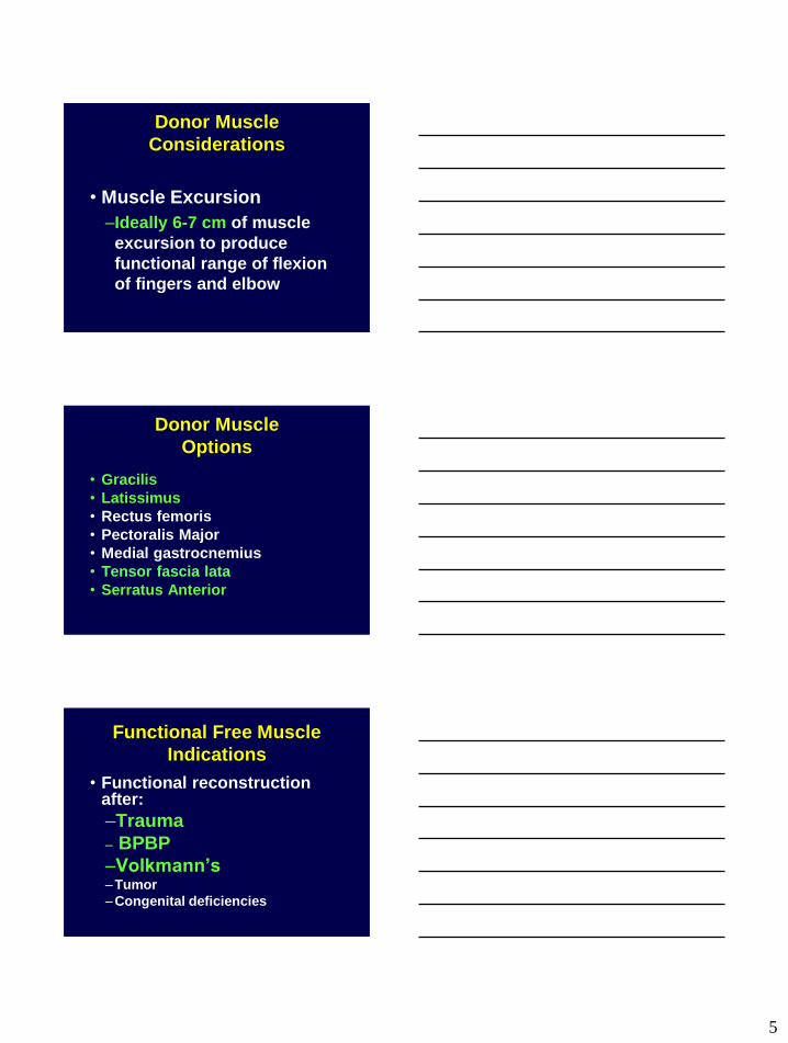
Donor Muscle
Considerations
• Muscle Excursion
–Ideally 6-7 cm of muscle
excursion to produce
functional range of flexion
of fingers and elbow
Donor Muscle
Options
• Gracilis
• Latissimus
• Rectus femoris
• Pectoralis Major
• Medial gastrocnemius
• Tensor fascia lata
• Serratus Anterior
Functional Free Muscle
Indications
• Functional reconstruction after:
–Trauma
– BPBP
–Volkmann’s–Tumor
–Congenital deficiencies

6
Traumatic C5-C6 BPI
2 years post-injury

7
Traumatic C5-C6 BPI
3 years post injury

8
CONGENITAL:
5 year old Arthrogrypotic
PROM
LATISSIMUS
RT LT

9
GRACILIS
6.5 Months F/U

10
Free gracilis for finger flexion
tumor
Free gracilis for finger flexion
Free gracilis for finger flexion

11
Free gracilis for finger flexion
10 month follow up
Volkmann’s Contracture
4 year old 2 years S/P SCH Fx
Flexor Origin Slide
to correct contracture

12
1 year after flexor origin slide
Nerve Graft

13
Independent fascicular territories
of gracilis
Cable grafting of severely
compromised median nerve
Functional results at one year

14
Harvesting Gracilis with a Skin Paddle

15
Median Nerve
AIN
Median nerve reconstruction

16
Options for Thumb Opposition
• Tendon transfer
• Huber muscle tranposition
• Functional free muscle
–Serratus
–Gracilis
–Medial plantar flap with adductor
hallucis

17
Operative

18
Operative
Serratus anterior
Free Serratus for thenar reconstruction
Operative

19
7 month follow up

20
Functional Free Muscle
Transfer
• Demanding procedure
• Meticulous technique
• Experience in microsurgery
Conclusions
Immediate Reconstruction
of
finger flexion after severe
Compartment
Syndrome with
liquifactive muscle
necrosis
Immediate Functional Reconstruction
Flexor Tendons
Median Nerve

21
22
Thank you

1
Medial femoral condyle
free flaps
Allen T. Bishop, MD
MFC flaps
Versatile flap for:
Periosteum: corticoperiosteal
flap
Bone: small segments
Cartilage: Osteoarticular graft
Skin: composite or buoy flap
Periosteal flap
Better than vascularized bone in certain locations and situationsClavicle50% nonunion with fibulaFailed grafting of nonunion radiation necrosis
Forearm: radius and ulna non-unionStructural allograft nonunionsAs a “patch” to form bone in larger
nonunion sites
2
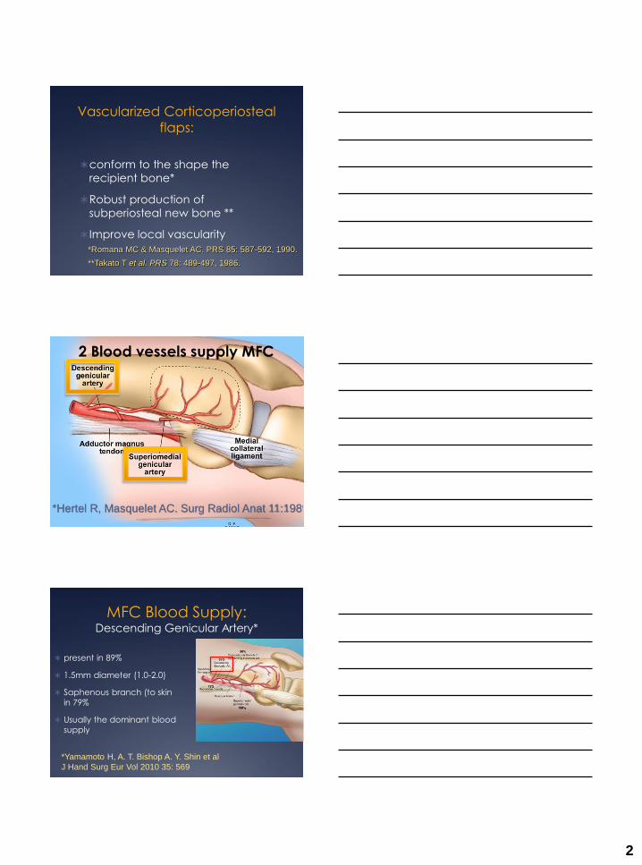
Vascularized Corticoperiosteal
flaps:
conform to the shape the
recipient bone*
Robust production of
subperiosteal new bone **
Improve local vascularity
*Romana MC & Masquelet AC. PRS 85: 587-592, 1990.
**Takato T et al. PRS 78: 489-497, 1986.
2 Blood vessels supply MFC
*Hertel R, Masquelet AC. Surg Radiol Anat 11:1989
MFC Blood Supply:Descending Genicular Artery*
present in 89%
1.5mm diameter (1.0-2.0)
Saphenous branch (to skin
in 79%
Usually the dominant blood
supply
*Yamamoto H, A. T. Bishop A. Y. Shin et al
J Hand Surg Eur Vol 2010 35: 569

3
MFC Blood SupplySuperomedial Genicular Artery
Present 100%
Origen: 5.2 cm proximal to knee
0.78 mm dia. (0.38-1.4)
Anastomosis with descending genicular a.
*Yamamoto H, A. T. Bishop A. Y. Shin et al
J Hand Surg Eur Vol 2010 35: 569
Blood supply of the medial femoral
condyle
Bone nutrient vessels
Highest # of
nutrient vessels in
distal-posterior
quadrant-
preferred for graft
harvest
*Yamamoto H, A. T. Bishop A. Y. Shin et al
J Hand Surg Eur Vol 2010 35: 569
Periosteal flap technique
Elevation of vastus medialis exposes condyle, descending genicular vessels
Condyle
Descending genicular

4
Periosteal flap technique
Osteotome used to
lift periosteal flap
containing
fragments of
cortical bone
Mean flap size 4.75
X 5.75 cm
Surgical Technique
Graft elevated on descending
genicular pedicle

5
Surgical Technique
Deep surface
includes
periosteum and
thin layer of bone
Dominant vascular
pedicle
Periosteal flap technique
flap is thin enough to bend: conforms to
recipient site
Periosteal flap: clavicle
Fuchs B, Bishop AT et al. J Shoulder Elbow Surg.
14(3):264-8, 2005

6
Periosteal flap: Clavicle
Flap wrapped around clavicle, radius
or ulna, secured with heavy sutures
Periosteal flaps
Clavicle
100% union
2 path. fractures
after radiation
1 infected
nonunion
Time to union 5
months
Fuchs B, Bishop AT et al. J Shoulder Elbow Surg.
14(3):264-8, 2005
Femoral allograft nonunion

7
Case:
Femoral allograft nonunion
Case:
Femoral allograft nonunion
MFC Vascularized bone graft:
Treatment of scaphoid nonunion
8
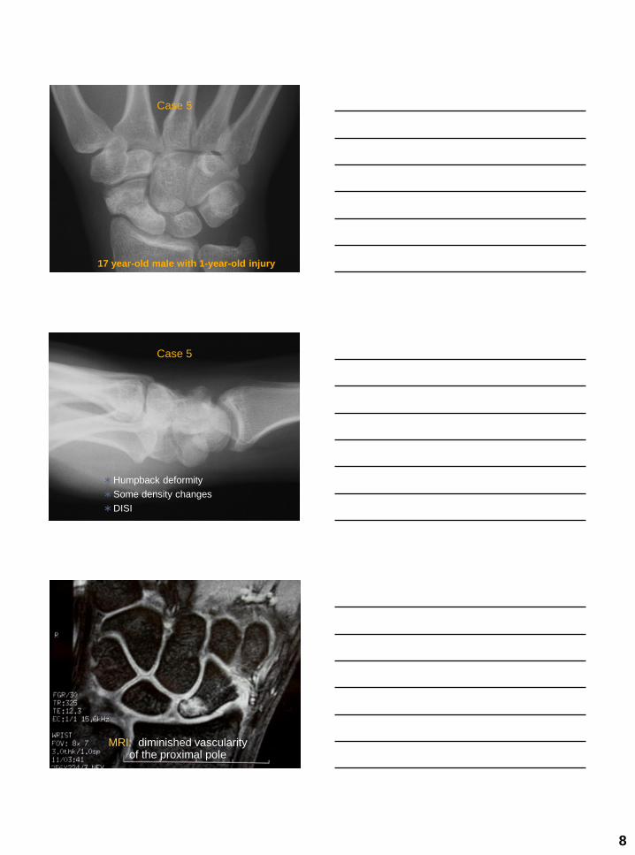
Case 5
17 year-old male with 1-year-old injury
Humpback deformity
Some density changes
DISI
Case 5
MRI: diminished vascularity of the proximal pole

9
Case 5
Density changes
suggestive of AVN
proximal pole
Case 5
Case 5
Exposure of medial condyle,

10
Case 5
Case 5
Case 5

11
Case 5
Case 5
Case 5

12
Case 5: 4 months
• 17 year
Union Rates
0
10
20
30
40
50
60
70
80
90
100
Union Rate
1,2
ICSRAN = 10
MFCN=12
100%
40%
p=0.039

13
Time to Union
0
2
4
6
8
10
12
14
16
18
20
Time to Union
1,2
ICSRA MFC
19.2 weeks 12.7
weeks
p=0.047
Osteoarticular MFC Flap
Uses in the wrist:
Replacement of damaged
proximal surfaces of scaphoid
and lunate
3D model of
proximal scaphoid
Medial patellar facet
14
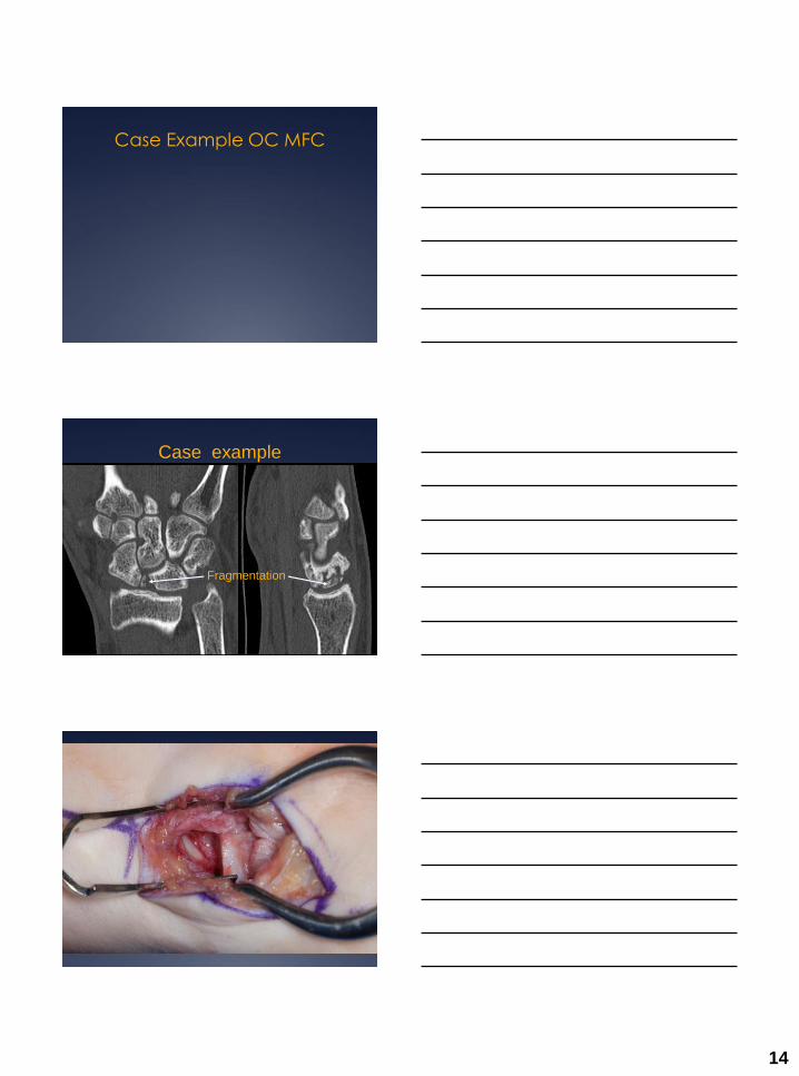
Case Example OC MFC
Case example
Fragmentation

15

16
16 cases of proximal scaphoid
replacement with 14 mo. Follow-
up
Healing in 15
12 with complete pain relief
No SL instability
Bürger, H et al.: J Hand Surg 38A:690, 2013
Conclusions
MFC grafts provide superior results in treating scaphoid nonunions with AVN and humpback deformity
Cortico-periosteal flaps allow salvage of recalcitrant nonunions of clavicle, forearm and allograft-host nonunions
The Osteochondral MFC flap is a new, promising method for irreparable damage to cartilage and subchondralbone
Thank You

2/9/2015
1
Toe-to-Hand Transfers
Neil Jones MD
Chief of Hand Surgery
Professor of Orthopedic Surgery
Professor of Plastic and Reconstructive Surgery
University of California - Irvine
Shriners Hospital Los Angeles
Toe-to-Hand Transfers
I have nothing to disclose
Toe-to-Hand Transfers
• Ultimate functioning free flap
• Osteocutaneous flap
• Mobile joints
• Sensate
• Growth in children

2/9/2015
2
Great Toe or Second ToeVascular Anatomy
• Dorsalis pedis artery - First dorsal metatarsal artery (FDMA)
- “Long” transfer
• First plantar metatarsal artery (FPMA) + vein graft - “Short” transfer
Toe-to-Hand TransfersPreoperative Investigations
• Preoperative angiogram?
• Doppler mapping of FDMA
Great Toe or Second ToeVascular Dissection
• Key point is to understand the relative size and dominance of
the dorsal and plantar arterial systems
• Proximal - distal dissection of FDMA
• Identify FDMA in 1st web space and dissect retrogradely (Wei)

2/9/2015
3
First Dorsal Metatarsal Artery(FDMA)
• Superficial to muscle
• Through the muscle
• Deep to muscle
• Relative position of FDMA with
respect to the muscle may be
evaluated by Doppler mapping
Great Toe or Second Toe Harvest
Great Toe or Second ToeAnatomy
• Dissection of extensor and flexor
tendons
• Tibial and fibular digital nerves
• Deep peroneal nerve
• Saphenous vein

2/9/2015
4
Toe-to-Hand TransfersSurgical Technique
• Bony osteosynthesis - 90-90 intraosseous wiring
• Flexor and extensor tendon repairs,
possible tendon grafts, tendon transfers
• Digital nerve repairs,
possible nerve grafts, end-to-side nerve transfers
• Venous anastomosis (end-to-end)
• Arterial anastomosis (end-to-end or end-to-side)
• Tension-free closure - liberal use of skin grafts
• Pulse-oximetry for post-operative monitoring
Toe-to-Thumb TransfersSurgical Technique
Toe-to-Hand Transfers
• Post-traumatic
• Congenital

2/9/2015
5
Toe-to-Thumb Transfers
• Great toe
“Wrap-around” great toe
“Trimmed” great toe
• Second toe
• Partial toe
Toe-to-Thumb Transfers
Toe-to-Hand TransfersPost-Traumatic Reconstruction
• Isolated thumb amputations
- from the metacarpal base to the mid-point P1,
with intact CMC joint and thenar muscles
• Multiple digit amputations
• Multiple digit and thumb amputations
• Metacarpal hand

2/9/2015
6
Post-Traumatic Reconstruction of the ThumbIndications for Toe-to-Thumb Transfers
• Isolated thumb amputation
(from CMC joint to mid-proximal phalanx)
• Thumb and multiple finger amputations
• Amputation of all five digits - “metacarpal hand”
Toe Transfers and the Level of Amputation
Great toe or variations Second toe transfer
Great Toe Transfer
• Reasonably similar appearance to thumb
• Can be debulked secondarily
• Best IP joint flexion
• Most significant donor defect in the foot

2/9/2015
7
Thumb Amputation at MCP Joint
Great Toe TransferBuncke, Cobbett 1969
Great Toe Transfer

2/9/2015
8
“Wrap-around” Great Toe TransferMorrison 1984, Steichen modification
• Minimizes donor defect in foot
• Soft tissues and nail and distal phalanx are transferred to minimize resorption of bone graft
• Intervening length made up by iliac crest bone graft
• Excellent appearance
• No IP joint motion
• No growth in children
Thumb Amputation through Proximal Phalanx
“Wrap-around” Great Toe Transfer

2/9/2015
9
“Wrap-around” Great Toe Transfer

2/9/2015
10
Trimmed Toe TransferWei, Upton 1988
• Reduces width of P1 and P2,
but preserves IP joint motion
Thumb Amputation base of Proximal Phalanx
“Trimmed” Toe Transfer

2/9/2015
11
Second Toe TransferYang 1977
• Narrower than a thumb
• Less satisfactory appearance
• Tend to develop DIP joint flexion deformity
• Relatively inconspicuous donor site
Thumb Amputations Proximal to mid-Metacarpal
• Second toe provides length of second metatarsal
• May require preliminary soft tissue coverage
and secondary opposition tendon transfer

2/9/2015
12
Isolated Thumb Amputation
just distal to CMC joint
4 years old
Second Toe-to-Thumb Transfer
Second Toe-to-Thumb Transfer
2 years postop

2/9/2015
13
Crush Injury Thumb
Immediate Custom Toe-to-Thumb Transfer
Immediate Custom Toe-to-Thumb Transfer

2/9/2015
14
Custom Toe-to-Thumb Transfer
• Minimize the amount of tissue harvested from the foot
• Make the toe transfer the same size as the contralateral normal thumb
Post-traumatic Reconstruction of the Fingers Indications for Toe-to-Hand transfers
• Multiple amputated fingers with a thumb present U4 R1
• Amputation of all 5 digits R5
U4 R1 R5
Toe Transfers for Post-Traumatic Reconstruction
of the Fingers
Either:
•Single second toe transfer
•Bilateral second toe transfers
•Combined second-third toes

2/9/2015
15
Amputation of Thumb and all 4 Fingers“Metacarpal Hand”
4 years old
Metacarpal Hand
Either:
•Great toe for thumb reconstruction and
combined second-third toes for finger reconstruction
•Bilateral second toes for thumb and finger reconstruction
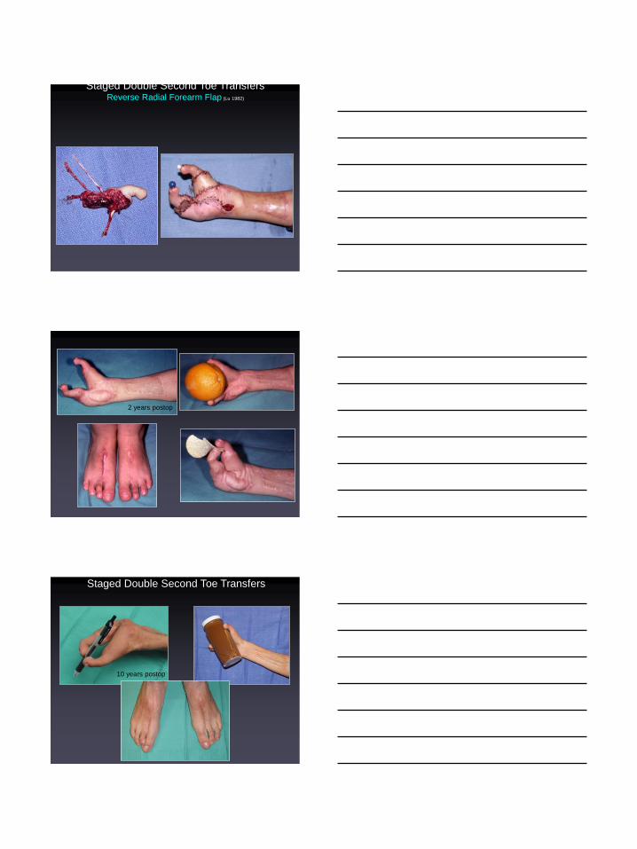
Staged Double Second Toe TransfersReverse Radial Forearm Flap (Lu 1982)
2/9/2015
16
Staged Double Second Toe TransfersReverse Radial Forearm Flap (Lu 1982)
2 years postop
Staged Double Second Toe Transfers
10 years postop

2/9/2015
17
“Metacarpal” Hand8 year old with burns
Right Great Toe-to-Right Thumb Transfer
Right Great Toe-to-Right Thumb Transfer

2/9/2015
18
Combined 2nd-3rd Toe Transfers
for Finger Reconstruction
Great Toe and Combined 2nd-3rd Toe Transfers
to Right Hand
Toe-to-Thumb TransfersResults
• Success > 97%
• Sensation: 2pd < 10mm in 50% patients (better than contralateral toe!)
• MCP joint motion 250, IP joint motion 290
• Grip strength 80-100%, key pinch 65-169%
• Pinch strength: second toe < 50% great toe
• Appearance: wrap-around and trimmed toe > great toe > second toe
• Donor foot: gait analysis close to normal

2/9/2015
19
Decisions in Post-Traumatic Toe TransfersConclusions
• Harvest the minimal amount of bone and soft tissue from the foot to minimize morbidity in the donor foot
• Second toe has most inconspicuous donor site
• Trimmed toe and Morrison wrap-around variations provide the best appearance of the reconstructed thumb
Decisions in Post-Traumatic Toe TransfersConclusions
• Explain all the options to the patient, both conventional and toe transfers
• Explain the morbidity of each donor site
• Show pre-operative and post-operative photographs
• Have patient meet with a patient who has had a similar reconstruction
• Individualize the reconstruction, don’t depend on a algorithm !
Toe-to-Hand Transfers
• Post-traumatic
• Congenital
2/9/2015
20
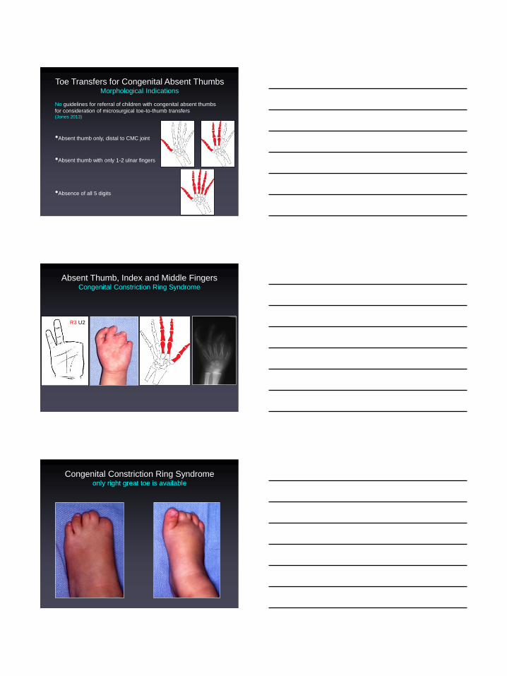
Toe Transfers for Congenital Absent ThumbsMorphological Indications
No guidelines for referral of children with congenital absent thumbs
for consideration of microsurgical toe-to-thumb transfers (Jones 2013)
•Absent thumb only, distal to CMC joint
•Absent thumb with only 1-2 ulnar fingers
•Absence of all 5 digits
Absent Thumb, Index and Middle FingersCongenital Constriction Ring Syndrome
R3 U2
Congenital Constriction Ring Syndromeonly right great toe is available

2/9/2015
21
Congenital Constriction Ring Syndrome“Wrap-Around” Great Toe-to-Thumb Transfer
Congenital Constriction Ring Syndrome“Wrap-Around” Great Toe-to-Thumb Transfer
5 years postop
Toe-to-Hand Transfers for Finger ReconstructionIndications
• Normal thumb but four absent fingers - U4R1
Symbrachydactyly
Transverse failure of formation
Congenital constriction ring syndrome
Ulnar longitudinal deficiency
U4R1

2/9/2015
22
Toe Transfers for Finger Reconstruction
• Second toe into ring or small finger position
(ulnar side of hand allows grasp and pinch)
• Two second toes into middle and small finger positions
allows 3-point pinch
• 2 stage procedure
• Simultaneous one stage procedure
Thumb but Absent Fingers
• 2 second toes into middle and small finger positions sequentially
• Simultaneous one-stage procedure
Simultaneous Double Second Toe Transfers
2/9/2015
23
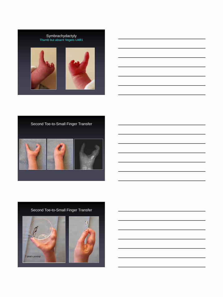
SymbrachydactylyThumb but absent fingers U4R1
Second Toe-to-Small Finger Transfer
Second Toe-to-Small Finger Transfer
7 years postop

2/9/2015
24
Toe-to-Hand Transfers for Finger ReconstructionIndications
• Prosthesis
•2 second toe transfers into thumb and small finger positions
•Two-stage procedure or simultaneously
•Complete absence of all 5 digits - R5
Complete Absence of all 5 Digits R5
• 2 second toe transfers into thumb and small finger positions
• Two-stage procedure or simultaneously
1st stage Second Toe Transfer into Thumb Position

2/9/2015
25
2nd stage Second Toe Transfer
into Small Finger Position
Staged Double Second Toe Transfers
5 years postop
Toe-to-Hand Transfers
for Congenital Hand Anomalies Conclusions
• All children have regained subjective sensibility
• MTP joint motion better than PIP and DIP joint motion
• All but 2 children have improved pinch and grasp function
• Toe-to-thumb transfers have better function than toe-to-finger transfers
• Epiphyses remain open
• Growth is equal to contralateral toe
• Very high parental satisfaction

2/9/2015
26
Toe-to-Hand Transfers for Congenital Absent Digits Conclusions
• Embryological classifications are not helpful in guiding reconstruction
• Morphological and radiographic anatomy of the hand and level of absence
of the digits are the most logical criteria for conventional or microsurgical
reconstruction
R3U2 U4R1 R5R2U3
R4U1