Microstructure and Mechanical Properties of In Situ ... · Microstructure and Mechanical Properties...
6
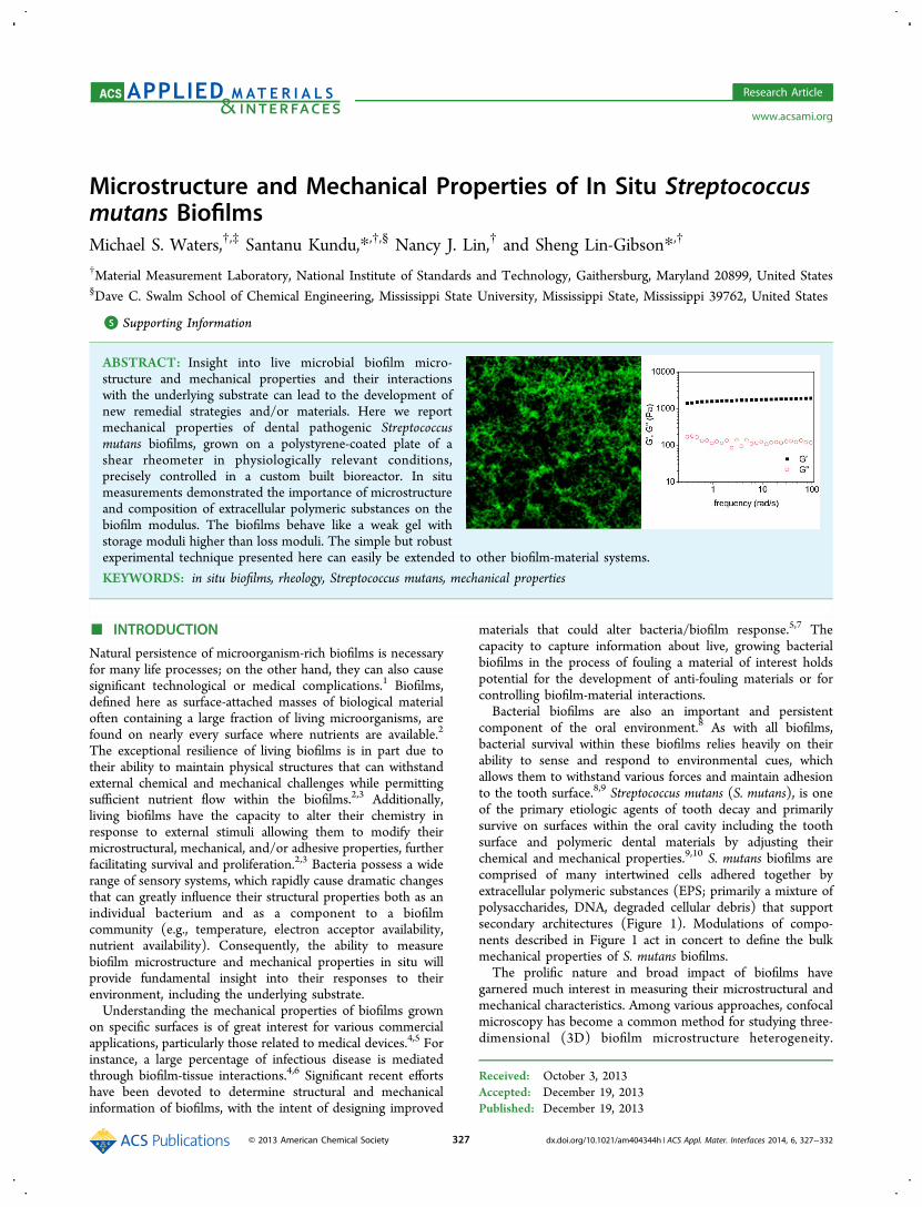
Microstructure and Mechanical Properties of In Situ Streptococcus mutans Biofilms Michael S. Waters, †,‡ Santanu Kundu,* ,†,§ Nancy J. Lin, † and Sheng Lin-Gibson* ,† † Material Measurement Laboratory, National Institute of Standards and Technology, Gaithersburg, Maryland 20899, United States § Dave C. Swalm School of Chemical Engineering, Mississippi State University, Mississippi State, Mississippi 39762, United States * S Supporting Information ABSTRACT: Insight into live microbial biofilm micro- structure and mechanical properties and their interactions with the underlying substrate can lead to the development of new remedial strategies and/or materials. Here we report mechanical properties of dental pathogenic Streptococcus mutans biofilms, grown on a polystyrene-coated plate of a shear rheometer in physiologically relevant conditions, precisely controlled in a custom built bioreactor. In situ measurements demonstrated the importance of microstructure and composition of extracellular polymeric substances on the biofilm modulus. The biofilms behave like a weak gel with storage moduli higher than loss moduli. The simple but robust experimental technique presented here can easily be extended to other biofilm-material systems. KEYWORDS: in situ biofilms, rheology, Streptococcus mutans, mechanical properties ■ INTRODUCTION Natural persistence of microorganism-rich biofilms is necessary for many life processes; on the other hand, they can also cause significant technological or medical complications. 1 Biofilms, defined here as surface-attached masses of biological material often containing a large fraction of living microorganisms, are found on nearly every surface where nutrients are available. 2 The exceptional resilience of living biofilms is in part due to their ability to maintain physical structures that can withstand external chemical and mechanical challenges while permitting sufficient nutrient flow within the biofilms. 2,3 Additionally, living biofilms have the capacity to alter their chemistry in response to external stimuli allowing them to modify their microstructural, mechanical, and/or adhesive properties, further facilitating survival and proliferation. 2,3 Bacteria possess a wide range of sensory systems, which rapidly cause dramatic changes that can greatly influence their structural properties both as an individual bacterium and as a component to a biofilm community (e.g., temperature, electron acceptor availability, nutrient availability). Consequently, the ability to measure biofilm microstructure and mechanical properties in situ will provide fundamental insight into their responses to their environment, including the underlying substrate. Understanding the mechanical properties of biofilms grown on specific surfaces is of great interest for various commercial applications, particularly those related to medical devices. 4,5 For instance, a large percentage of infectious disease is mediated through biofilm-tissue interactions. 4,6 Significant recent efforts have been devoted to determine structural and mechanical information of biofilms, with the intent of designing improved materials that could alter bacteria/biofilm response. 5,7 The capacity to capture information about live, growing bacterial biofilms in the process of fouling a material of interest holds potential for the development of anti-fouling materials or for controlling biofilm-material interactions. Bacterial biofilms are also an important and persistent component of the oral environment. 8 As with all biofilms, bacterial survival within these biofilms relies heavily on their ability to sense and respond to environmental cues, which allows them to withstand various forces and maintain adhesion to the tooth surface. 8,9 Streptococcus mutans (S. mutans), is one of the primary etiologic agents of tooth decay and primarily survive on surfaces within the oral cavity including the tooth surface and polymeric dental materials by adjusting their chemical and mechanical properties. 9,10 S. mutans biofilms are comprised of many intertwined cells adhered together by extracellular polymeric substances (EPS; primarily a mixture of polysaccharides, DNA, degraded cellular debris) that support secondary architectures (Figure 1). Modulations of compo- nents described in Figure 1 act in concert to define the bulk mechanical properties of S. mutans biofilms. The prolific nature and broad impact of biofilms have garnered much interest in measuring their microstructural and mechanical characteristics. Among various approaches, confocal microscopy has become a common method for studying three- dimensional (3D) biofilm microstructure heterogeneity. Received: October 3, 2013 Accepted: December 19, 2013 Published: December 19, 2013 Research Article www.acsami.org © 2013 American Chemical Society 327 dx.doi.org/10.1021/am404344h | ACS Appl. Mater. Interfaces 2014, 6, 327−332
Transcript of Microstructure and Mechanical Properties of In Situ ... · Microstructure and Mechanical Properties...
Microstructure and Mechanical Properties of In Situ Streptococcusmutans BiofilmsMichael S. Waters,†,‡ Santanu Kundu,*,†,§ Nancy J. Lin,† and Sheng Lin-Gibson*,†
†Material Measurement Laboratory, National Institute of Standards and Technology, Gaithersburg, Maryland 20899, United States§Dave C. Swalm School of Chemical Engineering, Mississippi State University, Mississippi State, Mississippi 39762, United States
*S Supporting Information
ABSTRACT: Insight into live microbial biofilm micro-structure and mechanical properties and their interactionswith the underlying substrate can lead to the development ofnew remedial strategies and/or materials. Here we reportmechanical properties of dental pathogenic Streptococcusmutans biofilms, grown on a polystyrene-coated plate of ashear rheometer in physiologically relevant conditions,precisely controlled in a custom built bioreactor. In situmeasurements demonstrated the importance of microstructureand composition of extracellular polymeric substances on thebiofilm modulus. The biofilms behave like a weak gel withstorage moduli higher than loss moduli. The simple but robustexperimental technique presented here can easily be extended to other biofilm-material systems.
KEYWORDS: in situ biofilms, rheology, Streptococcus mutans, mechanical properties
■ INTRODUCTION
Natural persistence of microorganism-rich biofilms is necessaryfor many life processes; on the other hand, they can also causesignificant technological or medical complications.1 Biofilms,defined here as surface-attached masses of biological materialoften containing a large fraction of living microorganisms, arefound on nearly every surface where nutrients are available.2
The exceptional resilience of living biofilms is in part due totheir ability to maintain physical structures that can withstandexternal chemical and mechanical challenges while permittingsufficient nutrient flow within the biofilms.2,3 Additionally,living biofilms have the capacity to alter their chemistry inresponse to external stimuli allowing them to modify theirmicrostructural, mechanical, and/or adhesive properties, furtherfacilitating survival and proliferation.2,3 Bacteria possess a widerange of sensory systems, which rapidly cause dramatic changesthat can greatly influence their structural properties both as anindividual bacterium and as a component to a biofilmcommunity (e.g., temperature, electron acceptor availability,nutrient availability). Consequently, the ability to measurebiofilm microstructure and mechanical properties in situ willprovide fundamental insight into their responses to theirenvironment, including the underlying substrate.Understanding the mechanical properties of biofilms grown
on specific surfaces is of great interest for various commercialapplications, particularly those related to medical devices.4,5 Forinstance, a large percentage of infectious disease is mediatedthrough biofilm-tissue interactions.4,6 Significant recent effortshave been devoted to determine structural and mechanicalinformation of biofilms, with the intent of designing improved
materials that could alter bacteria/biofilm response.5,7 Thecapacity to capture information about live, growing bacterialbiofilms in the process of fouling a material of interest holdspotential for the development of anti-fouling materials or forcontrolling biofilm-material interactions.Bacterial biofilms are also an important and persistent
component of the oral environment.8 As with all biofilms,bacterial survival within these biofilms relies heavily on theirability to sense and respond to environmental cues, whichallows them to withstand various forces and maintain adhesionto the tooth surface.8,9 Streptococcus mutans (S. mutans), is oneof the primary etiologic agents of tooth decay and primarilysurvive on surfaces within the oral cavity including the toothsurface and polymeric dental materials by adjusting theirchemical and mechanical properties.9,10 S. mutans biofilms arecomprised of many intertwined cells adhered together byextracellular polymeric substances (EPS; primarily a mixture ofpolysaccharides, DNA, degraded cellular debris) that supportsecondary architectures (Figure 1). Modulations of compo-nents described in Figure 1 act in concert to define the bulkmechanical properties of S. mutans biofilms.The prolific nature and broad impact of biofilms have
garnered much interest in measuring their microstructural andmechanical characteristics. Among various approaches, confocalmicroscopy has become a common method for studying three-dimensional (3D) biofilm microstructure heterogeneity.
Received: October 3, 2013Accepted: December 19, 2013Published: December 19, 2013
Research Article
www.acsami.org
© 2013 American Chemical Society 327 dx.doi.org/10.1021/am404344h | ACS Appl. Mater. Interfaces 2014, 6, 327−332

Techniques for determining the mechanical properties ofbiofilms include atomic force microscopy,11 microindenta-tion,12 particle tracking methods,13 microfluidic devices,14 andshear rheometry.11,15−17 Indentation and particle trackingmethods generally provide more localized properties, whereasshear rheology provides the average properties of the entirebiofilm. Shear rheology has been used to determine a thresholdfor disruptive debulking of live S. mutans biofilms underconstant stress.16 Pavlovsky et al. have studied the mechanicalproperties of Staphylococcus epidermidis (an antibiotic resistantinfectious bacteria causing skin lesions in immune compro-mised patients and medical device contamination) biofilm usingin situ rheology.17 Using oscillatory rheology, they were able tocapture the biofilm growth process.To evaluate biofilm properties that better correlate to in vivo
situations, it is critical to precisely control the environment inwhich a biofilm is grown and measured. The primary goal ofthis research is to measure mechanical properties of in situ S.mutans biofilms grown in a custom-designed, environment-controlled bioreactor. Microstructural and rheological proper-ties of intact biofilms were also compared to those fromdisrupted biofilms (centrifuged/spin down biofilms that aremacroscopically homogeneous) to assess contributions ofsecondary structures and physicochemical properties to biofilmmechanical properties.
■ MATERIALS AND METHODSBacterial Strains and Culturing. S. mutans strain UA159 starter
cultures (3 mL) were inoculated from a single colony struck on ToddHewitt Broth (THB; Becton Dickson) agar plates grown at 37 °C in a5 % (by volume) CO2 in air incubator as previously described.18
Biofilms were grown within the bioreactor by inoculating 10 mL ofsterile THB or THB with 1 % sucrose by mass, 1:100 with a 24 hstationary-phase starter culture, and incubating at 37 °C in a premixedmedical gas standard STM1702 equivalent to 5 % CO2 in air atatmospheric pressure.
Cultures for spin-down disrupted biofilm measurements weregrown as described above in 500 mL THB or THB with 1 % sucroseby mass in a 1 L Erlenmeyer flask. The entire volume along with thesurface-attached biofilm (removed by scraping) was collected by 10min centrifugation at 560 rad/s (3200 RCF) at room temperature.The pellet was used for rheological experiments.
Bioreactor Assembly and Rheological Measurement. Rheo-logical measurements were performed on an ARES rheometer using 50mm parallel plate geometry. The biofilm-receiving lower plate wascoated with polystyrene (via spin-coated using 1% by mass polystyrenein toluene19,20). The entire assembly was cleaned with water andsterilized with 100% ethanol. The zero position was determined at theexperimental temperature of 37 °C.
The bioreactor was assembled surrounding the lower plate (Figure2A). After sterile assembly and inoculation, the bioreactor was jacketedwith an insulated circulating water heating coil to maintain 37 °C andwas flushed with gas. Temperature was monitored by a thermocouplecontacting the bottom of the lower plate. Gas was passed through 0.2μm filtered inlet and exhaust ports to maintain internal sterility of thebioreactor. The bioreactor was disassembled after 24 h of growth.Superficial media was allowed to flow into an absorbent towel,
Figure 1. Description of the chemical and structural attributes influencing the mechanical properties of biofilms. (A) Confocal image of a DNA-stained S. mutans biofilm grown in the presence of sucrose. The brightest spots within the image are bacterial clusters and the segmented strings areindividual bacteria, which together make the biofilm. (B) Model of a S. mutans biofilm. The clear coating represents extracellular polymeric substance(EPS). (C) Each streptococcus bacterium that makes up the biofilm has a highly adhesive coating of EPS made primarily of secreted glucans. Thecross-sectioned cocci depict the structural makeup of an individual cell. (D) Model depicting the cell wall composition. Peptidoglycan comprises N-acetyl glucosamine and N-acetyl murine, linked by β-1,4 glycosidic bonds and peptide bridges. (E) 3D heat map of S. mutans biofilm-mediatedhuman tooth enamel erosion, measured by interferometric optical profilometry. Tooth enamel surface was masked (left) and exposed (right) to a 24h sucrose-fed S. mutans biofilm, which was removed revealing the biofilm-decayed enamel (right). The contrasting circular structures visible in theexposed area are enamel rods.
ACS Applied Materials & Interfaces Research Article
dx.doi.org/10.1021/am404344h | ACS Appl. Mater. Interfaces 2014, 6, 327−332328

revealing the biofilm (Figure 2B). The upper plate was brought intocontact with the biofilm by gradually lowering to (80 ± 1) μm gap. Tominimize the effect of compression as the upper plate is brought incontact with the biofilm, the upper plate was moved very slowly todissipate the compressive stress. The compressive stress value wasconstantly monitored during the process. A layer of low-viscosity oil(Nikon type A immersion oil) was wiped around the edge of the gapto reduce desiccation. The rheometer oven was then rapidly closedand was brought back to 37 °C. The time between disassembly andmeasurement was less than 2 min.Strain and frequency sweep experiments were performed on fresh
samples. Strain sweeps were performed at strains from 0.1 to 100%and at a constant frequency of 6.28 rad/s. Frequency sweeps wereperformed at a constant strain of 0.3%, which falls in the linearviscoelastic region, in the frequency range of 0.3−100 rad/s. At leastthree separate biofilms were evaluated in situ.Spin-down pellets of bacteria were applied directly to the lower 50
mm polystyrene-coated plate with a spatula as quickly and evenly aspossible. Measurements of spin-down biofilms were performed using
∼400 μm gap with oil applied as described above to preventdesiccation. Measurements of spin-down biofilms were repeated withvarying gap sizes and results were found to be independent of the gap.The experiment was repeated at least three times each time with afresh sample.
Confocal Biofilm Characterization. Confocal images andmeasurements were acquired on a Zeiss LSM 510 confocal microscopeand analyzed using Zeiss LSM Image Browser software. S. mutansbiofilms that were grown for 24 h were stained for 10 min withfluorescent DNA stain SYBR Green (Invitrogen) and imaged aspreviously described.18 Z-stacks at 10 μm and 1 μm intervals coveringthe entire fluorescent depth were acquired using 5× and 40× objectivelenses to evaluate distributions of biofilm height and structuralfeatures. These images indicated that an 80 μm gap provides near100% contact between the biofilm and the top plate.
Measurement of Biofilm-Mediated Tooth Dissolution. Toconfirm pathogenicity, cultures of S. mutans UA195 were prepared andincubated in the same conditions described above together withpolished, partially masked human tooth enamel chips for 24 h, in the
Figure 2. Bioreactor design and biofilm growth. (A) Assembled bioreactor surrounding a polystyrene-coated 50 mm standard disposable parallelplate, featuring gas-control and temperature feedback. (B) S. mutans biofilm (pasty cream-colored material) covered with a thin layer of media grownin the presence of sucrose on a polystyrene-coated plate directly on the rheometer, immediately before measurement. (C, D) Confocal measurementto determine gap parameters for parallel-plate rheological measurement, highlighting (C) microstructural features such as the ridges (brighterregions) and channels (darker regions) and (D) a height heat map of the confocal stack determining gap settings for biofilm measurement (arrow;∼80 μm), balancing full upper plate contact with the least biofilm structural damage. (E, F) Confocal images of DNA-stained S. mutans biofilmsgrown directly on the rheometer in (E) the absence of sucrose and (F) presence of sucrose. (G, H) Confocal images of centrifuged (spin-down)DNA-stained S. mutans biofilms grown in (G) the absence of sucrose and (H) the presence of sucrose.
ACS Applied Materials & Interfaces Research Article
dx.doi.org/10.1021/am404344h | ACS Appl. Mater. Interfaces 2014, 6, 327−332329

presence of 1% sucrose by mass. Tooth preparations andinterferometric optical profilometry measurements of biofilm-mediatedtooth dissolution were conducted as previously described.21
■ RESULTS AND DISCUSSION
Bioreactor Design. The bioreactor we designed andfabricated (Figure 2A) for this study features an environmentalcontrol unit enclosing the lower plate of the rheometer. Thisset up permits the measurement of biofilms grown directly onthe lower plate with increased measurement confidence for thefollowing reasons. The design allows precise gap control, sincethe gap is calibrated within the instrument just prior to biofilmgrowth. Precise gap control is important for the measurementsof thin biofilms. The in situ biofilm structure is highlynonuniform in the vertical direction (i.e., the biofilm becomesless dense further away from the substrate;see Figure S2 in theSupporting Information). As evident from Figures 2 and FigureS2 in the Supporting Information, over a dimension of a fewsquare millimeters, the height of the S. mutans biofilm varies insome extent. To determine the height-variation, we capturedimages of biofilm using confocal microscopy at different depths(Z-profile). We observed plume as high as 270 μm and somebiofilm growth up to a height of 170 μm. However, most of thebiofilm has a thickness of approximately ≥80 μm, which isapparent in Figure 2 and Figure S2 in the SupportingInformation in complete coverage of image area of the biofilm.Hence, we used a gap of 80 μm for all rheological studies.Measurements using a larger gap did not generate meaningfuldata due to insufficient contact between cellular materials andupper plate; measurements using smaller gaps compressed thebiofilm, which also introduced errors. As biofilms are grown insitu, perturbation of biofilms was minimal. Gas is designed togently circulate within the headspace, ensuring an eventurnover for stable absorbance within the media withoutagitation. The heated jacket covering the bioreactor contactsthe thermoconductive aluminum base, ensuring temperaturestability for the duration of the experiment. This bioreactordesign allow for facile control of conditions physiologicallyrelevant to bacteria (e.g., nutrient medium, gas, temperature,interacting surface, etc.).This design platform is also versatile for studying biofilm
interactions with various materials. In this study, polystyrenewas selected as the model surface as it is nonreactive and themost commonly used surface on which to evaluate biofilms.22
Similar techniques can be used to coat the bottom plate with a
broad range of materials, including polymers, ceramics, metals,and biological materials.
Biofilm Characterization. S. mutans biofilms cultivated onthe prophylactic polystyrene surface were grown either with orwithout sucrose supplementation in commercially availablegroup-A streptococcal media (THB) at temperatures and gasconditions similar to the environment within the mouth,demonstrating growth consistent with previous studies.18 S.mutans biofilms grown in the presence or absence of sucrosehave distinctly different morphologies. Confocal measurementsreveal that S. mutans biofilms grown in the absence of sucroselacked clear bridge structures between the bright punctateclusters of aggregated cells, contrasted by a black background(areas lacking DNA/bacteria), giving the biofilm a moredispersed appearance (Figure 2E). Conversely, biofilms grownin presence of sucrose (Figure 2C, D, F) have a distinct andheavily interconnected structural network showing linkagebetween filamentous and aggregated bacterial structures whilemaintaining a porous network structure, reminiscent of aheterogeneous polymeric gel, permitting flow of nutrients andother important survival components.S. mutans biofilms were also grown under similar conditions
(with and without sucrose) and then centrifuged to producespin-down cell pellets for evaluating the effect of physicochem-ical composition on mechanical properties. After centrifugationand pellet formation, secondary structural features within thebiofilms (e.g., channels and plumes) are severely diminished, ascan be seen by comparing images F and H in Figure 2.Although disrupted, pelleted bacterial cultures still reveal visiblestructural differences, with sucrose-grown pellets containingaggregated bacterial structures and those grown withoutsucrose appearing more homogeneous (Figure 2G, H). Thebright patches visible in images G and H in Figure 2 are due toa coalescence of green fluorescently stained bacteria. The darkpatches between the bacteria are the void space that indirectlyrepresents the level of biofilm hydration observed in thecentrifuged samples. Dehydration experiments revealed thatspin-down biofilms are comprised of (89.3 ± 2.6)% and (85.5± 1.7)% water by mass for no sucrose and sucrose-grownbiofilms (see the Supporting Information, Figure S1),respectively.The differences in microstructure between biofilms grown in
the presence or absence of sucrose are a result of many complexphysical and chemical interactions that are under the control ofseveral regulatory systems. These structures are primarily
Figure 3. Storage (G′) and loss modulus (G″) as a function of frequency for (A) in situ S. mutans biofilms grown in the presence of sucrose and (B)spin-down pelleted S. mutans samples grown in the presence or absence of sucrose. The applied strain was 0.3 %.
ACS Applied Materials & Interfaces Research Article
dx.doi.org/10.1021/am404344h | ACS Appl. Mater. Interfaces 2014, 6, 327−332330

maintained and reinforced by the secretion of alpha-1,3 andalpha-1,6 glucan (polysaccharides), produced by glucosyltrans-ferase-mediated conversion of sucrose. These polysaccharidesreinforce the biofilm microstructure, converting the biofilms torigid, desiccation-tolerant structures that can firmly attach tothe tooth surface. The heat map shows that biofilm plumesreached as high as 270 μm and the thickness of the confluentregion of the biofilm from the polystyrene surface to the base ofthe plumes was ∼80 μm (Figure 2D and Figure S2 in theSupporting Information). As complete contact between thesample and the top plate is important in obtaining meaningfuland reproducible rheological results, 80 μm was selected for allin situ sucrose-fed S. mutans biofilm measurements.Upon intake, S. mutans also metabolizes sucrose through a
series of enzymes, producing lactic acid as a by-product. Theacid reduces the pH of the overall environment to give S.mutans a competitive edge over acid-intolerant bacteria, andallows the biofilm to form a protective pocket into the toothenamel surface.9,23 To confirm that the sucrose growthconditions were indeed pathogenic, in situ biofilms wereprobed for their pathology to human tooth enamel byinterferometric optical profilometry. Within the 24 h cultureperiod, the environmental pH was reduced from pH 7.01 to pH4.02. When a premeasured flat enamel surface was masked onone side and left exposed to biofilm growth on the other,dramatic effects were observed with respect to the dissolutionof the tooth enamel (Figure 1E). These evaluations confirmedthat the strain of S. mutans evaluated here retains itspathogenicity in its sucrose fed state.Rheological Characterization of S. mutans Biofilms.
The storage modulus (G′, representing the elastic component)is consistently higher than the loss modulus (G″, representingthe viscous component) over the frequency range tested for insitu biofilms grown in the presence of sucrose (Figure 3A).These biofilms behaved like a weak gel (G′ > G″) and arerelatively frequency independent over the entire frequencyrange probed. Such behavior is typical of soft elastic materialsand signifies the presence of a network structure consistentwith the confocal images of sucrose biofilms. Moduli of biofilmsgrown in situ without sucrose could not be measured, likelybecause of their low rigidities/interconnectivities.Rheology measurements for centrifuged biofilms grown with
and without sucrose also showed G′ greater than G″ and wererelatively frequency-independent over the entire frequencyrange (Figure 3B). Although a similar rheological response was
observed for in situ biofilm and spin-down sucrose biofilm, thetwo samples were drastically different in their hydration state.The in situ biofilm contained significantly more water than thespin-down pellets as shown by the confocal images (Figure 2),which indicates that the fluid-filled interstitial space between insitu sucrose-fed S. mutans biofilms is much larger than sucrose-fed spin-down biofilms. The enhanced mechanical property forthe intact biofilm must be due to its network structure, whichresults in the modulus to the level comparable to a much densematerial.Comparison of spin-down sucrose and no sucrose biofilms
revealed significant differences in mechanical properties, as G′values for sucrose samples are significant higher than thatobtained for no sucrose samples. Confocal observations ofinterstitial space and desiccation mass experiments indicate thatthese spin-down biofilms had comparable amount of watercontent yet were one order of magnitude different in moduluswhen measured by rheology. These differences are attributed tochemical differencesthe presence of large amount of rigidhydrophobic extracellular alpha-1,3 glucan, produced bysucrose-fed biofilms.24,25
Strain sweep experiments were performed to determine thelinear viscoelastic region and to provide information about thelarge strain responses of biofilms on both in situ and spin-downbiofilms. The linear viscoelastic region for in situ biofilm was upto ∼1 % strain; further increases in strain resulted in strongstrain-softening (Figure 4A). Additionally, the modulus of thein situ biofilm does not recover appreciably when themeasurement was repeated on the same sample. In fact, theintact biofilm (first run) has a G′ that was about two orders ofmagnitude higher than the disrupted biofilm (second run). Thisbehavior is consistent with disruption of the interconnectedstructure at higher strains. Note that the second run was startedimmediately, without any wait time to avoid potential repairingof the live biofilm network.The linear viscoelastic region extended to ∼10 % strain for all
spin-down biofilms (Figure 4B). Consistent with frequencysweep experiments, G′ for biofilms grown with sucrose washigher than those grown without sucrose. The strain sweepexperiments are highly repeatable when the sample is runmultiple times, indicating a lack of structural change uponimposed strain. The recovery is likely due to direct measure-ment of the mechanical properties of the overall biomass,without significant contribution from larger structures.Interestingly, there is a G′ to G″ crossover at higher strains
Figure 4. Reversibility of strain sweep experiments. G′ and G″ as a function of strain for (A) in situ S. mutans biofilm grown in the presence ofsucrose, and (B) spin-down pelleted S. mutans samples grown in the presence or absence of sucrose. The applied frequency was 6.28 rad/s.
ACS Applied Materials & Interfaces Research Article
dx.doi.org/10.1021/am404344h | ACS Appl. Mater. Interfaces 2014, 6, 327−332331

for both spin-down samples, indicating the material willbecome more viscous-like under large strain deformations.The moduli were quite different for spin-down samples grownwith or without sucrose despite the fact that the interstitialspaces between the bacteria have been largely removed for bothsamples. The large difference in G′ indicates that thecomponents holding these bacteria together are quite differentin composition and/or distribution. As described above, theshifting of EPS properties by secretion of extracellular glucanvia the metabolism of sucrose by glucosyltransferase enzymes isexpected to be the most important reason for this modulusshift.24,25
■ CONCLUSIONSIn this study, we demonstrate that oscillatory shear rheologycan be used to evaluate in situ mechanical properties of liveacidogenic S. mutans biofilms. Biofilms were grown directlywithin the rheometer on a standard plate coated with a materialof our choosing, in a custom bioreactor that acts as a stand-alone, environmentally controlled culturing system. Thisapproach eliminates mechanical damage inherent to trans-ferring biofilms on plates to the rheometer and greatly reducesthe time between culturing and measurement to provide higherdegrees of repeatability. We show that the biofilm micro-structures play a key role in the overall mechanical properties,i.e., disrupted biofilm due to shear reduced G′ by approximatelytwo orders of magnitude. We were also able to discern G′differences due to biofilm EPS variations. This method tomeasure biofilm−material interactions can be easily adapted tomost biofilms or materials for advancing medical or environ-mental applications, particularly in the development ofmaterials that modify biofilm interactions.
■ ASSOCIATED CONTENT*S Supporting InformationWater retention of biofilms, confocal micrographs, andheterogeneity of biofilms are included in the SupportingInformation. This material is available free of charge via theInternet at http://pubs.acs.org/.
■ AUTHOR INFORMATIONCorresponding Authors*E-mail: [email protected].*E-mail: [email protected] Address‡M.S.W. is currently at Office of In Vitro Diagnostics andRadiological Health, Center for Devices and RadiologicalHealth, FDA, Silver Springs, MD 20993.NotesThe authors declare no competing financial interest.
■ ACKNOWLEDGMENTSThis work was in part supported by a National ResearchCouncil (NRC) ARRA postdoctoral Fellowship and anInteragency Agreement between NIDCR/NIH and NIST[Y1-DE-7005-01]. This is an official contribution of theNational Institute of Standards and Technology, and is notsubject to copyright in the United States. Certain commercialequipment, instruments, or materials are identified in this paperin order to specify the experimental procedure adequately. Suchidentification is not intended to imply recommendation orendorsement by the National Institute of Standards and
Technology, nor is it intended to imply that the materials orequipment identified are necessarily the best available for thepurpose.
■ REFERENCES(1) Community Structure and Co-Operation in Biofilms; Allison, D. G.,Gilbert, P., Lapin-Scott, H.M., Wilson, M., Eds.; Society for GeneralMicrobiology Symposia; Cambridge University Press: Cambridge,U.K., 2000.(2) Davey, M. E.; O’toole, G. A. Microbiol. Mol. Biol. Rev. 2000, 64,847−867.(3) Nadell, C. D.; Xavier, J. B.; Foster, K. R. FEMS Microbiol. Rev.2009, 33, 206−224.(4) Bryers, J. D. Biotechnol. Bioeng. 2008, 100, 1−18.(5) Biofilms in Medicine, Industry and Environmental Biotechnology:Characteristics, Analysis and Control; Lens, P., O’flaherty, V., Moran, A.P., Stoodley, P., Mahony, T., Eds.; International Water Association:London, 2003.(6) Wolcott, R.; Dowd, S. Plast. Reconstr. Surg. 2011, 127 (Suppl 1),28S−35S.(7) Houari, A.; Picard, J.; Habarou, H.; Galas, L.; Vaudry, H.; Heim,V.; Di Martino, P. Biofouling 2008, 24, 235−240.(8) Bowden, G. H. W.; Hamilton, I. R. Crit. Rev. Oral. Biol. Med.1998, 9, 54−85.(9) Nobbs, A. H.; Lamont, R. J.; Jenkinson, H. F. Microbiol. Mol. Biol.Rev. 2009, 73, 407−450.(10) Klein, M. I.; Duarte, S.; Xiao, J.; Mitra, S.; Foster, T. H.; Koo, H.Appl. Environ. Microbiol. 2009, 75, 837−841.(11) Lau, P. C. Y.; Dutcher, J. R.; Beveridge, T. J.; Lam, J. S. Biophys.J. 2009, 96, 2935−2948.(12) Cense, A. W.; Peeters, E. A. G.; Gottenbos, B.; Baaijens, F. P. T.;Nuijs, A. M.; van Dongen, M. E. H. J. Microbiol. Methods 2006, 67,463−472.(13) Cheong, F. C.; Duarte, S.; Lee, S.-H.; Grier, D. G. Rheol. Acta2009, 48, 109−115.(14) Hohne, D. N.; Younger, J. G.; Solomon, M. J. Langmuir 2009,25, 7743−7751.(15) Lieleg, O.; Caldara, M.; Baumgartel, R.; Ribbeck, K. Soft Matter2011, 7, 3307−3314.(16) Vinogradov, A. M.; Winston, M.; Rupp, C. J.; Stoodley, P.Biofilms 2004, 1, 49−56.(17) Pavlovsky, L.; Younger, J. G.; Solomon, M. J. Soft Matter 2013,9, 122−131.(18) Waters, M. S. Ph.D. Dissertation, University of SouthernCalifornia, Los Angeles, CA, 2009.(19) Stafford, C. M.; Guo, S.; Harrison, C.; Chiang, M. Y. M. Rev. Sci.Instrum. 2005, 76, 062207-1−062207-5.(20) Torres, J. M.; Stafford, C. M.; Vogt, B. D. Polymer 2010, 51,4211−4217.(21) Waters, M. S.; Yang, B.; Lin, N. J.; Lin-Gibson, S. In OpticalMeasurements, Modeling, and Metrology; Proulx, T., Ed.; Springer: NewYork, 2011; Vol. 5, pp 337−344.(22) Li, Y.-H.; Lau, P. C. Y.; Tang, N.; Svensater, G.; Ellen, R. P.;Cvitkovitch, D. G. J. Bacteriol. 2002, 184, 6333−6342.(23) Nicolas, G. G.; Lavoie, M. C. Can. J. Microbiol. 2011, 57, 1−20.(24) Hamada, S.; Kobayashi, Y.; Slade, H. D. Microbiol. Immunol.1978, 22, 279−282.(25) Mukasa, H.; Slade, H. D. Infect. Immun. 1973, 8, 555−562.
ACS Applied Materials & Interfaces Research Article
dx.doi.org/10.1021/am404344h | ACS Appl. Mater. Interfaces 2014, 6, 327−332332