Microsart AMP Mycoplasma - Sartorius
28
Instructions for Use Manufactured by: Minerva Biolabs GmbH | Schkopauer Ring 13 | 12681 Berlin | Germany Microsart ® AMP Mycoplasma Mycoplasma Detection Kit for qPCR Prod. No. SMB95-1001 | SMB95-1002 Reagents for 25 | 100 reactions For use in research and quality control
Transcript of Microsart AMP Mycoplasma - Sartorius

Instructions for Use
Manufactured by:
Minerva Biolabs GmbH | Schkopauer Ring 13 | 12681 Berlin | Germany
Microsart® AMP MycoplasmaMycoplasma Detection Kit for qPCR Prod. No. SMB95-1001 | SMB95-1002
Reagents for 25 | 100 reactionsFor use in research and quality control

Symbols
Lot No.
Order No.
Expiry date
Store at
Contains reagents for 25 or 100 reactions
Manufacturer

3Contents
Contents1. Intended Use . . . . . . . . . . . . . . . . . . . . . . . . . . . . . . . . . . . . . . . . . . . .52. Explanation of the Test . . . . . . . . . . . . . . . . . . . . . . . . . . . . . . . . . . . . .53. Test Principle. . . . . . . . . . . . . . . . . . . . . . . . . . . . . . . . . . . . . . . . . . . .64. Notes on the Test Procedure . . . . . . . . . . . . . . . . . . . . . . . . . . . . . . . . 75. Reagents . . . . . . . . . . . . . . . . . . . . . . . . . . . . . . . . . . . . . . . . . . . . . .86. Needed but not included . . . . . . . . . . . . . . . . . . . . . . . . . . . . . . . . . . .97. Specimen for Compliant Testing . . . . . . . . . . . . . . . . . . . . . . . . . . . . . 10
7.1 Sample collection and concentration . . . . . . . . . . . . . . . . . . . . . . . . . . . . . 10
7.2 Sample storage for later DNA extraction. . . . . . . . . . . . . . . . . . . . . . . . . . . . 11
7.3 DNA extraction . . . . . . . . . . . . . . . . . . . . . . . . . . . . . . . . . . . . . . . . . . . . 12
8. Test Procedure . . . . . . . . . . . . . . . . . . . . . . . . . . . . . . . . . . . . . . . . . 138.1 Rehydration of the Reagents. . . . . . . . . . . . . . . . . . . . . . . . . . . . . . . . . . . 13
8.2 Preparation of the Reaction Mix . . . . . . . . . . . . . . . . . . . . . . . . . . . . . . . . 14
8.3 Addition of Samples and Controls . . . . . . . . . . . . . . . . . . . . . . . . . . . . . . . 15
8.4 Start of the qPCR Reaction . . . . . . . . . . . . . . . . . . . . . . . . . . . . . . . . . . . 15
8.5 Analysis . . . . . . . . . . . . . . . . . . . . . . . . . . . . . . . . . . . . . . . . . . . . . . . . . 15
9. Interpretation of Results. . . . . . . . . . . . . . . . . . . . . . . . . . . . . . . . . . . 1610. Appendix. . . . . . . . . . . . . . . . . . . . . . . . . . . . . . . . . . . . . . . . . . . . . 17
ABI Prism® 7500 . . . . . . . . . . . . . . . . . . . . . . . . . . . . . . . . . . . . . . . . . . . . . . 17
Mx3005P™ . . . . . . . . . . . . . . . . . . . . . . . . . . . . . . . . . . . . . . . . . . . . . . . . . 19
Bio-Rad CFX96 Touch™ / CFX96 Touch™ deep well . . . . . . . . . . . . . . . . . . . . . 20
11. Related Products . . . . . . . . . . . . . . . . . . . . . . . . . . . . . . . . . . . . . . . 23 Short Instructions . . . . . . . . . . . . . . . . . . . . . . . . . . . . inside back cover


Intended Use | Explanation of the Test
1. Intended UseMicrosart® AMP Mycoplasma is used for the direct detection of Mollicutes (Myco-plasma, Acholeplasma, Spiroplasma) in cell cultures, cell culture media components and cell culture derived biologicals according to European Pharmacopoeia (EP) 2.6.7 “Mycoplasmas”.
2. Explanation of the TestMicrosart® AMP Mycoplasma utilizes real-time PCR (qPCR) as the method of choice for sensitive and robust detection of mycoplasma contaminations. The assay can be performed with any type of real-time PCR cycler able to detect the fluorescent dyes FAM™ and ROX™. The detection procedure can be performed within 3 hours. In con-trast to the detection by luminescence-based enzyme assays, fluorescent staining, or culture methods, samples do not need to contain living mycoplasma.
The kit was validated according to EP 2.6.7 “Mycoplasmas” in combination with EP 2.6.21 “Nucleic Acid Amplification Techniques” with respect to detection limit for all listed mycoplasma species, specificity and robustness for cell cultures and auto-logous cell transplants (e.g. chondrocytes). The kit complies fully with the require-ments of EP 2.6.7. The validation report is available on request. However, please note that these validation data are provided for information purposes only. EP 2.6.7 clearly states “Where commercial kits are used …, documented validation points already co-vered by the kit manufacturer can replace validation by the user. Nevertheless, the performance of the kit with respect to its intended use has to be demonstrated by the user (e.g. detection limit, robustness, cross-detection of other classes of bacteria)”. Please feel free to contact us if you need further assistance.
5

3. Test PrincipleMycoplasma are specifically detected by amplifying a highly conserved rRNA operon, or more precisely, a 16S rRNA coding region in the mycoplasma genome. The myco-plasma-specific amplification is detected at 520 nm (FAM™ channel). The kit includes primers and FAM™ labeled probes which allow the specific detection of all mollicute species mentioned in EP 2.6.7 and many more. The polymerase is part of the Myco-plasma Mix.
False negative results due to PCR inhibitors or improper DNA extraction are detected by the internal amplification control. The Internal Control DNA can be added directly to the PCR master mix to act as a PCR control or used to monitor the extraction process. The amplification of the internal amplification control is detected at 610 nm (ROX™ channel).
The kit contains dUTP instead of dTTP, so the option to degrade amplicons from previ-ous analysis by use of uracil-DNA glycosylase (UNG) is available. Thus, the occurrence of false-positive results can be minimized. UNG is not included in the kit.
6 Test Principle

4. Notes on the Test Procedure1. For in vitro use in research and quality control. This kit should be used by trained
staff, only.
2. All samples should be considered potentially infectious and handled according to local or national regulations. This kit does not contain hazardous substances and may be disposed of according to local regulations.
3. This leaflet must be fully understood in order to successfully use Microsart® AMP Mycoplasma kit. The reagents supplied should not be mixed with reagents from different lots but used as an integral unit. The reagents of the kit should not be used beyond their shelf life.
4. Any deviation from the test method can affect the results.
5. Assay inhibition may be caused by the sample matrix but also by sample elution buffers of incompatible or non-validated DNA extraction kits. If DNA extraction is performed, the negative controls should always be prepared with the elution buffer used for DNA extraction.
6. For each test setup, at least one negative control should be included. Positive con-trols facilitate the evaluation of the test. Typical Ct values for the internal control and positive control are shown on the Certificate of Analysis and can be used as a guideline for quality control.
7. The use of control samples is advised to ensure the day-to-day validity of re-sults. The controls should be handled in the same manner as the samples. It is recommended to run laboratory specific control samples with a high, medial and low level (e.g. 3 × LOD95), or established commercial controls, for example Sartorius Microsart® Validation Standards or Microsart® Calibration Reagents, available for all mycoplasma species listed in EP 2.6.7 (see Related Products).
8. We recommend the participation in external quality control programs, such as those offered by Minerva Biolabs.
7Notes on the Test Procedure

5. ReagentsEach kit contains reagents for 25 or 100 reactions. The expiry date of the unopened package is specified on the package label. The kit components are stored at +2 to +8 °C until use and must be stored at ≤ -18 °C after opening and rehydration. Protect the Mycoplasma Mix from light. The lot specific Certificate of Analysis can be downloaded from the manufacturer’s website (www.minerva-biolabs.com).
Quantity
Kit ComponentLabel Information
25 ReactionsOrder No. SMB95-1001
100 ReactionsOrder No. SMB95-1002
Cap Color
Mycoplasma Mix 1 × lyophilized 4 × lyophilized red
Rehydration Buffer 1 × 1.5 ml 4 × 1.5 ml blue
Positive Control DNA 1 × lyophilized 4 × lyophilized green
Internal Control DNA 1 × lyophilized 4 × lyophilized yellow
PCR grade Water 1 × 1.0 ml 4 × 1.0 ml white
8 Reagents

6. Needed but not includedMicrosart® AMP Mycoplasma contains all the reagents, including negative and positive controls, and polymerase (as a component of the Mycoplasma Mix) to perform the test. General industrial supplies and reagents, usually available in PCR laboratories, are not included:
� qPCR device with filter sets for the detection of the fluorescent dyes FAM™ and ROX™ and suitable for 100 µl PCR reaction volumes
� Vortex
� PCR reaction tubes for the specific qPCR device
� 1.5 ml reaction tubes, DNA- and RNA-free
� Microcentrifuge for 1.5 ml PCR reaction tubes
� Pipettes with corresponding filter tips to prepare and dispense the reaction mix (10, 100 and 1000 µl)
Optional:
� Microsart® AMP Extraction (Prod. No. SMB95-2003)
� Vivaspin® 6 or 20 (Prod. No. VS0641 / VS2041) and compatible centrifuges
� Microsart® AMP Coating Buffer (Prod. No. SMB95-2002)
� Microsart® Validation Standard available for all EP-listed mycoplasma species (see Related Products)
� Microsart® Calibration Reagent available for all EP-listed mycoplasma species (see Related Products)
9Needed but not included

7. Specimen for Compliant Testing
7.1 Sample collection and concentration
Please note that it is highly recommended to use preferably native cell culture samples with cells to detect intracellular mycoplasma as well. The kit has been validated using a collection of several representative cell culture samples (e.g. cell supernatants, sus-pensions etc.) provided by our customers. The assay can be successfully performed with different types of cell culture-derived material. Therefore, the maximum cell num-ber can vary significantly according to the specific characteristics of the sample (e.g. medium, cell type, and a combination of both factors) and suitability should be che-cked as part of each matrix specific validation.
Standard protocol: Use 200 µl of the native cell culture or cell culture supernatant directly for DNA ext-raction (7.3).
Concentration step for sample volumes > 200 µl (alternative protocols):For samples from 200 µl to 18 ml, an enrichment step is recommended to take advan-tage of the large sample volume and to increase the sensitivity of the test. Please note that this protocol is suitable for native samples only. For the following test procedures the Microsart® AMP Extraction kit (Prod. No. SMB95-2003) is recommended. This kit was especially designed to complement the Microsart® AMP Mycoplasma qPCR.
Test procedure for sample volumes > 200 µl to 5 ml1. Add 1 ml Microsart® Coating Buffer to the sample and vortex the mixture briefly.
Transfer the mixture into a Vivaspin® 6 unit.
2. Centrifuge for up to 30 min depending on the maximum speed of the availa-ble centrifuge and the viscosity of the sample matrix (see Vivaspin® operation instructions for details). The dead stop volume of the Vivaspin® 6 is 30 µl. The sample is concentrated to a volume < 200 µl.
3. Estimate the volume of the retentate during transfer into a fresh reaction tube.
4. Rinse the Vivaspin® 6 unit with the lysis buffer of the DNA extraction kit (Buffer A1 in case of Microsart® AMP Extraction). The wash volume depends on the volume of the retentate. In case of Microsart® AMP Extraction: wash volume = 400 µl – volume retentate. The wash is added to the retentate.
5. The complete sample can be used for DNA extraction. The sample (400 µl) con-tains the starting material (cell culture) and the lysis buffer. Proceed with adding buffer A2 and follow further instructions according to Microsart® AMP Extraction manual (see also 7.3).
10 Specimen

Test procedure for sample volumes > 5 to 18 ml
1. Add 2 ml Microsart® AMP Coating Buffer to the sample and vortex the mixture briefly. Transfer the mixture into a Vivaspin® 20 unit.
2. Centrifuge for up to 40 min depending on the maximum speed of the available centrifuge and the viscosity of the sample matrix (see Vivaspin® operation inst-ructions for details). The dead volume of the Vivaspin® 20 is 50 µl. The sample is concentrated to a volume < 200 µl.
3. Estimate the volume of the retentate during transfer into a fresh reaction tube.
4. Rinse the Vivaspin® 20 unit with the lysis buffer of the DNA extraction kit (Buffer A1 in case of Microsart® AMP Extraction). The wash volume depends on the volu-me of the retentate. In case of Microsart® AMP Extraction: wash volume = 400 µl – volume retentate. The wash is added to the retentate.
5. The complete sample can be used for DNA extraction. The sample (400 µl) con-tains the starting material (cell culture) and the lysis buffer. Proceed with adding buffer A2 and follow further instructions according to Microsart® AMP Extraction manual (see also 7.3).
7.2 Sample storage for later DNA extraction
Samples directly retrieved from cell culture contain DNases which can degrade myco-plasma DNA even at lower temperatures. If PCR analysis cannot be performed imme-diately after sampling, it is advised to stabilize the sample material by freezing or heat inactivation:
1. Transfer an appropriate volume of cell culture (e.g. 500 µl) or cell culture su-pernatant to a sterile reaction tube. The lid should be sealed tightly to prevent opening during heating.
2. Boil or incubate the sample at 95 °C for 10 min.
3. Centrifuge the sample briefly (5 sec) at approx. 13,000 × g to pellet cellular de-bris.
4. The supernatant (max. 200 µl) can now be used for DNA extraction (see also 7.3).
Heat-inactivated samples can be stored at +2 to +8 °C for 6 days. Longer storage times require a temperature of ≤ -18 °C. Repeated freezing and thawing should be avoided.
11Specimen

7.3 DNA extraction
Several studies show that DNA extraction is required to achieve highest sensitivity du-ring testing of any kind of sample. For most types of specimen, a standard DNA ext-raction method can be applied to obtain suitable PCR templates. However, such DNA extraction method must be applicable to the purification of mycoplasma genomic DNA. Furthermore, the obtained template must be validated in combination with the kit. Therefore, for an optimal performance, we recommend Microsart® AMP Extraction kit (Prod. No. SMB95-2003), which was extensively validated in combination with this testing procedure. The protocol for DNA purification is described in detail in the ma-nual of the DNA extraction kit.
Optional: The EP 2.6.7 requires a test sensitivity of 10 CFU/ml. Therefore, the sample can be spiked with mycoplasma particles by using special reference materials (Micro-sart® Validation Standard, Mollicutes see Related Products) and processed in parallel.
Recommended: The Internal Control DNA of Microsart® AMP Mycoplasma can be used to monitor the extraction process as follows. Add 5 µl of Internal Control DNA directly to each sample, vortex briefly and proceed to the DNA extraction as descri-bed. Please note that only the final elution volume (60 µl DNA extract) is relevant here, whereas the sample volume to be used does not affect the amount of Internal Con-trol DNA. Once added, the Internal Control DNA does not need to be pipetted in the reaction mix any longer (see chapter 8.2). Extracts can be stored at +2 to +8 °C for 6 days. Longer storage requires a temperature of ≤ -18 °C. Repeated freezing and tha-wing should be avoided.
12 Specimen

8. Test ProcedureThe test should be carried out with negative and positive controls and samples in du-plicates. For quantification, a dilution series of an appropriate standard should be pre-pared. All reagents and samples must be equilibrated to +2 to +8 °C prior to use.
8.1 Rehydration of the reagents
Reagents should be stored at ≤ -18 °C after reconstitution. We recommend the storage of small aliquots rather than performing repeated cycles (> 4) of freezing and thawing.
1. Mycoplasma Mix Internal Control DNA Positive Control DNA
red capyellow cap green cap
Spin all lyophilized components for 5 sec at maximum speed
2. Mycoplasma Mix red cap Add 1275 μl Rehydration Buffer (blue cap)
3. Internal Control DNA yellow cap Add 300 µl PCR grade Water (white cap)
4. Positive Control DNA green cap Add 300 µl PCR grade Water (white cap)
5. Mycoplasma Mix Internal Control DNA Positive Control DNA
red cap yellow cap green cap
Incubate 5 min at room temperature
6. Mycoplasma Mix Internal Control DNA Positive Control DNA
red cap yellow cap green cap
Vortex briefly and spin for 5 sec
13Test Procedure

8.2 Preparation of the reaction mix
The preparation of the reaction mix and sample loading should not take longer than 45 minutes to avoid a reduction in the fluorescence signal. The pipetting sequence should be followed strictly and the tubes should be closed after each sample has been loaded.
If the Internal Control DNA was not added to the sample to monitor the DNA extrac-tion process, follow this protocol:
1. Prepare the required amount of reaction mix at room temperature in a 1.5 ml reaction tube for all control and test reactions.
Cap Color For 1 reaction For 25 reactions
Mycoplasma Mix Internal Control DNA
redyellow
49.0 μl1 μl
1225.0 μl25 μl
2. Mix by gently tapping the tube.
3. Add 50 µl to each PCR tube.
Attention: If the Internal Control DNA was added to the sample prior to DNA extraction, add 50 µl of the Mycoplasma Mix (red cap) directly to each PCR tube.
14 Test Procedure

8.3 Addition of samples and controls
1. Negative controls: add 50 μl elution buffer from the DNA extraction kit (see chapter „Specimen“) or 50 μl PCR grade Water (white cap).
2. Sample: add 50 µl of sample.
3. Positive control: add 50 µl Positive Control DNA (green cap). Important: If the Internal Control DNA was added to the samples during DNA extraction, add 1 μl of Internal Control DNA to each negative and positive control.
4. Close tightly and spin all PCR tubes briefly.
8.4 Start of the qPCR reaction
1. Load the qPCR cycler, check the PCR tubes, and the cycler lid for tight fit.
2. Program the qPCR cycler or load an appropriate stored temperature profile. See Appendix for temperature profiles of selected qPCR cyclers.Programs for additional cyclers might be available on request.
3. Start the program and data reading.
8.5 Analysis
1. Save the data at the end of the run.
2. Analyze the FAM™ and ROX™ channels and examine the linear representation of the obtained amplification plots.
3. Set the thresholds following the specific instructions provided in the manual of your cycler or those given in the Appendix, for selected devices. Check fluores-cence data individually for typical amplification curves (logarithmic increase of fluorescence) and correct threshold settings manually, if needed.
4. Analyze the calculation of the Ct values for the negative controls, positive con-trols, and samples.
15Test Procedure

9. Interpretation of ResultsThe presence of mollicutes in the sample is indicated by an increasing fluorescence signal in the FAM™ channel during PCR. A successfully performed PCR without inhi-bition is indicated by an increasing fluorescence signal in the internal control channel. Mollicute DNA and Internal Control DNA are competitors in PCR. Because of the very low concentration of Internal Control DNA in the PCR mix, the signal strength in this channel is reduced with increasing mollicute DNA load in the sample.
Detection of MollicutesFAM™ channel
Internal ControlROX™ channel
Interpretation
positive (Ct < 40) irrelevant Mollicutes positive
negative (Ct ≥ 40)* negative** PCR inhibition
negative (Ct ≥ 40)* positive (Ct < 40) Mollicutes negative
* Samples showing Ct values ≥ 40 need to be evaluated individually. First, check their amplification curves for significant fluorescence increase in comparison to the back-ground noise of the negative control. In case of substantial amplification, the curve should display the typical shape associated with a logarithmic “amplification” function. In general, we recommend repeating the testing of samples with such Ct values. In fact, even if a Ct ≥ 40 is not necessarily indicating amplification of mycoplasma DNA, it can reflect sample matrix effects caused by incorrect sample preparations or setup errors.
** If used as PCR control, the internal control of negative samples (FAM™ ≥ 40) must show Ct values in the range of +/- 2 cycles (ROX™) of the negative control (master mix control, NTC). If used as process control, the internal control of negative samples (FAM™ ≥ 40) must show Ct values in the range of +/- 3 cycles (ROX™) of the NTC.
16 Interpretation of Results

10. AppendixThe protocol can be performed with any type of real-time PCR cycler able to detect the fluorescent dyes FAM™ and ROX™. The following qPCR cyclers were used for the validation of Microsart® AMP Mycoplasma kit:
ABI Prism® 7500Mx3005P™
According to the device specifications provided by the manufacturer, the following cy-cler is compatible with the Microsart® AMP Mycoplasma assay:
CFX96 Touch™ deep well
ABI Prism® 7500
1. Check the correct settings for the filter combination:
Target Mollicutes Internal Control
Filter FAM™ ROX™
Wavelength 470-510 nm 585-610 nm
Quencher none none
Important:The ROX™ Reference needs to be disabled. Activate both detectors for each well. Measurement of fluorescence during extension.
2. Program the Cycler:
Program Step 1: Pre-incubation
Setting Hold
Temperature 95 °C
Incubation time 3:00 min
17Appendix

Program Step 2: Amplification
Cycles 45
Setting Cycle
Denaturing 95 °C for 30 sec
Annealing 55 °C for 30 sec
Extension 60 °C for 45 sec
Analysis: � Enter the following basic settings at the right task bar:
Data: Delta RN vs. Cycle
Detector: FAM™ and ROX™
Line Colour: Well Colour
� Open a new window for the graph settings by clicking the right mouse button. � Select the following settings and confirm with ok:
Real Time Settings: Linear
Y-Axis Post Run Settings: Linear and Auto
Scale X-Axis Post Run Settings: Auto Scale
Display Options: 2
� Initiate the calculation of the Ct values and the graph generation by clicking on ”Analyse” within the report window.
� Pull the threshold line into the graph. FAM™: adapt the threshold line to the initial linear section of the positive control reaction. ROX™: adapt the threshold line to the initial linear section of the NTCs.
� Samples showing no Ct value can be considered as negative.
18 Appendix
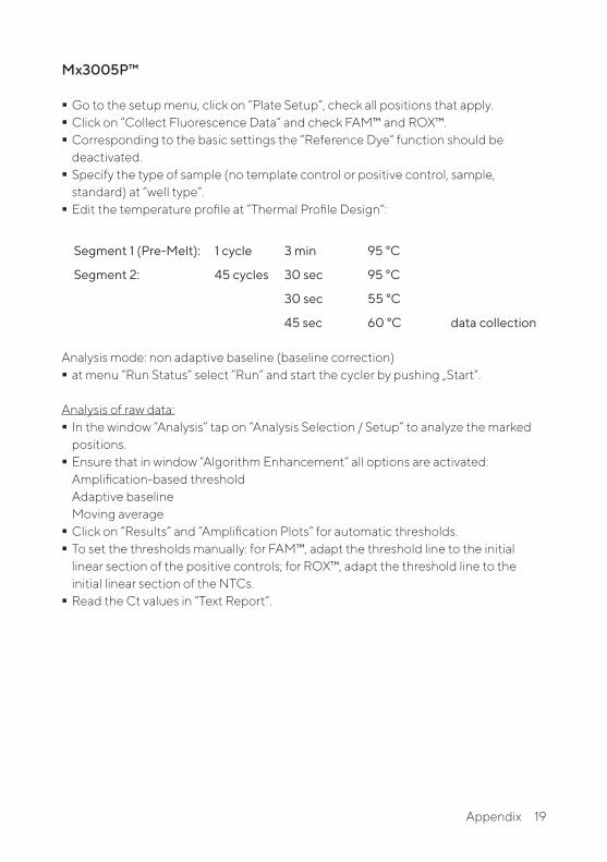
Mx3005P™
� Go to the setup menu, click on “Plate Setup“, check all positions that apply. � Click on “Collect Fluorescence Data“ and check FAM™ and ROX™. � Corresponding to the basic settings the “Reference Dye“ function should be deactivated.
� Specify the type of sample (no template control or positive control, sample, standard) at “well type“.
� Edit the temperature profile at ”Thermal Profile Design“:
Segment 1 (Pre-Melt): 1 cycle 3 min 95 °C
Segment 2: 45 cycles 30 sec 95 °C
30 sec 55 °C
45 sec 60 °C data collection Analysis mode: non adaptive baseline (baseline correction)
� at menu “Run Status“ select ”Run“ and start the cycler by pushing „Start“.
Analysis of raw data: � In the window “Analysis” tap on ”Analysis Selection / Setup” to analyze the marked positions.
� Ensure that in window “Algorithm Enhancement“ all options are activated: Amplification-based threshold Adaptive baseline Moving average
� Click on “Results“ and ”Amplification Plots“ for automatic thresholds. � To set the thresholds manually: for FAM™, adapt the threshold line to the initial linear section of the positive controls; for ROX™, adapt the threshold line to the initial linear section of the NTCs.
� Read the Ct values in “Text Report“.
19Appendix

Bio-Rad CFX96 Touch™ / CFX96 Touch™ deep well
Run Setup Protocol Tab: � Click File --> New --> Protocol to open the Protocol Editor and create a new protocol. � Select any step in either the graphical or text display. � Click the temperature or well time to directly edit the value.
Segment 1: 1 cycle 3 min 95 °C
Segment 2: 30 sec 95 °C
Segment 3: 30 sec 55 °C
Segment 4: 45 sec 60 °C data collection
GO TO Step 2, 44 more cycles
20 Appendix

Run Setup Plate Tab: � Click File --> New --> Plate to open the Plate Editor and create a new plate. � Specify the type of sample at Sample Type. � Name your samples at Sample Name. � Use the Scan Mode dropdown menu in the Plate Editor Toolbar to designate the data acquisition mode to be used during the run. Select All Channels mode.
� Click Select Fluorophores to indicate the fluorophores that will be used in the run. � Choose FAM™ for the detection of mycoplasma amplification and ROX™ for monitoring the amplification of the internal control. Within the plate diagram, select the wells to load.
� Choose the fluorophore data you want to display by clicking the fluorophore checkboxes located under amplification chart. Select FAM™ to display data of mycoplasma detection and ROX™ to display internal control amplification data.
21Appendix

Data Analysis: � Select Settings in the menu and select Baseline Subtracted Curve Fit as baseline setting and Single Threshold mode as Cq determination.
� View amplification curves of FAM™ channel by selecting the FAM™ checkbox under the amplification plot.
� To enable thresholds setting, please follow the specific instructions provided in the manual of your cycler.
� FAM™: adapt the threshold line to the initial linear section of the positive controls. ROX™: adapt the threshold line to the initial linear section of the NTCs.
� Evaluate the Ct values according to chapter 9.
22 Appendix

11. Related Products
Detection Kits for qPCR
SMB95-1003/1004 SMB95-1005/1006 SMB95-1007SMB95-1008SMB95-1009SMB95-1012SMB95-1014/1013
Microsart® ATMP MycoplasmaMicrosart® RESEARCH Mycoplasma Microsart® ATMP Sterile Release Microsart® ATMP BacteriaMicrosart® RESEARCH Bacteria Microsart® ATMP FungiMicrosart® RESEARCH Fungi
25/100 tests25/100 tests
10 samples100 tests
25 tests100 tests
25/100 tests
Vivaspin® and Coating Buffer
SMB95-2002VS0641VS0642VS2041VS2042
Microsart® AMP Coating BufferVivaspin® 6 Polyethesulfone 100,000 MWCOVivaspin® 6 Polyethesulfone 100,000 MWCOVivaspin® 20 Polyethesulfone 100,000 MWCOVivaspin® 20 Polyethesulfone 100,000 MWCO
20 × 2 ml25 units
100 units12 units
48 units
Microsart® Calibration Reagent, 1 vial, 108 genomes / vial (bacteria, including Mollicutes)
SMB95-2021 SMB95-2022 SMB95-2023 SMB95-2024 SMB95-2025 SMB95-2026 SMB95-2027 SMB95-2028 SMB95-2029 SMB95-2030 SMB95-2031 SMB95-2032 SMB95-2033 SMB95-2034 SMB95-2035 SMB95-2036
Mycoplasma argininiMycoplasma oraleMycoplasma gallisepticumMycoplasma pneumoniaeMycoplasma synoviaeMycoplasma fermentansMycoplasma hyorhinisAcholeplasma laidlawiiSpiroplasma citriBacillus subtilisPseudomonas aeruginosaKocuria rhizophilaClostridium sporogenesBacteroides vulgatusStaphylococcus aureusMycoplasma salivarium
Microsart® Calibration Reagent, 1 vial, 106 genomes / vial (fungi)
SMB95-2044 SMB95-2045 SMB95-2046 SMB95-2047 SMB95-2048SMB95-2049SMB95-2050
Candida albicansAspergillus brasiliensisAspergillus fumigatusPenicillium chrysogenumCandida glabrataCandida kruseiCandida tropicalis
23Related Products

Microsart® Validation Standard, 3 vials each, 10 CFU / vial (Mollicutes)
SMB95-2011 SMB95-2012SMB95-2013 SMB95-2014 SMB95-2015SMB95-2016SMB95-2017 SMB95-2018 SMB95-2019 SMB95-2020
Mycoplasma argininiMycoplasma oraleMycoplasma gallisepticum Mycoplasma pneumoniaeMycoplasma synoviaeMycoplasma fermentansMycoplasma hyorhinisAcholeplasma laidlawiiSpiroplasma citriMycoplasma salivarium
Microsart® Validation Standard, 6 vials each, 99 CFU / vial (bacteria* and fungi)
SMB95-2005SMB95-2006SMB95-2007SMB95-2008SMB95-2009SMB95-2010SMB95-2037SMB95-2038SMB95-2039SMB95-2040SMB95-2041SMB95-2042SMB95-2043
Bacillus subtilisPseudomonas aeruginosaKocuria rhizophilaClostridium sporogenesBacteroides vulgatusStaphylococcus aureusCandida albicansAspergillus brasiliensisAspergillus fumigatusPenicillium chrysogenumCandida glabrataCandida kruseiCandida tropicalis
* except for Mollicutes
DNA Extraction Kit
SMB95-2001SMB95-2003 56-0002
Microsart® ATMP Extraction (for bacteria and fungi)Microsart® AMP Extraction (for mycoplasma)Proteinase K**
50 extractions50 extractions50 extractions
PCR Clean™ **
15-202515-2200
DNA Decontamination Reagent, spray bottleDNA Decontamination Reagent, refill bottles
250 ml4 x 500 ml
PCR Clean™ Wipes**
15-200115-2002
DNA Decontamination WipesDNA Decontamination Wipes, refill sachets
50 wipes5 x 50 wipes
** Distributed by Minerva Biolabs
24 Related Products

Limited Product WarrantyThis warranty limits our liability for replacement of this product. No warranties of any kind, express or implied, including, without limitation, implied warranties of merchantability or fitness for a particular purpose, are provided. Sartorius Stedim Biotech GmbH and Minerva Biolabs shall have no liability for any direct, indirect, consequential, or incidental damages arising out of the use, the results of use, or the inability to use this product.
TrademarksABI Prism is a registered trademark of Applera Corporation or its subsidiaries in the US and certain other countries. CFX96 Touch is a trademark of Bio-Rad Laboratories, Inc. Mx3005P is a trademark of Agilent Technologies. FAM and ROX are trademarks of Applera Corporation or its subsidiaries in the US and certain other countries. Microsart is a registered trademark of Sartorius Stedim Biotech GmbH. PCR Clean is a trademark of Minerva Biolabs GmbH.
Last technical revision: 2020-01-15

Copyright by Sartorius Stedim Biotech GmbH, Goettingen, Germany. All rights reserved. No part of this publication may be reprinted or translated in any form or by any means without the prior written permission of Sartorius Stedim Biotech GmbH. The status of the information, specifications and illustrations in this manual is indicated by the date given below. Sartorius Stedim Biotech GmbH reserves the right to make changes to the technology, features, specifications and design of the equipment without notice.
Status:August 2020,Sartorius Stedim BiotechGmbH, Goettingen, Germany
Printed in Germany on paper thathas been bleached without any useof chlorine. | WPublication No.: SM-6109-e150102Order No.: 85037-546-74Ver. 08 | 2020
Sartorius Stedim Biotech GmbHAugust-Spindler-Str. 1137079 Goettingen, Germany
Phone +49 551 308 0Fax +49 551 308 3289 www.sartorius.com

Close lid tightlybriefly
ST_SI_Microsart®-AMP-Mycoplasma_03_ENThis procedure overview is not a substitute for the detailed manual.
1275 µl
Mycoplasma Mix
for 5 min RT
briefly
for 5 sec
Mycoplasma Mix
Positive Control DNA
Internal Control DNA
storage +2 - +8 °Cafter rehydration ≤ -18 °C
Rehydration BufferMycoplasma MixPCR grade WaterPositive ControlInternal Control
1. Rehydration of Reagents
4. Start of the qPCR Reaction
incubatevortexcentrifugeadd
Start PCRprogram
55°C60°C
3 min95°C 95°C
30 sec
30 sec45 sec
45 cycles
50 μl sample50 μl Positive Control (green cap)50 μl elution buffer orPCR grade Water (white cap)(negative control)
2. Preparation of the Reaction Mix
b) Internal Control not added during DNA extraction
Reaction Mix (for 1 reaction) 49 µl Mycoplasma Mix (red cap) 1 µl Internal Control (yellow cap)
a) Internal Control added during DNA extraction
50 µl Mycoplasma Mix (red cap)
50 µl Reaction Mix
do not forget to add 1 µl Internal Control to NTC and PC
*
3. Addition of Samples and Controls
Positive Control
300 µl
Internal Control
300 µl
Short Instructions

Sartorius Stedim Biotech GmbHAugust-Spindler-Str. 1137079 Goettingen, Germany
Phone +49 551 308 0Fax +49 551 308 3289 www.sartorius.com
Copyright by Sartorius Stedim Biotech GmbH, Goettingen, Germany. All rights reserved. No part of this publication may be reprinted or translated in any form or by any means without the prior written permission of Sartorius Stedim Biotech GmbH. The status of the information, specifications and illustrations in this manual is indicated by the date given below. Sartorius Stedim Biotech GmbH reserves the right to make changes to the technology, features, specifications and design of the equipment without notice.
Status:August 2020,Sartorius Stedim BiotechGmbH, Goettingen, Germany
Printed in Germany on paper thathas been bleached without any useof chlorine. | WPublication No.: SM-6109-e150102Order No.: 85037-546-74Ver. 08 | 2020