
Microfocus X-ray Sources for Chemical and High-Pressure...
24
Microfocus X-ray Sources for Chemical and High-Pressure Crystallography Jürgen Graf Incoatec GmbH, Geesthacht Bruker User‘s Group Meeting SC-XRD 2010
Transcript of Microfocus X-ray Sources for Chemical and High-Pressure...

Microfocus X-ray Sources for Chemical and High-Pressure Crystallography
Jürgen Graf
Incoatec GmbH, Geesthacht
Bruker User‘s Group Meeting SC-XRD 2010

2/23 Jürgen Graf – Karlsruhe 2010
Incoatec on the GKSS Research Center, 30 km South of Hamburg
History and Background
• Company founded in Jan 2002
• Spin-off of the Department of Thin Film Technology, GKSS Research Center, located in Geesthacht near Hamburg
• Joint venture together with Bruker AXS
• Development and manufacturing of multilayer X-ray optics by thin film technology
• Own application lab and R&D activities
Incoatec GmbH @ GITZ: Geesthachter Innovation and Technology Center
Incoatec GmbH:INnovative COAting TEChnologies

3/23 Jürgen Graf – Karlsruhe 2010
d = 2 – 6 nm
m 1.0° (Cu-K)
m 0.5° (Mo-K)
Multilayer X-ray Optics
W/C Multilayer (TEM, Uni Kiel, Prof. Jäger)
Capture angle Source
X-tal

4/23 Jürgen Graf – Karlsruhe 2010
Microfocus X-ray Sources
• Mirror should “see” the whole source:- Multilayer mirrors work best with microfocus X-ray sources
1.2 kW Microfocus Rotating Anode 30 W Microfocus Sealed Tube

5/23 Jürgen Graf – Karlsruhe 2010
• Small spot
• 2D heat flow allows more
efficient cooling
• ~ 5 kW/mm2
• Rel. brightness: <10X
Power loading of X-ray sources
e-
micro-focus sealed tube
e-
micro-focus sealed tube
• Large spot
• Quasi-1D heat flow limits
power density
• ~0.5 kW/mm2
• Rel. brightness: 1
e-
conventional sealed tube
e-
conventional sealed tube
e-
rotating anode
e-
rotating anode
• Large or small spot
• Heat spread by rotation
• >10 kW/mm2
• Rel. brightness: <100X
Power loading in all solid-target X-ray sources is limited by heat dissipation
R. Durst, A. Storm, Bruker AXS Technical Note 4 SCD, 2007.

6/23 Jürgen Graf – Karlsruhe 2010
Incoatec Microfocus Source – IµS
• High-brilliance microfocus sealed tube X-ray source
• Spot size < 50 µm
• Low power: max. 30 W• Air-cooled• Family of 2D beam shaping Montel optics:
The Quazar Optics (focusing or parallel beam)• Implemented in many new Bruker AXS instruments• Upgrade on older diffractometers
• Cu, Cr, Mo and Ag radiation 3 yearswarranty
> 100 soldworldwide
7/23 Jürgen Graf – Karlsruhe 2010
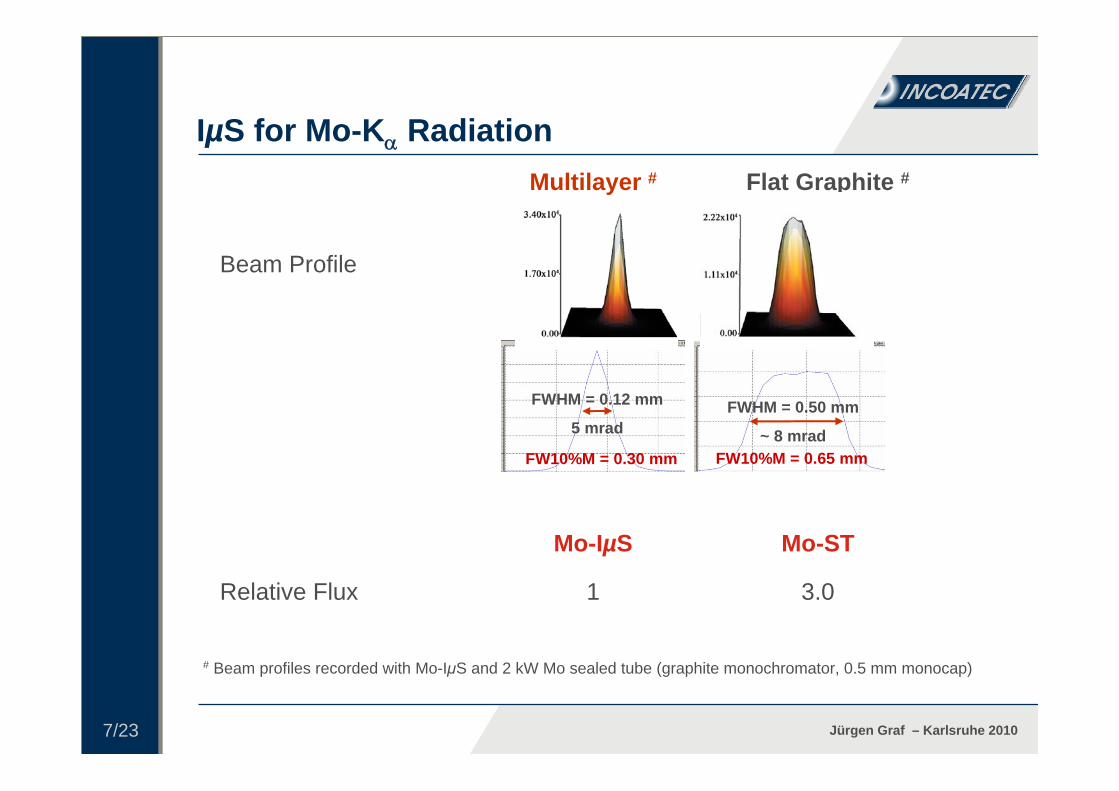
Mo-ST Mo-IµS
Relative Flux
Beam Profile
3.01
Flat Graphite #Multilayer #
# Beam profiles recorded with Mo-IµS and 2 kW Mo sealed tube (graphite monochromator, 0.5 mm monocap)
FWHM = 0.12 mm
5 mradFWHM = 0.50 mm
~ 8 mrad
IµS for Mo-K Radiation
FW10%M = 0.30 mm FW10%M = 0.65 mm
8/23 Jürgen Graf – Karlsruhe 2010
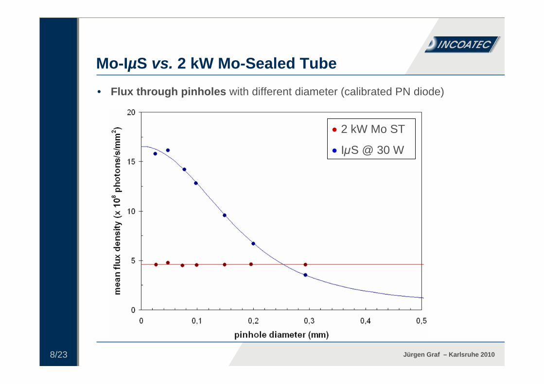
• Flux through pinholes with different diameter (calibrated PN diode)
2 kW Mo ST
IµS @ 30 W
Mo-IµS vs. 2 kW Mo-Sealed Tube
9/23 Jürgen Graf – Karlsruhe 2010
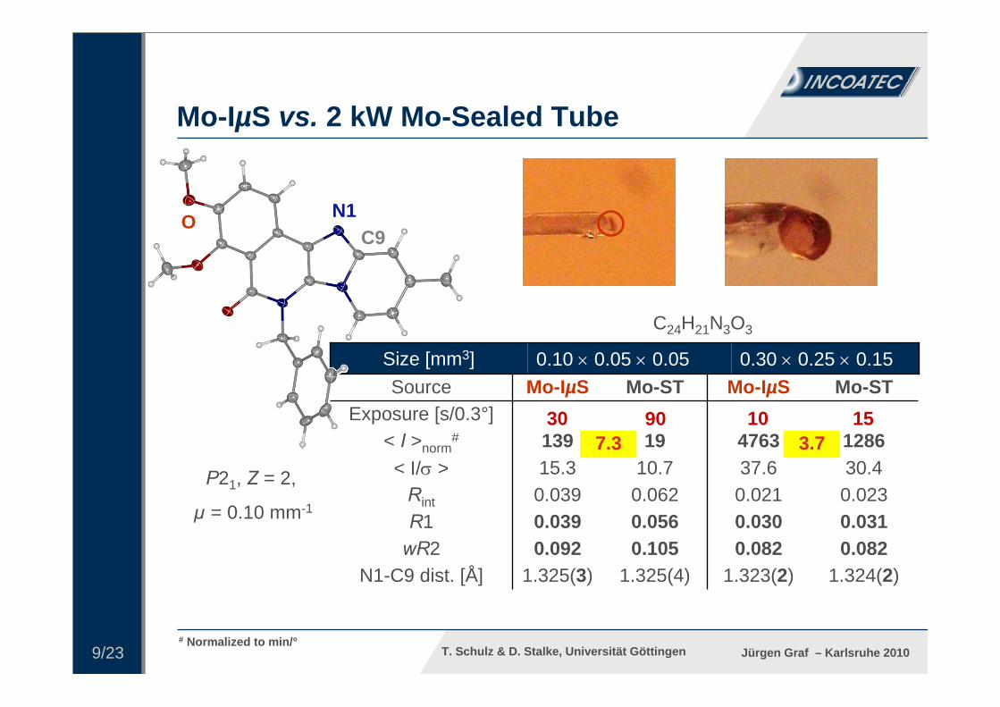
0.0310.0300.0560.039R1
1.324(2)1.323(2)1.325(4)1.325(3)N1-C9 dist. [Å]0.0820.0820.1050.092wR2
0.0230.0210.0620.039Rint
30.437.610.715.3< I/ >1286476319139< I >norm
#15109030Exposure [s/0.3°]
Mo-STMo-IµSMo-STMo-IµSSource0.30 0.25 0.150.10 0.05 0.05Size [mm3]
C24H21N3O3
Mo-IµS vs. 2 kW Mo-Sealed Tube
# Normalized to min/°
N1C9
O
T. Schulz & D. Stalke, Universität Göttingen
P21, Z = 2,
µ = 0.10 mm-1
7.330 90
3.710 15
10/23 Jürgen Graf – Karlsruhe 2010
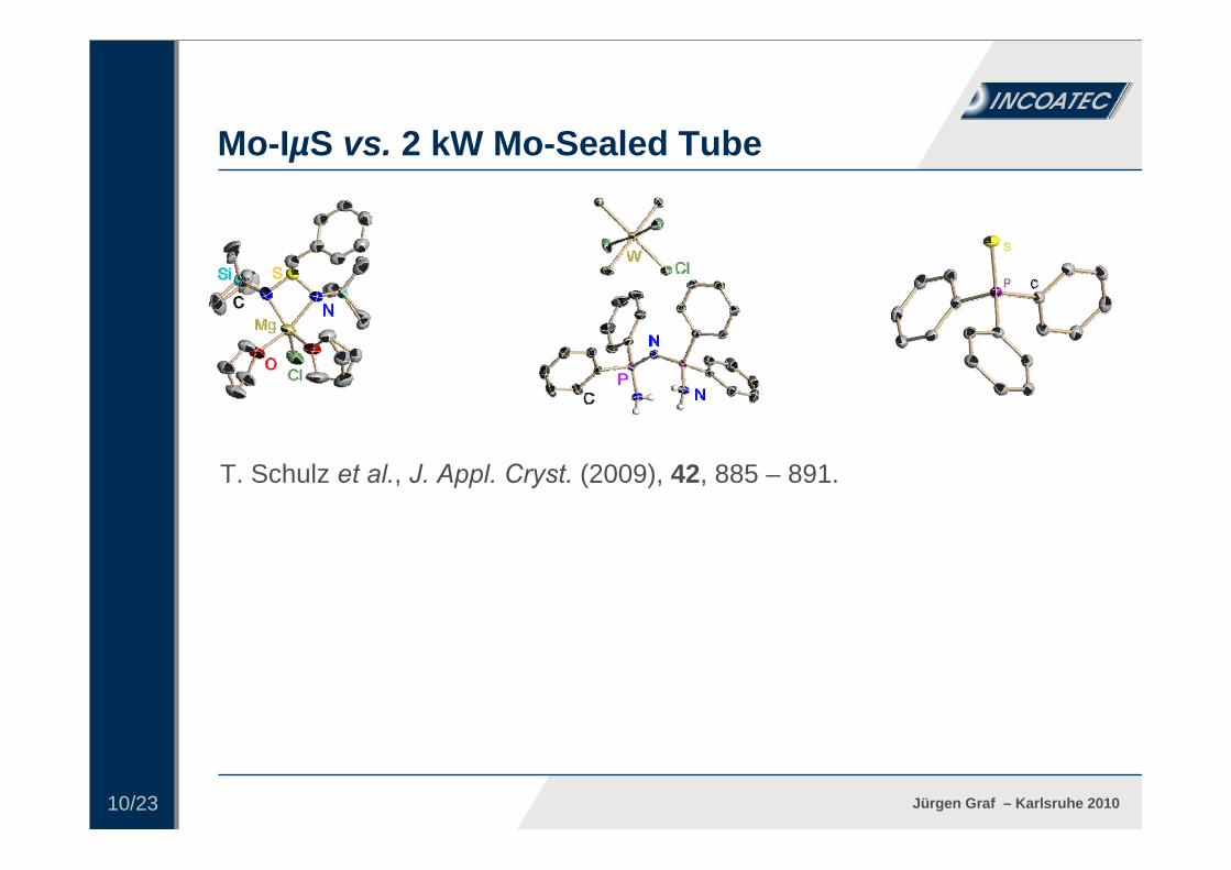
Conclusions:
• Mo-IμS always superior for small crystals (< 0.15 mm)• Comparable results for larger crystals (> 0.20 mm), often with
shorter exposure times• Precise crystal centring and scaling are essential for data quality
T. Schulz et al., J. Appl. Cryst. (2009), 42, 885 – 891.
Mo-IµS vs. 2 kW Mo-Sealed Tube

11/23 Jürgen Graf – Karlsruhe 2010
CuSO4
2.9
0.0390.0350.0900.084R10.1220.1150.2410.240wR2
3.35.21.11.6< >80.6175.65.917.1< I >20 20 150 150 Exposure time [s/°]4.00.02*4.00.03Power [kW]
FR 591Mo-IµSFR 591Mo-IµSSource
0.07 0.05 0.050.04 0.04 0.02Size [mm3]
SiO2
# FR591 plus flat graphite monochromator, 0.3 mm collimatorAll data recorded with a Nonius Kappa CCD goniometer
* Approximation for 30 W: < I > = 263.4, < > = 6.4
3.3263 81
Got Small Crystals? – Inorganics
C. W. Lehmann, Max-Planck-Institut Mülheim
• Comparison Mo-IµS vs. 4 kW Mo RAG # plus flat graphite monochromator

12/23 Jürgen Graf – Karlsruhe 2010
„C40H24N8Zn”
Got Small Crystals? – MOF
P42/n, Z = 8
a = b = 23.69 Å, c = 14.99 Å
µ (Mo) = ~ 0.5 mm-1
Mo-IµS
120 s/0.5°, APEX IIDX = 41 mm, 2 = 11°
1.26

13/23 Jürgen Graf – Karlsruhe 2010
Got Small Crystals? – MOF • Typical diffraction patterns recorded with Cu-IµS MX (FWHM = 0.12 mm)
Cu-IµS MX Cu-IµS MX
60 s/0.5°, APEX IIDX = 51 mm, 2 = -32°
120 s/0.5°, APEX IIDX = 51 mm, 2 = -93°
1.47
0.88

14/23 Jürgen Graf – Karlsruhe 2010
„C40H24N8Zn”
0.0545 (0.2330)Rint
6.5 (2.4)<Redundancy>
~ 2.5Total time [d]
16.2 (3.4)< I/ >0.88 (0.98 – 0.88)Resolution [Å]
60 - 120Exposure time [s/0.5°]Cu-IµS MXSource
0.04 0.03 0.01Size [mm3]
Got Small Crystals? – MOF
P42/n, Z = 8
a = b = 23.69 Å, c = 14.99 Å
µ (Cu) = ~ 4 mm-1
15/23 Jürgen Graf – Karlsruhe 2010
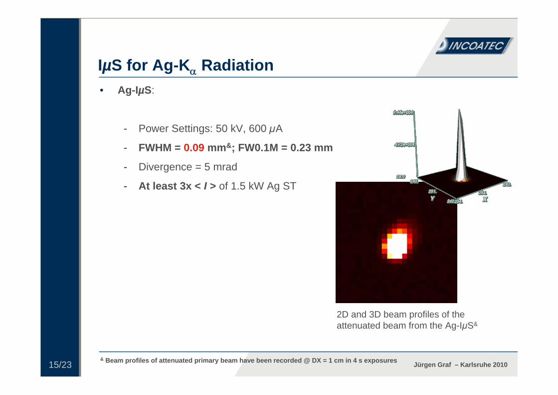
• Ag-IµS:
- Power Settings: 50 kV, 600 µA
- FWHM = 0.09 mm&; FW0.1M = 0.23 mm
- Divergence = 5 mrad
- At least 3x < I > of 1.5 kW Ag ST
• Benefits of Ag-Radiation:
- Less absorption, less extinction
- Solid State Chemistry
- Charge Density Studies
- “Compressed” reciprocal space
- High-pressure Crystallography
IµS for Ag-K Radiation
& Beam profiles of attenuated primary beam have been recorded @ DX = 1 cm in 4 s exposures
2D and 3D beam profiles of theattenuated beam from the Ag-IµS&

16/23 Jürgen Graf – Karlsruhe 2010
38.420.6µ [mm-1]
0.0048(10) 0.0028(4) Ueq (O1)0.0184, 0.05100.0176, 0.0450R1, wR2
0.77 (0.87 – 0.77)0.61 (0.71 – 0.61)Max. resolution [Å]
2.296(6)2.290(3)d(Pb1-O1) [Å]
187.7 (159.7)216.6 (192.7)< I/ >(0.77 Å)69 (17)127 (43)Unique data
187.7 (159.7)186.8 (143.5)< I/ >
1010Exposure [s/0.3°]
Mo-IµSAg-IµSSource0.11 0.09 0.06Size [mm3]
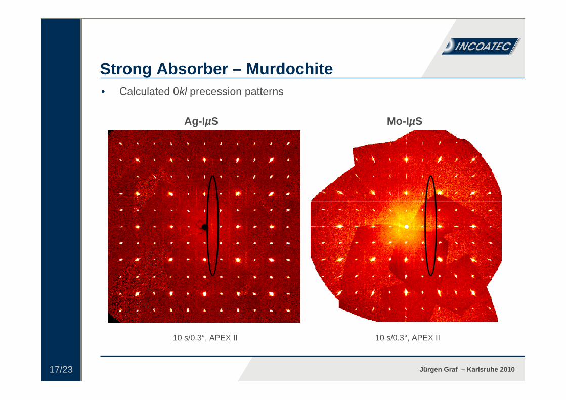
Cu6PbO8-x(Cl, Br)x
Strong Absorber – Murdochite
Fd-3m, Z = 4
17/23 Jürgen Graf – Karlsruhe 2010
Strong Absorber – Murdochite• Calculated 0kl precession patterns
10 s/0.3°, APEX II 10 s/0.3°, APEX II
Ag-IµS Mo-IµS

18/23 Jürgen Graf – Karlsruhe 2010
0.0342 (0.1489)0.0306 (0.1636)Rint705 (523)860 (630)Data used (I > 2(I))
18.3 (4.7)19.6 (3.2)< I/ >0.90 (1.00 – 0.90)0.90 (1.00 – 0.90)Resolution [Å]
33.7 (22.6)40.6 (28.9)<Completeness>
0.0532; 0.12320.0487; 0.1025R1; wR2
721 (135)866 (170)Unique data1.1 (0.7)1.5 (0.9)<Redundancy>
2020Exposure [s/0.3°]2 kW Mo-STAg-IµSSource
0.25 0.20 0.20Size [mm3]
C9H17NO2. 2 H2O
F. P. A. Fabbiani, Universität Göttingen
GabapentinHeptahydrate,
P-1, Z = 2,
µ = 0.12 mm-1
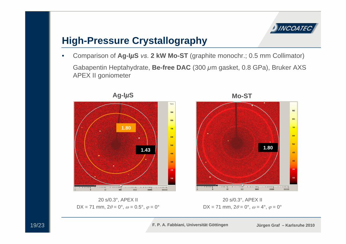
High-Pressure Crystallography
Gabapentincrystal in 300 µm gasket of Be-freeDAC
19/23 Jürgen Graf – Karlsruhe 2010
High-Pressure Crystallography
1.80
20 s/0.3°, APEX IIDX = 71 mm, 2 = 0°, = 0.5°, = 0°
20 s/0.3°, APEX IIDX = 71 mm, 2 = 0°, = 4°, = 0°
• Comparison of Ag-IµS vs. 2 kW Mo-ST (graphite monochr.; 0.5 mm Collimator)
Gabapentin Heptahydrate, Be-free DAC (300 µm gasket, 0.8 GPa), Bruker AXS APEX II goniometer
Mo-STAg-IµS
1.80
1.80
1.43
F. P. A. Fabbiani, Universität Göttingen

20/23 Jürgen Graf – Karlsruhe 2010
High-Pressure Crystallography
20 s/0.3°, APEX IIDX = 71 mm, 2 = -15°, = -160.1°, = 180°
• Ag vs. Mo: Gain in resolution by “Compression” of the Reciprocal Space
Gabapentin Heptahydrate, Be-free DAC (300 µm gasket, 0.8 GPa), Bruker AXS APEX II goniometer
Ag-IµS
1.36 1.36
1.36 1.36
F. P. A. Fabbiani, Universität Göttingen

21/23 Jürgen Graf – Karlsruhe 2010
IµS for Mo and Ag – High flux density in small, convergent beam
• Ideal for small and medium sized crystals
• Ideal for high-pressure experiments
• Mo-IµS: at least 4-fold intensity gain compared to 2 kW Mo sealed tube- div = 5 mrad, FWHM = 0.12 mm; FW10%M = 0.30 mm
• Ag-IµS: at least 3-fold intensity gain compared to 1.5 kW Ag sealed tube- div = 5 mrad, FWHM = 0.09 mm; FW10%M = 0.23 mm- Ideal for crystals showing strong absorption or extinction- Ideal for high-resolution data sets and de-novo phase determination using
high-pressure cells
Summary
• Cu-IµS powerful allrounder for Small Molecules, Proteins and Material Research

22/23 Jürgen Graf – Karlsruhe 2010
• The Göttingen people: - @ IAC: T. Schulz, D. Stalke- @ GWZ: F. P. A. Fabbiani
• C. Hauf, G. Eickerling, W. Scherer, University of Augsburg
• A. Dreier, C. W. Lehmann, Max-Planck-Institut, Mülheim
• Th. Malcherek (Hamburg), R. Seidel (Bochum)
• The Bruker AXS people: - R. Durst, M. Ruf, H. Ott, D. Stern, M. Nüsse
• The Incoatec people
Acknowledgement

23/23 Jürgen Graf – Karlsruhe 2010
Materials Center Leoben, Leoben
THANK YOU
Biozentrum, Univ. Basel
MPI, StuttgartNovartis AG, Basel
Sanofi Aventis GmbH, Frankfurt
Institute Le Bel, StrasbourgChemistry Dep., Univ.
Goettingen
Incoatec [email protected]

24/23 Jürgen Graf – Karlsruhe 2010
Please contact for more information:
Incoatec GmbHMax-Planck-Str. 2 • 21502 Geesthacht • GermanyTel: +49(0)41 52 - 88 93 81 • www.incoatec.de


![Mainframe Migration Macrosoft – Leaders in Mainframe Migration [ Lift & Shift *] “Lift & Shift” is a trademark of Microfocus.](https://static.fdocuments.in/doc/165x107/56649d235503460f949f9b08/mainframe-migration-macrosoft-leaders-in-mainframe-migration-lift-shift.jpg)


![Preparation,Modification,andApplicationof …downloads.hindawi.com/journals/jnm/2011/573687.pdf · 2019-07-31 · angle X-ray microfocus scattering [4, 5], ... nanocrystals by acid](https://static.fdocuments.in/doc/165x107/5f81279bafd8ec5abd5ed74d/preparationmodiicationandapplicationof-2019-07-31-angle-x-ray-microfocus-scattering.jpg)
