Microflora and Chemical Composition of Dental Plaque from
7
INFECTION AND IMMUNITY, June 1980, p. 853-859 0019-9567/80/06-0853/07$02.00/0 Vol. 28, No. 3 Microflora and Chemical Composition of Dental Plaque from Subjects with Hereditary Fructose Intolerance CHARLES I. HOOVER,' ERNEST NEWBRUN,I* GERRALD METTRAUX,2 AND HANS GRAF2 Department of Oral Medicine and Hospital Dentistry, University of California San Francisco, San Francisco, California 94143,' and Department of Periodontology, University of Bern, CH-3010, Bern, Switzerland2 We compared the microbiological and chemical composition of dental plaque from subjects with hereditary fructose intolerance who restrict their dietary sugar intake with that of control subjects who do not. The two groups showed no significant differences in chemical composition of plaque: the mean protein, carbohydrate, calcium, magnesium, and phosphate contents were similar. Dental plaque from both groups contained similar numbers of total colony-forming units per microgram of plaque protein, and Streptococcus sanguis, an indigenous nonpathogen, was isolated with equal frequency from plaque samples of both groups. However, potentially odontopathic Streptococcus mutans and Lactoba- cillus were isolated three to four times more frequently from plaque samples of control subjects than from plaque samples of subjects with hereditary fructose intolerance. Clearly, diet (sucrose in particular) influences the colonization and multiplication of specific cariogenic organisms in dental plaque. In 1890, W. D. Miller first postulated that dental caries resulted from the fermentation of carbohydrate by acidogenic microorganisms (39). During the early 1900s, researchers sug- gested that Lactobacillus acidophilus caused dental caries and reported that dietary restric- tion of carbohydrate concomitantly reduced sal- ivary counts of L. acidophilus and the develop- ment of new carious lesions (3, 23, 28). Recently, Streptococcus mutans has been rec- ognized as the prime cariogenic microorganism in humans (13, 26, 30, 36). Dietary restriction of carbohydrate also results in the reduction of S. mutans and iodophilic polysaccharide-storing microorganisms in dental plaque (14, 50). Chil- dren with congenital sucrase deficiency restrict the amount of sucrose in their diet and have fewer caries than normal children of similar age (51). S. mutans could not be detected in dental plaque from 40% of these children. Hereditary fructose intolerance (HFI), first described in 1956 by Chambers and Pratt (11), is characterized by severely reduced activity of fructose 1-phosphate aldolase (41, 43). Ingestion of items containing fructose or sucrose or both results in severe hypoglycemia, nausea, and vomiting. People who have HFI develop a strong aversion to all sweets, sucrose-containing foods, and fruits, and adhere to a lifelong, self-imposed diet that virtually excludes sucrose. We have compared the diet, oral hygiene, caries experi- ence, plaque microflora, and plaque chemical composition of subjects who have HFI with those of controls. Dietary analyses, oral hygiene status, and caries experience of our HFI and control subjects are reported in detail elsewhere (E. Newbrun, C. Hoover, G. Mettraux, and H. Graf, submitted for publication). This report assesses the effect of dietary sucrose restriction on the bacterial colonization and chemical com- position of dental plaque. MATERLS AND METHODS Study population. On four, in some cases five, separate occasions over a 2-year period, we examined 17 HFI subjects (3 from the United States, 14 from Switzerland) and 14 control subjects (2 from the United States, 12 from Switzerland). Oral examina- tions and plaque collections took place between 8 and 11 a.m. and were usually performed on a pair of subjects, one from each group. The HFI group con- sisted of 10 male and 7 female subjects whose mean age was 29.1 years (range, 6 to 54 years). The control group consisted of 3 male and 11 female subjects whose mean age was 26.5 years (range, 9 to 55 years). Eight control subjects were blood relatives of persons in the HFI group. Control subjects consumed more than 20 times as much sucrose as HFI subjects and had significantly greater DMFS scores (control, 36.1; HFI, 3.3). Microbiological analyses of dental plaque. Su- pragingival dental plaque was collected and assayed from four specific sites in each subject on each occa- sion. Sterile needles were used to collect plaque from the occlusal surface of the right first mandibular molar and the palatal groove of the left first maxillary molar. Interproximal plaque was obtained from between the left first and second mandibular molars and the right 853 Downloaded from https://journals.asm.org/journal/iai on 29 November 2021 by 1.215.31.114.
Transcript of Microflora and Chemical Composition of Dental Plaque from
INFECTION AND IMMUNITY, June 1980, p. 853-8590019-9567/80/06-0853/07$02.00/0
Vol. 28, No. 3
Microflora and Chemical Composition of Dental Plaque fromSubjects with Hereditary Fructose Intolerance
CHARLES I. HOOVER,' ERNEST NEWBRUN,I* GERRALD METTRAUX,2 AND HANS GRAF2Department of Oral Medicine and Hospital Dentistry, University of California San Francisco, SanFrancisco, California 94143,' and Department of Periodontology, University of Bern, CH-3010, Bern,
Switzerland2
We compared the microbiological and chemical composition of dental plaquefrom subjects with hereditary fructose intolerance who restrict their dietary sugarintake with that of control subjects who do not. The two groups showed nosignificant differences in chemical composition of plaque: the mean protein,carbohydrate, calcium, magnesium, and phosphate contents were similar. Dentalplaque from both groups contained similar numbers of total colony-forming unitsper microgram of plaque protein, and Streptococcus sanguis, an indigenousnonpathogen, was isolated with equal frequency from plaque samples of bothgroups. However, potentially odontopathic Streptococcus mutans and Lactoba-cillus were isolated three to four times more frequently from plaque samples ofcontrol subjects than from plaque samples of subjects with hereditary fructoseintolerance. Clearly, diet (sucrose in particular) influences the colonization andmultiplication of specific cariogenic organisms in dental plaque.
In 1890, W. D. Miller first postulated thatdental caries resulted from the fermentation ofcarbohydrate by acidogenic microorganisms(39). During the early 1900s, researchers sug-gested that Lactobacillus acidophilus causeddental caries and reported that dietary restric-tion of carbohydrate concomitantly reduced sal-ivary counts of L. acidophilus and the develop-ment of new carious lesions (3, 23, 28).
Recently, Streptococcus mutans has been rec-ognized as the prime cariogenic microorganismin humans (13, 26, 30, 36). Dietary restriction ofcarbohydrate also results in the reduction of S.mutans and iodophilic polysaccharide-storingmicroorganisms in dental plaque (14, 50). Chil-dren with congenital sucrase deficiency restrictthe amount of sucrose in their diet and havefewer caries than normal children of similar age(51). S. mutans could not be detected in dentalplaque from 40% of these children.
Hereditary fructose intolerance (HFI), firstdescribed in 1956 by Chambers and Pratt (11),is characterized by severely reduced activity offructose 1-phosphate aldolase (41, 43). Ingestionof items containing fructose or sucrose or bothresults in severe hypoglycemia, nausea, andvomiting. People who have HFI develop a strongaversion to all sweets, sucrose-containing foods,and fruits, and adhere to a lifelong, self-imposeddiet that virtually excludes sucrose. We havecompared the diet, oral hygiene, caries experi-ence, plaque microflora, and plaque chemicalcomposition of subjects who have HFI with
those of controls. Dietary analyses, oral hygienestatus, and caries experience of our HFI andcontrol subjects are reported in detail elsewhere(E. Newbrun, C. Hoover, G. Mettraux, and H.Graf, submitted for publication). This reportassesses the effect of dietary sucrose restrictionon the bacterial colonization and chemical com-position of dental plaque.
MATERLS AND METHODS
Study population. On four, in some cases five,separate occasions over a 2-year period, we examined17 HFI subjects (3 from the United States, 14 fromSwitzerland) and 14 control subjects (2 from theUnited States, 12 from Switzerland). Oral examina-tions and plaque collections took place between 8 and11 a.m. and were usually performed on a pair ofsubjects, one from each group. The HFI group con-sisted of 10 male and 7 female subjects whose meanage was 29.1 years (range, 6 to 54 years). The controlgroup consisted of 3 male and 11 female subjectswhose mean age was 26.5 years (range, 9 to 55 years).Eight control subjects were blood relatives of personsin the HFI group. Control subjects consumed morethan 20 times as much sucrose as HFI subjects andhad significantly greater DMFS scores (control, 36.1;HFI, 3.3).
Microbiological analyses of dental plaque. Su-pragingival dental plaque was collected and assayedfrom four specific sites in each subject on each occa-sion. Sterile needles were used to collect plaque fromthe occlusal surface of the right first mandibular molarand the palatal groove of the left first maxillary molar.Interproximal plaque was obtained from between theleft first and second mandibular molars and the right
853
Dow
nloa
ded
from
http
s://j
ourn
als.
asm
.org
/jour
nal/i
ai o
n 29
Nov
embe
r 20
21 b
y 1.
215.
31.1
14.
854 HOOVER ET AL.
first and second maxillary molars with sterile unwaxeddental floss (John 0. Buttler Co., Chicago, Ill.) held ina sterile plastic support (E-Z Floss, Palm Springs,Calif.). When any of these teeth were carious, restored,or missing, we substituted appropriate alternativesites.The collected plaque samples were immediately
placed in A/S Nunc vials (Almac Cryogenics Inc.,Oakland, Calif.) containing 1.0 ml of reduced transportfluid (47). The vials were introduced into an anaerobicglove box (12) containing 85% N2, 10% H2, and 5% C02,and the samples were dispersed, serially diluted,plated, cultured, and enumerated for total colony-forming units (CFU) of S. sanguis, S. mutans, andLactobacillus as previously described (27).We picked three to five representative S. mutans
colonies at random from S. mutans-positive plaquesamples and isolated them in pure culture. When morethan one colony morphology was apparent, we isolatedrepresentative colonies of each morphology for bio-chemical identification and biotyping. The isolateswere identified as S. mutans by Facklam's scheme (17)and biochemically typed by the scheme of Shklair andKeene (45).To compare the numbers of S. mutans colonizing
the plaque of control subjects with numbers in HFIsubjects, we analyzed the protein content of 109 inter-proximal samples collected for microbiological analysisby using a Coomassie brilliant blue colorimetric assay(37). The volume of each plaque suspension was meas-ured and recorded. The sample was dried in an ovenovernight at 859C and then resuspended in 50 ul ofdistilled water. The suspensions were spotted drop bydrop with capillary tubes on 25-mm glass fiber filters(Gilman Instrument Co., Ann Arbor, Mich.) and im-mediately dried with a heat lamp. Protein standards(bovine serum albumin, 1 to 10 ug) were also spottedand dried on filters. The dried filters were placed inice-cold 20% trichloroacetic acid for 5 to 10 min andstained for 20 min in a cold aqueous solution contain-ing 0.25% Coomassie brilliant blue R (Sigma ChemicalCo., St. Louis, Mo.), 7.5% acetic acid, and 5% methanol.To remove the stain, the filters were immersed for 10min three times in aqueous 7.5% acetic acid-5% meth-anol, then transferred to a vacuum filtration manifoldand rinsed with the stain removal solution until theblank filter was white. Stained protein spots were cutfrom the filters with a cork borer and placed in micro-centrifuge tubes (Beckman Instruments, Fullerton,Calif.) containing 0.6 ml of 0.12 N NaOH in 20% water-80% methanol (elution buffer). We calculated proteincontent by linear regression analysis and determinedthe total CFU per microgram of protein and S. mutansCFU per microgram of protein by matching the micro-biological analysis of each sample to the protein con-tent.
Plaque samples collected from subjects in theUnited States were processed and plated on growthmedia within 1 h. Plaque samples collected from sub-jects in Switzerland were shipped by air freight to ourlaboratory. Transit time between collection of samplesin Switzerland and processing in our laboratory wasusually 3 days. We have previously reported the trans-port conditions and the survival of selected oral bac-teria (27).Chemical analyses of dental plaque. We com-
pared the amounts of protein, carbohydrate, phos-phate, calcium, and magnesium in supragingival dentalplaque from HFI and control subjects. Plaque wascollected in plastic "scoops" held in sterile hemostats,pooled from as many sites as possible from each sub-ject, and placed in separate, preweighed Nunc vials.The wet weight of each sample was measured, and thespecimen was stored at -40°C until use. Samplescollected in Switzerland were stored and shipped inliquid nitrogen (-196°C). Before chemical analysis,the plaque was suspended in 1.0 ml of twice-distilledwater and dispersed by ultrasonic oscillation at 75 Wfor 30 s.We measured protein content by the fluorescamine
(Fluram) method (4) and used the method of Duboiset al. (16) to measure the carbohydrate content.To measure the phosphate content, we used a mod-
ified King (31) colorimetric assay. A 0.1-ml portion ofplaque suspension was pipetted into platinum cruci-bles containing 1.0 ml of CaO. The specimens weredried overnight at 85°C and then ashed in a furnace at575°C for 2 to 3 h (the CaO fixes the phosphate duringashing). The ashed samples were dissolved in 0.6 ml of30% HCl04, brought to a final volume of 1.1 ml withtwice-distilled water, and transferred to test tubes.While the tube contents were mixing on a Vortexmixer, 0.5 ml of 2.5% ammonium molybdate and 0.25ml of reducing agent (1-amino-2 naphthol-sulfonicacid) were added to each tube. The absorbance (660nm) of each sample was immediately measured againsta water reagent blank with a Beckman DB-G spectro-photometer. We calculated the phosphate content bylinear regression analysis with absorbance readings ofKH2PO4 standards. The range of the assay is from 0to 40Mug.We analyzed the calcium and magnesium content
of dental plaque by atomic absorption spectrophotom-etry. A. 0.2-ml portion of each suspended plaque sam-ple was placed in a preweighed platinum crucible.After the samples dried overnight at 85°C, they wereashed in a furnace for 2 to 3 h at 575°C, allowed toequilibrate to room temperature, and dissolved in 0.1ml of concentrated HCl followed by 0.9 ml of twice-distilled water. Each suspension was then divided into0.5-ml portions: one for measurement of Ca2" and theother for measurement of Mg2+.To measure Mg2e content, we aspirated the sample
directly into a Perkin-Elmer model 290 B atomic ab-sorption spectrophotometer (Perkin-Elmer Co., Nor-walk, Conn.). We calculated the Mg2+ content by linearregression analysis with atomic absorption readings ofMg2+ standards (0 to 2 Mg of Mg2+ per ml).To measure Ca2+ content, we added 0.12 ml of 5%
La2O3-25% HCl to each 0.5-ml sample before it wasaspirated into the atomic absorption spectrophotom-eter. We calculated the Ca2+ content by linear regres-sion analysis with atomic absorption readings of Ca2+standards (0 to 10lg of Ca2+ per ml).Both calcium and magnesium standards were pre-
pared from certified atomic absorption standards(Fisher Scientific Co., Fair Lawn, N.J.).
RESULTSMicrobiological analyses of dental
plaque. Samples of supragingival dental plaque
INFECT. IMMUN.
Dow
nloa
ded
from
http
s://j
ourn
als.
asm
.org
/jour
nal/i
ai o
n 29
Nov
embe
r 20
21 b
y 1.
215.
31.1
14.

DENTAL PLAQUE MICROFLORA IN HFI SUBJECTS 855
(271 from 17 HFI subjects, 220 from 14 controlsubjects) were collected and analyzed for thepresence of S. sanguis, S. mutans, and Lacto-bacillus (Fig. 1). The isolation frequency of S.sanguis was similar for both groups (HFI, 86.0%;control, 84.1%). However, S. mutans was isolatedfrom only 26.2% of plaque samples from HFIsubjects, compared with 72.3% from controls. S.mutans could not be detected in 5 of 17 HFIsubjects, but was present in 13 of 14 controlsubjects. Similarly, Lactobacillus was isolatedfrom only 10.3% of plaque samples from HFIsubjects, compared with 40% of samples fromcontrols. Lactobacillus was not detected in 5 of17 HFI subjects, but was present in 12 of 14control subjects. We isolated Lactobacillusmore often from interproximal plaque samplesthan from smooth surface samples in bothgroups (Table 1). No such variation according tosite of plaque collection was observed in eithergroup in the isolation frequency of S. sanguis orS. mutans.To express the extent of S. mutans coloniza-
tion quantitatively, we compared the S. mutansCFU per microgram of plaque protein of HFIsubjects with that of control subjects. Dentalplaque from both HFI and control subjects con-tained similar total CFU per microgram of pro-
100 r-
U)
Q
E0
.O-0
0~
I-
U
zuu
aU-
z0
0V.)
75 1-
50
25 F-
233(85'220
HFI
Control [
( )2209
( 71
271
2827 1
288220
n1
S. SANGUIS S. MUTANS LACTOBACILLUS
FIG. 1. Isolation frequency of S. sanguis, S. mu-
tans, and Lactobacillus from supragingival dental
plaque of 17 HFI and 14 control subjects. Numbers in
parentheses: numerator = number of isolations, de-nominator = number ofplaques sampled.
TABLE 1. Isolation frequency of S. sanguis, S.mutans, and Lactobacillus according to site of
plaque collectionIsolation fre-quency (%,)
Bacterium Population Inter-Smooth proxi-surfaces mal
spaces
S. sanguis HFI 82.4 89.1Control 77.5 88.7
S. mutans HFI 21.8 27.8Control 62.2 78.6
Lactobacillus HFI 3.7 16.6Control 20.5 58.6
Represents same data as shown in Fig. 1.
tein (Table 2). But nearly all HFI subjects, onan individual basis, had lower proportions of S.mutans in their dental plaque than the meanvalue for controls (Table 3). Plaque samplesfrom one HFI subject contained proportions ofS. mutans similar to those found for controls.One other HFI subject was also heavily colo-nized with S. mutans, but protein determina-tions were not performed on any of these sam-ples. In making these comparisons we analyzedonly data from those HFI and control subjectswhose interproximal plaque harbored S. mutans,excluding many samples (mainly from HFI sub-jects) in which S. mutans could not be detected.We did not consider it valid to use zero values tocalculate the mean proportions of S. mutans inplaque samples. Had all samples from HFI sub-jects been used, the differences between the twogroups in proportions of S. mutans would haveappeared much larger.The distribution and incidence of various se-
rotypes of S. mutans are of interest. We ran-domly selected 308 S. mutans isolates (fromboth HFI and control subjects) and assignedthem to biotypes (Table 4). Biotype I (serotypesc, e, and f ) was the most prevalent and could befound in all but one S. mutans-positive subject.Biotypes II (serotype b) and III (serotype a)were not isolated from any of the subjects. Thedistribution of biotype V (melibiose-negative se-rotype e), which was isolated from 23.1% ofcontrol subjects and 18.2% of HFI subjects, wasnot markedly different. However, control sub-jects appeared to be colonized with biotype IV(serotypes d and g) to a greater extent than HFIsubjects (control, 38.5%; HFI, 18.2%).Chemical composition of dental plaque.
The wet weight chemical composition of dentalplaque from HFI subjects and control subjectswas similar in most respects (Table 5). Theprotein content of dental plaque from bothgroups was similar, but 1.5 to 2 times larger than
VOL. 28, 1980
Dow
nloa
ded
from
http
s://j
ourn
als.
asm
.org
/jour
nal/i
ai o
n 29
Nov
embe
r 20
21 b
y 1.
215.
31.1
14.
856 HOOVER ET AL.
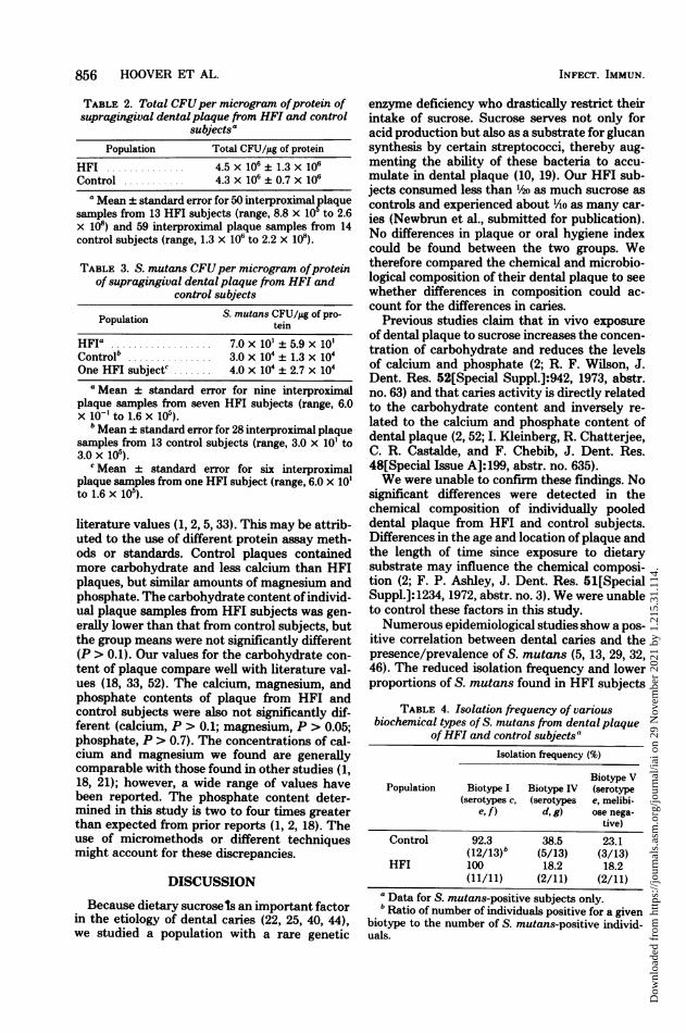
TABLE 2. Total CFUper microgram ofprotein ofsupragingival dentalplaque from HFI and control
subjectsPopulation Total CFU/,ug of protein
HFI 4.5 x 10⁶ ± 1.3 x 10⁶Control ... 4.3 x 10⁶ ± 0.7 x 10⁶
a Mean ± standard error for 50 interproximal plaquesamples from 13 HFI subjects (range, 8.8 x 10⁵ to 2.6x 10⁸) and 59 interproximal plaque samples from 14control subjects (range, 1.3 X 10⁶ to 2.2 x 10⁸).
TABLE 3. S. mutans CFUper microgram ofproteinof supragingival dental plaque from HFI and
control subjects
Population S. mutans CFU/jg of pro-tein
HFIG .. ...... 7.0 x 10 ± 5.9 x 10'Control....."... 3.0 X 10⁴± 1.3 x 10⁴One HFI subjects. 4.0 X 10⁴± 2.7 X 10⁴
a Mean ± standard error for nine interproximalplaque samples from seven HFI subjects (range, 6.0x 10-' to 1.6 x 10⁵).bMean ± standard error for 28 interproximal plaque
samples from 13 control subjects (range, 3.0 x 10' to3.0 x 105).
C Mean ± standard error for six interproximalplaque samples from one HFI subject (range, 6.0 x 10'to 1.6 x 10⁵).
literature values (1, 2, 5, 33). This may be attrib-uted to the use of different protein assay meth-ods or standards. Control plaques containedmore carbohydrate and less calcium than HFIplaques, but similar amounts of magnesium andphosphate. The carbohydrate content of individ-ual plaque samples from HFI subjects was gen-erally lower than that from control subjects, butthe group means were not significantly different(P > 0.1). Our values for the carbohydrate con-tent of plaque compare well with literature val-ues (18, 33, 52). The calcium, magnesium, andphosphate contents of plaque from HFI andcontrol subjects were also not significantly dif-ferent (calcium, P > 0.1; magnesium, P > 0.05;phosphate, P > 0.7). The concentrations of cal-cium and magnesium we found are generallycomparable with those found in other studies (1,18, 21); however, a wide range of values havebeen reported. The phosphate content deter-mined in this study is two to four times greaterthan expected from prior reports (1, 2, 18). Theuse of micromethods or different techniquesmight account for these discrepancies.
DISCUSSION
Because dietary sucrosels an important factorin the etiology of dental caries (22, 25, 40, 44),we studied a population with a rare genetic
enzyme deficiency who drastically restrict theirintake of sucrose. Sucrose serves not only foracid production but also as a substrate for glucansynthesis by certain streptococci, thereby aug-menting the ability of these bacteria to accu-mulate in dental plaque (10, 19). Our HFI sub-jects consumed less than 1/2o as much sucrose ascontrols and experienced about lAo as many car-ies (Newbrun et al., submitted for publication).No differences in plaque or oral hygiene indexcould be found between the two groups. Wetherefore compared the chemical and microbio-logical composition of their dental plaque to seewhether differences in composition could ac-count for the differences in caries.
Previous studies claim that in vivo exposureof dental plaque to sucrose increases the concen-tration of carbohydrate and reduces the levelsof calcium and phosphate (2; R. F. Wilson, J.Dent. Res. 52[Special Suppl.]:942, 1973, abstr.no. 63) and that caries activity is directly relatedto the carbohydrate content and inversely re-lated to the calcium and phosphate content ofdental plaque (2, 52; I. Kleinberg, R. Chatterjee,C. R. Castalde, and F. Chebib, J. Dent. Res.48[Special Issue A]:199, abstr. no. 635).We were unable to confirm these findings. No
significant differences were detected in thechemical composition of individually pooleddental plaque from HFI and control subjects.Differences in the age and location of plaque andthe length of time since exposure to dietarysubstrate may influence the chemical composi-tion (2; F. P. Ashley, J. Dent. Res. 51[SpecialSuppl.]:1234, 1972, abstr. no. 3). We were unableto control these factors in this study.Numerous epidemiological studies show a pos-
itive correlation between dental caries and thepresence/prevalence of S. mutans (5, 13, 29, 32,46). The reduced isolation frequency and lowerproportions of S. mutans found in HFI subjects
TABLE 4. Isolation frequency of variousbiochemical types of S. mutans from dental plaque
ofHFI and control subjectsaIsolation frequency (%)
Biotype VPopulation Biotype I Biotype IV (serotype
(serotypes c, (serotypes e, melibi-e, f) d, g) ose nega-
tive)Control 92.3 38.5 23.1
(12/13)b (5/13) (3/13)HFI 100 18.2 18.2
(11/11) (2/11) (2/11)Data for S. mutans-positive subjects only.Ratio of number of individuals positive for a given
biotype to the number of S. mutans-positive individ-uals.
INFECT. IMMUN.
Dow
nloa
ded
from
http
s://j
ourn
als.
asm
.org
/jour
nal/i
ai o
n 29
Nov
embe
r 20
21 b
y 1.
215.
31.1
14.

DENTAL PLAQUE MICROFLORA IN HFI SUBJECTS 857
TABLE 5. Chemical composition' of dental plaque from HFI and control subjects
Population Protein (JAg/mg) Carbohydrate Calcium (fig/ Magnesium (ug/ Phosphate (Jag/Population ~~~~~(Jn/mg) mg) mg) mg)
HFI (n = 17) 144.3 ± 8.6 18.1 ± 2.3 3.0 ± 0.9 0.3 ± 0.05 10.2 ± 1.2Control (n = 14) 141.7 ± 5.4 23.8 ± 3.1 1.5 ± 0.2 0.4 ± 0.03 8.4 ± 0.5
a Average of two to four analyses for each patient; micrograms per milligram (wet weight) ± standard error;n = number of individuals.
are consistent with their minimal caries experi-ence. S. mutans is widespread in human popu-lations and is present in 60% of sucrase-deficientchildren (51) and 70% of our HFI subjects. Po-tentially cariogenic microorganisms are presentin almost all people, even those who have se-verely limited the amount of sucrose in theirdiet for their entire lives. Thus, restriction ofsucrose intake neither totally prevents coloni-zation by S. mutans nor completely eliminatesit from the oral cavity. But, although sucrose isnot essential for colonization of dental plaque byS. mutans, it greatly facilitates their colonizationon the teeth and increases their proportions inplaque (48, 49). These observations suggest thatS. mutans is indigenous to the oral cavity andthat excessive sucrose consumption is the primecause of dental caries.
Quantitative recovery (30 to 80%) of S. san-guis, S. mutans, and Lactobacillus may be ac-complished from dental plaque for up to 72 hafter collection (27). Selective media (mitis-sal-ivarius, bacitracin agar) afford less recovery ofS. mutans CFU than do nonselective media (34).However, because S. mutans often constitutes asmall percentage of total CFU (especially in HFIsubjects), the use of selective media increasesdetection (isolation frequency). Plaque samplesfrom both groups were examined under identicalculture conditions. The limit of detection for thevarious organisms is difficult to define for any-thing but in vitro-grown stock cultures and mayhave little bearing on the detection limit of wild-type organisms from dental plaque. To arguethat potentially cariogenic microorganismswould escape detection because they are presentin lower numbers in HFI subjects than in con-trols would be tautological.
S. mutans comprises a serologically and bio-chemically heterogenous group of microorga-nisms. Bratthall (6, 8) characterized five serolog-ical types of S. mutans (a, b, c, d, e). Perch et al.(42) found two additional serotypes (f and g).Shklair and Keene (45) have devised a biochem-ical scheme that separates S. mutans into fivebiotypes and correlates them with the recog-nized serotypes. In early reports, when fluores-cent-antibody techniques were used to recognizeand enumerate S. mutans serotypes directly inplaque smears, serotypes a and b were most
commonly found (7, 15, 20). Because of immu-nological cross-reactivity or salivary masking ofantigens or both, these reports were probablyerroneous. Recent studies (9, 24, 42) indicatethat serotype c predominates in most popula-tions. Our results (Table 4) are consistent withthese studies in that biotype I (which includesserotype c) was the most prevalent and waspresent in 11/11 S. mutans-positive HFI sub-jects and 13/14 S. mutans-positive control sub-jects.
Several studies have postulated that variousbiotypes of S. mutans may preferentially causesmooth surface caries or be more cariogenic thanother biotypes (30, 36, 38, 48; D. S. Harper andW. J. Loesche, J. Dent. Res. 57[Special IssueA]:142, 1977, abstr. no. 269; W. H. van Palen-stein Helderman, P. Sampaio Camaergo, and J.H. J. Huis In't Veld, J. Dent. Res. 57[SpecialIssue A]:142, 1977, abstr. no. 271). We isolatedbiotype IV more often from control subjects (5/14) than from HFI subjects (2/11). Four of thefive control subjects who were colonized withbiotype IV had greater caries scores than themean for the control group. One HFI subject,who exhibited the highest caries scores, the high-est isolation frequency of S. mutans, and thehighest proportion of S. mutans of any HFIsubject, also was heavily colonized with biotypeIV. Detailed studies with gnotobiotic rats in-fected with multiple strains of each biotype arerequired before any definitive correlations be-tween biotype and cariogenic potential can bemade. This information is important for thedesign and production of an anticaries vaccine.The infrequent isolation of biotype IV from HFIsubjects might be explained by the observationthat biotype IV S. mutans requires sucrose tocolonize and remain on teeth of gnotobiotic rats(49). The habitually low sugar intake of HFIsubjects could minimize colonization by biotypeIV.
Our results support the specific plaque hy-pothesis of caries formation (35) which statesthat owing to the presence of specific pathogensor relative increases in the levels of certain in-digenous plaque organisms, only certain plaquescan cause infection. Dietary sucrose plays a se-lective role in facilitating colonization and mul-tiplication of specific cariogenic microorganisms
VOL. 28, 1980
Dow
nloa
ded
from
http
s://j
ourn
als.
asm
.org
/jour
nal/i
ai o
n 29
Nov
embe
r 20
21 b
y 1.
215.
31.1
14.

858 HOOVER ET AL.
in dental plaque. Caries activity occurs when thesucrose content of the diet and the ingestionfrequency exceed a certain level.
ACKNOWLEDGMENTS
This work was supported by Public Health Service contractDE-62498 from the National Institute of Dental Research.We thank Rachel Heiblum for her valued technical assist-
ance. The editorial assistance of Evangeline Leash is muchappreciated.
LITERATURE CITED
1. Ashlev, F. P., and R. F. Wilson. 1977. Dental plaqueand caries: a 3-year longitudinal study in children. Br.Dent. J. 142:85-91.
2. Ashley, F. P., and R. F. Wilson. 1977. The relationshipbetween dietary sugar experience and the quantity andbiochemical composition of dental plaque in man. Arch.Oral Biol. 22:409-414.
3. Becks, H., A. L. Jensen, and C. B. Millarr. 1944.Rampant dental caries: prevention and prognosis. J.Am. Dent. Assoc. 31:1189-1200.
4. Bohlen, P., S. Stein, W. Dairman, and S. Udenfriend.1973. Fluorometric assay of proteins in the nanogramrange. Arch. Biochem. Biophys. 155:213-220.
5. Bowen, W. H., H. Velez, M. Aguila, L. Velasquez, L.I. Sierra, and G. Gillespie. 1977. The microbiologyand biochemistry of drinking water from two commun-ities with contrasting levels of caries in Colombia, S. A.J. Dent. Res. 56(Special Issue C):C32-C39.
6. Bratthall, D. 1970. Demonstration of the five serologicalgroups of streptococcal strains resembling Streptococ-cus mutans. Odontol. Revy 21:143-152.
7. Bratthall, D. 1972. Demonstration of Streptococcus mu-tans strains in some selected areas of the world. Odon-tol. Revy 23:401-410.
8. Bratthall, D. 1972. Immunofluorescent identification ofStreptococcus mutans. Odontol. Revy 23:181-196.
9. Bright, J. S., S. Rosen, and F. W. Chorpenning. 1977.Survey of the seven serological types of Streptococcusmutans in six-year-old children. J. Dent. Res. 56:1421.
10. Carlsson, J. 1968. Plaque formation and streptococcalcolonization of teeth. Odontol. Revy 19(Suppl. 14):1-14.
11. Chambers, R. A., and R. T. C. Pratt. 1956. Idiosyncrasyto fructose. Lancet ii:340.
12. Cox, M. E., and J. I. Mangels. 1976. Improved chamberfor the isolation of anaerobic micro-organisms. J. Clin.Microbiol. 4:40-45.
13. de Stoppelaar, J. D., J. van Houte, and 0. BackerDirks. 1969. The relationship between extracellularpolysaccharide-producing streptococci and smooth sur-face caries in 13-year-old children. Caries Res. 3:190-199.
14. de Stoppelaar, J. D., J. van Houte, and 0. BackerDirks. 1970. The effect of carbohydrate restriction onthe presence of Streptococcus mutans, Streptococcussanguis and iodophilic polysaccharide-producing bac-teria in human dental plaque. Caries Res. 4:114-123.
15. Duany, L. F., J. M. Jablon, and D. D. Zinner. 1972.Epidemiologic studies of caries-free and caries-activestudents. I. Prevalence of potentially cariogenic strep-tococci. J. Dent. Res. 51:723-726.
16. Dubois, M., K. A. Giles, J. K. Hamilton, P. A. Rebers,and F. Smith. 1956. Colorimetric method for determin-ing sugars and related substances. Anal. Chem. 28:350-356.
17. Facklam, R. R. 1977. Physiological differentiation ofviridans streptococci. J. Clin. Microbiol. 5:184-201.
18. Geddes, D. A. M., J. A. Cooke, W. M. Edgar, and G.N. Jenkins. 1978. The effect of frequent sucrose
mouth-rinsing on the induction in vivo of caries-likechanges in human dental enamel. Arch. Oral Biol. 23:663-665.
19. Gibbons, R. J., and R. J. Fitzgerald. 1969. Dextran-induced agglutination of Streptococcus mutans and itspotential role in the formation of microbial dentalplaques. J. Bacteriol. 98:341-346.
20. Grenier, E. M., W. C. Eveland, and W. J. Loesche.1973. Identification of Streptococcus mutans serotypesin dental plaque by fluorescent antibody techniques.Arch. Oral Biol. 18:707-715.
21. Gron, P., K. Yao, and M. Spinelli. 1969. A study ofinorganic constituents in dental plaque. J. Dent. Res.48:799-805.
22. Gustafsson, B. E., C. E. Quensel, L. Lanke, C. Lund-quist, H. Grahnen, B. E. Bonow, and B. Krasse.1954. The Vipeholm dental caries study. The effect ofdifferent levels of carbohydrate intake on caries activityin 436 individuals observed for five years. Acta Odontol.Scand. 11:232-388.
23. Hadley, F. P., R. W. Bunting, and E. A. Delves. 1930.Recognition of Bacillus acidophilus associated withdental caries. J. Am. Dent. Assoc. 17:2041-2058.
24. Hamada, S., N. Masuda, T. Ooshima, S. Sobue, andS. Kotani. 1976. Epidemiological survey of Streptococ-cus mutans among Japanese children. Jpn. J. Microbiol.20:33-34.
25. Harris, R. 1963. Biology of the children of HopewoodHouse, Bowral, Australia. IV. Observations of dentalcaries experience extending over five years (1957-1961).J. Dent. Res. 42:1387-1398.
26. Hoermann, K. C., H. J. Keene, I. L. Shklair, and J. A.Burmeister. 1972. The association of Streptococcusmutans with early carious lesions in human teeth. J.Am. Dent. Assoc. 85:1349-1352.
27. Hoover, C. I., and E. Newbrun. 1977. Survival of bac-teria from human dental plaque under various transportconditions. J. Clin. Microbiol. 6:212-218.
28. Jay, P. 1947. The reduction of oral Lactobacillus aci-dophilus counts by the periodic restriction of carbohy-drate. Am. J. Orthodont. Oral Surg. 33:162-184.
29. Jordan, H. V., H. R. Englander, and S. Lim. 1969. Thepresence of potentially cariogenic streptococci in var-ious population groups. J. Am. Dent. Assoc. 78:1331-1335.
30. Keene, H. J., I. L. Shklair, D. M. Anderson, and G. J.Mickel. 1977. Relationship of Streptococcus mutansbiotypes to dental caries prevalence in Saudi Arabiannaval men. J. Dent. Res. 56:356-361.
31. King, E. J. 1932. The colorimetric determination of phos-phorus. Biochem. J. 26:292-297.
32. Krasse, B., H. V. Jordan, S. Edwardsson, I. Svens-son, and L. Trell. 1968. The occurrence of certain"caries-inducing" streptococci in human dental plaquematerial. Arch. Oral Biol. 13:911-918.
33. Krembel, J., R. M. Frank, and A. Deluzarche. 1969.Fractionation of human dental plaque. Arch. Oral Biol.14:563-565.
34. Little, W. A., D. C. Korts, L A. Thomson, and W. H.Bowen. 1977. Comparative recovery of Streptococcusmutans on ten isolation media. J. Clin. Microbiol. 5:578-583.
35. Loesche, W. J. 1976. Chemotherapy of dental plaqueinfections. Oral Sci. Rev. 9:65-107.
36. Masuda, N., N. Tsutsumi, S. Sobue, and H. Hamada.1979. Longitudinal survey of the distribution of variousserotypes of Streptococcus mutans in infants. J. Clin.Microbiol. 10:497-502.
37. McKnight, G. S. 1977. A colorimetric method for thedetermination of submicrogram quantities of protein.Anal. Biochem. 78:86-92.
38. Michalek, S. M., J. McGhee, and J. Navia. 1975. Vir-
INFECT. -IMMUN.
Dow
nloa
ded
from
http
s://j
ourn
als.
asm
.org
/jour
nal/i
ai o
n 29
Nov
embe
r 20
21 b
y 1.
215.
31.1
14.

DENTAL PLAQUE MICROFLORA IN HFI SUBJECTS 859
ulence of Streptococcus mutans: a sensitive method forevaluating cariogenicity in young gnotobiotic rats. In-fect. Immun. 12:69-75.
39. Miller, W. D. 1890. The microorganisms of the humanmouth, p. 102-144. Reprinted by S. Karger, Basel, Swit-zerland.
40. Newbrun, E. 1979. Dietary carbohydrates: their role incariogenicity. Med. Clin. N. Am. 63:1069-1086.
41. Nordmann, Y., F. Schapira, and J. C. Dreyfus. 1968.A structurally modified liver aldolase in fructose intol-erance: immunological and kinetic evidence. Biochem.Biophys. Res. Commun. 31:884-889.
42. Perch, B., E. Kjems, and T. Ravn. 1974. Biochemicaland serological properties of Streptococcus mutansfrom various human and animal sources. Acta Pathol.Microbiol. Scand. Sect. B 82:357-370.
43. Schapira, F., Y. Nordmann, and C. Gregori. 1972.Hereditary alterations of fructose metabolizing en-zymes. Acta Med. Scand. Suppl. 542:77-83.
44. Scheinin, A., and K. K. Makinen. 1975. Turku sugarstudies. I-XXI. Acta Odontol. Scand. 33:Suppl. 70.
45. Shklair, I. L, and H. J. Keene. 1976. Biochemicalcharacterization and distribution of Streptococcus mu-
tans in three diverse populations, p. 201-210. In H. M.Stiles, W. J. Loesche, and T. C. O'Brien (eds.), Proceed-ings: Microbial Aspects of Dental Caries (a special
supplement to Microbiology Abstracts), vol. 1. Infor-mation Retrieval Inc., Washington, D.C.
46. Shklair, I. L., H. J. Keene, and P. Cullen. 1974. Thedistribution of Streptococcus mutans on the teeth oftwo groups of naval recruits. Arch. Oral Biol. 19:199-202.
47. Syed, S. A., and W. J. Loesche. 1972. Survival ofhumandental plaque flora in various transport media. Appl.Microbiol. 24:638-644.
48. Tanzer, J. M. 1979. Essential dependence of smoothsurface caries on, and augmentation of fissure caries by,sucrose and Streptococcus mutans infection. Infect. Im-mun. 25:526-531.
49. Thomson, L. A., W. H. Bowen, W. A. Little, H. M.
Kuzmiak-Jones, and I. M. Gomez. 1979. Simultane-ous implantation of five serotypes of Streptococcusmutans in gnotobiotic rats. Caries Res. 13:9-17.
50. van Houte, J. 1964. Relationship between carbohydrateintake and polysaccharide-storing micro-organisms indental plaque. Arch. Oral Biol. 9:91-93.
51. van Houte, J., and S. Duchin. 1975. Streptococcus mu-
tans in the mouths of children with congenital sucrase
deficiency. Arch. Oral Biol. 20:771-773.52. Wood, J. M. 1967. The amount, distribution and metab-
olism of soluble polysaccharides in human dentalplaque. Arch. Oral Biol. 12:849-858.
VOL. 28, 1980
Dow
nloa
ded
from
http
s://j
ourn
als.
asm
.org
/jour
nal/i
ai o
n 29
Nov
embe
r 20
21 b
y 1.
215.
31.1
14.