Micro-Nutrient Needs in Down Syndrome: A peer … 3-arm chromosome (trisomy) increases production by...
80
Micro-Nutrient Needs in Down Syndrome: A peer-reviewed science base © 2014 Joan Jory, MSc, PhD, RD
Transcript of Micro-Nutrient Needs in Down Syndrome: A peer … 3-arm chromosome (trisomy) increases production by...

Micro-Nutrient Needs in Down Syndrome:
A peer-reviewed science base © 2014
Joan Jory, MSc, PhD, RD


Down Syndrome: the Implications
Down Syndrome results from a (third arm) on chromosome 21.
This trisomy is associated with changes in:
- neurological development
- physical growth
- muscle to fat ratio
- immunity and resistance to infection
- risk of leukemia
- risk of celiac disease
- risk of thyroid dysfunction
- risk of diabetes and blood sugar abnormalities
- precocious aging
- Alzheimer-like lesions

Down Syndrome: an Analogy
A 2-arm chromosome (normal) is like a recipe:
X volume of ingredients Y volume of product.
A 3-arm chromosome (trisomy) increases production by 50%:
Y + 50% volume of product requires X + 50% volume ingredients.
X + 50% ingredients faster depletion of ingredient stores
‘stealing’ ingredients from other functions
decreased ingredients for growth + development
decreased ingredients for disease prevention
decreased ingredients for healthy aging

Down Syndrome: the Functional Paradigm
1. 50% overproduction of products coded on Chromosome 21
(50% more products produced)
2. 50% increase in production causes a 50% increase in requirements for raw ingredients (vitamins, minerals, amino acids, etc)
(50% more ingredients needed)
3. There is also an increase in the byproducts (mess), requiring increased antioxidants to neutralize the excess byproducts
(50% more garbage produced + 50% more antioxidants needed).
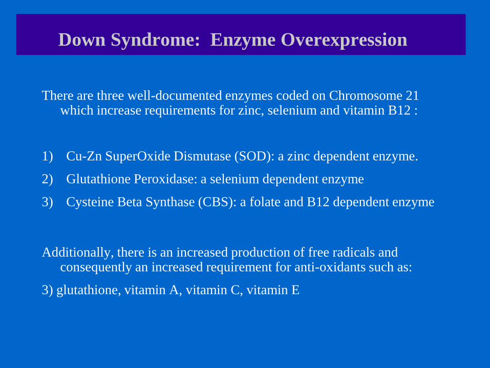
Down Syndrome: Enzyme Overexpression
There are three well-documented enzymes coded on Chromosome 21 which increase requirements for zinc, selenium and vitamin B12 :
1) Cu-Zn SuperOxide Dismutase (SOD): a zinc dependent enzyme.
2) Glutathione Peroxidase: a selenium dependent enzyme
3) Cysteine Beta Synthase (CBS): a folate and B12 dependent enzyme
Additionally, there is an increased production of free radicals and consequently an increased requirement for anti-oxidants such as:
3) glutathione, vitamin A, vitamin C, vitamin E
Copper-Zinc Superoxide Dismustase (SOD) in Down
Syndrome

Down Syndrome: What Evidence of Excess SOD?
Superoxide Dismutase (SOD) levels can be normal in mosaic Down Syndrome
(Biol Trace Elem Res 2001)
Superoxide Dismutase (SOD) levels are increased in full expression Down
Syndrome
(Mayo Clin Proc 2005)
Overproduction of Superoxide Dismutase (SOD) increases oxidative stress and
increases oxidative damage to proteins in children with Down Syndrome .
(Clin Chem Lab Med 2006)
Consequences of Low Zinc in Down Syndrome

Down Syndrome: Implications of Low Zinc
Low zinc status in Down Syndrome is associated with:
1) decreased immune status
2) decreased growth and lean tissue ratios
3) thyroid dysfunction
4) celiac disease
5) altered taste perception and appetite
Zinc and Immunity
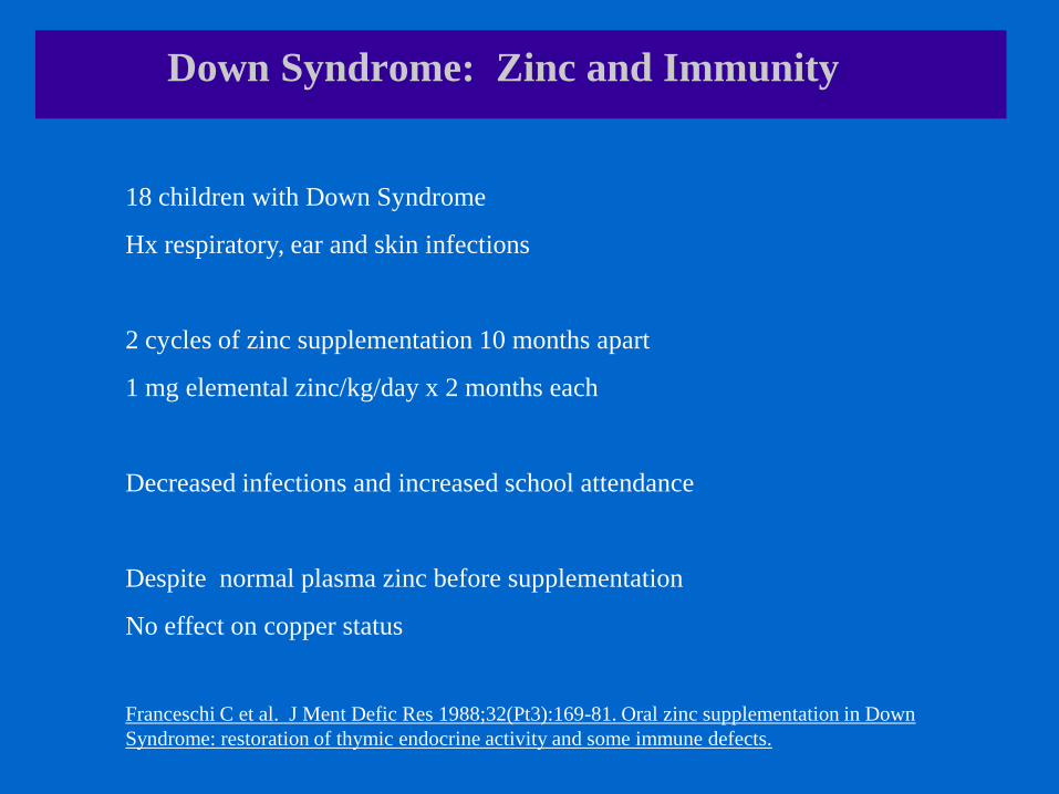
Down Syndrome: Zinc and Immunity
18 children with Down Syndrome
Hx respiratory, ear and skin infections
2 cycles of zinc supplementation 10 months apart
1 mg elemental zinc/kg/day x 2 months each
Decreased infections and increased school attendance
Despite normal plasma zinc before supplementation
No effect on copper status
Franceschi C et al. J Ment Defic Res 1988;32(Pt3):169-81. Oral zinc supplementation in Down
Syndrome: restoration of thymic endocrine activity and some immune defects.

Down Syndrome: Zinc and Immunity
12 children with Down’s Syndrome
Low serum zinc and immune deficiency
Zinc sulfate supplementation @ 135 mg zinc/d x 2 months
Immune improvements in 11/12 children
Bjorksten B et al. Acta Pediatr Scand 1990; .Zinc and Immune Function in Down’s Syndrome.
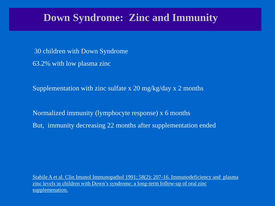
Down Syndrome: Zinc and Immunity
30 children with Down Syndrome
63.2% with low plasma zinc
Supplementation with zinc sulfate x 20 mg/kg/day x 2 months
Normalized immunity (lymphocyte response) x 6 months
But, immunity decreasing 22 months after supplementation ended
Stabile A et al. Clin Imunol Immunopathol 1991; 58(2): 207-16. Immunodeficiency and plasma
zinc levels in children with Down’s syndrome: a long-term follow-up of oral zinc
supplemenation.

Zinc and Growth

Down Syndrome: Zinc, Growth, Body Composition
Meta-analysis of 33 zinc supplementation studies and childhood growth.
Highly significant improvements in height
Highly significant improvements in weight
Greatest improvements among children with lowest height-for-age scores.
Brown KH et al. Am J Clin Nutr 2002; 75(6): 1062-71. Effect of supplemental zinc on the growth and
serum zinc concentrations of preburtal children: a meta-analysis of randomized controlled trials.

Down Syndrome: Zinc, Growth, Body Composition
35 children with Down Syndrome
No difference in protein intake, carb or fat intake
Zinc intake adequate in 40% of Down Syndrome and 67% controls
Lower plasma zinc in Down Syndrome.
Shorter height in Down Syndrome.
Lima AS et al. Biol Trace Elem Res 2010; 133(1): 20-8. Nutritional status of zinc in children with Down
Syndrome

Down Syndrome: Zinc, Growth, Body Composition
22 children with Down Syndrome
Supplemented with zinc sulphate for 6-9 months.
Increased growth percentile and increased growth hormone levels
Increased growth velocity (23.84 -> 40.80 mm/6 months).
Napolitano G et al. Am J Med Genet 1990;37(S7):63-65. Growth delay in Down Syndrome and zinc
sulphate supplementation.

Zinc and Body Composition

Down Syndrome: Zinc, Growth, Body Composition
9 female elite athletes
% fat mass is negatively associated with plasma zinc.
% fat mass is positive associated with the plasma copper/zinc ratio.
Koury JC et al. Biol Trace Elem Res 2007; 115(1): 23-30. Plasma zinc, copper, leptin, and body
composition are associated in elite female judo athletes.

Down Syndrome: Zinc, Growth, Body Composition
30 adolescents with Down Syndrome
No difference in protein, fat, carb or zinc intake
No difference in plasma zinc
Greater zinc losses in urine in Down Syndrome.
Greater overweight (26.7%) and obesity (6.6%) in Down Syndrome.
Marques RC et al. Biol Trace Elem Res 2007; 120(1-3): 11-8. Zinc nutritional status in adolescents with
Down Syndrome.

Zinc and Thyroid
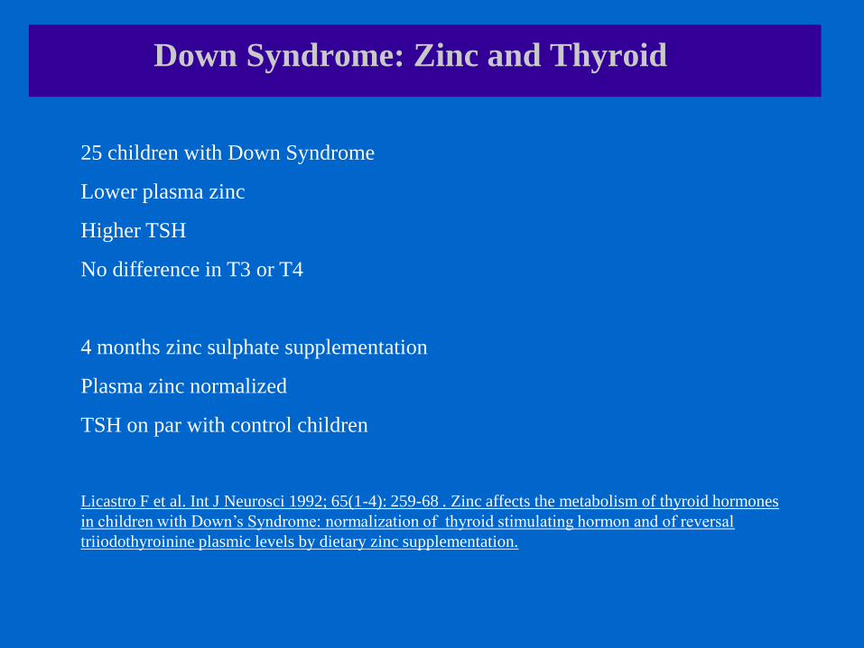
Down Syndrome: Zinc and Thyroid
25 children with Down Syndrome
Lower plasma zinc
Higher TSH
No difference in T3 or T4
4 months zinc sulphate supplementation
Plasma zinc normalized
TSH on par with control children
Licastro F et al. Int J Neurosci 1992; 65(1-4): 259-68 . Zinc affects the metabolism of thyroid hormones
in children with Down’s Syndrome: normalization of thyroid stimulating hormon and of reversal
triiodothyroinine plasmic levels by dietary zinc supplementation.

Down Syndrome: Zinc and Thyroid
51 children with Down Syndrome
4 months of zinc sulphate supplementation:
Normalized plasma zinc and TSH
1 year after zinc supplementation stopped:
Plasma zinc decreasing
TSH increasing
Licastro F et al. J Trace Elem Electrolytes Health Dis 1993; 7(4): 237-9. Modulation of the
neuroendocrine system and immune functions by zinc supplementation in children with Down’s
Syndrome.
Zinc and Celiac Disease

Down Syndrome: Zinc and Celiac Disease
43 children with Down Syndrome
Lower zinc
Lower immune function
Higher TSH
Higher coeliac disease
Zinc lowest in children with celiac disease and decreased immune status
Licastro F et al. Brain Res Bull 2001; 55(2): 313-7. Immune-endocrine status and coeliac disease in
children with Down’s Syndrome: relationship with zinc and cognitive efficiency.
Zinc and Taste/Appetite

Down Syndrome: Zinc and Taste Disorders
22 patients with altered taste acuity (hypogeusia)
Zinc acetate supplementation x 50 mg zinc/day
Improvement in plasma zinc
Improvement in taste perception for sweet, salt and bitter.
Mahaian SK et al. Am J Clin Nutr 1980’; 33(7): 1517-21. Improvement of uremic hypogeusia by zinc: a
double-blind study.

Zinc Safety
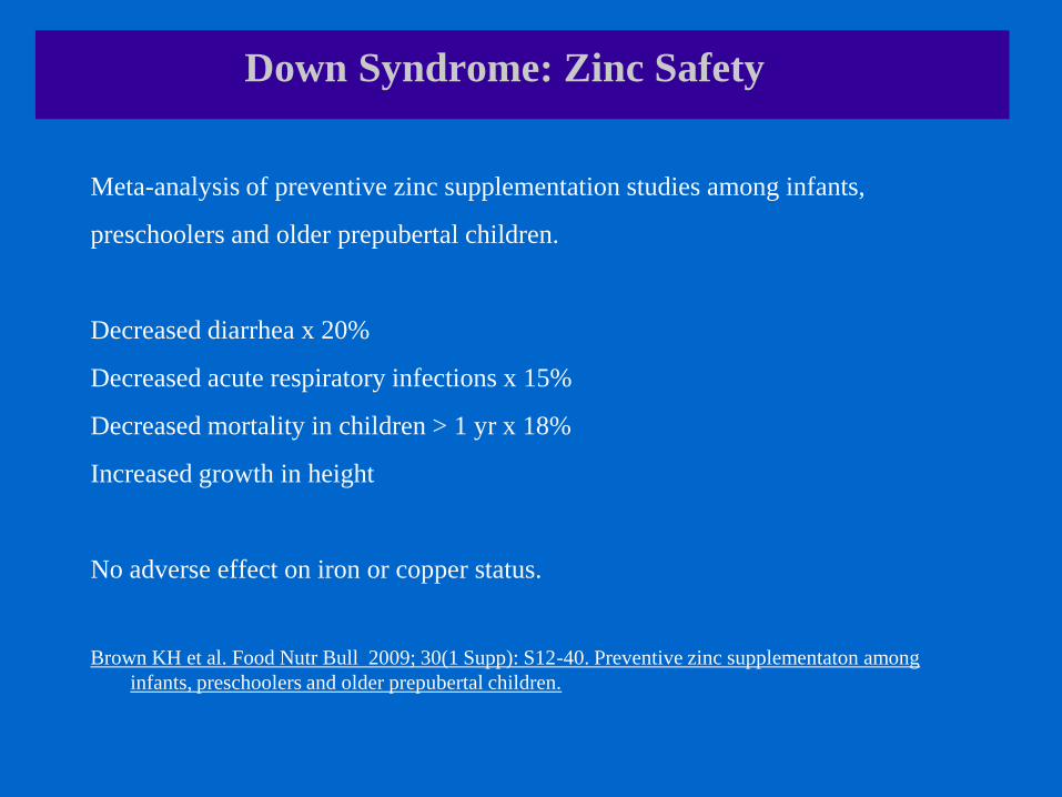
Down Syndrome: Zinc Safety
Meta-analysis of preventive zinc supplementation studies among infants,
preschoolers and older prepubertal children.
Decreased diarrhea x 20%
Decreased acute respiratory infections x 15%
Decreased mortality in children > 1 yr x 18%
Increased growth in height
No adverse effect on iron or copper status.
Brown KH et al. Food Nutr Bull 2009; 30(1 Supp): S12-40. Preventive zinc supplementaton among
infants, preschoolers and older prepubertal children.
Zinc Testing

Zinc Assessment
1) Serum/plasma zinc
-> recent intake only
-> many confounding variables
2) Alkaline phosphatase (zinc enzyme)
-> functional zinc status
-> unreliable if recent growth as alk phos is mobilized into blood during growth
3) Best approach
-> combination of serum/plasma zinc AND alkaline phosphatase

Dietary Sources of Zinc

Zinc Sources in Diet
Absorption blocked by calcium, iron, fibre, phytate.
Richest food sources:
Seafood
Fish
Liver
Beef, Pork, Chicken
Beans
Cashews
Cheese
US National Institutes of Health, 2013

Glutathione Peroxidase (GPx) in Down Syndrome
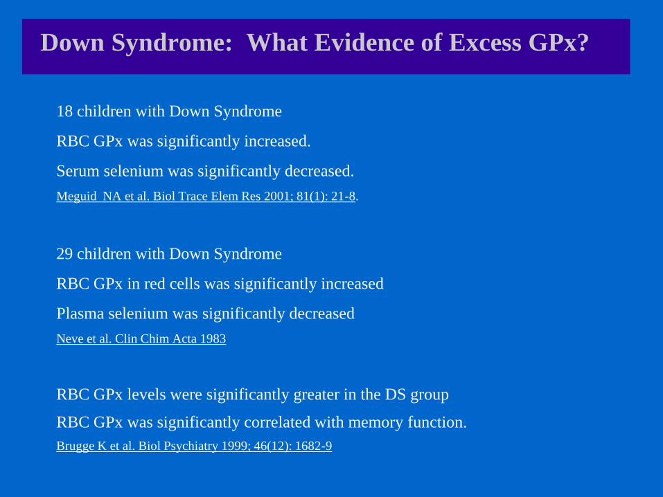
Down Syndrome: What Evidence of Excess GPx?
18 children with Down Syndrome
RBC GPx was significantly increased.
Serum selenium was significantly decreased.
Meguid NA et al. Biol Trace Elem Res 2001; 81(1): 21-8.
29 children with Down Syndrome
RBC GPx in red cells was significantly increased
Plasma selenium was significantly decreased
Neve et al. Clin Chim Acta 1983
RBC GPx levels were significantly greater in the DS group
RBC GPx was significantly correlated with memory function.
Brugge K et al. Biol Psychiatry 1999; 46(12): 1682-9

Consequences of Low Selenium in Down Syndrome

Down Syndrome: Implications of Low Selenium
Low selenium levels in Down Syndrome are associated with:
1) impaired thyroid status
2) decreased immune status

Selenium and Thyroid

Down Syndrome: Selenium and Thyroid
109 healthy individuals
Decreased serum selenium
Decreased T3/T4 ratio
Decreased conversion of T4 to T3 because of low selenium status
Olivieri O et al. Biol Trace Elem Res 1996; 51(1): 31-41. Selenium, zinc, and thyroid hormones in health
subjects.
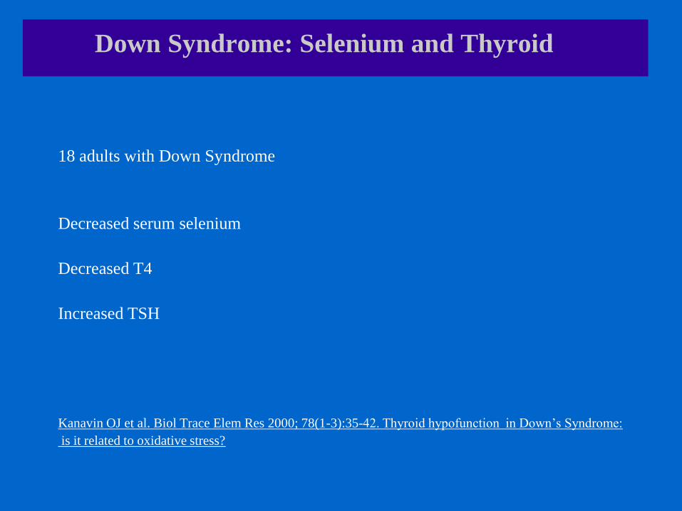
Down Syndrome: Selenium and Thyroid
18 adults with Down Syndrome
Decreased serum selenium
Decreased T4
Increased TSH
Kanavin OJ et al. Biol Trace Elem Res 2000; 78(1-3):35-42. Thyroid hypofunction in Down’s Syndrome:
is it related to oxidative stress?
Selenium and Immunity

Down Syndrome: Selenium and Immunity
1a) Natural Killer (NK) activity is low in Down Syndrome. Ugazio et al. Am J Med Genet Suppl 1990
1b) Supplementation with Se increases natural killer (NK) cell activity in the mouse. Kiremidjian-Schumacker & Roy. Z Ernarhrungswis 1998
2a) The T-lymphocyte activation response is patients with dysmorphic disorders. Cruz et al. Ann Allergy Ashtma Immunol 2009
2b) Selenoprotein deficiency suppresses T-lymphocyte response. Shrimali et al. J Biol Chemi 2008.

Selenium Testing

Selenium Assessment
1) Serum/plasma selenium -> only recent selenium intake
2) RBC selenium -> longer term selenium status
3) But, there are no established ‘normal ranges’ for either test
Thomson CD. Eur J Clin Nutr 2004; 58: 391-402. Assessment of requirements for selenium and adequacy
of selenium status: a review.

Dietary Sources of Selenium
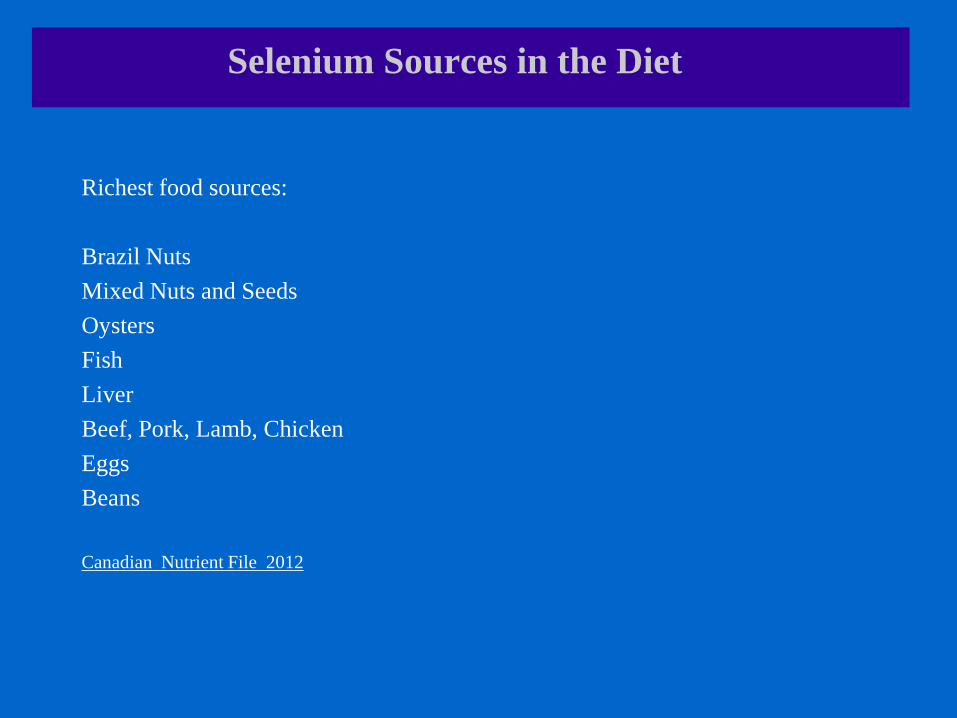
Selenium Sources in the Diet
Richest food sources:
Brazil Nuts
Mixed Nuts and Seeds
Oysters
Fish
Liver
Beef, Pork, Lamb, Chicken
Eggs
Beans
Canadian Nutrient File 2012
Cystathione Beta Synthase (CBS) in Down Syndrome

Down Syndrome: What Evidence of Excess CBS?
Cystathione beta synthase (CBS) levels are increased by approximately three
times in the Down Syndrome brain.
High CBS causes homocysteinuria, which is characterized by mental retardation
and vascular disease.
The high CBS may explain the cognitive abnormalities in Down Syndrome, and
the vulnerability to Alzheimer’s Disease
Ichinohe A et al. Biochem Biophys Res Commun 2005; 338(3): 1547-50. Cystathione beta-synthase is
enriched in the brains of Down’s patients.

Down Syndrome: What Evidence of Excess CBS?
42 children with Down Syndrome
CBS overexpression in lymphoblasts altered homocysteine metabolism with
decreased homocysteine content, and decreased glutathione.
Addition of methyl-folate, methyl-B12 or dimethylglycine improved this profile
Progribna M et al. Am J Hum Genet 2001; 69(1): 88-95 Homocystein metabolism in children with Down
Syndrome: in vitro modulation

Folate in Down Syndrome
Down Syndrome: Folate Status
Canadian Health Measures Survey
5248 people 6-79 years + 1162 women 15-45 years
‘Folate deficiency is virtually nonexistent in the Canadian population’
Very high folate among 40% of the Canadian population
New reference range for ‘normal’ RBC folate: 305-1360 nmol/L
Colapinto CK et al. CMAJ 2011; 183(13): 1519. Folate status of the population in the Canadian Health
Measures Survey.

Down Syndrome: Folate Status
Following folate fortification of flour in US :
Significant increase in maternal folate status
Normal serum and RBC folate in infants with Down Syndrome
Significant decrease in neural tube defects and cleft palate
7% increase in Down Syndrome (where no prenatal DS testing)
Canfield MA et al. Birth Defects Res A Clin Mol Teratol 2005; 73(10): 679-89. Changes in the birth
prevalence of selected birth defects after grain fortification with folic acid in the United States: findings
from a multi-state population based study.
Down Syndrome: Folate Status
10 children with Down Syndrome
No difference in serum and RBC folate between Down Syndrome and controls
Roizen and Amarose. Am J Med Genet 1993; 46(5): 510-2. Hematological abnormalities in children with
Down Syndrome
50 children with Down Syndrome
No difference in serum or RBC folate between Down Syndrome and controls
Onorata D et al. Pediatr Hematol-Oncol 1996; 13(3): 271-275. Hematological studies in children with
Down Syndrome
113 patients with Down Syndrome
No difference in serum of RBC folate between Down Syndrome and controls
Howell A et al. Scand J Haematol 1973; 11: 140-147. Red cell size and uric acid in Down Syndrome.
Folate Assessment
1) Serum/plasma folate -> only recent folate intake
2) RBC folate -> longer term folate status
Reference range for Canadians: 305-1360 nmol/L
Caution if RBC folate > 1360 nmol/L
Colapinto CK et al. CMAJ 2011; 183(13): 1519. Folate status of the population in the Canadian Health
Measures Survey.

Folate Sources in the Diet
All flour and flour products in Canada are fortified with flour
All multi-B, multivitamin and prenatal vitamins contain folate
Many protein powders and alternate non-dairy milks are folate fortified.
Richest unfortified dietary sources:
Liver
Beans and lentils
Dark green vegetables
Sunflower seeds
Potatoes
Fruit
Canadian Nutrient File 2010

B12 Status in Down Syndrome
Down Syndrome: B12 Status
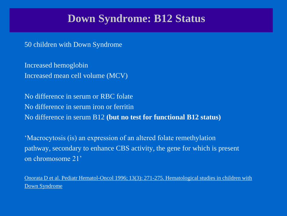
50 children with Down Syndrome
Increased hemoglobin
Increased mean cell volume (MCV)
No difference in serum or RBC folate
No difference in serum iron or ferritin
No difference in serum B12 (but no test for functional B12 status)
‘Macrocytosis (is) an expression of an altered folate remethylation
pathway, secondary to enhance CBS activity, the gene for which is present
on chromosome 21’
Onorata D et al. Pediatr Hematol-Oncol 1996; 13(3): 271-275. Hematological studies in children with
Down Syndrome

Down Syndrome: B12 Status
113 patients with Down Syndrome
Increased mean cell volume (MCV)
Decreased serum B12
No difference in serum of RBC folate
Howell A et al. Scand J Haematol 1973; 11: 140-147. Red cell size and uric acid in Down Syndrome.
10 children with Down Syndrome
Increased hematocrit
Increased mean cell volume (MCV)
Decreased white blood cells (WBC)
No B12 testing
No difference in serum and RBC folate
Roizen and Amarose. Am J Med Genet 1993
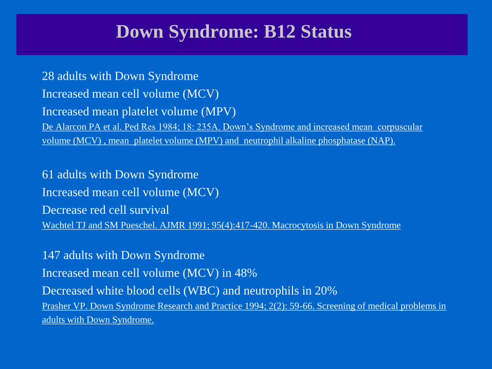
Down Syndrome: B12 Status
28 adults with Down Syndrome
Increased mean cell volume (MCV)
Increased mean platelet volume (MPV)
De Alarcon PA et al. Ped Res 1984; 18: 235A. Down’s Syndrome and increased mean corpuscular
volume (MCV) , mean platelet volume (MPV) and neutrophil alkaline phosphatase (NAP).
61 adults with Down Syndrome
Increased mean cell volume (MCV)
Decrease red cell survival
Wachtel TJ and SM Pueschel. AJMR 1991; 95(4):417-420. Macrocytosis in Down Syndrome
147 adults with Down Syndrome
Increased mean cell volume (MCV) in 48%
Decreased white blood cells (WBC) and neutrophils in 20%
Prasher VP. Down Syndrome Research and Practice 1994; 2(2): 59-66. Screening of medical problems in
adults with Down Syndrome.

B12 deficiency and Abnormal Hematology

Down Syndrome: B12 and Hematology
Vitamin B12 deficiency can cause profound alterations in the bone marrow.
These alterations can mimic the more serious diagnosis of acute leukemia.
Two patients were suspected of having acute leukemia or myelodysplasia
on the basis of bone marrow smear.
They were both found to have vitamin B12 deficiency
Parenteral vitamin B12 resulted in normalization of the bone marrow.
Aitelli C et al. South Med J 2004; 97(3): 295-7. Pernicous anemia: presentations mimicking acute
leukemia.
Down Syndrome: B12 and Hematology
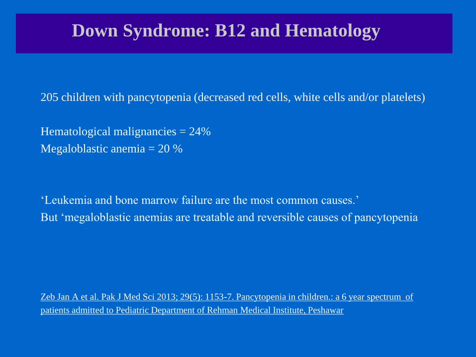
205 children with pancytopenia (decreased red cells, white cells and/or platelets)
Hematological malignancies = 24%
Megaloblastic anemia = 20 %
‘Leukemia and bone marrow failure are the most common causes.’
But ‘megaloblastic anemias are treatable and reversible causes of pancytopenia
Zeb Jan A et al. Pak J Med Sci 2013; 29(5): 1153-7. Pancytopenia in children.: a 6 year spectrum of
patients admitted to Pediatric Department of Rehman Medical Institute, Peshawar

Down Syndrome: B12 and Hematology
Plasma concentrations of methycobalamin was significantly lower in CML
patients than the reference population.
Low methylcobalamin was associated with a poor prognosis.
Gimsing P. Br J Haematol 1995; 89(4): 812-9. Cobalamin forms and analogues in plasma and myeloid
cells during chronic myelogenous leukaemia related to clinical condition.
In the mouse model of L1210 leukemia, vitamin B12 + C inhibited cell growth
and increased survival
Pydock ME. Am J Clin Nutr 1991; 54 (6 Suppl): 1261S-1265S. Effect of combined ascorbic acid and
B12 on survival of mice with implanted Ehrlich carcinoma and L1210 leukemia.

B12 and Myelination
Down Syndrome: B12 and Myelination
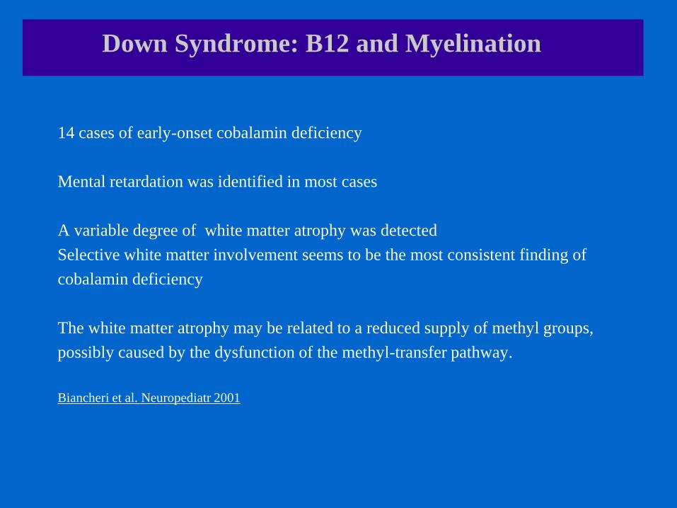
14 cases of early-onset cobalamin deficiency
Mental retardation was identified in most cases
A variable degree of white matter atrophy was detected
Selective white matter involvement seems to be the most consistent finding of
cobalamin deficiency
The white matter atrophy may be related to a reduced supply of methyl groups,
possibly caused by the dysfunction of the methyl-transfer pathway.
Biancheri et al. Neuropediatr 2001

Down Syndrome: B12 and Myelination
1)The brain of a child with Down Syndrome develops differently from a normal
one, attaining a form reduced in size and altered in configuration.
2) Directly related to the mental retardation are neuronal modifications manifest as
alterations in cortical lamination (myelination)
Becker et al. Prog Clin Biol Res 1991
120 children with Down Syndrome
Delayed myelination in 22.5%
Wisniewski & Schmidt-Sidor. Clin Neuropath 1989
18 month old infant with Down Syndrome
Brain myelination equivalent to an 11-month infant.
Koo et al. J Child Neurol 1992

B12 Assessment
1) No gold standard has emerged for the diagnosis of cobalamin deficiency.
2)Therapeutic trials with pharmacologic doses of cobalamin are suggested when
findings consistent with cobalamin deficiency are present regardless of the results
of diagnostic tests. Solomon LR. Blood Rev 2007
Normal serum B12 can be repeatedly normal in the presence of haematological
and neurological symptoms of B12 deficiency.
Mar N et al. Open J of Hematol 2012; 3: DOI-10.13055/ojhmt_3_1_2.120920. Pitfalls in the diagnosis of
B12 deficiency.
There is concern that high intakes of folic acid from fortified food and dietary
supplements might mask the macrocytic anemia of vitamin B12 deficiency,
thereby eliminating an important diagnostic sign.
Johnson MA. Nutr Rev 2007
Occult cobalamin deficiency could become a common disorder. Ray et al. Clin Biochem 2000

B12 Assessment
1) Serum B12
Recent intake -> will show high if supplementation
Caution if < 400 (Japanese cutoff for serum B12)
2) MCV and MCH
If high, may indicate folate or B12 deficiency.
Test for RBC folate to differentiate
3) White cell count (WBC) and subsets, red cell count (RBC) and platelets
If low, may indicate B12 deficiency.
Consider testing response to high dose B12 before considering leukemia
4) Homocysteine
Will be low in Down Syndrome because of CBS overexpression on chromosome 21

B12 Sources in the Diet
Only 1% of dietary B12 is passively absorbed.
99% requires intrinsic factor production in the gut
Best dietary sources:
Clams
Liver
Seafood
Trout and Salmon
Beef and wild game
Canadian Nutrient File 2010
US National Institutes of Health 2011

Down Syndrome: Importance of Iron
Human infants with iron deficiency anemia test lower in cognitive, motor, social-
emotional, and neurophysiologic development than comparison group infants.
B Lozoff and MKGeorgieff. Sem in Pediatr Neurol 2006; 13(3): 158-165. Iron Deficiency and Brain
Development.
Increased likelihood of mild or moderate mental retardation associated with
anemia, independent of birth weight, maternal education, sex, race-ethnicity,
the mother’s age, or the child’s age at entry into the US WIC (Women, Infants
and Children Supplementation program.
EK Hurtado et al. Am J Clin Nutr 1999; 69(1): 115-119; Early Childhood Anemia and Mild or Moderate
Mental Retardation

Down Syndrome: Iron
114 children with Down Syndrome
Iron deficiency in 10%
Iron deficiency anemia in 3%
”Screening should include CBC, transferrin saturation and ferritin”
Dixon NE et al. J Pediatr 2010; 157(6): 967-971. Prevalence of iron deficiency in children with Down
Syndrome.
149 children with Down Syndrome
Anemia in 8.1%
Iron deficiency in 50% with iron tests (19/38)
Tenenbaum et al. Int J Pediatrics 2011; ID 813541. doi: 10.1155/2011/813541.

Iron Assessment
Serum Ferritin
An acute phase reactant -> can be falsely high if infection or inflammation
Can be increased if insufficient B12 for red cell production
Caution if < 30
Mean Cell Volume (MCV) and Mean Cell Hemoglobin
Low MCV and MCH in uncomplicated iron deficiency
May be normal or high if co-existing B12 or folate deficiency
Caution if <75-80 or >90-95

Iron Assessment
Iron Saturation
May be low if low iron or B12
May be high if macrocytosis (swollen cells) from B12 or folate deficiency
Serum iron
Recent iron intake; not functional iron status
Hemoglobin and hematocrit
If low, may be due to iron, B12 or folate deficiency
Need to test ferritin and RBC folate to determine which deficiency

Iron Sources in the Diet
Heme iron has a higher absorption, and comes from animal products
Non-heme iron absorption may be compromised by the presence of fibre and
phytate.
Iron absorption is enhanced by vitamin C and decreased by calcium
Richest dietary sources:
Seafood
Fish
Liver and kidney
Red meat
Beans and Soybeans
Spinach and dark greens
Fortified cereals
Canadian Nutrient File 2010

Vitamin A in Down Syndrome

Down Syndrome: Vitamin A
38 children with Down Syndrome
Serum retinol deficiency (<20mg/dL) in 18.4%
Chavez CJ et al. An Pediatr (Barc) 2010; 72 (3): 185-90.
12 patients with Down Syndrome
Lower plasma and red cell retinol.
Shah SN and RD Johnson. Nutr Res 1989; 9(7): 709-715. Antioxidant vitamin (A and E) status of
Down’s Syndrome subjects.
33 patients with Down Syndrome
No difference in vitamin A intake, serum vitamin A or Vitamin A absorption
But skin symptoms of hypovitaminosis A
Pueschel SM et al. J Met Defic Res 1990; 34(Pt 3) : 269-75. Vitamin A gastrointestinal absorption in
persons with Down’s Syndrome.

Vitamin A
Testing
Serum Vitamin A (retinol): recent intake only
Vitamin A Sources in the Diet
Liver
Cod Liver Oil
Eggs
Goat cheese
Cow cheese
Orange vegetables (sweet potato, pumpkin, carrots)
Green vegetables (spinach, kale, swiss chard)
Canadian Nutrient File 2010

Nutrient-Rich Diet for Down Syndrome
High animal protein (liver, fish, beef, lamb) (unfortified non-GMO whey protein)
High intake of colourful vegetables (dark greens and dark oranges)
High intake of nuts and seeds and lentils
Minimally processed grains
Minimal fruit
Dairy x 2-3 servings/day
Cod liver oil (without Vitamin A and D removed)
¼ tsp of iodized salt/day

Baseline Bloodwork for Children with Down Syndrome
CBC
Serum ferritin -> separate test for serum iron and iron saturation if abnormal
RBC folate
AST & ALT
Alkalinine phosphatase
TSH & T4
Ammonia
Fasting blood sugar & triglycerides
HDL and LDL cholesterol (if older than 12 years)
Urinalysis (for occult urinary tract infections or dysurias)
Consider also: vitamin A, serum zinc and serum selenium if appropriate