micro lab
34
12/29/2012 1 Sessions: • Gram stain and Bacteriology • Bacillus, diphteroids,staph,and strept under the microscope • Medias • Sensitivity test • Normal flora bacteria and throat culture • Urine and stool culture • Agglutination and precipitation tests • Acid fast culture and stain, and yeast, candida, and Aspergillus.
-
Upload
allaa-haddoush -
Category
Documents
-
view
216 -
download
3
description
micro lab for dental students
Transcript of micro lab

12/29/2012
1
Sessions: • Gram stain and Bacteriology
• Bacillus, diphteroids,staph,and strept under the microscope
• Medias
• Sensitivity test
• Normal flora bacteria and throat culture
• Urine and stool culture
• Agglutination and precipitation tests
• Acid fast culture and stain, and yeast, candida, and Aspergillus.

12/29/2012
2
Gram stain and Bacteriology Bacillus, diphteroids, staph, and strept under the
microscope
Streptococcus pneumoniae gram postive ,diplococci, lancet shape

12/29/2012
3
Niesseriae Gram negative, diplococci, Kidney shaped
Niesseriae Spp. Intracellular

12/29/2012
4
Diphteroids Gram positive , Chinese letters ( rods)
Bacillus spp. Gram positive , rods, there are spores

12/29/2012
5
Staphylococci
Streptococci

12/29/2012
6
Hemophilus spp G-ve cocbacilli
Mixed gram stain Staph & Proteus

12/29/2012
7
Bacillus on blood agar dry colonies
Microbiological Midea

12/29/2012
8
Blood agar for –ve and +ve bacteria
Contains blood cells from an animal (e.g. a sheep); most bacteria will grow on this medium , but it is NOT Suitable for Student Use due to potential for contamination from human contact. Btw….. it so advantageous for distinguishing the pathogenic and the normal bacteria by noticing the Decomposition RBC which indicate to the pathogenic bacteria.
Alpha-type Hemolytic Streptococcus it turns into green.. alpha hemolytic >green beta hemolytic > yellow gamma hemolytic > no hemolysis

12/29/2012
9
Beta Hemolytic Bacteria it turns to yellow
The picture on the left is group A Beta hemolytic which is sensitive to Bacitracin and The picture on the right is group B Beta hemolytic which is resistanat to Bacitracin.

12/29/2012
10
MacConky Agar for some -ve
a culture medium designed to grow Gram-negative bacteria so it's a selective media** and we can called it indicator media **, also it is used for distinguishing between lactose fermental bacteria which appears pink in color and lactose none-fermental bacteria that is appears yellow in the same media .
CLED agar a selective media for the gram positive & it is used for differentiations and enumeration of microorganisms in urine and to distinguish between Staphylococcus aureus which produces green colonies on this medium and the staphulococcus intermedius which produces yellow colonies on the same medium .

12/29/2012
11
Hecton Enteric agar
Hecton enteric agar media it’s selective for salmonella and shegella , its color is yellowish to orange salmonella growth turns the color into blue shigella growth turns its color into green
TCBS
TCBS :Thiosulfate-citrate-bile salts-sucrose agar or TCBS agar is a type of selective agar culture plate that is used in microbiology laboratories to isolate Vibrio spp. TCBS Agar is highly selective for the isolation of Vibrio cholera

12/29/2012
12
kligler’s iron agar… is red in color, solid put these test tubes in the autoclave to make sure they are sterile after that we should tilt them and till the agar gets solid. SIM agar… we use this colorless media to show three things:H2S production ,, indole +/- and motility
Kigller’s agar Citrate +ve>> blue -ve >>green
Tubidity test (Motility)

12/29/2012
13
Sensitivity test
A plate containing Alpha hemolysis streptococcus viridans which is resistance to the antibiotic Optochin

12/29/2012
14
Normal flora bacteria and throat culture
Throat swab

12/29/2012
15
Staphylococcus Aureus culture on CLED agar at the bottom ( yellow) Staphylococcus epidermidis culture on CLED agar at the bottom ( white)
Staphylococcus Aureus culture on CLED agar ( yellow)

12/29/2012
16
Staphylococcus epidermidis culture on CLED agar ( white)
MSA (MANNITOL SALT AGAR): is a selective media for Gram positive staphylococci & Micrococcaceae The original color of the media is pink/red. - Staphylococcus Aureus (Coagulase +ve) turns the color to yellow -Staphylococcus Epidermidis (Coagulase -ve ) the color remains pink

12/29/2012
17
Dephtheroid it appears dry on a plate
Urine Culture

12/29/2012
18
E coli
E coli on Blood “ I guess “!!
E coli on CLED

12/29/2012
19
E Coli on MacConky
Enterobacter and Ecoli

12/29/2012
20
Klebsialla on Blood
Klebsialla is non-motile , weak fructose fermenter, and its colonies have a very mucoid appearance and that because this bacteria has a capsule
Klebs on MacConky
Klebsialla

12/29/2012
21
Stool Culture
Salmonella
a growth of salmonella with a lot of H2S; (black dots inside the colony)
Hikton Enteric agar

12/29/2012
22
Shigella
shegella colonies appears transparent
Hikton Enteric agar
Proteus

12/29/2012
23
In blood culture , it is highly motile , its make wavy appearance (the wavy appearance on media is called swarming phenomena).It has a bad smell.
TCBS media ,its color is green , if there is growth for vibrio ,the plate color is turned from green to golden(the color is changed
due to sucrose fermentation ).
Vibrio Cholera

12/29/2012
24
pseudomonas on cled has metalic appearence +silver color
Pseudomonas it has a good smell, its color is silver
Shigella E coli H2S Production
SIM agar The 1st one on the right >> H2S production the 2nd one >> Indole positive

12/29/2012
25
Proteus +ve and –ve
Collection of diff. tubes

12/29/2012
26
Agglutination and precipitation tests
counter-immuno electrophoresis(Qualitative method)
12/29/2012
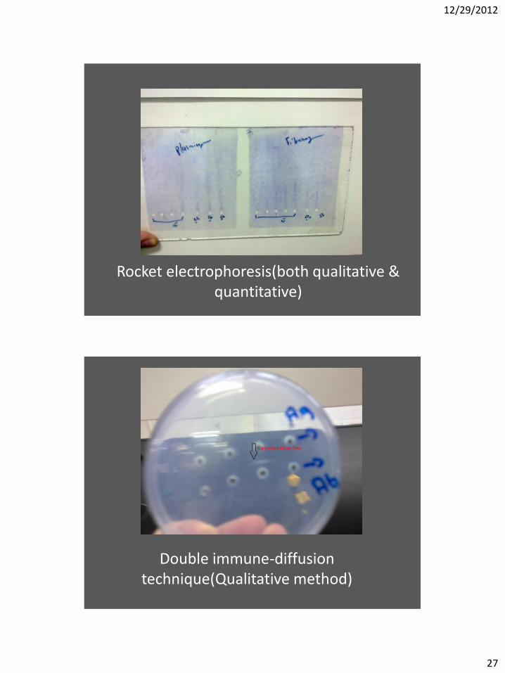
27
Rocket electrophoresis(both qualitative & quantitative)
Double immune-diffusion technique(Qualitative method)

12/29/2012
28
single radial immune diffusion both methods
Agglutination in pregnancy test

12/29/2012
29
Antibody antibiotic reaction
Acid fast culture and stain, and yeast, candida, and Aspergillus.

12/29/2012
30
TB culture

12/29/2012
31

12/29/2012
32
With gram stain

12/29/2012
33
Asperigellus nigra

12/29/2012
34