
Mice sans synaptotagmin - The Queen's Medical Center
2
NEWS AND VIEWS NEUROBIOLOGY--------------------------------------------------------------- Mice sans synaptotagmin Erwin Neher and Reinhold Penner IN the past two years we have come a long way in the biochemical characterization of synaptic proteins. It has emerged that some 6-8 of them interact to form the so-called fusion complex (see refs 1-3 for review); and that homologues of some of the proteins are located at different places in the cell interior, wherever intracellular membranes are believed to fuse with one a Two sensors Wild type Synaptotagmin b Wild type Synaptotagmin arrested at the 'active zone' (the site of neurotransmitter release) until an action potential arrives and increases the concen- tration of Ca 2 +. It was therefore argued that synaptic vesicles possess a 'fusion clamp' which prevents fusion otherwise. Synaptotagmin, by the mere fact of its presence at sites of regulated fusion, was held up as a candidate for carrying out the Mutated Mutated Models for the role of synaptotagmin in eliciting vesicle-membrane fusion. Each of the four diagrams shows a vesicle attached to the plasma membrane by a fusion complex. a, The two-sensor model 9 . Two Ca 2 + -sensors, a low-affinity one (LAS) on synaptotagmin and a high-affinity one (HAS), respectively provide for fast synchronous and asynchronous release. The LAS normally senses short intensive pulses of Ca 2 + when Ca 2 + -channels open, but not in the absence of synaptotagmin (right). The HAS is not necessarily a single binding site, but may represent several Ca 2 + -dependent steps preceding fusion. b, A speculative, alternative model, with only one low-affinity Ca 2 + -sensor. Here, synaptotagmin's function is to link a Ca 2 + -channel to the fusion machinery. In its absence (right), channels and release sites are randomly distributed, so that the Ca 2 + -sensor no longer experiences short intensive pulses of Ca 2 +. another. Synaptic proteins are conserved between man and yeast, so there is no doubt that the fusion complex is one of the very basic molecular machines. It not only mediates synaptic transmission (by letting synaptic vesicles fuse with the plasma membrane) but also enables membranes to flow from their site of synthesis to the cell surface through various cycles of vesicle formation and fusion. It seems, then, that we know which players take part in this game. But what does each of them do? A paper by Gep- pert et al. 4, published in Cell last week, provides some clues to help start assigning specific functions to specific proteins. Fusion of transmitter-containing vesi- cles during synaptic transmission is pre- cisely regulated by calcium, in contrast to 'constitutive exocytosis' which goes on continuously. Synaptic vesicles seem to be 316 clamping function. When it was found that each molecule has two so-called C2- domains (which in protein kinase C bind calcium), the obvious conclusion was that synaptotagmin is the Ca 2 + -sensor of regulated secretion- or, more precisely, a Ca 2 + -controlled fusion clamp 5 . Geppert et al. have now looked at hippocampal neurons from transgenic mice lacking functional synaptotagmin I, one of the two isoforms of the molecule. They conclude that synaptotagmin I is one of the main Ca 2 + -sensors for transmitter release. Mutational studies on Drosophila 6 • 7 and Caenorhabditis elegans 8 provided the initial indications that synaptotagmin is essential for neurotransmission, and that lack of the molecule leads to severe dys- function in secretion. But Geppert et al. find that, immediately after birth, mice homozygous for the knocked out synapto- tagmin gene are phenotypically indisting- uishable from heterozygotes or wild-type mice. Transgenic animals breathe nor- mally, and respond to tactile stimulation; the retina and brain structure show no morphological abnormalities. If one be- lieves that synaptotagmin I is central to neurotransmitter release, and that some functional interaction is required for es- tablishment of a correct body plan, these findings are quite unexpected. Nevertheless, Geppert et al. find that synaptic transmission in cultured hippo- campal neurons is severely perturbed, but in a more subtle way than would be expected if there were just a single Ca 2 +- sensor. They recorded from pairs of hip- pocampal neurons forming contacts in cell culture, stimulating the 'presynaptic' cells and then analysing responses in voltage- clamped 'postsynaptic' cells. With wild- type cells, large-amplitude, postsynaptic currents were observed, which were well synchronized with presynaptic action potentials. A slower 'asynchronous com- ponent', lasting some 100 milliseconds, contributed only little to the overall sig- nal. In cells from mutant mice, on the other hand, the fast synchronous com- ponent was almost completely absent. The asynchronous phase, however, was unaltered or slightly increased. Geppert et al. interpret the data in the light of a study by Goda and Stevens 9 , which describes two such components of neurotransmitter release: a fast compo- nent mediated by a highly efficient, low- affinity Ca 2 + receptor; and a slower com- ponent mediated by a less efficient, high- affinity receptor. In this scheme, synapto- tagmin would be the low-affinity sensor, which efficiently elicits secretion during the short period of the action potential when concentrations of Ca 2 + at the re- lease site reach very high levels (see left-hand part of a in the figure). Mutant mice lacking synaptotagmin (right-hand part of a) would be stimulated only through the inefficient (but high-affinity) receptor; they therefore show only a small, slow response, while residual Ca 2 +, after having spread out diffusionally, decays back to basal levels. This proposal explains the data very well, and provides a detailed picture of the events preceding fusion. But other inter- pretations are possible. One is offered bJ the so-called Ca 2 + -voltage hypothesis 1 , which holds that transmitter release is regulated both by a Ca 2 + -sensor and by a voltage sensor. It seems that the simple postulate that synaptotagmin is the vol- tage sensor would, by and large, explain the results (we are aware that its molecu- lar properties appear to be incompatible with this assumption, but it might be coupled to a voltage sensor). A study on synaptotagmin-deficient PC12 cells can be interpreted in terms of the Ca 2 + -voltage hypothesis, however, with the postulate NATURE · VOL 372 · 24 NOVEMBER 1994
Transcript of Mice sans synaptotagmin - The Queen's Medical Center

NEWS AND VIEWS NEUROBIOLOGY---------------------------------------------------------------
Mice sans synaptotagmin Erwin Neher and Reinhold Penner
IN the past two years we have come a long way in the biochemical characterization of synaptic proteins. It has emerged that some 6-8 of them interact to form the so-called fusion complex (see refs 1-3 for review); and that homologues of some of the proteins are located at different places in the cell interior, wherever intracellular membranes are believed to fuse with one
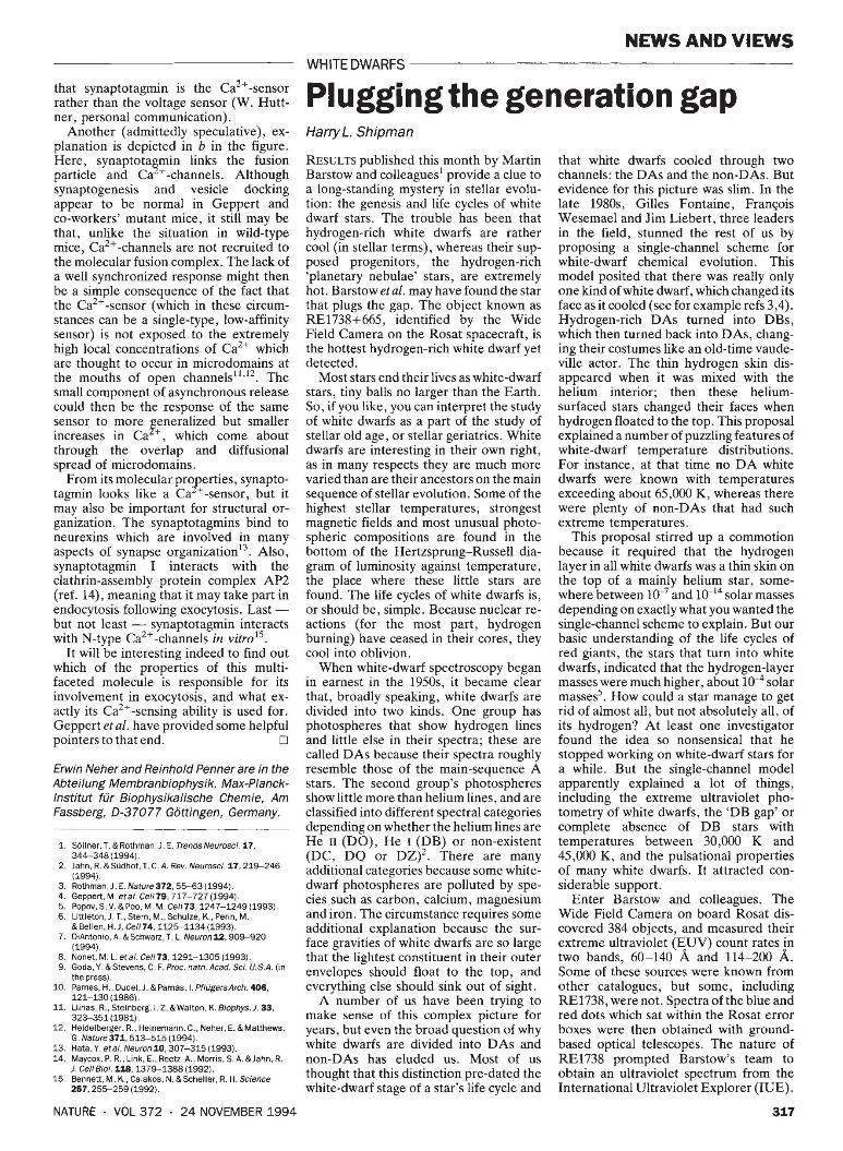
a Two sensors Wild type
Synaptotagmin
b Ca2+-~hannellink Wild type
Synaptotagmin
arrested at the 'active zone' (the site of neurotransmitter release) until an action potential arrives and increases the concentration of Ca2+. It was therefore argued that synaptic vesicles possess a 'fusion clamp' which prevents fusion otherwise. Synaptotagmin, by the mere fact of its presence at sites of regulated fusion, was held up as a candidate for carrying out the
Mutated
Mutated
Models for the role of synaptotagmin in eliciting vesicle-membrane fusion. Each of the four diagrams shows a vesicle attached to the plasma membrane by a fusion complex. a, The two-sensor model9
. Two Ca 2 + -sensors, a low-affinity one (LAS) on synaptotagmin and a high-affinity one (HAS), respectively provide for fast synchronous and asynchronous release. The LAS normally senses short intensive pulses of Ca 2 + when Ca 2 + -channels open, but not in the absence of synaptotagmin (right). The HAS is not necessarily a single binding site, but may represent several Ca 2 + -dependent steps preceding fusion. b, A speculative, alternative model, with only one low-affinity Ca 2 + -sensor. Here, synaptotagmin's function is to link a Ca2 + -channel to the fusion machinery. In its absence (right), channels and release sites are randomly distributed, so that the Ca 2 + -sensor no longer experiences short intensive pulses of Ca2 +.
another. Synaptic proteins are conserved between man and yeast, so there is no doubt that the fusion complex is one of the very basic molecular machines. It not only mediates synaptic transmission (by letting synaptic vesicles fuse with the plasma membrane) but also enables membranes to flow from their site of synthesis to the cell surface through various cycles of vesicle formation and fusion.
It seems, then, that we know which players take part in this game. But what does each of them do? A paper by Geppert et al. 4, published in Cell last week, provides some clues to help start assigning specific functions to specific proteins.
Fusion of transmitter-containing vesicles during synaptic transmission is precisely regulated by calcium, in contrast to 'constitutive exocytosis' which goes on continuously. Synaptic vesicles seem to be
316
clamping function. When it was found that each molecule has two so-called C2-domains (which in protein kinase C bind calcium), the obvious conclusion was that synaptotagmin is the Ca2+ -sensor of regulated secretion- or, more precisely, a Ca2+ -controlled fusion clamp5 . Geppert et al. have now looked at hippocampal neurons from transgenic mice lacking functional synaptotagmin I, one of the two isoforms of the molecule. They conclude that synaptotagmin I is one of the main Ca2+ -sensors for transmitter release.
Mutational studies on Drosophila6•7
and Caenorhabditis elegans8 provided the initial indications that synaptotagmin is essential for neurotransmission, and that lack of the molecule leads to severe dysfunction in secretion. But Geppert et al. find that, immediately after birth, mice homozygous for the knocked out synapto-
tagmin gene are phenotypically indistinguishable from heterozygotes or wild-type mice. Transgenic animals breathe normally, and respond to tactile stimulation; the retina and brain structure show no morphological abnormalities. If one believes that synaptotagmin I is central to neurotransmitter release, and that some functional interaction is required for establishment of a correct body plan, these findings are quite unexpected.
Nevertheless, Geppert et al. find that synaptic transmission in cultured hippocampal neurons is severely perturbed, but in a more subtle way than would be expected if there were just a single Ca2+sensor. They recorded from pairs of hippocampal neurons forming contacts in cell culture, stimulating the 'presynaptic' cells and then analysing responses in voltageclamped 'postsynaptic' cells. With wildtype cells, large-amplitude, postsynaptic currents were observed, which were well synchronized with presynaptic action potentials. A slower 'asynchronous component', lasting some 100 milliseconds, contributed only little to the overall signal. In cells from mutant mice, on the other hand, the fast synchronous component was almost completely absent. The asynchronous phase, however, was unaltered or slightly increased.
Geppert et al. interpret the data in the light of a study by Goda and Stevens9
,
which describes two such components of neurotransmitter release: a fast component mediated by a highly efficient, lowaffinity Ca2+ receptor; and a slower component mediated by a less efficient, highaffinity receptor. In this scheme, synaptotagmin would be the low-affinity sensor, which efficiently elicits secretion during the short period of the action potential when concentrations of Ca2+ at the release site reach very high levels (see left-hand part of a in the figure). Mutant mice lacking synaptotagmin (right-hand part of a) would be stimulated only through the inefficient (but high-affinity) receptor; they therefore show only a small, slow response, while residual Ca2+,
after having spread out diffusionally, decays back to basal levels.
This proposal explains the data very well, and provides a detailed picture of the events preceding fusion. But other interpretations are possible. One is offered bJ the so-called Ca2+ -voltage hypothesis1
,
which holds that transmitter release is regulated both by a Ca2+ -sensor and by a voltage sensor. It seems that the simple postulate that synaptotagmin is the voltage sensor would, by and large, explain the results (we are aware that its molecular properties appear to be incompatible with this assumption, but it might be coupled to a voltage sensor). A study on synaptotagmin-deficient PC12 cells can be interpreted in terms of the Ca2+ -voltage hypothesis, however, with the postulate
NATURE · VOL 372 · 24 NOVEMBER 1994
that synaptotagmin is the Ca2+ -sensor rather than the voltage sensor (W. Huttner, personal communication).
Another (admittedly speculative), explanation is depicted in b in the figure. Here, synaptota~min links the fusion particle and Ca +-channels. Although synaptogenesis and vesicle docking appear to be normal in Geppert and co-workers' mutant mice, it still may be that, unlike the situation in wild-type mice, Ca2+ -channels are not recruited to the molecular fusion complex. The lack of a well synchronized response might then be a simple consequence of the fact that the Ca2+ -sensor (which in these circumstances can be a single-type, low-affinity sensor) is not exposed to the extremely high local concentrations of Ca2+ which are thought to occur in microdomains at the mouths of open channels11
•12
• The small component of asynchronous release could then be the response of the same sensor to more feneralized but smaller increases in Ca +, which come about through the overlap and diffusional spread of microdomains.
From its molecular pro:Eerties, synaptotagmin looks like a Ca +-sensor, but it may also be important for structural organization. The synaptotagmins bind to neurexins which are involved in many aspects of synapse organization13
. Also, synaptotagmin I interacts with the clathrin-assembly protein complex AP2 (ref. 14), meaning that it may take part in endocytosis following exocytosis. Lastbut not least - synaptotagmin interacts with N-type Ca2+ -channels in vitro 15
.
It will be interesting indeed to find out which of the properties of this multifaceted molecule is responsible for its involvement in exocytosis, and what exactly its Ca2+ -sensing ability is used for. Geppert et al. have provided some helpful pointers to that end. D
Erwin Neher and Reinhold Penner are in the Abteilung Membranbiophysik, Max-Piancklnstitut tar Biophysikalische Chemie, Am Fassberg, D-37077 Gottingen, Germany.
1. Sellner, T. &Rothman, J. E. TrendsNeurosci.17, 344-348 ( 1994).
2. Jahn, R. &Siidhof, T. C.A. Rev. Neurosci.17, 219--246 (1994).
3. Rothman, J. E. Nature372, 55-63 (1994). 4. Geppert, M. eta/. Ce/179, 717-727 (1994). 5. Popov, S. V. &Poo, M. M. Ce//73, 1247-1249 (1993). 6. Littleton, J. T., Stern, M., Schulze, K., Perin, M.
&Bel len, H.J. Ce//74, 1125-1134 (1993). 7. DiAntonio, A. & Schwarz, T. L. Neuron 12, 909--920
(1994). 8. Nonet, M. L. eta/. Ce/173, 1291-1305 (1993). 9. Goda, Y. &Stevens, C. F. Proc. natn.Acad. Sci. U.S.A. (in
the press). 10. Parnas, H., Dudei,J. &Parnas, I. PfliigersArch. 406,
121-130 (1986). 11. Llinas, R., Steinberg, I. Z. &Walton, K. Biophys.J. 33,
323--351 (1981). 12. Heidelberger, R., Heinemann, C., Neher, E. & Matthews,
G. Nature371, 513--515 (1994). 13. Hata, Y. eta/. Neuron10, 307-315 (1993). 14. Maycox. P.R., Link, E., Reetz, A., Morris, S. A. &Jahn, R.
J. Cell Bioi. 118, 1379--1388 (1992). 15. Bennett, M. K., Calakos, N. &Scheller, R. H. Science
257,255-259 (1992).
NATURE · VOL 372 · 24 NOVEMBER 1994
NEWS AND VIEWS WHITE DWARFS-------------------
Plugging the generation gap Harry L. Shipman
RESULTS published this month by Martin Barstow and colleagues1 provide a clue to a long-standing mystery in stellar evolution: the genesis and life cycles of white dwarf stars. The trouble has been that hydrogen-rich white dwarfs are rather cool (in stellar terms), whereas their supposed progenitors, the hydrogen-rich 'planetary nebulae' stars, are extremely hot. Barstow et al. may have found the star that plugs the gap. The object known as RE1738+665, identified by the Wide Field Camera on the Rosat spacecraft, is the hottest hydrogen-rich white dwarf yet detected.
Most stars end their lives as white-dwarf stars, tiny balls no larger than the Earth. So, if you like, you can interpret the study of white dwarfs as a part of the study of stellar old age, or stellar geriatrics. White dwarfs are interesting in their own right, as in many respects they are much more varied than are their ancestors on the main sequence of stellar evolution. Some of the highest stellar temperatures, strongest magnetic fields and most unusual photospheric compositions are found in the bottom of the Hertzsprung-Russell diagram of luminosity against temperature, the place where these little stars are found. The life cycles of white dwarfs is, or should be, simple. Because nuclear reactions (for the most part, hydrogen burning) have ceased in their cores, they cool into oblivion.
When white-dwarf spectroscopy began in earnest in the 1950s, it became clear that, broadly speaking, white dwarfs are divided into two kinds. One group has photospheres that show hydrogen lines and little else in their spectra; these are called DAs because their spectra roughly resemble those of the main-sequence A stars. The second group's photospheres show little more than helium lines, and are classified into different spectral categories depending on whether the helium lines are He II (DO), He I (DB) or non-existent (DC, DQ or DZ)2 . There are many additional categories because some whitedwarf photospheres are polluted by species such as carbon, calcium, magnesium and iron. The circumstance requires some additional explanation because the surface gravities of white dwarfs are so large that the lightest constituent in their outer envelopes should float to the top, and everything else should sink out of sight.
A number of us have been trying to make sense of this complex picture for years, but even the broad question of why white dwarfs are divided into DAs and non-DAs has eluded us. Most of us thought that this distinction pre-dated the white-dwarf stage of a star's life cycle and
that white dwarfs cooled through two channels: the DAs and the non-DAs. But evidence for this picture was slim. In the late 1980s, Gilles Fontaine, Fram;ois Wesemael and Jim Liebert, three leaders in the field, stunned the rest of us by proposing a single-channel scheme for white-dwarf chemical evolution. This model posited that there was really only one kind of white dwarf, which changed its face as it cooled (see for example refs 3,4). Hydrogen-rich DAs turned into DBs, which then turned back into DAs, changing their costumes like an old-time vaudeville actor. The thin hydrogen skin disappeared when it was mixed with the helium interior; then these heliumsurfaced stars changed their faces when hydrogen floated to the top. This proposal explained a number of puzzling features of white-dwarf temperature distributions. For instance, at that time no DA white dwarfs were known with temperatures exceeding about 65,000 K, whereas there were plenty of non-DAs that had such extreme temperatures.
This proposal stirred up a commotion because it required that the hydrogen layer in all white dwarfs was a thin skin on the top of a mainly helium star, somewhere between 10,_7 and 10,_14 solar masses depending on exactly what you wanted the single-channel scheme to explain. But our basic understanding of the life cycles of red giants, the stars that turn into white dwarfs, indicated that the hydrogen-layer masses were much higher, about 10-4 solar masses5
• How could a star manage to get rid of almost all, but not absolutely all, of its hydrogen? At least one investigator found the idea so nonsensical that he stopped working on white-dwarf stars for a while. But the single-channel model apparently explained a lot of things, including the extreme ultraviolet photometry of white dwarfs, the 'DB gap' or complete absence of DB stars with temperatures between 30,000 K and 45,000 K, and the pulsational properties of many white dwarfs. It attracted considerable support.
Enter Barstow and colleagues. The Wide Field Camera on board Rosat discovered 384 objects, and measured their extreme ultraviolet (EUV) count rates in two bands, 60-140 A and 114-200 A. Some of these sources were known from other catalogues, but some, including RE1738, were not. Spectra ofthe blue and red dots which sat within the Rosat error boxes were then obtained with groundbased optical telescopes. The nature of RE1738 prompted Barstow's team to obtain an ultraviolet spectrum from the International Ultraviolet Explorer (IUE).
317