Meyers’ Dynamic Radiology of the Abdomen · of the Abdomen Normal and Pathologic Anatomy SIXTH...
30
Meyers’ Dynamic Radiology of the Abdomen SIXTH EDITION
Transcript of Meyers’ Dynamic Radiology of the Abdomen · of the Abdomen Normal and Pathologic Anatomy SIXTH...

Meyers’ Dynamic Radiology
of the Abdomen
SIXTH EDITION

Morton A. MeyersChusilp CharnsangavejMichael Oliphant
Meyers’ Dynamic Radiologyof the Abdomen
Normal and Pathologic AnatomySIXTH EDITION
With 531 Figures, in 729 Parts, 22 in Color
1 3

Morton A. Meyers, MD, FACR, FACGProfessor Emeritus of Radiology and MedicineDistinguished University ProfessorState University of New YorkStony Brook, NY 11794-8460USA
Chusilp Charnsangavej, MD, FSIRProfessor of RadiologyRobert D. Moreton DistinguishedChair in Diagnostic Radiology
The University of TexasM.D. Anderson Cancer CenterHouston, TX 77030USA
Michael Oliphant, MD, FACRProfessor of RadiologyDepartment of RadiologyWake Forest UniversitySchool of Medicine
Winston-Salem, NC 27157-1088USA
ISBN 978-1-4419-5938-6 e-ISBN 978-1-4419-5939-3DOI 10.1007/978-1-4419-5939-3Springer New York Dordrecht Heidelberg London
Library of Congress Control Number: 2010932339
# Springer ScienceþBusiness Media, LLC 2011
Print # 2005 Springer ScienceþBusiness Media, Inc.
Print # 2000, 1994, 1988, 1982, 1976 Springer-Verlag New York, Inc.
All rights reserved. This work may not be translated or copied in whole or in part without the written permission of the publisher
(Springer ScienceþBusiness Media, LLC, 233 Spring Street, New York, NY 10013, USA), except for brief excerpts in connectionwith reviews or scholarly analysis. Use in connection with any form of information storage and retrieval, electronic adaptation,computer software, or by similar or dissimilar methodology now known or hereafter developed is forbidden.The use in this publication of trade names, trademarks, service marks, and similar terms, even if they are not identified as such, is not
to be taken as an expression of opinion as to whether or not they are subject to proprietary rights.While the advice and information in this book are believed to be true and accurate at the date of going to press, neither the authorsnor the editors nor the publisher can accept any legal responsibility for any errors or omissions that may be made. The publisher
makes no warranty, express or implied, with respect to the material contained herein.
Printed on acid-free paper
Springer is part of Springer ScienceþBusiness Media (www.springer.com)

To Bea, Amy,
Richard, Karen,
Sarah, and Sam
I couldn’t wish for a more loving family
Morton A. Meyers
To my teachers: Professor Milton Elkin who encouraged me to use
multimodality approach and to apply physiology and pathology
in Diagnostic Imaging, and to Professor Sidney Wallace who taught me
how to be a clinician
To my wife and children: Tanitra, Chutapom, Tonyamas, Nalinda,
Sirynda, and Larissa who endured my long hours at work
To my parents: Chow and Usa who would like their children
to be successful and secure a better life
Chusilp Charnsangavej
To Phyllis, Melissa,
Jason, Bradley, and Ella
All my love always.
In memory of Molly Sara
Michael Oliphant
There are some things which cannot be learned quickly, and time,which is all we have, must be paid heavily for their acquiring. They arethe very simplest things; and, because it takes a man’s life to know
them, the little new that each man gets from life is very costly and theonly heritage he has to leave.
Ernest Hemingway
Death in the Afternoon
Preface to the Sixth
Edition
The preface to the first edition of Meyers’ DynamicRadiology of the Abdomen: Normal and PathologicAnatomy stated that this book introduces a systematicapplication of anatomic and dynamic principles to thepractical understanding and diagnosis of intraabdom-inal diseases. The clinical insights and rational systemof diagnostic analysis stimulated by an appreciation ofthe dynamic intraabdominal relationships outlined inprevious editions have been universally adopted.Literally thousands of scientific articles in the litera-ture have attested to their basic precepts. Formula-tions and analytic approaches introduced in the firstedition are now widely applied in clinical medicine sothat many of the terminologies, definitions, and con-cepts of pathogenesis have solidly entered the scientificdomain. These insights lead to the uncovering of clini-cally deceptive diseases, the evaluation of the effects ofdisease, the anticipation of complications, and thedetermination of the appropriate diagnostic and ther-apeutic approaches. Spanish, Italian, Japanese, andPortuguese editions have encouraged more wide-spread application of the principles, which in turnhas led to further contributions to our understandingof the features of spread and localization of intraab-dominal diseases. These principles have been appliedto the full range of imaging modalities – from plainfilms and conventional contrast studies to CT, US inall its modes (endoscopic, laparoscopic, and intra-operative), MRI, and PET-CT – leading to this sixthedition after 34 years.
In the pursuit of comprehending the pattern, allmethods of investigation have been used, including(a) anatomic cross-sectioning of cadavers frozen tomaintain relationships; (b) cadaver injections and dis-sections performed to determine preferential planes ofspread along ligaments, mesenteries, and extraperito-neal fascial compartments; (c) selected clinical caseswith the fullest range of imaging studies; (d) peritoneo-scopy and peritoneography; and (e) surgical opera-tions, surgical pathology, and autopsies.
The basic aims in writing this book have not chan-ged from the first edition, and it is produced in thesame spirit as its predecessors. The quest of science hasalways sought the identification of a pattern of cir-cumstances. With this recognition, there followsinsight and understanding into the nature anddynamics of events and thereby their predictability,management, and consequences. This book estab-lishes that the spread and localization of diseasesthroughout the abdomen and pelvis are not random,irrational occurrences but rather are governed by lawsof structural and dynamic factors.
In the past, radiology books have traditionallydealt with highly focused topics limited to a particularorgan or imagingmodality. Often, these have typicallybeen collections of cases illustrating the range of dis-eases affecting that organ or the advantages and lim-itations offered by a particular imaging technique.However, in a clinical setting, patients often presentin a manner challenging the physician’s thinking
patterns: to determine not only ‘‘what?’’ but ‘‘how?’’and ‘‘why?’’ and ‘‘where?’’
The first edition was hailed as ‘‘the book that revo-lutionized abdominal radiology.’’ One reviewerenthused: ‘‘In literature there are favorite 64 thousanddollar questions, namely, which three books would aman choose if he had to live alone on a deserted island.If one narrows the field to radiological abdominaltexts, I wouldn’t hesitate to take Meyers’ DynamicRadiology of the Abdomen. . .The book would be anintellectual challenge that would make the lonelinessbearable.’’ An author’s pride that critical insights hadbeen formulated was furthered by another reviewer’stribute: ‘‘Morton Meyers has opened up a whole newworld for many of us. . .Meyers on the abdomen is likeArmstrong on the moon.’’
While hewing to the fundamental theme, this sixthedition is not simply a revision, not merely a compen-dium of the observations and experiences reported byothers. Rather, it is decidedly a virtually new presenta-tion. Its authorship has been enlarged by two inter-national authorities who have pioneered ground-breaking perspectives in the precise recognition of awide spectrum of intraabdominal disease processes.To satisfy these aims, completely new chapters havebeen added and others have been extensively updatedand enlarged. This edition includes more than 680 newimages and illustrations.
The insights introduced by Morton Meyers in thefirst edition and developed over the subsequent edi-tions ensured the critical position of the radiologist inestablishing the diagnosis, often redirecting the courseof investigation, and in indicating the prognosis anddetermining the management. Clearly established arethe dynamics and pathways of spread and localizationof intraperitoneal infections andmalignancies, and theanatomic–pathologic delineation of the three extra-peritoneal spaces. What had been woefully describedas a ‘‘hinterland of straggling mesenchyme with itsshadowy fascial boundaries’’ is now seen as clearlydemarcated compartments with pathognomonicfeatures.
As useful as these have been, much has been gainedby broadening a vision to encompass global anatomiccontinuity throughout the abdomen and pelvis: just asa loop of ribbon twisted once or several times, as in aMobius strip, yields a structure with continuity ofplanes. The unifying concept of the subperitonealspace of the abdomen and pelvis devised and refinedby Michael Oliphant and colleagues in the scientificliterature, including the fifth edition, is here now ele-gantly elaborated for clinical applications. It servesboth to illuminate the potential of bidirectional spreadof disease – predominantly cancer but also benignconditions, e.g., inflammation and trauma – and to
explain what has long been thought of as illogical andmysterious circumstances. On this basis, the role ofdiagnostic imaging is vastly extended.
Many new chapters meticulously detail the patternof lymphatic spread of cancer from primary organs inthe abdomen and pelvis. Chusilp Charnsangavej illus-trates exquisitely precise identification based on ana-lysis of huge clinical material at the M.D. AndersonCancer Center in Houston.
With a known primary lesion, it may be critical toanticipate the likely sites of spread. On the other hand,a patient may present with a lesion at a remote site, inwhich case it becomes important to think backward inorder to reveal the occult primary site. Charnsangavejshows that an intimate knowledge of the vasculardistributions characteristic of each organ providesthe template for identifying its lymphatic pathways.He emphasizes that the benefits of understanding thepathways of lymphatic drainage of each individualorgan are threefold. First, when the primary site ofthe tumor is known, it allows precise identification ofthe expected sites of nodal metastases by following thearterial supply or venous drainage in the ligaments,mesentery, or mesocolon attached to that organ. Sec-ond, when the primary site of tumor is not clinicallyknown, identifying abnormal nodes allows trackingthe arterial supply or venous drainage in that regionto the primary source. Third, it also allows identifica-tion of the expected site of recurrent disease or nodalmetastasis or the pattern of disease progression aftertreatment by looking at the nodal station beyond thetreated site. Accurate assessment is crucial for plan-ning treatment regarding neoadjuvant therapy andsurgery and may impact the outcome of treatment.
Additional value is occasionally encountered. Anincidentally noted abnormal-appearing lymph nodenot in the expected pathway from a known primarysite may be discounted to represent a metastasis. Andtoday, with increased patient longevity achieved fol-lowing treatment of a primary cancer, second and eventhird primaries may arise. In this setting, if a lymphnode metastasis is identified at a distant site, knowl-edge of the pathways of spread may help in accuratelydetermining the particular primary site fromwhich therecurrence has taken origin.
As in previous editions, great care has been takenwith the layout to give prominence to selected illustra-tions and, most importantly, to position the figures asclosely as possible to their citation in the text so thatthe reader’s time and effort are not wasted referring topages some distance apart.
The color images detail anatomic features of clin-ical significance.
The references have been expanded and continue toinclude both classic articles and recent citations. They
viii � Preface to the Sixth Edition

are not restricted to the English language and, whenpertinent, refer to the original descriptions. A lengthyindex with cross-references provides immediate accessto the detailed material presented.
We wish to express our gratitude to the contribut-ing authors who have added luster to this edition:
� Drs. Yong Ho Auh of the Weill Cornell MedicalCollege – New York Presbyterian Hospital, NewYork City; Jae Hoon Lim of the SungkyunkwanUniversity School of Medicine, Samsung MedicalCenter, Seoul, Korea; and Sophia T. Kung ofthe Weill Cornell Medical College – New YorkPresbyterian Hospital, who contributed Chapter7 on The Extraperitoneal Pelvic Compartments;
� Drs. Maarten S. van Leeuwen and Michiel A.M.Feldberg of the University Medical Center,Utrecht, The Netherlands, who contributed thesection on Compartmentalization of the AnteriorPararenal Space in Chapter 6.
We also wish to thank Dr. Jae Hoon Lim for hisgenerous cooperation in providing many state-of-the-art images depicting extraperitoneal anatomy andpathology.
We have submitted this manuscript to Springer,confident that their skills have produced another edi-tion of high technical quality.
Morton A. Meyers, M.D., F.A.C.R, F.A.C.G.Stony Brook, New York
Chusilp Charnsangavej, M.D., F.S.I.R.Houston, Texas
Michael Oliphant, M.D., F.A.C.R.Winston-Salem, North Carolina
Preface to the Sixth Edition � ix

Contents
Preface to the Sixth Edition . . . . . . . . . . . . . . . . . . . . . . . . . . . . . . . . . . . . . . . . . . . . . . . . . . . . . . . . . . . . . . . . . . vii
1 A New Paradigm . . . . . . . . . . . . . . . . . . . . . . . . . . . . . . . . . . . . . . . . . . . . . . . . . . . . . . . . . . . . . . . . . . . 1
References . . . . . . . . . . . . . . . . . . . . . . . . . . . . . . . . . . . . . . . . . . . . . . . . . . . . . . . . . . . . . . . . . . . . . . . . 7
2 Clinical Embryology of the Abdomen . . . . . . . . . . . . . . . . . . . . . . . . . . . . . . . . . . . . . . . . . . . . . . . . . . . . 9
Introduction . . . . . . . . . . . . . . . . . . . . . . . . . . . . . . . . . . . . . . . . . . . . . . . . . . . . . . . . . . . . . . . . . . . . . . . 9
Early Embryonic Development. . . . . . . . . . . . . . . . . . . . . . . . . . . . . . . . . . . . . . . . . . . . . . . . . . . . . . . . . 9
Thoracoabdominal Continuum . . . . . . . . . . . . . . . . . . . . . . . . . . . . . . . . . . . . . . . . . . . . . . . . . . . . . . . . . 10
Subperitoneal Space. . . . . . . . . . . . . . . . . . . . . . . . . . . . . . . . . . . . . . . . . . . . . . . . . . . . . . . . . . . . . . . . . 12
Ventral Mesentery Specialization . . . . . . . . . . . . . . . . . . . . . . . . . . . . . . . . . . . . . . . . . . . . . . . . . . . . 13
Dorsal Mesentery Specialization. . . . . . . . . . . . . . . . . . . . . . . . . . . . . . . . . . . . . . . . . . . . . . . . . . . . . 14
Pelvic Specialization . . . . . . . . . . . . . . . . . . . . . . . . . . . . . . . . . . . . . . . . . . . . . . . . . . . . . . . . . . . . . . 18
Embryology of Specific Organs . . . . . . . . . . . . . . . . . . . . . . . . . . . . . . . . . . . . . . . . . . . . . . . . . . . . . . . . 19
Embryologic Rotation and Fixation of the Gut . . . . . . . . . . . . . . . . . . . . . . . . . . . . . . . . . . . . . . . . . 19
Hepatobiliary System . . . . . . . . . . . . . . . . . . . . . . . . . . . . . . . . . . . . . . . . . . . . . . . . . . . . . . . . . . . . . 20
Pancreas . . . . . . . . . . . . . . . . . . . . . . . . . . . . . . . . . . . . . . . . . . . . . . . . . . . . . . . . . . . . . . . . . . . . . . . 21
Spleen . . . . . . . . . . . . . . . . . . . . . . . . . . . . . . . . . . . . . . . . . . . . . . . . . . . . . . . . . . . . . . . . . . . . . . . . . 21
Adrenal Glands . . . . . . . . . . . . . . . . . . . . . . . . . . . . . . . . . . . . . . . . . . . . . . . . . . . . . . . . . . . . . . . . . . 21
Urinary System . . . . . . . . . . . . . . . . . . . . . . . . . . . . . . . . . . . . . . . . . . . . . . . . . . . . . . . . . . . . . . . . . . 21
References . . . . . . . . . . . . . . . . . . . . . . . . . . . . . . . . . . . . . . . . . . . . . . . . . . . . . . . . . . . . . . . . . . . . . . . . 22
3 Clinical Anatomy of the Abdomen . . . . . . . . . . . . . . . . . . . . . . . . . . . . . . . . . . . . . . . . . . . . . . . . . . . . . . 23
Introduction . . . . . . . . . . . . . . . . . . . . . . . . . . . . . . . . . . . . . . . . . . . . . . . . . . . . . . . . . . . . . . . . . . . . . . . 23
The Fundamental Concept of the Subperitoneal Space. . . . . . . . . . . . . . . . . . . . . . . . . . . . . . . . . . . . . . . 23
The Subperitoneal Space . . . . . . . . . . . . . . . . . . . . . . . . . . . . . . . . . . . . . . . . . . . . . . . . . . . . . . . . . . . . . 23

Ventral Mesogastric Derivatives . . . . . . . . . . . . . . . . . . . . . . . . . . . . . . . . . . . . . . . . . . . . . . . . . . . . . 24
Dorsal Mesogastric Derivatives . . . . . . . . . . . . . . . . . . . . . . . . . . . . . . . . . . . . . . . . . . . . . . . . . . . . . 25
Dorsal Mesentery Derivatives. . . . . . . . . . . . . . . . . . . . . . . . . . . . . . . . . . . . . . . . . . . . . . . . . . . . . . . 26
Continuity with the Female Organs . . . . . . . . . . . . . . . . . . . . . . . . . . . . . . . . . . . . . . . . . . . . . . . . . . 29
Central and Lateral Continuity. . . . . . . . . . . . . . . . . . . . . . . . . . . . . . . . . . . . . . . . . . . . . . . . . . . . . . 29
Anterior Continuity . . . . . . . . . . . . . . . . . . . . . . . . . . . . . . . . . . . . . . . . . . . . . . . . . . . . . . . . . . . . . . 30
Pelvic Continuity. . . . . . . . . . . . . . . . . . . . . . . . . . . . . . . . . . . . . . . . . . . . . . . . . . . . . . . . . . . . . . . . . 30
Thoracoabdominal Continuum . . . . . . . . . . . . . . . . . . . . . . . . . . . . . . . . . . . . . . . . . . . . . . . . . . . . . . . . . 31
Imaging Features . . . . . . . . . . . . . . . . . . . . . . . . . . . . . . . . . . . . . . . . . . . . . . . . . . . . . . . . . . . . . . . . . . . 32
The Peritoneal Cavity . . . . . . . . . . . . . . . . . . . . . . . . . . . . . . . . . . . . . . . . . . . . . . . . . . . . . . . . . . . . . . . 32
References . . . . . . . . . . . . . . . . . . . . . . . . . . . . . . . . . . . . . . . . . . . . . . . . . . . . . . . . . . . . . . . . . . . . . . . . 40
4 Mechanisms of Spread of Disease in the Abdomen and Pelvis . . . . . . . . . . . . . . . . . . . . . . . . . . . . . . . . . 41
Introduction . . . . . . . . . . . . . . . . . . . . . . . . . . . . . . . . . . . . . . . . . . . . . . . . . . . . . . . . . . . . . . . . . . . . . . . 41
Distinguishing Intraperitoneal Spread from Subperitoneal Spread . . . . . . . . . . . . . . . . . . . . . . . . . . . . . . 42
Subperitoneal Spread Along Mesenteric Planes . . . . . . . . . . . . . . . . . . . . . . . . . . . . . . . . . . . . . . . . . . . . 44
Subperitoneal Spread by Lymphatics and Lymph Node Metastasis . . . . . . . . . . . . . . . . . . . . . . . . . . . . . 55
Subperitoneal Spread by Periarterial and Perineural Spread . . . . . . . . . . . . . . . . . . . . . . . . . . . . . . . . . . 55
Subperitoneal Spread by Transvenous Spread . . . . . . . . . . . . . . . . . . . . . . . . . . . . . . . . . . . . . . . . . . . . . 55
Subperitoneal Spread by Intraductal Spread . . . . . . . . . . . . . . . . . . . . . . . . . . . . . . . . . . . . . . . . . . . . . . 66
Summary . . . . . . . . . . . . . . . . . . . . . . . . . . . . . . . . . . . . . . . . . . . . . . . . . . . . . . . . . . . . . . . . . . . . . . . . . 67
References . . . . . . . . . . . . . . . . . . . . . . . . . . . . . . . . . . . . . . . . . . . . . . . . . . . . . . . . . . . . . . . . . . . . . . . . 67
5 Intraperitoneal Spread of Infections and Seeded Metastases . . . . . . . . . . . . . . . . . . . . . . . . . . . . . . . . . . 69
Intraperitoneal Infections: Pathways of Spread and Localization . . . . . . . . . . . . . . . . . . . . . . . . . . . . . . 69
Anatomic Considerations . . . . . . . . . . . . . . . . . . . . . . . . . . . . . . . . . . . . . . . . . . . . . . . . . . . . . . . . . . . . . 69
The Posterior Peritoneal Attachments . . . . . . . . . . . . . . . . . . . . . . . . . . . . . . . . . . . . . . . . . . . . . . . . 69The Right Subhepatic Space . . . . . . . . . . . . . . . . . . . . . . . . . . . . . . . . . . . . . . . . . . . . . . . . . . . . . . . . . 71The Right Subphrenic Space. . . . . . . . . . . . . . . . . . . . . . . . . . . . . . . . . . . . . . . . . . . . . . . . . . . . . . . . . 72The Left Subphrenic Space . . . . . . . . . . . . . . . . . . . . . . . . . . . . . . . . . . . . . . . . . . . . . . . . . . . . . . . . . . 72The Lesser Sac. . . . . . . . . . . . . . . . . . . . . . . . . . . . . . . . . . . . . . . . . . . . . . . . . . . . . . . . . . . . . . . . . . . . 73
Radiologic Features . . . . . . . . . . . . . . . . . . . . . . . . . . . . . . . . . . . . . . . . . . . . . . . . . . . . . . . . . . . . . . . . . 76
The Spread and Localization of Intraperitoneal Abscesses . . . . . . . . . . . . . . . . . . . . . . . . . . . . . . . . 76Pelvic Abscesses . . . . . . . . . . . . . . . . . . . . . . . . . . . . . . . . . . . . . . . . . . . . . . . . . . . . . . . . . . . . . . . . . . 77Right Subhepatic and Subphrenic Abscesses . . . . . . . . . . . . . . . . . . . . . . . . . . . . . . . . . . . . . . . . . . . . 77
Hydrostatic Consideration . . . . . . . . . . . . . . . . . . . . . . . . . . . . . . . . . . . . . . . . . . . . . . . . . . . . . . . . . 82Lesser Sac Abscesses . . . . . . . . . . . . . . . . . . . . . . . . . . . . . . . . . . . . . . . . . . . . . . . . . . . . . . . . . . . . . . . 83Left Subphrenic Abscesses . . . . . . . . . . . . . . . . . . . . . . . . . . . . . . . . . . . . . . . . . . . . . . . . . . . . . . . . . . 83
Summary of Pathways. . . . . . . . . . . . . . . . . . . . . . . . . . . . . . . . . . . . . . . . . . . . . . . . . . . . . . . . . . . . . 87
Intraperitoneal Seeding: Pathways of Spread and Localization . . . . . . . . . . . . . . . . . . . . . . . . . . . . . . . . 87
Pathways of Ascitic Flow . . . . . . . . . . . . . . . . . . . . . . . . . . . . . . . . . . . . . . . . . . . . . . . . . . . . . . . . . . . . . 88
Seeded Sites . . . . . . . . . . . . . . . . . . . . . . . . . . . . . . . . . . . . . . . . . . . . . . . . . . . . . . . . . . . . . . . . . . . . . . . 89
Pouch of Douglas (Rectosigmoid Junction): Radiologic Features . . . . . . . . . . . . . . . . . . . . . . . . . . . 89
Lower Small Bowel Mesentery (Terminal Ileum and Cecum): Radiologic Features . . . . . . . . . . . . . 91
Sigmoid Colon: Radiologic Features . . . . . . . . . . . . . . . . . . . . . . . . . . . . . . . . . . . . . . . . . . . . . . . . . 92
Right Paracolic Gutter (Cecum and Ascending Colon) and Morison’s Pouch:
Radiologic Features . . . . . . . . . . . . . . . . . . . . . . . . . . . . . . . . . . . . . . . . . . . . . . . . . . . . . . . . . . . . . . 96
Seeded Perihepatic and Subdiaphragmatic Metastases. . . . . . . . . . . . . . . . . . . . . . . . . . . . . . . . . . . . 96
xii � Contents

Seeded Metastases on the Greater Omentum . . . . . . . . . . . . . . . . . . . . . . . . . . . . . . . . . . . . . . . . . . . 103
Two Unusual Sites of Peritoneal Carcinomatosis. . . . . . . . . . . . . . . . . . . . . . . . . . . . . . . . . . . . . . . . 103Sister Mary Joseph’s Nodule . . . . . . . . . . . . . . . . . . . . . . . . . . . . . . . . . . . . . . . . . . . . . . . . . . . . . . . . 103Krukenberg Tumors . . . . . . . . . . . . . . . . . . . . . . . . . . . . . . . . . . . . . . . . . . . . . . . . . . . . . . . . . . . . . . . 104
Mimicry of Carcinomatosis. . . . . . . . . . . . . . . . . . . . . . . . . . . . . . . . . . . . . . . . . . . . . . . . . . . . . . . . . 105
Instrumental, Operative, and Needle Track Seeding . . . . . . . . . . . . . . . . . . . . . . . . . . . . . . . . . . . . . 105
References . . . . . . . . . . . . . . . . . . . . . . . . . . . . . . . . . . . . . . . . . . . . . . . . . . . . . . . . . . . . . . . . . . . . . . . . 105
6 The Extraperitoneal Spaces: Normal and Pathologic Anatomy . . . . . . . . . . . . . . . . . . . . . . . . . . . . . . . . 109
Introduction . . . . . . . . . . . . . . . . . . . . . . . . . . . . . . . . . . . . . . . . . . . . . . . . . . . . . . . . . . . . . . . . . . . . . . . 109
Anatomic Considerations . . . . . . . . . . . . . . . . . . . . . . . . . . . . . . . . . . . . . . . . . . . . . . . . . . . . . . . . . . . . . 110
The Three Extraperitoneal Compartments and Perirenal Fasciae . . . . . . . . . . . . . . . . . . . . . . . . . . . 110
The Psoas Muscle . . . . . . . . . . . . . . . . . . . . . . . . . . . . . . . . . . . . . . . . . . . . . . . . . . . . . . . . . . . . . . . . 125
The Hepatic and Splenic Angles . . . . . . . . . . . . . . . . . . . . . . . . . . . . . . . . . . . . . . . . . . . . . . . . . . . . . 128
Anterior Pararenal Space . . . . . . . . . . . . . . . . . . . . . . . . . . . . . . . . . . . . . . . . . . . . . . . . . . . . . . . . . . . . 128
Roentgen Anatomy of Distribution and Localization of Collections. . . . . . . . . . . . . . . . . . . . . . . . . 128
Sources of Effusions . . . . . . . . . . . . . . . . . . . . . . . . . . . . . . . . . . . . . . . . . . . . . . . . . . . . . . . . . . . . . . 128Extraperitoneal Perforations of the Colon and Appendix . . . . . . . . . . . . . . . . . . . . . . . . . . . . . . . . . . 130Perforation of the Duodenum. . . . . . . . . . . . . . . . . . . . . . . . . . . . . . . . . . . . . . . . . . . . . . . . . . . . . . . . 131Retroduodenal and Intramural Duodenal Hematoma. . . . . . . . . . . . . . . . . . . . . . . . . . . . . . . . . . . . . 132Pancreatitis . . . . . . . . . . . . . . . . . . . . . . . . . . . . . . . . . . . . . . . . . . . . . . . . . . . . . . . . . . . . . . . . . . . . . . 132Bleeding from Bare Area of Spleen, Splenic Artery, or Hepatic Artery. . . . . . . . . . . . . . . . . . . . . . . . 147Pelvic and Mesenteric Continuities. . . . . . . . . . . . . . . . . . . . . . . . . . . . . . . . . . . . . . . . . . . . . . . . . . . . 151
Compartmentalization of the Anterior Pararenal Space. . . . . . . . . . . . . . . . . . . . . . . . . . . . . . . . . . . . . . 151
Maarten S. van Leeuwen, M.D., Ph.D., Michiel A.M. Feldberg, M.D., Ph.D
Fusional Fasciae . . . . . . . . . . . . . . . . . . . . . . . . . . . . . . . . . . . . . . . . . . . . . . . . . . . . . . . . . . . . . . . . . 151
Normal Imaging Features . . . . . . . . . . . . . . . . . . . . . . . . . . . . . . . . . . . . . . . . . . . . . . . . . . . . . . . . . . 152
Abnormal Imaging Features . . . . . . . . . . . . . . . . . . . . . . . . . . . . . . . . . . . . . . . . . . . . . . . . . . . . . . . . 152
Perirenal Space . . . . . . . . . . . . . . . . . . . . . . . . . . . . . . . . . . . . . . . . . . . . . . . . . . . . . . . . . . . . . . . . . . . . 158
Roentgen Anatomy of Distribution and Localization of Collections. . . . . . . . . . . . . . . . . . . . . . . . . 158Sources of Effusions . . . . . . . . . . . . . . . . . . . . . . . . . . . . . . . . . . . . . . . . . . . . . . . . . . . . . . . . . . . . . . . 158Perirenal Gas-Producing Infection . . . . . . . . . . . . . . . . . . . . . . . . . . . . . . . . . . . . . . . . . . . . . . . . . . . . 160Perirenal Abscess . . . . . . . . . . . . . . . . . . . . . . . . . . . . . . . . . . . . . . . . . . . . . . . . . . . . . . . . . . . . . . . . . 165Uriniferous Perirenal Pseudocyst (Urinoma) . . . . . . . . . . . . . . . . . . . . . . . . . . . . . . . . . . . . . . . . . . . . 169
Etiology and Pathogenesis . . . . . . . . . . . . . . . . . . . . . . . . . . . . . . . . . . . . . . . . . . . . . . . . . . . . . . . . . 169Clinical Signs and Symptoms. . . . . . . . . . . . . . . . . . . . . . . . . . . . . . . . . . . . . . . . . . . . . . . . . . . . . . . 170Radiologic Findings . . . . . . . . . . . . . . . . . . . . . . . . . . . . . . . . . . . . . . . . . . . . . . . . . . . . . . . . . . . . . . 170Treatment . . . . . . . . . . . . . . . . . . . . . . . . . . . . . . . . . . . . . . . . . . . . . . . . . . . . . . . . . . . . . . . . . . . . . 172
Distinction Between Perirenal and Subcapsular Collections . . . . . . . . . . . . . . . . . . . . . . . . . . . . . . . . 172Anatomic Considerations . . . . . . . . . . . . . . . . . . . . . . . . . . . . . . . . . . . . . . . . . . . . . . . . . . . . . . . . . . 175Etiology and Pathogenesis . . . . . . . . . . . . . . . . . . . . . . . . . . . . . . . . . . . . . . . . . . . . . . . . . . . . . . . . . 175Clinical Signs and Symptoms. . . . . . . . . . . . . . . . . . . . . . . . . . . . . . . . . . . . . . . . . . . . . . . . . . . . . . . 176Radiologic Findings . . . . . . . . . . . . . . . . . . . . . . . . . . . . . . . . . . . . . . . . . . . . . . . . . . . . . . . . . . . . . . 177Bridging Renal Septa . . . . . . . . . . . . . . . . . . . . . . . . . . . . . . . . . . . . . . . . . . . . . . . . . . . . . . . . . . . . . 179Treatment . . . . . . . . . . . . . . . . . . . . . . . . . . . . . . . . . . . . . . . . . . . . . . . . . . . . . . . . . . . . . . . . . . . . . 179
Perirenal Lymphoma . . . . . . . . . . . . . . . . . . . . . . . . . . . . . . . . . . . . . . . . . . . . . . . . . . . . . . . . . . . . . . 181Perirenal Retroperitoneal Fibrosis . . . . . . . . . . . . . . . . . . . . . . . . . . . . . . . . . . . . . . . . . . . . . . . . . . . . 182Perirenal Extramedullary Hematopoiesis . . . . . . . . . . . . . . . . . . . . . . . . . . . . . . . . . . . . . . . . . . . . . . . 182Perirenal Metastases . . . . . . . . . . . . . . . . . . . . . . . . . . . . . . . . . . . . . . . . . . . . . . . . . . . . . . . . . . . . . . . 182
Posterior Pararenal Space . . . . . . . . . . . . . . . . . . . . . . . . . . . . . . . . . . . . . . . . . . . . . . . . . . . . . . . . . . . . 183
Roentgen Anatomy of Distribution and Localization of Collections. . . . . . . . . . . . . . . . . . . . . . . . . 183
Contents � xiii

Clinical Sources of Effusions. . . . . . . . . . . . . . . . . . . . . . . . . . . . . . . . . . . . . . . . . . . . . . . . . . . . . . . . 185Hemorrhage . . . . . . . . . . . . . . . . . . . . . . . . . . . . . . . . . . . . . . . . . . . . . . . . . . . . . . . . . . . . . . . . . . . . . 185Abscess . . . . . . . . . . . . . . . . . . . . . . . . . . . . . . . . . . . . . . . . . . . . . . . . . . . . . . . . . . . . . . . . . . . . . . . . . 186
Diffuse Extraperitoneal Gas. . . . . . . . . . . . . . . . . . . . . . . . . . . . . . . . . . . . . . . . . . . . . . . . . . . . . . . . . . . 186
Rectal Perforation . . . . . . . . . . . . . . . . . . . . . . . . . . . . . . . . . . . . . . . . . . . . . . . . . . . . . . . . . . . . . . . . 190
Sigmoid Perforation . . . . . . . . . . . . . . . . . . . . . . . . . . . . . . . . . . . . . . . . . . . . . . . . . . . . . . . . . . . . . . 190
Extraperitoneal Gas of Supradiaphragmatic Origin. . . . . . . . . . . . . . . . . . . . . . . . . . . . . . . . . . . . . . 190
Differential Diagnosis of Small Amounts of Subdiaphragmatic Gas. . . . . . . . . . . . . . . . . . . . . . . . . 190
Psoas Abscess and Hematoma . . . . . . . . . . . . . . . . . . . . . . . . . . . . . . . . . . . . . . . . . . . . . . . . . . . . . . . . . 192
References . . . . . . . . . . . . . . . . . . . . . . . . . . . . . . . . . . . . . . . . . . . . . . . . . . . . . . . . . . . . . . . . . . . . . . . . 196
7 The Extraperitoneal Pelvic Compartments. . . . . . . . . . . . . . . . . . . . . . . . . . . . . . . . . . . . . . . . . . . . . . . . 203
Yong Ho Auh, M.D., Jae Hoon Lim, M.D., Ph.D., Sophia T. Kung, M.D.
Anatomy . . . . . . . . . . . . . . . . . . . . . . . . . . . . . . . . . . . . . . . . . . . . . . . . . . . . . . . . . . . . . . . . . . . . . . . . . 203
Prevesical Space. . . . . . . . . . . . . . . . . . . . . . . . . . . . . . . . . . . . . . . . . . . . . . . . . . . . . . . . . . . . . . . . . . 203
Perivesical Space . . . . . . . . . . . . . . . . . . . . . . . . . . . . . . . . . . . . . . . . . . . . . . . . . . . . . . . . . . . . . . . . . 207
Perirectal Space . . . . . . . . . . . . . . . . . . . . . . . . . . . . . . . . . . . . . . . . . . . . . . . . . . . . . . . . . . . . . . . . . . 207
Presacral Space . . . . . . . . . . . . . . . . . . . . . . . . . . . . . . . . . . . . . . . . . . . . . . . . . . . . . . . . . . . . . . . . . . 211
Abnormal Imaging Features. . . . . . . . . . . . . . . . . . . . . . . . . . . . . . . . . . . . . . . . . . . . . . . . . . . . . . . . . . . 211
Prevesical Fluid Collections . . . . . . . . . . . . . . . . . . . . . . . . . . . . . . . . . . . . . . . . . . . . . . . . . . . . . . . . 211
Perivesical Fluid Collections . . . . . . . . . . . . . . . . . . . . . . . . . . . . . . . . . . . . . . . . . . . . . . . . . . . . . . . . 215
Perirectal Pathology . . . . . . . . . . . . . . . . . . . . . . . . . . . . . . . . . . . . . . . . . . . . . . . . . . . . . . . . . . . . . . 215
Presacral Space Pathology . . . . . . . . . . . . . . . . . . . . . . . . . . . . . . . . . . . . . . . . . . . . . . . . . . . . . . . . . 219
Extension Across Fascial Planes . . . . . . . . . . . . . . . . . . . . . . . . . . . . . . . . . . . . . . . . . . . . . . . . . . . . . 219
References . . . . . . . . . . . . . . . . . . . . . . . . . . . . . . . . . . . . . . . . . . . . . . . . . . . . . . . . . . . . . . . . . . . . . . . . 221
8 Patterns of Spread of Disease from the Liver . . . . . . . . . . . . . . . . . . . . . . . . . . . . . . . . . . . . . . . . . . . . . . 223
Introduction . . . . . . . . . . . . . . . . . . . . . . . . . . . . . . . . . . . . . . . . . . . . . . . . . . . . . . . . . . . . . . . . . . . . . . . 223
Embryology and Anatomy of the Liver . . . . . . . . . . . . . . . . . . . . . . . . . . . . . . . . . . . . . . . . . . . . . . . . . . 223
Development of the Liver and Bile Duct . . . . . . . . . . . . . . . . . . . . . . . . . . . . . . . . . . . . . . . . . . . . . . 223
Peritoneal Ligaments. . . . . . . . . . . . . . . . . . . . . . . . . . . . . . . . . . . . . . . . . . . . . . . . . . . . . . . . . . . . . . 223
Anatomic Landmarks of Peritoneal Ligaments Attaching to the Liver . . . . . . . . . . . . . . . . . . . . . . . 224
Patterns of Spread of Disease from the Liver. . . . . . . . . . . . . . . . . . . . . . . . . . . . . . . . . . . . . . . . . . . . . . 224
Intraperitoneal Spread . . . . . . . . . . . . . . . . . . . . . . . . . . . . . . . . . . . . . . . . . . . . . . . . . . . . . . . . . . . . 224
Subperitoneal Spread . . . . . . . . . . . . . . . . . . . . . . . . . . . . . . . . . . . . . . . . . . . . . . . . . . . . . . . . . . . . . 227Contiguous Subperitoneal Spread . . . . . . . . . . . . . . . . . . . . . . . . . . . . . . . . . . . . . . . . . . . . . . . . . . . . 227Lymphatic Spread and Nodal Metastasis. . . . . . . . . . . . . . . . . . . . . . . . . . . . . . . . . . . . . . . . . . . . . . . 227
Pathways of Lymphatic Drainage of the Liver. . . . . . . . . . . . . . . . . . . . . . . . . . . . . . . . . . . . . . . . . . 228Periarterial and Perineural Spread . . . . . . . . . . . . . . . . . . . . . . . . . . . . . . . . . . . . . . . . . . . . . . . . . . . . 234Intravenous Spread . . . . . . . . . . . . . . . . . . . . . . . . . . . . . . . . . . . . . . . . . . . . . . . . . . . . . . . . . . . . . . . . 235Intraductal Spread . . . . . . . . . . . . . . . . . . . . . . . . . . . . . . . . . . . . . . . . . . . . . . . . . . . . . . . . . . . . . . . . 235
References . . . . . . . . . . . . . . . . . . . . . . . . . . . . . . . . . . . . . . . . . . . . . . . . . . . . . . . . . . . . . . . . . . . . . . . . 240
9 Patterns of Spread of Disease from the Distal Esophagus and Stomach. . . . . . . . . . . . . . . . . . . . . . . . . . 243
Introduction . . . . . . . . . . . . . . . . . . . . . . . . . . . . . . . . . . . . . . . . . . . . . . . . . . . . . . . . . . . . . . . . . . . . . . . . 243
Embryology and Anatomy of the Distal Esophagus and Stomach . . . . . . . . . . . . . . . . . . . . . . . . . . . . . . . 243
Peritoneal Ligaments of the Stomach . . . . . . . . . . . . . . . . . . . . . . . . . . . . . . . . . . . . . . . . . . . . . . . . . 243The Gastrosplenic Ligament and Splenorenal Ligament . . . . . . . . . . . . . . . . . . . . . . . . . . . . . . . . . . . 244The Gastrocolic Ligament and the Greater Omentum. . . . . . . . . . . . . . . . . . . . . . . . . . . . . . . . . . . . . 244
xiv � Contents
The Gastrohepatic and Hepatoduodenal Ligament . . . . . . . . . . . . . . . . . . . . . . . . . . . . . . . . . . . . . . . 244
Peritoneal Recesses Around the Stomach . . . . . . . . . . . . . . . . . . . . . . . . . . . . . . . . . . . . . . . . . . . . . . 245
Patterns of Spread of Disease from the Distal Esophagus and Stomach . . . . . . . . . . . . . . . . . . . . . . . . . . 245
Intraperitoneal Spread . . . . . . . . . . . . . . . . . . . . . . . . . . . . . . . . . . . . . . . . . . . . . . . . . . . . . . . . . . . . 246
Direct and Subperitoneal Mesenteric Spread of Gastric Cancer . . . . . . . . . . . . . . . . . . . . . . . . . . . . 247
Subperitoneal Lymphatic Spread and Pathways of Lymph Node Metastasis . . . . . . . . . . . . . . . . . . 249Paraesophageal and Paracardiac Nodes . . . . . . . . . . . . . . . . . . . . . . . . . . . . . . . . . . . . . . . . . . . . . . . . 252Nodal Metastases in the Gastrohepatic Ligament . . . . . . . . . . . . . . . . . . . . . . . . . . . . . . . . . . . . . . . . 252Nodal Metastases in the Gastrosplenic Ligament . . . . . . . . . . . . . . . . . . . . . . . . . . . . . . . . . . . . . . . . 252Nodal Metastases in the Gastrocolic Ligament . . . . . . . . . . . . . . . . . . . . . . . . . . . . . . . . . . . . . . . . . . 252Inferior Phrenic Nodal Pathways . . . . . . . . . . . . . . . . . . . . . . . . . . . . . . . . . . . . . . . . . . . . . . . . . . . . . 254
Periarterial and Perineural Invasion . . . . . . . . . . . . . . . . . . . . . . . . . . . . . . . . . . . . . . . . . . . . . . . . . . 254
Transvenous Spread . . . . . . . . . . . . . . . . . . . . . . . . . . . . . . . . . . . . . . . . . . . . . . . . . . . . . . . . . . . . . . 256
References . . . . . . . . . . . . . . . . . . . . . . . . . . . . . . . . . . . . . . . . . . . . . . . . . . . . . . . . . . . . . . . . . . . . . . . . 256
10 Patterns of Spread of Disease from the Pancreas . . . . . . . . . . . . . . . . . . . . . . . . . . . . . . . . . . . . . . . . . . . 259
Introduction . . . . . . . . . . . . . . . . . . . . . . . . . . . . . . . . . . . . . . . . . . . . . . . . . . . . . . . . . . . . . . . . . . . . . . . 259
Embryology and Anatomy of the Pancreas . . . . . . . . . . . . . . . . . . . . . . . . . . . . . . . . . . . . . . . . . . . . . . . 259
Development of the Pancreas . . . . . . . . . . . . . . . . . . . . . . . . . . . . . . . . . . . . . . . . . . . . . . . . . . . . . . . 259
Anatomy of the Pancreas and Peritoneal Ligaments Around the Pancreas, Mesentery,
and Mesocolon . . . . . . . . . . . . . . . . . . . . . . . . . . . . . . . . . . . . . . . . . . . . . . . . . . . . . . . . . . . . . . . . . . 260
Anatomic Landmarks of Ligaments and Peritoneal Folds Around the Pancreas . . . . . . . . . . . . . . . 261
Vascular Anatomy. . . . . . . . . . . . . . . . . . . . . . . . . . . . . . . . . . . . . . . . . . . . . . . . . . . . . . . . . . . . . . . . 261
Patterns of Spread of Disease from the Pancreas. . . . . . . . . . . . . . . . . . . . . . . . . . . . . . . . . . . . . . . . . . . 263
Intraperitoneal Spread . . . . . . . . . . . . . . . . . . . . . . . . . . . . . . . . . . . . . . . . . . . . . . . . . . . . . . . . . . . . 263
Subperitoneal Spread . . . . . . . . . . . . . . . . . . . . . . . . . . . . . . . . . . . . . . . . . . . . . . . . . . . . . . . . . . . . . 263Contiguous Subperitoneal Spread . . . . . . . . . . . . . . . . . . . . . . . . . . . . . . . . . . . . . . . . . . . . . . . . . . . . 263Lymphatic Spread and Nodal Metastasis. . . . . . . . . . . . . . . . . . . . . . . . . . . . . . . . . . . . . . . . . . . . . . . 265Periarterial and Perineural Spread . . . . . . . . . . . . . . . . . . . . . . . . . . . . . . . . . . . . . . . . . . . . . . . . . . . . 265Intravenous Spread . . . . . . . . . . . . . . . . . . . . . . . . . . . . . . . . . . . . . . . . . . . . . . . . . . . . . . . . . . . . . . . . 268Intraductal Spread . . . . . . . . . . . . . . . . . . . . . . . . . . . . . . . . . . . . . . . . . . . . . . . . . . . . . . . . . . . . . . . . 268
References . . . . . . . . . . . . . . . . . . . . . . . . . . . . . . . . . . . . . . . . . . . . . . . . . . . . . . . . . . . . . . . . . . . . . . . . 274
11 Patterns of Spread of Disease from the Small Intestine . . . . . . . . . . . . . . . . . . . . . . . . . . . . . . . . . . . . . . 275
Introduction . . . . . . . . . . . . . . . . . . . . . . . . . . . . . . . . . . . . . . . . . . . . . . . . . . . . . . . . . . . . . . . . . . . . . . . 275
Embryology and Anatomy of the Small Intestine . . . . . . . . . . . . . . . . . . . . . . . . . . . . . . . . . . . . . . . . . . . 275
Imaging Landmarks of the Mesentery of the Small Intestine . . . . . . . . . . . . . . . . . . . . . . . . . . . . . . . . . . 276
Patterns of Spread of Disease of the Small Intestine and Appendix . . . . . . . . . . . . . . . . . . . . . . . . . . . . . 277
Malrotation of the Small Intestine, Volvulus of the Mesentery, and Intestinal Obstruction . . . . . . . 277
Inflammatory Disease of the Small Intestine and Appendix . . . . . . . . . . . . . . . . . . . . . . . . . . . . . . . 279
Neoplasms of the Small Intestine and Appendix . . . . . . . . . . . . . . . . . . . . . . . . . . . . . . . . . . . . . . . . 283Adenocarcinoma of the Small Intestine . . . . . . . . . . . . . . . . . . . . . . . . . . . . . . . . . . . . . . . . . . . . . . . . 283Carcinoid Tumors. . . . . . . . . . . . . . . . . . . . . . . . . . . . . . . . . . . . . . . . . . . . . . . . . . . . . . . . . . . . . . . . . 283Tumors of the Appendix. . . . . . . . . . . . . . . . . . . . . . . . . . . . . . . . . . . . . . . . . . . . . . . . . . . . . . . . . . . . 289
Summary . . . . . . . . . . . . . . . . . . . . . . . . . . . . . . . . . . . . . . . . . . . . . . . . . . . . . . . . . . . . . . . . . . . . . . . . . 290
References . . . . . . . . . . . . . . . . . . . . . . . . . . . . . . . . . . . . . . . . . . . . . . . . . . . . . . . . . . . . . . . . . . . . . . . . 290
12 Patterns of Spread of Disease from the Large Intestine . . . . . . . . . . . . . . . . . . . . . . . . . . . . . . . . . . . . . . 293
Embryology and Anatomy of the Colon, Rectum, and Anal Canal . . . . . . . . . . . . . . . . . . . . . . . . . . . . . . 293
Contents � xv

Anatomic Consideration . . . . . . . . . . . . . . . . . . . . . . . . . . . . . . . . . . . . . . . . . . . . . . . . . . . . . . . . . . . 293The Cecum and Ascending Colon and Their Mesocolon. . . . . . . . . . . . . . . . . . . . . . . . . . . . . . . . . . . 294The Transverse Colon and Mesocolon . . . . . . . . . . . . . . . . . . . . . . . . . . . . . . . . . . . . . . . . . . . . . . . . . 294The Descending Colon and Mesocolon . . . . . . . . . . . . . . . . . . . . . . . . . . . . . . . . . . . . . . . . . . . . . . . . 294The Sigmoid Colon and Mesocolon . . . . . . . . . . . . . . . . . . . . . . . . . . . . . . . . . . . . . . . . . . . . . . . . . . . 295The Rectum and Mesorectum. . . . . . . . . . . . . . . . . . . . . . . . . . . . . . . . . . . . . . . . . . . . . . . . . . . . . . . . 295The Anal Canal . . . . . . . . . . . . . . . . . . . . . . . . . . . . . . . . . . . . . . . . . . . . . . . . . . . . . . . . . . . . . . . . . . . 295
Disease of the Colon and Rectum. . . . . . . . . . . . . . . . . . . . . . . . . . . . . . . . . . . . . . . . . . . . . . . . . . . . . . . 295
Diverticulitis and Colitis . . . . . . . . . . . . . . . . . . . . . . . . . . . . . . . . . . . . . . . . . . . . . . . . . . . . . . . . . . . 295
Neoplasms of the Colon, Rectum, and Anus . . . . . . . . . . . . . . . . . . . . . . . . . . . . . . . . . . . . . . . . . . . 297
Patterns of Disease Spread . . . . . . . . . . . . . . . . . . . . . . . . . . . . . . . . . . . . . . . . . . . . . . . . . . . . . . . . . . . 299
Intraperitoneal Spread . . . . . . . . . . . . . . . . . . . . . . . . . . . . . . . . . . . . . . . . . . . . . . . . . . . . . . . . . . . . 299
Contiguous Spread to Adjacent Organs and Structures . . . . . . . . . . . . . . . . . . . . . . . . . . . . . . . . . . . 299
Subperitoneal Spread . . . . . . . . . . . . . . . . . . . . . . . . . . . . . . . . . . . . . . . . . . . . . . . . . . . . . . . . . . . . . 300Nodal Metastasis . . . . . . . . . . . . . . . . . . . . . . . . . . . . . . . . . . . . . . . . . . . . . . . . . . . . . . . . . . . . . . . . . 301Periarterial and Perineural Spread . . . . . . . . . . . . . . . . . . . . . . . . . . . . . . . . . . . . . . . . . . . . . . . . . . . . 307Intravenous Spread . . . . . . . . . . . . . . . . . . . . . . . . . . . . . . . . . . . . . . . . . . . . . . . . . . . . . . . . . . . . . . . . 307
References . . . . . . . . . . . . . . . . . . . . . . . . . . . . . . . . . . . . . . . . . . . . . . . . . . . . . . . . . . . . . . . . . . . . . . . . 311
13 Patterns of Spread of Renal, Upper Urothelial, and Adrenal Pathology. . . . . . . . . . . . . . . . . . . . . . . . . . 313
Introduction . . . . . . . . . . . . . . . . . . . . . . . . . . . . . . . . . . . . . . . . . . . . . . . . . . . . . . . . . . . . . . . . . . . . . . . 313
Vascular Anatomy . . . . . . . . . . . . . . . . . . . . . . . . . . . . . . . . . . . . . . . . . . . . . . . . . . . . . . . . . . . . . . . . . . 314
Lymphatic Anatomy . . . . . . . . . . . . . . . . . . . . . . . . . . . . . . . . . . . . . . . . . . . . . . . . . . . . . . . . . . . . . . . . 314
Spread of Disease. . . . . . . . . . . . . . . . . . . . . . . . . . . . . . . . . . . . . . . . . . . . . . . . . . . . . . . . . . . . . . . . . . . 314
Renal Tumors . . . . . . . . . . . . . . . . . . . . . . . . . . . . . . . . . . . . . . . . . . . . . . . . . . . . . . . . . . . . . . . . . . . 314Renal Cell Carcinomas . . . . . . . . . . . . . . . . . . . . . . . . . . . . . . . . . . . . . . . . . . . . . . . . . . . . . . . . . . . . . 314
Mechanisms of Spread of Renal Cell Carcinoma . . . . . . . . . . . . . . . . . . . . . . . . . . . . . . . . . . . . . . . . 315Renal Lymphoma . . . . . . . . . . . . . . . . . . . . . . . . . . . . . . . . . . . . . . . . . . . . . . . . . . . . . . . . . . . . . . . . . 317Medullary Carcinoma of the Kidney and Perirenal Abscess . . . . . . . . . . . . . . . . . . . . . . . . . . . . . . . . 320
Urothelial Tumors. . . . . . . . . . . . . . . . . . . . . . . . . . . . . . . . . . . . . . . . . . . . . . . . . . . . . . . . . . . . . . . . 322Patterns of Spread of Upper Urinary Tract Urothelial Tumors . . . . . . . . . . . . . . . . . . . . . . . . . . . . . 322
Adrenal Tumors . . . . . . . . . . . . . . . . . . . . . . . . . . . . . . . . . . . . . . . . . . . . . . . . . . . . . . . . . . . . . . . . . 322Adrenocortical Carcinoma . . . . . . . . . . . . . . . . . . . . . . . . . . . . . . . . . . . . . . . . . . . . . . . . . . . . . . . . . . 323Pheochromocytomas. . . . . . . . . . . . . . . . . . . . . . . . . . . . . . . . . . . . . . . . . . . . . . . . . . . . . . . . . . . . . . . 324Neuroblastoma/Ganglioneuromas Complex . . . . . . . . . . . . . . . . . . . . . . . . . . . . . . . . . . . . . . . . . . . . 324
References . . . . . . . . . . . . . . . . . . . . . . . . . . . . . . . . . . . . . . . . . . . . . . . . . . . . . . . . . . . . . . . . . . . . . . . . 327
14 Patterns of Spread of Disease of the Pelvis and Male Urogenital Organs . . . . . . . . . . . . . . . . . . . . . . . . 329
Embryology . . . . . . . . . . . . . . . . . . . . . . . . . . . . . . . . . . . . . . . . . . . . . . . . . . . . . . . . . . . . . . . . . . . . . . . 329
Anatomy . . . . . . . . . . . . . . . . . . . . . . . . . . . . . . . . . . . . . . . . . . . . . . . . . . . . . . . . . . . . . . . . . . . . . . . . . 330
Bladder . . . . . . . . . . . . . . . . . . . . . . . . . . . . . . . . . . . . . . . . . . . . . . . . . . . . . . . . . . . . . . . . . . . . . . . . 330
Prostate Gland and Seminal Vesicles . . . . . . . . . . . . . . . . . . . . . . . . . . . . . . . . . . . . . . . . . . . . . . . . . 331
Penis and Urethra . . . . . . . . . . . . . . . . . . . . . . . . . . . . . . . . . . . . . . . . . . . . . . . . . . . . . . . . . . . . . . . . 331
Testis and Scrotum . . . . . . . . . . . . . . . . . . . . . . . . . . . . . . . . . . . . . . . . . . . . . . . . . . . . . . . . . . . . . . . 332
Disease of the Bladder, Prostate Gland, Urethra, Penis, and Testis . . . . . . . . . . . . . . . . . . . . . . . . . . . . . 332
Bladder Cancer . . . . . . . . . . . . . . . . . . . . . . . . . . . . . . . . . . . . . . . . . . . . . . . . . . . . . . . . . . . . . . . . . . 332
Inflammatory and Inflammatory-Like Bladder Masses . . . . . . . . . . . . . . . . . . . . . . . . . . . . . . . . . . . 333
Prostate Cancer . . . . . . . . . . . . . . . . . . . . . . . . . . . . . . . . . . . . . . . . . . . . . . . . . . . . . . . . . . . . . . . . . . 333
Testicular Cancer . . . . . . . . . . . . . . . . . . . . . . . . . . . . . . . . . . . . . . . . . . . . . . . . . . . . . . . . . . . . . . . . 334
Patterns of Disease Spread . . . . . . . . . . . . . . . . . . . . . . . . . . . . . . . . . . . . . . . . . . . . . . . . . . . . . . . . . . . 334
xvi � Contents
Intraperitoneal Spread . . . . . . . . . . . . . . . . . . . . . . . . . . . . . . . . . . . . . . . . . . . . . . . . . . . . . . . . . . . . 334
Subperitoneal Spread . . . . . . . . . . . . . . . . . . . . . . . . . . . . . . . . . . . . . . . . . . . . . . . . . . . . . . . . . . . . . 334Contiguous Extraperitoneal Spread . . . . . . . . . . . . . . . . . . . . . . . . . . . . . . . . . . . . . . . . . . . . . . . . . . . 334Lymph Node Metastasis . . . . . . . . . . . . . . . . . . . . . . . . . . . . . . . . . . . . . . . . . . . . . . . . . . . . . . . . . . . . 336Vascular and Perineural Invasion . . . . . . . . . . . . . . . . . . . . . . . . . . . . . . . . . . . . . . . . . . . . . . . . . . . . . 340
References . . . . . . . . . . . . . . . . . . . . . . . . . . . . . . . . . . . . . . . . . . . . . . . . . . . . . . . . . . . . . . . . . . . . . . . . 345
15 Patterns of Spread of Gynecologic Disease . . . . . . . . . . . . . . . . . . . . . . . . . . . . . . . . . . . . . . . . . . . . . . . 347
Introduction . . . . . . . . . . . . . . . . . . . . . . . . . . . . . . . . . . . . . . . . . . . . . . . . . . . . . . . . . . . . . . . . . . . . . . . 347
Vulva . . . . . . . . . . . . . . . . . . . . . . . . . . . . . . . . . . . . . . . . . . . . . . . . . . . . . . . . . . . . . . . . . . . . . . . . . . . . 349
Direct and Subperitoneal Spread of Vulvar Cancer . . . . . . . . . . . . . . . . . . . . . . . . . . . . . . . . . . . . . . 349
Vagina . . . . . . . . . . . . . . . . . . . . . . . . . . . . . . . . . . . . . . . . . . . . . . . . . . . . . . . . . . . . . . . . . . . . . . . . . . . 350
Direct and Subperitoneal Spread of Vaginal Carcinomas . . . . . . . . . . . . . . . . . . . . . . . . . . . . . . . . . 350
Uterus . . . . . . . . . . . . . . . . . . . . . . . . . . . . . . . . . . . . . . . . . . . . . . . . . . . . . . . . . . . . . . . . . . . . . . . . . . . 351
Invasive Cervical Cancer. . . . . . . . . . . . . . . . . . . . . . . . . . . . . . . . . . . . . . . . . . . . . . . . . . . . . . . . . . . 351
Cancer of the Uterine Body . . . . . . . . . . . . . . . . . . . . . . . . . . . . . . . . . . . . . . . . . . . . . . . . . . . . . . . . 352
Fallopian Tube . . . . . . . . . . . . . . . . . . . . . . . . . . . . . . . . . . . . . . . . . . . . . . . . . . . . . . . . . . . . . . . . . . . . . 352
Patterns of Spread of Fallopian Tube Carcinoma . . . . . . . . . . . . . . . . . . . . . . . . . . . . . . . . . . . . . . . 353
Ovary . . . . . . . . . . . . . . . . . . . . . . . . . . . . . . . . . . . . . . . . . . . . . . . . . . . . . . . . . . . . . . . . . . . . . . . . . . . . 353
Mechanisms for Spread of Ovarian Tumors. . . . . . . . . . . . . . . . . . . . . . . . . . . . . . . . . . . . . . . . . . . . 355
Pelvic Inflammatory Disease . . . . . . . . . . . . . . . . . . . . . . . . . . . . . . . . . . . . . . . . . . . . . . . . . . . . . . . . . . 356
References . . . . . . . . . . . . . . . . . . . . . . . . . . . . . . . . . . . . . . . . . . . . . . . . . . . . . . . . . . . . . . . . . . . . . . . . 360
16 Patterns of Extraabdominal and Extrapelvic Spread . . . . . . . . . . . . . . . . . . . . . . . . . . . . . . . . . . . . . . . . 363
Introduction . . . . . . . . . . . . . . . . . . . . . . . . . . . . . . . . . . . . . . . . . . . . . . . . . . . . . . . . . . . . . . . . . . . . . . . 363
The Diaphragm . . . . . . . . . . . . . . . . . . . . . . . . . . . . . . . . . . . . . . . . . . . . . . . . . . . . . . . . . . . . . . . . . . . . 363
Anatomy . . . . . . . . . . . . . . . . . . . . . . . . . . . . . . . . . . . . . . . . . . . . . . . . . . . . . . . . . . . . . . . . . . . . . . . 363
Patterns of Disease Spread from the Abdomen to the Chest. . . . . . . . . . . . . . . . . . . . . . . . . . . . . . . . . . . 364
Direct Contiguous Spread. . . . . . . . . . . . . . . . . . . . . . . . . . . . . . . . . . . . . . . . . . . . . . . . . . . . . . . . . . 364
Lymphatic Spread . . . . . . . . . . . . . . . . . . . . . . . . . . . . . . . . . . . . . . . . . . . . . . . . . . . . . . . . . . . . . . . . 364
Transvenous Spread . . . . . . . . . . . . . . . . . . . . . . . . . . . . . . . . . . . . . . . . . . . . . . . . . . . . . . . . . . . . . . 364
Abdominal Wall . . . . . . . . . . . . . . . . . . . . . . . . . . . . . . . . . . . . . . . . . . . . . . . . . . . . . . . . . . . . . . . . . . . . 365
Anatomy . . . . . . . . . . . . . . . . . . . . . . . . . . . . . . . . . . . . . . . . . . . . . . . . . . . . . . . . . . . . . . . . . . . . . . . 365
Patterns of Disease Spread from the Abdominal Cavity to the Anterior Abdominal Wall . . . . . . . . . . . . 369
Pelvis . . . . . . . . . . . . . . . . . . . . . . . . . . . . . . . . . . . . . . . . . . . . . . . . . . . . . . . . . . . . . . . . . . . . . . . . . . . . 371
Anatomy . . . . . . . . . . . . . . . . . . . . . . . . . . . . . . . . . . . . . . . . . . . . . . . . . . . . . . . . . . . . . . . . . . . . . . . 371
Patterns of Spread from Inside to Outside the Pelvis . . . . . . . . . . . . . . . . . . . . . . . . . . . . . . . . . . . . . . . . 373
Intraperitoneal Spread . . . . . . . . . . . . . . . . . . . . . . . . . . . . . . . . . . . . . . . . . . . . . . . . . . . . . . . . . . . . 373
Direct Contiguous Spread. . . . . . . . . . . . . . . . . . . . . . . . . . . . . . . . . . . . . . . . . . . . . . . . . . . . . . . . . . 373
References . . . . . . . . . . . . . . . . . . . . . . . . . . . . . . . . . . . . . . . . . . . . . . . . . . . . . . . . . . . . . . . . . . . . . . . . 380
17 Internal Abdominal Hernias . . . . . . . . . . . . . . . . . . . . . . . . . . . . . . . . . . . . . . . . . . . . . . . . . . . . . . . . . . . 381
Introduction . . . . . . . . . . . . . . . . . . . . . . . . . . . . . . . . . . . . . . . . . . . . . . . . . . . . . . . . . . . . . . . . . . . . . . . 381
Paraduodenal Hernias . . . . . . . . . . . . . . . . . . . . . . . . . . . . . . . . . . . . . . . . . . . . . . . . . . . . . . . . . . . . . . . 382
Anatomic Considerations . . . . . . . . . . . . . . . . . . . . . . . . . . . . . . . . . . . . . . . . . . . . . . . . . . . . . . . . . . 382Left Paraduodenal Hernias. . . . . . . . . . . . . . . . . . . . . . . . . . . . . . . . . . . . . . . . . . . . . . . . . . . . . . . . . . 382Right Paraduodenal Hernias . . . . . . . . . . . . . . . . . . . . . . . . . . . . . . . . . . . . . . . . . . . . . . . . . . . . . . . . 383
Contents � xvii

Clinical Features . . . . . . . . . . . . . . . . . . . . . . . . . . . . . . . . . . . . . . . . . . . . . . . . . . . . . . . . . . . . . . . . . 383
Imaging Features. . . . . . . . . . . . . . . . . . . . . . . . . . . . . . . . . . . . . . . . . . . . . . . . . . . . . . . . . . . . . . . . . 383
Internal Hernias Through the Foramen of Winslow . . . . . . . . . . . . . . . . . . . . . . . . . . . . . . . . . . . . . . . . . 387
Pericecal Hernias. . . . . . . . . . . . . . . . . . . . . . . . . . . . . . . . . . . . . . . . . . . . . . . . . . . . . . . . . . . . . . . . . . . 395
Intersigmoid Hernias . . . . . . . . . . . . . . . . . . . . . . . . . . . . . . . . . . . . . . . . . . . . . . . . . . . . . . . . . . . . . . . . 396
Transmesenteric, Transomental, and Transmesocolic Hernias . . . . . . . . . . . . . . . . . . . . . . . . . . . . . . . . . 397
Hernias Through the Falciform Ligament . . . . . . . . . . . . . . . . . . . . . . . . . . . . . . . . . . . . . . . . . . . . . . . . 400
Retroanastomotic Hernias . . . . . . . . . . . . . . . . . . . . . . . . . . . . . . . . . . . . . . . . . . . . . . . . . . . . . . . . . . . . 402
Supravesical and Pelvic Hernias . . . . . . . . . . . . . . . . . . . . . . . . . . . . . . . . . . . . . . . . . . . . . . . . . . . . . . . 402
Internal Hernia After Bariatric Surgery . . . . . . . . . . . . . . . . . . . . . . . . . . . . . . . . . . . . . . . . . . . . . . . . . 405
References . . . . . . . . . . . . . . . . . . . . . . . . . . . . . . . . . . . . . . . . . . . . . . . . . . . . . . . . . . . . . . . . . . . . . . . . 407
Index . . . . . . . . . . . . . . . . . . . . . . . . . . . . . . . . . . . . . . . . . . . . . . . . . . . . . . . . . . . . . . . . . . . . . . . . . . . . . . . . . . . . 411
xviii � Contents
A New Paradigm 1
Science is characterized by discoveries. While thediscovery of new facts is reportable, facts alone donot constitute the entirety of science. ‘‘Facts are theenemy of truth!’’ cried Don Quixote de la Mancha.Certainly, unprocessed facts, facts taken at facevalue, may limit our grasp of fundamental relation-ships. Understanding comes from making connec-tions between many disparate facts. Such patternrecognition need not require immense data sets. Inhis insightful The Art of Scientific Investigation,W.I.B. Beveridge declares: ‘‘More discoveries havearisen from intense observations of a very limitedmaterial than from statistics applied to large groups,for only by being familiar with the usual can wenotice something as being unusual or unexplained.’’1
This is especially true in the biological sciences, whereprogress is achieved not only by new information butalso by the improved understanding of puzzling phe-nomena, the removal of contradictions, the makingof better predictions, and the determination of con-nections between previously unconnected phenom-ena. Essential is the development of new conceptsoften integrating the new with the previously estab-lished facts.
A paradigm is a universal adoption of scientificachievements that for a period of time provides themodel for problem solving. One can become so investedinto the prevailing paradigm that revolutionaryadvances making their appearance are categoricallydenied. Nothing illustrates this more dramaticallythan the utterances of false prophets.
Hear the prediction of Yale Professor Irving Fisherjust before the 1929 stock market crash. Fisherdeclared that stocks had reached ‘‘what looks likea permanently high plateau.’’ As we all know, theplateau abruptly turned into an abyss.
Economics is accepted for its dubious accuracy, butscience is regarded as, well, scientific. But despite stun-ning breakthroughs in medicine over the past century,false prophets have long trumpeted the end of scien-tific advances.2 Consider these
X-rays will prove to be a hoax.
Lord Kelvin, English physicist and President of theRoyal Society, 1896
Everything that can be invented has been invented.
Charles H. Duell, commissioner of the U.S. PatentOffice, in a letter to President William McKinley,urging him to close the office, 1899
We can surely never hope to see the craft of surgerymade much more perfect than it is today. We are atthe end of a chapter.
Berkeley George Moynihan, Leeds UniversityMedical School, 1930
The great era of scientific discovery is over.. . .Further research may yield no more great revela-tions or revolutions, but only incremental, diminish-ing returns.
John Horgan, science journalist, 19963
Reality shows that such statements border on farce.A shift in paradigm occurs after new discoveries,
new facts, new problems concerning the facts cannotbe explained within the existing framework. This shiftcomes only after a reevaluation of traditional proce-dures indicates the inadequacy of underlying conceptsleading to an altering of perception and the introduc-tion of a new paradigm. It is the initiation of inquiryinto the reigning paradigm that is the most difficult
M.A. Meyers et al., Meyers’ Dynamic Radiology of the Abdomen, DOI 10.1007/978-1-4419-5939-3_1,� Springer ScienceþBusiness Media, LLC 2011
1
part in the transformation process. The difficulty liesin recognizing that a problem exists and in notingprecisely the point or points to direct the inquiry.
In many fields – most notably in physics – advancesduring the twentieth century have been made by dis-carding mechanistic principles of what came to beknown as the scientific method and adopting a newconcept. The world could no longer be viewed andunderstood as a multitude of individual objects butrather as one indivisible dynamic whole, whose partsare interrelated and understood as integrated parts ofthe whole.
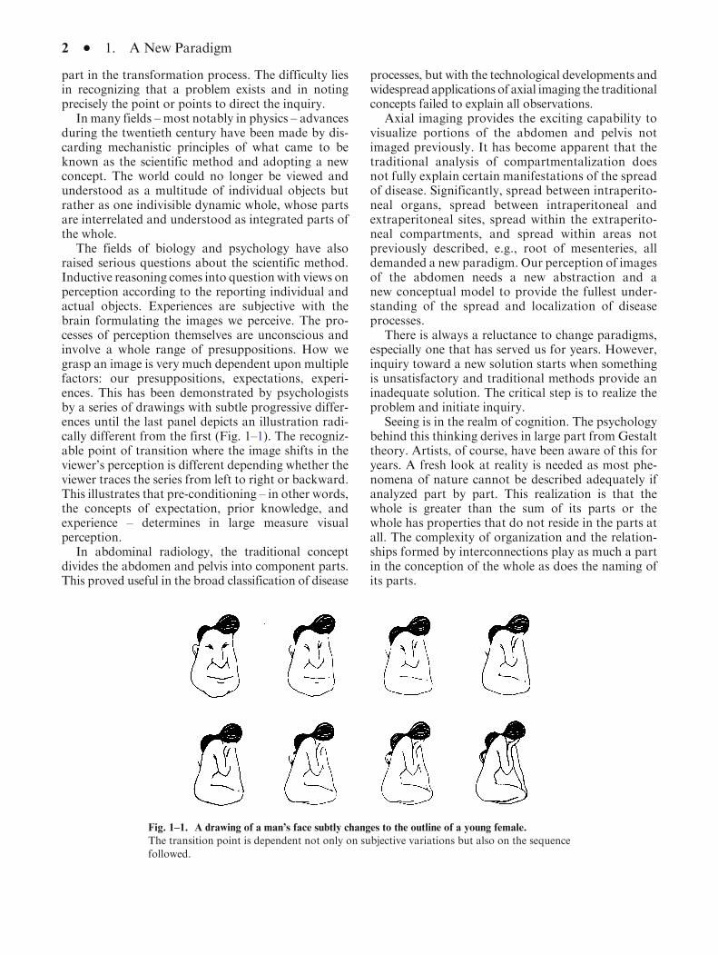
The fields of biology and psychology have alsoraised serious questions about the scientific method.Inductive reasoning comes into question with views onperception according to the reporting individual andactual objects. Experiences are subjective with thebrain formulating the images we perceive. The pro-cesses of perception themselves are unconscious andinvolve a whole range of presuppositions. How wegrasp an image is very much dependent upon multiplefactors: our presuppositions, expectations, experi-ences. This has been demonstrated by psychologistsby a series of drawings with subtle progressive differ-ences until the last panel depicts an illustration radi-cally different from the first (Fig. 1–1). The recogniz-able point of transition where the image shifts in theviewer’s perception is different depending whether theviewer traces the series from left to right or backward.This illustrates that pre-conditioning – in other words,the concepts of expectation, prior knowledge, andexperience – determines in large measure visualperception.
In abdominal radiology, the traditional conceptdivides the abdomen and pelvis into component parts.This proved useful in the broad classification of disease
processes, but with the technological developments andwidespread applications of axial imaging the traditionalconcepts failed to explain all observations.
Axial imaging provides the exciting capability tovisualize portions of the abdomen and pelvis notimaged previously. It has become apparent that thetraditional analysis of compartmentalization doesnot fully explain certain manifestations of the spreadof disease. Significantly, spread between intraperito-neal organs, spread between intraperitoneal andextraperitoneal sites, spread within the extraperito-neal compartments, and spread within areas notpreviously described, e.g., root of mesenteries, alldemanded a new paradigm. Our perception of imagesof the abdomen needs a new abstraction and anew conceptual model to provide the fullest under-standing of the spread and localization of diseaseprocesses.
There is always a reluctance to change paradigms,especially one that has served us for years. However,inquiry toward a new solution starts when somethingis unsatisfactory and traditional methods provide aninadequate solution. The critical step is to realize theproblem and initiate inquiry.
Seeing is in the realm of cognition. The psychologybehind this thinking derives in large part from Gestalttheory. Artists, of course, have been aware of this foryears. A fresh look at reality is needed as most phe-nomena of nature cannot be described adequately ifanalyzed part by part. This realization is that thewhole is greater than the sum of its parts or thewhole has properties that do not reside in the parts atall. The complexity of organization and the relation-ships formed by interconnections play as much a partin the conception of the whole as does the naming ofits parts.
Fig. 1–1. A drawing of a man’s face subtly changes to the outline of a young female.
The transition point is dependent not only on subjective variations but also on the sequence
followed.
2 � 1. A New Paradigm
Illustrative of this phenomenon are poet JohnGod-frey Saxe’s six blind men (from his poem ‘‘The BlindMen and the Elephant’’) observing different parts ofan elephant and coming to a very different but equallyerroneous conclusions about it. The first fell againstthe elephant’s side and concluded that it was a wall.The second felt the smooth, sharp tusk and mistook itfor a spear. The third held the squirming trunk andknew it was a snake. The fourth took the knee to be atree. The fifth touched the ear and declared it a fan.And the sixth seized the tail and thought he had a rope.One of the poem’s lessons: ‘‘Each was partly in theright, and all were in the wrong!’’4
The relevance of these views on cognition becomesevident in the art and science of imaging. We canview an image and yet perceive it in different ways.Figure 1–2 illustrates that the image first seen is deter-mined by the relationship between individual features.Both images are present in the one drawing. Theviewer sees either an old woman or a young lady.The perception of both images is determined by theirrelationships. Interestingly, one sees the young lady orthe old woman, but not both at once.
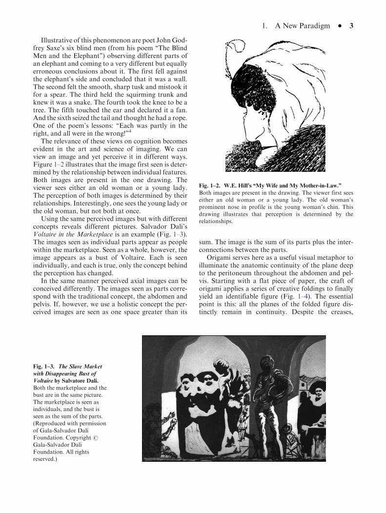
Using the same perceived images but with differentconcepts reveals different pictures. Salvador Dali’sVoltaire in the Marketplace is an example (Fig. 1–3).The images seen as individual parts appear as peoplewithin the marketplace. Seen as a whole, however, theimage appears as a bust of Voltaire. Each is seenindividually, and each is true, only the concept behindthe perception has changed.
In the same manner perceived axial images can beconceived differently. The images seen as parts corre-spond with the traditional concept, the abdomen andpelvis. If, however, we use a holistic concept the per-ceived images are seen as one space greater than its
sum. The image is the sum of its parts plus the inter-connections between the parts.
Origami serves here as a useful visual metaphor toilluminate the anatomic continuity of the plane deepto the peritoneum throughout the abdomen and pel-vis. Starting with a flat piece of paper, the craft oforigami applies a series of creative foldings to finallyyield an identifiable figure (Fig. 1–4). The essentialpoint is this: all the planes of the folded figure dis-tinctly remain in continuity. Despite the creases,
Fig. 1–2. W.E. Hill’s ‘‘My Wife and My Mother-in-Law.’’
Both images are present in the drawing. The viewer first seeseither an old woman or a young lady. The old woman’sprominent nose in profile is the young woman’s chin. This
drawing illustrates that perception is determined by therelationships.
Fig. 1–3. The Slave Market
with Disappearing Bust of
Voltaire by Salvatore Dali.
Both the marketplace and thebust are in the same picture.
The marketplace is seen asindividuals, and the bust isseen as the sum of the parts.
(Reproduced with permissionof Gala-Salvador DalıFoundation. Copyright #
Gala-Salvador DalıFoundation. All rightsreserved.)
1. A New Paradigm � 3

bends, overlaps, and projections, the surface of theoriginal flat paper is uninterrupted.
Similarly, as detailed in the following chapters onthe embryology of the abdomen and pelvis, the planedeep to the peritoneum is continuous throughout. Torecognize this is to vastly extend the clinical contribu-tions of abdominal imaging. In the planes formed bythe subperitoneal space course connective tissue,blood vessels, nerves, and lymphatics.5 It thusbecomes evident that the roots of the mesenteries –transversemesocolon, small bowelmesentery, sigmoidmesocolon, broad ligament – provide avenues of ana-tomic continuity (Figs. 1–5 and 1–6).
This holistic concept underscores the viewing of thefundamental structures of the abdomen and pelvis asone space – the subperitoneal space. This spaceincludes the extraperitoneal space, and the ligamentsandmesenteries of the abdomen and pelvis. The abdo-men and pelvis conceived within this holistic paradigmreadily explain the interconnections between all theorgans, mesenteries, roots of mesenteries, and extra-peritoneum in any conceivable combination.
A curved line, viewed from one side, is convex, butviewed from the other side, is concave. Two conceptsapplied to the same perceived image yield two pic-tures. Put another way, inherent in the grouping oflines and shadows in the illustrated drawings are two
individual perceptions, co-existing and reflective ofeach other. Conceiving the images as individual partsis most useful in explaining confinement of a diseaseprocess and differential diagnosis based on location.However, conceiving the image as a holistic anatomicconcept illuminates a new revolutionary paradigm.The abdomen and pelvis are constituted by one inter-connected space. This is of critical use in explainingthe pathways of spread of disease.
The introduction and acceptance of a new para-digm is made more difficult if vocabulary from a pre-vious paradigm continues to be used. This is due to thepotentially misleading implications the term carriesfrom its use in the previous paradigm. While it is bestto use new terms, this is not always possible. A clear setof definitions of how a word or words are used in theholistic concept is useful:
Subperitoneal Space: Extraperitoneal space and theligaments/mesenteries of the abdomen andpelvis.
Extraperitoneal: The circumferential space aroundthe abdomen and pelvis lying beneath the parie-tal peritoneum, stratified in the abdomen byrenal fascia and in the pelvis by umbilicovesicalfascia.
Retroperitoneum: Posterior portion of the extraper-itoneum in the abdomen.
Ligaments/mesenteries: Formed by two peritoneallayers (visceral peritoneum) in continuity with theparietal peritoneum. The structures enclosed –connective tissue, blood vessels, nerves, andlymphatics – are in continuity with theextraperitoneum.
A major purpose of Meyers’ Dynamic Radiology ofthe Abdomen is in explaining the pathways of diseasespread. The subperitoneal space provides the avenuesfor spread interconnecting all the organs. The peritonealcavity provides the potential pathways for intraperito-neal spread. The benefits of this cognitive framework aremultiple. When the primary site of disease – whetherinfectious, traumatic, or neoplastic in nature – is known,precise identification can bemade of the expected sites ofspread and localization. On the other hand, when apatient presents with a remote lesion, the primary site –whichmay be clinically occult – can be inferred. Further-more, such basic understanding facilitates identificationof the expected site of recurrent disease or the pattern ofprogression after treatment.
By developing focused search patterns, the radiol-ogist serves in a critical position to direct the course ofinvestigation, to evaluate the extent of disease, to indi-cate the prognosis, and to determine the appropriatemanagement.
Fig. 1–4. Origami Monkey.
(Courtesy of Annemarie Johnson.)
4 � 1. A New Paradigm

Fig. 1–5. Abdominal viscera.
The stomach has been removed from the cardia to the pylorus, revealing the lesser sac (omental bursa) and structures on theposterior wall.(Reproduced with permission from Putz.6 Copyright Elsevier [Churchill Livingstone Imprint].)
1. A New Paradigm � 5

Fig. 1–6. Retroperitoneum of an adult female.
(Reproduced with permission from Putz.6 Copyright Elsevier [Churchill Livingstone], 2006.)
6 � 1. A New Paradigm

References
1. BeveridgeWIB: The Art of Scientific Investigation.W. Heinemann, London, 1957, p 105.
2. Meyers MA: Back to the future. AJR 2008;190:561–564.
3. Horgan J: The End of Science. Addison–Wesley,Reading, MA, 1996.
4. Saxe JG: The Blind Men and the Elephant.McGraw-Hill, New York, 1963.
5. Oliphant M, Berne AS, Meyers MA: The subper-itoneal space of the abdomen and pelvis: planes ofcontinuity. AJR 1996; 167:1433–1439.
6. Putz R: Sobotta – Atlas of Human Anatomy SingleVolume Edition: Head, Neck, Upper Limb,Thorax, Abdomen, Pelvis, Lower Limb, 14th ed.Churchill Livingstone, The Netherlands 2006.
1. A New Paradigm � 7

Clinical Embryology
of the Abdomen 2
Introduction
Conventional distinction between intraperitonealand extraperitoneal sites is often helpful in differen-tial diagnostic considerations.1 Yet it should beunderstood that the abdomen and pelvis constitutean anatomic continuum that is punctuated by themesenteries, ligaments, and fasciae, which may eitherconfine pathology or actually provide avenues fordisease spread. It is essential to recognize the ana-tomic continuity of subserous connective tissue withits vessels and lymphatics as an extension of theextraperitoneal space that underlies the holistic con-cept of the subperitoneal space. A scaffold with pre-cise anatomic planes is provided for spread of diseasenot only between intraperitoneal structures but alsobetween extraperitoneal and intraperitoneal sites.2
This unifying concept is the basis for understandingthe dissemination of intraabdominal disease, includ-ing malignancies and inflammatory and traumaticprocesses, both focally and at areas distant from thesite of origin.
The subperitoneal space’s continuity with thethorax provides access for the bidirectional spread ofdisease involving these regions.3–7 It is the continuitybetween and within the abdomen and thorax thatprovides the rationale for understanding the paradox-ical clinical appearance of disease at a distance from itssite of origin. The graphic display of the anatomy withmodern imaging modalities coupled with currentknowledge of the morphology of the subperitonealspace provide a comprehensive clinical delineation of
disease processes and an improved understanding ofthe pathogenesis of direct spread of disease.
The knowledge of the development of the subper-itoneal space is a prerequisite to recognizing patholo-gic conditions and understanding the pathogenesis ofdisease spread.8–11 The conceptualization of the abdo-men and pelvis as one space, the subperitoneal space,and its continuity with the thorax requires the reexa-mination of standard embryology from a holisticperspective.
Early Embryonic Development
After fertilization, the zygote rapidly develops into atrilaminar sphere with three distinct layers: ento-derm, mesoderm, and ectoderm. Various body partsare then derived by progressive differentiation anddivergent specialization. The entoderm becomes thelining of the gastrointestinal tract, the liver, and pan-creatic glandular tissue. The ectoderm becomes thenervous system and epidermis. The mesoderm devel-ops into the remaining tissue including the visceraland parietal peritoneum, visceral and parietal pleura,as well as the ligaments and the mesenteries of theabdomen.
The lateral portion of the mesodermal layer of theembryo divides by the 4th week (Fig. 2–1). The lateralmargins move ventrally and medially and encompassthe yolk sac (Fig. 2–2). This incorporates the intraem-bryonic coelom, forming a tube within a tube. Theouter tube is the body cavity, and the inner tube is
M.A. Meyers et al., Meyers’ Dynamic Radiology of the Abdomen, DOI 10.1007/978-1-4419-5939-3_2,� Springer ScienceþBusiness Media, LLC 2011
9
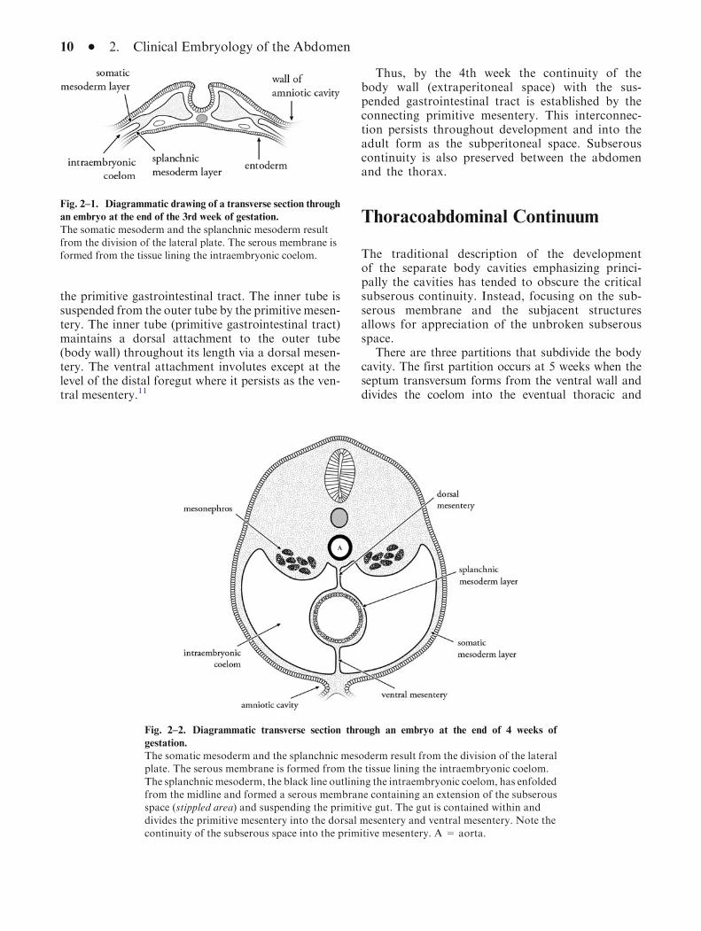
the primitive gastrointestinal tract. The inner tube issuspended from the outer tube by the primitive mesen-tery. The inner tube (primitive gastrointestinal tract)maintains a dorsal attachment to the outer tube(body wall) throughout its length via a dorsal mesen-tery. The ventral attachment involutes except at thelevel of the distal foregut where it persists as the ven-tral mesentery.11
Thus, by the 4th week the continuity of thebody wall (extraperitoneal space) with the sus-pended gastrointestinal tract is established by theconnecting primitive mesentery. This interconnec-tion persists throughout development and into theadult form as the subperitoneal space. Subserouscontinuity is also preserved between the abdomenand the thorax.
Thoracoabdominal Continuum
The traditional description of the developmentof the separate body cavities emphasizing princi-pally the cavities has tended to obscure the criticalsubserous continuity. Instead, focusing on the sub-serous membrane and the subjacent structuresallows for appreciation of the unbroken subserousspace.
There are three partitions that subdivide the bodycavity. The first partition occurs at 5 weeks when theseptum transversum forms from the ventral wall anddivides the coelom into the eventual thoracic and
Fig. 2–1. Diagrammatic drawing of a transverse section through
an embryo at the end of the 3rd week of gestation.
The somatic mesoderm and the splanchnic mesoderm resultfrom the division of the lateral plate. The serous membrane isformed from the tissue lining the intraembryonic coelom.
Fig. 2–2. Diagrammatic transverse section through an embryo at the end of 4 weeks of
gestation.
The somatic mesoderm and the splanchnic mesoderm result from the division of the lateralplate. The serous membrane is formed from the tissue lining the intraembryonic coelom.
The splanchnic mesoderm, the black line outlining the intraembryonic coelom, has enfoldedfrom the midline and formed a serous membrane containing an extension of the subserousspace (stippled area) and suspending the primitive gut. The gut is contained within and
divides the primitive mesentery into the dorsal mesentery and ventral mesentery. Note thecontinuity of the subserous space into the primitive mesentery. A = aorta.
10 � 2. Clinical Embryology of the Abdomen

abdominal cavities. The persistent openings on eachside of the coelomic cavity are called the pericardioper-itoneal canals. These are potential spaces defined by thesubserous lining. The developing organs are subjacentto this lining and project into the potential space of thecoelomic cavity. The lung buds form in the thorax fromthe primitive gut and grow laterally. The lungs projectinto the pericardioperitoneal canals enclosed by theserous membrane (Fig. 2–3).
Dorsal to the septum transversum, the heart is con-fluent with the gut and liver. The gut and liver areenclosed by the serous membrane and suspended ontheir mesenteries. A portion of the liver developswithin the caudal side of the septum transversum.This forms a barrier preventing the developing lungfrom expanding into the abdomen. The lungs developcovered by the serous membranes (parietal and visc-eral pleura) and project laterally forming the pleuralcavities. As the lungs and pleura develop, a secondpartition is formed: the pleuropericardial folds(Fig. 2–4). The portion of the serous membranebetween the lung and heart grows medially and fusesat the midline separating the pericardial cavity. Thepleural cavities remain connected dorsally with theperitoneal cavity due to the incomplete developmentof the diaphragm.
The isolation of the pleura and peritoneal cavitiesoccurs by 7 weeks as the diaphragm is completed bythe third partition – the pleuroperitoneal folds. Thesefuse with the esophageal mesentery, dividing thepleural and peritoneal cavities (Fig. 2–5).
The diaphragm is covered by the serous membrane:the thoracic side by the pleura and the abdominal sideby the peritoneum. The diaphragm, although separat-ing the body cavities, allows continuity of the subser-ous space mainly through the esophageal and aortichiatuses.12
The esophageal hiatus is ventral and cranial to theaortic hiatus. The esophageal hiatus contains areolartissue and the esophagus, vagus nerves, esophagealvessels and lymphatics as they course between thethorax and abdomen. The aortic hiatus is an osseoa-poneurotic opening between the diaphragm and ver-tebral column. The aorta, azygous vein, thoracicduct, and lymphatics course through this aperture.Thus, the esophageal hiatus and the aortic hiatusallow continuity of the subserous space of thoraxand abdomen. The vena cava foramen is the mostventral of the three main diaphragmatic aperturesand transmits only the inferior vena cava. Thecaval wall is adherent to the margins of the foramenand interrupts continuity of the subserous space.Small apertures reside ventrally between the sternumand the costal cartilage and allow the superior
Fig. 2–3. Diagrammatic drawing transverse section through a
4-week embryo in which the pleural and pericardial regions are
forming.
The lung buds are growing into the pericardioperitoneal folds,
and the heart is forming. The continuous serous membranelines that portion of the coelomic that will become the pleuralcavity and pericardial cavity. The subserous space is the
stippled area subjacent to the serous membrane.
Fig. 2–4. Diagrammatic drawing of transverse section through a
5-week embryo in which the pleural and pericardial regions of the
coelom become separated.
Complexity of the serous membrane results as it fuses ventrally
forming the pericardial cavity. The pleuropericardial folds fusebilaterally at the root of the lungs. The serous membrane linesthe pleural cavities as the visceral and parietal pleura. The
subserous space is subjacent to this lining. A=aorta.
Thoracoabdominal Continuum � 11
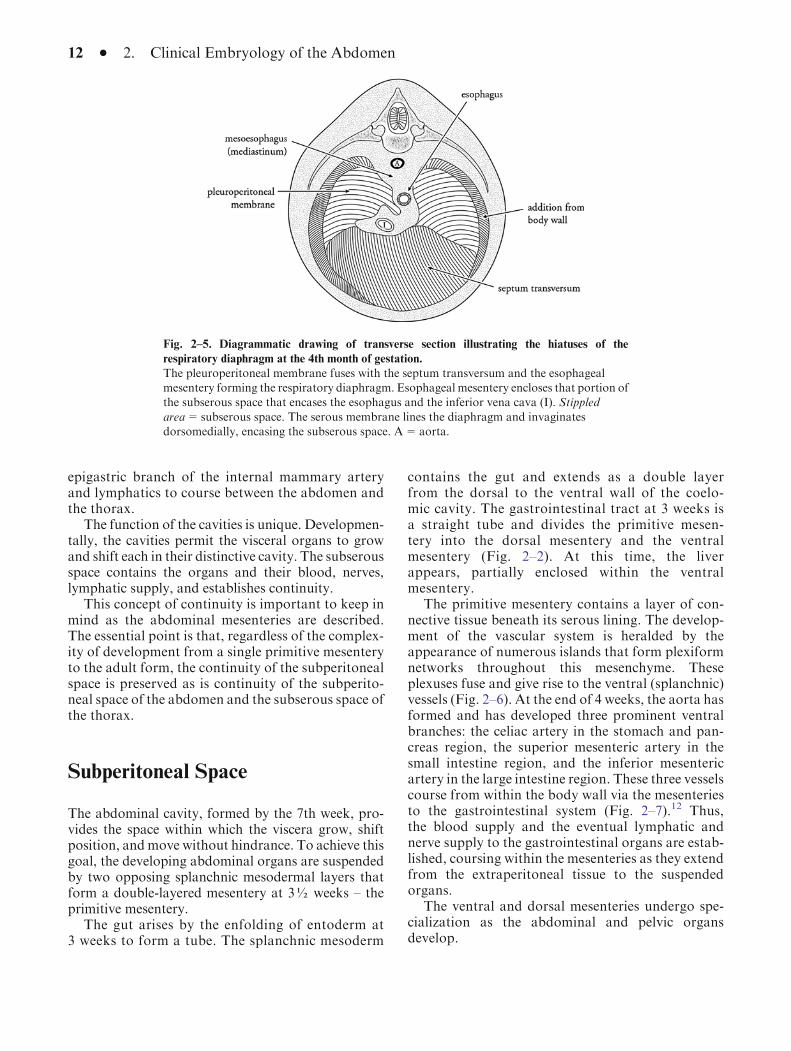
epigastric branch of the internal mammary arteryand lymphatics to course between the abdomen andthe thorax.
The function of the cavities is unique. Developmen-tally, the cavities permit the visceral organs to growand shift each in their distinctive cavity. The subserousspace contains the organs and their blood, nerves,lymphatic supply, and establishes continuity.
This concept of continuity is important to keep inmind as the abdominal mesenteries are described.The essential point is that, regardless of the complex-ity of development from a single primitive mesenteryto the adult form, the continuity of the subperitonealspace is preserved as is continuity of the subperito-neal space of the abdomen and the subserous space ofthe thorax.
Subperitoneal Space
The abdominal cavity, formed by the 7th week, pro-vides the space within which the viscera grow, shiftposition, andmove without hindrance. To achieve thisgoal, the developing abdominal organs are suspendedby two opposing splanchnic mesodermal layers thatform a double-layered mesentery at 3½ weeks – theprimitive mesentery.
The gut arises by the enfolding of entoderm at3 weeks to form a tube. The splanchnic mesoderm
contains the gut and extends as a double layerfrom the dorsal to the ventral wall of the coelo-mic cavity. The gastrointestinal tract at 3 weeks isa straight tube and divides the primitive mesen-tery into the dorsal mesentery and the ventralmesentery (Fig. 2–2). At this time, the liverappears, partially enclosed within the ventralmesentery.
The primitive mesentery contains a layer of con-nective tissue beneath its serous lining. The develop-ment of the vascular system is heralded by theappearance of numerous islands that form plexiformnetworks throughout this mesenchyme. Theseplexuses fuse and give rise to the ventral (splanchnic)vessels (Fig. 2–6). At the end of 4 weeks, the aorta hasformed and has developed three prominent ventralbranches: the celiac artery in the stomach and pan-creas region, the superior mesenteric artery in thesmall intestine region, and the inferior mesentericartery in the large intestine region. These three vesselscourse from within the body wall via the mesenteriesto the gastrointestinal system (Fig. 2–7).12 Thus,the blood supply and the eventual lymphatic andnerve supply to the gastrointestinal organs are estab-lished, coursing within the mesenteries as they extendfrom the extraperitoneal tissue to the suspendedorgans.
The ventral and dorsal mesenteries undergo spe-cialization as the abdominal and pelvic organsdevelop.
Fig. 2–5. Diagrammatic drawing of transverse section illustrating the hiatuses of the
respiratory diaphragm at the 4th month of gestation.
The pleuroperitoneal membrane fuses with the septum transversum and the esophagealmesentery forming the respiratory diaphragm. Esophageal mesentery encloses that portion ofthe subserous space that encases the esophagus and the inferior vena cava (I). Stippled
area=subserous space. The serous membrane lines the diaphragm and invaginatesdorsomedially, encasing the subserous space. A=aorta.
12 � 2. Clinical Embryology of the Abdomen

Ventral Mesentery Specialization
The ventral mesentery that initially attaches the entirelength of the primitive gut to the ventral abdominal wallregresses except in the region of the lower esophagus,stomach, upper duodenum, and liver. The liver appearsat 3–4 weeks and rapidly enlarges as it projects from theseptum transversum into the ventral mesentery. Theliver splits the ventral mesentery into anterior and pos-terior portions, the falciform ligament and gastrohepaticligament (lesser omentum), respectively (Fig. 2–8a, b).The free margin of the falciform ligament contains theleft umbilical vein, forming the ligamentum teres. The
free margin of the gastrohepatic ligament contains thecommon bile duct, portal vein, and hepatic artery and istermed the hepatoduodenal ligament
The liver capsule, formed by the visceral perito-neum, is continuous with the peritoneum, exceptwhere the liver is embedded within the septum trans-versum, known as the ‘‘bare area.’’ The peritoneallining reflects from this area as the coronary ligamentand attaches to the lateral abdominal wall as the tri-angular ligaments. The liver ligaments are in continu-ity with the falciform ligament and the gastrohepaticligament as derivates of the ventral mesentery (ventralmesogastrium) (Fig. 2–9).
Fig. 2–6. Diagrammatic
drawing of transverse section
through an embryo at the end of
4 weeks of gestation.
The ventral (splanchnic)
artery has formed and suppliesthe primitive gut. The ventralartery extends within the
mesentery from the aorta (A)to the suspended gut. Notecontinuity of subperitoneal
space (stippled area) within themesentery as well ascontinuity from right to left
and dorsal to ventral.
Fig. 2–7. Three-dimensional
drawing of a 5-week embryo.
The entire gut as well as theliver, pancreas, and spleen areencased within the mesentery.
The organs of the foregut arewithin the ventral and dorsalmesentery; the organs of the
midgut and hindgut are withinthe dorsal mesentery. All theorgans are supplied by the
aorta and its three ventralarteries (celiac, superiormesenteric, inferior
mesenteric arteries) as theyextend within the mesentery tothe suspended organs.
Subperitoneal Space � 13
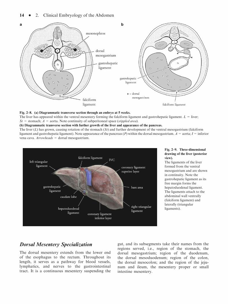
Dorsal Mesentery Specialization
The dorsal mesentery extends from the lower endof the esophagus to the rectum. Throughout itslength, it serves as a pathway for blood vessels,lymphatics, and nerves to the gastrointestinaltract. It is a continuous mesentery suspending the
gut, and its subsegments take their names from theregions served, i.e., region of the stomach, thedorsal mesogastrium; region of the duodenum,the dorsal mesoduodenum; region of the colon,the dorsal mesocolon; and the region of the jeju-num and ileum, the mesentery proper or smallintestine mesentery.
Fig. 2–9. Three-dimensional
drawing of the liver (posterior
view).
The ligaments of the liverformed from the ventralmesogastrium and are shown
in continuity. Note thegastrohepatic ligament as itsfree margin forms the
hepatoduodenal ligament.The ligaments attach to theabdominal wall ventrally(falciform ligament) and
laterally (triangularligaments).
a b
Fig. 2–8. (a) Diagrammatic transverse section through an embryo at 5 weeks.
The liver has appeared within the ventral mesentery forming the falciform ligament and gastrohepatic ligament. L = liver;St = stomach; A = aorta. Note continuity of subperitoneal space (stippled area).(b) Diagrammatic transverse section with further growth of the liver and appearance of the pancreas.
The liver (L) has grown, causing rotation of the stomach (St) and further development of the ventral mesogastrium (falciformligament and gastrohepatic ligament). Note appearance of the pancreas (P) within the dorsal mesogastrium.A=aorta; I= inferiorvena cava. Arrowheads = dorsal mesogastrium.
14 � 2. Clinical Embryology of the Abdomen
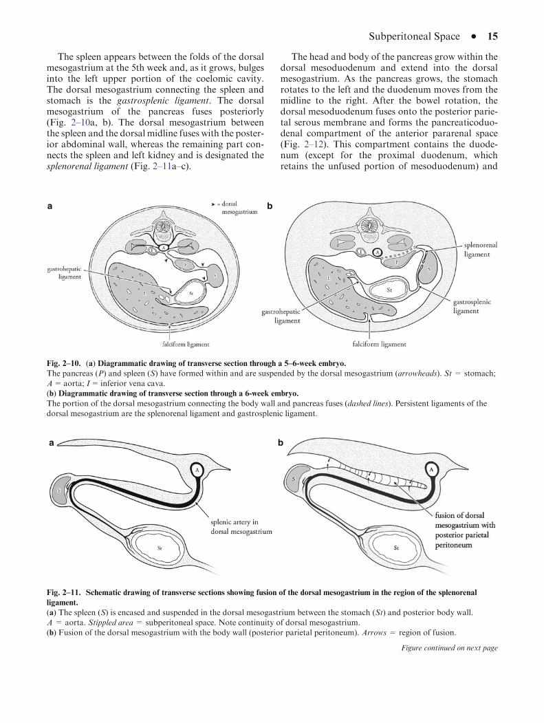
The spleen appears between the folds of the dorsalmesogastrium at the 5th week and, as it grows, bulgesinto the left upper portion of the coelomic cavity.The dorsal mesogastrium connecting the spleen andstomach is the gastrosplenic ligament. The dorsalmesogastrium of the pancreas fuses posteriorly(Fig. 2–10a, b). The dorsal mesogastrium betweenthe spleen and the dorsal midline fuses with the poster-ior abdominal wall, whereas the remaining part con-nects the spleen and left kidney and is designated thesplenorenal ligament (Fig. 2–11a–c).
The head and body of the pancreas grow within thedorsal mesoduodenum and extend into the dorsalmesogastrium. As the pancreas grows, the stomachrotates to the left and the duodenum moves from themidline to the right. After the bowel rotation, thedorsal mesoduodenum fuses onto the posterior parie-tal serous membrane and forms the pancreaticoduo-denal compartment of the anterior pararenal space(Fig. 2–12). This compartment contains the duode-num (except for the proximal duodenum, whichretains the unfused portion of mesoduodenum) and
a b
Fig. 2–10. (a) Diagrammatic drawing of transverse section through a 5–6-week embryo.
The pancreas (P) and spleen (S) have formed within and are suspended by the dorsal mesogastrium (arrowheads). St = stomach;A=aorta; I=inferior vena cava.(b) Diagrammatic drawing of transverse section through a 6-week embryo.
The portion of the dorsal mesogastrium connecting the body wall and pancreas fuses (dashed lines). Persistent ligaments of thedorsal mesogastrium are the splenorenal ligament and gastrosplenic ligament.
a b
Fig. 2–11. Schematic drawing of transverse sections showing fusion of the dorsal mesogastrium in the region of the splenorenal
ligament.
(a) The spleen (S) is encased and suspended in the dorsal mesogastrium between the stomach (St) and posterior body wall.
A = aorta. Stippled area = subperitoneal space. Note continuity of dorsal mesogastrium.(b) Fusion of the dorsal mesogastrium with the body wall (posterior parietal peritoneum). Arrows = region of fusion.
Figure continued on next page
Subperitoneal Space � 15
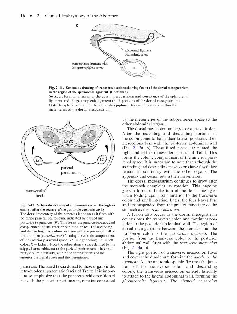
pancreas. The fused fascia dorsal to these organs is theretroduodenal pancreatic fascia of Treitz. It is impor-tant to emphasize that the pancreas, while positionedbeneath the posterior peritoneum, remains connected
by the mesenteries of the subperitoneal space to theother abdominal organs.
The dorsal mesocolon undergoes extensive fusion.After the ascending and descending portions ofthe colon come to lie in their lateral positions, theirmesocolons fuse with the posterior abdominal wall(Fig. 2–13a, b). These fused fascia are named theright and left retromesenteric fascia of Toldt. Thisforms the colonic compartment of the anterior para-renal space. It is important to note that although theascending and descending mesocolons have fused theyremain in continuity with the other organs. Theappendix and cecum retain their mesenteries.
The dorsal mesogastrium continues to grow afterthe stomach completes its rotation. This ongoinggrowth forms a duplication of the dorsal mesogas-trium folding upon itself anterior to the transversecolon and small intestine. Later, the four leaves fuseand are suspended from the greater curvature of thestomach as the greater omentum.
A fusion also occurs as the dorsal mesogastriumcourses over the transverse colon and continues pos-terior to the posterior abdominal wall. The region ofdorsal mesogastrium between the stomach and thetransverse colon is the gastrocolic ligament. Theportion from the transverse colon to the posteriorabdominal wall fuses with the transverse mesocolon(Fig. 2–14a, b).
The right portion of transverse mesocolon fusesand covers the duodenum forming the duodenocolicligament. At the anatomic splenic flexure (the junc-tion of the transverse colon and descendingcolon), the transverse mesocolon extends laterallyto attach to the lateral abdominal wall, forming thephrenicocolic ligament. The sigmoid mesocolon
c
Fig. 2–11. Schematic drawing of transverse sections showing fusion of the dorsal mesogastrium
in the region of the splenorenal ligament. (Continued)
(c) Adult form with fusion of the dorsal mesogastrium and persistence of the splenorenalligament and the gastrosplenic ligament (both portions of the dorsal mesogastrium).Note the splenic artery and the left gastroepiploic artery as they course within themesenteries of the dorsal mesogastrium.
Fig. 2–12. Schematic drawing of a transverse section through an
embryo after the reentry of the gut to the coelomic cavity.
The dorsal mesentery of the pancreas is shown as it fuses withposterior parietal peritoneum, indicated by dashed lineposterior to pancreas (P). This forms the pancreaticoduodenal
compartment of the anterior pararenal space. The ascendingand descending mesocolons will fuse with the posterior wall ofthe abdomen (curved arrows) forming the colonic compartment
of the anterior pararenal space. RC = right colon; LC = leftcolon;K=kidney. Note the subperitoneal space defined by thestippled area subjacent to the parietal peritoneum is in conti-
nuity circumferentially, within the compartments of theanterior pararenal space and the mesenteries.
16 � 2. Clinical Embryology of the Abdomen