Metodi computazionali in fisica medica delle radiazioni ed...
99
Metodi computazionali in fisica medica delle radiazioni ed in radioprotezione Dott. Ernesto Amato, Ph.D. Dipartimento di Scienze Biomediche e delle Immagini Morfologiche e Funzionali Sezione di Scienze Radiologiche [email protected]
Transcript of Metodi computazionali in fisica medica delle radiazioni ed...

Metodi computazionali in fisica medica delle radiazioni ed in radioprotezione
Dott. Ernesto Amato, Ph.D.
Dipartimento di Scienze Biomediche e delleImmagini Morfologiche e Funzionali
Sezione di Scienze Radiologiche
The Monte Carlo method
“Monte Carlo methods” are a vast series of statistical computational methods, based on the generation of “pseudo-random” numbers, with several of applications in mathematics, physics, chemistry, economy and social sciences.

The Monte Carlo method
“Monte Carlo methods” are a vast series of statistical computational methods, based on the generation of “pseudo-random” numbers, with several of applications in mathematics, physics, chemistry, economy and social sciences.

Generation of “pseudo-random” numbers
• True random numbers: outcomes of a physics experiment;• Pseudo-random numbers: deterministic sequence of number that
seem to be random if the source algorithm is unknown.
Properties of a pseudo-random number generator:
• Uncorrelated sequences
• Long period
• Uniformity
• Efficiency

Monte Carlo in nuclear physics
Computer simulation of the interaction of particles with particles or with atoms and nuclei of the traversed matter.
It employes theroretical models of forces and interactions known from nuclear and particle physics, and computational statistical methods in order to track the “history” of each simulated particle and retrieve the statistical quantities of interest (e.g. angular distributions, deposited energy, number and type of secondaries...) in a generic geometry.
Main “general purpose” Monte Carlo codes available:• Geant4• Fluka• EGS• MCNP• Penelope

6
Multiple scattering BremsstrahlungIonisationAnnihilationPhotoelectric effect Compton scattering Rayleigh effect
γ conversion e+e- pair production
Synchrotron radiationTransition radiationCherenkovAbsorptionScintillationFluorescenceAuger
Interactions Geometries
Low energy extensionsLow energy extensions fundamental for space and medical applications, dark
matter and ν experiments, spectroscopy etc.
electrons and positrons■ γ, X-ray and optical photons■ muons■ charged hadrons■ ions
ATLAS
KamLAND
Courtesy of H.Ikeda (Tohoku)
Definition and handling of complex geometries. Modeling of nuclear and particle experiments in accelerators, storage rings and reactors.Astroparticle physics detectors.

Particle tracking

Depending on the nature and energy of the primary particles, and on the composition, density and dimension of the traversed materials, it is of paramount importance a proper choice of:
1. active interaction channels2. accurate parametrizations or look-up tables of the total and differential cross sections in the range of energy explored3. tracking step4. cut in range and/or in energy in each material
Simulation set-up

Applications in medical radiation physics
Dedicated interfaces and tools of a general purpose MC:Examples: GAMOS and GATE for Geant4
Dedicated codes. Example: SIMIND, for nuclear medicine

MC application in medical radiation physics
Design of novel instruments (diagnosis and therapy)
Radiation dosimetry (internal or external)
Microdosimetry and biological effect

MC applications in nuclear medicine Design of novel detectors
− new crystals or crystal-photodetector assemblies
− new geometries for PET, SPECT or dedicated scanners
Internal dosimetry
− development of models
− 3D internal dosimetry through direct MC simulation
Microdosimetry
− tissue and cell models
− biological effect of each radiation type (alpha, beta, Auger...)
Production of radioisotopes
− improvements in cyclotron or reactor targetry

MC applications in radiology
Design of novel X-ray sources and detectors
− new solid state detectors and CT geometries
− emission spectra from X-ray tubes
External dosimetry
− development of models
− dosimetry in presence of contrast agents

MC applications in radiotherapy Design and characterization of sources
− Linacs
− brachytherapy sources (radioactive, miniature X-ray tubes)
− hadrontherapy sources
External dosimetry
− development of models
− Treatment Planning Systems through direct MC simulation
Microdosimetry
− tissue and cell models
− biological effect of each radiation type (photons, electrons, protons, light nuclei)

Interdisciplinarity
Medicine (specialists in radiology, nuclear medicine, radiotherapy, oncology, anatomy, fisiology, pathology...)
Biology
Physics
Chemistry
Pharmacy and radiopharmacy
Engineering

Examples of applications in internal dosimetry of nuclear medicine
Calculation of self-doses in simple geometries (spheres, ellipsoids)
Calculation of dose factors for organs in simplified anthropomorphic models
Calculation of the three-dimensional dose distribution at the voxel level

Nuclear medicine imaging
Dual head tomographic gamma camera
Nuclear medicine therapy
Purpose: to deliver lethal radiation dose to pathologic tissues (tumors, hyperfunctioning thyroid nodules...), while preserving the remnant of the body.
Agents: radiopharmaceuticals labeled with beta, Auger or alpha emitters. Associated gamma emission is useful for imaging.
Internal dosimetry: assessment of radiation absorbed dose for risk/benefit evaluation and quantification of the activity to be administered to obtain the therapeutic effect.
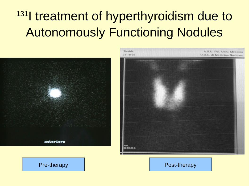
131I treatment of hyperthyroidism due to Autonomously Functioning Nodules
Pre-therapy Post-therapy

NET therapy with 90Y-Dotatoc

Dose in spheres from nuclides
• Stabin and Konijnenberg “Re-evaluation of absorbed fractions for photons and electrons in spheres of various sizes” J Nucl Med 2000; 41:149
• Bardies and Chatal “Absorbed doses for internal radiotherapy from 22 beta-emitting radionuclides: beta dosimetry of small spheres” Phys Med Biol 1994 39:961
Absorbed fractions

Generalization to ellipsoids:1. validation of our MC

Prolate, oblate and scalene ellipsoids; spheres.Radiation: alpha, beta, electrons, X, gamma
Generalization to ellipsoids:2. simulations and model development


● E. Amato, D. Lizio and S. Baldari, “Absorbed fractions in ellipsoidal volumes for β − radionuclides employed in internal radiotherapy”, Phys. Med. Biol. 54 (2009) 4171
● E. Amato, D. Lizio and S. Baldari, “Absorbed fractions for photons in ellipsoidal volumes”, Phys. Med. Biol. 54 (2009) N479
● E. Amato, D. Lizio and S. Baldari, “Absorbed fraction for electrons in ellipsoidal volumes”, Phys. Med. Biol. 56 (2011) 357
● E. Amato, A. Italiano and S. Baldari, “Absorbed fraction for alpha particles in ellipsoidal volumes”, Phys. Med. Biol. 58 (2013) 5449
● E. Amato, A. Italiano, S. Baldari, “An analytical model to calculate absorbed fractions for internal dosimetry with alpha, beta and gamma emitters”, AAPP, Classe di Scienze Fisiche, Matematiche e Naturali 92 (2014) A1-1

Comparison of the S factor included in the Olinda/EXM software and with the constant value in the Snyder formula.
The Snyder formula subtendsa constant value for the
energy deposited by e- and gammas
Application to the hyperthyroidism due to toxic nodular goiter
E. Amato, D. Lizio, R. M. Ruggeri, M. Raniolo, A. Campennì, S. Baldari. “An Analytical Model for Improving Absorbed Dose Calculation Accuracy in non Spherical AFTN”, Quart. J. Nucl. Med. Mol. Imag. 63 (2011) 560

MC dosimetric study for 90-Y brachytherapy of the prostate
F. Botta, M. Cremonesi, M. E. Ferrari, E. Amato, F. Guerriero, A. Vavassori, A. Sarnelli, S. Severi, G. Pedroli, G. Paganelli. “Investigation of Y90-avidin for prostate cancer brachytherapy: a dosimetric model for phase I-II clinical study” Eur J Nucl Med Mol Imaging 40 (2013) 1047-1056

Comparison PENELOPE-GEANT4

Organ dosimetry in anthropomorphic phantoms
s
t
Several phantoms (adult, pediatrics...)
ICRPMIRDORNL Cristy ed EckermannKramer
Handbook of anatomical models for radiation dosimetry, Ed. Xu e Eckermann, CRC Press, 2010

MIRD approach at organ level: “S” factors (MIRD Pamphlet 16)
Snyder, et al. MIRD Pamphlet 11: S, Absorbed Dose per Unit Cumulated Activity for Selected Radionuclides and Organs. 1975; Society of Nuclear Medicine, Reston, VA.
Snyder,et al. MIRD Pamphlet #5 Revised: Estimates of Absorbed Fractions for Monoenergetic Photon Sources Uniformly Distributed in Various Organs of a Heterogeneous Phantom. 1969; J Nucl Med Suppl Number 3

Voxel dosimetry
s
t
MIRD Pamphlet 17 (1999)

Calculation of S factors for a generic l and electron spectrum
For monoenergetic electrons (E) in a given voxel side (l), S factors can be calculated interpolating the fit parameters at (E,l):
For a generic electron spectrum dn(E)/dE, S factors can be derived by integration:

Monoenergetic electrons: 10-2000 keV

Monoenergetic photons: 20-1000 keV

Log(a)
b
c
Fit parameters in (l,E) space for electrons
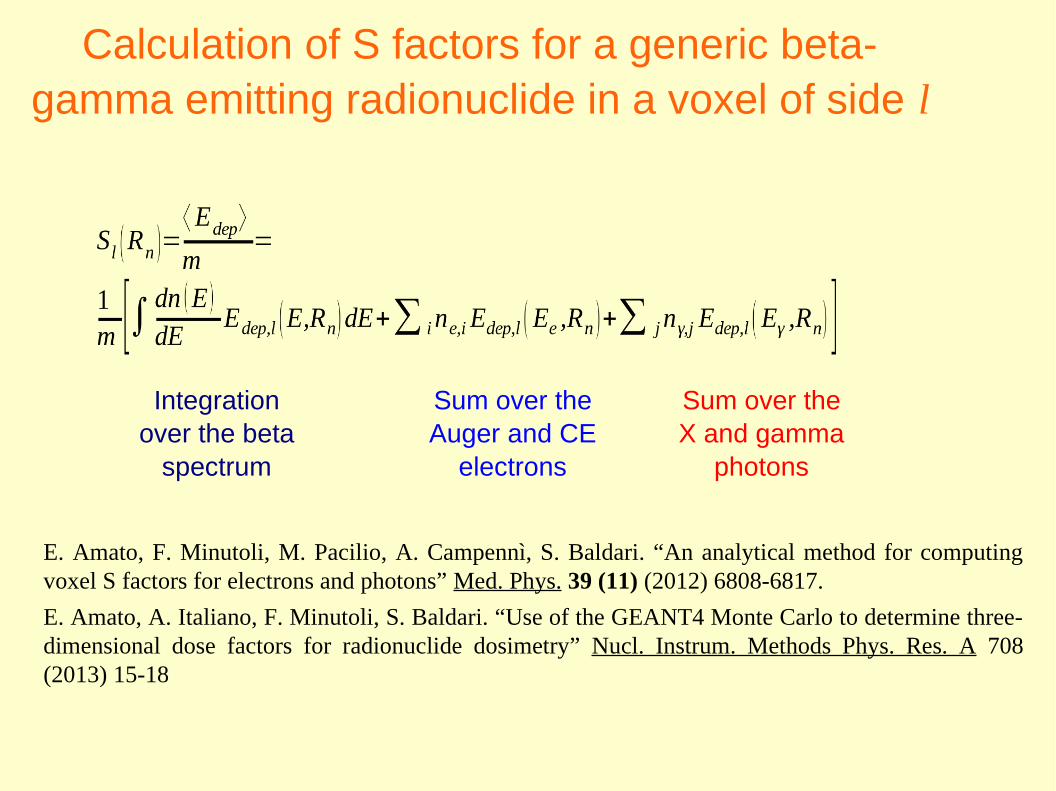
Calculation of S factors for a generic beta-gamma emitting radionuclide in a voxel of side l
Integrationover the beta
spectrum
Sum over the Auger and CE
electrons
Sum over theX and gamma
photons
E. Amato, F. Minutoli, M. Pacilio, A. Campennì, S. Baldari. “An analytical method for computing voxel S factors for electrons and photons” Med. Phys. 39 (11) (2012) 6808-6817.
E. Amato, A. Italiano, F. Minutoli, S. Baldari. “Use of the GEANT4 Monte Carlo to determine three-dimensional dose factors for radionuclide dosimetry” Nucl. Instrum. Methods Phys. Res. A 708 (2013) 15-18

131-iodine: analytical calculation / direct MC simulation / reference data (website)
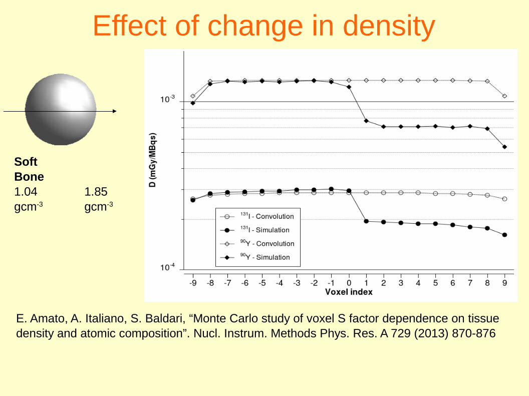
Effect of change in density
SoftBone1.04 1.85gcm-3 gcm-3
E. Amato, A. Italiano, S. Baldari, “Monte Carlo study of voxel S factor dependence on tissue density and atomic composition”. Nucl. Instrum. Methods Phys. Res. A 729 (2013) 870-876
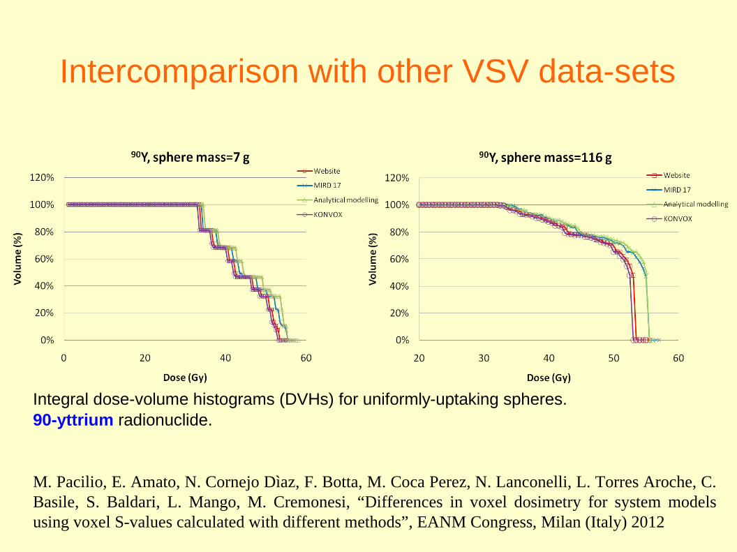
Intercomparison with other VSV data-sets
Integral dose-volume histograms (DVHs) for uniformly-uptaking spheres.90-yttrium radionuclide.
M. Pacilio, E. Amato, N. Cornejo Dìaz, F. Botta, M. Coca Perez, N. Lanconelli, L. Torres Aroche, C. Basile, S. Baldari, L. Mango, M. Cremonesi, “Differences in voxel dosimetry for system models using voxel S-values calculated with different methods”, EANM Congress, Milan (Italy) 2012

Intercomparison with other VSV data-sets
Integral dose-volume histograms (DVHs) for uniformly-uptaking spheres.131-iodine radionuclide.
M. Pacilio, E. Amato, N. Cornejo Dìaz, F. Botta, M. Coca Perez, N. Lanconelli, L. Torres Aroche, C. Basile, S. Baldari, L. Mango, M. Cremonesi, “Differences in voxel dosimetry for system models using voxel S-values calculated with different methods”, EANM Congress, Milan (Italy) 2012

Intercomparison on patient data:SPECT images for IART and SIRT therapies
M. Pacilio, F. Botta, L. Torres Aroche, E. Amato, N. Cornejo Dìaz, N. Lanconelli, M. Coca Perez, C. Basile, M. Cremonesi, “Impact on voxel S-values calculation method on 3D dosimetry for radionuclide therapy: application to SIRT and IART treatments”, EANM Congress, Milan (Italy) 2012
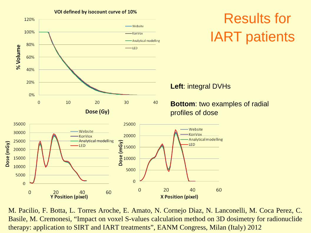
Results for IART patients
Left: integral DVHs
Bottom: two examples of radial profiles of dose
M. Pacilio, F. Botta, L. Torres Aroche, E. Amato, N. Cornejo Dìaz, N. Lanconelli, M. Coca Perez, C. Basile, M. Cremonesi, “Impact on voxel S-values calculation method on 3D dosimetry for radionuclide therapy: application to SIRT and IART treatments”, EANM Congress, Milan (Italy) 2012

Microdosimetric applications
Simulation of the tissutal micro-structure
bone
solid tumours wit hypoxic areas
capillar microstructure (anti-angiogenic effect)
Multi-regional cell models
dose to the mithocondria, nucleus, membrane...
Molecular “nanodosimetry”
relative biological effectiveness of the radiations on DNA, RNA and other biomolecules

90Y: high cross-fireD = 2 mm D(est) = 2 cm D(int) = 2 mm

177Lu: modest cross-fireD = 2 mm D(est) = 2 cm D(int) = 2 mm

Anti-angiogenic effectTumours with rapidly growing neo-vascularization can require a combination of cytotoxic and anti-angiogenic effects.
Maximum dose to the capillary endothelium low diffusion range of the RFlow range of radiations (Auger, α)
Maximum dose to the viable tumour low diffusion range of the RF high range of radiations (β) OR high range of diffusion of the RF
X. Zhu,...A. Kassis “Solid-Tumor Radionuclide Therapy Dosimetry: New Paradigms in View of Tumor Microenvironment and Angiogenesis” Med. Phys. 2010
Monte Carlo
EGS
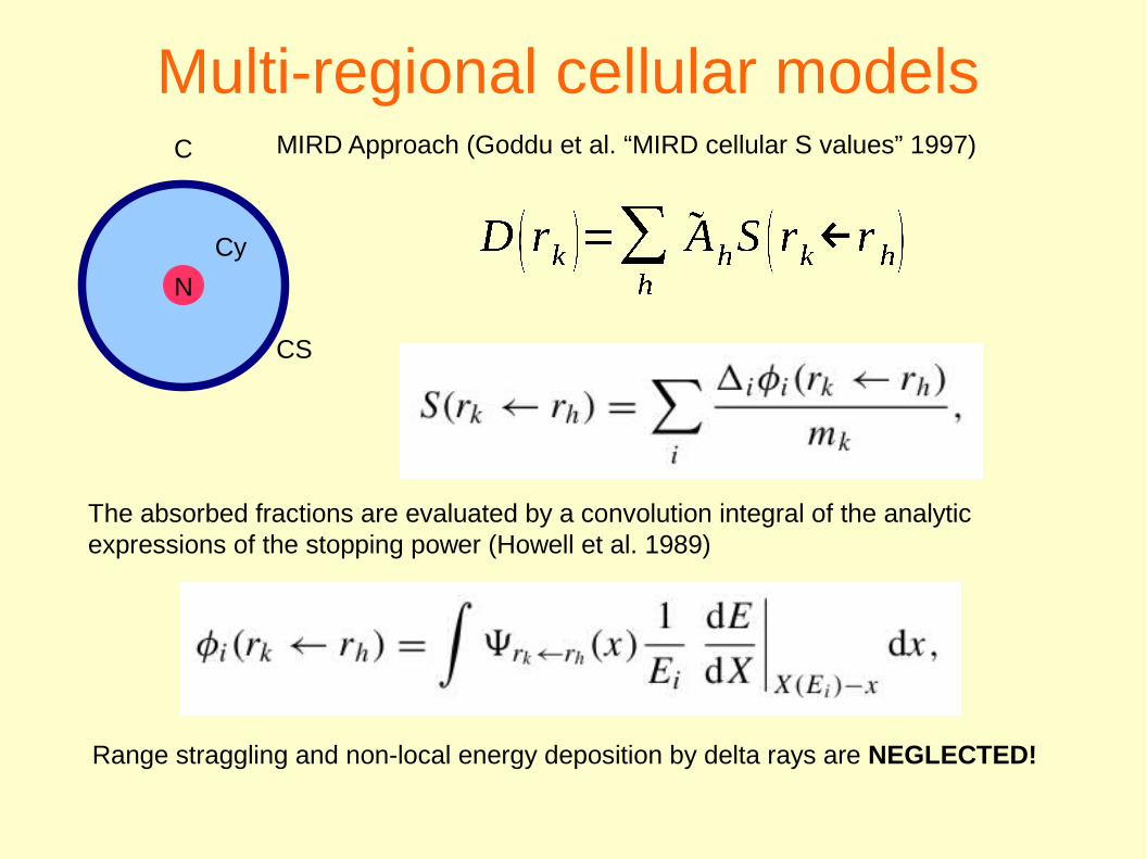
Multi-regional cellular modelsMIRD Approach (Goddu et al. “MIRD cellular S values” 1997)
The absorbed fractions are evaluated by a convolution integral of the analytic expressions of the stopping power (Howell et al. 1989)
Range straggling and non-local energy deposition by delta rays are NEGLECTED!
N
C
CS
Cy

MC contribution
Bousis et al. “A Monte Carlo study of cellular S-factors for 1 keV to 1 MeV electrons” Phys. Med. Biol. 2009, 54 5023

Demonstration of the invariance of the number of SSB vs. LET and of the dependence of DSB vs. LET
M. Bernal et al. “The invariance of the total direct DNA strand break yield” Med. Phys. 38 4147 (2011)
Monte Carlo: Geant4-DNA

Which parametrizations and cuts towards eV and nm?
S. Incerti et al. “Comparison of GEANT4 very low energy cross section models with experimental data in water” Med. Phys. 37 4692 (September 2010)
Elastic scattering

Scarce data for some processesS. Incerti et al. “Comparison of GEANT4 very low energy cross section models with experimental data in water” Med. Phys. 37 4692 (September 2010)
Geant4-DNA: Born model for electron excitation in liquid water. Data rescaled from the water vapor state.
NEW and MORE ACCURATE DATA are needed to parametrize processes at extremely low energies. It is difficult to set up experiments.

Which is the low-energy limit of “classic” MC codes?
Thomson e Kawrakow “On the Monte Carlo simulation of electron transport in the sub-1 keV energy range” Med. Phys. 38 4531 (August 2011)
Heisenberg principle:
s, lenght of interest p, particle momentum
Maximum uncertainties in position and momentum:

εc small for heavy or neutral particles (e.g.: classical limit OK for thermal
neutrons);
Light charged particles with high cross section (electrons and positrons) have ε
c higher:
In LIQUID water:● below 1 keV, uncertainties higher
than 5%.● below 100 eV, higher than
20% !!!
In AIR: 1% also at eV !!
Below few keV thede Broglie wavelenght is comparable with the inter-atomic distances in condensed matter, hence:
It is inaccurate to rescale for density the cross sections from vapour to liquid below few keV.

Applications in Radiology
Study of the dose increment in contrast-enhanced computed
tomography (CT)

Computed tomography

55
Doses in diagnostic radiology in presence of iodinated contrast media
This geometry, reproduced in MC

Simulation of CT irradiation: anthropomorphic phantoms
Abdomen Neck
a. spine d. left kidneyb. liver e. pancreasc. spleen
a. thyroidb. cervical spine
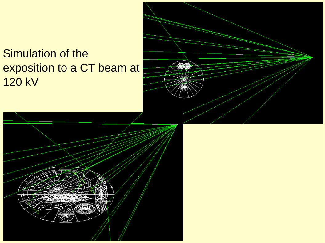
Simulation of the exposition to a CT beam at 120 kV
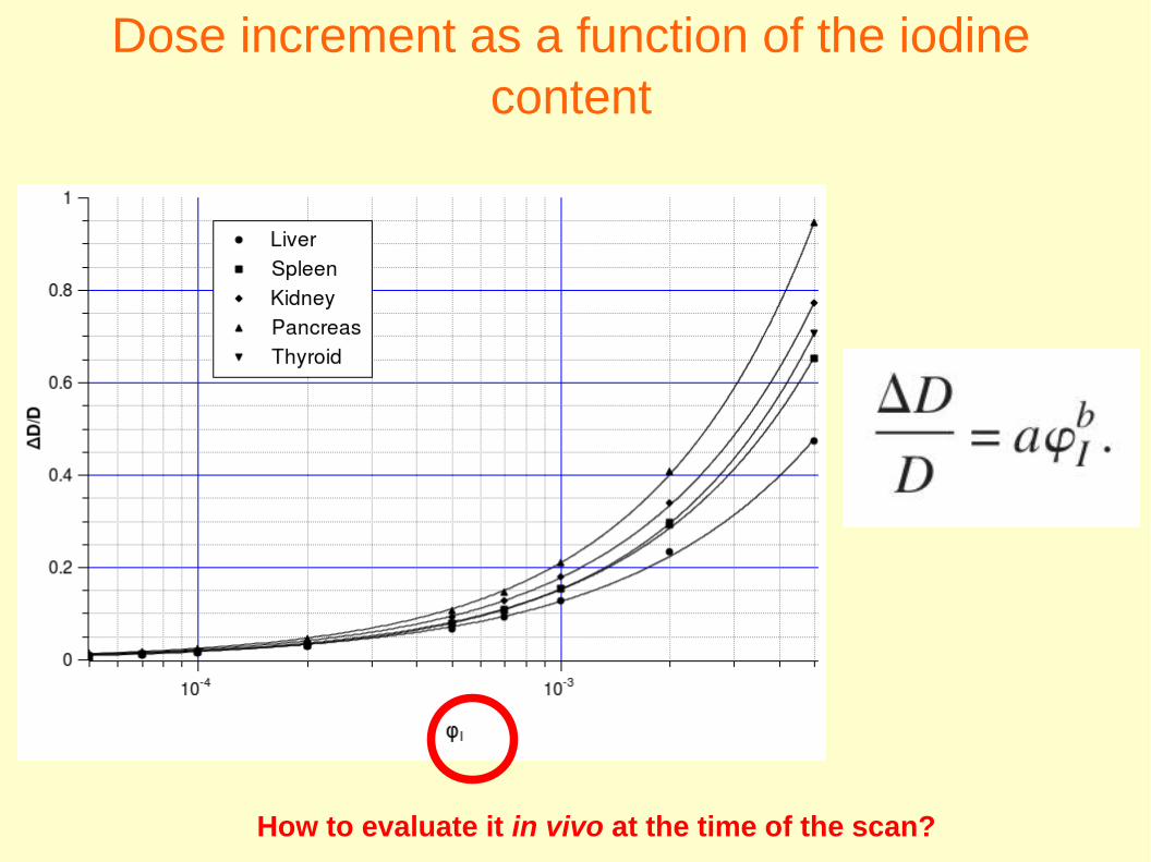
Dose increment as a function of the iodine content
How to evaluate it in vivo at the time of the scan?

Measurement on a CT of the HU increment vs. iodine concentration
We found a relationship between the HU increment and the iodine mass fraction.we determined the parameter gamma, which depends on kV and tube filtration:
= 2.89 104
E. Amato, D. Lizio, N. Settineri, A. Di Pasquale, I. Salamone, I. Pandolfo, A method to evaluate the dose increase in CT with iodinated contrast medium, Med Phys 37 (August 2010) 4249

Final result: dose increment as a function of the HU increment

Results on 40 patients
E. Amato, I. Salamone, S. Naso, A. Bottari, M. Gaeta, A. Blandino. “Can contrast medium increase organ radiation doses in CT examinations? A clinical study” American Journal of Roentgenology 200 (2013) 1288-1293
Applications in radiotherapy
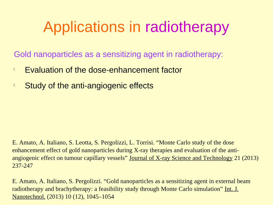
Gold nanoparticles as a sensitizing agent in radiotherapy:
Evaluation of the dose-enhancement factor
Study of the anti-angiogenic effects
E. Amato, A. Italiano, S. Leotta, S. Pergolizzi, L. Torrisi. “Monte Carlo study of the dose enhancement effect of gold nanoparticles during X-ray therapies and evaluation of the anti-angiogenic effect on tumour capillary vessels” Journal of X-ray Science and Technology 21 (2013) 237-247
E. Amato, A. Italiano, S. Pergolizzi. “Gold nanoparticles as a sensitizing agent in external beam radiotherapy and brachytherapy: a feasibility study through Monte Carlo simulation” Int. J. Nanotechnol. (2013) 10 (12), 1045–1054

Doses in GNP-loaded tissue for 150-kVp irradiation. Depth-dose profiles

Dose Enhancement Factors vs. target depth

Anti-angiogenic effect:influence of the radial profile of GNP
diffusion out of a capillary vessel

Applications in radioprotection
Occupational protection
Protection of the population
Environmental protection

✔ External irradiation
✔ Internal contamination
✔ Development of (standard) models
✔ Calculation of dose factors
✔ Simulation of complex layouts:
✔ dimensioning of the shields
✔ estimation of the doses to workers and population
Applications in radioprotection

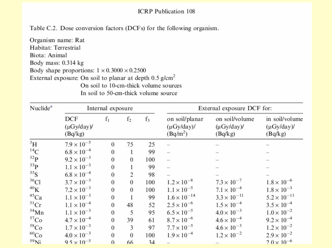
Environmental protection: the concept and use of reference animals and plantsICRP Publication 108 (2008)

Definition of the reference population

Assumption of ellipsoidal shape

Calculation of dose conversion factors (DCF)
Approach developed by Ulanowsky and Prohl
η = S/S0
S: surface of the ellipsoidS
0: surface of the sphere of equal mass
φ0: absorbed fraction in the sphere of
equal mass


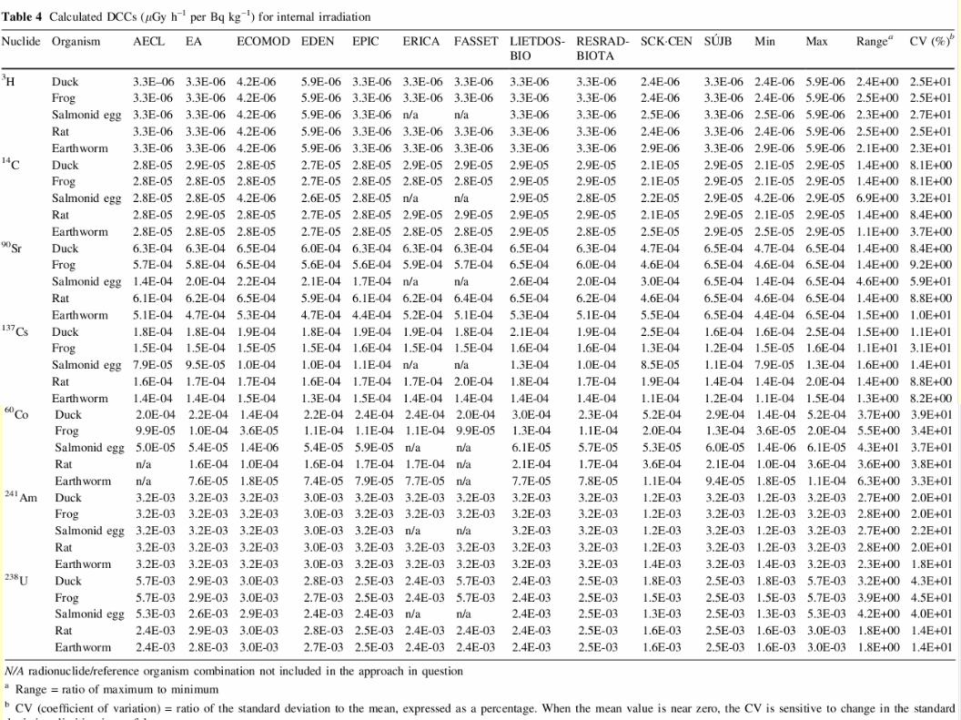
Importance of independent cross-check and validation
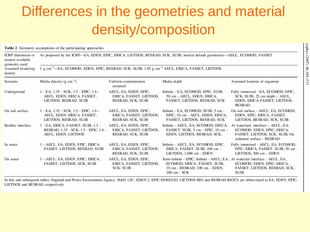
Differences in the geometries and material density/composition

Differences in the sources hypothesized: emission spectra and daughters
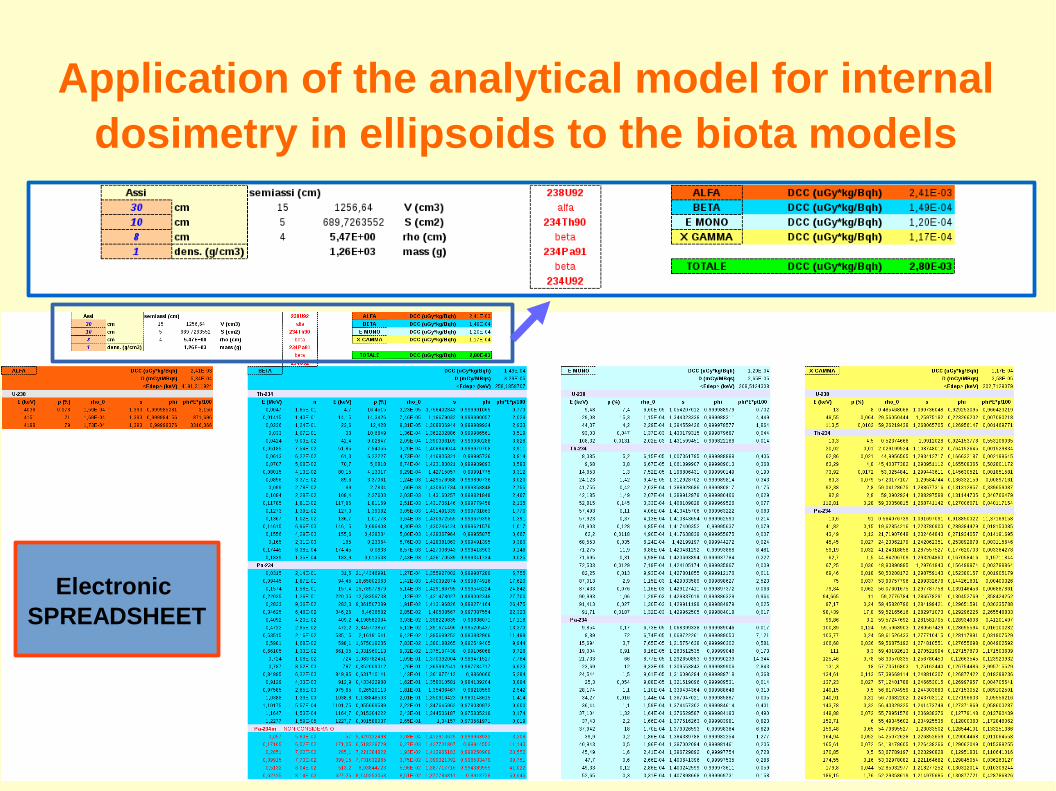
Application of the analytical model for internal dosimetry in ellipsoids to the biota models
Electronic SPREADSHEET

E. Amato, A. Italiano, “An analytical model for calculating internal dose conversion coefficients (DCCs) for non-human biota”, Radiation and Environmental Biophysics, 53 (2014) March (Online First)

Examples of shield design and optimization
Stopping of electrons in plastic materials
Plastic shields for beta radioactiove sources
ELI-Beamlines (preliminary studies):
beam dump and shielding for electrons
beam dump and shielding for protons

Comparative study of the absorption of beta particles in plastic materials
Conditions of “good geometry”
E. Amato and D. Lizio, “Plastic materials as a radiation shield for beta- sources: a comparative study through Monte Carlo calculation”, J. Radiol. Prot. 29 (2009) 239
E. Amato and D. Lizio, “Shielding of ionizing radiations with PTFE”, Advances in Chemistry Research - Vol. 7, chapter 8 (2011) 231-239, Nova Publishers Co. (USA).

90-Yttrium
Attenuation curves in PET, PTFE, PP and PMMA, in comparison with Al and Water.
Bremsstrahlung X-rays emission spectra.

Maximum range and number of bremsstrahlung X-ray photons emitted in the materials.
90-Yttrium

Optimization of real shields
Simulation of real-like geometries:
self-absorption inside the liquid source multi-layer shields with holes, capillars... shape, position and composition of the “target”: hand, fingertip, whole body...

90Y source in glass vial with PTFE shield
Dimensions (mm): vial: D=16 H=20 thick=1 water source: half vial PTFE shield thick= 5 “finger”: D=10 V=1 cm3 at 30 from the source centre.
Opaque, but high-temperature resistant
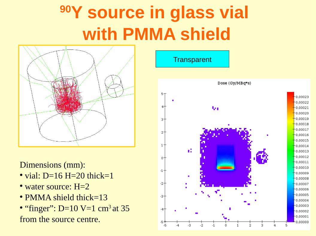
90Y source in glass vial with PMMA shield
Dimensions (mm): vial: D=16 H=20 thick=1 water source: H=2 PMMA shield thick=13 “finger”: D=10 V=1 cm3 at 35 from the source centre.
Transparent

177Lu source in glass vial with PMMA and W shields
Dimensions (mm): vial: D=16 H=20 thick=1 water source: H=2 PMMA shield thick=13 W shield thick=4 “finger”: D=10 V=1 cm3 at 35 from the source centre.

177Lu or 90Y sources in syringe with PMMA and W shields
syringe: D=16 L=60 thickness=1, material: PET PMMA shield thick=10 lateral shield in W thick=3 “hand” and “finger” represented by a parallelepiped and a cylinder.


Radiation field characterization and shielding studies for the
ELI Beamlines facility (Prague)
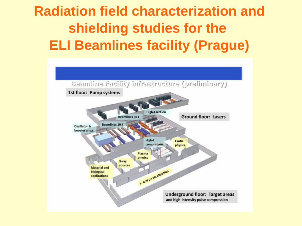
Radiation field characterization and shielding studies for the
ELI Beamlines facility (Prague)

Radiation field characterization and shielding studies for the
ELI Beamlines facility (Prague)

Radiation field characterization and shielding studies for ELI Beamlines:
1. proton sources
A. Ferrari, E. Amato, D. Margarone, T. Cowan, G. Korn. “Radiation field characterization and shielding studies for the ELI Beamlines Facility” Applied Surface Science 272 (2013) 138-144
A. Ferrari, E. Amato, D. Margarone. “Radiation field characterization and shielding studies”, ELIMED C.D.R., AIP Conference Proceedings 1546 (2013) 57-62
Preliminary studies (2011-2012): Fluka-Geant4 comparisons

Radiation field characterization and shielding studies for ELI Beamlines:
2. electron sources

ELI - 50 GeV e- beam dump

ELI – 3 GeV protons beam dump

Preliminary comparison: Fluka/Geant4

Conclusions
MC simulation is a powerful and versatile tool that can be applied in all fields of medical radiation physics and radiological protection
It is particularly useful when complex geometries and radiation fields are expected
It is a “laboratory” with unlimited possibilites, available to everyone
Intercomparisons are necessary for validation
It should be used with awareness and, possibly, with creativity!