Metode sinteze i oksidacije piridilporfirina
77
Metode sinteze i oksidacije piridilporfirina Pavletić, Pegi Master's thesis / Diplomski rad 2019 Degree Grantor / Ustanova koja je dodijelila akademski / stručni stupanj: University of Rijeka / Sveučilište u Rijeci Permanent link / Trajna poveznica: https://urn.nsk.hr/urn:nbn:hr:193:167322 Rights / Prava: In copyright Download date / Datum preuzimanja: 2021-10-08 Repository / Repozitorij: Repository of the University of Rijeka, Department of Biotechnology - BIOTECHRI Repository
Transcript of Metode sinteze i oksidacije piridilporfirina

Metode sinteze i oksidacije piridilporfirina
Pavletić, Pegi
Master's thesis / Diplomski rad
2019
Degree Grantor / Ustanova koja je dodijelila akademski / stručni stupanj: University of Rijeka / Sveučilište u Rijeci
Permanent link / Trajna poveznica: https://urn.nsk.hr/urn:nbn:hr:193:167322
Rights / Prava: In copyright
Download date / Datum preuzimanja: 2021-10-08
Repository / Repozitorij:
Repository of the University of Rijeka, Department of Biotechnology - BIOTECHRI Repository
UNIVERSITY OF RIJEKA
DEPARTMENT OF BIOTECHNOLOGY
Graduate studies
Research and development of drugs
Methods of pyridylporphyrin oxidation and
synthesis
Pegi Pavletić
Rijeka, 2019.

SVEUČILIŠTE U RIJECI
ODJEL ZA BIOTEHNOLOGIJU
Diplomski studij
Istraživanje i razvoj lijekova
Metode sinteze i oksidacije piridilporfirina
Pegi Pavletić
Rijeka, 2019.

Special thanks to my family who supported me throughout my studies,
my mentor Nela Malatesti, phD. for supporting me
in my research and ambitions
and to Martina Mušković for the experimental guidance
and invaluable contribution to my work.
Experimental work was performed using scientific equipment obtained
from the project: “Razvoj istraživačke infrastrukture na Kampusu
Sveučilišta u Rijeci”, financed by the European Union, within the European
Regional Development Fund.
Research work was financed through University of Rijeka Research
Support: “uniri-prirod-18-173”.

Graduate degree thesis was defended on the date: 12th September 2019.,
in front of the members of the evaluation committee:
1. Assistant professor dr. Ivana Ratkaj– head of the committee
2. Assistant professor dr. Rozi Andretić- Waldowski- member
3. Associate professor dr. Nela Malatesti– member, supervisor

Summary
Porphyrins are organic compounds that can be used as photosensitizers in
photodynamic therapy (PDT). PDT involves photosensitizer (PS)
administration into the body, where they penetrate tumour tissues and,
once illuminated, cause tumour degradation.
Most common way of porphyrin synthesis in our laboratory includes
modified Adler-Longo method, since it is relatively easy to conduct, it is fast
and gives us products with impurities separable with a column
chromatography. Recently we have found that zwitterionic N-oxidised
pyridylporphyrins as PSs in PDT exhibit low “dark” toxicity, as opposed to
their cationic analogues, N-methylated pyridylporphyrins, thus we decided
to focus our research on this perspective group of porphyrin PSs.
One of the requirements for porphyrins to be used in PDT efficiently, is their
high uptake into targeted cells. Therefore, N-oxidised pyridylporphyrins we
are particularly interested in have lipophilic component, and their synthesis
in this work includes adding long alkyl chains derived from dodecanoyl and
hexadecanoyl chlorides.
In this work different methods of pyridylporphyrin N-oxidation were
analysed to investigate which method is the best in terms of yields and ease
of purification for preparation of new potential photosensitisers for the use
in PDT. Four methods were assessed for N-oxidations of porphyrins using:
meta-chloroperoxybenzoic acid (m-CPBA), dimethyldioxirane (DMD);
hydrogen peroxide, sodium hydroxide and meta-chloroperoxybenzoic acid;
or acetic acid. Only methods using m-CPBA and DMD showed promising
results, providing high yields of oxidised pyridylporphyrins (65 % for m-
CPBA and 70 % for DMD) in the reactions that were easy to optimize and
conduct. Method including H2O2, NaOH and m-CPBA gave a yield of
(oxydo)pyridylporphyrin of 3,92 % and the reaction using CH3COOH did not
occur.

UV/ Vis spectroscopic analysis showed no difference between the spectra of
symmetric porphyrin oxidised by the different methods.
We are concluding from this work that both methods 1 (m-CPBA) and 2
(DMD) provide good yields. Although method 2 is more complicated to
perform due to constant pH adjustments, the product contains less
impurities, needs less additional purification and the yield is up to 5 %
higher in comparison to the method 1.
Key words: porphyrin, m-CPBA, dimethyldioxirane, PDT, synthesis

Sažetak
Porfirini su organski spojevi koji se mogu koristiti kao fotoosjetljivi spojevi
(PS) u fotodinamičkoj terapiji (PDT). PDT uključuje administraciju PS-a u
tijelo, gdje se isti lokalizira u tumorskom tkivu te, nakon osvjetljavanja
svjetlošću određene valne duljine, uzrokuje odumiranje tumorskog tkiva.
Najčešća metoda sinteze porfirina u našem laboratoriju je modificirana
Adler- Longo reakcija koja se relativno jednostavno izvodi, brza je i daje
nečistoće koje se mogu pročistiti stupčanom kromatografijom. Zwitterionski
oksidirani porfirini imaju malu toksičnost u “mraku”, što je bitno za
fotodinamičku terapiju (PDT), u usporedbi sa svojim kationskim analozina,
N-metiliranim piridilporfirinima. Stoga, smo naše istraživanje usmjerili na
proučavanje upravo ove grupe porfirinskih PS-ova.
Jedan od zahtjeva za porfirine koji se efikasno primjenjuju u PDT, je njihov
visoki unos u ciljane stanice. Stoga, N-oksidirani piridilporfirini koji su nam
od interesa imaju lipofilnu komponentu, a njihova sinteza u okviru ovog
istraživanja uključuje adiciju dugih alkilnih lanaca koji potječu od dodekanoil
klorida ili heksadekanoil klorida.
U ovom radu, različite metode N-oksidacije piridilporfirina su analizirane
kako bi se istražilo koja metoda rezultira najvećim iskorištenjem oksidiranih
porfirina s minimalnom prisutnošću nečistoća za potencijalnu primjenu u
PDT-u. Četiri analizirane metode uključuju N-oksidaciju pomoću: meta-
kloroperbenzojeve kiseline (m- CPBA); dimetildioksirana (DMD); hodikovog
peroksida, natrijeva hidroksida i meta- kloroperbenzojeve kiseline; ili
pomoću octene kiseline. Samo su se metode N-oksidacije pomoću m-CPBA
i DMD-a pokazale optimalnima, jer su rezultirale visokim iskorištenjem
piridilporfirina (65 % za m-CPBA i 70 % za DMD) u reakcijama koje su se
lagano mogle optimizirati i izvesti. Metoda koja uključuje upotrebu H2O2,
NaOH i m-CPBA, dala je iskorištenje od 3,92% a reakcija s CH3COOH nije
dala produkte.

UV/Vis spektroskopska analiza nije pokazala razliku između spektara
simetričnog porfirina oksidiranima upotrebom različitih metoda.
Zaključak istraživanja jest kako metoda 1 (m-CPBA) i 2 (DMD) daju
zadovoljavajuća iskorištenja. Iako je oksidacija metodom 2 zahtjevnija zbog
potrebe za stalnim podešavanjem pH vrijednosti, daje produkt s manje
onečišćenja, te je iskorištenje veće za do 5%.
Ključne riječi: porfirin, m-CPBA, dimetildioksiran, PDT, sinteza

Table of contents
1. Porphyrins in photodynamic therapy .......................................... 1
1.1. Historical overview of porphyrin discovery and research .......... 1
1.2. Porphyrin synthesis ............................................................ 3
a) Rothemund`s method......................................................... 3
b) Adler-Longo method ........................................................... 4
c) Lindsey’s method ............................................................... 4
1.3. N-oxidation of pyridylporphyrins .......................................... 5
a) N-oxidation using meta-chloroperoxybenzoic acid ..................... 6
b) N-oxidation using dimethyldioxirane ....................................... 7
1.4. Photochemical properties of porphyrins ................................ 10
2. Photosensitizers ..................................................................... 13
3. Research objective ................................................................. 17
4. Materials and methods ............................................................ 18
4.1. General information ........................................................... 18
4.2. Porphyrin synthesis ........................................................... 20
a) 5,10,15,20-tetrakis(3-pyridyl)porphyrin ............................... 20
b) 5-(4-acetamydophenyl)-10,15,20-tris(3-pyridyl)porphyrin ..... 21
c) 5-(4-dodecanamidophenyl)-10,15,20-tris(3-pyridyl)porphyrin 22
d) 5-(4-hexadecanamidophenyl)-10,15,20-tris(3-pyridyl)porphyrin
24
e) 5,10,15,20-tetrakis(1-oxydopyrid-3-yl)porphyrin .................. 25
f) 5-(4-dodecanamidophenyl)-10,15,20-tris(1-oxopyrid-3-
yl)porphyrin ............................................................................. 28
g) 5-(4-hexadecanamidophenyl)-10,15,20-tris(1-oxopyrid-3-
yl)porphyrin ............................................................................. 30
5. Results and discussion ............................................................ 33
5.1. Porphyrin synthesis ........................................................... 33
5.2. Methods of N-oxidation of pyridylporphyrins and optimization . 35
a) Oxidation of pyridylporphyrins using meta-chloroperoxybenzoic
acid 35
b) Oxidation of pyridylporphyrins using dimethyldioxirane .......... 37

c) Oxidation of pyridylporphyrins using hydrogen peroxide and
meta-chloroperoxybenzoic acid .................................................. 39
d) Oxidation of pyridylporphyrins using acetic acid and hydrogen
peroxide .................................................................................. 40
5.3. UV/Vis spectroscopy .......................................................... 41
5.4. Nuclear Magnetic Resonance .............................................. 50
6. Conclusion ............................................................................ 58
Literature...................................................................................... 60
Table of Figures ............................................................................. 63
Curriculum vitae ............................................................................ 65

1
1. Porphyrins in photodynamic therapy
1.1. Historical overview of porphyrin discovery and research
Porphyrins, as naturally occurring compounds, were mentioned for the first
time in 460-370 BC, by the famous philosopher and scientist Hippocrates,
who had described a case of a young woman, who had symptoms
resembling menstrual-related acute porphyria.(1) Following the modern
scientific interest in the field, porphyrins are mentioned for the first time
during the 19th century, in a medical context, by a researcher named
Laennec who had diagnosed a patient with anaemia caused by lead
poisoning. In 1841, Scherer isolated the first iron-free hemine compound,
and after the discovery of haemoglobin in 1864, porphyrins have been
isolated and researched.
First visible spectrum of porphyrins was taken in 1867, alongside red
fluorescence, by Thudichum who had called porphyrin: “cruentine“ due to
the blood-red fluorescence colour of the compound. Fluorescence of the
compound was not found interesting at that time, and it will take more than
a decade until it was assessed again by Hoppe-Seyler (1879) who had
studied chlorophylls at that time and noticed the resemblance between the
stated compounds. He had also, in 1871, prepared porphyrins from blood
samples and noticed their tetrapyrrolic nature. Hoppe-Seyler named
isolated compounds “Hämatoporphyrin“, meaning “blood-purple“, and used
different prefixes on the “porphyrin“ name base, which has become an
official nomenclature of the researched compounds.
Soret discovered in 1883 that hemins absorb light at the wavelength of 400
nm, which is characteristic trait for all porphyrins. At this time, from 1890s
to 1930s, lots of research of porphyrins were connected to physiological
effects, and porphyria was first recognised and categorised as a disease by
Günther. The structure of porphyrins was at this time still unknown,
although protoporphyrin had been successfully synthesized and
2
coproporphyrin, meso-porphyrin and haemoglobin were known to exist and
were isolated.
In 1912, W. Küster derived a haemin formula in which he had proposed a
structure of hemin, constituted of four pyrrole rings linked with 4 methine
bridges on the third position in the pyrrole ring. In the centre of the
molecule, connected to the inward-facing nitrogen atoms of pyrroles is a
proposed FeCl group.(2) In 1929, Fischer and Zeile had successfully
synthesized hemine that confirmed Küster`s proposed porphyrin structure
and connections to the structures of heme and chlorophyll were made.
Fischer prepared uroporphyrin ester from urine of his patient named Petry,
who suffered from porphyria. He received Nobel prize in 1930 for his work
on porphyrins.(3) He also found that uroporphyrin has lower
photosensitivity in comparison to the hematoporphyrin. Körbler had
noticed, in 1931, that there is a significant uptake of porphyrin in the
tumour tissue, which marked the era of research of porphyrins as a possible
therapy for tumours.
Looking back at the historical timeline of porphyrin research, it is evident
that the long initial research period of porphyrin-based compounds was a
result of not only the lack in technology, but mostly due to use of different
terminology for the compounds.(3) In this regard, Hoppe-Seyler marked a
beginning of porphyrin era, after which most significant scientists for the
era were Günter, whom after congenital porphyria was named Günter`s
disease due to his classification of porphyrias, and Fischer. Fischer
contributed greatly to the field by introducing the first porphyrin
nomenclature (trivial names and isomer nomenclature) (4) and establishing
a research facility for this type of compounds. After him, porphyrin research
had developed fast in structure research and nomenclature optimization.
Porphyrin is built of four pyrrolic rings linked by four methine bridges, and
it is substituted on one of the twelve possible positions on the inner ring.
Eight of those positions are referred to as the β- positions and only four are
located on the methine bridges, called meso positions. Accordingly, β-

3
substituted porphyrins are named β-porphyrins and meso-substituted
porphyrins are called meso-porphyrins.
Figure 1: meso-substitution position on porphyrin (left) and β-substitution positions on
porphyrin (right)
If the porphyrin is not substituted, its structure is called porphine. Naturally
occurring porphyrins are mostly β-substituted, but meso-porphyrins are
easier to produce and are so far used more as potential drugs in PDT.
1.2. Porphyrin synthesis
There are several ways to synthesize porphyrins, and throughout the years,
scientists have tried to find new ways of synthesis, that would result in
higher yields and the ones that require less purification to isolate clean
products. Depending on the structure of porphyrin product we are trying to
synthesize, there are different methods we can use to produce them. Two
types of occurring porphyrins are meso-substituted and β-substituted, latter
of which are more similar to the natural porphyrin, such as protoporphyrin
IX. The focus of the research in this work are meso-substituted porphyrins
with applications in photodynamic therapy (PDT).
a) Rothemund`s method
In 1936, scientist Paul Rothemund concluded that the best method for
porphyrin synthesis is a reaction of a pre-synthesized pyrrole and a
substance with aldehyde function (formaldehyde, acetaldehyde etc.) at
different temperatures (mostly 90- 95 C). This refers only to the meso-

4
substitution of aldehyde groups to the α,β,γ and δ positions in a porphyrin
ring.(5) The reaction occurs in a presence of nitrogen and in an absence of
oxidant. Rothemund method for porphyrin synthesis results in creation of
parallel products to the desired porphyrin, mostly chlorins, which results in
lower porphyrin yields.(5),(6) Chlorins can be oxidized to related porphyrins
afterwards. Yield of porphyrin in this reaction is up to 10%.
b) Adler-Longo method
After Rothemund had developed a method for symmetric porphyrin
synthesis, in 1960s, Adler and Longo used the condensation reaction of
pyrrole and benzaldehyde in acid (mainly acetic acid or acetic acid with
metal salts). The reaction was conducted under reflux.(6) Reaction has two
steps.
First step is a self-assembly of the four pyrrole and four aldehyde molecules
into porphyrinogen. In this, concentration-dependent process, eight C-C
bonds are formed.
Second step includes oxidation of porphyrinogen to porphyrin in the
presence of an oxidant. Light and oxygen must be present in the reaction
system.(7)
Nowadays, this method is improved by the use of propionic acid as a
solvent, and this reaction requires oxygen for the formation of porphyrins,
as it oxidises the porphyrinogen intermediate.(8) Yields of porphyrins using
this method are up to 20%, thus higher in comparison to the Rothemund`s
method.(7)
c) Lindsey’s method
About 20 years after the Adler-Longo method, Lyndsey has created a
method for porphyrin synthesis under milder conditions than Rothemund
and Adler-Longo. It is a reaction in the presence of oxygen-donor and
includes pyrrole condensation in dichloromethane (DCM) or chloroform,
catalysed by a Lewis acid. Reaction undergoes reflux conditions for one hour

5
after the addition of an oxidant. This allows for the formation of a
tetrapyrrolmethane. Yields in this reaction can be up to 40 % or 50%.(8)
Products in the reaction are a mix of different asymmetric porphyrins that
are meso-substituted.
1.3. N-oxidation of pyridylporphyrins
Previous research has found that N-oxides of pyridylporphyrins, exhibit
lower “dark” toxicity in comparison to the N-methylated
pyridylporphyrins.(9) However, their synthesis proved to be difficult,
especially purification of the products, thus we wanted to study current and
investigate new methods for the preparation of this group of compounds.
N-oxidation of pyridylporphyrins includes preparation of
(oxido)pyridylporphyrins using different oxidants. Oxidation results in the
formation of different products, thus there is a need for the isolation of the
wanted products and its purification. Success in the oxidation reactions
toward preparation of (oxido)pyridylporphyrins is followed on Thin Layer
Chromatography (TLC), and the isolation from the reaction mixture and
product purification is carried out by column chromatography, using the
appropriate solvent ratios.
In the aforementioned oxidation reactions, the addition of an excess oxidant
can lead to the degradation of the pyridylporphyrin, thus we have to take
this into account when considering existing and new methods, and their
optimisation. Furthermore, (oxido)pyridylporphyrins are more soluble than
their parent porphyrins in polar solvents.(10)
In this work, N-oxidations were carried out using following reagents:
- meta-chloroperoxybenzoic acid (m-CPBA)
- Oxone
- Hydrogen peroxide
- m-CPBA and hydrogen peroxide

6
a) N-oxidation using meta-chloroperoxybenzoic acid
m-CPBA is an abbreviation and a trivial name for the compound 3-
chloroperbenzoic acid, whose molecular mass is 156,57 g/mol. It belongs
to the class of organic compounds named halobenzoic acids, due to the
position of the chloride on the aromatic ring (Figure 2). It is a white solid
soluble in aqueous solutions and in some organic solvents, such as DCM.
Figure 2: Structure of m-CPBA
In 1999. Posakony et al. used m-CPBA for N-oxidation of porphyrins
containing one to four 4-pyridyl groups. Mixed aldehyde condensation was
used to synthesize meso-substituted porphyrin, and then N-oxidation was
performed using m-CPBA to produce five (oxido)pyridylporphyrin
compounds and seven porphyrin-N-oxides. m-CPBA was used in a solution
of CH2Cl2 and CH3OH at room temperature, its aliquot being 1-2 equivalents
of porphyrin. This resulted in formation of (oxido)pyridylporphyrin, whilst
the addition of several aliquots resulted in the formation of porphyrin-N-
oxides. Using m-CPBA as an oxidant included the use of triethylamine to
quench the reaction to prevent over-oxidation. After the reaction
completion, some by-products appeared, such as meta-chlorobenzoic acid
which had to be removed from the reaction mixture with the extraction
using NaOH solution. Triethylamine was absorbed on the silica gel of the
column, straight after the N-oxidation, to remove it from the mixture and
to stop its further oxidation. Addition of m-CPBA after the completion of the
reaction led to the degradation of porphyrin.(10)

7
In the same paper, the authors state that using Lindsey method of
porphyrin synthesis had given impure products, resulting in the formation
of unknown compounds, resulting in lower porphyrin yields.
Oxidopyridyl group is, in comparison with phenyl group, less susceptible to
oxidative degradation, which means that (oxido)pyridylporphyrins are
stable enough to be isolated. m-CPBA was shown to provide stable products
and this method is reproducible on large number of compounds. Expected
values of 1H NMR of (oxido)pyridylporphyrins created using m-CPBA include
a complex β-hydrogens region, caused by the proximity of different meso-
substituents, which appears as broad area of one or more peaks in the
spectrum. The hydrogens from within the porphyrin ring appear as singlets
at ~-2,85. (10)
b) N-oxidation using dimethyldioxirane
Dimethyldioxirane (DMD) was first synthesized in 1985. by Robert W.
Murray and Ramasubbu Jeyaraman who defined dioxiranes as the smallest
cyclic peroxide systems.(11) Caroate-acetone system was used to produce
DMD in situ with the yield up to 90% at room temperature. The caroate
addressed here is peroxymonosulfate, potassium caroate or trivially-
Oxone. DMD is yellow in a solution and they have determined that the
maximum absorption peak of DMD is at λ= 335 nm. Proton NMR analysis
showed single peak at δ = 1,65, and 13C NMR analysis shows peaks at 22,72
(quartet) and 214,4 ppm (singlet). They have also noticed stereospecificity
in reactions they have conducted.(11)
In 1988. Curci and Edwards have created Methyl(trifluoromethyl)dioxirane,
a form of dioxirane compound up to 1000 times more reactive than DMD,
by the reaction of caroate and acetone. Curci also discovered that
fluorinated ketones have an impact on the stability and reactivity of the
synthesized dioxirane.(12) It is also recognised that the stability of the DMD
is not high, therefore more of the reactants are used to produce it; however,
the cost of these compounds is low.(12) After this, in 1992. Murray

8
developed a new method for DMD synthesis from cyclic compounds, such
as cyclohexane.
DMD is often used in the reactions of epoxidation (13), but it is also an
effective oxidising agent for the oxidation of porphyrins.(14) DMD exhibits
stereoselectivity and chemoselectivity and is desirable oxidant for the
creation of oxidised porphyrin compounds. The preparation of DMD in situ
is achieved in the reaction of Oxone with ketone under hydrolytic
conditions.(15) DMD undergoes the conversion to a solvent during the
reaction of its preparation.(16) Preparing DMD prior to its use in reactions
allows for its use in non-aqueous reactions. However, for the purpose of
this research, we opted for in situ generation of DMD in a non-miscible one
pot (CH2Cl2 and H2O) reaction system, after which the purification of the
product was conducted. In such preparation procedure, the pH needs to be
between 7,0 and 8,0 to assure the formation of (oxido)pyridylporphyrin,
and buffers such as sodium bicarbonate or phosphate buffer are mostly
used in a solution with excess ketone. Reaction lasts anywhere between 0,5
and 72 hours, and many variables effect the success of the reaction. Besides
this method of synthesis, it is possible to derive DMD in a system using
benzene.(17) Isolation method for the DMD is used only if the substrate or
the product are unstable in biphasic conditions, which was not the case in
this research. The excess of Oxone and the excess of ketone do not affect
the rate of conversion to DMD, neither increase the product yield. The
mechanism of dioxirane formation involves the nucleophilic attack of Oxone
at the carbonyl carbon, resulting in the loss of potassium hydrogen
sulphate.(17)

9
Figure 3: Conversion of Oxone to DMD
Paredes et al. have compared diastereoselectivity between products
obtained with the use of DMD and m-CPBA in 1997 and have concluded that
the difference is not very high between the two.(18) However, DMD is more
sensitive on the steric interactions and its regiospecificity is determined by
the electronic effects in the molecule subjected to oxidation, whilst the latter
does not influence the oxidation using m-CPBA.
Conditions that effect production of DMD are: pH, time of oxidant addition,
ketone type and lipophilicity and steric interactions. (17) Temperature is
not considered to have an impact on the production of oxidised compounds
nowadays, although some previous research stated that it has an
impact.(17)
In literature, there are divided opinions on the temperature impact on the
success of the reaction. Some of the new sources (15) have provided a
detailed research into this topic, where it was concluded that the reactions
at room temperature give the same yields of products as do the reactions
at 0 C. However, in the older research and sometimes, in the new
processes for porphyrin oxidations, it is advised to conduct the reactions at
0 C throughout the time of the oxidant addition to the mixture.(17) In our
research, no metalloporphyrins were used, so it was concluded that the
temperature of the reaction will be kept at room temperature, since only
metalloporphyrins undergo thermal decomposition in solutions in the
presence of dioxiranes.(19)

10
1.4. Photochemical properties of porphyrins
Photodynamic therapy (PDT) is a non-invasive form of photosensitized
reactions that have a positive effect on human health. Ancient Egyptians,
Chinese and Indians were known to use plants orally for the treatment of
vitiligo and psoriasis in direct sunlight. Scientist Niels Finsen used light
therapy (red part of the spectrum) to limit spreading of smallpox pustules
in patients, during the end of 18th century. For his research, he was awarded
the Nobel prize in 1903.(20) Many consider Oscar Raab to be the first to
study PDT, having discovered that acridine dyes are the cause of death of
mono cellular organisms, such as Paramecium. Scientists R. L. Lipson and
S. Schwartz who worked in 1960 at the Mayo Clinic are the first who have
discovered that preparations containing hematoporphyrin’s cause
fluorescence in tumour tissue of the treated patients.(21)
Modern understanding of PDT involves three components: photosensitizing
agent, light of the specific wavelength and molecular oxygen. The
photochemistry behind the PDT can be explained with the use of Jablonski
diagram (Figure 4), which serves as a visual representation of the PDT
principle.
Figure 4: Scheme of modified Jablonski diagram(22)
Chromophore (an atom or a molecular group that gives colour to the
compound), is illuminated with the light of a specific wavelength that excites

11
chromophore to one of the excited singlet states. The excited chromophore
may relax to the singlet ground state by the emission of photon in a process
called fluorescence, or it can convert to the excited triplet state in a process
called intersystem crossing. Relaxation from the triplet state back to the
ground state is named phosphorescence, and it is caused by the emission
of photon from the excited triplet state of the chromophore. The loss of
energy, except from phosphorescence, may happen if the excited
chromophore transfers its energy to another molecule. In PDT of
porphyrins, that molecule is triplet ground-state oxygen (3O2) that converts
to the singlet oxygen (1O2).(23) Ian J. Macdonald and Thomas J. Dougherty
state that: “The energy required for the triplet to singlet transition in oxygen
is 22 kcal/mol, which corresponds to a wavelength of 1274 nm (infrared
light). Thus relatively low energy is needed to produce singlet oxygen.
Photochemical reactions of this type are known as Type-II photoreactions
and are characterized by dependence on the oxygen concentration (O2)“.
Type I photoreactions involve a reaction between a triplet-state
photosensitizer with superoxide radicals to produce superoxide anions. Type
I and type II photoreactions are depicted in the Figure 5. They can then
react with other molecules to create hydroxide radicals.(23) Type I
sensitizers are subject to photoinduced electron transfer, upon which O2•−
and HO−• are being produced. In comparison to that, type II reactions are
characterized as energy-transfer process to oxygen, causing the formation
of 1O2. Both types are oxygen-dependent reactions.(24)

12
Figure 5: Type I and type II photoreactions (25)
Singlet oxygen (product of the type II reaction) that forms in PDT is formed
by the inversion of the electron’s spin on the outer electron of the molecule.
In the magnetic field, there are three different configurations of electrons
possible, which are indistinguishable when magnetic field is not present.
Those are:
- Electronic configuration with both spins oriented downwards
- Electronic configuration with both spins oriented upwards
- Electronic configuration with one spin oriented upwards and the other
downwards
Due to these three possibilities, oxygen is a triplet in its ground state.
Oxygen is very reactive due to two electrons pairing into π*2p antibonding
orbitals in the same spin, which is not in accordance to the Hund`s rule and
the Pauli`s exclusion principle, and it is destabilising the molecule. Lifetime
of this oxygen is quite long, up to 10-100 µs in organic solvents.(23)

13
2. Photosensitizers
Photosensitizers (PS) used in PDT should have as much as possible
absorption in the red field of electromagnetic spectrum and have the
possibility of producing singlet oxygen. Red light allows for deeper
penetration into the tissue thus having better healing effect in tumour
application. Most used PDT photosensitizers are mostly planar
tetrapyrroles: porphyrin, bacteriochlorin and chlorin. Porphyrins mostly
absorb visible light at approximately 400-450 nm, visible on the
spectroscopy chromatogram, and known as Soret band. This wavelength,
however, since it is in the blue area of visible light on the electromagnetic
spectrum, is not so relevant for PDT applications, as this type of light does
not penetrate the tissue as deep as the red light, thus not achieving the
desirable effect. Aside from Soret band, there are four more bands
characteristic for porphyrins, ranging from 600-800 nm on chromatogram,
known as Q-bands. They are in the red part of the spectra and are more
relevant for most of the PDT applications. These compounds successfully
create singlet oxygen in line with the singlet oxygen quantum yield, marked
with the symbol φ. This yield determines the number of oxygen molecules
formed per photon upon the absorption of energy.(23)
The properties of PSs differ significantly between solvents and biological
systems, making them harder to study. Ideal PS exhibits no effect until the
application of light, i.e. they should not express the so called “dark” toxicity.
PSs do not bind to the receptors within the body, but there is a link between
lipophilicity and drug uptake into serum by binding to low density lipoprotein
(LDL) serum constituents. LDL is increased in cancer tissue due to growth
of cancer`s vasculature. From the circulation, PSs move into cells. After the
irradiation of the tumour tissue, immunological response occurs in the
accumulation of platelets, macrophages, and the release of prostaglandins
and cytokines, which cause the damage to the tumour vasculature.
Apoptosis is triggered in tumour cells, but it is not achieved fully only by
PDT. Besides apoptosis, necrosis may occur in tumour tissue. Duration and

14
intensity of irradiation as well as the amount of PS used have impact on the
effectiveness of tumour PDT.(26)
Apart from LDL, some researchers believe that low pH value and the
presence of macrophages can also contribute to the PS localisation in
tumour tissue.(27) With the increase in porphyrin hydrosolubility, their
accumulation in tumour tissue grows. On the other side, if lipophilic carriers
are used for PSs transport at the temperature of 37 C, binding to LDL is
the primary path of their tissue distribution. Tumour cells possess a high
number of LDL receptors on their surface, making them even more
susceptible to the binding of PS. By endocytosis, PSs pass through the
membrane and bind to the mitochondrial membrane, the Golgi apparatus
and the endoplasmic reticulum. It is debatable which transporters to the
tumour tissue are better, for there are examples of good transporters that
do not bind to LDL (meso-tetraphenylporphin tetrasulfonate is one
example) and of some poor transporters, such as hematoporphyrin.
Additional factor to take into account is the availability of oxygen in the
tumour tissues, which can drop significantly in vivo if PS are applied in full
dose and irradiated. Photobleaching helps by limiting the initial response of
the applied PS, which causes PS to activate through a certain period, thus
helping in maintaining sufficient oxygen levels in tumour tissue. Oxygen
levels can be controlled by modifying the duration and repetition of light
during therapy, for example 15-20 seconds with pauses between the
application. (27)
PS must exhibit properties such as: having an absorption peak in the red
area of electromagnetic spectra (>650 nm), having high quantum yield for
the formation of triplet state and singlet oxygen production, low toxicity in
the dark, selectivity for tumour tissues, fast reactivity, fast and easy
elimination from the body, easy and affordable synthesis, optimal
pharmacokinetics, low to no side-effects for the patient, long-term
beneficiary effect for the patient, or easy administration.

15
- First generation PSs
First generation photosensitizers include only porphyrin compounds,
amongst which Photofrin IX and Hematoporphyrin. These compounds were
created for the purpose of suboptimal tissue penetration and prolonged skin
photosensitization, mostly for the detection of cancer due to their
fluorescent nature.(28) These compounds were not selective enough, so
their ingestion caused displacement in different tissues and unwanted
photoactivation wherever PS was absorbed. The problem which occurred
with Photofrin was that it had to be administered in large dose to be
effective in tumour therapy, and it had an exceptionally long half-life (up to
452 hours), which caused undesirable photosensitization in patients
amongst other side-effects. Photofrin is still used today as a golden
standard, despite its many flaws, such as low absorption in the red spectrum
and shallow tissue penetration (1,5 mm).(28) Mechanism of PDT action of
these PSs includes the formation of ROS (mostly singlet oxygen) that
damage tumour cells and tumour tissue vasculature.
- Second generation PSs
Second generation PSs were created to try and resolve the deficiencies of
the first generation of PSs. Bacteriochlorins and chlorins are such compound
types. PS Tookad is a bacteriochlorin and it has better properties than
Photofrin. It penetrates the tissue moderately (up to 4,0 mm), clears from
the body quicker and has a much shorter half-life (up to 20 minutes).(28)
These compounds target tumour vasculature causing hypoxia, necrosis and
cell death. Foscan, a chlorin compound, has similar properties to Tookad,
as well as Protoporphyrin IX. Foscan, in comparison to Tookad, had longer
half-life and could cause moderate skin problems weeks after its
administration. Second generation PSs could not completely resolve the
problems of the first generation, but only limit certain parameters, such as
tissue penetration, clearance, higher generation of ROS, length of half-life,
dark-toxicity, higher yields etc. Second generation PSs include both
porphyrin and non- porphyrin compounds.
16
- Third generation PSs
Specificity of a third generation PSs is targeted delivery of the second
generation of PSs to the tumour tissues in carrier molecules such as
monoclonal antibodies, polymer bodies and liposomes. The goal of the third
generation is to make PSs more hydrophilic at physiological pH for a more
efficient delivery.
Due to occurrence of specific antigens on the surface of tumour cells,
antibodies can detect them and bind to their receptors on the surface. In
this way, healthy tissues are protected from the targeted action of PS.
Polyethyleneglycol (PEG) nanocarriers are used widely, and PEG liposomes
are being developed. Photofrin is an example of a third generation PS that
was used incorporated into polyacrylamide nanoparticles and applied in the
therapy of a rat brain tumour. Encapsulating photofrin caused better
efficiency in tumour therapy which significantly reduced the time of action
of the drug. Besides PEG, phthalocyanines and amine-functionalized
polyacrylamide are used as carriers.(28) Third generation PSs include both
porphyrin and non-porphyrin compounds, and they are still being developed
today. Although they seem very promising in therapy, there is a possibility
that closing the PS in a capsule may lead to the lower level of produced
ROS.(29)

17
3. Research objective
The purpose of this research is to find, implement and optimize the reaction
of N-oxidation of pyridylporphyrins.
Four methods will be tested and compared in terms of product yields and
ease of purification. These methods will use following oxidants:
- meta-chloroperoxybenzoic acid
- dimethyldioxirane
- meta-chloroperoxybenzoic acid (elution with NaOH)
- H2O2 and acetic acid
All aforementioned methods will be tested on symmetric 5,10,15,20-
tetrakis(3-pyridyl)porphyrin to evaluate the success of the reaction
(percentage of yield), whereas only the most successful method, or the one
most similar to m-CPBA set as the standard, will be further tested on
porphyrins substituted with dodecanoyl chloride and palmitoyl chloride.
Asymmetric pyridylporphyrins with long alkyl chains are more effective in
photodynamic therapy, as they can penetrate membranes more
successfully than symmetric hydrophilic porphyrin, thus it is our aim to
optimise their synthesis and preparation.

18
4. Materials and methods
4.1. General information
Reagents and chemical substances used in this work are commercially
available, and originate from manufacturers: Sigma-Aldrich, VWR
Chemicals, Acros Chemicals and Carlo Erba Reagents. For Thin Layer
Chromatography (TLC), aluminium plates with thin layer of silica gel were
used (Macherey-Nagel, 0.20 mm silica gel 60 Å with fluorescent indicator
UV254), alongside UV lamp at wavelengths of 254 and 365 nm. Column
chromatography was performed in columns of different diameters filled with
silica gel from Macherey-Nagel (Silica 60 Å, 0.04-0.06). Elution was carried
out with dichloromethane and methanol solution in different ratios.
Ultra-violet and visible (UV/Vis) spectrometry was performed on the Agilent
Cary 60 UV-Vis Spectrophotometer in the visible spectre, between 350 nm
and 750 nm. The dilutions of the compounds were prepared in the following
manner, from the 10 µM solution of porphyrin:
a) First vial: 10 µM solution of porphyrin
b) Second vial: 8 µM solution of porphyrin
c) Third vial: 6 µM solution of porphyrin
d) Fourth vial: 4 µM solution of porphyrin
e) Fifth vial: 2 µM solution of porphyrin
f) Sixth vial: 1 µM solution of porphyrin
g) Seventh vial: BLANK, CH2Cl2
Quartz cuvette was used for the analysis of the porphyrin samples.
Agilent Cary Eclipse Fluorescence Spectrophotometer was used for
measuring the fluorescence of the synthesized porphyrins, at the maximum
wavelength of the Soret band of the mentioned porphyrin. Range in which
the fluorescence emission was recorded was between 500 nm and 800 nm,
for the concentration of 1µM porphyrin.
19
Nuclear magnetic resonance (NMR) analysis was conducted at the
Chemistry Department of the University of Zagreb, Faculty of science; in
the Laboratory for NMR spectroscopy, with Bruker Avance III HD 400 MHz.
Solvents used for the preparation of samples were deuterated chloroform
and deuterated methanol. Analysis of the NMR spectres was performed
using the program MestReNova 14.0.1.
Computational chemistry involved the use of ChemAxon`s Marvin Sketch
programme. 3D projections of the compounds were done in Avogadro.

20
4.2. Porphyrin synthesis
a) 5,10,15,20-tetrakis(3-pyridyl)porphyrin
Molecular weight: 618,704 g/ mol
Molecular formula: C40H26N8
Polar surface area: 108,92
5,10,15,20-tetrakis(3-pyridyl)porphyrin (porphyrin 1) is prepared by
dissolving 4-acetaminobenzaldehyde (0,90 g; 5,52 mmol; 1 eq.) and 3-
pyridinecarboxaldehyde (1,78 g; 0,02 mol; 3 eq.) in propionic acid (70 ml).
The reaction is conducted at two temperatures, whilst the reaction mixture
is being gradually heated:
- When the temperature reaches 20°C - 30°C, distilled pyrrole (1,54
ml; 0.02 mol; 4 eq.) is added to the mixture slowly and in small amounts
within 10 minutes
- When the temperature reaches 90°C, it is maintained for 45 minutes
approximately, after which the reaction is stopped.
Light and oxygen are necessary during this process to allow the oxidation
of the porphyrinogen to porphyrin.

21
After the confirmation of product formation on TLC, solvent is removed in
vacuo. Two column chromatography procedures are conducted
subsequently for product purification. On the column, porphyrin 1 is
separated as dark purple, crystalline solid (0,182 g; 5,12%).
UV/ Vis spectroscopy analysis showed the presence of Soret band at 418
nm, with Q bands at next λmax/ nm: 514, 549, 589, 645. Extinction
coefficient was calculated: Ɛ= 0,31. Fluorescence measured at the
excitation wavelength of the Soret band gave two peaks at λmax/nm= 650,
715.
1H NMR was measured in solvent CD3Cl and next peaks were observed:
δ/ppm 9,49 (d; J = 2,2 Hz; 3H; Py-2-H); 9,10 (dd; J = 4.9; 1.7 Hz; 3H;
Py-4-H); 8,89 (s; 8H; β-H); 8,56 (d, J = 7.7 Hz; 3H; Py-6-H); 7,81 (dd, J
= 7,8; 4,9 Hz; 3H; Py-5-H); -2,81 (s, 2H, pyrrole NH).
b) 5-(4-acetamydophenyl)-10,15,20-tris(3-pyridyl)porphyrin
Molecular weight: 674,768 g/ mol
Molecular formula: C43H30N8O
Polar surface area: 125,13

22
5-(4-acetamydophenyl)-10,15,20-tris(3-pyridyl)porphyrin (porphyrin 2) is
separated from the same column as porphyrin 1, as a second fraction. This
fraction is also isolated as a dark purple crystalline matter (0,131 g;
3,74%).
UV/ Vis spectroscopy analysis shows the presence of Soret band at 418 nm,
with Q bands at next λmax/ nm: 515, 550, 590, 647. Extinction coefficient
was calculated: Ɛ= -0,29. Fluorescence measured at the excitation
wavelength of the Soret band gave two peaks at λmax/nm= 651, 717.
c) 5-(4-dodecanamidophenyl)-10,15,20-tris(3-pyridyl)porphyrin
Molecular weight: 815,038 g/ mol
Molecular formula: C53H50N8O
Polar surface area: 125,13
5-(4-dodecanamidophenyl)-10,15,20-tris(3-pyridyl)porphyrin (porphyrin
3) is synthesized by adding dodecanoyl chloride (0,083 g; 0,377 mmol; 5
eq.) to porphyrin 2 (0,051 g; 0,078 mmol; 1 eq.) in dichloromethane at 0
°C through 45 minutes in a single round flask. The reaction completion was
followed by TLC. Solvent for TLC was CH2Cl2: CH3OH= 9:1. The oxidised

23
compound is purified by extraction with water (3 times) and Na2SO4, filtered
and the solvent is removed in vacuo. Column chromatography is used for
purification of the synthesized porphyrin 3, obtained from the column as
dark purple fraction (0,073 g; 87,95%).
UV/ Vis spectroscopy analysis shows the presence of Soret band at 419 nm,
with Q bands at next λmax/ nm: 515, 551, 591, 646. Extinction coefficient
was calculated: Ɛ= 0,38. Fluorescence measured at the excitation
wavelength of the Soret band gave two peaks at λmax/nm= 649, 715.
1H NMR was measured in solvent CD3Cl and next peaks were observed:
δ/ppm 9,49 (t; J= 2,5 Hz; 3H; Py-2-H); 9,09 (d; J= 3,7 Hz; 3H; Py-4-H);
8,99- 8,80 (m; 8H; β-H); 8,55 (d, J= 5,76 Hz, 3H; Py-6-H); 8,20 (d; J=
6,6 Hz; 2H; Ar-2,6-H); 7,96 (bd, 2H; Ar-3,5-H); 7,79 (t; J=5,13 Hz; 3H;
Py-5-H); 7,74 (s; 1H; amide N-H); 2,57 (t; J = 5,64 Hz; 2H; C2H2); 1,91
(t; J= 3,02 Hz; 2H; R-C3H2); 1,49 – 1,29 (m; 18H; R-H: C4H2, C5H2, C6H2,
C7H2, C8H2, C9H2, C10H2, C11H2); 0,92 (t; J= 5,19 Hz; 3H; R-C-H3); -2,78
(s; 2H; pyrrole N-H)

24
d) 5-(4-hexadecanamidophenyl)-10,15,20-tris(3-
pyridyl)porphyrin
Molecular weight: 871,146 g/ mol
Molecular formula: C57H58N8O
Polar surface area: 125,13
5-(4-hexadecanamidophenyl)-10,15,20-tris(3-pyridyl)porphyrin (porphyrin
4) is synthesized in a reaction of hexadecanoyl chloride (palmitoyl chloride)
(0,107 g; 0,388 mmol; 5 eq.) and porphyrin 2 (0,052 g; 0,078 mmol; 1
eq.) in dry CH2Cl2 at 0 °C through 45 minutes in a single round flask. The
reaction completion was followed by TLC. Solvent for TLC was CH2Cl2:
CH3OH= 9:1. The oxidised compound was extracted with water (3 times)
and dried over Na2SO4. Drying agent was filtered and the solvent was
removed in vacuo. Column chromatography was used for purification of the
synthesized porphyrin 4, which was obtained from the column as dark
purple fraction (0,047 g; 90,38%).
UV/ Vis spectroscopy analysis showed the presence of Soret band at 419
nm, with Q bands at next λmax/ nm: 517, 554, 593, 648. Extinction
coefficient was calculated: Ɛ= 0,40. Fluorescence measured at the

25
excitation wavelength of the Soret band gave two peaks at λmax/nm= 658,
722.
1H NMR was measured in solvent CD3Cl and next peaks were observed:
δ/ppm 9,49 (s; 3H; Py-2-H); 9,09 (d; J=3,75 Hz; 3H; Py-4-H); 8,98- 8,83
(m; 8H; β-H); 8,55 (d, J= 5,67 Hz, 3H; Py-6-H); 8,20 (d; J= 6,6 Hz; 2H;
Ar-2,6-H); 7,964 (bd, 2H; Ar-3,5-H); 7,79 (t; J= 2,90 Hz; 3H; Py-5-H);
7,70 (s; 1H; amide N-H); 2,57 (t; J = 5,64 Hz; 2H; C2H2); 1,91 (t; J= 5,78
Hz; 2H; R-C3H2); 1,42 – 1,28 (m; 24H; R-H: C4H2, C5H2, C6H2, C7H2, C8H2,
C9H2, C10H2, C11H2, C12H2, C13H2, C14H2, C15H2); 0,90 (t; J= 5,19 Hz; 3H; R-
C-H3); -2,78 (s; 2H; pyrrole N-H).
e) 5,10,15,20-tetrakis(1-oxydopyrid-3-yl)porphyrin
Molecular weight: 682,702 g/ mol
Molecular formula: C40H26N4[N+]4[O‐]4
Polar surface area: 165,12
Method 1:
Porphyrin 1 is oxidised into 5,10,15,20-tetrakis(1-oxydopyrid-3-
yl)porphyrin (porphyrin 5) using m-CPBA in a round flask, on room
temperature, under nitrogen atmosphere. Porphyrin 1 (0,080 g; 0,119

26
mmol; 1 eq.) was dissolved in dry CH2Cl2 and m-CPBA (0,615 g; 3,567
mmol; 30 eq.) was added slowly over 45 minutes. The oxidation reaction
was followed on TLC. Upon completed oxidation, reaction was quenched by
triethylamine (1,5 ml). Solvent was removed in vacuo, and the reaction
mixture was purified on the column using CH2Cl2 and CH3OH in ratio 20:1,
with the addition of small amount of triethylamine. Porphyrin 5 was
obtained as purple compound (0,052 g; 65,0 %).
Method 2:
We undertook a few steps in the oxidation reaction of porphyrin using DMD.
In a round flask, under room temperature, porphyrin 1 (0,020 g; 0,032
mmol; 1 eq.) was dissolved in solvent CH2Cl2: CH3OH= 1:1 (10 ml) and
stirred on a magnetic stirrer. Oxone (0,180 g; 0,650 mmol; 20 eq.) was
dissolved separately in 0,01 M NaOH (1 ml). Acetone (3 ml) was added in
the flask in excess and the pH in the flask was set to approximately 7,0
using 0,01M NaOH (the flask was set under nitrogen conditions). Oxone was
injected slowly into the flask over the course of 30 minutes and the pH value
was reset periodically.
After the addition of Oxone, the oxidation reaction was followed on TLC. To
purify the product from the watery layer, filtration is conducted, followed
by the extraction with water (3x) and drying the organic extract over
Na2SO4. The dried solvent was removed in vacuo. To purify the product
additionally, column chromatography was performed using the solvent
mixture CH2Cl2:CH3OH = 20:1 for elution. Porphyrin 5 (0,014 g; 70,0%)
was obtained as a purple solid.
Method 3:
H2O2 was used in the reaction of oxidation of porphyrin 1 as an additional
oxidant. In a round flask, under nitrogen conditions, porphyrin 1 (0,051 g;

27
0,082 mmol; 1 eq.) is dissolved in the solvent (dry CH2Cl2), and m-CPBA
(0,423 g; 2,450 mmol; 30 eq.) was added to the reaction mixture within
45 minutes. 2 ml of H2O2 were sufficient for full oxidation of 5,10,15,20-
tetrakis(3-pyridyl)porphyrin in 45 min. TLC was used to confirm that the
reaction was completed. To remove the excess of H2O2, extraction was
performed using 0,01 M NaOH (1x), H2O (2x) and the product was dried
with Na2SO4. The excess of solvent was removed in vacuo. Column
chromatography was performed twice with the ratio of solvents CH2Cl2:
CH3OH= 15:1 to purify the porphyrin 5 (0,002 g; 3,92%) additionally.
Method 4:
The reaction was performed in a round flask, wherein porphyrin 1 (0,012
g; 0,019 mmol; 1 eq.) was dissolved in CH3COOH (10 ml). To this mixture,
H2O2 (0,013 g; 0,384 mmol; 20 eq.) was added in small amounts through
45 minutes. TLC was used to check the progress of the reaction. No
oxidation occurred in the flask and no product was isolated.
UV/ Vis spectroscopy analysis showed the presence of Soret band at 418
nm, with Q bands at next λmax/ nm: 513, 547, 589, 647. Extinction
coefficient was calculated: Ɛ= 0,32. Fluorescence measured at the
excitation wavelength of the Soret band gave two peaks at λmax/nm= 648,
712.
1H NMR was measured in solvent CD3Cl and next peaks were observed:
δ/ppm 9,02 (s; 4H; Py-2-H); 8,92 (bs; 8H; β-H); 8,70 (d; J= 4,02 Hz; 4H;
Py-4-H); 8,20 (d; 4H; Py-6-H); 7,84 (t; J= 4,86 Hz; 4H; Py-5-H)

28
f) 5-(4-dodecanamidophenyl)-10,15,20-tris(1-oxopyrid-3-
yl)porphyrin
Molecular weight: 863,035 g/ mol
Molecular formula: C53H50N5[N+]3O[O‐]3
Polar surface area: 167,28
Method 1:
Porphyrin 3 was oxidised into 5-(4-dodecanamidophenyl)-10,15,20-tris(1-
oxopyrid-3-yl)porphyrin (porphyrin 6) using m-CPBA in a round flask, at
room temperature, under nitrogen atmosphere. Porphyrin 3 (0,034 g;
0,042 mmol; 1 eq.) was dissolved in dry CH2Cl2 and m-CPBA (0,144 g;
0,836 mmol; 20 eq.) was added slowly over 45 minutes. The success of the
oxidation was tracked on TLC. Upon completion, the reaction was quenched
using triethylamine (1 ml). Solvent was removed in vacuo, and the
compound was then isolated by purification through the column using
CH2Cl2 and CH3OH in ratio 20:1, with the addition of small amount of
triethylamine. Porphyrin 6 was obtained as purple solid (0,024 g; 70,59 %).
29
Method 2:
We undertook following steps in porphyrin N-oxidation using DMD: in a
single round flask, under room temperature, porphyrin 3 (0,011 g; 0,014
mmol; 1 eq.) was dissolved in solvent CH2Cl2: CH3OH= 1:1 (10 ml) and
stirred on a magnetic stirrer; after which Oxone (0,129 g; 0,421 mmol; 25
eq.) was dissolved separately in 0,01 M NaOH (1 ml). Acetone (3 ml) is
added in the flask in excess and the pH in the flask was set to approximately
7,0 using 0,01M NaOH, and the flask was set under nitrogen conditions.
Oxone was injected slowly into the flask over the course of 30 minutes and
the pH value is reset periodically.
After the addition of Oxone, the success of the oxidation was followed on
TLC. To extract the product from the organic layer, and remove the water
and hydrophilic impurities, filtration was conducted, followed by the
extraction with water (3x) and Na2SO4 (1x). The rest of the organic solvent
after the removal of water solvent was removed in vacuo. To purify the
product additionally, column chromatography was performed using the
solvent CH2Cl2:CH3OH = 20:1. Porphyrin 6 (0,008 g; 72,72%) was obtained
as a purple solid.
UV/ Vis spectroscopy analysis showed the presence of Soret band at 420
nm, with Q bands at next λmax/ nm: 514, 550, 589, 646. Extinction
coefficient was calculated: Ɛ= 0,25. Fluorescence measured at the
excitation wavelength of the Soret band gave two peaks at λmax/nm= 649,
715.
1H NMR was measured in solvent CD3Cl and next peaks were observed:
δ/ppm 9,09 (s; 3H; Py-2-H); 9,03 – 8,87 (m; 8H; β-H); 8,72 (d; J=4,77
Hz; 3H; Py-4-H); 8,18 (d; J= 5,97 Hz; 3H; Py-6-H); 8,12 (bd; 2H; Ar-
2,6H); 7,96 (bd; 2H; Ar-3,5-H); 7,74 (t; J= 4,86 Hz; 3H; Py-5-H); 7,67 (s;
1H; amide N-H); 2,59 (t; J= 5,69 Hz;2H; C2H2); 1,53 (t; J= 5,69 Hz; 2H;
C3H2); 1,48- 1,29 (m; 16H; R-H: C4H2, C5H2, C6H2, C7H2, C8H2, C9H2, C10H2,
C11H2); 0,91 (t; J= 5,18 Hz; 3H; R-C-H3); -2,94 (s; 2H; pyrrole N-H)

30
g) 5-(4-hexadecanamidophenyl)-10,15,20-tris(1-oxopyrid-3-
yl)porphyrin
Molecular weight: 919,143 g/ mol
Molecular formula: C57H58N5[N+]3O[O‐]3
Polar surface area: 167,28
Method 1:
Porphyrin 4 was oxidised into 5-(4-hexadecanamidophenyl)-10,15,20-
tris(1-oxopyrid-3-yl)porphyrin (porphyrin 7) using m-CPBA in a round flask,
at room temperature, under nitrogen atmosphere. Porphyrin 4 (0,057 g;
0,065 mmol; 1 eq.) was dissolved in dry CH2Cl2 and m-CPBA (0,225 g;
1,300 mmol; 20 eq.) was added slowly over 45 minutes. The success of the
oxidation was followed on TLC. Reaction was stopped using triethylamine
(1 ml). Solvent was removed in vacuo, and the compound was isolated by
column chromatography using CH2Cl2 and CH3OH in ratio 20:1, with the
addition of small amount of triethylamine. Porphyrin 7 was obtained as
purple solid (0,041 g; 71,93 %).

31
Method 2:
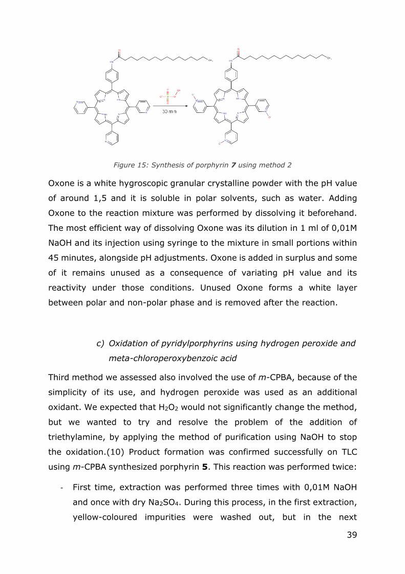
The steps we undertook in porphyrin N-oxidation using DMD are: in a round
flask, under room temperature, porphyrin 4 (0,017 g; 0,019 mmol; 1 eq.)
was dissolved in solvent CH2Cl2: CH3OH= 1:1 (10 ml) and stirred on a
magnetic stirrer and Oxone (0,180 g; 0,584 mmol; 25 eq.) was dissolved
separately in 0,01 M NaOH (1 ml). Acetone (3 ml) was added in the flask in
excess and the pH in the flask is set to approximately 7,0 using 0,01M
NaOH. Flask was set under nitrogen conditions and Oxone was injected
slowly into the flask over the course of 30 minutes and the pH value was
adjusted periodically.
After the addition of Oxone, the success of the oxidation was followed on
TLC. To extract the product from the organic layer, and remove the water
and hydrophilic impurities, filtration was conducted, followed by the
extraction with water (3x) and Na2SO4 (1x). The rest of the organic solvent
after the removal of water solvent was removed in vacuo. To purify the
product additionally, column chromatography was performed using the
solvent mixture CH2Cl2: CH3OH= 20:1. Porphyrin 7 (0,013 g; 76,47 %) was
obtained as a purple solid.
UV/ Vis spectroscopy analysis showed the presence of Soret band at 421
nm, with Q bands at next λmax/ nm: 514, 552, 591, 650. Extinction
coefficient was calculated: Ɛ= 0,19. Fluorescence measured at the
excitation wavelength of the Soret band gave two peaks at λmax/nm= 648,
716.
1H NMR was measured in solvent CD3Cl and next peaks were observed:
δ/ppm 9,09 (s; 3H; Py-2-H); 9,04- 8,87 (m; 8H; β-H); 8,72 (d; J= 5,04
Hz; 3H; Py-4-H); 8,18 (d, J= 6,60 Hz, 3H; Py-6-H); 8,12 (bd; J= 6,6 Hz;
2H; Ar-2,6-H); 8,01 (bd; 3H; Ar-3,5-H); 7,75 (d; J= 2,67 Hz; 3H; Py-6-H);
7,73 (s; 1H; amide N-H); 2,59 (t; J= 5,69 Hz; 2H; C2H2); 1,53 (t; J= 6,32
Hz; 2H; C3H2); 1,46- 1,26 (m; 24H; R-H: C4H2, C5H2, C6H2, C7H2, C8H2,

32
C9H2, C10H2, C11H2, C12H2, C13H2, C14H2, C15H2); 0,90 (t; J= 5,18 Hz; 3H; R-
C-H3); -2,94 (s; 2H; pyrrole N-H)

33
5. Results and discussion
5.1. Porphyrin synthesis
Modified Adler-Longo method was chosen for the condition reaction to
prepare starting material porphyrins, since it is easy to conduct, it is
relatively fast and gives us products whose impurities is possible to separate
on a column. Adler-Longo method gives better yields of porphyrins in
comparison to the Rothemund method.(30) Propionic acid under the reflux
conditions and the oxygen from the air creates the initial mixture of
porphyrins from pyrrole, 4- acetamidobenzaldehyde and 3-
pyridylbenzaldehyde; afterwards separated by the column
chromatography.(31) Acetamidobenzaldehyde gives 4-acetamidophenyl
group and 3-pyridylbenzaldehyde gives three pyridyl groups on the meso
positions on the porphyrin ring.
Two porphyrins were isolated from the reaction mixture:
- Symmetric porphyrin/ porphyrin 1: 5,10,15,20-tetrakis(3-
pyridyl)porphyrin
- Asymmetric porphyrin/ porphyrin 2: 5-(4-acetamydophenyl)-
10,15,20-tris(3-pyridyl)porphyrin
The yield of porphyrin 1 (Figure 6) was calculated to be 5,12 % and the
yield of porphyrin 2 (Figure 7) is 3,74 %.
Figure 6: Synthesis of porphyrin 1

34
Figure 7: Synthesis of porphyrin 2
Hydrolysis of the amide group on the porphyrin 2 is performed in order to
add longer alkyl chains onto the phenyl meso substituent of the porphyrin.
Hydrolysis is conducted using 18% hydrochloric acid under reflux and the
product with free amino group is then used for the addition of chains
deriving from dodecanoyl chloride or hexadecanoyl chloride/ palmitoyl
chloride.
The reaction of dodecanoyl chloride and palmitoyl chloride with amino group
on the porphyrin leads to formation of lipophilic porphyrins, which should
penetrate biological cells better then porphyrin 1 in PDT applications.
Metabolism of lipids in tumour cells is also affected by the use of conjugated
fatty acids onto the porphyrin structures. Yields in addition of the chains are
high: for the reaction with dodecanoyl chloride it amounts to 87,95%
(Figure 8) and with palmitoyl chloride amounts to 90,38 % (Figure 9). The
correct structure of each new porphyrin was confirmed using 1H NMR
spectroscopy. Obtained data for porphyrins 1 and 2 is in accordance with
published results.(9)

35
Figure 8: Synthesis of porphyrin 3
Figure 9: Synthesis of porphyrin 4
Both synthesized porphyrins (porphyrin 3 and 4) have higher polarity than
the compounds they originate from, which is visible from the polar surface
calculated in Chemaxon Marvin Sketch programme.
5.2. Methods of N-oxidation of pyridylporphyrins and
optimization
Four methods were assessed for N-oxidation of the synthesized
pyridylporphyrins, based on the literature research.
a) Oxidation of pyridylporphyrins using meta-
chloroperoxybenzoic acid
First method includes m-CPBA use as an oxidant of asymmetric porphyrins
(porphyrins 3 and 4). This method has been previously often used in the
laboratories and the parameters for a successful reaction were known. m-
CPBA acts as an oxidant in the reactions of N-oxidation with pyridyl

36
compounds, and the surplus of m-CPBA per reaction varies between 60%
and 85% per reaction. Regarding porphyrin compounds, oxidation is
successful if several equivalents of m-CPBA are added, resulting in relatively
good yields of porphyrin-N-oxides.(10) Syntheses of porphyrin 5 (Figure
10), porphyrin 6 (Figure 11) and porphyrin 7 (Figure 12) using m-CPBA are
depicted below.
Figure 10: Synthesis of porphyrin 5 using method 1
Figure 11: Synthesis of porphyrin 6 using method 1
Figure 12: Synthesis of porphyrin 7 using method 1

37
Following this oxidation, immediate purification on the column was
performed to remove the excess of oxidant, making the oxidation process
last up to 13 hours or more, to properly purify the porphyrin. Similar to
reported autoxidation of trimethylamine (32), triethylamine is autoxidised
in aqueous solutions, and this is why dry DCM is used. In the presence of a
strong oxidant such as m-CPBA, triethylamine is converted to triethylamine
oxide, which is very hard to remove and presents an obstacle for successful
use of our asymmetric porphyrins. This was a motivation to try other
methods of oxidation that could give pure products with less or no
impurities. Impurities were visible on the NMR spectra of porphyrins 3 and
4.
b) Oxidation of pyridylporphyrins using dimethyldioxirane
Second assessed method included oxidation using DMD, for which we took
several parameters into account during the in situ preparation of DMD from
Oxone. It took some time before the optimal method was created that could
work both on the porphyrin 1 and porphyrins 3 and 4. Primarily, decision
on which temperature will be used for the addition of the oxidant was
discussed. It was decided room temperature will be assessed first and in
the repeated oxidation procedure, 0 °C will be applied. The reaction was
optimized in 4 consecutive attempts, and it was found that the temperature
had no impact on the yield of the oxidised products (porphyrins 6 and 7).
Secondly, pH had to be set between 7,0 and 8,0, and 0,01M NaOH was
used, which was easy to prepare and to add into the reaction mixture. The
only problem encountered was the lack of pH meter to calculate the exact
pH of the coloured mixture. pH strips were used, but the colour of porphyrin
was, at times, too intensive to read out the most probable pH value with
certainty. Non-miscible layers had different pH values, which could cause
confusion if the pH strip was not in contact with the bottom, non-polar layer
where the porphyrin and DMD were dissolved. Upper, polar layer had

38
slightly higher pH value, even if the reaction was stirred, and it could cause
false reading of the pH strip to presume the pH value is around 7,5 when in
fact, it is lower and insufficient for Oxone conversion. We found that the pH
value is the most important parameter to be set for in situ generation of
DMD and adding more Oxone and/or ketone had no influence on the
increase in DMD formation. Acetone had to be added to assure the DMD
generation, and a surplus of acetone was not a big problem for the reaction
system. However, acetone changed the pH value of the reaction, so adding
acetone had to be followed by the adjustment of pH. Syntheses of porphyrin
5 (Figure 13), porphyrin 6 (Figure 14) and porphyrin 7 (Figure 15) using
DMD formed in situ are depicted below.
Figure 13: Synthesis of porphyrin 5 using method 2
Figure 14: Synthesis of porphyrin 6 using method 2
39
Figure 15: Synthesis of porphyrin 7 using method 2
Oxone is a white hygroscopic granular crystalline powder with the pH value
of around 1,5 and it is soluble in polar solvents, such as water. Adding
Oxone to the reaction mixture was performed by dissolving it beforehand.
The most efficient way of dissolving Oxone was its dilution in 1 ml of 0,01M
NaOH and its injection using syringe to the mixture in small portions within
45 minutes, alongside pH adjustments. Oxone is added in surplus and some
of it remains unused as a consequence of variating pH value and its
reactivity under those conditions. Unused Oxone forms a white layer
between polar and non-polar phase and is removed after the reaction.
c) Oxidation of pyridylporphyrins using hydrogen peroxide and
meta-chloroperoxybenzoic acid
Third method we assessed also involved the use of m-CPBA, because of the
simplicity of its use, and hydrogen peroxide was used as an additional
oxidant. We expected that H2O2 would not significantly change the method,
but we wanted to try and resolve the problem of the addition of
triethylamine, by applying the method of purification using NaOH to stop
the oxidation.(10) Product formation was confirmed successfully on TLC
using m-CPBA synthesized porphyrin 5. This reaction was performed twice:
- First time, extraction was performed three times with 0,01M NaOH
and once with dry Na2SO4. During this process, in the first extraction,
yellow-coloured impurities were washed out, but in the next
40
extractions, porphyrin was eluted in the polar phase and this led to
the loss of product. It was decided to limit the number of extractions
with NaOH.
- The products in the second reaction were only once washed with the
0,01 M NaOH and impurities were washed without porphyrins in the
polar phase. Two additional extractions were performed with water
and eventually excess water was removed with Na2SO4.
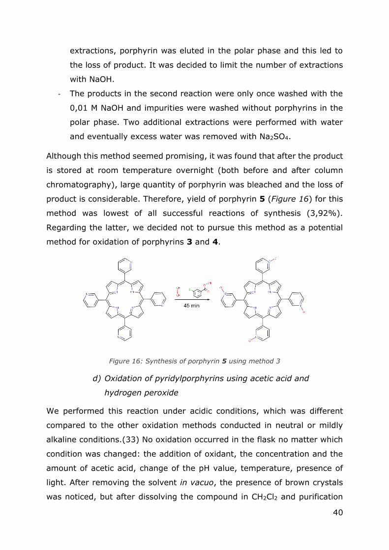
Although this method seemed promising, it was found that after the product
is stored at room temperature overnight (both before and after column
chromatography), large quantity of porphyrin was bleached and the loss of
product is considerable. Therefore, yield of porphyrin 5 (Figure 16) for this
method was lowest of all successful reactions of synthesis (3,92%).
Regarding the latter, we decided not to pursue this method as a potential
method for oxidation of porphyrins 3 and 4.
Figure 16: Synthesis of porphyrin 5 using method 3
d) Oxidation of pyridylporphyrins using acetic acid and
hydrogen peroxide
We performed this reaction under acidic conditions, which was different
compared to the other oxidation methods conducted in neutral or mildly
alkaline conditions.(33) No oxidation occurred in the flask no matter which
condition was changed: the addition of oxidant, the concentration and the
amount of acetic acid, change of the pH value, temperature, presence of
light. After removing the solvent in vacuo, the presence of brown crystals
was noticed, but after dissolving the compound in CH2Cl2 and purification

41
on the column, product was stretched, and TLC could not confirm any
presence of porphyrin. This method was abandoned as potential method for
oxidation (Figure 17).
Figure 17: Synthesis of porphyrin 5 using method 4
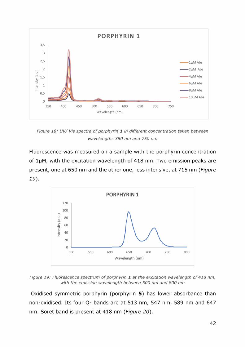
5.3. UV/Vis spectroscopy
Spectroscopic analysis was done to evaluate the absorbance of the
porphyrin compounds in the visible spectrum of electromagnetic radiation.
Soret band of the porphyrin 1 has a maximum at 418 nm, which is
consistent with the known value of Soret band at 420 nm. Four Q-bands
are spread between 450 and 670 nm, with maximum absorbances at 514
nm, 549 nm, 589 nm and 645 nm (Figure 18).
42
Figure 18: UV/ Vis spectra of porphyrin 1 in different concentration taken between
wavelengths 350 nm and 750 nm
Fluorescence was measured on a sample with the porphyrin concentration
of 1µM, with the excitation wavelength of 418 nm. Two emission peaks are
present, one at 650 nm and the other one, less intensive, at 715 nm (Figure
19).
Figure 19: Fluorescence spectrum of porphyrin 1 at the excitation wavelength of 418 nm,
with the emission wavelength between 500 nm and 800 nm
Oxidised symmetric porphyrin (porphyrin 5) has lower absorbance than
non-oxidised. Its four Q- bands are at 513 nm, 547 nm, 589 nm and 647
nm. Soret band is present at 418 nm (Figure 20).
0
0,5
1
1,5
2
2,5
3
3,5
350 400 450 500 550 600 650 700 750
Inte
nsi
ty (
a.u
.)
Wavelength (nm)
PORPHYRIN 1
1µM Abs
2µM Abs
4µM Abs
6µM Abs
8µM Abs
10µM Abs
0
20
40
60
80
100
120
500 550 600 650 700 750 800
Inte
nsi
ty (
a.u
.)
Wavelength (nm)
PORPHYRIN 1
43
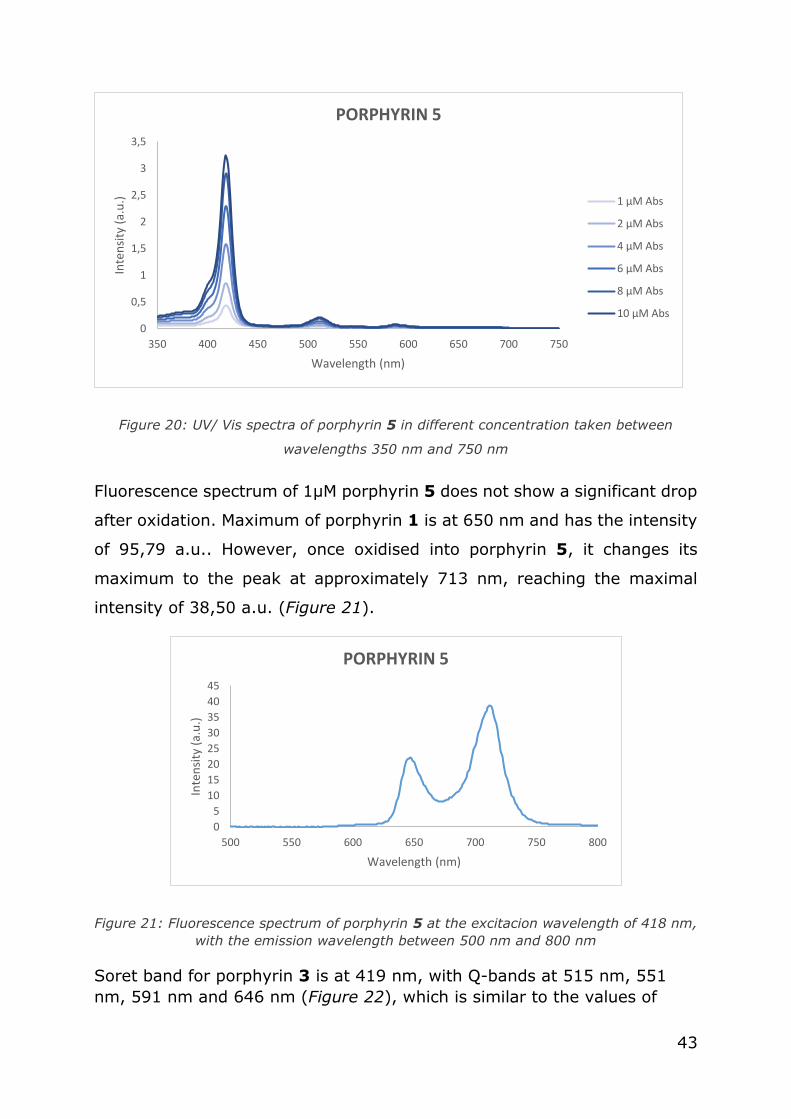
Figure 20: UV/ Vis spectra of porphyrin 5 in different concentration taken between
wavelengths 350 nm and 750 nm
Fluorescence spectrum of 1µM porphyrin 5 does not show a significant drop
after oxidation. Maximum of porphyrin 1 is at 650 nm and has the intensity
of 95,79 a.u.. However, once oxidised into porphyrin 5, it changes its
maximum to the peak at approximately 713 nm, reaching the maximal
intensity of 38,50 a.u. (Figure 21).
Figure 21: Fluorescence spectrum of porphyrin 5 at the excitacion wavelength of 418 nm,
with the emission wavelength between 500 nm and 800 nm
Soret band for porphyrin 3 is at 419 nm, with Q-bands at 515 nm, 551
nm, 591 nm and 646 nm (Figure 22), which is similar to the values of
0
0,5
1
1,5
2
2,5
3
3,5
350 400 450 500 550 600 650 700 750
Inte
nsi
ty (
a.u
.)
Wavelength (nm)
PORPHYRIN 5
1 µM Abs
2 µM Abs
4 µM Abs
6 µM Abs
8 µM Abs
10 µM Abs
0
5
10
15
20
25
30
35
40
45
500 550 600 650 700 750 800
Inte
nsi
ty (
a.u
.)
Wavelength (nm)
PORPHYRIN 5

44
porphyrin 1. Soret band for porphyrin 4 was detected at 419 nm, and Q
bands were detected at 517 nm, 554 nm, 593 nm and 648 nm (Figure
23).
Figure 22: UV/ Vis spectra of porphyrin 3 in different concentration taken between
wavelengths 350 nm and 750 nm
Figure 23: UV/ Vis spectra of porphyrin 4 in different concentration taken between
wavelengths 350 nm and 750 nm
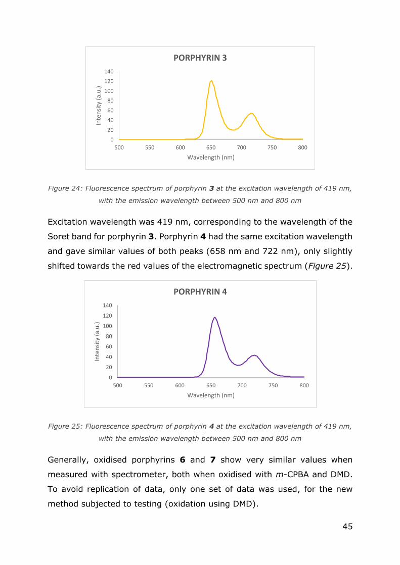
Fluorescence of the porphyrin 3 was spread between 600 and 800 nm,
like in symmetric porphyrin. Maximum peak was at 649 nm and the lower
peak was at 715 nm (Figure 24).
0
0,5
1
1,5
2
2,5
3
3,5
350 400 450 500 550 600 650 700 750
Inte
nsi
ty (
a.u
.)
Wavelength/ nm
PORPHYRIN 3
1 µM Abs
2 µM Abs
4 µM Abs
6 µM Abs
8 µM Abs
10 µM Abs
0
0,5
1
1,5
2
2,5
3
3,5
4
350 400 450 500 550 600 650 700 750
Inte
nsi
ty (
a.u
.)
Wavelength (nm)
PORPHYRIN 4
1µM Abs
2µM Abs
4µM Abs
6µM Abs
8µM Abs
10µM Abs
45
Figure 24: Fluorescence spectrum of porphyrin 3 at the excitation wavelength of 419 nm,
with the emission wavelength between 500 nm and 800 nm
Excitation wavelength was 419 nm, corresponding to the wavelength of the
Soret band for porphyrin 3. Porphyrin 4 had the same excitation wavelength
and gave similar values of both peaks (658 nm and 722 nm), only slightly
shifted towards the red values of the electromagnetic spectrum (Figure 25).
Figure 25: Fluorescence spectrum of porphyrin 4 at the excitation wavelength of 419 nm,
with the emission wavelength between 500 nm and 800 nm
Generally, oxidised porphyrins 6 and 7 show very similar values when
measured with spectrometer, both when oxidised with m-CPBA and DMD.
To avoid replication of data, only one set of data was used, for the new
method subjected to testing (oxidation using DMD).
0
20
40
60
80
100
120
140
500 550 600 650 700 750 800
Inte
nsi
ty (
a.u
.)
Wavelength (nm)
PORPHYRIN 3
0
20
40
60
80
100
120
140
500 550 600 650 700 750 800
Inte
nsi
ty (
a.u
.)
Wavelength (nm)
PORPHYRIN 4

46
Porphyrin 6`s Soret band has a maximum peak at 420 nm, with Q bands
at 514 nm, 550 nm, 589 nm and 646nm (Figure 26).
Figure 26: UV/ Vis spectra of porphyrin 6 in different concentration taken between
wavelengths 350 nm and 750 nm
Total intensity of porphyrin 6 is lower than the intensity of porphyrin 3.
Porphyrin 7 has a Soret band at 421 nm, with Q bands at 514 nm, 552 nm,
591 nm and 650 nm (Figure 27).
Figure 27: UV/ Vis spectra of porphyrin 7 in different concentration taken between
wavelengths 350 nm and 750 nm
0,00
0,50
1,00
1,50
2,00
2,50
350 400 450 500 550 600 650 700 750
Inte
nsi
ty/
a.u
.
Wavelength/ nm
PORPHYRIN 6
1µM Abs
2µM Abs
4 µM Abs
6µM Abs
8 µM Abs
10 µM Abs
0
0,5
1
1,5
2
350 400 450 500 550 600 650 700 750
Inte
nsi
ty (
a.u
.)
Wavelength (nm)
PORPHYRIN 7
1µM Abs
2µM Abs
4µM Abs
6µM Abs
8µM Abs
10µM Abs
47
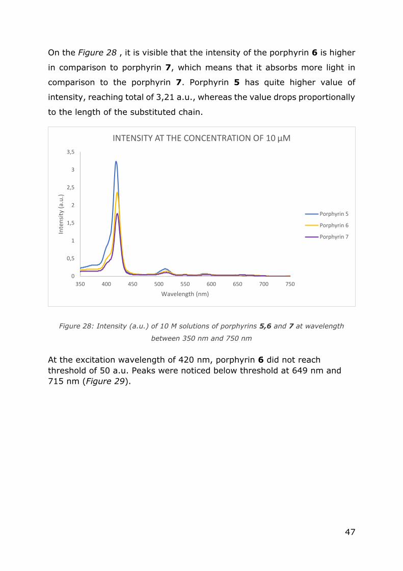
On the Figure 28 , it is visible that the intensity of the porphyrin 6 is higher
in comparison to porphyrin 7, which means that it absorbs more light in
comparison to the porphyrin 7. Porphyrin 5 has quite higher value of
intensity, reaching total of 3,21 a.u., whereas the value drops proportionally
to the length of the substituted chain.
Figure 28: Intensity (a.u.) of 10 M solutions of porphyrins 5,6 and 7 at wavelength
between 350 nm and 750 nm
At the excitation wavelength of 420 nm, porphyrin 6 did not reach
threshold of 50 a.u. Peaks were noticed below threshold at 649 nm and
715 nm (Figure 29).
0
0,5
1
1,5
2
2,5
3
3,5
350 400 450 500 550 600 650 700 750
Inte
nsi
ty (
a.u
.)
Wavelength (nm)
INTENSITY AT THE CONCENTRATION OF 10 µM
Porphyrin 5
Porphyrin 6
Porphyrin 7

48
Figure 29: Fluorescence spectrum of porphyrin 6 at the excitation wavelength of 420 nm,
with the emission wavelength between 500 nm and 800 nm
Fluorescence of the porphyrin 7 was also beneath the threshold of 50 a.u.,
and it was measured for the concentration of 1µM at the 421 nm
excitation wavelength. Measured maximums of the two peaks were at 648
nm and 716 nm (Figure 30).
Figure 30: Fluorescence spectrum of porphyrin 7 at the excitacion wavelength of 421 nm,
with the emission wavelength between 500 nm and 800 nm
Lower fluorescence of the porphyrins is noticed after the addition of the
chains to the structure of porphyrin 2, and the subsequent oxidation of the
compounds to porphyrins 6 and 7.
0
2
4
6
8
10
12
500 550 600 650 700 750 800
Inte
nsi
ty (
a.u
.)
Wavelength (nm)
PORPHYRIN 6
0
5
10
15
20
500 550 600 650 700 750 800
Inte
nsi
ty (
a.u
.)
Wavelength (nm)
PORPHYRIN 7

49
If the values of fluorescence intensity for porphyrins 5, 6 and 7 are
compared, same trend is noticeable as with the intensity of measured
absorbance: the values of fluorescence drop proportionally to the length of
the substituted chain on the porphyrin structure. Oxidised products have
lower intensities. The difference between the heights of two peaks for the
same porphyrin in oxidised and non-oxidised states vary: the difference is
inverted in favour of the peak between 700 nm and 750 nm for symmetric
porphyrin, whereas porphyrin 6 and 7 exhibit smaller growth of the same
peak but it does not pass the value of the peak between 630 nm and 670
nm (Figure 31).
Figure 31: Fluorescence spectra of oxidised porphyrins (porphyrins 5,6 and 7) at
wavelength between 500 nm and 800 nm
No difference was evident between spectroscopy results obtained from
compounds oxidised with m-CPBA and those oxidised with DMD. All graphs
gave one Soret band and four Q-bands at approximately the same
wavelength and of approximately the same intensity, making it impossible
to distinguish the difference between methods and measurements.
Ɛ were higher for porphyrins 3 and 4 than for porphyrins 6 and 7, while the
value of Ɛ remained similar for both porphyrins 1 and 5. This indicates that
the amount of light absorbed by a porphyrin is higher in non-oxidised
0
5
10
15
20
25
30
35
40
45
500 550 600 650 700 750 800
Inte
nsi
ty (
a.u
.)
Wavelength (nm)
FLUORESCENCE OF OXIDISED PORPHYRINS
Porphyrin 5
Porphyrin 6
Porphyrin 7

50
asymmetric porphyrins than in oxidised compounds. This is in correlation
with the UV/Vis spectra which shows that the oxidised compounds absorb
less visible light.
5.4. Nuclear Magnetic Resonance
NMR spectra were collected for porphyrins 3,4,5,6 and 7.
Two spectra were obtained for porphyrin 5, one representing method 2
(Figure 32), and the other one representing method 3 (Figure 33).
In the Figure 32, where DMD was used as an oxidant, it is visible that
impurities are present in the synthesized porphyrin 5, and the spectrum
peaks are visible at higher values than the ones in Figure 33. Impurities
have lower values quantitatively than hydrogen atoms present in the
molecule of porphyrin. In the Figure 33, peak of triethylamine oxide
hydrogens from CH3 group is present at approximately 3,35 ppm, and
hydrogens from CH2 groups are located between 3,35 and 3,90 ppm.
Mentioned hydrogens are present as an impurity from triethylamine that
could not be efficiently separated after the reaction of the N-oxidation using
m-CPBA was performed. Such peak is not visible when DMD is used as an
oxidising agent, which can, after the purification of porphyrin 5, give a clean
product.
In the Figures 34 and 35 aromatic hydrogens appear as broad doublets.
Shift is noticed towards lower values, and remarkable similarities are
noticed between both spectra (porphyrin 6 and 7) in the position of the
peaks. β-hydrogens are shifted towards higher values and peaks
representing impurities are in the following range: 7,37; 4,38; 2,98-2,91
(d). Water is present from 1,95-1,80 and CDCl3 is at 7,28 ppm. The peak
of methine protons might appear at 2,98 and 2,94 ppm on 1H NMR
spectrum, the peak of methine protons might appear, but it is unclear why
those peaks would not appear also on the spectra with oxidation method
1.(34)

51
Comparing the NMR spectra of porphyrin 3 and porphyrin 6, due to
proximity of hydrogen atoms both to oxygen molecules and nitrogen in the
pyridyl rings it is visible that the values (ppm) of hydrogen atoms are
slightly shifted towards the higher values. Oxidation of porphyrins 3 (Figure
36) and 4 (Figure 37) with DMD resulting in formation of porphyrins 6 and
7 gives products with less impurities, which is visible in the spectra of the
synthesized porphyrins (Figure 34 and 35). β-hydrogens are shifted right,
towards lower shift values of the spectrum, as oxygen added to the
molecule does not impact them as much as pyridyl hydrogens. Part of the
spectrum in which aliphatic hydrogens are detected does not change
significantly. Only the hydrogen on the nitrogen from the amide bond shifts
towards higher values.

52
Figure 32: NMR spectrum of porphyrin 5 oxidised using method 2 (DMD)
4H; Py-2-H
8H; β-H
4H; Py-4-H 4H; Py-6-H
4H; Py-5-H

53
Figure 33: NMR spectrum of porphyrin 5 oxidised using method 3 (m-CPBA and H2O2)
4H; Py-2-H
8H; β-H
4H; Py-4-H
4H; Py-6-H
4H; Py-5-H

54
Figure 34: NMR spectrum of porphyrin 6 oxidised using method 2 (DMD)
3H; Py-2-H
8H; β-H
3H; Py-4-H
4H; Py-6-H
4H; Py-5-H
2H; Ar-2,6-H
2H; Ar-3,5-H
1H; amide N-H
18H; R-H
2H; C2H2
3H; R-C-H3 2H; C3H2
2H; pyrrole N-H

55
Figure 35: NMR spectrum of porphyrin 7 oxidised using method 2 (DMD)
8H; β-H
3H; Py-4-H
4H; Py-6-H
2H; Ar-3,5-H
1H; amide N-H
18H; R-H
2H; C2H2 3H; R-C-H3
2H; C3H2
2H; pyrrole N-H
3H; Py-2-H
2H; Ar-2,6-H
4H; Py-5-H

56
Figure 36: NMR spectrum of porphyrin 3
4H; Py-2-H
4H; Py-4-H
8H; β-H
2H; Ar-2,6-H
4H; Py-6-H
2H; Ar-3,5-H
1H; amide
N-H
18H; R-H
2H; C2H2
3H; R-C-H3 2H; C3H2
2H; pyrrole N-H
4H; Py-5-H

57
Figure 37: NMR spectrum of porphyrin 4
4H; Py-2-H
4H; Py-4-H
8H; β-H
2H; Ar-2,6-H
4H; Py-6-H
4H; Py-5-H
2H; Ar-3,5-H
1H; amide N-H
18H; R-H
2H; C2H2 3H; R-C-H3
2H; C3H2
2H; pyrrole N-H

58
6. Conclusion
The aim of this work was to develop an alternative method for N-oxidation
of pyridylporphyrins, and to assess different oxidation methods currently
used.
Four different methods were assessed on the symmetric pyridylporphyrin:
- Method 1: Oxidation using m-CPBA (current standard)
- Method 2: Oxidation using DMD
- Method 3: Oxidation using m-CPBA and NaOH (H2O2 was used as an
oxidant)
- Method 4: Oxidation using CH3COOH
Out of all methods of oxidation, method using DMD stood out as the reaction
was relatively quick to perform and gave satisfying porphyrin yields
(70,00%). Yield of porphyrin 1 oxidised using m-CPBA was 65,00%. After
optimization of the reaction, it was concluded that the most important factor
for it was the pH value. Neither the fact that the reaction is performed in a
non-miscible reaction system, or that the acetone is added, did not cause
unsuccessful reaction. DMD was prepared successfully in situ from Oxone.
Reaction of oxidation using method 1 was faster in comparison to the
method 2, however, it required more steps and was more sensitive than the
method 1 to the conditions of the reaction. Purification of the product using
method 2 is significantly shorter (up to several hours shorter), due to the
fact triethylamine is not used in the reaction. Triethylamine oxidises when
added to reaction mixture with m-CPBA and porphyrin, to triethylamine
oxide, which sticks to the column and cannot be subsequently removed
from the product, as seen on the NMR spectrum (Figure 33). Method 2
involves different methods of purification (filtration, extraction, second
filtration, column chromatography), more than method 1 (extraction,
filtration, column chromatography).
59
We measured yields of porphyrin 6 and 7, and for porphyrin 6, yield using
method 1 was 70,59% and using method 2 was 72,72%, while for porphyrin
7, yield using method 1 was 71,93% and using method 2 was 76,47%.
Oxidation process in which m-CPBA and NaOH were used gave a poor yield
of oxidised symmetric porphyrin (3,92%), and no product was formed with
the use of CH3COOH. These reactions were not pursued for optimization as
a new potential method of oxidation.
Analysis of UV/Vis spectres of porphyrins oxidised by m-CPBA and by DMD
has shown no difference between the spectra; therefore UV/Vis analysis
was not a criterion in deciding which method is more convenient for
porphyrin oxidation.
NMR analysis was performed on porphyrins 3,4,5,6, and 7, leading to the
conclusion that the method 2 contributes less to the creation of total amount
of impurities, and those impurities can subsequently be removed. Method 1
gave expected results, giving less different impurities, but exhibited
impurities originating from triethylamine oxide, which cannot be removed
from the final product effectively. Part of the NMR spectrum in which
aromatic hydrogens can be found changes significantly by the oxidation of
porphyrins, shifting the spectrum peaks towards higher values. Aliphatic
hydrogens do not change significantly.
Considering all the outcomes, we conclude that the method 2 is better in
the terms of producing compounds with less impurities and in much shorter
period than method 1. However, economically, more resources are used,
which can be a negative side of this method.
More research should be dedicated to the optimization of a method 2,
making it more practical and reducing the need for repeated purification of
a product. Future steps would be to investigate and optimize methods 3 and
4 to give the wanted product and to be able to fully assess the methods of
porphyrin oxidation.

60
Literature
1. Smith Alison, Warren Martin, editor. An Historical Introduction to
Porphyrin and Chlorophyll Synthesis. In: Tetrapyrroles: Birth, Life and
Death [Internet]. Landes bioscience; 2009. p. 0–115.
2. Fischer, Hans. On haemin and the relationships between haemin and
chlorophyll [Internet]. 1930.
3. K. With Toben. A short history of porphyrins and the porphyrias. Int J
Biochem. 1979 Aug 17;11:189–200.
4. Maria da G.H. Vicente, Kevin M. Smith. Syntheses and Functionalizations of Porphyrin Macrocycles. Curr Org Synth.
2014;11(1):3–28.
5. Paul Rothemund. Porphyrin Studies. III. The Structure of the Porphine
Ring System. J Am Chem Soc. 1939 Jul 17;61(10):2912–5.
6. Parul Angrish. Syntheses of meso-substituted porphodimethenes and
poorphyrins with exocyclic ring systems [Internet] [Master thesis].
[State of Florida]: University of Florida; 2003.
7. F. Montanari, L. Casella. Metalloporphyrins Catalyzed Oxidation. Vol.
17. Italy: Springer- Science+ Business Media, B. V.; 1994.
8. Areej Merhi. Synthesis of new organic and organometallic Porphyrin
Assemblies for Optics [Internet] [phD]. [France]: l’Université
européenne de Bretagne; 2013.
9. Mateo Jelovica, Petra Grbčić, Martina Mušković, Mirela Sedić, Sandra Kraljević, Pavelić, Martin Lončarić, Nela Malatesti. In vitro
photodynamic activity of N-methylated and N-oxidised tripyridyl porphyrins with a long alkyl chain and their inhibitory activity in
sphingolipid metabolism. ChemMedChem. 13(4):360–72.
10. Jeffrey J. Posakony, Russell C. Pratt, Steven J. Rettig, Brian R. James,
Kirsten A. Skov. Porphyrins incorporating heterocyclic N-oxides: (oxidopyridyl)porphyrins, porphyrin-N-oxides, and a tirapazamine–
porphyrin conjugate. Can J Chem. 1999;77(2):182–98.
11. Robert W. Murray, Ramasubbu Jeyaraman. Dioxiranes: Synthesis and Reactions of Methyldioxiranes. J Org Chem. 1985 Apr 3;50(16):2847–
53.
12. Andrew Harned, editor. Nonnitrogenous Organocatalysis [Internet].
CRC Press; 2018.

61
13. Robert W. Murray, Megh Singh. Synthesis Of Epoxides Using
Dimethyldioxirane: Trans-Stilbene Oxide. Org Synth [Internet].
14. Marta Ferrer, Francisco Slinchez-Baeza, Angel Messeguer. On the
Preparation of Amine N-Oxides by Using Dioxiranes. Tetrahedron.
1997 Jul 31;53(46):15877–1588.
15. Victoria L. Waddington. Applications and mechanisms of dioxirane
oxidations [phD]. [United Kingdom]: Loughborough University; 2013.
16. Valerie Stone, Davita P. McTush, Al L. Baumstark, Pedro Vasquez.
Computational Studies of the Epoxidation of cis/trans-Alkenes by Dimethyldioxirane. Colon Acad Alliance Undergrad Res J.
2015;4(1):1–12.
17. Scott E. Denmark, David C. Forbes, David S. Hays, Jeffrey S. DePue,
Richard G. Wilde. Catalytic Epoxidation of Alkenes with Oxone. J Org
Chem. 1994 Jun 12;60(5).
18. Waldemar Adam, Rodrigo Paredes, Alexander K. Smerz, L. Angela Veloza. Probing for Steric and Electronic Effects in Diastereoselective
Dioxirane Epoxidations Compared to the Oxygen Transfer by Peroxy
Acids. Inst Org Chem Univ Wurzburg. 1996 Apr 11;547–51.
19. Stanisław Wołowiec, Lechoslaw Latos-Grazynski. Oxidation of Iron(III) Tetramesitylporphyrin with Dimethyldioxirane. Inorg Chem. 1997 Dec
26;37(12):2984–8.
20. Dennis E.J.G.J. Dolmans, Dai Fukumura, Rakesh K. Jain.
Photodynamic therapy for cancer. Nat Rev Cancer. 2003;3(5):380–7.
21. H. B. Gregorie, Jr., Edgar O. Horger, John L. Ward, James F. Green, Thomas Richards, H. C. Robertson, Jr., T. B. Stevenson.
Hematoporphyrin-Derivative Fluorescence in Malignant Neoplasms.
Ann Surg. 1968;167(6):820–8.
22. Photodynamic Therapy: An overview [Internet]. The Photochemistry
Portal. 2012.
23. Ian J. MacDonald, Thomas J. Dougherty. Basic principles of photodynamic therapy. J Porphyr Phthalocyanines. 2000 Aug
6;5(2):105–29.
24. Maurício da Silva Baptista, Jean Cadet, Paolo Di Mascio, Ashwini A.
Ghogare, Alexander Greer, Michael R. Hamblin, Carolina Lorente, Silvia Cristina Nunez, Martha Simões Ribeiro, Andrés H. Thomas,
Mariana Vignoni, Tania Mateus Yoshimura. Type I and II
Photosensitized Oxidation Reactions: Guidelines and Mechanistic
Pathways. Photochem Photobiol. 2018 Jan 7;93(4):912–9.

62
25. Cal State LA, Department of Chemistry and Biochemistry. Research -
What is Singlet Oxygen? [Internet]. Cal State LA.
26. Ethan D. Sternberg, David Dolphin. Porphyrin-based Photosensitizers
for Use in Photodynamic Therapy. Tetrahedron. 1998;54(447):4151–
202.
27. Ana P. Castano, Tatiana N. Demidova, Michael R. Hamblin.
Mechanisms in photodynamic therapy: Part three-Photosensitizer pharmacokinetics, biodistribution, tumor localization and modes of
tumor destruction. Photodiagnosis Photodyn Ther. 2014 Jul
23;2(2):91–106.
28. Aisling E. O’Connor, William M. Gallagher, Annette T. Byrne. Porphyrin and Nonporphyrin Photosensitizers in Oncology: Preclinical and Clinical
Advances in Photodynamic Therapy. Photochem Photobiol. 2009 Apr
16;85:1053–74.
29. Leanne B. Josefsen; Ross W. Boyle. Photodynamic Therapy and the Development of Metal-Based Photosensitisers. Met-Based Drugs. 2007
Oct 30;2008:24.
30. A.M. d`A. Rocha Gonsalves, Jorge M. T. B. Varejao, Mariette M.
Pereira. Some New Aspects Related to the Synthesis of meso-
Substituted Porphyrins. J Heterocycl Chem. 1991 May;28:635–40.
31. Alan D. Adler. A Simplified Synthesis for meso-Tetraphenylporphin. J
Org Chem. 32(2):476–476.
32. Michael J. Chen, John C. Linehan, Jerome W. Rathke. Autoxidation of
Trimethylamine in Aqueous Solutions. J Org Chem. 1989 Aug
30;55(10):3233–6.
33. Shaker Youssif. Recent trends in the chemistry of pyridine N-oxides.
Arch Org Chem. 2001 Dec 18;2001(1):242–68.
34. Alfred Hassner, Meir Stern, Hugo E. Gottlieb. Utility of a Polymeric Azide Reagent in the Formation of Di- and Triazidomethane. Their
NMR Spectra and the X-ray Structure of Derived Triazoles. J Org
Chem. 1989 Sep 18;55.
63
Table of Figures
Figure 1: meso-substitution position on porphyrin (left) and β-substitution
positions on porphyrin (right) ............................................................. 3
Figure 2: Structure of m-CPBA ........................................................... 6
Figure 3: Conversion of Oxone to DMD ................................................ 9
Figure 4: Scheme of modified Jablonski diagram(22)........................... 10
Figure 5: Type I and type II photoreactions (25) ................................ 12
Figure 6: Synthesis of porphyrin 1 .................................................... 33
Figure 7: Synthesis of porphyrin 2 .................................................... 34
Figure 8: Synthesis of porphyrin 3 .................................................... 35
Figure 9: Synthesis of porphyrin 4 .................................................... 35
Figure 10: Synthesis of porphyrin 5 using method 1 ........................... 36
Figure 11: Synthesis of porphyrin 6 using method 1 ........................... 36
Figure 12: Synthesis of porphyrin 7 using method 1 ........................... 36
Figure 13: Synthesis of porphyrin 5 using method 2 ........................... 38
Figure 14: Synthesis of porphyrin 6 using method 2 ........................... 38
Figure 15: Synthesis of porphyrin 7 using method 2 ........................... 39
Figure 16: Synthesis of porphyrin 5 using method 3 ........................... 40
Figure 17: Synthesis of porphyrin 5 using method 4 ........................... 41
Figure 18: UV/ Vis spectra of porphyrin 1 in different concentration taken
between wavelengths 350 nm and 750 nm ........................................ 42
Figure 19: Fluorescence spectrum of porphyrin 1 at the excitation
wavelength of 418 nm, with the emission wavelength between 500 nm
and 800 nm ................................................................................... 42
Figure 20: UV/ Vis spectra of porphyrin 5 in different concentration taken
between wavelengths 350 nm and 750 nm ........................................ 43
Figure 21: Fluorescence spectrum of porphyrin 5 at the excitacion
wavelength of 418 nm, with the emission wavelength between 500 nm
and 800 nm ................................................................................... 43
Figure 22: UV/ Vis spectra of porphyrin 3 in different concentration taken
between wavelengths 350 nm and 750 nm ........................................ 44

64
Figure 23: UV/ Vis spectra of porphyrin 4 in different concentration taken
between wavelengths 350 nm and 750 nm ........................................ 44
Figure 24: Fluorescence spectrum of porphyrin 3 at the excitation
wavelength of 419 nm, with the emission wavelength between 500 nm
and 800 nm ................................................................................... 45
Figure 25: Fluorescence spectrum of porphyrin 4 at the excitation
wavelength of 419 nm, with the emission wavelength between 500 nm
and 800 nm ................................................................................... 45
Figure 26: UV/ Vis spectra of porphyrin 6 in different concentration taken
between wavelengths 350 nm and 750 nm ........................................ 46
Figure 27: UV/ Vis spectra of porphyrin 7 in different concentration taken
between wavelengths 350 nm and 750 nm ........................................ 46
Figure 28: Intensity (a.u.) of 10 M solutions of porphyrins 5,6 and 7 at
wavelength between 350 nm and 750 nm .......................................... 47
Figure 29: Fluorescence spectrum of porphyrin 6 at the excitation
wavelength of 420 nm, with the emission wavelength between 500 nm
and 800 nm ................................................................................... 48
Figure 30: Fluorescence spectrum of porphyrin 7 at the excitacion
wavelength of 421 nm, with the emission wavelength between 500 nm
and 800 nm ................................................................................... 48
Figure 31: Fluorescence spectra of oxidised porphyrins (porphyrins 5,6
and 7) at wavelength between 500 nm and 800 nm ............................ 49
Figure 32: NMR spectrum of porphyrin 5 oxidised using method 2 (DMD)
.................................................................................................... 52
Figure 33: NMR spectrum of porphyrin 5 oxidised using method 3 (m-
CPBA and H2O2) .............................................................................. 53
Figure 34: NMR spectrum of porphyrin 6 oxidised using method 2 (DMD)
.................................................................................................... 54
Figure 35: NMR spectrum of porphyrin 7 oxidised using method 2 (DMD)
.................................................................................................... 55
Figure 36: NMR spectrum of porphyrin 3 ........................................... 56
Figure 37: NMR spectrum of porphyrin 4 ........................................... 57

65
Curriculum vitae
PERSONAL
INFORMATION
Pegi Pavletić
Corrado Ilijassich 3, 51000 Rijeka (Croatia)
0912266551
Skype pegi.pavletic.
WORK EXPERIENCE
01/07/2012–
01/09/2012
Pharmacist, specialization
Private health institution: Pharmacy Mira Đukić, Ana-
Marija Đukić,
Rijeka (Croatia)
18/12/2013–
18/12/2014
Pharmaceutical technician
ZU: Ljekarna Pablo, Baštijanova 26, Rijeka (Croatia)
01/04/2016–
01/06/2016
Demonstrator on subject: "Analitical chemistry"
University of Rijeka, Department of biotechnology,
Rijeka (Croatia)
01/08/2018–
Present
Student job: pharmacist
Jadran Pharmacy, Cres, Rijeka (Croatia)
01/12/2016–Present
Board for project tracking and evaluation for 2016.
Student Council at the University of Rijeka, Rijeka
(Croatia)
EDUCATION AND
TRAINING

66
01/09/2009–
20/05/2013
Pharmaceutical technician
Medical High School in Rijeka, Rijeka (Croatia)
01/10/2010–Present
Alumni representative for Croatia, Europe and
Euroasia in GLOBE project
Globe project, Rijeka (Croatia)
20/07/2017–01/09/2017
Certificate of completion Climate KIC EIT
European institute of innovation and technology
5 week course in climate change in Copenhagen, Zurich
and Wroclaw
01/10/2014–01/10/2017
Bacolumn chromatographyhelor in
Biotechnology and drug research
University of Rijeka, Rijeka (Hrvatska)
PERSONAL SKILLS
Mother tongue(s) Croatian
Foreign
language(s) UNDERSTANDING SPEAKING
WRITING
Listening Reading Spoken
interaction
Spoken
production
English C2 C2 C2 C2 C2
Levels: A1 and A2: Basic user - B1 and B2:
Independent user - C1 and C2: Proficient user Common European Framework of Reference for
Languages
ADDITIONAL
INFORMATION
Acolumn
chromatographyomplishments
- Stipend of the Ministry of war veterans in academic years
2017/2018 and 2018/2019
- Rector`s award for student activism