methods of determining drug absorption ppt
54
Seminar on Methods of determining absorption Tenzin Jangchuk 1 st M.Pharm Dept.- Pharmaceutics
-
Upload
tenzin-jangchuk -
Category
Education
-
view
2.462 -
download
1
Transcript of methods of determining drug absorption ppt

Seminar
on
Methods of determining
absorption
Tenzin Jangchuk
1st M.Pharm
Dept.- Pharmaceutics

Absorption
Absorption is defined as the process of movement
of unchanged drug from the site of administration
to the systemic circulation.

Different methods of determining drug
absorption are:
A.In vitro method
B.In Vivo method
C.In situ method
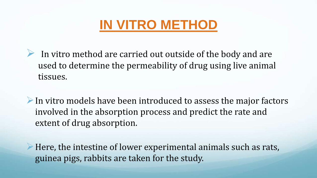
IN VITRO METHOD
In vitro method are carried out outside of the body and are used to determine the permeability of drug using live animal tissues.
In vitro models have been introduced to assess the major factors involved in the absorption process and predict the rate and extent of drug absorption.
Here, the intestine of lower experimental animals such as rats, guinea pigs, rabbits are taken for the study.

The different in vitro methods are: Physicochemical methods
1. Partition coefficient
2. Artificial membranes
3. Chromatographic retention indices4. Brush border membrane vesicles(BBMV)
5. Isolated intestinal cells
6. Tissue techniques:
a). Everted small intestinal sac technique
b) Everted sac modification
c) Circulation techniques
d) Everted intestinal ring or slice techniques
7. Diffusion cell method
8. Cell culture techniques

1. PARTITION COEFFICIENT
Partition coefficient between an oil and water phase, log P, is one of the easiest property of a drug molecule that can be determined.
It provides a measure of the lipophilicity of a molecule and can be used to predict to what extent it will cross the biological membrane.
eg. Octanol is selected as an oil phase as it has similar properties to biological membranes.
It’s important to note that log P does not take the degree of ionization into consideration and hence log D is used.

log D is the distribution coefficient where aqueous phase is at a particular pH and thus it takes into account the ionization of the molecule at this pH.
The log D measured at intestinal pH will give a much better idea about extent of drug permeability across GI membrane than log P.

2. ARTIFICAL MEMBRANES Artificial membranes are very useful in studying passive membrane
permeability as they are reproducible and are suitable for high throughput screening.
In this method, PAMPA model is used.
Parallel artificial membrane permeability assay (PAMPA)
PAMPA is a method which determines the permeability of substances from a donor compartment, through a lipid infused artificial membrane into an acceptor compartment.
The artificial membrane is like a phospholipid membranes supported by filter material.
It is prepared by pipetting a solution of lipids in an inert organic solvent on a supporting filter material which is placed on 96- well microtitre plate.

Procedure
test compound is added to the donor compartment in buffer solution of pH 7.4 and permeation takes place through artificial membrane into the acceptor compartment.
An aliquot is collected and the concentration of drug permeated is measured by suitable assay technique.
Fig. Parallel Artificial Membrane Permeation assay(PAMPA)

Uses
mainly used to determine the intestinal permeability
Prediction of passive transport
Trans-cellular permeability of drugs
Advantage
Used for screening large no. of compounds.
Provides information on solubility, lipophilicity and ionization status of a drug.
It is much lesser labor intensive than cell culture methods.
Disadvantage
They are based on approximation and oversimplification of the actual in vivo conditions.

A modification of this system is immobilized liposome chromatography(ILC) and on ILC, many compounds with same log P have been shown to demonstrate variable membrane partitioning based on their logs.
Liposomes , lipid bilayers produced from mixture of lipids are also useful in investigating passive diffusion of drugs through lipid membranes and also used to study passive absorption of many monocarboxylic acids.
A modified version of this is the filter-immobilized artificial membrane (filter-IAM) permeability assay.
This coupled with an instrument PSR4p (Permeability solubility retention, pION, USA) can be used for high throughput permeability screens

3. CHROMATOGRAPHIC RETENTION INDICES
Immobilized artificial membranes(IAM) chromatography along with physicochemical parameters is used for evaluation of passive intestinal absorption.
IAM packings are prepared by covalently immobilizing monolayers of membrane phospholipids to silica particles.
Micellar liquid chromatography(MLC) is also used for the prediction of passive drug absorption and in this system retention of drug mainly depends on hydrophobic, electronic and steric interactions.
In general, chromatographic techniques are easy in operation and have high analytical sensitivity.

4.BRUSH BORDER MEMBRANE VESICLES (BBMV)
A brush border is the name for the microvilli covered surface of simple cuboidal epithelium and simple columnar epithelial cells, found in the small intestine.
Both animal and human tissue can be used for this.
Procedure:
- intestinal tissues are treated with calcium chloride precipitation method using centrifugation.
- the pellets obtained after centrifugation is resuspended in buffer which results in the formation of vesicles.

Fig.1: Brush border membrane vesicles Fig.2: Structure of microvilli

Vesicles are mixed with drug in buffer solution and filtered after a period of time
The amount of drug taken up by the vesicles gives an account of drug absorption.
Advantage
useful for mechanic studies of drug absorption process.

5. USING ISOLATED INTESTINAL CELLS
Here, the small intestine is perfused with enzyme solutions that release the cells and the cells are treated with chelating agents or enzymes.
The freshly isolated cells are suspended in buffer solution.
At the time of experiment, the cells are separated, resuspended in buffer containing the drug under O2/CO2 and shaken well.
After a specific period of time, the cells are separated by filtration, extracted and drug absorbed is determined.
6.TISSUE TECHNIQUES
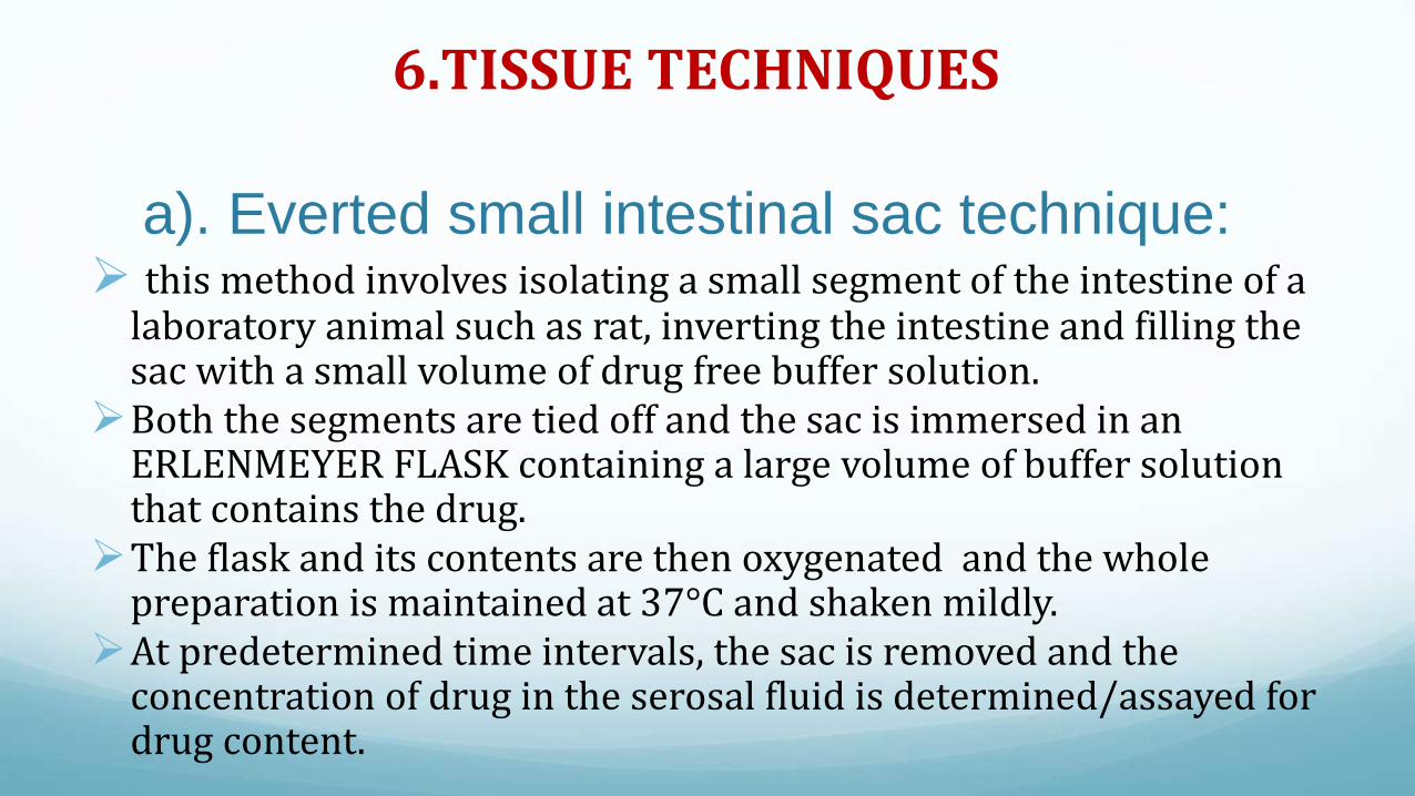
a). Everted small intestinal sac technique: this method involves isolating a small segment of the intestine of a
laboratory animal such as rat, inverting the intestine and filling the sac with a small volume of drug free buffer solution.
Both the segments are tied off and the sac is immersed in an ERLENMEYER FLASK containing a large volume of buffer solution that contains the drug.
The flask and its contents are then oxygenated and the whole preparation is maintained at 37°C and shaken mildly.
At predetermined time intervals, the sac is removed and the concentration of drug in the serosal fluid is determined/assayed for drug content.

Advantage-the epithelial cells of the mucosal surface are exposed directly to the oxygenated mucosal fluid.
-Prolongs the viability and integrity of the preparation after removal from the animal.
-Convenience and accuracy with respect to drug analysis.
Disadvantage
- Difficulty in obtaining more than one sample per intestinal segment

Fig. Everted sac technique

b)- EVERTED SAC MODIFICATION
In this method, the test animal is fasted for a period of 20-24 hrand water is allowed.
The animal is killed and the entire small intestine is everted. Segments, 5-15 cm in length are cut from a specific region of the intestine.
The distal end of the segment is tied and the proximal end is attached to the cannula. The segment is suspended in a mucosal solution which contains the drug.
A drug free buffer is then placed in the serosal compartment.

Fig. Everted sac modification
For determining the rate of drug transfer, the entire volume of serosal solution is removed from the sac at each time interval with the help of a syringe and replaced with fresh buffer solution.
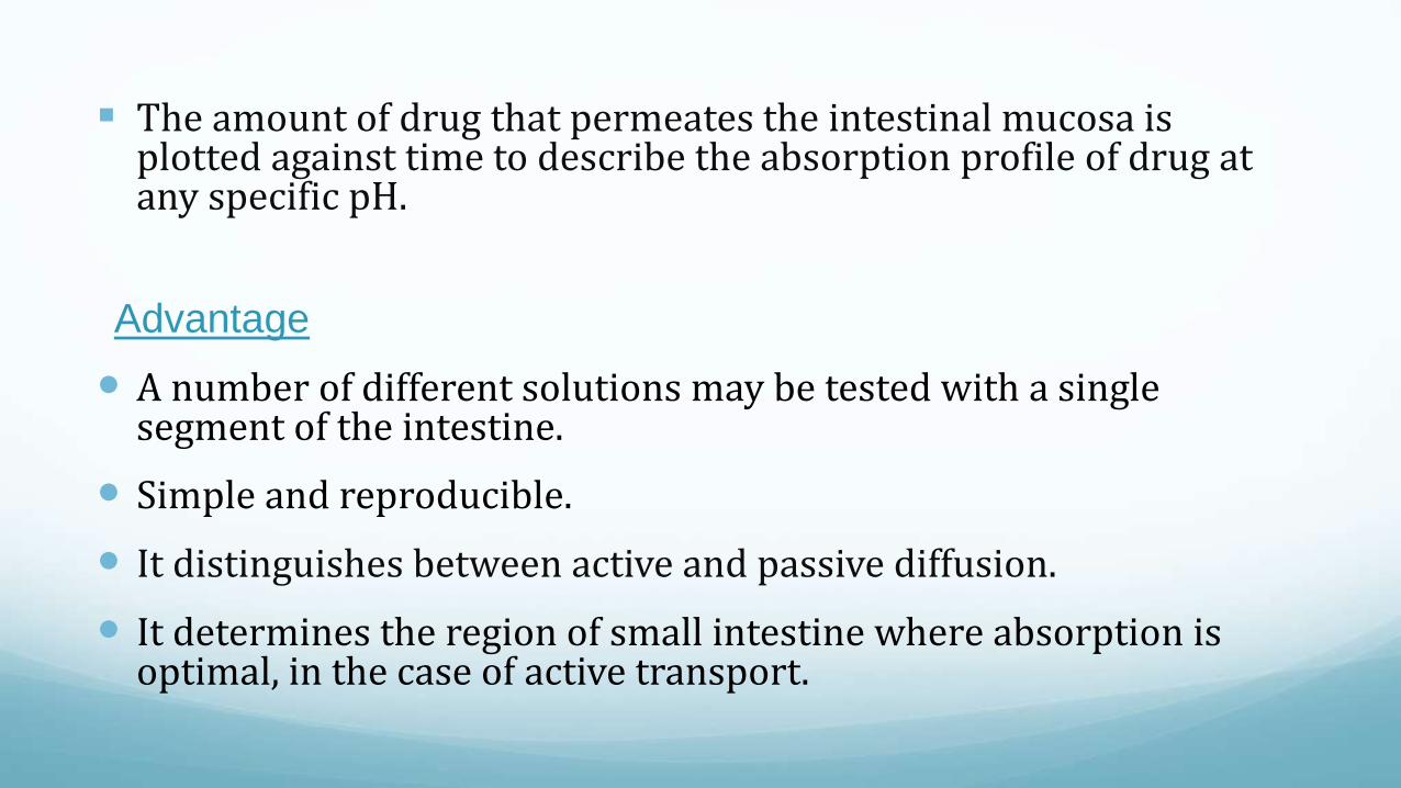
The amount of drug that permeates the intestinal mucosa is plotted against time to describe the absorption profile of drug at any specific pH.
Advantage
A number of different solutions may be tested with a single segment of the intestine.
Simple and reproducible.
It distinguishes between active and passive diffusion.
It determines the region of small intestine where absorption is optimal, in the case of active transport.

Used to study the effect of pH, surface active agents, complexation and enzymatic hydrolysis.
Disadvantage
- The intestinal preparation is removed from the animal as
well as from its normal blood supply. Under these conditions, the permeability characteristics of the membrane are significantly altered.
- The rate of transport of drug as determined from the everted sac technique, may be slower than in the intact animal.
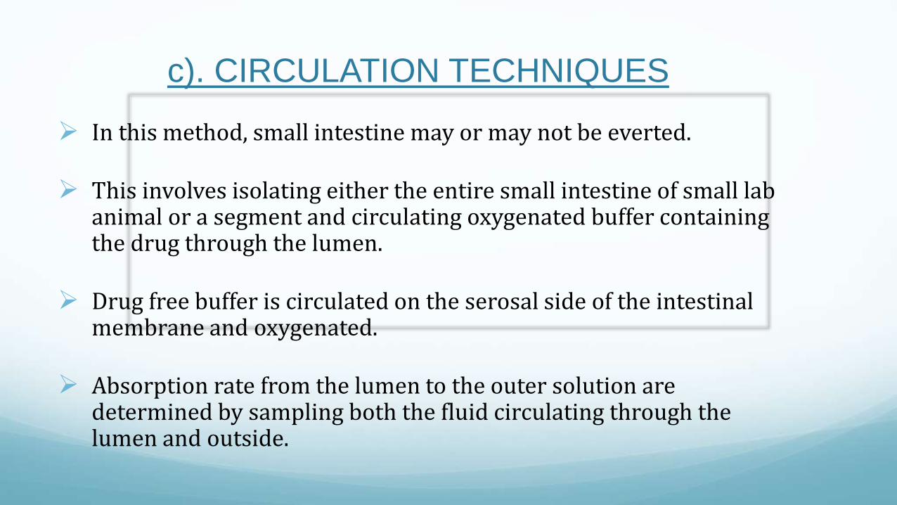
c). CIRCULATION TECHNIQUES
In this method, small intestine may or may not be everted.
This involves isolating either the entire small intestine of small lab animal or a segment and circulating oxygenated buffer containing the drug through the lumen.
Drug free buffer is circulated on the serosal side of the intestinal membrane and oxygenated.
Absorption rate from the lumen to the outer solution are determined by sampling both the fluid circulating through the lumen and outside.

Advantage
• This method is applicable to kinetic studies of the factors affecting drug absorption.
• Both surfaces are oxygenated.
• Eversion is not necessary.
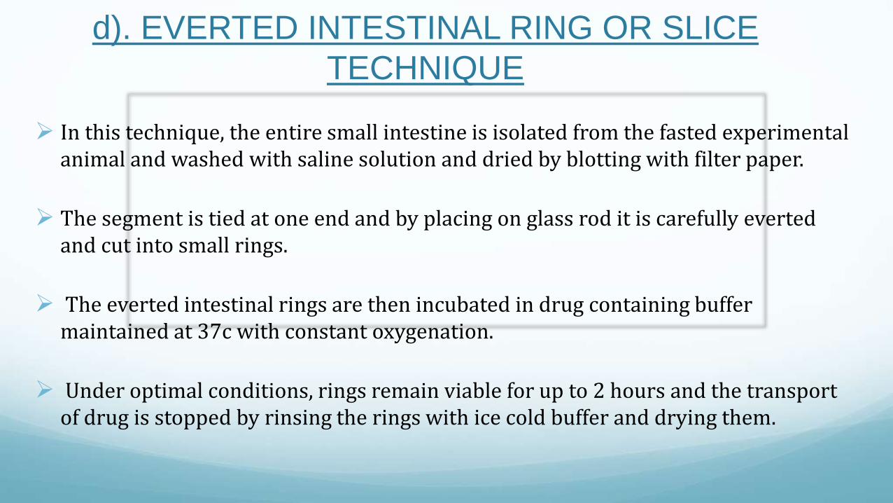
d). EVERTED INTESTINAL RING OR SLICE
TECHNIQUE
In this technique, the entire small intestine is isolated from the fasted experimental animal and washed with saline solution and dried by blotting with filter paper.
The segment is tied at one end and by placing on glass rod it is carefully evertedand cut into small rings.
The everted intestinal rings are then incubated in drug containing buffer maintained at 37c with constant oxygenation.
Under optimal conditions, rings remain viable for up to 2 hours and the transport of drug is stopped by rinsing the rings with ice cold buffer and drying them.

At selected time interval, the tissue slices are assayed for drug content and expressed as mol/gm/time.
Advantage• Simple and reproducible.• Kinetic studies can be performed.• Each animal can act as its own control as many rings can be prepared
from each segment of the intestine isolated.• Mechanism of drug absorption can be studied by changing the
experimental conditions.
Disadvantage• Extreme care is needed to maintain to viability of the tissue
throughout the experiment.• tissue needs to be disrupted completely for the determination of drug
contents, which complicates the assay procedure.• polarity of the absorption cannot be assayed.
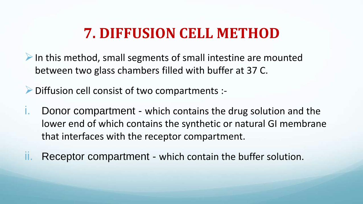
7. DIFFUSION CELL METHOD
In this method, small segments of small intestine are mounted between two glass chambers filled with buffer at 37 C.
Diffusion cell consist of two compartments :-
i. Donor compartment - which contains the drug solution and the lower end of which contains the synthetic or natural GI membrane that interfaces with the receptor compartment.
ii. Receptor compartment - which contain the buffer solution.

Fig. Diffusion cell for studying drug uptake from GIT

8. CELL CUTURE TECHNIQUESCell culture is the complex process by which cells are grown under
controlled conditions, generally outside their natural environment.
In this technique, differentiated cells of the intestine, originating from CaCo2 cells ( cells of carcinoma of colon) are placed on synthetic polycarbonate membrane previously treated with an appropriate material such as collagen which on incubation aids reproduction of cells while not retarding drug permeation characteristics.
These models are based on the assumption that passage of drugs across the intestinal epithelium is the main barrier for drugs to reach the circulation.
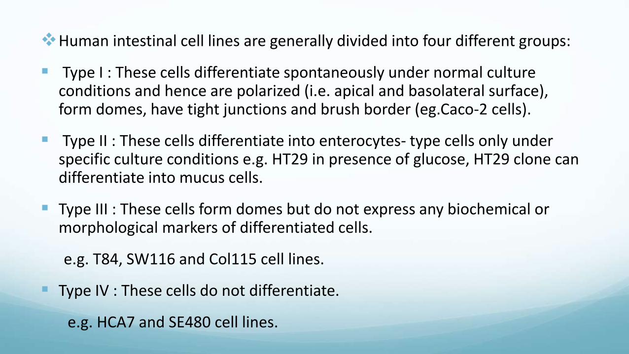
Human intestinal cell lines are generally divided into four different groups:
Type I : These cells differentiate spontaneously under normal culture conditions and hence are polarized (i.e. apical and basolateral surface), form domes, have tight junctions and brush border (eg.Caco-2 cells).
Type II : These cells differentiate into enterocytes- type cells only under specific culture conditions e.g. HT29 in presence of glucose, HT29 clone can differentiate into mucus cells.
Type III : These cells form domes but do not express any biochemical or morphological markers of differentiated cells.
e.g. T84, SW116 and Col115 cell lines.
Type IV : These cells do not differentiate.
e.g. HCA7 and SE480 cell lines.
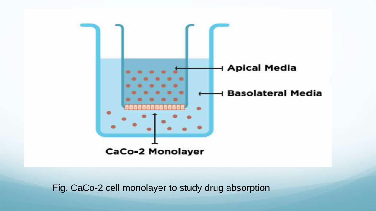
Fig. CaCo-2 cell monolayer to study drug absorption
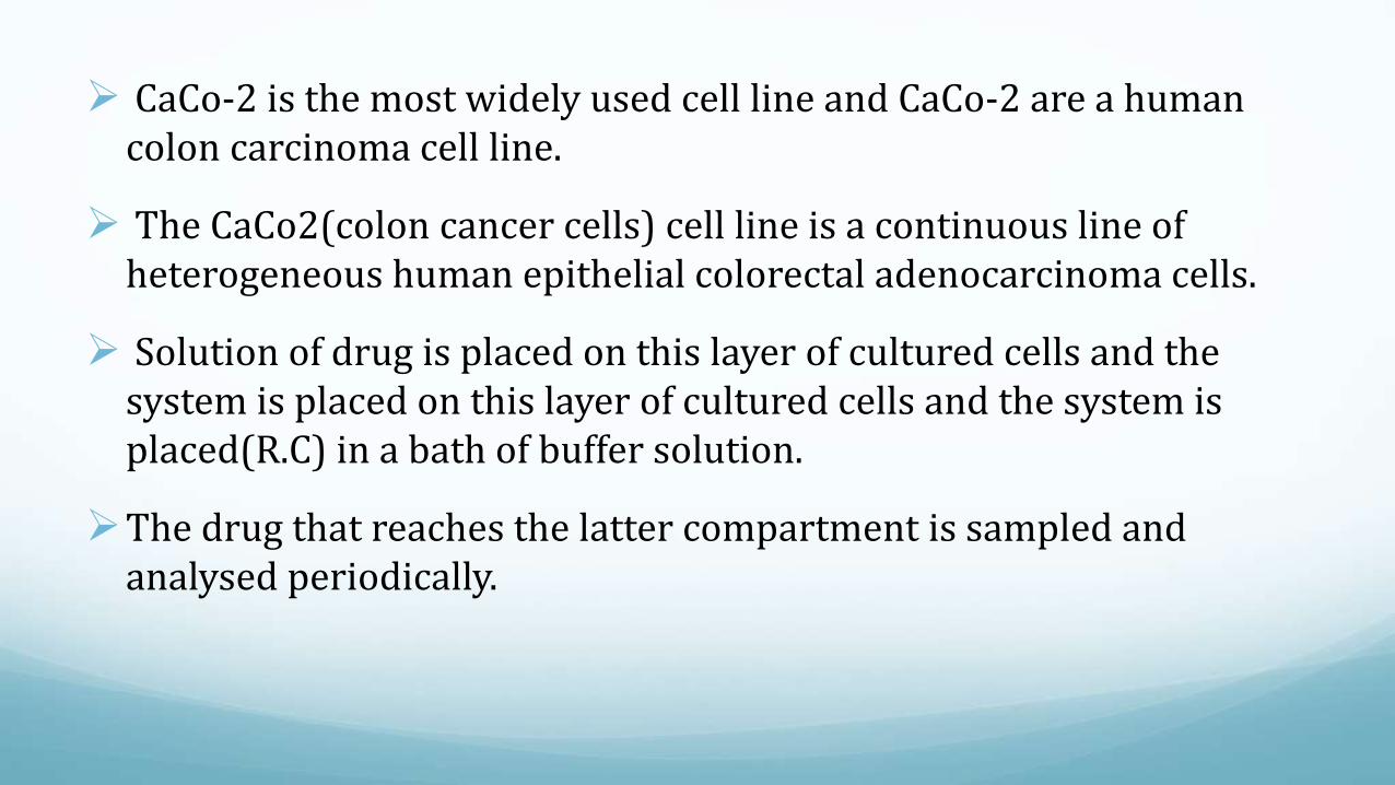
CaCo-2 is the most widely used cell line and CaCo-2 are a human colon carcinoma cell line.
The CaCo2(colon cancer cells) cell line is a continuous line of heterogeneous human epithelial colorectal adenocarcinoma cells.
Solution of drug is placed on this layer of cultured cells and the system is placed on this layer of cultured cells and the system is placed(R.C) in a bath of buffer solution.
The drug that reaches the latter compartment is sampled and analysed periodically.

CaCo2 cells express tight junctions, microvilli and a number of enzymes and transporters that are characteristic of enterocytes.
CaCo2 monolayer is widely used across the pharmaceutical industry as an in vitro model of the human small intestine mucosa to predict the absorption of orally administered drugs.
The correlation between the in vitro apparent permeability across CaCo2 monolayers and the in vivo fraction absorbed is well established.
Cell culture models have been employed in the screening of the intestinal permeability of libraries of new drug entities that have been generated through combinatorial chemistry and high throughput pharmacological screening.
IN VIVO METHODS
In vitro and in situ techniques gives us an idea about
absorption, but in vivo method gives us an idea about some
important factor that influence absorption such as gastric
emptying, intestinal motility and the effects of drugs on the GIT
can be determined.
The in vivo method can be classified into:
1. Direct method
2. Indirect method

Direct method
The drug levels in blood or urine is determined as a function of time. For this, a suitable sensitive reproducible analytical procedure should be developed to determine the drug in the biological fluid.
In this method, blank urine or blood sample is taken from the test animal before the experiment.
The test dosage form is administered to the animal and at appropriate intervals of time the blood or urine sample are collected and assayed for the drug content.
From the data, we can determine the rate and extent of drug absorption.
In this method, the experimental animal chosen should bear some resemble to man.
It is reported that pigs most closely resemble to man but are not used due to the handling problems.
The other animal that can be used are dogs, rabbits and rats.

Indirect method
When the measurement of drug concentration in blood or urine is difficult or not possible, but a sensitive method is available to test the activity, then absorption studies can be done by this indirect method.
In this method, pharmacological response of the drug is related to the amount of drug in the body.
The response is determined after the administration of a test dosage form, LD 50 appears to be dependent on the rate of the absorption of drug.
IN SITU METHOD
It simulates the in vivo conditions for drug absorption and are based on perfusion of a segment of GIT by drug solution and determination of amount of drug diffused through it.
In situ refers to those method in which the animal’s blood supply remains intact in which the rate of absorption determined from these methods may be more realistic than those determined from in vitro techniques.
These models are powerful tools to study the mechanistic aspects of these important process.
Acts as a bridge between in vivo and in vitro methods.

I- Absorption of drug from small intestine
Absorption of drug from small intestine is described in two methods:
i. Perfusion technique:
Doluisio method
Single pass perfusion method
ii. Intestinal loop techniques

1. Perfusion technique
a) Doluisio method
In this technique, adult male rats are fasted for about 16-24 hours before the experiments.
A midline abdominal incision is made and the intestinal segment to be perfused is identified.
A L shaped glass inlet cannula is secured into the segment and the outlet cannula is placed approx. 15-50cm from the inlet cannula. These are secured with sutures and the intestine is replaced in the abdominal cavity.
Dissected rats are connected by means of tubing to syringes of capacity of 10-30ml.
After washing the intestinal segment with normal saline , the syringe is filled with a solution of drug which is delivered to the intestinal segment and is then collected in the second syringe and analysed for the drug content.
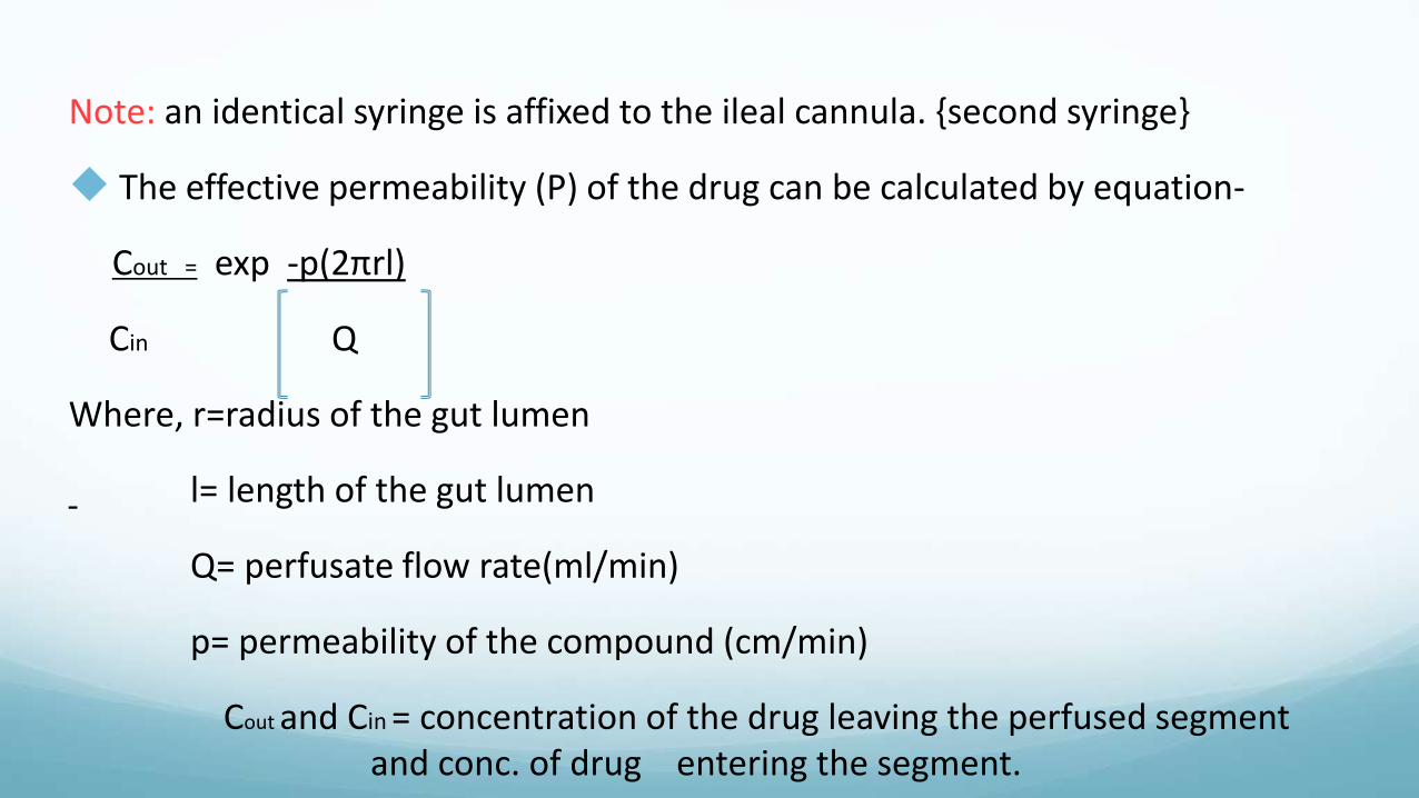
Note: an identical syringe is affixed to the ileal cannula. {second syringe}
The effective permeability (P) of the drug can be calculated by equation-
Cout = exp -p(2πrl)
Cin Q
Where, r=radius of the gut lumen
l= length of the gut lumen
Q= perfusate flow rate(ml/min)
p= permeability of the compound (cm/min)
Cout and Cin = concentration of the drug leaving the perfused segment and conc. of drug entering the segment.
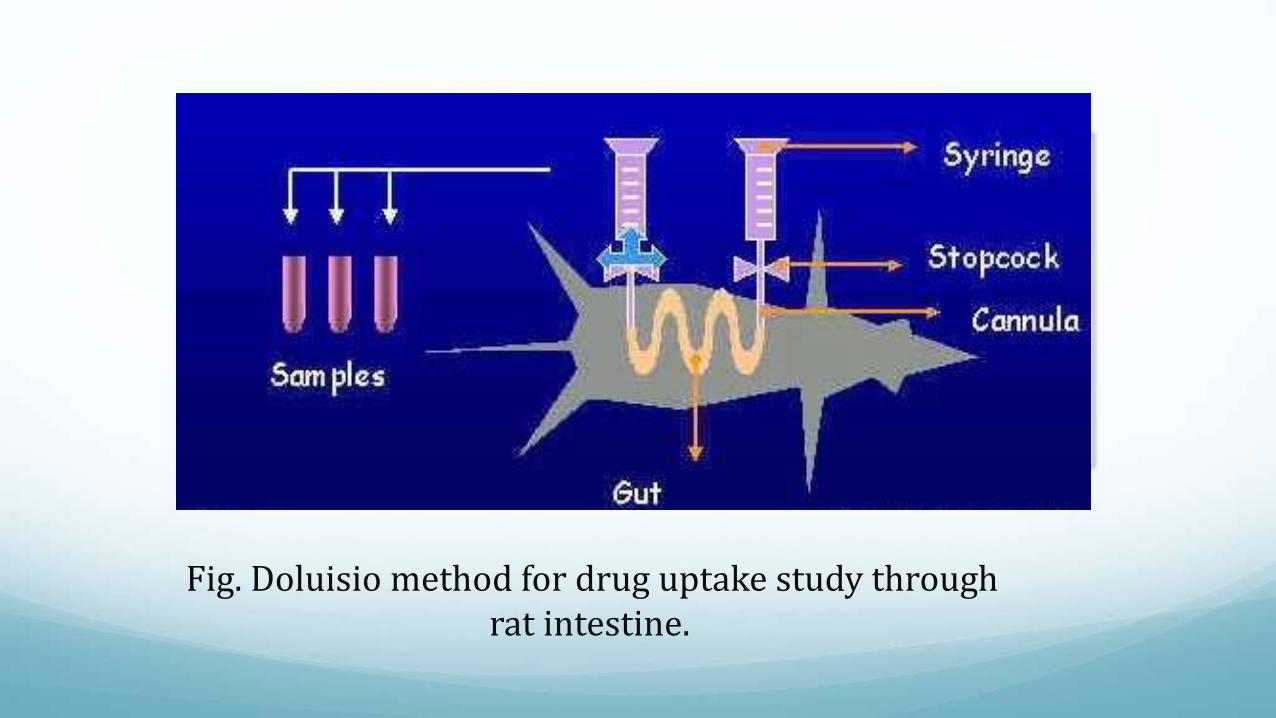
Fig. Doluisio method for drug uptake study through rat intestine.

b. Single pass perfusion
In this technique, the drug solution passes through the intestinal segment just once.
This technique is superior to Doluisio method in that precise adjustment of hydrodynamic conditions that can influence blood circulation and puts stress on intestinal wall can be controlled.
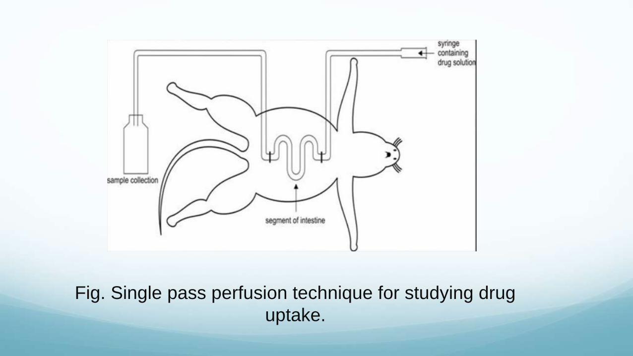
Fig. Single pass perfusion technique for studying drug
uptake.

2. Intestinal loop technique
Here, single or multiple intestinal loops are used for studying the absorption.
Adult male rats are fasted before the experiment under the anaesthesia and abdominal midline incision is made and the small intestine is exposed.
A proximal ligature is placed around the intestine from the pylorus and a distal ligature is secured at a distance of 4’’ distal to the proximal ligature.

The drug solution is introduced into the lumen of the loop by means of a syringe which is secured by the proximal ligature.
After injection, the needle is removed, the proximal ligature is tightened and the loop is replaced into the abdominal cavity and the incision is closed.
After a specific time, the animal is sacrificed, the intestinal loop is rapidly excised and homogenized and the amount of unabsorbed is determined.

Advantage Simple and reproducible
Disadvantage Only one sample can be obtained from the experimental
animal.

II- Absorption from the stomach
Fasted adult male rats are anaesthetized, stomach is exposed and the cardiac end is ligated.
An incision is made in the pylorus in which the cannula is introduced and ligated.
The lumen is washed several times with saline and subsequently with 0.1N HCl solution containing 0.15M NaCl.
The drug solution of known concentration is introduced into the stomach and after 1 hour, the solution is removed from the gastric pouch and assayed for drug content.

The gastric pouch may also be homogenized and analysed for drug.
In order to obtain number of samples as a function of time, the
following modifications is done:
Drug solution is introduced into the gastric lumen via the cardiac cannula.
A polyethylene tubing connected to a 2ml syringe equipped with a three way stopcock is attached to the duodenal L shaped glass cannula.
At specific intervals, the stomach contents are sampled by withdrawing the solution into the syringe.
Advantage:
has the capability of absorbing the most acidic drug form and the very weakly basic drugs.
E.g. salicylic acid, aspirin, thiopental, and antipyrine, which are un-dissociated in the acidic gastric contents were readily absorbed.
Disadvantage:
In situ techniques equate absorption with the loss of drug from the GI lumen.
A drug is accumulated or metabolized in the gut wall and one will get an overestimate of the amount of drug absorbed.
Reference:
o Biopharmaceutics and pharmacokinetics
- Dr. Shobha Rani R.
o Biopharmaceutics and pharmacokinetics
- DM Brahmankar and Sunil B. Jaiswal
- J.S Kulkarni, A.P. Pawar, V.P. Shedbalkar

Important questions:
1. In vitro - in vivo correlation
2. Explain in vitro methods for determination of drug absorption.