Methods for the Isolation, Differentiation, and ... · Methods for the Isolation, Differentiation,...
8
Methods for the Isolation, Differentiation, and Characterization of CD4 + T Cell Subsets Characterize Differentiate Isolate PBMC Splenocytes
Transcript of Methods for the Isolation, Differentiation, and ... · Methods for the Isolation, Differentiation,...
Methods for the Isolation, Differentiation, and Characterization of CD4+ T Cell Subsets
Characterize
Differentiate
Isolate
PBMC Splenocytes
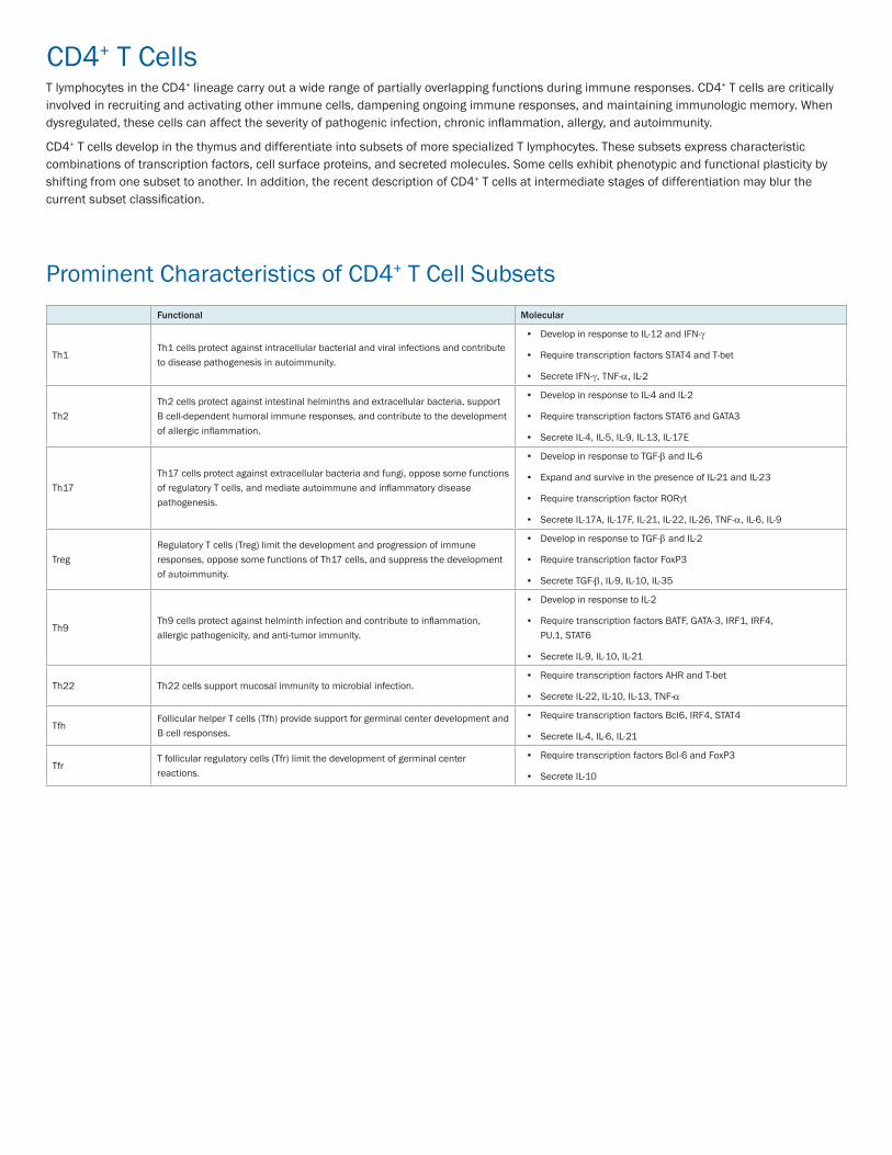
CD4+ T CellsT lymphocytes in the CD4+ lineage carry out a wide range of partially overlapping functions during immune responses. CD4+ T cells are critically involved in recruiting and activating other immune cells, dampening ongoing immune responses, and maintaining immunologic memory. When dysregulated, these cells can affect the severity of pathogenic infection, chronic inflammation, allergy, and autoimmunity.
CD4+ T cells develop in the thymus and differentiate into subsets of more specialized T lymphocytes. These subsets express characteristic combinations of transcription factors, cell surface proteins, and secreted molecules. Some cells exhibit phenotypic and functional plasticity by shifting from one subset to another. In addition, the recent description of CD4+ T cells at intermediate stages of differentiation may blur the current subset classification.
Prominent Characteristics of CD4+ T Cell SubsetsFunctional Molecular
Th1Th1 cells protect against intracellular bacterial and viral infections and contribute to disease pathogenesis in autoimmunity.
• Develop in response to IL-12 and IFN-g
• Require transcription factors STAT4 and T-bet
• Secrete IFN-g, TNF-a, IL-2
Th2Th2 cells protect against intestinal helminths and extracellular bacteria, support B cell-dependent humoral immune responses, and contribute to the development of allergic inflammation.
• Develop in response to IL-4 and IL-2
• Require transcription factors STAT6 and GATA3
• Secrete IL-4, IL-5, IL-9, IL-13, IL-17E
Th17Th17 cells protect against extracellular bacteria and fungi, oppose some functions of regulatory T cells, and mediate autoimmune and inflammatory disease pathogenesis.
• Develop in response to TGF-b and IL-6
• Expand and survive in the presence of IL-21 and IL-23
• Require transcription factor RORgt
• Secrete IL-17A, IL-17F, IL-21, IL-22, IL-26, TNF-a, IL-6, IL-9
Treg Regulatory T cells (Treg) limit the development and progression of immune responses, oppose some functions of Th17 cells, and suppress the development of autoimmunity.
• Develop in response to TGF-b and IL-2
• Require transcription factor FoxP3
• Secrete TGF-b, IL-9, IL-10, IL-35
Th9Th9 cells protect against helminth infection and contribute to inflammation, allergic pathogenicity, and anti-tumor immunity.
• Develop in response to IL-2
• Require transcription factors BATF, GATA-3, IRF1, IRF4, PU.1, STAT6
• Secrete IL-9, IL-10, IL-21
Th22 Th22 cells support mucosal immunity to microbial infection.• Require transcription factors AHR and T-bet
• Secrete IL-22, IL-10, IL-13, TNF-a
TfhFollicular helper T cells (Tfh) provide support for germinal center development and B cell responses.
• Require transcription factors Bcl6, IRF4, STAT4
• Secrete IL-4, IL-6, IL-21
TfrT follicular regulatory cells (Tfr) limit the development of germinal center reactions.
• Require transcription factors Bcl-6 and FoxP3
• Secrete IL-10
+
Undesired cells Desired cells
CD3-
PE
100
101
100
102
103
104
101 102 103 104
CD4-FITC
A
CD3-
PE
100
101
100
102
103
104
101 102 103 104
CD4-FITC
B
CD3-
PE
100
101
100
102
103
104
101 102 103 104
CD4-FITC
A
CD3-
PE
100
101
100
102
103
104
101 102 103 104
CD4-FITC
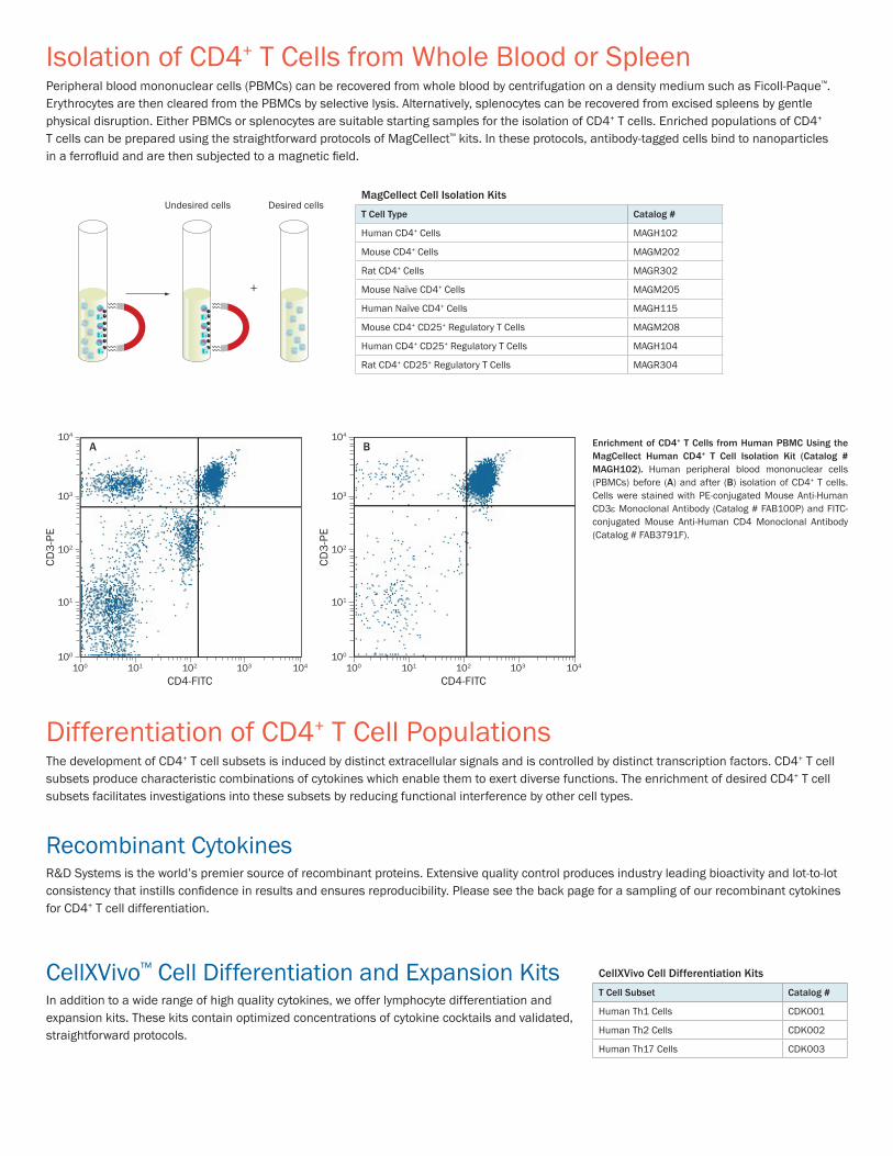
B Enrichment of CD4+ T Cells from Human PBMC Using the MagCellect Human CD4+ T Cell Isolation Kit (Catalog # MAGH102). Human peripheral blood mononuclear cells (PBMCs) before (A) and after (B) isolation of CD4+ T cells. Cells were stained with PE-conjugated Mouse Anti-Human CD3e Monoclonal Antibody (Catalog # FAB100P) and FITC-conjugated Mouse Anti-Human CD4 Monoclonal Antibody (Catalog # FAB3791F).
CellXVivo™ Cell Differentiation and Expansion KitsIn addition to a wide range of high quality cytokines, we offer lymphocyte differentiation and expansion kits. These kits contain optimized concentrations of cytokine cocktails and validated, straightforward protocols.
Isolation of CD4+ T Cells from Whole Blood or SpleenPeripheral blood mononuclear cells (PBMCs) can be recovered from whole blood by centrifugation on a density medium such as Ficoll-Paque™. Erythrocytes are then cleared from the PBMCs by selective lysis. Alternatively, splenocytes can be recovered from excised spleens by gentle physical disruption. Either PBMCs or splenocytes are suitable starting samples for the isolation of CD4+ T cells. Enriched populations of CD4+ T cells can be prepared using the straightforward protocols of MagCellect™ kits. In these protocols, antibody-tagged cells bind to nanoparticles in a ferrofl uid and are then subjected to a magnetic fi eld.
Differentiation of CD4+ T Cell PopulationsThe development of CD4+ T cell subsets is induced by distinct extracellular signals and is controlled by distinct transcription factors. CD4+ T cell subsets produce characteristic combinations of cytokines which enable them to exert diverse functions. The enrichment of desired CD4+ T cell subsets facilitates investigations into these subsets by reducing functional interference by other cell types.
Recombinant CytokinesR&D Systems is the world’s premier source of recombinant proteins. Extensive quality control produces industry leading bioactivity and lot-to-lot consistency that instills confi dence in results and ensures reproducibility. Please see the back page for a sampling of our recombinant cytokines for CD4+ T cell differentiation.
CellXVivo Cell Differentiation Kits
T Cell Subset Catalog #
Human Th1 Cells CDK001
Human Th2 Cells CDK002
Human Th17 Cells CDK003
MagCellect Cell Isolation Kits
T Cell Type Catalog #
Human CD4+ Cells MAGH102
Mouse CD4+ Cells MAGM202
Rat CD4+ Cells MAGR302
Mouse Naïve CD4+ Cells MAGM205
Human Naïve CD4+ Cells MAGH115
Mouse CD4+ CD25+ Regulatory T Cells MAGM208
Human CD4+ CD25+ Regulatory T Cells MAGH104
Rat CD4+ CD25+ Regulatory T Cells MAGR304
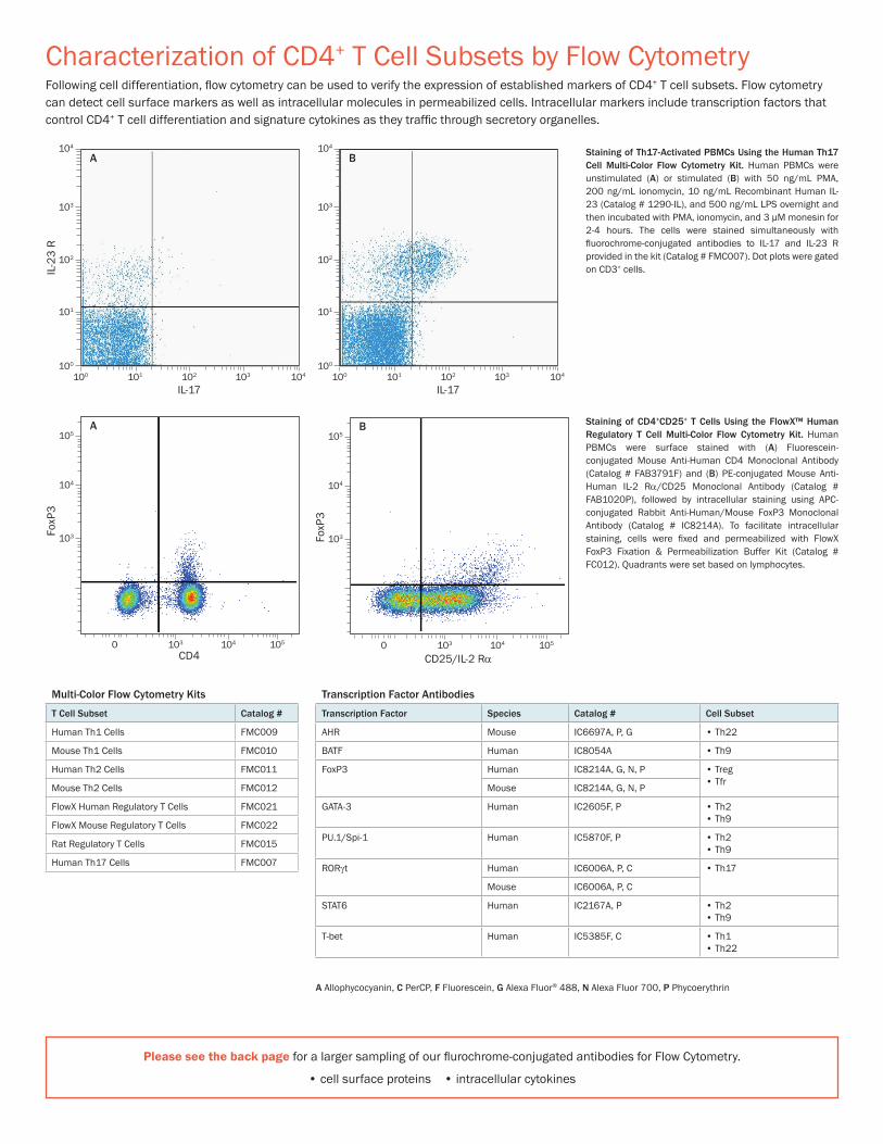
Characterization of CD4+ T Cell Subsets by Flow CytometryFollowing cell differentiation, fl ow cytometry can be used to verify the expression of established markers of CD4+ T cell subsets. Flow cytometry can detect cell surface markers as well as intracellular molecules in permeabilized cells. Intracellular markers include transcription factors that control CD4+ T cell differentiation and signature cytokines as they traffi c through secretory organelles.
IL-2
3 R
100
101
100
102
103
104
101 102 103 104
IL-17
A
100
101
100
102
103
104
101 102 103 104
IL-17
B
IL-2
3 R
100
101
100
102
103
104
101 102 103 104
IL-17
A
100
101
100
102
103
104
101 102 103 104
IL-17
B
105
104
103
0 103 104 105
CD25/IL-2 RαCD4
A
FoxP
3
FoxP
3
105
104
103
0 103 104 105
B105
104
103
0 103 104 105
CD25/IL-2 RαCD4
A
FoxP
3
FoxP
3
105
104
103
0 103 104 105
B
Staining of Th17-Activated PBMCs Using the Human Th17 Cell Multi-Color Flow Cytometry Kit. Human PBMCs were unstimulated (A) or stimulated (B) with 50 ng/mL PMA, 200 ng/mL ionomycin, 10 ng/mL Recombinant Human IL-23 (Catalog # 1290-IL), and 500 ng/mL LPS overnight and then incubated with PMA, ionomycin, and 3 μM monesin for 2-4 hours. The cells were stained simultaneously with fl uorochrome-conjugated antibodies to IL-17 and IL-23 R provided in the kit (Catalog # FMC007). Dot plots were gated on CD3+ cells.
Staining of CD4+CD25+ T Cells Using the FlowX™ Human Regulatory T Cell Multi-Color Flow Cytometry Kit. Human PBMCs were surface stained with (A) Fluorescein-conjugated Mouse Anti-Human CD4 Monoclonal Antibody (Catalog # FAB3791F) and (B) PE-conjugated Mouse Anti-Human IL-2 Ra/CD25 Monoclonal Antibody (Catalog # FAB1020P), followed by intracellular staining using APC-conjugated Rabbit Anti-Human/Mouse FoxP3 Monoclonal Antibody (Catalog # IC8214A). To facilitate intracellular staining, cells were fi xed and permeabilized with FlowX FoxP3 Fixation & Permeabilization Buffer Kit (Catalog # FC012). Quadrants were set based on lymphocytes.
Multi-Color Flow Cytometry Kits
T Cell Subset Catalog #
Human Th1 Cells FMC009
Mouse Th1 Cells FMC010
Human Th2 Cells FMC011
Mouse Th2 Cells FMC012
FlowX Human Regulatory T Cells FMC021
FlowX Mouse Regulatory T Cells FMC022
Rat Regulatory T Cells FMC015
Human Th17 Cells FMC007
Transcription Factor Antibodies
Transcription Factor Species Catalog # Cell Subset
AHR Mouse IC6697A, P, G • Th22
BATF Human IC8054A • Th9
FoxP3 Human IC8214A, G, N, P • Treg• Tfr
Mouse IC8214A, G, N, P
GATA-3 Human IC2605F, P • Th2• Th9
PU.1/Spi-1 Human IC5870F, P • Th2 • Th9
RORgt Human IC6006A, P, C • Th17
Mouse IC6006A, P, C
STAT6 Human IC2167A, P • Th2• Th9
T-bet Human IC5385F, C • Th1 • Th22
Please see the back page for a larger sampling of our fl urochrome-conjugated antibodies for Flow Cytometry.
• cell surface proteins • intracellular cytokines
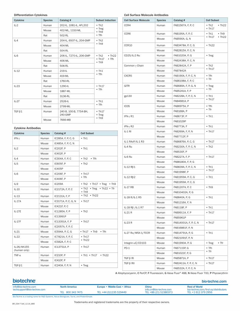
A Allophycocyanin, C PerCP, F Fluorescein, G Alexa Fluor® 488, N Alexa Fluor 700, P Phycoerythrin

Characterization of Cytokine Secretion by MultiplexingThe culture medium from CD4+ T cell differentiation procedures should be tested to confirm that the cells are secreting cytokines relevant to the desired cell subset. Multi-analyte detection techniques enable efficient screening for many cytokines simultaneously. Both Proteome Profiler™ Antibody Arrays and Luminex®-based Flow Cytometry Assays are optimized for maximum specificity and sensitivity of analyte detection.
Proteome Profiler Antibody ArraysProteome Profiler Antibody Arrays allow for the measurement of up to 111 proteins in a single sample. These arrays do not require specialized equipment and eliminate the need for multiple Western blot experiments. Antibody array kits contain buffers, detection antibodies, and membranes spotted in duplicate with high-quality capture antibodies. The arrays utilize chemiluminescence for detection, and membranes can be assessed for protein levels in the same manner as traditional Western blots. Select arrays are also suitable for use with the LI-COR® detection system.
IFN-γ IL-10 TNF-α
Naïve Human CD4+ T Cells
Th1-Polarized Human CD4+ T CellsCh
itina
se 3
-like
1
EMM
PRIN
/CD
147
Fas
Liga
nd
GM
-CSF
CXCL
1/G
ROα
IFN
-γ
IL-6
CXCL
8/IL
-8
IL-1
0
IL-1
2 p7
0
LIF
MIF
CCL5
/RAN
TES
TfR
TNF-α
50000
40000
30000
20000
10000
Mea
n Pi
xel D
ensi
ty
Detection of Cytokines Secreted by Th1-Polarized Human CD4+ T Cells Using the Proteome Profiler XL Cytokine Array. Naïve T cells were isolated from human PBMCs using the MagCellect Human Naïve CD4+ T Cell Isolation Kit (Catalog # MAGH115). These cells were either untreated or treated with CellXVivo Human Th1 Cell Differentiation Kit (Catalog #
CDK001) reagents. Array images show the detection of secreted cytokines. The densitometric profile of other cytokines before (green bars) and after (blue bars) Th1 polarization is shown in the histogram.
Proteome Profiler Antibody Arrays
Cytokine Array Analytes Catalog # LI-COR Chemiluminescence
Human Cytokine Array Kit, Panel A 36 ARY005 ✓ ✓
Mouse Cytokine Array Kit, Panel A 40 ARY006 ✓ ✓
Rat Cytokine Array Kit 29 ARY008 ✓ ✓
Proteome Profiler Mouse XL Cytokine Array Kit 111 ARY028 ✓ ✓
Proteome Profiler Human XL Cytokine Array Kit 102 ARY022 ✓ ✓
Learn more > rndsystems.com/ProteomeProfiler
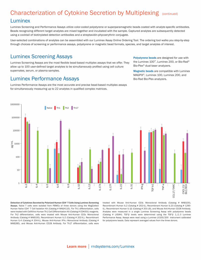
Luminex Screening AssaysLuminex Screening Assays are the most flexible bead-based multiplex assays that we offer. They allow up to 100 user-defined target analytes to be simultaneously profiled using cell culture supernates, serum, or plasma samples.
Luminex Performance AssaysLuminex Performance Assays are the most accurate and precise bead-based multiplex assays for simultaneously measuring up to 22 analytes in qualified complex matrices.
Detection of Cytokines Secreted by Polarized Human CD4+ T Cells Using Luminex Screening Assays. Naïve T cells were isolated from PBMCs of three donors using the MagCellect Human Naïve CD4+ T Cell Isolation Kit (Catalog # MAGH115). For Th1 differentiation, cells were treated with CellXVivo Human Th1 Cell Differentiation Kit (Catalog # CDK001) reagents. For Th2 differentiation, cells were treated with Mouse Anti-Human CD3e Monoclonal Antibody (Catalog # MAB100), Recombinant Human IL-2 (Catalog # 202-IL), Recombinant Human IL-4 (Catalog # 204-IL), Mouse Anti-Human IFN-g Monoclonal Antibody (Catalog # MAB285), and Mouse Anti-Human CD28 Antibody. For Th17 differentiation, cells were
treated with Mouse Anti-Human CD3e Monoclonal Antibody (Catalog # MAB100), Recombinant Human IL-2 (Catalog # 202-IL), Recombinant Human IL-23 (Catalog # 1290-IL), Recombinant Human IL-1b (Catalog # 201-LB), and Mouse Anti-Human CD28 Antibody. Analytes were measured in a single Luminex Screening Assay with polystyrene beads (Catalog # LXSAH). TGF-b levels were determined using the TGF-b 1,-2,-3 Luminex Performance Assay. Assays were read using a Luminex LX100/200 instrument calibrated for polystyrene beads. Data represent averaged values from the three donors.
Learn more │ rndsystems.com/Luminex
LuminexLuminex Screening and Performance Assays utilize color-coded polystyrene or superparamagnetic beads coated with analyte-specific antibodies. Beads recognizing different target analytes are mixed together and incubated with the sample. Captured analytes are subsequently detected using a cocktail of biotinylated detection antibodies and a streptavidin-phycoerythrin conjugate.
User-selected combinations of analytes can be assembled with our Luminex Assay Online Ordering Tool. The ordering tool walks you step-by-step through choices of screening or performance assays, polystyrene or magnetic bead formats, species, and target analytes of interest.
Polystyrene beads are designed for use with the Luminex 100™, Luminex 200, or Bio-Rad® Bio-Plex® dual-laser analyzers.
Magnetic beads are compatible with Luminex MAGPIX®, Luminex 100, Luminex 200, and Bio-Rad Bio-Plex analyzers.
IL-2
IL-4
IL-5
IL-6
IL-9
IL-1
0
IL-1
3
IL-1
7A
IL-1
7F
IL-2
2
IL-3
1
IFN
-g
TGF-
b1
TNF-
a
CCL1
7/TA
RC
1000000
pg/m
L
100000
10000
1000
100
10
1
Naïve Th2Th1 Th17
CXCL
13/
BLC/
BCA
Characterization of Cytokine Secretion by Multiplexing (continued)
Learn more │ rndsystems.com/ELISAs
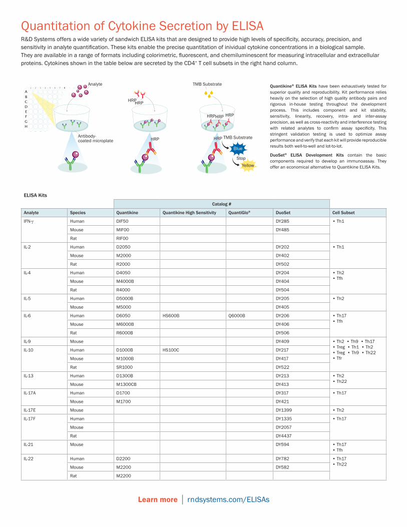
Quantitation of Cytokine Secretion by ELISAR&D Systems offers a wide variety of sandwich ELISA kits that are designed to provide high levels of specifi city, accuracy, precision, and sensitivity in analyte quantifi cation. These kits enable the precise quantitation of inividual cytokine concentrations in a biological sample. They are available in a range of formats including colorimetric, fl uorescent, and chemiluminescent for measuring intracellular and extracellular proteins. Cytokines shown in the table below are secreted by the CD4+ T cell subsets in the right hand column.
HRPHRP
Antibody-coated microplate HRP
TMB Substrate
Blue
Yellow
TMB Substrate
Stop
HRP
HRP
HRP HRP
Analyte Quantikine® ELISA Kits have been exhaustively tested for superior quality and reproducibility. Kit performance relies heavily on the selection of high quality antibody pairs and rigorous in-house testing throughout the development process. This includes component and kit stability, sensitivity, linearity, recovery, intra- and inter-assay precision, as well as cross-reactivity and interference testing with related analytes to confi rm assay specifi city. This stringent validation testing is used to optimize assay performance and verify that each kit will provide reproducible results both well-to-well and lot-to-lot.
DuoSet® ELISA Development Kits contain the basic components required to develop an immunoassay. They offer an economical alternative to Quantikine ELISA Kits.
ELISA Kits
Catalog #
Analyte Species Quantikine Quantikine High Sensitivity QuantiGlo® DuoSet Cell Subset
IFN-g Human DIF50 DY285 • Th1
Mouse MIF00 DY485
Rat RIF00
IL-2 Human D2050 DY202 • Th1
Mouse M2000 DY402
Rat R2000 DY502
IL-4 Human D4050 DY204 • Th2 • Tfh
Mouse M4000B DY404
Rat R4000 DY504
IL-5 Human D5000B DY205 • Th2
Mouse M5000 DY405
IL-6 Human D6050 HS600B Q6000B DY206 • Th17 • Tfh
Mouse M6000B DY406
Rat R6000B DY506
IL-9 Mouse DY409 • Th2 • Th9 • Th17 • Treg • Th1 • Th2• Treg • Th9 • Th22• Tfr
IL-10 Human D1000B HS100C DY217
Mouse M1000B DY417
Rat SR1000 DY522
IL-13 Human D1300B DY213 • Th2• Th22
Mouse M1300CB DY413
IL-17A Human D1700 DY317 • Th17
Mouse M1700 DY421
IL-17E Mouse DY1399 • Th2
IL-17F Human DY1335 • Th17
Mouse DY2057
Rat DY4437
IL-21 Mouse DY594 • Th17• Tfh
IL-22 Human D2200 DY782 • Th17• Th22
Mouse M2200 DY582
Rat M2200
A Allophycocyanin, C PerCP, F Fluorescein, G Alexa Fluor® 488, N Alexa Fluor 700, P Phycoerythrin
Differentiation Cytokines
Cytokine Species Catalog # Subset Induction
IL-2 Human 202-IL, 1081-IL, AFL202 • Th2• Treg• Th9• Tfh
Mouse 402-ML, 1150-ML
Rat 502-RL
IL-4 Human 204-IL, 6507-IL, 204-GMP • Th2• Th9
Mouse 404-ML
Rat 504-RL
IL-6 Human 206-IL, 7270-IL, 206-GMP • Th2• Th17• Th9
• Th22• Tfh
Mouse 406-ML
Rat 506-RL
IL-12 Human 219-IL • Th1• Tfh
Mouse 419-ML
Rat 1760-RL
IL-23 Human 1290-IL • Th17• Tfh
Mouse 1887-ML
Rat 3136-RL
IL-27 Human 2526-IL • Th1 • Tfh
Mouse 2799-ML
TGF-b1 Human 240-B, 100-B, 7754-BH, 240-GMP
• Th17• Treg• Th9
Mouse 7666-MB
Cell Surface Molecule Antibodies
Cell Surface Molecule Species Catalog # Cell Subset
CCR4 Human FAB1567A, F, P, C • Th2• Th17
• Th22
CCR6 Human FAB195A, F, P, C • Th1• Th17
• Th9• Th22
Mouse FAB590A, G, N
CCR10 Human FAB3478A, P, C, G • Th22
Mouse FAB2815A, P, C, N
CD25/IL-2 Ra Human FAB1020A, P, G • Treg
Mouse FAB2438A, P, C, N
Common g Chain Human FAB2842A, F, P • Th2• Th17
Mouse FAB7842A
CXCR5 Human FAB190A, F, P, C, N • Tfh• Tfr
Mouse FAB6198A, F, P, C
GITR Human FAB689A, F, P, G, N • Treg • Tfr
Mouse FAB5241A, F, P
gp130 Human FAB228A, F, P, C, N • Th1• Th17
Mouse FAB4681A, P
ICOS Human FAB6975A, P • Tfh• Tfr
Mouse FAB168A, P
IFN-g R1 Human FAB673F, P • Th1
Mouse FAB1029P
IFN-g R2 Human FAB773A, F • Th1
IL-1 RI Human FAB269A, F, P, N • Th17
Mouse FAB7712F, P
IL-1 RAcP/IL-1 R3 Human FAB6876A, P, C, G • Th17
IL-4 Ra Human FAB230A, F, P, C, N • Th2
Mouse FAB530F, P
IL-6 Ra Human FAB227A, F, P • Th17
Mouse FAB8180A, F, P, G
IL-12 Rb1 Human FAB839A, F, P, C, N • Th1• Th17
Mouse FAB1998F, P
IL-12 Rb2 Human FAB1959A, P, C, G • Th1
Mouse FAB1959A, P, C, G
IL-17 RB Human FAB1207A, P, C • Th9
Mouse FAB10402A, P, G
IL-18 R/IL-1 R5 Human FAB840A, P, G • Th1
Mouse FAB1216A, F, N
IL-18 Rb /IL-1 R7 Human FAB118F, P • Th1
IL-21 R Human FAB9911A, F, P • Th17
Mouse FAB5961P
IL-23 R Human FAB14001A, F, P, C, N • Th17
Mouse FAB16861F, P, N
IL-27 Ra/WSX-1/TCCR Human FAB14791A, P, G • Th1
Mouse FAB21091F, P, N
Integrin aE/CD103 Mouse FAB1990A, P, G • Treg • Tfr
PD-1 Human FAB7115P, G • Tfh• Tfr
Mouse FAB1021F, P, G
TGF-b RI Mouse FAB5871A, P • Th17
TGF-b RII Human FAB2411A, F, P, C, N • Th17
Mouse FAB532A, F, P, C, N
Cytokine Antibodies
Cytokine Species Catalog # Cell Subset
IFN-g Human IC285A, F, P, C, G • Th1
Mouse IC485A, F, P, C, N
IL-2 Human IC202F, P • Th1
Mouse IC402F, P
IL-4 Human IC304A, F, P, C • Th2 • Tfh
IL-5 Human IC605F, P • Th2
Mouse IC405P
IL-6 Human IC206F, P • Th17 • Tfh
Mouse IC406F, P
IL-9 Human IC209A • Th2 • Th17 • Treg • Th9
IL-10 Human IC2172A, F, P, C • Th2 • Th9
• Treg • Th22 • Tfr
IL-13 Human IC2131A, F, P • Th2 • Th22
IL-17A Human IC3171A, P, C, G, N • Th17
Mouse IC421F, P, C
IL-17E Human IC1285A, F, P • Th2
Mouse IC13991P
IL-17F Human IC13351A, F, P • Th17
Mouse IC2057A, F, P, C
IL-21 Mouse IC594A, P, C, G • Th17 • Th9 • Tfh
IL-22 Human IC7821A, F, P, C • Th17• Th22
Mouse IC582A, F, P, C
IL-26/AK155 (human only)
Human IC13751A, P • Th17
TNF-a Human IC210F, P • Th1 • Th17 • Th22
Mouse IC410F, P
TGF-b1 Human IC240A, F, P, N • Treg
BR_CD4 T Cell_12.14_866Trademarks and registered trademarks are the property of their respective owners.
Bio-Techne is a trading name for R&D Systems, Novus Biologicals, Tocris, and ProteinSimple.
Rest of Worldbio-techne.com/find-us/distributorsTEL +1 612 379 2956
[email protected]@bio-techne.com
North America
TEL 800 343 7475
Europe • Middle East • Africa
TEL +44 (0)1235 529449
[email protected] +86 (21) 52380373
bio-techne.com