Methods for studying root colonization by introduced ...
13
HAL Id: hal-00886192 https://hal.archives-ouvertes.fr/hal-00886192 Submitted on 1 Jan 2003 HAL is a multi-disciplinary open access archive for the deposit and dissemination of sci- entific research documents, whether they are pub- lished or not. The documents may come from teaching and research institutions in France or abroad, or from public or private research centers. L’archive ouverte pluridisciplinaire HAL, est destinée au dépôt et à la diffusion de documents scientifiques de niveau recherche, publiés ou non, émanant des établissements d’enseignement et de recherche français ou étrangers, des laboratoires publics ou privés. Methods for studying root colonization by introduced beneficial bacteria Elisa Gamalero, Guido Lingua, Graziella Berta, Philippe Lemanceau To cite this version: Elisa Gamalero, Guido Lingua, Graziella Berta, Philippe Lemanceau. Methods for studying root colonization by introduced beneficial bacteria. Agronomie, EDP Sciences, 2003, 23 (5-6), pp.407-418. 10.1051/agro:2003014. hal-00886192
Transcript of Methods for studying root colonization by introduced ...
HAL Id: hal-00886192https://hal.archives-ouvertes.fr/hal-00886192
Submitted on 1 Jan 2003
HAL is a multi-disciplinary open accessarchive for the deposit and dissemination of sci-entific research documents, whether they are pub-lished or not. The documents may come fromteaching and research institutions in France orabroad, or from public or private research centers.
L’archive ouverte pluridisciplinaire HAL, estdestinée au dépôt et à la diffusion de documentsscientifiques de niveau recherche, publiés ou non,émanant des établissements d’enseignement et derecherche français ou étrangers, des laboratoirespublics ou privés.
Methods for studying root colonization by introducedbeneficial bacteria
Elisa Gamalero, Guido Lingua, Graziella Berta, Philippe Lemanceau
To cite this version:Elisa Gamalero, Guido Lingua, Graziella Berta, Philippe Lemanceau. Methods for studying rootcolonization by introduced beneficial bacteria. Agronomie, EDP Sciences, 2003, 23 (5-6), pp.407-418.�10.1051/agro:2003014�. �hal-00886192�
407Agronomie 23 (2003) 407–418© INRA, EDP Sciences, 2003DOI: 10.1051/agro:2003014
Review article
Methods for studying root colonization by introducedbeneficial bacteria
Elisa GAMALEROa*, Guido LINGUAa, Graziella BERTAa, Philippe LEMANCEAUb
a Università del Piemonte Orientale “Amedeo Avogadro”, Department of Science and Advanced Technology, Corso Borsalino 54, 15100 Alessandria, Italyb UMR “Microbiologie et Géochimie des Sols”, INRA-CMSE, BP 86510, 21065 Dijon Cedex, France
(Received 24 October 2002; accepted 4 March 2003)
Abstract – Some free-living rhizobacteria are considered as potential biocontrol and plant growth-promoting agents. Successful application ofbeneficial bacteria as microbial inoculants requires their presence and activity at the appropriate level, but even more, at the right time and place.Various markers are described in the literature to differentiate introduced bacteria from indigenous microflora and to visualize them. Thesemarkers are presented together with the methods currently applied to quantifying bacterial densities and to characterizing the distribution ofintroduced bacteria. The methods of quantifying bacterial densities are either based on bacterial cultivation or not. Different types ofmicroscopic observations, allowing the characterization of the bacterial distribution and location in the rhizosphere, are also described. Therespective advantages and limitations of these markers and methods are discussed.
rhizosphere / bacterization / methodology
Résumé – Méthodes d’étude de la colonisation racinaire par des bactéries bénéfiques introduites. Certaines rhizobactéries libres sontconsidérées comme des agents potentiels de lutte biologique et de stimulation de la croissance des plantes. Le succès de leur applicationnécessite la présence et l’activité des bactéries à un niveau suffisamment élevé, mais également au bon moment et au bon endroit. Différentsmarqueurs permettant la différentiation des bactéries introduites de la microflore indigène ainsi que leur observation dans la rhizosphère, sontdécrits dans la littérature. Ces marqueurs ainsi que les méthodes, appliqués pour quantifier la densité et pour caractériser la distribution desbactéries introduites, sont présentés. Les méthodes de quantification de la densité bactérienne sont basées ou non sur la culture des bactéries.Différentes méthodes d’observations microscopiques permettent de caractériser la distribution et la localisation bactérienne dans la rhizosphère.Les avantages et les inconvénients respectifs des marqueurs et des méthodes décrits sont discutés.
rhizosphère / bactérisation / méthodologie
1. INTRODUCTION
The rhizosphere was defined in 1904 by Hiltner [40] asbeing the volume of soil, influenced by the presence of livingplant roots, whose extension may vary with soil type, plantspecies, age and other factors [34]. Plant roots release anenormous amount of root exudates that may represent up to10–20% of the photosynthethates (see the review by C.Nguyen in the present issue), leading to a significantstimulation of the microbial density and activity. Specificpopulations are more favored than others in the rhizospheredue to the level of adequation of their metabolic activities withthe composition of the root exudates. The structure anddiversity of microbial populations in the rhizosphere thus
differ significantly from those of soilborne populations [55,64, 67]. These quantitative and qualitative variations of thesoilborne microflora are described as the rhizosphere effect.This rhizosphere effect varies according to the root exudatecomposition, which is affected by the plant physiology [26],the stage of plant development [115] and the position on theroot system [57].
The microflora associated with the roots affect plant growthand health. Indeed, some bacterial populations are pathogenic,whereas others are beneficial. Beneficial rhizobacteria includeboth symbiotic and free-living microorganisms. Among thelatter, special attention has been given to the fluorescentpseudomonad group. Positive effects on plant growth andhealth of inoculations of bacterial strains belonging to this
* Correspondence and [email protected]
Communicated by Yves Dessaux (Gif-sur-Yvette, France)
408 E. Gamalero et al.
bacterial group have been reported since the late ‘70s [7, 18,36, 46, 56, 116]. However, overall biological control ofsoilborne diseases achieved by microbial inoculants is ofteninconsistent [86]. This has been especially illustrated forfluorescent pseudomonads. This inconsistency has beenpartially associated with inefficient root colonization by theintroduced bacteria [54, 116]. Indeed, a clear relationship hasbeen established between suppression of the wheat rootdisease take-all and that of fusarium-wilts by different strainsof fluorescent pseudomonads, and the densities of thesebacteria in the corresponding host plants [16, 78]. In order tomake biological control more consistent, there is a need for abetter knowledge of bacterial traits promoting rhizospherecompetence.
Biocontrol of soilborne diseases is ascribed to microbialantagonism and/or to induced resistance of the host plant [25,108]. Microbial antagonism results from the suppression ofsaprophytic growth of plant pathogens mediated by antibioticsand siderophores. The concentration of these metabolites inthe rhizosphere is expected to be related to the density ofactive bacteria. Moreover, the synthesis of some of thesemetabolites (phenazines and pyoverdines) was demonstratedto be regulated by quorum-sensing [75, 94]. Besides the totalbacterial density, which is related to the survival kinetics ofthe introduced strain, the antagonistic metabolites and thus thebacterial cells should be located at the infection courts of thesoilborne pathogens. To summarize, the expression of thebeneficial effects by the introduced bacteria requires theirpresence at a density high enough and at the time and locationthat are favorable for root infection by the pathogens.
The methods required for analyzing bacterial traitsinvolved in the rhizosphere competence and plant-microbeinteractions therefore must allow the quantification of bacte-rial density but also the characterization of the bacterial celldistribution and location. Moreover, these methods shouldtake into account not only culturable, but also total bacterialcells, since the frequency of viable but non-culturable cellswould vary according to the environmental conditions [100].
The aim of the present review is to present methods ofquantification and characterization of the distribution alongthe roots of introduced bacteria in the soil and rhizosphere.
2. MARKERS USED FOR TRACKING INTRODUCED BACTERIA
Tracking bacteria introduced into complex environmentssuch as soils requires the ability to discriminate them from theindigenous microflora. Markers used for that purpose shouldtherefore fulfil several prerequisites. These markers shouldobviously be specific. This specificity must be checked in theenvironment in which bacteria are introduced. The markersshould also be stable in soil and with time. The relativestability of the marker is required both to avoid its loss and/orits transfer to other bacteria. Since the aim of the markers is toperform ecological studies on the introduced bacteria, theyshould affect as little as possible the behavior of these bacteria.Surprisingly, there are few studies comparing the fitness of themarked and wild-type strains [14, 28, 35, 72, 73, 106]. In the
same way, since the perturbation of the system should be keptas low as possible, the expression of the marker should avoidsubstrate amendment. More generally, the markers chosenshould be easy to track in a wide range of soils, andenvironmental conditions (pO2, pH, etc.) favorable for theirexpression should be considered [43].
In this section different markers are presented together withtheir properties.
2.1. Serological markers
The primary immunological tool used in environmentalmicrobiology is the antibody. Immunoassays are analyticalmethods used to detect and/or quantify the antigen-antibodyinteraction. The conjugation of a signal molecule (fluoro-chromes, enzymes and radioisotopes) to the antibody isrequired to visualize the antigen-antibody interaction. Immu-nological techniques are relevant especially for the detection,enumeration and localization of introduced bacterial strains inthe soil and rhizosphere. The critical aspect of serologicalmethods is the specificity of the antibodies used. Polyclonal ormonoclonal antibodies may be applied according to their spe-cificity. Monoclonal antibodies are obviously more expensiveto raise but are more specific. The specificity of the antibodies,especially polyclonal ones, should be checked to decrease theoccurrence of possible cross-reactions. Usually, a high enoughspecificity may be obtained for fluorescent pseudomonadstrains with polyclonal antibodies raised against membraneproteins [35].
2.2. Molecular markers
Various molecular markers such as antibiotic resistance[35, 53, 105], chromogenic (xylE, gusA and lacZ) [48, 116],luminescent (luxAB and luc) [50, 60, 79, 80, 81, 102] andfluorescent markers (gfp and unstable gfp) [13, 58, 71, 95]have been developed and widely applied to studying rootcolonization.
2.2.1. Antibiotic resistance
Antibiotic resistances have been widely used as markers inmicrobial ecology. Although various plasmids and transposonshave been used [27, 69, 72, 76, 109], most of the studies on bac-terial survival kinetics are based on the use of spontaneouslyoccurring antibiotic-resistant mutants. Rifampicin resistance iscommonly used as a marker to study survival kinetics of intro-duced bacteria in the rhizosphere [35, 68, 105]. Stability ofrifampicin resistance was checked with Pseudomonas putidaWCS358 in field conditions [35]. Kanamycin and streptomy-cin resistance obtained by Tn5 mutagenesis with the suicideplasmid method of Simon et al. [90] was also described as apossible marker [105]. The maintenance of Tn5 in the mutantJM218 was ascertained by comparing bacterial densities of thismutant in root suspensions, estimated by serology, with bacte-rial density estimated by plate count on King’s B medium sup-plemented with kanamycin [53]. As stressed above, the levelof resistance of the indigenous microflora to the antibiotic usedas a marker must be determined in order to check the specificityof the marker used. As an example, Wilson et al. [118] have
Bacterial root colonization 409
estimated that the background of naturally kanamycin-resist-ant bacteria in a Dutch soil was 2���104 cfu per gram.
Antibiotic resistance is often used for studies on survivalkinetics of introduced bacteria [35, 68, 76, 105] since the cor-responding detection method (plate counting, see Sect. 3.1) isquite sensitive, cost effective, reliable and easy to perform.However, possible genetic changes associated with chromo-somal-mediated antibiotic resistance may affect several eco-logically important traits [14, 28, 63, 91]. Moreover, the useof antibiotic tagged bacteria carries with it the risk of contrib-uting to the indesiderable spread of antibiotic resistance innature [41].
2.2.2. Chromogenic markers
Several genes encoding metabolic enzymes have been usedas markers to detect, quantify and localize introduced bacteria.The xylE gene encodes for a catechol 2,3-dioxygenase catalyz-ing the formation of hydroxymuconic acid that, reacting witha catechol, forms a yellow semialdehyde derivative. ThegusA and lacZ genes encode for a �-glucuronidase and �-galactosidase, respectively: in the presence of the adequatesubstrates (5-bromo-4-chloro-3-indolyl-�-D-glucopyranosideand 5-bromo-4-chloro-3-indolyl-beta-D-galactopyranoside),they produce a blue pigment [41].
Although the use of these chromogenic markers is simple,not expensive and well defined, several disadvantages havebeen widely discussed [32, 43, 92]. For example, the xylE geneapplicability is restricted due to inactivation of cathechol 2,3-dioxygenase by oxygen [85]. The gusA and lacZ markers havelimited application in soil because of the presence of high back-ground (105 cfu·g–1 of soil) from the indigenous microflora[33, 118]. Moreover, these chromogenic markers require sub-strates or reactives to be expressed or detected; the stability ofthese gene products is usually measured by hours or days [43].
2.2.3. Luminescent markers: luxAB and luc genes
Another very sensitive approach is the transfer of biolumi-nescence marker genes to bacteria, providing them with thecapability of emitting light. Prokaryotic bioluminescencegenes have been cloned from Vibrio fischeri and Vibrio har-veyi [9, 31], while those eukaryotic (luc genes) have beencloned by the firefly Photinus pyralis [29]. The lux operon ofV. fischeri includes five genes: two genes (luxAB) encode thesubunits of the luciferase enzyme and the other three (lux-CDE) encode enzymes involved in the synthesis of the aldehy-dic substrate (n-decanal) [43].
The requirement for molecular oxygen limits the use andthe interpretations of this kind of physiological reportersystem, but if oxygen is present, the monitoring of light mayresult in very useful information on bacterial activity anddistribution in different environments such as the plantrhizosphere [70]. The minimum detection limit for fully activecells has been reported as 445 cells per gram of soil [81].Although eukaryotic luc genes show some advantages incomparison with the prokaryotic counterpart (i.e. higherspecificity and absence of background in native microflora),the substrate luciferin is expensive and sometimes not readily
taken up by the cells. The stability of the gene product isusually measured by minutes or hours [43].
2.2.4. Fluorescent markers: stable and unstable green fluorescent protein
Another attractive marker system for monitoring bacterialcells in the environment is the green fluorescent protein(GFP). The GFP is a 27 KDa polypeptide which convertsthe blue chemiluminescence of the Ca+2-sensitive photopro-tein (aequorin from the jellyfish Aequorea victoria) into greenlight [20]. A series of red shifted GFP mutants, 20–35 timesstronger than the wild type, with various excitation and emis-sion wavelengths such as the ECFP (enhanced cyan), EGFP(enhanced green) and EYFP (enhanced yellow), have beenrecently developed [101].
The advantages and disadvantages of this marker have beenextensively discussed by Errampalli et al. [32]. Some of themost relevant advantages are that GFP is extremely stable andresistant to proteases, is easily detectable, does not requireexogenous substrate and allows the monitoring of single cellseven in real time. Moreover, GFP is continuously synthesizedand there is no background in indigenous bacterial populations.However, the interference of soil particles, the variability ofGFP expression in different species, the inability to work inanaerobic conditions and the instability of the plasmid shouldbe considered. In order to overcome the latest limitation andreduce the risk of a plasmid transfer to other microorganisms,bacterial strains used are preferentially chromosomallymarked. For that purpose, several Tn5 transposon suicidedelivery vectors have been developed [95, 98]. The stability ofthe GFP varies according to the variants and plasmid con-structs in the range of hours or days [44, 95].
Recently, Andersen et al. [4] developed a new variant ofGFP characterized by its short half-life. The unstable GFP hasbeen constructed by the addition of a short peptide sequence tothe C-terminal end of the intact GFP: this modification allowsits degradation by bacterial endogenous proteases. Since theGFP produced during bacterial growth does not accumulate, itis possible to perform real-time analysis of the bacterial meta-bolic activity [58, 93]. However, different levels of proteasesmay be expressed depending on the microorganisms, thegrowth phase and environmental factors, and care must beapplied in the interpretation of the results [44].
2.2.5. Specific primers and oligonucleotidic probes
Introduced bacteria can be monitored using primers orprobes that allow amplification or hybridization of sequenceswhich are strain-specific. Specific probes can be used tohybridize bacterial colonies after in vitro growth [117] or bac-terial cells for in situ studies. Probes are usually covalentlylinked to a fluorochrome such as fluorescein, rhodamine,Texas red, Cy3 and Cy5 [3].
Specific sequences may be introduced by a geneticconstruction. As an example, a specific primer amplifyingacross nptII-lacZ junctions on the Tn5B20 construct was usedto follow the survival kinetics in the soil and rhizosphere of thestrain P. fluorescens R2f tagged by the lacZ-nptII marker gene
410 E. Gamalero et al.
[109]. However, as stressed before, genetic constructs mayaffect the ecological behavior of bacterial strains.
Another strategy consists of identifying sequences specificto the strains in order to design primers and probes. Differentapproaches have been proposed to develop this identification.One is to compare homologous nucleic acid sequences ofribosomal RNA (rRNA) to sequences available in databases.Since rRNA are present in all living microorganisms in highcopy number and are quite stable, oligonucleotidic probes canbe applied [3]. They are either species-specific or even strain-specific in some cases [5, 24]. Pseudomonas specific primerhas been designed by Braun-Howland [15]. This PSMg primerwas applied to describing the dynamic of indigenous popula-tions of Pseudomonas in soil hot-spots [45] and to character-izing the succession of Pseudomonas on barley root in aperturbed environment [96]. Analysis of the 16S rDNA of thePaenibacillus azotofixans strain with that of 2000 bacteria alsoenabled Rosado et al. [84] to identify the presence of threehighly variable regions that were used to design primers forstudying the kinetics of this bacterial strain in the soil andwheat rhizosphere. Another approach to defining primers andprobes is (i) to characterize the diversity of populationsbelonging or not to the same group by Random Amplificationof Polymorphic DNA-Polymerase Chain Reaction (RAPD-PCR), in order (ii) to identify discriminating bands, then (iii)to pick them from the gel, and (iv) to re-amplify and test themfor specificity.
Monitoring introduced bacteria on the basis of its specificRAPD-PCR pattern has also been proposed but is very time-consuming [21, 51].
3. METHODS TO QUANTIFY DENSITIESOF INTRODUCED BACTERIA
Methods to quantify introduced bacteria can be classifiedinto two major types depending upon whether they are basedon the cultivation of the bacteria or not. Obviously, the cul-ture-dependent methods will not allow the detection of viablebut not culturable bacteria (VBNC). Since microorganismsintroduced into soil can go through different processes(conversion to the non-culturable state and phase changes)doubts have been raised about the representativity of the viewgiven by data yielded with culture-dependent methods of thereal processes in soil. Despite this limitation, culture-depend-ent methods remain widely used, mainly because they are easyto apply. The culture-independent methods can provide a morecomplete picture of the kinetics of the total number of micro-bial cells [105]. However, the major limitation of these meth-ods is that they may not allow differentiation between viableand not-viable cells.
In this section, major culture-dependent and culture-inde-pendent methods of microbial quantification in the rhizo-sphere are presented.
3.1. Culture-dependent methods
These methods are based on the suspension-dilution of soiland/or root samples and on inoculation of growing media
(solid or liquid) with adequate dilutions. They requiretherefore the use of labeled strains (see Sects. 2.1 and 2.2). Theculture-dependent methods differ according to the type ofmarker used giving the specificity to the growing media. Thistype of method is quite simple to perform, not too expensiveand quite sensitive (102–103 cfu per g), but labor-intensiveand shows some limitations [44]. This type of methodunderestimates the number of bacteria present in soil or in therhizosphere. Bacteria may remain physically attached to thesoil particles, may be killed in the dilution medium or may failto grow on growth media [47]. Some of them may remainaggregated even during the dilution process in such a way thata cfu may be originated by more than one cell. Suspension-dilution can either be plated on solid media or introduced intoliquid media with various dilutions in order to determine fromwhich dilution there is no more bacterial growth. This lastmethod, named Most Probable Numbers (MPN), requires theuse of probability tables to process data that contribute toreducing the sensitivity of the analysis compared withplating [61].
The most basic method consists of plating mutants resistantto antibiotics on solid growth medium supplemented with thecorresponding antibiotic and with an anti-eucaryotic com-pound such as cycloheximide. This method is widely used,especially for survival kinetics of introduced bacteria and forcompetition studies between wild-type strains and mutantsimpaired in specific phenotypes [67, 68, 72, 106].
The sensitivity of this type of plating method may be signif-icantly lowered by combining plating and serologicalapproaches with the immunofluorescence colony-staining(IFC) technique. Detection limits as low as 20 cfu of Erwiniaspp. per gram of soil have been reported by Van Vuurde andVan der Wolf [111]. The IFC technique, developed by theseauthors, is based on the use of fluorescein isothiocyanate(FITC)-conjugated IgG antibodies specific to the introducedbacterial strain to discriminate target from non-target colonies.Bacterial colonies remain viable and IFC positive colonies canbe subcultured to confirm their identity by other biochemicalor molecular methods. Direct IFC has been used by severalinvestigators [52, 53, 63, 77, 110] to track and quantify bacte-rial strains introduced into soil or onto plants. Since the IFCdoes not require any alteration in the phenotype or genotype ofthe wild-type strain, this technique allows the comparison ofan unaltered wild-type strain to a genetically modified deriva-tive of the wild-type strain. The main restriction of direct IFCis the necessity of a good quality fluorescent conjugate againstthe target bacteria. However, outside the medical field, spe-cific conjugates are usually not commercially available. Forthis reason, Veena and Van Vuurde [113] recently developedan indirect IFC using diluted specific antiserum and commer-cial conjugate to detect bacterial pathogens on tomato seeds.Indirect IFC is suitable for routine applications with facilitiesfor fluorescence microscopy and does not require much exper-tise. As for any serological methods, the main limitation is therisk of false positive reactions due to cross-reacting bacteria.
Reporter genes may also be applied for culture-dependentmethods. Lux-luc tagged bacteria can be detected and enumer-ated by plate counting, luminometry or scintillation countersand by imaging. Luminometry is an easy and sensitive methodthat has been applied to evaluating the density of luminescent
Bacterial root colonization 411
bacteria on the root surface [8] and rhizosphere [81]. These twoinstruments are sensitive but they are not specifically desig-nated for bioluminescence application [17]. Bioluminescentcolonies can be counted directly by color photography (auto-photography), by exposure to X-ray film, by direct microscopywith a CCD camera enhancement (imaging) or alternatively, ifthe amount of light emitted is high, it is possible to visualizethem by eye [41].
Green fluorescence due to GFP-tagged bacteria can beobserved in colonies cultured on agar media under a hand-heldlong-wave UV lamp. This is a simple and cheap way toenumerate colonies but the potential DNA damage of UV overtime may be a limitation [32]. Fluorimetric detection of GFP-labeled bacteria is useful for screening or confirmation of cellgrowth. A detection limit of 103 cells per ml of P. putida in soilhas been reported by Burlage and Kuo [17]. Recently, Cassidyet al. developed a MPN method to evaluate cell density ofGFP-marked Pseudomonas in the soil, rhizosphere andrhizoplane [19].
Finally, colonies grown on solid media can be hybridizedwith specific oligonucleotidic probes [117].
3.2. Culture-independent methods
Culture-independent methods can be distributed into threedifferent categories: serological, molecular and cytologicalmethods.
3.2.1. Serological methods
Among the serological methods, the enzyme-linked immu-nosorbent assay (ELISA) is a very sensitive immunoassay forthe detection of antigens. ELISA is based on direct or indirectsandwich methods. The ELISA method has been used to studyand quantify the external and internal root colonization ofmaize by two P. fluorescens strains [11] and the distribution oftwo diazotrophic enterobacterial strains, Pantoea agglomer-ans and Klebsiella pneumoniae, on cereal shoots and roots[82]. The ELISA method is quite sensitive (103 cells per ml inpure cultures and 104–105 cells per g of soil) and associatedwith a standard curve relating the amount of signal given to thedirect counts by microscopic enumeration, can provide quan-titative information. Disadvantages are related to possiblecross-reactivity and non-specific signal production.
3.2.2. Molecular methods: detection of nucleic acids
Detection methods based on nucleic acids extracted fromthe soil offer the possibility of monitoring specific bacterialgenotypes (gene or genomic markers), providing a picture ofthe dynamics of total numbers of microbial cells [45]. Thepolymerase chain reaction (PCR), primarily used as a qualita-tive method to confirm the presence or absence of a specificDNA sequence, has been recently applied to obtaining quanti-tative information. Up to now, PCR has been the most sensi-tive method for detection of specific DNA in environmentalsamples; sensitivities of 1–100 cells per gram of soil have beenreported [42].
Before amplification, microbial DNA is extracted fromsoil. Various methods have been described for this extraction
[65]. All DNA extraction methods present potential biasdepending on the soil properties (i.e. humic substances) [107].The final goal of DNA extraction methods is to obtain DNAwith a quality good enough for PCR amplification and foryielding consistent data.
Three PCR methods for quantification have been developedand applied to evaluating bacterial population in the soil orrhizosphere: the most probable number PCR (MPN-PCR), thecompetitive PCR (C-PCR) and quantitative PCR (Q-PCR).
In the case of MPN-PCR, the quantification of targetsequences is based on the serial dilution of the PCR productsin order to identify from which dilution the target sequence isno longer detectable by electrophoresis. The detectable limitof the product is calibrated by an external standard and the ini-tial amount of the target molecules is estimated using the dilu-tion factor of the positive sample. This method was firstdeveloped by Picard et al. with Agrobacterium tumefaciensand Frankia spp. [74]. Using this method, Picard et al. wereable (i) to detect the A. tumefaciens strain when inocula rangedfrom 103 to 107 cells, and (ii) to estimate the indigenous pop-ulations of Frankia spp. at 0.2���105 genomes per gram of soil[74]. A detection limit of 102 cfu of Paenibacillus azotofixansper gram of rhizospheric soil was reported by Rosado et al.[84]. However, as indicated by Jansson and Leser [42], a lim-itation of the MPN-PCR method is the probabilistic evalua-tion, based on several dilutions and replicates, whichcontribute to reducing the precision of the estimation.
In C-PCR, a DNA fragment containing the same primersequences (internal standard) as the target fragment is allowedto compete in the same tube with the target for primer bindingand amplification. Experimentally, PCR reaction tubescontaining the target samples are spiked with a dilution seriesof the competitor fragment. When the molar ratio of PCRproducts generated from the target and competitor is equal toone, the amount of the target is equal to the competitor. Sincethe amount of the competitor is known, the amount of the tar-get can be determined. However, this technique is labor-inten-sive and its accuracy is dependent on an internal competitor,which must possess the same amplification efficiency as thetarget [42]. Thirup et al. [96] applied this type of PCR to stud-ying the effects of P. fluorescens DR54 and the fungicide Ima-zalil on the succession of indigenous Pseudomonas spp. andActinomyces on barley roots. Martin-Laurent et al. [66] havequantified with C-PCR the amount of the atzC gene known tobe involved in atrazine mineralization in a soil treated with thisherbicide.
Finally, Q-PCR is the direct measurement of the amount ofproducts generated from different samples by a calibratedinstrument. The initial amount of the target molecules in thesamples is estimated by the determination of the PCRamplification efficiency defined by the amplification of aknown amount of the same target (external standard). Q-PCRhas certain limitations, such as accounting for the variationbetween samples during the reaction and the fact thatquantification is only possible during the exponential phase ofthe amplification [42].
The major advantages of the PCR-based methods are theirsensitivity and specificity. Theoretically, a single copy of thetarget nucleic acid sequence can be detected; normally, at least

412 E. Gamalero et al.
103 copies of the target are required for PCR methods innucleic acids isolated from environmental samples. Thesemethods allow one to take into consideration both cultivableand not cultivable organisms, but do not allow differentiationbetween viable and non-viable organisms.
3.2.3. Cytological method: flow cytometry
Flow cytometry is a cytological tool valid for evaluatingroot colonization by introduced bacteria. This instrumentmeasures and analyzes the optical properties of hundreds ofsingle cells per second, passing through a focused laser beam.As each cell or particle passes through the flow cytometer, itis monitored by forward scatter (detects each particle accord-ing to its size), side scatter (measures simultaneously the sizeand the shape of the particle) and fluorescence (evaluates thefluorescence intensity) detectors. Flow cytometry allows thedetection and quantification of both the individual fluorescentcells within a population and the fluorescent intensity frommore than one bacterial group. For environmental samples,bacteria from the particulate matter and an internal standardmust be used to quantify the cell number [44]. A limitation ofthe technique is related to its low sensitivity due to the abun-dance of fluorescence particles present in most environmentalsamples. However, the assay is rapid and simple and thou-sands of cells can be analyzed in a short time, allowing theprocessing of the data by various statistical procedures.
Flow cytometry can be applied to evaluating the densityand characterizing the kinetics of introduced bacteria taggedby fluorescent antibodies [23], GFP [98] and specific oligonu-cleotidic probes [96]. A detection limit of 103 cells per ml hasbeen recorded evaluating the density of fluorescent antibody-tagged Xanthomonas campestris in Brassica oleracea [23],and of 3���104 cells per gram of dry soil in the characterizationof the kinetics of the fluorescent oligonucleotidic probe-tagged Sphingomonas spp. strain 107 in soil [97].
The use of different dyes or fluorochrome provides anextremely powerful way to characterize the physiologicalstate, activity or degree of viability of bacteria [37, 114], andthus to quantify by flow cytometry the viable and non-viablebacterial cells. Various criteria have been proposed to discrim-inate the viable from non-viable cells. Impermeability of thebacterial membrane to dyes is the basis of the dye exclusiontest. Propidium iodide (PI) and other dyes characterized by thepresence of quaternary ammonium groups and two or morepositive charges, such as Sytox green, TO-PRO-1 and TO-PRO-3, are membrane-impermeant. Cells retaining these dyesare usually considered as non-viable cells. Moreover, usingsimultaneously permeant (SYBR green) and non-permeant(PI) dyes makes possible the discrimination of cells with acompromised, slightly damaged or intact membrane [37]. Theuse of a membrane permeant substrate such as fluorescein dia-cetate (FDA) and 5-cyano-2,3-ditolyl tetrazolium chloride(CTC), that are cytoplasmic enzymatically cleaved to form afluorescent impermeant product (fluorescein and formazan,respectively) allows the discrimination of cells with intact(retaining the product of the reaction) and damaged mem-branes (losing the product of the reaction) [112]. Membranepotential (MP) is the most used vitality parameter in microbialflow cytometry. A 100 mV bacterial transmembrane electrical
potential gradient, due to selective permeability and ionictransport, is usually reported. Variations in the MP measure-ment can be recorded using lypophilic charged dyes that canbe accumulated or excluded by the cell [88].
4. METHODS TO CHARACTERIZE DISTRIBUTION AND LOCALIZATIONOF INTRODUCED BACTERIA
Distribution and localization of introduced bacteria requirethe use of fluorescent antibodies, fluorescent markers and oli-gonucleotic probes (see Sects. 2.1, 2.2.4 and 2.2.5). Thesestudies are sometimes only possible in gnotobiotic conditions.
Immunolocalization is based on the use of fluorescent sig-nal molecules conjugated to the antibodies; the emission offluorescent light indicates the presence of a specific antigen.The basic procedure consists of the reaction between a fluores-cent specific antibody with the antigen attached to a slide, andof the observation of the sample using a fluorescence micro-scope. To enhance the signals and the specificity of the reac-tion, an indirect immunolocalization, using a fluorescentsecondary antibody, is usually performed. Simultaneous local-ization of different antigens can be obtained using antibodiescoupled to different fluorochromes. Immunolocalization hasbeen successfully used to study root colonization. Examplesinclude the analysis of the spatial competition between P. flu-orescens Ag1 and Ralstonia eutropha (formerly Alcaligeneseutrophus) during barley root colonization [49], the cell distri-bution of P. fluorescens DF57 on barley roots [38], the aut-oecology of the biocontrol agent P. fluorescens CHA0 in therhizosphere of different crops [100] and the endophytic colo-nization of spruce by Paenibacillus polymyxa (formerly Bacil-lus polymyxa) Pw-2R and P. fluorescens Sm3-RN [89].Advantages of immunolocalization are the simplicity of its useand the short time required to obtain results. On the other hand,several problems, such as autofluorescence of the sample,non-specific staining, antigen instability and the inability tocheck viability have to be considered.
Lux genes have been widely used in the study of bacterialroot colonization and activity [50, 60, 79, 83, 103], while lucgenes have been only recently applied for monitoring activityof P. fluorescens 31K3 in forest soil [12] and of Sinorhizobiumarboris in the Acacia senegal rhizosphere [80].
GFP and its derivatives have been applied to characterizingthe distribution of the biocontrol agent P. chlororaphisMA342 on barley seeds [99], the localization, the viability andthe activity of P. fluorescens DR54 on the barley rhizosphere[71] and the colonization pattern of P. fluorescens WCS365 ontomato roots [13]. The use of different GFP color variants,allowing the simultaneous monitoring of multiple bacterialspecies, opens new perspectives in the study of complexmicrobial communities [13]. The visualization of GFP-taggedcells using microscopy assures a single cell detection level.
Fluorescent in situ hybridization (FISH) involves the use offluorescence-labeled oligonucleotidic probes, constructed onthe basis of the 16S rRNA sequence, to target rRNA withinmorphologically intact cells [2]. The FISH technique can be
Bacterial root colonization 413
used to detect all bacterial cells, using a universal probe, or asingle population, using a strain-specific probe. The in situlocalization of Azospirillum brasilense in the wheat rhizo-sphere was described by Assmus et al. [5], the distribution ofP. syringae and Rhodococcus fascians on the tomato root sur-face was characterized by Macnaughton et al. [62] and potatotissue infection by Ralstonia solanacearum was studied byWullings et al. [119]. The in situ hybridization method is fur-ther detailed in the present issue by Schumpp et al. [87].
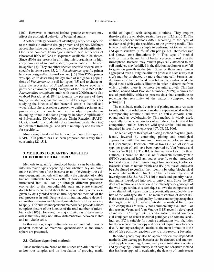
Bacteria marked by fluorescent antibodies, fluorescentmarkers and by oligonucleotic probes, can be detected bydirect microscopy using an epifluorescent microscope with anadequate filter kit. The method is simple and the counts arerapid and precise, but the limit of detection is related to thefield of view and to the matrix of the sample. Interfacing anepifluorescent microscope with a charged-coupled device(CCD) camera and image analysis software can enhance thesensitivity to a single cell level (Fig. 1). The main problemusing direct microscopy is the high background offluorescence coming from the root, the soil particles and thecontaminants.
However, the recent development of confocal laser scan-ning microscopy (CLSM) has significantly reduced some ofthese limitations. CLSM is a powerful apparatus for visualiz-ing with high resolution microbial cells labeled by fluorescentantibodies, GFP or oligonucleotidic probes. Because three-dimensional views can be generated, CLSM readily lendsitself to digital processing, by which images of thin opticalsections can be reassembled into a composite, 3D image. Themajor advantage of CLSM is that the confocal imaging systemallows the detection of signals only from the focused plane,limiting background fluorescence arising from materials such
as plant tissue, soil particles or organic debris. Moreover, byusing different fluorescence channels, CLSM allows thesimultaneous detection of different bacterial populations and/or secondary metabolites. For all these advantages, there is anincreasing use of CLSM to localize introduced microorgan-isms on plant roots [5, 13, 38, 99]. The limitation in the use ofCLSM is the cost of the instrument.
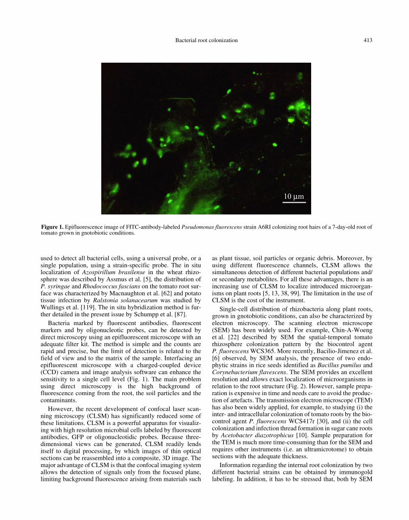
Single-cell distribution of rhizobacteria along plant roots,grown in gnotobiotic conditions, can also be characterized byelectron microscopy. The scanning electron microscope(SEM) has been widely used. For example, Chin-A-Woenget al. [22] described by SEM the spatial-temporal tomatorhizosphere colonization pattern by the biocontrol agentP. fluorescens WCS365. More recently, Bacilio-Jimenez et al.[6] observed, by SEM analysis, the presence of two endo-phytic strains in rice seeds identified as Bacillus pumilus andCorynebacterium flavescens. The SEM provides an excellentresolution and allows exact localization of microorganisms inrelation to the root structure (Fig. 2). However, sample prepa-ration is expensive in time and needs care to avoid the produc-tion of artefacts. The transmission electron microscope (TEM)has also been widely applied, for example, to studying (i) theinter- and intracellular colonization of tomato roots by the bio-control agent P. fluorescens WCS417r [30], and (ii) the cellcolonization and infection thread formation in sugar cane rootsby Acetobacter diazotrophicus [10]. Sample preparation forthe TEM is much more time-consuming than for the SEM andrequires other instruments (i.e. an ultramicrotome) to obtainsections with the adequate thickness.
Information regarding the internal root colonization by twodifferent bacterial strains can be obtained by immunogoldlabeling. In addition, it has to be stressed that, both by SEM
Figure 1. Epifluorescence image of FITC-antibody-labeled Pseudomonas fluorescens strain A6RI colonizing root hairs of a 7-day-old root oftomato grown in gnotobiotic conditions.
414 E. Gamalero et al.
and TEM, only very small root samples can be analyzed. Toallow the investigation of a more general root bacterial distri-bution, electron microscopy should be combined with othermethods [1].
5. CONCLUSIONS AND PERSPECTIVES
During the present review, different methodologies toquantify and localize introduced bacterial strains have beenpresented. These methods have their own advantages and lim-itations. Some only allow the quantification of cultivable bac-teria, putting aside the so-called VBNC (culture-dependentmethods). Others allow the quantification of the cultivable andnon-cultivable cells of the introduced bacterial strain; how-ever, they do not allow the discrimination of the viable andnon-viable cells (immunofluorescence and PCR). Taking intoconsideration the limitations of both types of methods, a com-bination of culture-dependent methods and serological meth-ods (IFC) or a combination of culture-dependent andmolecular methods (colony hybridization) have been pro-posed. Combining colony counts of an antibiotic-resistantstrain with the immunofluorescence technique has been suc-cessfully applied to monitoring the distribution and dynamicof bacteria in soil or on the root [39, 49].
Taking into account the advantages and the limitations ofthe different methods, a polyphasic approach based on the useof different enumeration methods (conventional plate count-ing, luminometry, fluorimetry, flow cytometry and quantita-tive PCR) has been proposed by Cassidy et al. [19] todiscriminate the total number, and the number of viable andcultivable bacterial cells.
A polyphasic approach could also be proposed to bothcharacterize the localization and the activity of the cells of theintroduced bacterial strain. As an example, Lubeck et al. [59],applied the combination of fluorescent antibodies and FISH tostudying sugar beet root localization of P. fluorescens DR54by CLSM; this dual staining protocol allowed cellular activityto be recorded in both single cells and microcolonies duringthe bacterial establishment on the root. Similarly, Unge et al.[103] developed a dual gfp-luxAB marker system to monitorsimultaneously the cell number and activity of specificbacterial populations. They recently applied this dual markerto characterizing the population size, the metabolic activityand the distribution pattern of P. fluorescens SBW25 alongwheat roots by luminometry, flow cytometry and CLSM[104].
Over the last few years, there has been an increased interestin microscopic observations of the microflora in the rhizo-sphere [13, 38, 98], renewing the early studies of Foster [34].This revival of interest is related to the progress made in themicroscopy apparatus and in the molecular and serologicalmarkers.
In conclusion, different techniques combining multiplestaining/tagging methods should provide more insight into thereciprocal interactions between the plant and the microorgan-isms in the rhizosphere and about the spatial-temporal coloni-zation pattern and the physiological status of a microbialinoculant along the root.
Acknowledgments: The authors are grateful to Sylvie Mazurier andChristophe Mougel for the interesting and stimulating discussions and AnnaFusconi for the photo obtained by SEM.
Figure 2. SEM image of Pseudomonas fluorescens A6RI colonizing the root surface of a 7-day-old root of tomato grown in gnotobioticconditions.

Bacterial root colonization 415
REFERENCES
[1] Achouak W., Heulin T., Villemin G., Balandreau J., Root coloni-zation by symplasmata-forming Enterobacter agglomerans,FEMS Microbiol. Ecol. 13 (1994) 287–294.
[2] Amann R., Ludwig W., Ribosomal RNA-targeted nucleic acidprobes for studies in microbial ecology, FEMS Microbiol. Rev. 24(2000) 555–565.
[3] Amann R., Fuchs B., Behrens S., The identification of microor-ganisms by fluorescence in situ hybridization, Curr. Opin. Biotech.12 (2001) 231–236.
[4] Andersen J.B., Sternberg C., Poulsen L.K., Bjørn S.P., GivskovM., Molin S., New unstable variants of green fluorescent proteinfor studies of transient gene expression in bacteria, Appl. Environ.Microbiol. 64 (1998) 2240–2246.
[5] Assmus B., Hutzler P., Kirchhof G., Amann R., Lawrence J.R.,Hartmann A., In situ localization of Azospirillum brasilense in therhizosphere of wheat with fluorescently labeled rRNA-targetedoligonucleotide probes and scanning confocal microscopy, Appl.Environ. Microbiol. 61 (1995) 1013–1019.
[6] Bacilio-Jiménez M., Aguilar-Flores S., del Valle M.V., Pérez A.,Zepeda A., Zenteno E., Endophytic bacteria in rice seeds inhibitearly colonization of roots by Azospirillum brasiliense, Soil Biol.Biochem. 33 (2001) 167–172.
[7] Bakker P.A.H.M., Bakker A.W., Marugg J.D., Weisbeek P.J.,Schippers B., Bioassay for studying the role of siderophores inplant growth stimulation by fluorescent Pseudomonas spp. in shortpotato rotations, Soil Biol. Biochem. 19 (1987) 443–449.
[8] Beauchamp C.J., Kloepper J.W., Lewke P.A., Luminometric anal-yses of plant root colonization by bioluminescent pseudomonads,Can. J. Microbiol. 39 (1993) 434–441.
[9] Belas R., Mileham A., Cohn D., Hilmen M., Simon M., SilvermanM., Bacterial bioluminescence: isolation and expression of theluciferase genes from Vibrio harveyi, Science 218 (1982) 791–793.
[10] Bellone C.H., De Bellone S.D.V.C., Pedraza R.O., Monzon M.A.,Cell colonization and infection thread formation in sugar caneroots by Acetobacter diazotrophicus, Soil Biol. Biochem. 29(1997) 965–967.
[11] Benizri E., Schoeny A., Picard C., Courtade A., Guckert A., Exter-nal and internal root colonization of maize by two Pseudomonasstrains: enumeration by enzyme–linked immunosorbent assay(ELISA), Curr. Microbiol. 34 (1997) 297–302.
[12] Björklöf K., Jørgensen K.S., Applicability of non antibiotic resist-ance marker genes in ecological studies of introduced bacteria inforest soil, FEMS Microbiol. Ecol. 38 (2001) 179–188.
[13] Bloemberg G.V., Wijfjes A.H.M., Lamers G.E.M., Stuurman N.,Lugtenberg B.J.J., Simultaneous imaging of Pseudomonas fluo-rescens WCS365 populations expressing three different autofluo-rescent proteins in the rhizosphere: new perspectives for studyingmicrobial communities, Mol. Plant-Microbe Interact. 13 (2000)1170–1176.
[14] Blot M., Hauer B., Monnet G., Tn5 bleomycin resistance geneconfers improved survival growth advantage in Escherichia coli,Mol. Gen. Genet. 242 (1994) 595–601.
[15] Braun-Howland E.B., Vescio P.A., Nierzwicki-Bauer S.A., Use ofa semplified cell blot technique and 16S rRNA-directed probes foridentification of common environmental isolates, Appl. Environ.Microbiol. 59 (1993) 3219–3224.
[16] Bull C.T., Weller D.M., Thomashow L.S., Relationship betweenroot colonisation and suppression of Gaeumannomyces graminisvar. tritici by Pseudomonas fluorescens strain 2-79, Phytopathol-ogy 81 (1991) 954–959.
[17] Burlage R.S., Kuo C.-T., Living biosensors for the managementand manipulation of microbial consortia, Ann. Rev. Microbiol. 48(1994) 291–309.
[18] Burr T.J., Schroth M.N., Suslow T., Increased potato yield bytreatment of seed pieces with specific strain of Pseudomonas fluo-
rescens and Pseudomonas putida, Phytopathology 68 (1978)1377–1383.
[19] Cassidy M.B., Leung K.T., Lee H., Trevors J.T., Comparison ofenumeration methods for culturable Pseudomonas fluorescenscells marked with green fluorescent protein, J. Microbiol. Methods40 (2000) 135–145.
[20] Chalfie M., Tu Y., Euskirchen G., Ward W.W., Prasher D.C.,Green fluorescent protein as a marker for gene expression, Science263 (1994) 802–805.
[21] Chapon A., Guillerm A.Y., Delalande L., Lebreton L., SarniguetA., Dominant colonization of wheat roots by Pseudomonas fluo-rescens Pf29A and selection of the indigenous microflora in thepresence of the take-all fungus, Eur. J. Plant Pathol. 108 (2002)449–459.
[22] Chin-A-Woeng T.F.C., de Priester W., Van der Bij A., LugtenbergB.J.J., Description of the colonization of a gnotobiotic tomatorhizosphere by Pseudomonas fluorescens biocontrol strainWCS365, using scanning electron microscopy, Mol. Plant-Microbe Interact. 10 (1997) 79–86.
[23] Chitarra L.G., Langerak C.J., Bergervoet J.H.W., Van der BulkR.W., Detection of the plant pathogenic bacterium Xanthomonascampestris pv. campestris in seed extracts of Brassica sp. applyingfluorescent antibodies and flow cytometry, Cytom. 47 (2002) 118–126.
[24] Christensen H., Hansen M., Sorensen J., Counting and size classi-fication of active soil bacteria by fluorescence in situ hybridizationand rRNA oligonucleotide probe, Appl. Environ. Microbiol. 65(1999) 1753–1760.
[25] Cook R.J., Thomashow L.S., Weller D.M., Fujimoto D., MazzolaM., Bangera G., Kim D.-S., Molecular mechanisms of defence byrhizobacteria against root disease, Proc. Natl. Acad. Sci. USA 92(1995) 4197–4201.
[26] De Leij F.A.A.M., Whipps J.M., Lynch J.M., The use of colonydevelopment for the characterization of the bacterial community insoil and on roots, Microb. Ecol. 27 (1994) 81–97.
[27] De Lorenzo V., Designing microbial systems for gene expressionin the field, Trends Biotechnol. 12 (1994) 365–371.
[28] Devanas M.A., Rafaeli-Eshkol D., Stotzky G., Survival of plasmidcontaining strains of Escherichia coli in soil: effect of plasmid sizeand nutrients on survival of hosts and maintenance of plasmids,Curr. Microbiol. 13 (1986) 269–277.
[29] De Wet J.R., Wood K.V., Helinski D.R., De Luca M., Cloning offirefly luciferase cDNA and the expression of active luciferase inEscherichia coli, Proc. Natl. Acad. Sci. USA 82 (1985) 7870–7873.
[30] Duijff B.J., Gianinazzi-Pearson V., Lemanceau P., Involvement ofouter membrane lipopolysaccharides in the endophytic coloniza-tion of tomato roots by biocontrol Pseudomonas fluorescens strainWCS417r, New. Phytol. 35 (1997) 325–334.
[31] Engebrecht J., Nealson K.H., Silverman M., Bacterial biolumines-cence: isolation and genetic analysis of functions from Vibriofischeri, Cell 32 (1983) 773–781.
[32] Errampalli D., Leung K., Cassidy M.B., Kostrzynska M., BlearsM., Lee H., Trevors J.T., Applications of the green fluorescent pro-tein as a molecular marker in environmental microorganisms,J. Appl. Microbiol. Methods 35 (1999) 187–199.
[33] Flemming C.A., Leung K.T., Lee H., Trevors J.T., Greer C.W.,Survival of lux-lac marked biosurfactant producing Pseudomonasaeruginosa UG2L in soil monitored by non selective plating andPCR, Appl. Environ. Microbiol. 60 (1994) 1606–1613.
[34] Foster R.C., Microenvironments of soil microorganisms, Biol.Fertil. Soils 6 (1988) 189–203.
[35] Glandorf D.C.M., Brand I., Bakker P.A.H.M., Stability ofrifampicin resistance as a marker for root colonization studies ofPseudomonas putida in field, Plant and Soil 147 (1992) 135–142.
[36] Glick B.R., The enhancement of plant growth by free-living bac-teria, Can. J. Microbiol. 41 (1995) 109–117.
416 E. Gamalero et al.
[37] Gregori G., Citterio S., Ghiani R., Labra M., Sgorbati S., Brown S.,Denis M., Resolution of viable and membrane compromised bac-teria in freshwater and marine waters based on analytical flowcytometry and nucleic acid double staining, Appl. Environ. Micro-biol. 67 (2001) 4662–4670.
[38] Hansen M., Kragelund L., Nybroe O., Sørensen J., Early coloniza-tion of barley roots by Pseudomonas fluorescens studied byimmunofluorescence technique and confocal laser scanningmicroscopy, FEMS Microbiol. Ecol. 23 (1997) 353–360.
[39] Hase C., Mascher F., Moënne-Loccoz Y., Défago G., Nutrientdeprivation and subsequent survival of biocontrol Pseudomonasfluorescens CHA0 in soil, Soil Biol. Biochem. 31 (1999) 1181–1188.
[40] Hiltner L., Über neuere erfahrungen und problem auf dem gebeitder bodenbakteriologie und unter besonderer berucksichtigung dergrundungung und brache, Arb. Dtsch. Landwirt. Ges. 98 (1904)59–78.
[41] Jansson J.K., Tracking genetically engineered microorganisms innature, Curr. Opin. Biotechnol. 6 (1995) 275–283.
[42] Jansson J.K., Leser T., Quantitative PCR of environmental sample,in: Akkermans A.D.L., van Elsas J.D., de Bruijn F.J. (Eds.),Molecular Microbial Ecology Manual, Kluwer Academic Publish-ers, Dordrecht, The Netherlands 1996, pp. 1–19.
[43] Jansson J.K., Reporter genes for monitoring microbial cell activityand/or the environment. An opinion, MAREP (Marker/Reportergenes in microbial ecology): A concerted action: European Com-mission Biotechnology Programme, DGXII, Boras, Sweden,1998.
[44] Jansson J.K., Molin S., Romantschuk M., Methods for monitoringgenetically modified microorganisms (GMMs) in nature, NT TechReport 455, 2000.
[45] Johnsen K., Enger O., Jacobsen C.S., Thirup L., Torsvik V., Quan-titative selective PCR of 16S ribosomal DNA correlates well withselective agar plating in describing population dynamics of indig-enous Pseudomonas spp. in soil hot spot, Appl. Environ. Micro-biol. 65 (1999) 1786–1789.
[46] Kloepper J.W., Schroth M.N., Plant growth promoting rhizobacte-ria and plant growth under gnotobiotic conditions, Phytopathology71 (1981) 642–664.
[47] Kloepper J.W., Beauchamp C.J., A review of issues related tomeasuring colonization of plant roots by bacteria, Can. J. Micro-biol. 38 (1992) 1219–1232.
[48] Kozaczuk M.M., Kopciñska J., Lotocka B., Golinowski W.,Skorupska A., Infection of clover by plant growth promotingPseudomonas fluorescens strain 267 and Rhizobiumleguminosarum bv. trifolii studied by mTn5-gusA, Ant. van Leew.78 (2000) 1–11.
[49] Kragelund L., Nybroe O., Competition between Pseudomonas flu-orescens Ag1 and Alcaligenes eutrophus JMP134 (pJP4) duringcolonization of barley roots, FEMS Microbiol. Ecol. 20 (1996) 41–51.
[50] Kragelund L., Hosbond C., Nybroe O., Distribution of metabolicactivity and phosphate starvation response of lux-tagged Pseu-domonas fluorescens reporter bacteria in the barley rhizosphere,Appl. Environ. Microbiol. 63 (1997) 4920–4928.
[51] Latour X., Philippot L., Corberand T., Lemanceau P., The estab-lishment of an introduced community of fluorescent pseudomon-ads is affected both by the soil-type and the rhizosphere, FEMSMicrobiol. Ecol. 30 (1999)163–170.
[52] Leeman M., Raaijmakers J.M., Bakker P.A.H.M., Schippers B.,Immunofluorescence colony staining for monitoring pseudomon-ads introduced into soil, in: Beemster A.B.R., Bollen G.J., GerlaghM., Ruissen M.T., Schippers B., Tempel A. (Eds.), Biotic Interac-tions and Soilborne diseases, Elsevier, Amsterdam, 1991, pp. 374–380.
[53] Lemanceau P., Bakker P.A.H.M., De Kogel W.J., Alabouvette C.,Schippers B., Effect of pseudobactin 358 produced by Pseu-domonas putida WCS358 on suppression of fusarium wilt of car-
nations by non pathogenic Fusarium oxysporum Fo47, Appl.Environ. Microbiol. 58 (1992) 2978–2982.
[54] Lemanceau P., Alabouvette C., Suppression of fusarium-wilts byfluorescent pseudomonads: mechanisms and applications, Biocon-trol Sci. Technol. 3 (1993) 219–234.
[55] Lemanceau P., Corberand T., Gardan L., Latour X., Laguerre G.,Boeufgras J.M., Alabouvette C., Effect of two plant species, flax(Linum usitatissimum L.) and tomato (Lycopersicon esculentumMill.) on the diversity of soilborne populations of fluorescentPseudomonads, Appl. Environ. Microbiol. 61 (1995) 1004–1012.
[56] Lifshitz R., Kloepper R.W., Kozlowsli M., Simonson C.,Carlstone J., Tipping E.M., Zaleska I., Growth promotion ofcanola (rapeseed) seedlings by a strain of Pseudomonas putidaunder gnotobiotic conditions, Can. J. Microbiol. 33 (1987) 390–395.
[57] Liljeroth E., Burgers S.L.G.E., Van Veen J.A., Changes in bacte-rial populations along the roots in wheat (Triticum aestivum L.)seedlings, Biol. Fertil. Soil 10 (1991) 276–280.
[58] Lowder M., Oliver D.J., The use of modified GFP as a reporter formetabolic activity in Pseudomonas putida, Microb. Ecol. 41(2001) 310–313.
[59] Lübeck P.S., Hansen M., Sørensen J., Simultaneous detection ofthe establishment of seed inoculated Pseudomonas fluorescensstrain DR54 and native soil bacteria on sugar beet rot surfacesusing fluorescence antibody and in situ hybridization techniques,FEMS Microbiol. Ecol. 33 (2000) 11–19.
[60] Ma W., Zalec K., Glick B.R., Biological activity and colonizationpattern of the bioluminescence-labeled plant growth-promotingbacterium Kluyvera ascorbata SUD165/26, FEMS Microbiol.Ecol. 35 (2001) 137–144.
[61] MacCrady M.H., The numerical interpretation of fermentationtube results, J. Infect. Dis. 17 (1915) 183–212.
[62] Macnaughton S.J., Booth T., Embley T.M., O’Donnell A.G.,Physical stabilization and confocal microscopy of bacteria on rootsusing 16S rRNA targeted, fluorescent-labeled oligonucleotideprobes, J. Microbiol. Methods 26 (1996) 279–285.
[63] Mahaffee W.F., Bauske E.M., Van Vuurde J.W.L., Van der WolfJ.M., Van der Brink M., Kloepper J.W., Comparative analysis ofantibiotic resistance, immunofluorescent colony staining, and atransgenic marker (bioluminescence) for monitoring the environ-mental fate of rhizobacterium, Appl. Environ. Microbiol. 63(1997) 1617–1622.
[64] Maloney P.E., Van Bruggen A.H.C., Hu S., Bacterial communitystructure in relation to the carbon environment in lettuce andtomato rhizospheres and bulk soil, Microb. Ecol. 34 (1997) 109–117.
[65] Martin-Laurent F., Philippot L., Hallet S., Chaussod R., GermonJ.-C., Soulas G., Catroux G., DNA extraction from soils: old biasfor new microbial diversity analysis methods, Appl. Environ.Microbiol. 67 (2001) 2354–2359.
[66] Martin-Laurent F., Piutti S., Hallet S., Wagschal I., Philippot L.,Catroux G., Soulas G. Monitoring of atrazine treatment on soilbacterial, fungal, and atrazine degrading communities by quantita-tive competitive PCR, Pest Management Science, in press.
[67] Mavingui P., Laguerre G., Berge E., Heulin T., Genetic and phe-notypic diversity of Bacillus polymyxa in soil and in the wheatrhizosphere, Appl. Environ. Microbiol. 58 (1992) 1894–1903.
[68] Mirleau P., Delorme S., Philippot L., Meyer J.M., Mazurier S.,Lemanceau P., Fitness in soil and rhizosphere of Pseudomonas flu-orescens C7R12 compared with a C7R12 mutant affected in pyo-verdine synthesis and uptake, FEMS Microbiol. Ecol. 34 (2000)35–44.
[69] Mirleau P., Philippot L., Corberand T., Lemanceau P., Involve-ment of nitrate reductase and pyoverdine in competitiveness ofPseudomonas fluorescens C7R12 in soil, Appl. Environ. Micro-biol. 67 (2001) 2627–2635.
Bacterial root colonization 417
[70] Molin S., Givskov M., Application of molecular tools for in situmonitoring of bacterial growth activity, Environ. Microbiol. 1(1999) 383–391.
[71] Normander B., Hendriksen N.B., Nybroe O., Green fluorescentprotein marked Pseudomonas fluorescens: localization, viabilityand activity in the natural barley rhizosphere, Appl. Environ.Microbiol. 65 (1999) 4646–4651.
[72] Orvos D.R., Lacy G.H., Cairns J. Jr., Genetically engineeredErwinia carotovora: survival, intraspecific competition andeffects upon selected bacterial genera, Appl. Environ. Microbiol.56 (1990) 1689–1694.
[73] Philippot L., Clays-Josserand A., Lensi R., Use of Tn5 mutants toassess the role of dissimilatory nitrite reductase in the competitiveabilities of two strains in soil, Appl. Environ. Microbiol. 61 (1995)1426–1430.
[74] Picard C., Ponsonnet C., Paget E., Nesme X., Simonet P., Detec-tion and enumeration of bacteria in soil by direct DNA extractionand polymerase chain reaction, Appl. Environ. Microbiol. 58(1992) 2717–2722.
[75] Pierson L.S. III, Keppenne V.D., Wodd D.W., Phenazine antibi-otic biosynthesis in Pseudomonas aureofaciens 30-84 is regulatedby PhzR in response to cell density, J. Bacteriol. 176 (1994) 3966–3974.
[76] Prosser J.I., Molecular marker systems for detection of geneticallyengineered microorganisms in the environment, Microbiol. 140(1994) 5–17.
[77] Raaijmakers J.M., Wilbert B., Punte H.L.M., Siderophore receptorPupA as a marker to monitor wild-type Pseudomonas putidaWCS358 in natural environments, Appl. Environ. Microbiol. 60(1994) 1184–1190.
[78] Raaijmakers J.M., Leeman M., Van Oorshot P., Van der Sluis L.,Schippers B., Bakker P.A.H.M., Dose-response relationships inbiological control of fusarium wilt of radish by Pseudomonas spp.,Phytopathology 85 (1995) 1075–1081.
[79] Ramos C., Molina L., Mølbak L., Ramos J.L., Molin S., A biolu-minescent derivative of Pseudomonas putida KT2440 for deliber-ate release into the environment, FEMS Microbiol. Ecol. 34 (2000)91–102.
[80] Räsänen L.A., Elväng A.M., Jansson J., Lindström K., Effect ofheat stress on cell activity and cell morphology of the tropicalrhizobium Sinorhizobium arboris, FEMS Microbiol. Ecol. 34(2001) 267–278.
[81] Rattray E.A.S., Prosser J.I., Glover L.A., Killham K., Characteri-zation of rhizosphere colonization by luminescent Enterobactercloacae at the population and single-cell levels, Appl. Environ.Microbiol. 61 (1995) 2950–2957.
[82] Remus R., Ruppel S., Jacob H.J., Hecht-Bucholz C., Merbach W.,Colonization behavior of two enterobacterial strains on cereals,Biol. Fertil. Soils 30 (2000) 550–557.
[83] Roberts D.P., Kobayashi D.Y., Dery P.D., Short N.M., An imageanalysis method for determination of spatial colonization patternsof bacteria in plant rhizosphere, Appl. Microbiol. Biotechnol. 51(1999) 653–658.
[84] Rosado A.S., Seldin L., Wolters A.C., Van Elsas J.D., Quantitative16S rDNA-targeted polymerase chain reaction and oligonucle-otide hybridization for the detection of Paenibacillus azotofixansin soil and the wheat rhizosphere, FEMS Microbiol. Ecol. 19(1996) 153–164.
[85] Saunders J.R., Pickup R.W., Morgan J.A., Winstanley C.,Saunders V.A., XylE as a marker for microorganisms, in:Akkermans A.D.L., Van Elsas J.D., de Bruijn F.J. (Eds.), Molecu-lar Ecology Manual, Kluwer Academic Publisher.
[86] Schippers B., Bakker A.W., Bakker P.A.H.M., Interactions of del-eterious and beneficial rhizospheric microorganisms and the effecton cropping practices, Ann. Rev. Phytopathol. 25 (1987) 339–358.
[87] Schumpp O., Gherbi H., Escoute J., Payre H., Drevon J.-J., In situhybridization of a radioactive RNA probe on resin-embedded leg-
ume root-nodule sections: a tool for observing gene expression inthe rhizosphere?, Agronomie, submitted for publication.
[88] Shapiro H.M., Microbial analysis at the single-cell level: task andtechniques, J. Microbiol. Methods 42 (2000) 3–16.
[89] Shishido M., Breuil C., Chanway C.P., Endophytic colonization ofspruce by plant growth-promoting rhizobacteria, FEMS Micro-biol. Ecol. 29 (1999) 191–196.
[90] Simon R., Priefer U., Pühler A., A broad host range mobilizationsystem for in vivo genetic engineering: transposon mutagenesis inGram negative bacteria, Biotechnology 1 (1983) 784–791.
[91] Smit E., Wolters A., Van Elsas J.D., Genetic stability, conjugaltransfer and expression of heterologous DNA inserted into differ-ent plasmids and the genome of Pseudomonas fluorescens in soil,Rev. Microbiol. 26 (1995) 169–179.
[92] Sørensen J., Jensen L.E., Nybroe O., Soil and rhizosphere as hab-itats for Pseudomonas inoculants: new knowledge on distribution,activity and physiological state derived from micro-scale and sin-gle cell studies, Plant and Soil 232 (2001) 97–108.
[93] Sternberg C., Christensen B.B., Johansen T., Nielsen A.T.,Andersen J.B., Givskov M., Molin S., Distribution of bacterialgrowth activity in flow-chamber biofilms, Appl. Environ. Micro-biol. 65 (1999) 4108–4117.
[94] Stintzi A., Evans K., Meyer J.-M., Poole K., Quorum-sensing andsiderophore biosynthesis in Pseudomonas aeruginosa: lasR/lasImutant exhibit reduced pyoverdine biosynthesis, FEMS Micro-biol. Lett. 166 (1998) 341–345.
[95] Suarez A., Guttler A., Stratz M., Staendner L.H., Timmis K.N.,Guzman C.A., Green fluorescent protein based reporter systemsfor genetic analysis of bacteria including monocopy applications,Gene 196 (1997) 69–74.
[96] Thirup L., Johnesn K., Winding A., Succession of indigenousPseudomonas spp. and Actinomycetes on barley roots affected bythe antagonistic strain Pseudomonas fluorescens DR54 and thefungicide imazalil, Appl. Environ. Microbiol. 67 (2001) 1147–1153.
[97] Thomas J.C., Desrosiers M., St Pierre Y., Lirette P., BisaillonJ.-G., Baudet R., Villemur R., Quantitative flow cytometric detec-tion of specific microorganisms in soil samples using rRNA tar-geted fluorescent probes and ethidium bromide, Cytom. 27 (1997)224–232.
[98] Tombolini R., Unge A., Davey M.E., de Bruijn F.J., Jansson J.K.,Flow cytometric and microscopic analysis of GFP-tagged Pseu-domonas fluorescens bacteria, FEMS Microb. Ecol. 22 (1997) 17–28.
[99] Tombolini R., Van der Gaag D.J., Gerhardson B., Jansson J.K.,Colonization pattern of the biocontrol strain Pseudomonas chloro-raphis MA342 on barley seeds visualized by using green fluores-cent protein, Appl. Environ. Microbiol. 65 (1999) 3674–3680.
[100] Troxler J., Zala M., Natsch A., Moënne-Loccoz Y., Défago G.,Autoecology of the biocontrol strain Pseudomonas fluorescensCHA0 in the rhizosphere and inside roots at later stage of plantdevelopment, FEMS Microbiol. Ecol. 23 (1997) 119–130.
[101] Tsien R.Y., The green fluorescent protein, Ann. Rev. Biochem. 67(1998) 509–544.
[102] Turnbull G.A., Morgan J.A.W., Whipps J.M., Saunders J.R., Therole of bacterial motility in the survival and spread of Pseu-domonas fluorescens in soil and in the attachment and colonizationof wheat roots, FEMS Microbiol. Ecol. 36 (2001) 21–31.
[103] Unge A., Tombolini R., Mølback L., Jansson J.K., Simultaneousmonitoring of cell number and metabolic activity of specific bac-terial populations with a dual gfp-luxAB marker system, Appl.Environ. Microbiol. 65 (1999) 813–821.
[104] Unge A., Jansson J., Monitoring population size, activity and dis-tribution of gfp-luxAB tagged Pseudomonas fluorescens SBW25during colonization of wheat, Microb. Ecol. 41 (2001) 290–300.
[105] Van Elsas J.D., Dijhstra J.M., Govaert J.M., Van Veen J.A., Sur-vival of Pseudomonas fluorescens and Bacillus subtilis introduced
418 E. Gamalero et al.
To access this journal online: www.edpsciences.org
into soil of different texture in field microplots, FEMS Microbiol.Ecol. 38 (1986) 151–160.
[106] Van Elsas J.D., Van Overbeek L.S., Feldmann A.M., DullemansA.M., de Leeuw O., Survival of genetically engineered Pseu-domonas fluorescens in soil in competition with the parent strain,FEMS Microbiol. Ecol. 85 (1991) 53–64.
[107] Van Elsas J.D., Duarte G.F., Rosado A.S., Smalla K., Microbio-logical and molecular biological methods for monitoring microbialinoculants and their effects in the soil environment, J. Microbiol.Methods 32 (1998) 133–154.
[108] Van Loon L.C., Bakker P.A.H.M., Pieterse C.M.J., Systemicresistance induced by rhizosphere bacteria, Ann. Rev. Plant Phys-iol. 26 (1998) 453–483.
[109] Van Overbeek L.S., Van Veen J.A., Van Elsas J.D., Inducedreporter gene activity, enhanced stress resistance and competitiveability of a genetically modified Pseudomonas fluorescens strainreleased into a field pot planted with wheat, Appl. Environ. Micro-biol. 63 (1997) 1965–1973.
[110] Van Vuurde J.W.L., Roozen N.J.M., Comparison of immunofluo-rescence colony-staining in media, selective isolation on pectatemedium, ELISA, and immunofluorescence cell staining for detec-tion of Erwinia carotovora subsp. atroseptica and E. chrysanthemiin cattle manure slurry, Neth. J. Plant Path. 96 (1990) 75–89.
[111] Van Vuurde J.W.L., Van Der Wolf J.M., The use of the antibodytechniques in soil, in: Alef K., Nannipieri P. (Eds.), Methods inapplied soil microbiology and biochemistry, Academic Press, Inc.New York, 1995, pp. 452–457.
[112] Veal D.A., Deere D., Ferrari B., Piper J., Attfield P.V., Fluores-cence staining and flow cytometry for monitoring microbial cells,J. Immunol. Methods 243 (2000) 191–210.
[113] Veena M.S., Van Vuurde J.W.L., Indirect immunofluorescencecolony staining method for detecting bacterial pathogens oftomato, J. Microbiol. Methods 49 (2002) 11–17.
[114] Von Caron G.N., Stephens P.J., Hewitt C.J., Powell J.R., BadleyR.A., Analysis of bacterial function by multi colour fluorescenceflow cytometry and single cell sorting, J. Microbiol. Methods 42(2000) 97–114.
[115] Waisel Y., Eshel A., Kafkafi U., Plant roots. The hidden half, Mar-cel Dekker (Ed.), 1991.
[116] Weller D.M., Biological control of soilborne plant pathogen in therhizosphere with bacteria, Annu. Rev. Phytopathol. 26 (1988)379–407
[117] Werner D., Batinic T., Feder I.S., Kosch K., Redecker D., SchulzU., Streit W., Fleischman P.V., Competitiveness of symbiotic andantagonistic soil bacteria-Development of reporter gene fusionsand specific gene probes, Proceedings of the Fourth InternationalWorkshop on Plant Growth Promoting Rhizobacteria, 1996,pp. 44–48.
[118] Wilson K.J., Molecular techniques for the study of rhizobial ecol-ogy in the field, Soil Biol. Biochem. 27 (1994) 501–514.
[119] Wullings B.A., Van Beuningen A.R., Janse J.D., AkkermansA.D.L., Detection of Ralstonia solanacearum, which causesbrown rot of potato, by fluorescent in situ hybridisation with 23SrRNA-targeted probes, Appl. Environ. Microbiol. 64 (1998)4546–4554.