METHOD OF RETENTIVE FOR MAXILLOFACIAL PROSTHESIS : A …
23
www.ijcrt.org © 2020 IJCRT | Volume 8, Issue 6 June 2020 | ISSN: 2320-2882 IJCRT2006397 International Journal of Creative Research Thoughts (IJCRT) www.ijcrt.org 2893 METHOD OF RETENTIVE FOR MAXILLOFACIAL PROSTHESIS : A REVIEW 1 Dr Shivani Kotewar , 2 Dr Nazish Baig , 3 Dr Vivek Jadhav , 4 Dr Ruchi Kasat 1 MDS II student , 2 professor and guide , 3 professor and guide, 4 professor and guide 1 muhs ABSTRACT : One of the most important factors that determines the success of a maxillofacial prosthesis is retention. Retention has always been a problem in prosthodontics. Increased retention improves comfort as well as the confidence in the patient while wearing a facial prosthesis at work and in social settings. The journey from using metal bands to using adhesives to placing implants for retaining a maxillofacial prosthesis has been fascinating and satisfying to many, but, the aim of achieving the full potential still remains incomplete. The present article tries to describe different types of retentive aids in maxillofacial prosthesis. KEYWORDS : Retention, maxillofacial prosthesis INTRODUCTION :- Retention as per The Glossary of Prosthodontic Terms-9 is defined as, “retention is that quality inherent in the dental prosthesis acting to resist the forces of dislodgement along the path of placement”. 1 The success of a prosthetic restoration of any part of the body depends on the availability of a method of attaching the artificial substitute securely to its proper place without discomfort and irritation to the tissues with which
Transcript of METHOD OF RETENTIVE FOR MAXILLOFACIAL PROSTHESIS : A …
www.ijcrt.org © 2020 IJCRT | Volume 8, Issue 6 June 2020 | ISSN: 2320-2882
IJCRT2006397 International Journal of Creative Research Thoughts (IJCRT) www.ijcrt.org 2893
METHOD OF RETENTIVE FOR
MAXILLOFACIAL PROSTHESIS : A REVIEW 1Dr Shivani Kotewar , 2Dr Nazish Baig , 3Dr Vivek Jadhav , 4Dr Ruchi Kasat
1MDS II student , 2professor and guide , 3professor and guide, 4professor and guide 1muhs
ABSTRACT :
One of the most important factors that determines the success of a
maxillofacial prosthesis is retention. Retention has always been a problem
in prosthodontics. Increased retention improves comfort as well as the
confidence in the patient while wearing a facial prosthesis at work and
in social settings. The journey from using metal bands to using adhesives to
placing implants for retaining a maxillofacial prosthesis has been fascinating
and satisfying to many, but, the aim of achieving the full potential still
remains incomplete. The present article tries to describe different types of
retentive aids in maxillofacial prosthesis.
KEYWORDS :
Retention, maxillofacial prosthesis
INTRODUCTION :-
Retention as per The Glossary of Prosthodontic Terms-9 is defined as,
“retention is that quality inherent in the dental prosthesis acting to resist the
forces of dislodgement along the path of placement”.1
The success of a prosthetic restoration of any part of the body depends
on the availability of a method of attaching the artificial substitute securely
to its proper place without discomfort and irritation to the tissues with which
www.ijcrt.org © 2020 IJCRT | Volume 8, Issue 6 June 2020 | ISSN: 2320-2882
IJCRT2006397 International Journal of Creative Research Thoughts (IJCRT) www.ijcrt.org 2894
it comes in contact. The materials used for rehabilitation have travelled a long
way from wood to polymers and the retentive aids used from metal bands to
implants.
Methods of Retention used for maxillofacial prostheses fall into four
categories: 2
1. Anatomical, in which the retentive contours existing at the site of
deformity are used to retain the prosthesis.
2. Chemical, in which adhesive materials are used to retain the prosthesis.
3. Mechanical.
4. Implant, in which implant fixtures anchored into the bone are used to
retain the facial prosthesis.
1. ANATOMIC RETENTION:
Anatomic undercut areas can always be created by planning before
and after surgery as a mode of retention for maxillofacial prosthesis.
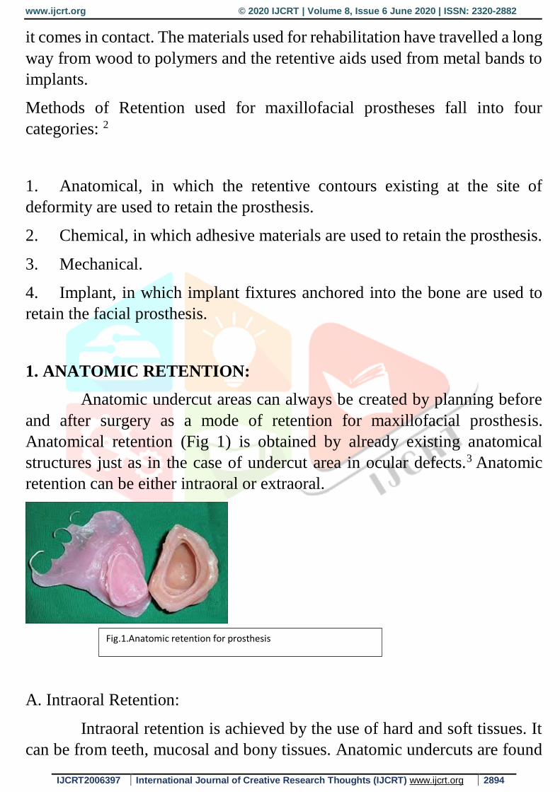
Anatomical retention (Fig 1) is obtained by already existing anatomical
structures just as in the case of undercut area in ocular defects.3 Anatomic
retention can be either intraoral or extraoral.
A. Intraoral Retention:
Intraoral retention is achieved by the use of hard and soft tissues. It
can be from teeth, mucosal and bony tissues. Anatomic undercuts are found
Fig.1.Anatomic retention for prosthesis
www.ijcrt.org © 2020 IJCRT | Volume 8, Issue 6 June 2020 | ISSN: 2320-2882
IJCRT2006397 International Journal of Creative Research Thoughts (IJCRT) www.ijcrt.org 2895
in the palatal area, cheek, retromolar area, remaining teeth, alveolar ridge,
septum and anterior nasal aperture. 4 Large alveolar ridge and high arched
palate provides greater retention than flat ridges and palate. Intraoral
retentive aids are usually considered comfortable for the patient for easy
removal and for the examination of the surgical site by the dentist in order to
check for recurrence of tumour.
B. Extraoral Retention:
Extraoral retention can be achieved from hard and soft tissues of
maxillofacial and neck region. Deep undercuts create difficulties in insertion
and removal of prosthesis. Soft tissues create problem due to their mobility
and lesser resistance to displacement when a force is applied. 4 Soft tissue
undercuts are usually in the maxillary sinus, nasal cavity and orbital regions.
The advantages of prosthesis used in these areas are that they are cost
effective, aesthetic and easy to fabricate.
2. CHEMICAL:
Chemical retention is achieved by adhesives. According to GPT-9,
maxillofacial prosthetic adhesive is “a material used to adhere external
prosthesis to the skin and associated structures around the periphery of an
external anatomic defect.” Ideal properties of adhesives for maxillofacial
prostheses: 5
1. The adhesive must be biocompatible, non toxic and non irritating.
2. The material should be odourless and moisture repellent.
3. The dried adhesive should be porous and absorbent to allow passage of
secretions.
4. The patient should find it easy to apply.
5. The material should dry quickly.
6. The bond should hold the prosthesis in place for atleast 12hours daily.
7. The adhesive must be easy to remove without injuring the skin and
prosthesis.
www.ijcrt.org © 2020 IJCRT | Volume 8, Issue 6 June 2020 | ISSN: 2320-2882
IJCRT2006397 International Journal of Creative Research Thoughts (IJCRT) www.ijcrt.org 2896
8. The adhesive should be available in a travel sized package.
Adhesives are considered as the most popular retentive aid in maxillofacial
prosthesis retention. The selection of an adhesive is based on certain criteria.
They include:
1. Bond strength of the adhesive to the prosthetic material and recipient
tissues.
2. Biocompatibility
3. Prosthesis design.
4. Type and quality of patient’s skin.
5. Composition and viscosity.
6. Handling, storage and shelf life. 4
These are available as acrylic or silicone based adhesives, latex, spirit
gum or water based adhesives. 4,6
A. Acrylic resin adhesives:
Acrylic resin adhesives consists of acrylic resin dispersed in a water
solvent which when evaporated leaves a rubber like substance. Dispersions
of synthetic resins and rubbers have recently been termed latex adhesives.
The addition of surfactants and the attainment of the proper particle allow for
controlled penetration and wetting of these adhesives. In order for these
adhesives to be successful, one surface must be permeable to water to dry the
dispersion and develop the bond.
B. Silicone adhesive:
Silicone adhesives are a form of room temperature vulcanizing (RTV)
silicones usually dissolved in a solvent. Once the adhesive is applied, the
solvent evaporates and a tacky adhesive results, which may then be contact
bonded to another surface such as a skin. These adhesive develop good
resistance to moisture and weathering with low water sorption. They can
withstand the effects of sunlight, ozone, contact with many oils and
www.ijcrt.org © 2020 IJCRT | Volume 8, Issue 6 June 2020 | ISSN: 2320-2882
IJCRT2006397 International Journal of Creative Research Thoughts (IJCRT) www.ijcrt.org 2897
chemicals and bio-deterioration. A disadvantage of this material is a low
adhesive strength.
C. Pressure sensitive tapes:
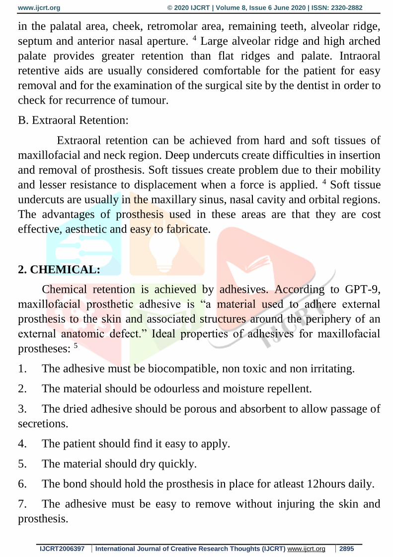
Pressure sensitive tapes (Fig 2) used in the retention of facial prostheses
are applied by finger pressure in the absence of heat or solvents. These tapes
consists of a backing strip composed of cloth, paper, film, foil or a laminate
strip coated with a pressure – sensitive adhesive.The tape has adhesive on
both surfaces. The bond of the Bi face tape to skin is weaker than the acrylic
resin adhesive. The bi-Face tape can be used on materials with poor
flexibility and for patients whose defects demonstrate little or no movement.
D. Rubber based liquid adhesive:
Rubber occurs in nature as latex, which is obtained by tapping the bark
of rubber trees. The latex thus obtained is readily soluble in organic solvent,
such as benzene or petroleum spirits, to form a natural rubber adhesive. This
mixture quickly gels because of atmospheric oxidation. Subsequent
vulcanization with sulphur converts the sticky rubber into hardened state.
Dissolving the reclaimed rubber in naphtha forms a rubber cement with
excellent adhesive qualities. These natural rubber adhesive are known for
their dry tack or their ability to adhere two fresh non-sticky surfaces together.
Fig.2. Presssure sensitive tape
www.ijcrt.org © 2020 IJCRT | Volume 8, Issue 6 June 2020 | ISSN: 2320-2882
IJCRT2006397 International Journal of Creative Research Thoughts (IJCRT) www.ijcrt.org 2898
This property of dry tack makes natural rubber adhesives useful for contact
adhesives or pressure sensitive adhesives.eg: Bard Appliance Adhesive.
E. Combination of adhesives:
The previously described adhesives can be used alone or in
combination. In most clinical practices, only one adhesive system is used to
simplify the instructions and procedures for the patient. However, the
combination of one or more adhesives can serve to solve retention problems
in various situations.
Some of the adhesives available are: 7
• Silastic MDX4-4210 medical grade elastomer
• Silastic medical adhesive silicone type A
• Secure2 Medical Adhesive
• Epithane-3 Adhesive ES
• Skin-Prep protective dressing (Fig 3)
• Uni-Solve adhesive remover
• Pros-Aide adhesive (Fig 4)
• Epithane-3 adhesive
• Telesis Silicone Adhesive (Fig 5)
• 3M bifaceis
• Hollister Medical Adhesive

www.ijcrt.org © 2020 IJCRT | Volume 8, Issue 6 June 2020 | ISSN: 2320-2882
IJCRT2006397 International Journal of Creative Research Thoughts (IJCRT) www.ijcrt.org 2899
Advantages:
Adhesives are cost effective and easy to manipulate and apply.
Maxillofacial defect patients who are not willing for implant surgical
procedures consider adhesives as a retentive aid. 8
Disadvantages:
Certain adhesives require solvents to clean after removal of prosthesis.
It provides an unreliable retention. Its degradation to the prosthetic material
adds to its disadvantages apart from irritation, perspiration and movement
that compromises the bond. In some patients, it may cause allergic reactions.8
The rationale for use of adhesives in combination is based on
overcoming the limitation of one adhesive system by combining it with
another adhesive system. The end result is a good adhesive bond between
the prosthesis and the skin.
Fig 5 : Silicone Adhesive
Fig 3: Skin Prep Wipes Fig 4: Pros-Aide Adhesive
www.ijcrt.org © 2020 IJCRT | Volume 8, Issue 6 June 2020 | ISSN: 2320-2882
IJCRT2006397 International Journal of Creative Research Thoughts (IJCRT) www.ijcrt.org 2900
3. MECHANICAL RETENTION:
Mechanical retention of facial prostheses is the oldest method of
retention reported in the field of facial prosthesis. Ambrose Pare reported the
retention of an artificial nose to the face by means of strings. Pare also
reported the retention of an artificial ear and an orbital prosthesis by a metal
or leather band worn around the head.
Mechanical Retention Mechanical anchorage includes:8
1. Eye glasses and frames.
2. Extension from denture.
3. Precision attachments.
4. Elastic and non-elastic straps.
5. Magnets.
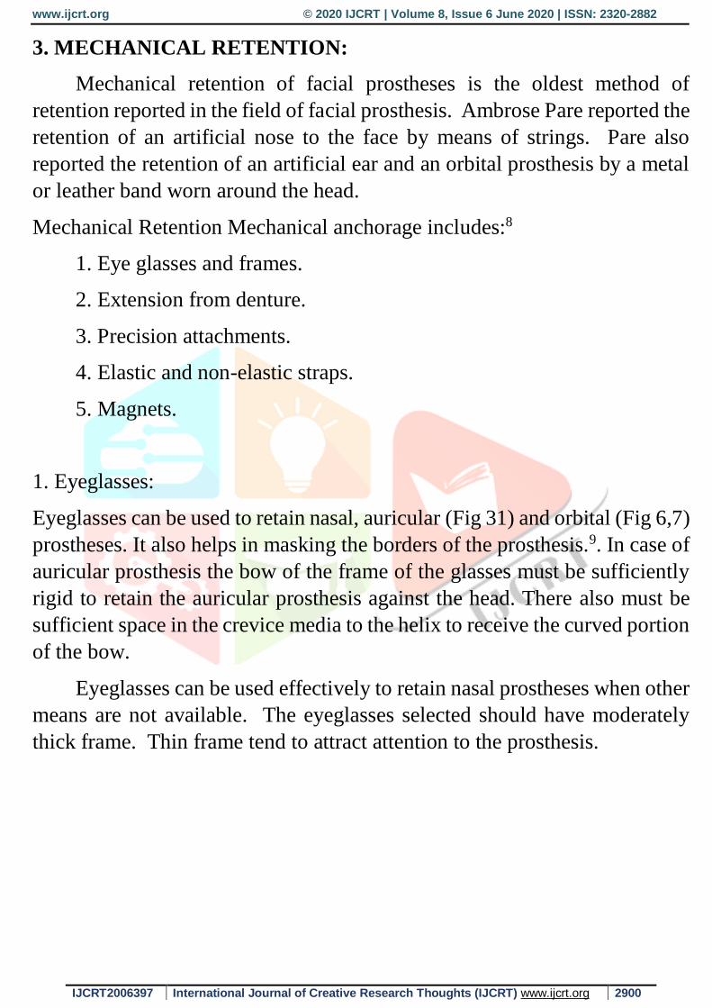
1. Eyeglasses:
Eyeglasses can be used to retain nasal, auricular (Fig 31) and orbital (Fig 6,7)
prostheses. It also helps in masking the borders of the prosthesis.9. In case of
auricular prosthesis the bow of the frame of the glasses must be sufficiently
rigid to retain the auricular prosthesis against the head. There also must be
sufficient space in the crevice media to the helix to receive the curved portion
of the bow.
Eyeglasses can be used effectively to retain nasal prostheses when other
means are not available. The eyeglasses selected should have moderately
thick frame. Thin frame tend to attract attention to the prosthesis.
www.ijcrt.org © 2020 IJCRT | Volume 8, Issue 6 June 2020 | ISSN: 2320-2882
IJCRT2006397 International Journal of Creative Research Thoughts (IJCRT) www.ijcrt.org 2901
It is advantageous, if the eyeglasses frame is made of acrylic resin,
which will enable a chemical bond by using auto polymerizing resin between
the glasses and some of the currently available types of facial materials. The
eyeglass frame should be opaque in color rather than translucent to prevent
retention marks from becoming visible.4 The attachment of a nasal prosthesis
to eyeglasses frames as a permanent fixation should be avoided since removal
of the glasses by necessity causes removal of the prosthesis, which can be
very embarrassing.
Fig 6: Ear prosthesis supported by
eyeglasses. Fig 7: Eye prosthesis supported by eyeglasses
Fig 8: Eye prosthesis supported by
eyeglasses
www.ijcrt.org © 2020 IJCRT | Volume 8, Issue 6 June 2020 | ISSN: 2320-2882
IJCRT2006397 International Journal of Creative Research Thoughts (IJCRT) www.ijcrt.org 2902
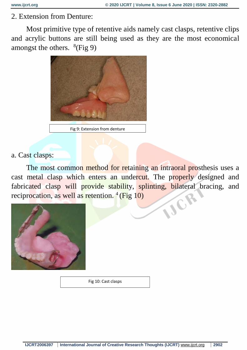
2. Extension from Denture:
Most primitive type of retentive aids namely cast clasps, retentive clips
and acrylic buttons are still being used as they are the most economical
amongst the others. 8(Fig 9)
a. Cast clasps:
The most common method for retaining an intraoral prosthesis uses a
cast metal clasp which enters an undercut. The properly designed and
fabricated clasp will provide stability, splinting, bilateral bracing, and
reciprocation, as well as retention. 4 (Fig 10)
Fig 9: Extension from denture
Fig 10: Cast clasps
www.ijcrt.org © 2020 IJCRT | Volume 8, Issue 6 June 2020 | ISSN: 2320-2882
IJCRT2006397 International Journal of Creative Research Thoughts (IJCRT) www.ijcrt.org 2903
b. Retentive clips:
Retentive clips are metallic or plastic clips that snap over the bar used
as a superstructure connected to the implants. Retentive clips have more
retentive ability in terms of breakaway retentive force than magnets.
However, retentive clips tend to wear at a faster rate than the magnets.
Retentive clips have an advantage over magnets in that they are not subject
to the effects to bodily fluids as magnets are.
Retentive clips are useful in retaining facial prostheses in patients with
good dexterity and where retention is to be maximized in areas with little
muscle force. An example of such a clinical situation would be for retention
of an auricular prosthesis.
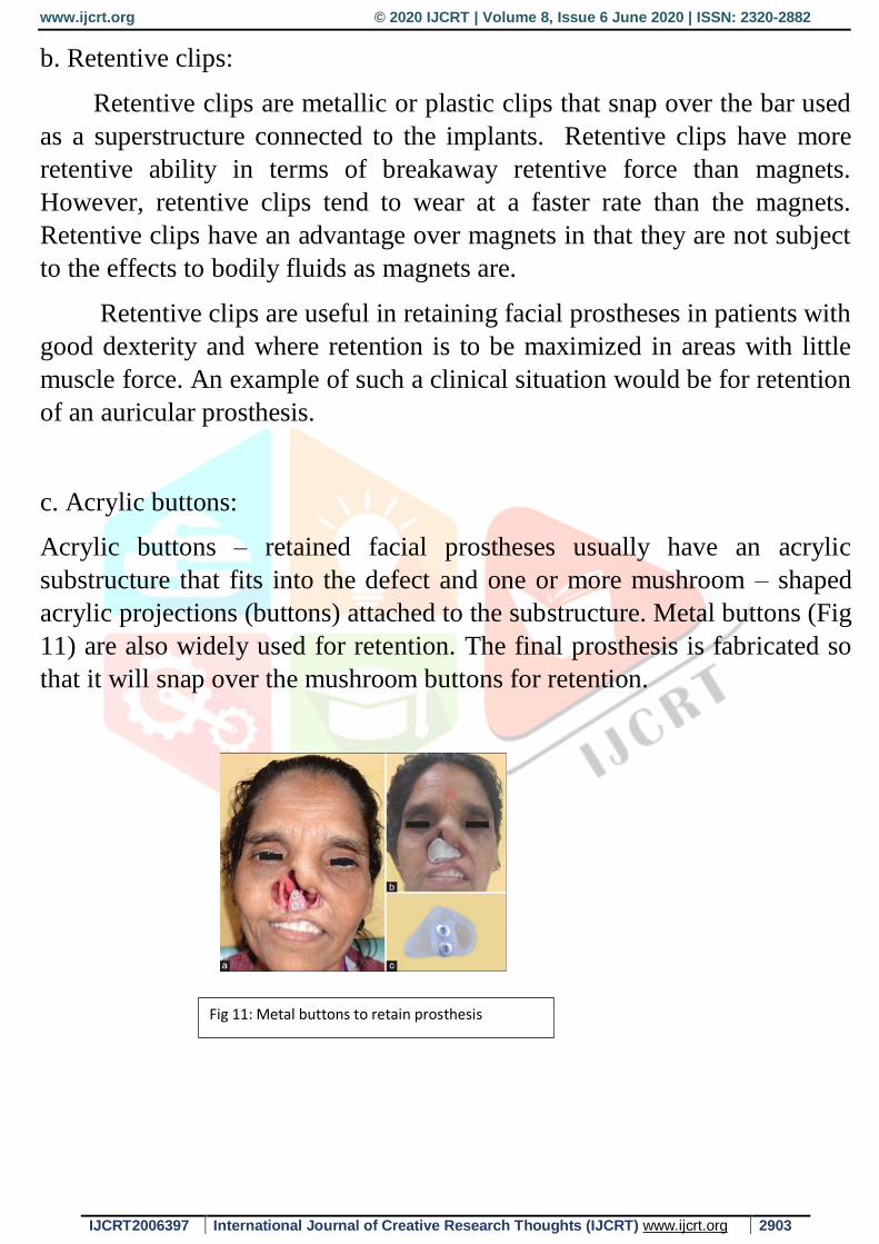
c. Acrylic buttons:
Acrylic buttons – retained facial prostheses usually have an acrylic
substructure that fits into the defect and one or more mushroom – shaped
acrylic projections (buttons) attached to the substructure. Metal buttons (Fig
11) are also widely used for retention. The final prosthesis is fabricated so
that it will snap over the mushroom buttons for retention.
Fig 11: Metal buttons to retain prosthesis
www.ijcrt.org © 2020 IJCRT | Volume 8, Issue 6 June 2020 | ISSN: 2320-2882
IJCRT2006397 International Journal of Creative Research Thoughts (IJCRT) www.ijcrt.org 2904
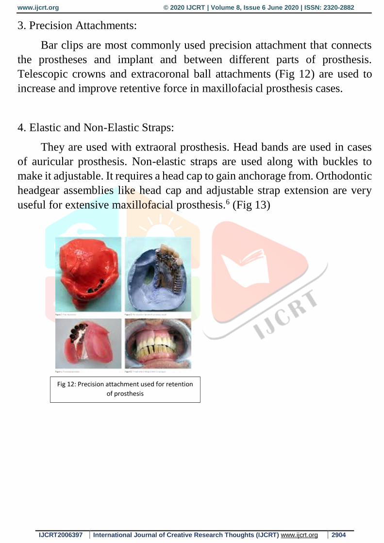
3. Precision Attachments:
Bar clips are most commonly used precision attachment that connects
the prostheses and implant and between different parts of prosthesis.
Telescopic crowns and extracoronal ball attachments (Fig 12) are used to
increase and improve retentive force in maxillofacial prosthesis cases.
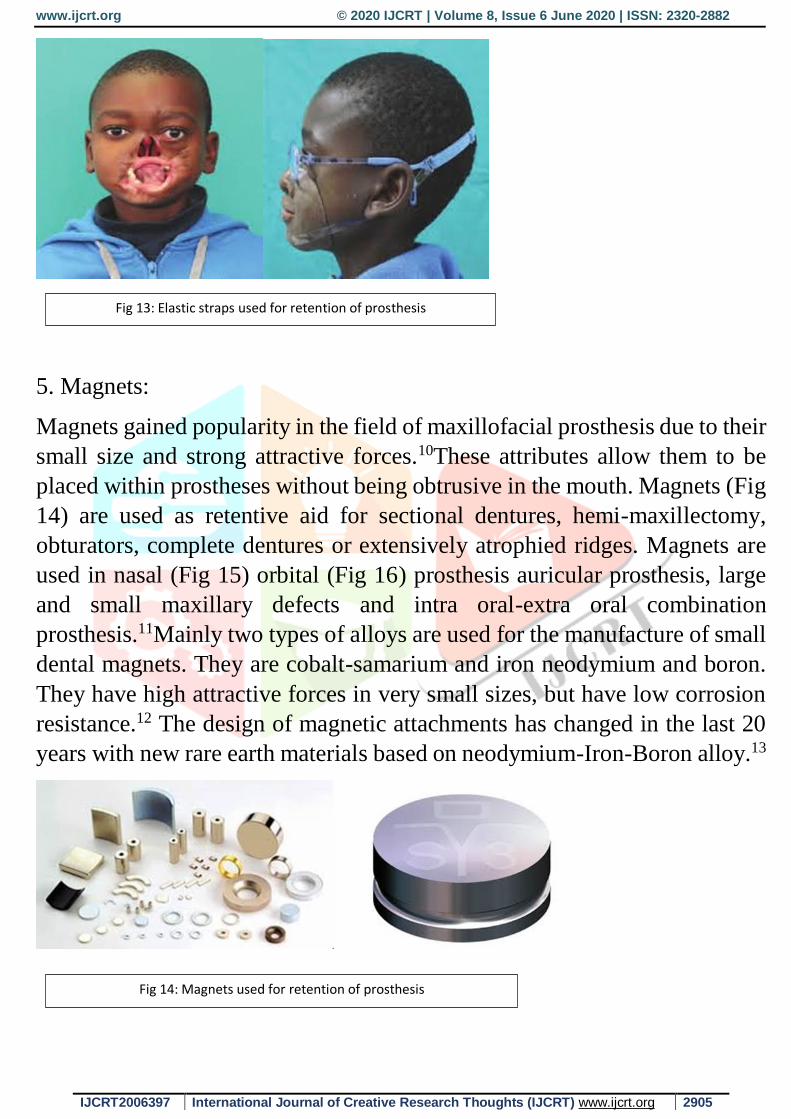
4. Elastic and Non-Elastic Straps:
They are used with extraoral prosthesis. Head bands are used in cases
of auricular prosthesis. Non-elastic straps are used along with buckles to
make it adjustable. It requires a head cap to gain anchorage from. Orthodontic
headgear assemblies like head cap and adjustable strap extension are very
useful for extensive maxillofacial prosthesis.6 (Fig 13)
Fig 12: Precision attachment used for retention
of prosthesis
www.ijcrt.org © 2020 IJCRT | Volume 8, Issue 6 June 2020 | ISSN: 2320-2882
IJCRT2006397 International Journal of Creative Research Thoughts (IJCRT) www.ijcrt.org 2905
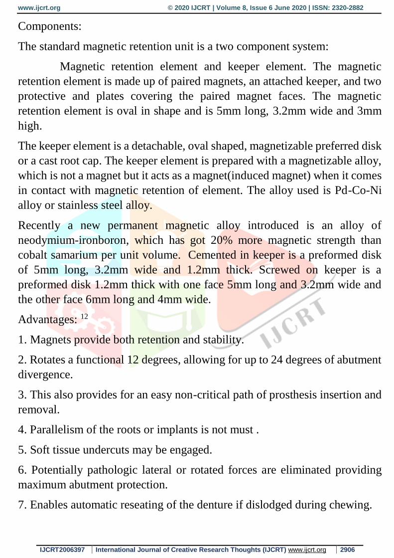
5. Magnets:
Magnets gained popularity in the field of maxillofacial prosthesis due to their
small size and strong attractive forces.10These attributes allow them to be
placed within prostheses without being obtrusive in the mouth. Magnets (Fig
14) are used as retentive aid for sectional dentures, hemi-maxillectomy,
obturators, complete dentures or extensively atrophied ridges. Magnets are
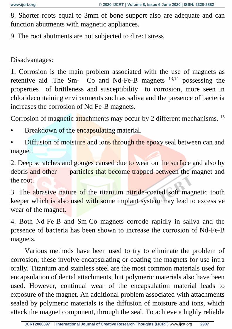
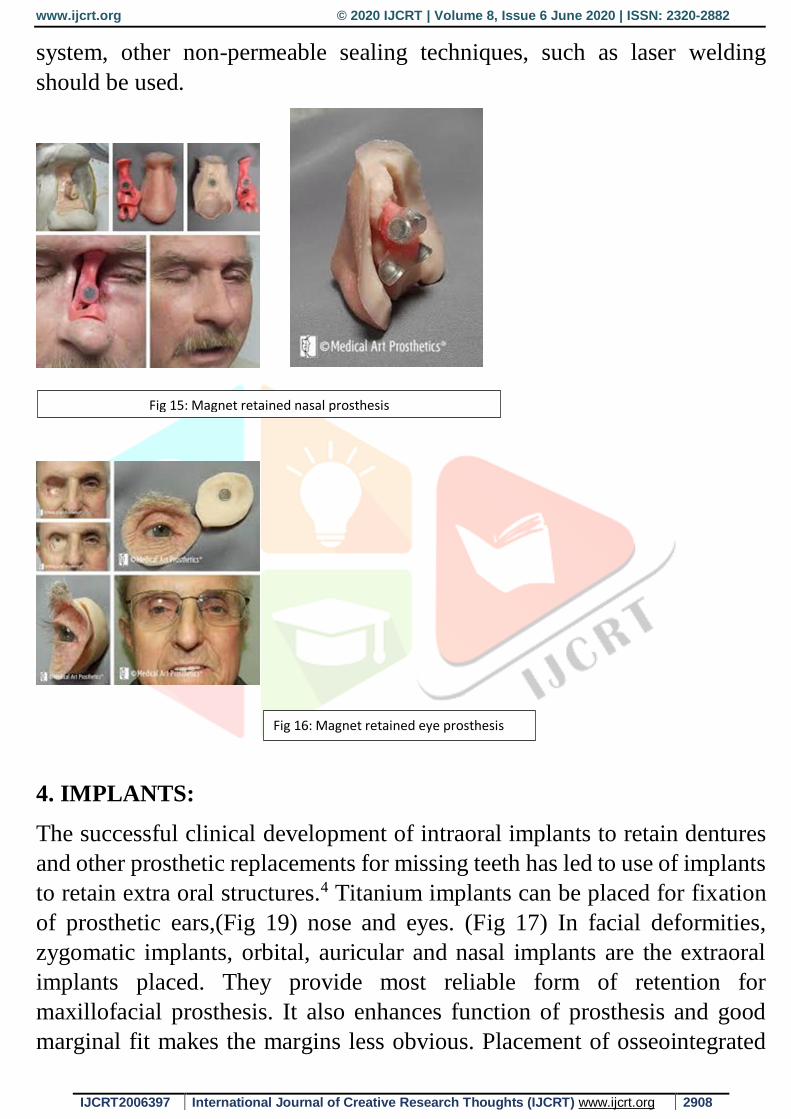
used in nasal (Fig 15) orbital (Fig 16) prosthesis auricular prosthesis, large
and small maxillary defects and intra oral-extra oral combination
prosthesis.11Mainly two types of alloys are used for the manufacture of small
dental magnets. They are cobalt-samarium and iron neodymium and boron.
They have high attractive forces in very small sizes, but have low corrosion
resistance.12 The design of magnetic attachments has changed in the last 20
years with new rare earth materials based on neodymium-Iron-Boron alloy.13
Fig 13: Elastic straps used for retention of prosthesis
Fig 14: Magnets used for retention of prosthesis
www.ijcrt.org © 2020 IJCRT | Volume 8, Issue 6 June 2020 | ISSN: 2320-2882
IJCRT2006397 International Journal of Creative Research Thoughts (IJCRT) www.ijcrt.org 2906
Components:
The standard magnetic retention unit is a two component system:
Magnetic retention element and keeper element. The magnetic
retention element is made up of paired magnets, an attached keeper, and two
protective and plates covering the paired magnet faces. The magnetic
retention element is oval in shape and is 5mm long, 3.2mm wide and 3mm
high.
The keeper element is a detachable, oval shaped, magnetizable preferred disk
or a cast root cap. The keeper element is prepared with a magnetizable alloy,
which is not a magnet but it acts as a magnet(induced magnet) when it comes
in contact with magnetic retention of element. The alloy used is Pd-Co-Ni
alloy or stainless steel alloy.
Recently a new permanent magnetic alloy introduced is an alloy of
neodymium-ironboron, which has got 20% more magnetic strength than
cobalt samarium per unit volume. Cemented in keeper is a preformed disk
of 5mm long, 3.2mm wide and 1.2mm thick. Screwed on keeper is a
preformed disk 1.2mm thick with one face 5mm long and 3.2mm wide and
the other face 6mm long and 4mm wide.
Advantages: 12
1. Magnets provide both retention and stability.
2. Rotates a functional 12 degrees, allowing for up to 24 degrees of abutment
divergence.
3. This also provides for an easy non-critical path of prosthesis insertion and
removal.
4. Parallelism of the roots or implants is not must .
5. Soft tissue undercuts may be engaged.
6. Potentially pathologic lateral or rotated forces are eliminated providing
maximum abutment protection.
7. Enables automatic reseating of the denture if dislodged during chewing.
www.ijcrt.org © 2020 IJCRT | Volume 8, Issue 6 June 2020 | ISSN: 2320-2882
IJCRT2006397 International Journal of Creative Research Thoughts (IJCRT) www.ijcrt.org 2907
8. Shorter roots equal to 3mm of bone support also are adequate and can
function abutments with magnetic appliances.
9. The root abutments are not subjected to direct stress
Disadvantages:
1. Corrosion is the main problem associated with the use of magnets as
retentive aid .The Sm- Co and Nd-Fe-B magnets 13,14 possessing the
properties of brittleness and susceptibility to corrosion, more seen in
chloridecontaining environments such as saliva and the presence of bacteria
increases the corrosion of Nd Fe-B magnets.
Corrosion of magnetic attachments may occur by 2 different mechanisms. 15
• Breakdown of the encapsulating material.
• Diffusion of moisture and ions through the epoxy seal between can and
magnet.
2. Deep scratches and gouges caused due to wear on the surface and also by
debris and other particles that become trapped between the magnet and
the root.
3. The abrasive nature of the titanium nitride-coated soft magnetic tooth
keeper which is also used with some implant system may lead to excessive
wear of the magnet.
4. Both Nd-Fe-B and Sm-Co magnets corrode rapidly in saliva and the
presence of bacteria has been shown to increase the corrosion of Nd-Fe-B
magnets.
Various methods have been used to try to eliminate the problem of
corrosion; these involve encapsulating or coating the magnets for use intra
orally. Titanium and stainless steel are the most common materials used for
encapsulation of dental attachments, but polymeric materials also have been
used. However, continual wear of the encapsulation material leads to
exposure of the magnet. An additional problem associated with attachments
sealed by polymeric materials is the diffusion of moisture and ions, which
attack the magnet component, through the seal. To achieve a highly reliable
www.ijcrt.org © 2020 IJCRT | Volume 8, Issue 6 June 2020 | ISSN: 2320-2882
IJCRT2006397 International Journal of Creative Research Thoughts (IJCRT) www.ijcrt.org 2908
system, other non-permeable sealing techniques, such as laser welding
should be used.
4. IMPLANTS:
The successful clinical development of intraoral implants to retain dentures
and other prosthetic replacements for missing teeth has led to use of implants
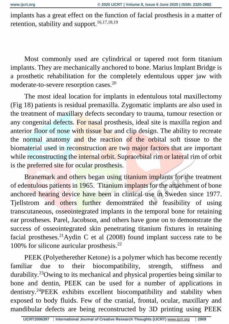
to retain extra oral structures.4 Titanium implants can be placed for fixation
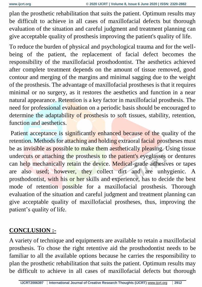
of prosthetic ears,(Fig 19) nose and eyes. (Fig 17) In facial deformities,
zygomatic implants, orbital, auricular and nasal implants are the extraoral
implants placed. They provide most reliable form of retention for
maxillofacial prosthesis. It also enhances function of prosthesis and good
marginal fit makes the margins less obvious. Placement of osseointegrated
Fig 15: Magnet retained nasal prosthesis
Fig 16: Magnet retained eye prosthesis
www.ijcrt.org © 2020 IJCRT | Volume 8, Issue 6 June 2020 | ISSN: 2320-2882
IJCRT2006397 International Journal of Creative Research Thoughts (IJCRT) www.ijcrt.org 2909
implants has a great effect on the function of facial prosthesis in a matter of
retention, stability and support.16,17,18,19
Most commonly used are cylindrical or tapered root form titanium
implants. They are mechanically anchored to bone. Marius Implant Bridge is
a prosthetic rehabilitation for the completely edentulous upper jaw with
moderate-to-severe resorption cases.20
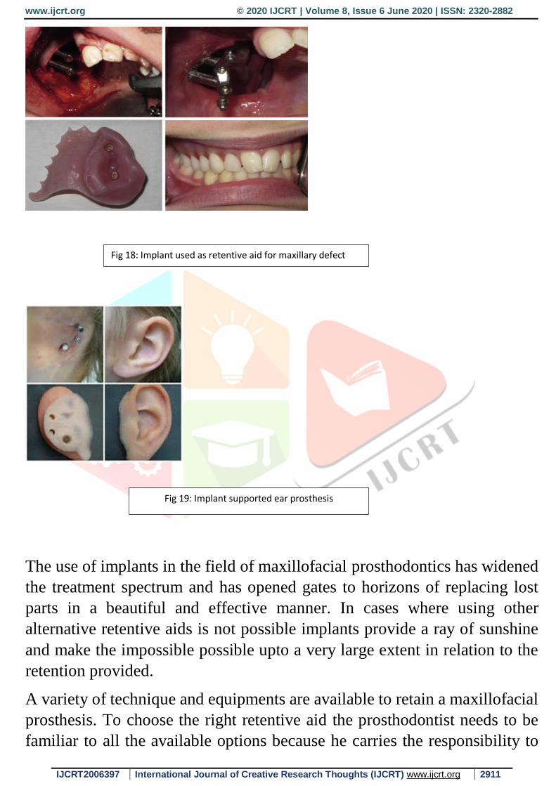
The most ideal location for implants in edentulous total maxillectomy
(Fig 18) patients is residual premaxilla. Zygomatic implants are also used in
the treatment of maxillary defects secondary to trauma, tumour resection or
any congenital defects. For nasal prosthesis, ideal site is maxilla region and
anterior floor of nose with tissue bar and clip design. The ability to recreate
the normal anatomy and the reaction of the orbital soft tissue to the
biomaterial used in reconstruction are two major factors that are important
while reconstructing the internal orbit. Supraorbital rim or lateral rim of orbit
is the preferred site for ocular prosthesis.
Branemark and others began using titanium implants for the treatment
of edentulous patients in 1965. Titanium implants for the attachment of bone
anchored hearing device have been in clinical use in Sweden since 1977.
Tjellstrom and others further demonstrated the feasibility of using
transcutaneous, osseointegrated implants in the temporal bone for retaining
ear prostheses. Parel, Jacobson, and others have gone on to demonstrate the
success of osseointegrated skin penetrating titanium fixtures in retaining
facial prostheses.21Aydin C et al (2008) found implant success rate to be
100% for silicone auricular prosthesis.22
PEEK (Polyetherether Ketone) is a polymer which has become recently
familiar due to their biocompatibility, strength, stiffness and
durability.23Owing to its mechanical and physical properties being similar to
bone and dentin, PEEK can be used for a number of applications in
dentistry.24PEEK exhibits excellent biocompatibility and stability when
exposed to body fluids. Few of the cranial, frontal, ocular, maxillary and
mandibular defects are being reconstructed by 3D printing using PEEK
www.ijcrt.org © 2020 IJCRT | Volume 8, Issue 6 June 2020 | ISSN: 2320-2882
IJCRT2006397 International Journal of Creative Research Thoughts (IJCRT) www.ijcrt.org 2910
(Polyetherether ketone). PEEK implants can be machined to many organic
shapes and fixated to the adjacent bone standard screws and plates.25
Through the years it has become obvious that this complex field of
dental implantology would require the optimization of several important
variables to enhance the chances of success. These include:
1. Proper material selection and design.
2. Understanding and evaluation of the biologic interaction at the interface
between the implant and tissue
3. Evaluation of the quality of the existing bone.
4. Careful and controlled surgical technique.
5. Joint approach between the various specialties to optimize patient
selection.
6. Prosthodontic geometry
7. Follow up care.
The retention provided by the implants makes it possible to fabricate large
maxillofacial prostheses that rest on movable tissue bed. The advent and
increasing availability of 3-D cone beam computerized tomography (CT) and
3-D digital panoramic imaging machines makes it easier, timely and less
costly to obtain images C.T. images are extremely useful as a visualization
and diagnostic tool.
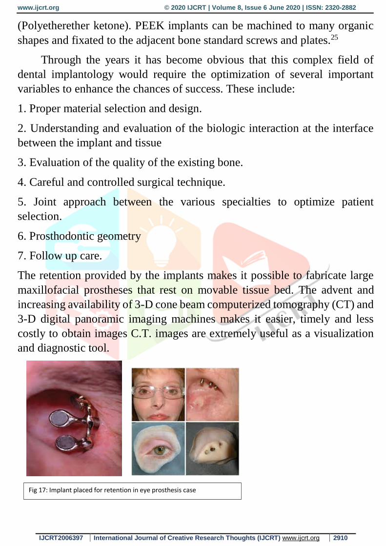
Fig 17: Implant placed for retention in eye prosthesis case
www.ijcrt.org © 2020 IJCRT | Volume 8, Issue 6 June 2020 | ISSN: 2320-2882
IJCRT2006397 International Journal of Creative Research Thoughts (IJCRT) www.ijcrt.org 2911
The use of implants in the field of maxillofacial prosthodontics has widened
the treatment spectrum and has opened gates to horizons of replacing lost
parts in a beautiful and effective manner. In cases where using other
alternative retentive aids is not possible implants provide a ray of sunshine
and make the impossible possible upto a very large extent in relation to the
retention provided.
A variety of technique and equipments are available to retain a maxillofacial
prosthesis. To choose the right retentive aid the prosthodontist needs to be
familiar to all the available options because he carries the responsibility to
Fig 18: Implant used as retentive aid for maxillary defect
Fig 19: Implant supported ear prosthesis
www.ijcrt.org © 2020 IJCRT | Volume 8, Issue 6 June 2020 | ISSN: 2320-2882
IJCRT2006397 International Journal of Creative Research Thoughts (IJCRT) www.ijcrt.org 2912
plan the prosthetic rehabilitation that suits the patient. Optimum results may
be difficult to achieve in all cases of maxillofacial defects but thorough
evaluation of the situation and careful judgment and treatment planning can
give acceptable quality of prosthesis improving the patient's quality of life.
To reduce the burden of physical and psychological trauma and for the well-
being of the patient, the replacement of facial defect becomes the
responsibility of the maxillofacial prosthodontist. The aesthetics achieved
after complete treatment depends on the amount of tissue removed, good
contour and merging of the margins and minimal sagging due to the weight
of the prosthesis. The advantage of maxillofacial prostheses is that it requires
minimal or no surgery, as it restores the aesthetics and function in a near
natural appearance. Retention is a key factor in maxillofacial prosthesis. The
need for professional evaluation on a periodic basis should be encouraged to
determine the adaptability of prosthesis to soft tissues, stability, retention,
function and aesthetics.
Patient acceptance is significantly enhanced because of the quality of the
retention. Methods for attaching and holding extraoral facial prostheses must
be as invisible as possible to make them aesthetically pleasing. Using tissue
undercuts or attaching the prosthesis to the patient's eyeglasses or dentures
can help mechanically retain the device. Medical-grade adhesives or tapes
are also used; however, they collect dirt and are unhygienic. A
prosthodontist, with his or her skills and experience, has to decide the best
mode of retention possible for a maxillofacial prosthesis. Thorough
evaluation of the situation and careful judgment and treatment planning can
give acceptable quality of maxillofacial prostheses, thus, improving the
patient’s quality of life.
CONCLUSION :-
A variety of technique and equipments are available to retain a maxillofacial
prosthesis. To chose the right retentive aid the prosthodontist needs to be
familiar to all the available options because he carries the responsibility to
plan the prosthetic rehabilitation that suits the patient. Optimum results may
be difficult to achieve in all cases of maxillofacial defects but thorough
www.ijcrt.org © 2020 IJCRT | Volume 8, Issue 6 June 2020 | ISSN: 2320-2882
IJCRT2006397 International Journal of Creative Research Thoughts (IJCRT) www.ijcrt.org 2913
evaluation of the situation and careful judgment and treatment planning can
give acceptable quality of prosthesis improving the patient's quality of life.
The journey from using metal bands to using adhesives to placing implants
for retaining a maxillofacial prosthesis has been fascinating and satisfying to
many, but, the aim of achieving the full potential still remains incomplete.
REFERENCES :-
1. Glossary of Prosthodontic terms. 9th ed. J Prosthet
Dent.2017;117(55):e1-e105.
2. Mckinstry RE. Retention and facial prostheses. In: Mckinstry RE.
Fundamental of facial prosthetics. Arlington (USA): ABI
Professional Publications; 1995. p. 19–30.
3. Karthikeyan I, Khatree M, Gaddale R, et al. A review on prosthetic
rehabilitation of maxillofacial region. Journal of Scientific
Dentistry 2016;6(1):6-12.
4. Rajesh Gurjar, Sunil Kumar M.V., Harikesh Rao, Alok Sharma,
Sumit Bhansali. Retentive Aids in Maxillofacial Prosthodontics -
A Review. IJCD 2011; 2(3):84-88.
5. Maxillofacial Rehabilitation. John Beumer, Mark T. Marunick,
Salvatore J. Esposito.3rd Edition.pg no-267.
6. Yeshwante B, Patil SJ, Baig N. Retentive aids used in
maxillofacial prosthesis. IJCDS 2014;5(2):12-20.
7. Sudarat Kiat-amnuay, Lawrence Gettleman, Zafrulla Khan, and L.
Jane Goldsmith. Effect of adhesive retention on maxillofacial
prostheses. Part I: Skin dressings and solvent removers. jpd
2000;84(3)-335-340
8. Kanathila H, Pangi A. The changing concepts in the retention of
maxillofacial prosthesis from past to present- a review. J.
Evolution Med. Dent. Sci. 2017;6(84):5879-5883
9. Muddugangadhar BC, Sonika R, Chheda PS, et al. Rehabilitation
of an orbital defect: a simplified technique. J Int Oral Health
2015;7(7):121-3.
10. Bhat VS, Shenoy KK, Premkumar P, et al. Magnets in
dentistry. AMHS 2013;1(1):73-9.
www.ijcrt.org © 2020 IJCRT | Volume 8, Issue 6 June 2020 | ISSN: 2320-2882
IJCRT2006397 International Journal of Creative Research Thoughts (IJCRT) www.ijcrt.org 2914
11. Bhat.V : A close-up on obturators using magnets : Part 11.
J.Indian.Pros Society Sep2006:6(3); 148-53.
12. Highton R, Caputo AA, Pezzoli M, Matyas J. Retentive
characteristics of different magnetic system for dental applications.
J Prosthet Dent 1986;56:104–6.
13. Melissa Alessandra Riley et al: Magnets in prosthetic
dentistry. J Prosthet Dent 2001; 86:137-42.
14. Coey J.M.D: Magnetism in future. J.Magnetism and
Magnetic Materials 226-30(2001)2107-2112.
15. Riley MA, Williams AJ, Speight JD, Walmsley AD, Harris
JR. Investigations into the failure of dental magnets. Int J
Prosthodont 1999;12:249–54.
16. Kantola R. Use of fibre-reinforced composite framework and
thermochromic pigment in facial prosthesis. Turun Yliopisto
University of Turku. Turku 2014: p. 13.
17. Parel SM, Tjellström A. The United States and Swedish
experience with osseointegration and facial prostheses. Int J Oral
Maxillofac Implants 1991;6(1):75-9.
18. Watson RM, Coward TJ, Forman GH. Results of treatment
of 20 patients with implants-retained auricular prostheses. Int J
Oral Maxillofac Implants 1995;10(4):445-9.
19. Arcuri MR, Rubenstein JT. Facial implants. Dent Clin North
Am 1998;42(1):161-75.
20. Fortin Y, Sullivan RM, Rangert BR. The Marius implant
bridge: surgical and prosthetic rehabilitation for the completely
edentulous upper jaw with moderate to severe resorption: a 5-year
retrospective clinical study. Clin Implant Dent Relat Res
2002;4(2):69-77.
21. Schaaf NG, Kielch M. Implant retained facial prostheses. In:
Mckinstry RE. Fundamental of facial prosthetics. Arlington
(USA): ABI Professional Publications; 1995. p. 169–79.
22. Yoon HI, Han JS. Prosthetic rehabilitation with an implant-
supported fixed prosthesis using computeraided design and
www.ijcrt.org © 2020 IJCRT | Volume 8, Issue 6 June 2020 | ISSN: 2320-2882
IJCRT2006397 International Journal of Creative Research Thoughts (IJCRT) www.ijcrt.org 2915
computer-aided manufacturing dental technology for a patient with
a mandibulectomy: a clinical report. Journal of Prosthetic Dentistry
2016;115(2):133-6.
23. Kim MM. Use of customized polyetheretheretone (PEE)
implants in the reconstruction of comple maxillofacial defects.
Arch Facial Plas Surg 2009;11(1):53-7.
24. Najeeb S, Zafar MS, Khurshid Z, et al. Applications of
polyetheretherketone (PEEK) in oral implantology and
prosthodontics. Journal of Prosthodontic Research 2016;60(1):12-
9.
25. Herford AS, Miller M, Lauritano F, et al. The use of virtual
surgical planning and navigation in the treatment of orbital trauma.
Chinese Journal of Traumatology 2017;20(1):9-13.