Metallic nanoparticles and their medicinal potential. Part II: aluminosilicates, nanobiomagnets,...
18
1179 ISSN 2041-5990 10.4155/TDE.13.74 © 2013 Future Science Ltd Ther. Deliv. (2013) 4(9), 1179–1196 REVIEW Nanostructured metallic films approximately 200–500 nm thick have been a part of ceramic decorations ‘luster’ since the medieval period. The homogeneous dispersion of silver and cop- per nanoparticles over glazed pottery results in a colored iridescence called luster, a technique very popular in the Middle East, Egypt, Persia and Spain. In 1685, Andreas Cassius invented a recipe of glass coloring pigment called Purple of Cassius. He made the purple precipitates by dissolving gold particles in aqua regia and then added a piece of tin to it. The pigment is famous for its use in high-quality porcelain ware. A Vien- nese chemist, Richard Zsigmondy was awarded the Nobel Prize in Chemistry in 1925 for discov- ering that colloidal gold adsorbed on stannous hydroxide base makes Purple of Cassius. Michael Faraday (1857) prepared gold sols, whose size was found out by JM Thomas in 1988 to be 3–30 nm in diameter, when he reproduced those gold nanoparticles. Industrial nanotechnology was initiated in 1930 with the manufacturing of sil- ver coatings for photographic films. At around this time imaging techniques such as ultrasound, MRI, computed tomography, positron emission tomography and surface enhanced raman spec- troscopy were becoming popular for imaging various disease states and magnetic nanopar- ticles came to the rescue as contrast agent with unique physicochemical properties. The 1980s took nanomaterials to higher strata by introduc- ing fullerenes and scanning tunnel microscopes. In the mid-1980s, when growth technologies such as molecular beam epitaxy tied nuptials with electron-beam lithography to confine electron motion in all three (x-y-z) directions, quantum dots (Q-dots) were produced. These metallic nanoparticles have been embraced by nanotechnology for more than four decades now. Today, with Q-dots as new nanoball bearings, aluminosilicates as nanowire gauges, cochle- ates as nanocrystalline delivery trucks and iron nanoparticles as new biomagnets, it is no wonder that nanoscience thinks about metals in terms of size control, spatial resolution, chemical reac- tivity and engineering their relationship at the cellular level in real time. Metal nanoparticles can be easily prepared to the nanometer scale, they possess fundamentals of light matter interaction and are highly suitable Metallic nanoparticles and their medicinal potential. Part II: aluminosilicates, nanobiomagnets, quantum dots and cochleates Metallic miniaturization techniques have taken metals to nanoscale size where they can display fascinating properties and their potential applications in medicine. In recent years, metal nanoparticles such as aluminium, silicon, iron, cadmium, selenium, indium and calcium, which find their presence in aluminosilicates, nanobiomagnets, quantum dots (Q-dots) and cochleates, have caught attention of medical industries. The increasing impact of metallic nanoparticles in life sciences has significantly advanced the production techniques for these nanoparticles. In this Review, the various methods for the synthesis of nanoparticles are outlined, followed by their physicochemical properties, some recent applications in wound healing, diagnostic imaging, biosensing, assay labeling, antimicrobial activity, cancer therapy and drug delivery are listed, and finally their toxicological impacts are revised. The first half of this article describes the medicinal uses of two noble nanoparticles – gold and silver. This Review provides further information on the ability of aluminum, silicon, iron, selenium, indium, calcium and zinc to be used as nanoparticles in biomedical sciences. Aluminosilicates find their utility in wound healing and antibacterial growth. Iron-oxide nanoparticles enhance the properties of MRI contrast agents and are also used as biomagnets. Cadmium, selenium, tellurium and indium form the core nanostructures of tiny Q-dots used in cellular assay labeling, high-resolution cell imaging and biosensing. Cochleates have the bivalent nano ions calcium, magnesium or zinc imbedded in their structures and are considered to be highly effective agents for drug and gene delivery. The aluminosilicates, nanobiomagnets, Q-dots and cochleates are discussed in the light of their properties, synthesis and utility. Leena Loomba 1 & Tiziano Scarabelli* 2,3 1 Punjab Agricultural University, Ludhiana, Punjab, India 2 Center for Heart & Vessel Preclinical Studies, St. John Hospital & Medical Center, Wayne State University School of Medicine, Detroit, MI, USA 3 Medical Center, Wayne State University School of Medicine, MI, USA *Author for correspondence: E-mail: [email protected] For reprint orders, please contact [email protected]
Transcript of Metallic nanoparticles and their medicinal potential. Part II: aluminosilicates, nanobiomagnets,...

1179ISSN 2041-599010.4155/TDE.13.74 © 2013 Future Science Ltd Ther. Deliv. (2013) 4(9), 1179–1196
Review
Nanostructured metallic films approximately 200–500 nm thick have been a part of ceramic decorations ‘luster’ since the medieval period. The homogeneous dispersion of silver and cop-per nanoparticles over glazed pottery results in a colored iridescence called luster, a technique very popular in the Middle East, Egypt, Persia and Spain. In 1685, Andreas Cassius invented a recipe of glass coloring pigment called Purple of Cassius. He made the purple precipitates by dissolving gold particles in aqua regia and then added a piece of tin to it. The pigment is famous for its use in high-quality porcelain ware. A Vien-nese chemist, Richard Zsigmondy was awarded the Nobel Prize in Chemistry in 1925 for discov-ering that colloidal gold adsorbed on stannous hydroxide base makes Purple of Cassius. Michael Faraday (1857) prepared gold sols, whose size was found out by JM Thomas in 1988 to be 3–30 nm in diameter, when he reproduced those gold nanoparticles. Industrial nanotechnology was initiated in 1930 with the manufacturing of sil-ver coatings for photographic films. At around this time imaging techniques such as ultrasound, MRI, computed tomography, positron emission
tomography and surface enhanced raman spec-troscopy were becoming popular for imaging various disease states and magnetic nanopar-ticles came to the rescue as contrast agent with unique physicochemical properties. The 1980s took nanomaterials to higher strata by introduc-ing fullerenes and scanning tunnel microscopes. In the mid-1980s, when growth technologies such as molecular beam epitaxy tied nuptials with electron-beam lithography to confine electron motion in all three (x-y-z) directions, quantum dots (Q-dots) were produced. These metallic nanoparticles have been embraced by nanotechnology for more than four decades now. Today, with Q-dots as new nanoball bearings, aluminosilicates as nanowire gauges, cochle-ates as nanocrystalline delivery trucks and iron nanoparticles as new biomagnets, it is no wonder that nanoscience thinks about metals in terms of size control, spatial resolution, chemical reac-tivity and engineering their relationship at the cellular level in real time.
Metal nanoparticles can be easily prepared to the nanometer scale, they possess fundamentals of light matter interaction and are highly suitable
Metallic nanoparticles and their medicinal potential. Part II: aluminosilicates, nanobiomagnets, quantum dots and cochleates
Metallic miniaturization techniques have taken metals to nanoscale size where they can display fascinating properties and their potential applications in medicine. In recent years, metal nanoparticles such as aluminium, silicon, iron, cadmium, selenium, indium and calcium, which find their presence in aluminosilicates, nanobiomagnets, quantum dots (Q-dots) and cochleates, have caught attention of medical industries. The increasing impact of metallic nanoparticles in life sciences has significantly advanced the production techniques for these nanoparticles. In this Review, the various methods for the synthesis of nanoparticles are outlined, followed by their physicochemical properties, some recent applications in wound healing, diagnostic imaging, biosensing, assay labeling, antimicrobial activity, cancer therapy and drug delivery are listed, and finally their toxicological impacts are revised. The first half of this article describes the medicinal uses of two noble nanoparticles – gold and silver. This Review provides further information on the ability of aluminum, silicon, iron, selenium, indium, calcium and zinc to be used as nanoparticles in biomedical sciences. Aluminosilicates find their utility in wound healing and antibacterial growth. Iron-oxide nanoparticles enhance the properties of MRI contrast agents and are also used as biomagnets. Cadmium, selenium, tellurium and indium form the core nanostructures of tiny Q-dots used in cellular assay labeling, high-resolution cell imaging and biosensing. Cochleates have the bivalent nano ions calcium, magnesium or zinc imbedded in their structures and are considered to be highly effective agents for drug and gene delivery. The aluminosilicates, nanobiomagnets, Q-dots and cochleates are discussed in the light of their properties, synthesis and utility.
Leena Loomba1 & Tiziano Scarabelli*2,3
1Punjab Agricultural University, Ludhiana, Punjab, India 2Center for Heart & Vessel Preclinical Studies, St. John Hospital & Medical Center, Wayne State University School of Medicine, Detroit, MI, USA 3Medical Center, Wayne State University School of Medicine, MI, USA *Author for correspondence: E-mail: [email protected]
For reprint orders, please contact [email protected]

Review | Loomba & Scarabelli
Ther. Deliv. (2013) 4(9)1180 future science group
for conjugation with drugs, ligands, antibod-ies and genes, as well as functional groups of interest. Excitation and relaxation of conduction band electrons in metallic nanoparticles results in plasmon resonance – a unique phenomenon responsible for energy dissipation and optical effects. An effective cellular communication, the invincible ability of nanometals provides immense possibility for their growth and devel-opment in biomedicine. Nanoparticles have been synthesized, tested, used and modified over the years to function as agents for gene therapy, DNA sequencing, cancer detection, cellular tracking, targeted drug delivery and biomedical imaging. The versatile attitude of metallic nanoparticles attracts scientific research and clinical appli-cations under the stream of nanotechnology (Table 1).
Aluminosilicate nanoparticles�� Types, properties & synthesis of
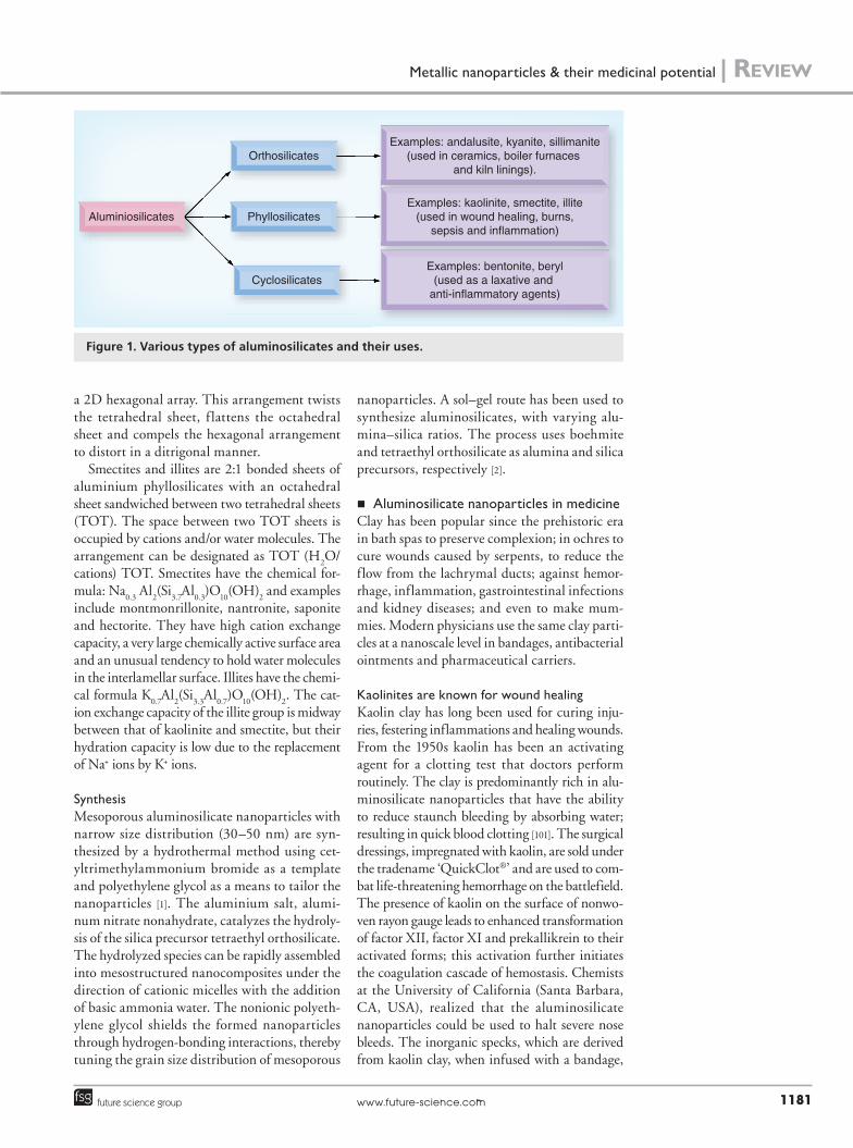
aluminosilicate nanoparticlesTypesAluminosilicates are broadly classified into the following categories (FiguRe 1):
��Orthosilicates with [AlO6]3- anions connected
by isolated (SiO4)4- clusters; for example,
andalusite (Al2SiO
5) and its polymorphs,
kyanite and sillimanite;
��Phyllosilicates with tetrahedral and octahedral layers in two dimensions; for example, kaolinite, smectite and illite;
��Cyclosilicates with tetrahedral clusters of (Si
3O
7)6-, (Si
4O
12)8- or (Si
6O
18)12- arranged in
a cyclic manner; for example, bentonite (BaTi[Si
3O
9]) and beryl (Be
3Al
2[Si
6O
18]).
PropertiesAluminosilicates are minerals consisting of alu-minium and silicon oxides. Silicates are tetra-hedrally clustered polymers of (SiO
4)4- anions.
The positively charged ions of Al3+ can either substitute silica atoms in the silicate tetrahedra
or connect outside the anionic framework, to form aluminosilicates. In nature, magma solid-ifies to form aluminosilicates such as feldspar (xAl[Al,Si]
3O
8, where x can be Na, K or Ca),
mica, beryl or wollastonite. In feldspar the Al3+ ion replaces the Si4+ cation of (SiO4)4-, leaving behind a negative charge on the 3D framework. The positively charged ions neutralize this negative charge, for example, K+ in microcline (KAlSi
3O
8) and Na+ in albite (NaAlSi
3O
8),
both K+ and Na+ in sanidine ([K,Na]AlSi3O
8]
4)
and Ca2+ in Anorthite (Ca[AlSi2O
8]). The
weather plays its role to convert feldspar to clay kaolin (Al
2Si
2O
5[OH]
4) or montmorillonite
([Na,Ca]0.33
[Al,Mg]2[Si
4O
10][OH]
2.nH
2O).
Naturally occurring aluminosilicate nanopar-ticles exist as nanotubes called imogolites, or hollow 3–5 nm spherical allophanes. Both these aluminosilicates have on identical chemical com-position (Al
2SiO
3[OH])
4, but with different
structures depending on the Al/Si ratio.Clay has negatively charged sites that can
attract and hold positively charged particles and this is called ‘cation exchange capacity’; it is the measure of how many negatively charged sites are available on a nanoparticles surface. These exchange reactions are rapid, reversible and stoichiometric with respect to charge:
2{K+-Soil} + Ca2+ → 2K+ + Ca2+–(Soil)2
equaTion 1
Aluminosilicate nanoparticles undergo ion exchange readily, that is, adsorbed cations can be replaced by a large quantity of other com-peting ions, which superimpose their strength and resistance. The layered sheet-like structure of aluminosilicate nanoparticles provides addi-tional surface area as well as the ability to hold substances for targeted delivery.
Kaolinites are 1:1 aluminium phyllosilicates having the chemical formula Al
2Si
2O
5(OH)
4.
Clays such as kaolinite, dichite, nacrite and hal-loysite fall under this category. They have SiO
4
tetrahedrons and AlO4 octahedrons arranged in
Table 1. Examples of metallic nanoparticles used as drugs and diagnostic agents.
Metallic nanoparticles Element Use
Aluminosilicate nanoparticles Al, Si Faster blood clotting in open woundsNano biomagnets Fe Helps in targeted drug deliveryQ-dots Cd, Se, In Medical imagingCochleates Ca, Zn, Mg Oral drug delivery of drugs encapsulated in a
nanocrystalline structure
Metallic nanoparticles & their medicinal potential | Review
www.future-science.com 1181future science group
a 2D hexagonal array. This arrangement twists the tetrahedral sheet, f lattens the octahedral sheet and compels the hexagonal arrangement to distort in a ditrigonal manner.
Smectites and illites are 2:1 bonded sheets of aluminium phyllosilicates with an octahedral sheet sandwiched between two tetrahedral sheets (TOT). The space between two TOT sheets is occupied by cations and/or water molecules. The arrangement can be designated as TOT (H
2O/
cations) TOT. Smectites have the chemical for-mula: Na
0.3 Al
2(Si
3.7Al
0.3)O
10(OH)
2 and examples
include montmonrillonite, nantronite, saponite and hectorite. They have high cation exchange capacity, a very large chemically active surface area and an unusual tendency to hold water molecules in the interlamellar surface. Illites have the chemi-cal formula K
0.7Al
2(Si
3.3Al
0.7)O
10(OH)
2. The cat-
ion exchange capacity of the illite group is midway between that of kaolinite and smectite, but their hydration capacity is low due to the replacement of Na+ ions by K+ ions.
SynthesisMesoporous aluminosilicate nanoparticles with narrow size distribution (30–50 nm) are syn-thesized by a hydrothermal method using cet-yltrimethylammonium bromide as a template and polyethylene glycol as a means to tailor the nanoparticles [1]. The aluminium salt, alumi-num nitrate nonahydrate, catalyzes the hydroly-sis of the silica precursor tetraethyl orthosilicate. The hydrolyzed species can be rapidly assembled into mesostructured nanocomposites under the direction of cationic micelles with the addition of basic ammonia water. The nonionic polyeth-ylene glycol shields the formed nanoparticles through hydrogen-bonding interactions, thereby tuning the grain size distribution of mesoporous
nanoparticles. A sol–gel route has been used to synthesize aluminosilicates, with varying alu-mina–silica ratios. The process uses boehmite and tetraethyl orthosilicate as alumina and silica precursors, respectively [2].
�� Aluminosilicate nanoparticles in medicineClay has been popular since the prehistoric era in bath spas to preserve complexion; in ochres to cure wounds caused by serpents, to reduce the flow from the lachrymal ducts; against hemor-rhage, inflammation, gastrointestinal infections and kidney diseases; and even to make mum-mies. Modern physicians use the same clay parti-cles at a nanoscale level in bandages, antibacterial ointments and pharmaceutical carriers.
Kaolinites are known for wound healingKaolin clay has long been used for curing inju-ries, festering inflammations and healing wounds. From the 1950s kaolin has been an activating agent for a clotting test that doctors perform routinely. The clay is predominantly rich in alu-minosilicate nanoparticles that have the ability to reduce staunch bleeding by absorbing water; resulting in quick blood clotting [101]. The surgical dressings, impregnated with kaolin, are sold under the tradename ‘QuickClot®’ and are used to com-bat life-threatening hemorrhage on the battlefield. The presence of kaolin on the surface of nonwo-ven rayon gauge leads to enhanced transformation of factor XII, factor XI and prekallikrein to their activated forms; this activation further initiates the coagulation cascade of hemostasis. Chemists at the University of California (Santa Barbara, CA, USA), realized that the aluminosilicate nanoparticles could be used to halt severe nose bleeds. The inorganic specks, which are derived from kaolin clay, when infused with a bandage,
Aluminiosilicates
Orthosilicates
Phyllosilicates
Cyclosilicates
Examples: andalusite, kyanite, sillimanite(used in ceramics, boiler furnaces
and kiln linings).
Examples: kaolinite, smectite, illite(used in wound healing, burns,
sepsis and inflammation)
Examples: bentonite, beryl(used as a laxative and
anti-inflammatory agents)
Figure 1. Various types of aluminosilicates and their uses.

Review | Loomba & Scarabelli
Ther. Deliv. (2013) 4(9)1182 future science group
trigger the body’s natural clotting process. The bandage stops the bleeding immediately, when rolled up and inserted in the nose [102].
Smectites & illites possess antibacterial abilitySmectites are famous for their tendency to absorb the carcinogenic metabolite af latoxin B1, produced by the fungi Aspergillus flavus in animal diet [3]. The nanoparticles of smectites–illites and reduced iron present in natural clay have the potential to eliminate Escherichia coli and even antibiotic-resistant bacteria such as methicillin-resistant Staphylococcus aureus. The hydrated clay leaches into the bacterial cell mem-brane to increase bacterial iron and phosphorous levels and metabolic activity of the membrane. The regulatory proteins subsequently come into action to oxidize Fe2+ to Fe3+ and even produce hydroxyl radicals, which enter the cytoplasm and cause cell death [4].
The in vitro antibacterial activity of clay min-erals has proven effective against Buruli ulcer and b-lactamase E. coli. The mineral surfaces of aluminosilicates in clay alter pH and oxidation states in bacterial membranes to control redox reactions, resulting in cell lysis [5]. The layered metal hydroxides of clay behave as excellent pharmaceutical carriers. The lamellar surfaces of various layers can easily hybridize nano-medicines in their 2D structure. For example, methotrextate – a folate antagonist anticancer drug – is unstable and also has a short plasma half-life. The drug, when layered in Mg and Al hydroxides of clay, specifically suppresses growth of human osteosarcoma cancer cells [6].
Bentonite: a versatile aluminisilicateBentonite is a chemically inert, absorbent alu-minium phyllosilicate consisting of montmoril-lonite. Bentonite supports good digestion and acts as a laxative. In the gastrointestinal tract of animals bentonite reduces bacterial mucolysis and inflammation. The granular form of benton-ite is used under the commercial name ‘Wound-Stat™’, in battlefields for wound dressings. It reduces pain associated with stings, burns and cuts, promotes detoxification and also shields against urushiol – the oil found in poison ivy.
Nanobiomagnets�� Types, properties & synthesis of
nanobiomagnetsTypesThe nanosized, biocompatible, paramag-netic iron oxides that serve as biomagnets are
magnetite (Fe3O
4), maghemite (g-Fe
2O
3) and
haematite (a-Fe2O
3); of which magnetite,
because of its biocompatibility, is very promis-ing. Iron oxide nanoparticles (IONps) are avail-able in various dimensions and shapes such as nanorods, nanotubes, hollow fibers, rings and snowflakes.
The iron oxide nanorods demonstrate higher incident photon-to-current conversion compared with nanospheres, which is further improved by surface modification and doping with Zn. The nanoparticle size imposes a huge impact on superparamagnetism and, in turn, their usage. Generally, iron oxide nanoparticles ranging from 1 to 25 nm are highly efficient models. Super-paramagnetic iron oxide nanoparticles are of particular interest in MRI; examples include:
��AMI-227 (Sinerem, Combidex®) and SHU-55C – a 20 nm sized iron oxide nanoparticle coated with carbodextran. It demonstrates excellent T2 relaxivities of 151.0 mmol/sec and has been used for lymph node and bone marrow imaging.
��OMP (Abdoscan®) and AMI-121 (Lumirem®, GastroMARK®) are 300 nm sized iron oxide nanoparticles (IONps) coated with silica. Their oral administration finds utility as a gastrointestinal contrast.
PropertiesIn magnetite, Fe3+ ions are placed at all tetrahe-dral sites, whereas both ferrous and ferric ions occupy octahedral sites of inverse spinel struc-ture. Maghemite is the oxidized form of mag-netite having 56 ions in each unit cell, of which 32 are O
2- ions, eight Fe3+ ions in tetrahedral
sites and 16 Fe3+ in octahedral sites. Magne-tite is a spin-polarized black crystal containing both Fe (II), Fe (III) and absorbs throughout the UV–vis–IR spectrum, while maghemite is an insulator. Both phases are ferrimagnetic. Haematite, a-Fe
2O
3, has a 3D framework built
up of trigonally distorted octahedra FeO6, with
oxygens in hexagonal closest-packing. The tri-valent iron ions are closely packed between two oxygen layers. This arrangement makes the structure neutral with no excess charge. Haematite has antiferromagnetic properties and an absorption spectrum in the visible range between 295 and 600 nm. The magnetic behav-ior of these oxides is due to their stereochem-istry that triggers internal superexchange com-petition between tetrahedral and octahedral

Metallic nanoparticles & their medicinal potential | Review
www.future-science.com 1183future science group
sites. At room temperature both magnetite and maghemite are superpara magnetic, which means an external magnetic field can easily magnetize the nanoparticles.
The IONps need to be superparamagnetic, biocompatible and nontoxic to be useful for molecular imaging purposes. They also need to bind to a range of metabolites. The zero point charge value of seven makes oxides stable only in highly acidic or basic aqueous media. This drawback in their surface chemistry causes con-siderable aggregation and precipitation in solu-tion phase. Also, low hole mobility, electron-hole recombination and electon-trapping, and oxygen-deficient iron sites yield poor photocur-rent efficiency. However, coating the particles with silica, dextran, carbodextran, poloxamines or poly(ethylene glycol) followed by their bio-conjugation with various ligands gives them both stability and specificity. Magnetic iron-oxides need high r1 and r2 relaxivities, as well as surface engineering, to fine tune their size and structure, before being used for in vivo applications [7].
The ferromagnetic nanoparticles magnetiza-tion fluctuates with temperature, fluctuations are generally larger at higher temperatures and smaller at lower temperatures. When the time between two magnetization fluctuations (Néel relaxation time) is shorter than the time used to measure the magnetization of the nanoparticles, in the absence of external magnetic field, the nanoparticles show an average zero magnetiza-tion. This is called superparamagnetism. Mate-rials having superparamagnetism have a high saturation magnetization and zero coercivity and remanence.
The Néel relaxation time is highly tempera-ture dependent, it fluctuates randomly by ther-mal fluctuation at high enough temperatures. The thermal energy decreases at lower tempera-tures and blocks the magnetic moments. This temperature is called the blocking temperature. It is a function of the particle size and increases with increasing particle size. Thus, superpara-magnetism increases with the decrease in size of the nanoparticle. Below blocking tempera-ture, the preferred direction of magnetization of superparamagnetic material is lost in zero magnetic fields. When the temperature rises above the blocking temperature, the nanopar-ticles show no hysteresis. With these fascinating superparamagnetic properties, IONps find their utility in ferrofluids, hyperthermia and MRI contrast agents.
SynthesisReverse micelle and precipitation are two com-monly used techniques for the synthesis of iron oxides [8]. The simplest of all the methods to prepare IONps is the coprecipitation of a 2:1 stoichiometric mixture of Fe2+/Fe3+ salts in an aqueous medium of pH between 8 and 14. The magnetite forms black colored precipitates. The overall reaction is written as:
Fe2+ + 2Fe3+ + 8OH- → Fe3O
4 + 4H
2O
equaTion 2
The particle size depends on numerous fac-tors such as Fe3+/Fe2+ ratio, temperature, ionic strength, nature of salts, pH and addition of che-lating agents. Generally, the nanoparticle size decreases with an increase in the pH, Fe3+/Fe2+ ratio and ionic strength of the medium.
The aqueous iron salt solutions essentially form reverse micelles with the hydrophilic head towards the core of the micelle and the hydro-phobic tail directed outwards. Reverse micelles solubilize large amounts of water, which can be controlled, for nanoparticle production. A wide range of iron oxide nanoparticles can be syn-thesized by altering the nature and amount of surfactant, solvent and cosurfactant.
They can also be synthesized using techniques such as sonochemistry, microwave irradiation and autogenic pressure reactor [9]. A new method to produce nanocrystals is glass crystallization [10]. In total, 15–20 nm sized, monodisperse, Fe
3O
4 nanoparticles are synthesized by decom-
position of iron (II) acetate at 400°C. IONps of desired size and dispersity are also synthesized by heating iron-oleic complex at 320°C in 1-octa-decene for 30 min. Hydrothermal treatment of iron powder and iron chloride solution in urea solution for 20 h at 130–150°C yields iron rods of nearly 80 nm.
�� Photoelectrochemical applications of biomagnetsThe chemistry of iron oxide nanoparticles can be manipulated to have magnetic properties that find their importance in magnetic reso-nance imaging, biotechnology and effective hyperthermia (FiguRe 2).
Iron-oxide nanoparticles as hyperthermia & MRI contrast agentsMRI is a noninvasive technique that combines the characteristics of high spatial resolution,

Review | Loomba & Scarabelli
Ther. Deliv. (2013) 4(9)1184 future science group
nonionizing radiation and multiplanar tomog-raphies in cellular imaging. Superparamagnetic iron oxide nanoparticles comprise a class of novel MRI contrast agents that are composed of a ferrous iron (Fe2+) and ferric iron (Fe3+) core, and a layer of dextran or other polysaccharide coating [11]. The iron nanoparticles have a very large magnetic moment, which leads to local magnetic field inhomogeneity. Consequently, they serve to enhance the image contrast and, thus, improve the sensitivity and specificity of MRI in mapping information from tissues [12]. In vivo, nonspecific superparamagnetic iron oxide nanoparticles are mainly captured by the reticuloendothelial system, and they are more suitable for liver, spleen and lymph node imaging [13]. Because of their long plasma half-life, super-paramagnetic iron oxides are also used as blood pool agents in magnetic resonance angiography.
Haematite nanoparticles, 1.8 nm in size, when coated with polysaccharides such as chi-tosan and alginate, respond superparamagneti-cally with very low coercivity. These nanopar-ticles can either be converted to magnetite by reduction or used directly for imaging [14]. The intensity of magnetic field of iron-based nanoparticles, having a layer of bis-carboxyl-ter-minated poly(ethylene glycol) on them, induces more effective hyperthermia than uncoated iron particles. They are far better MRI contrast agents and provide a focused approach for in vivo applications and cancer therapy [15]. Iron oxide nanoparticles manipulated with Herceptin® – an antibody present in breast cancer cells – or chlorotoxin – a peptide that binds MMP-2 in gliomas, show enhanced in vivo tumor-targeting properties [16]. Mammary tumors contain over-expressed levels of urokinase-type plasminogen activator. Amino-terminal fragment conjugated IONps can effectively bind the over expressed receptors in breast cancer tissues and help in vivo imaging [17]. The nanocomposites of maghemite,
such as those with bentonite and raffinose-mod-ified trypsin, are used as MRI contrast agents for the gastrointestinal tract and magnetic carriers for trypsin immobilization, respectively [18]. Flu-orescence and magnetism can be uniquely com-bined over maghemite nanoparticles. Congo-red or rhodamine dyes hybridize with g-Fe
2O
3 to
serve as biomarkers for in vivo Alzheimer’s dis-ease diagnosis [19]. Ferrite nanoparticles ranging from 20 to 200 nm in diameter are being used for biosensing and as contrast agents for MRI, when attached with europium these spheres can emit f luorescent radiations at 618 nm to help detect cancer [20].
Nanobiomagnets in biotechnologyBiotechnology can rely on the magnetic powers of IONps to separate specific proteins from a group of biomolecules. For example, dopamine grafted IONps can be used for protein separa-tion. The bidentate enediol ligands of the dopa-mine molecule tightly bind with unsaturated iron sites. The nanostructures so produced enhance specificity for protein separation and provide tremendous stability to heating and high salt concentrations. In the same manner, mag-netic nanoparticles are ideal candidates for gene detection. In the diagnosis of diseases involving genetic expression, the separation of rare DNA/mRNA targets with single-base mismatches in a mixture of various bio complexes is criti-cally important. Genomagnetic nano capturers have been formulated using IONps to detect DNA/RNA molecules with one single-base dif-ference. Nanobiomagnets can transfer drugs into the body and are held at the target site by an external magnet. The purpose of this is to concentrate the drug at the tumor site for long enough for it to be absorbed and release the drug on demand. The control of drug delivery using biomagnets can reduce the dosage by 60–75%, thus enhancing drug efficacy while decreasing
Nanobiomagnets
Magnetite
Maghemite
Haematite
Used in hyperthermia, in biotechnology as genomagnetic capturer, as MRI contrast agents
and to magnetically focusthe drug at tumor sites
Photoelectrochemicalapplications
Figure 2. Nanobiomagnets and their photochemical applications.

Metallic nanoparticles & their medicinal potential | Review
www.future-science.com 1185future science group
unwanted systemic uptake. This mechanism can find its utility in control of insulin-dependent diabetes. Recent studies report that the iron oxide nanoparticles can adhere to red blood cell’s surface for nearly 4 months. This can help to release drugs slowly into the body and can lead to controlled treatment of many immunogenic diseases.
Q-dots�� Types, properties & synthesis of Q-dots
TypesQ-dots are tiny particles, traditionally chal-cogenides (selenides or sulfides) of metal such as cadmium or zinc (CdSe/ZnS) ranging from 2 to 10 nm. The electrons and holes of the semiconductor cores being confined to a point significantly modifies the energy spec-trum of the carriers. Q-dots have a metallic core made of semiconductors, noble metals, and magnetic transition metals, shielded by a shell. Depending on the variation in the con-stituents of the core, Q-dots are classified into various groups:
��Group II–IV series Q-dots contains ZnS, ZnSe, CdSe and CdTe cores;
��Group III–V series Q-dots have InAs, InP, GaAs and GaN cores;
��Group IV-VI series Q-dots have PbTe, SnTe, SnS and SnS
2 cores.
Q-dots are also classified as Type-I, Reverse Type-I and Type-II:
��Type-I Q-dots have a core that simultaneously traps electrons and holes giving rise to
contravariant band layout so that both the conduction and valence band edges of the core lie within the bandgap of the shell; for example, CdSe/CdS, CdSe/ZnS and InAs/CdSe;
��For reverse Type-I Q-dots, the bandgap of the core is wider than the shell, and the conduc-tion and valence band edges of the shell lie in the core; for example, CdS/HgS, CdS/CdSe and ZnSe/CdSe;
��Type-II Q-dots have one type of charge carrier in the core while the shell carries the other type. It maintains covariant band layout in which the valence and conduction band edge are either lower or higher than the band edges of the shell; for example, Type-II Q-dots that attract holes are GaSb/GaAs and Ge/Si, and those that attract electrons are InP/InGaP and InP/GaAs.
PropertiesQ-dots are basically made of three parts – a core, a shell and the outer coating (FiguRe 3). The core region, when excited by a photon, triggers its electron in the semiconductor band gap, leaving behind a positive hole in the lower energy band. An increase in excitation increases the absorption in the band gap giving rise to broad absorption spectrum. Since the energy gap between higher and lower energy bands is responsible for emission energy, and the energy gap is low, the emission spectrum is narrow. The shell covers the surface defects of the elec-tron–hole nanocore, and thus protects it from oxidation, fluorescence and chemical reactions. The shells having large energy gaps increase the quantum yield and enhance photostability. A
Q-dot core(CdSe)
Shell (ZnS)
Cap (disulfide bridge, silane)
SBiomolecule
(protein/DNA)
Biological applications
Cellular and assay labeling
High-resolutioncell imaging
Q-dot-FRETbiosensing
Figure 3. A biofunctional quantum dot and its biological applications. FRET: Fluorescence resonance energy transfer; Q-dot: Quantum dot; S: Sulfide bridge.

Review | Loomba & Scarabelli
Ther. Deliv. (2013) 4(9)1186 future science group
coating of functional ligands over the Q-dot shell improves their solubility in polar solvents and also labels them. The mono or dithiol dihy-drolipoic acid ligands improve stability for over 1–2 years; phospholipids induces stability over a wide pH range while thiolated peptides or poly histidine residues provide both dispersion and bio-functionalization.
The electronic properties of Q-dots are inter-mediate between those of bulk semiconductors and discrete molecules. The most apparent of these is the emission of photons under excitation, which are visible to the human eye as light. The wavelength of these photon emissions depends on their size. The smaller the dot, the closer it is to the blue end of the spectrum and the larger the dot, the closer to the red end. The charac-teristics of Q-dots that attract the attention of biomedicine are brightness, time resolved imag-ing because of 20 s lifetime of fluorescence, and the ability to image many colors simultaneously without overlapping, due to narrow fluorescence emission. Moreover, Q-dots require a mini-mum amount of energy to induce fluorescence, resulting in high quantum yields. Their core-shell structure makes them highly stable against photobleaching.
SynthesisThe binary semiconductor nanocrystals such as cadmium selenide, cadmium sulfide, indium arsenide and indium phosphide could be synthesized by fabrication, colloidal syn-thesis or as viral assembly. Bulk quantities of semiconductor dots are produced by colloidal synthesis based on a three-component system composed of precursors, organic surfactants and solvents. The high temperature turns the reaction medium to monomers [21]. Fabrica-tion produces 5–50 nm sized dots, defined by lithographically patterned gate electrodes, or by etching on 2D electron gases in semiconductor heterostructures [22]. The biocomposite struc-tures of Q-dots could be genetically engineered using bacteriophage viruses (TMV, M13 or Fd) [23]. Subjecting the organometallic precursors (CdO, Cd-acetate) and solvent-ligand (trioctyl phosphine–tri octyl phosphine oxide) mixture to high temperatures yields CdSe Q-dots with high crystalline cores.
�� The utility of Q-dots in medicineThe initial Q-dot bioconjugate was reported in 1998. Over the past decade, the study of Q-dots has extended from high-resolution
cellular imaging to labeling, tumor targeting, and diagnostics.
Q-dots in cellular & assay labelingWhen introduced in cells, Q-dots found applica-tions in cell tracking, immunoassays, determin-ing the metastatic potential of cells and unleash-ing various cellular and metabolic processes. The labeling of cells and assays with Q-dots is an initial step of imaging processes and can be achieved by extracellular or intracellular modes.
The proteins as well as receptors associated with membranes help in extracellular labeling of Q-dots to understand biological pathways such as signal transduction, chemotaxis, cel-lular organization and diffusion behavior of metabolites. Studies have been successfully carried out with biotinylated-coated dots and glycosyl–phosphatidyl–inositol conjugated avi-din Q-dots to understand the diffusive behavior of the plasma membrane [24].
To demonstrate intracellular labeling, cells can be microinjected or incubated with Q-dots via nonspecific endocytosis. The peptide-medi-ated intracellular delivery of Q-dots allows pas-sive intake of biomolecules, such as cytokeratin, mortalin, microtubules, liposomes and oligonu-cleotides, into the cells. The streptavidin–biotin complex links easily, through covalent bonding to the Q-dot surface to control intracellular delivery.
The difference between invasive and non-invasive cancer cell lines can be demonstrated by in vitro cell motility assay based on the phago-kinetic uptake of Q-dots. The cell lines move across the homogeneous layer of Q-dots and leave a fluorescent-free trail. On calculating the ratio of trail-to-cell area, the tumor invasiveness can be easily distinguished.
Q-dots coated with DNA serve as probes for the detection of multiallele DNA and human metaphase chromosomes. They also act as spe-cific DNA labels for highly sensitive in situ hybridizations [25]. Multiple toxin ana lysis in immunoassays and marking Her2 breast cancer cells has been possible by conjugating Q-dots with antibodies [26]. Q-dots have also been used to diffuse glycine receptors in neurons and in near-infrared emission identification of lymph nodes during live animal surgery [27].
Recently, CdTe Q-dots have been reported to control the nerve cells. Light energy excites electrons in the Q-dot, which causes the immediate environment to become negatively charged. This cause the ion channels to open

Metallic nanoparticles & their medicinal potential | Review
www.future-science.com 1187future science group
in the cancerous tissue, allowing the thorough-fare of ions in and out of the cells. The ion channel openings generate action potential over nerve cells, which in turn can be controlled by external voltage on Q-dots to depolarize the unwanted cells [28].
High-resolution cell imaging with the help of Q-dotsQ-dots have the ability to overcome the limi-tations of fluorescence imaging of live tissues, which is greatly hindered by the poor trans-mission of visible light. Q-dots act as the inor-ganic fluorophore for intra-operative detection of tumors using f luorescence spectroscopy as they are 20-times brighter and 100-times more stable than the traditional f luorescent report-ers [29]. The improved photostability of Q-dots allows the acquisition of many consecutive focal plane images that can be reconstructed into a high-resolution 3D image. The extraordinary stability makes them a probe to track cells or molecules over extended periods of time. The ability to image single-cell migration in real-time renders their importance in embryogen-esis, cancer metastasis, stem-cell therapeutics, lymphocyte immunology and in vitro imaging of prelabeled cells [30]. Fluorescent Q-dots can be tagged to antibodies that target cancerous cells or cells infected with tuberculosis or HIV [31], and could also be used to diagnose malaria by making them target the protein that forms a mesh in the blood cell’s inner membrane. The shape of this protein network changes when cells are infected with malaria, so scientists are able to spot malaria infection from the shape produced by the dots [32]. Q-dots have earned success in sentinel lymph node biopsy, a tech-nique that locates the first draining lymph node at the cancer site. The background tissue auto-fluorescence is an avid limitation of blue dye and radioisotopes used in biopsy, which is overcome by Q-dots emitting at the near-IR range. This allows surgeons to undertake biopsy with high accuracy and minimum invasiveness.
Q-dot-fluorescence resonance energy transfer biosensorsA f luorescence resonance energy transfer (FRET) is an energy transfer between two chro-mophores through dipole–dipole interactions. The process of energy transfer enjoys an inverse relationship to the sixth power of the distance between donor and acceptor molecules. FRET can wonderfully detect molecular interactions
and conformations in biological systems. Q-dots can transfer their energy to quencher analytes through FRET, thus minimizing the f luores-cence from the Q-dot donor. Gold rods read-ily quench the f luorescence from the Q-dots. This exciting property of FRET between Q-dots and the surface of gold nanoparticles helps to explore many DNA properties [33]. The friend-ship between FRET technology and Q-dots can reduce background signal due to time-gating and increase the possibility of measuring long distances.
FRET-based Q-dots biosensors have been developed to detect Aspergillus amstelodami. The Q-dots conjugated to IgG antibodies transfer their energy to quencher-labeled ana-lytes through FRET. The high-affinity target analytes replace the quencher analytes during detection to increase Q-dot f luorescence sig-nal. The sandwich immunoassay then detects Aspergillus, as low as 103 spores/ml, in 5 min. The idea can be further exploited to detect other biological threats [34]. Multiple colored Q-dots can tag various antibodies uniquely. Recently, researchers demonstrated a novel idea to multi-plex the utility of Q-dot biosensors, by applying simultaneous FRET to five different Q-dots on terbium complex with emission maxima at 529, 565, 604, 653 and 712 nm [35].
CdSe-ZnS core-shell Q-dots coated with dihydrolipoic acid and conjugated with human phosphoinositide-dependent protein kinase-1 have been designed to identify selective inhibi-tors of protein kinases. The response of this bio-sensor is tested in molecular dyad incorporating an ATP ligand and a chromophore. The organic dye allows nonspecific adsorption on the surface of nanoparticles promoting FRET from Q-dot to quencher dye. The assay demands study of new strategies to prevent energy adsorption on the nanoparticle donor surface [36].
Cochleates�� Types, properties & synthesis of
cochleatesTypesCochleates are multilayered delivery vehicles made of alternating layers of divalent counter ions (Ca2+/Zn2+) and bridging phospholipid bilayers, all rolled up in a spiral [37]. They are made up of three constituents: the lipid bilay-ers, the cations and the agent to be delivered; on varying one or more of these constituents, various permutation combinations are possible, as shown in box 1.

Review | Loomba & Scarabelli
Ther. Deliv. (2013) 4(9)1188 future science group
PropertiesCochleates are rod-like, rigid, internally hydro-phobic sheets made from small unilamellar lipo-somes condensed by bivalent cations. The positive charge on cations such as Zn2+, Ca2+, Mg2+ and Ba2+ interacts with negatively charged lipid to con-dense it and rolling further makes them resistant to their immediate environment. The high ten-sion at the bilayer edges of cochleates is the driv-ing force of cochleate’s interaction with the tissue membrane [38]. The cell membranes fuse with the lipid bilayer structure of cochleates, which unfolds to release the internal contents into cells. Another hypothesis put forward is the idea of phagocytosis for nanocochleates’ delivery. The phosphatidylser-ine receptors are common between the liposomal membranes of macrophages as well as those of cochleates. When in close proximity, the liposome membrane and the outer cochleate layer fuse to release the drugs into cell cytoplasm.
The alternating lipid layers entrap the drug molecules without chemically bonding to it and potentially protect it from digestive enzymes in the stomach. Encochleation is a medium to extend the shelf-life of drugs because the cochle-ate cores are resistant to water and oxygen, two components that act as leading agents of drug decomposition and degradation.
SynthesisCochleates can be produced in submicron size using methods known as hydrogel-isolated cochleation, trapping, binary aqueous–aqueous emulsion, liposome before cochleates dialysis, direct calcium dialysis, or simply by increasing the ratio of multivalent cationic peptides over nega-tively charged liposomes. The hydrogel method immerses unilamellar liposomes loaded with drug in two sets of immiscible polymers. The polymer miscibility results in a two-phase aqueous system, which is crosslinked by a cation salt. The tiny
cochleates so formed are washed and then sus-pended in a buffer. The trapping method involves dropwise addition of calcium salt and water phase to the formative layer of phosphatidylserine lipo-somes. The binary aqueous–aqueous emulsion method injects the primary dextran–liposome phase into a secondary non-miscible polyeth-ylene glycol polymer. The divalent cations are then diffused from one phase to another forming cochleates, less than 100 nm in size.
The liposome before cochleates dialysis method suspends a detergent–lipid mixture in a two-phase polymer system. The mixture is dia-lyzed with a buffer to form protein–lipid vesi-cles. The cochleate precipitates from the vesicles by addition of calcium ions. Large needle-shaped cochleates are formed by the direct calcium dial-ysis method, in which lipid detergent mixture is dialyzed against CaCl
2 solution [39].
�� The cochleate technology for nanomedicineCochleate means spiral shell. In 1975 Papah-adjopoulos and Wilschut discovered cigar-like nanocochleates nearly 500 nm in size. Since then, cochleates have been used to formulate a variety of biologically active molecules, mediate effective oral drug bioavailability and reduce toxicity. There is a budding interest of scientists to explore cochleate efficacy in gene delivery.
Effective drug delivery by cochleatesCochleate technology is a new means of over-coming the poor oral absorption of drugs such as amphotericin B and to facilitate the bioral drug delivery of cochleate-administered oral doses of amphotericin B, ranging from 0 to 40 mg/kg of body weight/day fortnightly in a murine model of systemic aspergillosis. This leads to a reduction of more than two logs of colony counts in hepatic, pulmonary and renal organs [40]. Cochleates increase the efficacy of antibacterial drugs such as clofazimine used against tuberculosis. To protect mice from lethal acute graft-versus-host disease the immunosuppressive, water-insoluble com-pound 3-(2-ethylphenyl)-5-(3-methoxyphenyl)-1H-1,2,4-triazole was subcutaneously adminis-tered through an oily vehicle. The oral admin-istration of 10 mg/kg of this compound after encochleation reduced lethality, and increased the survival rate to 100%, whereas the control with empty nanocochleates was inactive [41].
Cochleates serve as delivery vehicles for anti-inflammatory drugs such as naproxen, ibupro-fen and acetaminophen. Macrophages use the
Box 1. The various constituents of a cochleate.
Cations Lipids Drugs
Zn2+, Ca2+, Mg2+, Ba2+ Phosphotidylserinephosphatidic acidPhosphotidylinosotolPhosphotidyl GlycerolPhosphotidylcholinePhosphotidylethanolamineDiphosphotidylglycerolDioleoyl phosphatidic acidDistearoyl phosphatidyl serineDipalmitoylphosphatidylglycerol
ProteinPeptidePolynucleotideAntiviral agentAnaesthetic agentAnticancer agentImmunosuppressantAnti-inflammatory agentTranquilizerNutritional supplementVitamins orVasodilator agent

Metallic nanoparticles & their medicinal potential | Review
www.future-science.com 1189future science group
enzyme nitric oxide synthase during inflamma-tion. In vitro studies of encochleated aspirin and acetaminophen show tenfold more effectiveness against nitric acid synthase in the macrophage-derived cell line J774. In vivo data of a third of aspirin cochleate in carrageenan rat model dem-onstrated a reduction in gastric inflammation by nearly 70% compared with the free drug [103].
A naturally occurring, HDL ApoA1 mediates enzymatic esterification of cholesterol, trans-porting cholesteryl esters to the liver, thus low-ering the risk of atherosclerosis and other coro-nary heart diseases. ApoA1 being a protein, is ingested by gastrointestinal enzymes, but when delivered through nanocochleates ApoA1 read-ily controls high blood cholesterol levels. The bridging counterions can be easily extracted out of the inter-bilayer spaces to convert cochleates to liposomes, making it an excellent delivery system for insoluble ingredients [42].
Gene delivery through cochleatesGene therapy is an emerging field of medicine that combines a DNA plasmid and proteins with a delivery vehicle to insert into the genome of a defective cell. Gene transfer was first reported in animals in 1988. The treatment holds the poten-tial to cure many genetic disorders such as sickle cell anemia, cystic fibrosis, hemophilia, Gauch-er’s disease and many other immunodeficiency diseases. DNA protein cochleates are handsome candidates for gene therapy applications. Cochle-ate formulations facilitate stable gene transfer in CD34+ neonatal cord blood cells. In vitro gene transfer ability of cochleate complexes in human hematopoietic cells holds latent potential to treat many genetic and infectious diseases [103]. Scientists have tested for the feasibility of using rhodamine-labeled cochleates to deliver a green fluorescent protein (GFP) plasmid in cell lines and primary cells. They found that cochleates are efficiently accumulated by macrophages in both cell lines and primary cells in vitro and in vivo [43]. In animal models, cochleates have been reported to be proficient in the delivery of antisense oligonucleotide for chronic lymphocytic leukemia [44].
Toxicity of metallic nanoparticlesEven the inert metals gold and silver, at nanoscale dimensions, bustle with energy compared with their larger counterparts. A decrease in particle size increases the surface area to volume ratio, and in turn raises chemical and physiological activity. The determination of nanotoxicity is a
crucial subject as it highly affects the relation-ship of nanometals with humans and the envi-ronment. A precise knowledge of adverse effects of nanostructures can help in overcoming health risks and potential cytotoxicity if inadvertently released into the human body during clinical applications. The toxicological consideration of the aluminosilicates, iron-oxides, Q-dots and cochleates are presented in the following sections.
�� Aluminosilicate nanoparticlesAluminosilicates being used in military goods, as surgical gauges, coatings and propellants, increase the probability of their exposure to military personnel. The cellular interaction of aluminosilicates demonstrates the release of free radicals and oxidative stress.
Kaolin dust causes in vitro toxicity and DNA damage in lavaged rat pulmonary alveolar mac-rophages. The cytotoxic effects are delayed when the same dust is treated with dipalmitoyl phospha-tidylcholine – a pulmonary surfactant [45]. Alu-minosilicate clay kaolin, at concentrations from 0.25 to 1.0 mg/ml, is twice as active as quartz on a mass basis and about half as active on a surface area basis. The phagocytotic ability and viability of cells is greatly hindered. Fetal bovine serum greatly reduces the toxic effects of the clay parti-cles [46]. Oscillating bubble tensiometry techinque has been used to study the influence of benton-ite, halloysite and mont morillonite nanoparticles on biophysical activity of pulmonary surfactant. The study indicates that the aluminosilicate particles at concentrations 0.1 mg cm-3 or above in the pulmonary liquid deviate the minimum surface tension, stability index and the size of surface tension hysteresis of the alveolar region. This alters the dynamic biophysical activity of the pulmonary surfactant leading to undesirable health issues [47]. Aluminosilicate nanoparticles, when exposed to HeLa cells, are known to express toxicity depending on their size, composition, dose and shape. Nanozeolites containing alumi-num-like, ZSM-5, LTL and LTA, demonstrate a dose-dependent toxicity by inducing cell necrosis rather than cell apoptosis by the damnification for the cell membranes. By contrast, pure silica nanozeolite silicalite-1 at the same concentration is found to be nontoxic [48]. Microarray-based toxicogenomics studies on 75 cell cycle-related genes of adult Drosophila melanogaster, after being fed with nanoparticles-mixed media for 15 days, show that silver, zinc oxide and alumino silicate nano particles adversely influence cell cycle genes,

Review | Loomba & Scarabelli
Ther. Deliv. (2013) 4(9)1190 future science group
whereas gold and silica nanoparticles exert the least effect on these genes [49].
The opposing face of the aluminosilicates coin reveals a different story. Raw aluminosilicate nanoparticles pretreated with iron and manga-nese salts serve as selective sorbents for anionic contaminants of As/Sb/Se polluted aqueous systems. Silica-coated, bismuth-doped, alumi-nosilicates nanoparticles are not only simple to prepare, but also exhibit long-lived photolumi-nescence, high photostability, low toxicity and smooth penetration into tissues. Pregnant rats fed on an aflatoxin-contaminated diet (2.5 mg kg(-1) diet) and then on a diet mixed with 0.5% (w/w) aluminosilicates and bentonite for 20 days have not shown any major malformations. However, af latoxin alone can lead to toxicity and growth deformities [50]. The nanosilicate platelets derived from natural montmorillonite clay have been tested for genotoxicity using five strains – TA98, TA100, TA102, TA1535 and TA1537, of Salmonella typhimurium. The Comet assay test on Chinese hamster ovary cells in vitro involving incubation with 1000 µg/ml plate-lets for 24 h indicates no DNA damage. The micronucleus assay in vivo and the Salmonella gene mutation assay results in no micronucleus induction in the Chinese hamster ovary cells. Even 3-(4,5-dimethylthiazol-2-yl)-2,5-diphen-yltetrazolium bromide and lactate dehydroge-nase release assays on Chinese hamster ovary cells, after 12 h incubation, demonstrate feeble cytotoxicity under 1000 µg/ml [51].
Another study conducted recently, involving lipophilically coated silica nanoparticles and alumina nanoparticles in the hexagonal close-packed a structure, found that the nanoparticles have potential to fight Grasserie, a lethal poly-organotrophic disease caused by Bombyx mori nucleopolyhedrovirus, the fifth instar silkworm larvae. The nanoparticles cause morphological transformation of Bombyx mori nucleopolyhe-drovirus polyhedra, and thereby reduce virus-induced cytopathic effect and plaque formation. This study can contribute to the development of new antiviral nanomedicines [52]. In vitro cyto-toxicity experiments conducted on nanozeolites and monodisperse amorphous silica nanopar-ticles of similar size range (25–100 nm) and concentration (500 µg/ml) report lower toxic-ity in nanozeolites [53]. The potential toxicity of organophyllosilicates on cells from lung epithe-lial cancer, colon epithelial cancer, lung fibro-blast and skin fibroblast is not apparent even in the concentration range of 500–1000 µg/ml [54].
Despite the good documentation of respira-ble-sized aluminosilicates being responsible for pulmonary diseases, for generations, alumino-silicates have also been considered as versatile and benign. A careful methodology involved in the preparation of aluminosilicates, to avoid their aggregation or adsorption in the biologi-cal media, could render them more stable in nanomedicine.
�� Iron oxide nanoparticlesThe different types of iron oxides hold different cytotoxic levels at varying concentrations. Sci-entists recently studied potential adverse biologi-cal effects of four different iron nanoparticles, namely dextran-coated Endorem®, carboxydex-tran-coated Resovist®, lipid-coated magnetoli-posomes and citrate-coated very small iron oxide particles. They found magnetoliposomes have the highest level of safe concentrations in cells followed by Endorem, Resovist and small iron oxide particles. This study is significant in evalu-ation of nontoxic concentrations and choice of particles for cell labeling techniques [55]. Exces-sive levels of nearly 1000 mg Fe/ml, of these iron oxide nanoparticles localized in perinuclear sur-faces of murine C17.2 neural progenitor cells and human blood outgrowth endothelial cells dem-onstrate detrimental effects on cell migration and differentiation [56].
Superparamagnetic iron oxide nanoparticles (SPIO), which are considered as the gold stan-dard for MRI cell tracking, have the potential to perturb the actin cytoskeleton, alter gene/cel-lular expression, impair cell-cycle regulation and disturb iron homeostasis and pancreatic func-tioning. In some cases SPIO-labeled cells pro-duce hyperintensities on T
2/T
2-weighted MRI,
making it difficult to distinguish from other hypointense regions such as damaged tissues or blood clots. Also, SPIO are known to cause inf lammation, mitochondrial disintegration, chromosome condensation and formation of apoptotic bodies and reactive oxygen species [57]. The SPIOs, Feridex® (also known as Endorem or ferumoxide) and Resovist (also known as clia-vist) have been known for their utility as T
2 MRI
contrast agents due to their ability to shorten T2
relaxation times in the liver and spleen, by selec-tive uptake and accumulation in the cells of the reticuloendothelial system. These SPIOs being synthesized in aqueous media have poor crystal-linity and, in turn, relatively low r
2 relaxivities,
which makes them unfit for ultrasensitive MRI. Since 2009, both Feridex and Resovist have been

Metallic nanoparticles & their medicinal potential | Review
www.future-science.com 1191future science group
abandoned from their use in humans due to their adverse side effects such as severe back pain, leg pain, vasodilation and paraesthesia [58].
Scientists believe that it is chemical instability or surface reactivity of metallic nanoparticles that causes them to produce in vitro toxicity. Nanopar-ticles that are readily oxidized, reduced or dis-solved carry good potential to be genotoxic or cytotoxic. Oxidation of zero valent iron nanopar-ticles into lepidocrocite (gFeOOH) and magnetite (Fe
3O
4) in contact with E. coli leads to the genera-
tion of reactive oxygen species through the fenton reaction inducing oxidative stress [59]. Oxidative stress is considered the main mechanism behind cytotoxicity and this can be suppressed to some extent by use of surface coatings such as citrate, polyvinyl alcohol and meso-2,3-dimercaptosuc-cinic acid, which reduces the number of oxida-tive sites and also prevents direct contact between nanoparticles and cells. Tetramethylammonium 11-aminoundecanoate-coated iron nanoparticles are found to be nontoxic at 0.110 mg/ml and cyto-toxic at 100 mg/ml [60]. For clinical utility, efforts should be made to improve magnetic properties of iron oxide nanoparticles, and thereby increase relaxivities by controlling the composition, oxida-tion state, aggregation and toxicity due to pres-ence of other harmful transition elements such as Co and Mn. Very recently, magnetite nano cubes of 40–120 nm, encapsulated in polyethylene glycol-phospholipids, have been used as highly sensitive contrast agents for MRI of single cells both in vitro and in vivo [61]. Magnetic iron oxide nanoparticles are toxic at 300 g/ml, but are very promising in orthopaedic applications at a low concentration of 20 g/ml [62]. Manganese-doped SPIO nanocomposites of nearly 80 nm shows T
2
relaxivity of 270 (Mn+Fe) mM-1s-1 in a magnetic field of 1.5 T. The relaxivity is much higher than a single manganese-doped SPIO nanoparticle con-taining lipid–PEG micelles and is highly useful to form ultrasensitive MRI contrast agents for liver imaging [63]. These high-yield designs, along with suitable detoxification pathways of nanoparticles, hold immense implications for safe utility in the human body.
�� Q-dotsThe health hazards posed by Q-dots depend on their size, concentration [64], type of metal-lic cores [65], bioactivity of outer coating [66] and their oxidative or photolytic stability [67]. The cytotoxicity of cellular Q-dots is mainly because of photobleaching in an in vivo aero-bic environment through irradiation. The high
photobleaching rate occurs in oxygen-rich condi-tions and at low Q-dot concentration subjected to high irradiation power density. The photo-chemical process results in transfer of an elec-tron to an excited Q-dot oxygen molecule, which leads to the production of a superoxide anion that corrodes the nanoparticle surface. The photooxidation of CdTe has been observed in Euglena gracilis (EG 277) and human embryonic kidney (HEK 293) cells [68].
Cadmium-, selenium- and tellurium-contain-ing Q-dots are toxic due to release of free metal, production of free radical species, and also intra-cellular distribution of Q-dot nanoparticles in cells, causing toxicity in the kidneys, lungs and nervous system, in addition to DNA damage. CdSe-incubated Bacillus subtilis have shown the heavy metal damage [69]. Moreover, each Q-dot is unique in its composition and physi-cochemical properties, which makes it difficult to generalize their biological and toxicological influence in various models. One study on a luminous bacteria Photobacterium phosphoreum, involving a series of Q-dots (CdSe, CdTe and ZnS-AgInS2), holds surface coating such as with MAA or dihydrolipoic acid, responsible for cytotoxicity [70].
Inhibition of the respiratory mechanism or membrane depolarization, as a result of interac-tion of metallic components of Q-dots with com-ponents of the electron transport chain, might be another cause of toxicity [71]. Dose-dependent cell motility has been found in Chagas’ disease caus-ing protozoan Trypanosoma cruzi, where CdTe Q-dots with concentrations below 20 µM did not affect cell division of parasites, while 2–200 µM CdTe Q-dots caused a reduction in the percent-age of DNA-duplicated parasites. T. cruzi epimas-tigotes incubated with 200 µM Q-dots, under ultrastructural analysis, reveal mitochondrial swelling, intense vacuolization in the cytosol, the appearance of endoplasmic reticulum profiles sur-rounding organelles and other cytosolic degraded structures. The presence of the nanoparticle could be observed inside the cytosolic vesicles and also on the epimastigote surface [72]. In vitro vascular endothelial toxicity, through activation of the mitochondrial death pathway and induction of endothelial apoptosis, of mercaptosuccinic acid (2-sulfanylbutanedioic acid)-capped Q-dots has been observed in plant cells [73].
The study of cytotoxicity of aqueous synthe-sized Q-dots, CdTe/CdS and CdTe/CdS/ZnS, reveals that the cytotoxicity of these Q-dots not only comes from the release of Cd2+ ions,

Review | Loomba & Scarabelli
Ther. Deliv. (2013) 4(9)1192 future science group
but also intracellular distribution of Q-dots in cells and the associated nanoscale effects [74]. The cadmium leaching from the more soluble Q-dots is found on examination of multiple end points on nematodes using core (CdSe) and core–shell (CdSe/ZnS) Q-dots, at low (10 mg/l Cd), medium (50 mg/l Cd) and high concen-trations (100 mg/l Cd). The core–shell Q-dots exhibit no effect on lifespan, fertility and growth while the core Q-dots cause acute effects simi-lar to those found for cadmium salts. The lower lifecycle exposures to core Q-dots did not carry enough adverse biological attributes to the prog-eny compared with those subjected to higher concentrations [75].
Most of the studies on Q-dots cited in the literature are in vitro; however, a recent in vivo study of animal toxicity of CdSe–ZnS Q-dots does not report significant adverse effects [76]. CdSe Q-dots having a protective coating of tri-n-octylphosphine oxide, one or two monolayers of ZnS and bovine serum albumin on their surface, which virtually eliminates their cytotoxicity [77]. The mobile charges present on the surface of Q-Dots cause photo brightening and blinking. A rapid alternation of Q-dots among emitting and nonemitting states produce blinking, on the other hand, excessive excitation increases fluo-rescence intensity to cause photo brightening. These photophysical limitations, once reduced, can further enhance the efficiency of Q-dots.
�� CochleatesCochleates are considered safe oily vehicles. The AFCo1 cochleate produced from proteo-liposome Neisseria meningitidis B, when tested for toxicological relevance in Sprague Dawley rats, was found to be nontoxic and fit for human consumption through the nasal route [78]. The efficacy of oral cochleate-amphotericin B to treat systemic candidiasis is well known. Adminis-tration of low doses (0.5 mg/kg body weight) through cochleates completely clears the fungal infection from kidneys and lungs, leaving behind no trace of toxicity. Cochleates have introduced a novel way to empower multidrug resistant bacte-ria, by working in coordination with membrane active oligo-acyl-lysyl sequence. The oligo-acyl-lysy-based cochleates act synergistically with various antibiotics, to decrease drug toxicity and increase dose-dependent therapeutic efficacy.
The cochleates encapsulating amphotericin B do not induce hemolysis of human red blood cells even at high concentrations of 500 µg of ampho-tericin B/ml. These cochleates were also found
to be highly effective in treating mice infected with Candida albicans when administered intra-peritoneally at doses as low as 0.1 g/kg/day [79]. Cochleates are considered to be highly efficient lipid-based modules for the treatment of vis-ceral leishmaniasis, and also a highly reliable source to deliver nucleic acid-based therapies. When compared with the conventional drug amphotericin B deoxycholate, the in vitro activ-ity of cochleates encapsulating amphotericin B is similar, having an ED
50 of 0.021 µg/ml and
the latter of 0.017 µg/ml; however, cochleates encapsulating amphotericin B unlike ampho-tericin B deoxycholate deoxycholate is not toxic to macrophages at concentrations as high as 2.5 µg/l [80]. Lipid formulations of amphoteri-cin B deoxycholate (fungizone) circumscribe the nephrotoxicity and infusion-related side effects of this broad-spectrum antifungal drug. The formulations are available under the names of AmBisome®, a true liposome; Abelcet®, ribbon-shaped; and Amphocil®, disc-shaped drug. They differ in size, shape, visceral diffusion and retic-uloendothelial clearance but all of formulations contain amphotericin B with an effective dose range of 3–5 mg/kg/day [81]. Cochleates encap-sulating a plasmid (3.75 µg/ml DNA) expressing GFP, when incubated in vitro in 4T1 murine mammary adenocarcinoma cells, exhibit GFP expression of 19.49–10.12% 96 h after addi-tion; with GFP expression seen for up to 7 days postaddition [82].
ConclusionCurrently, nanometals are a highly promising class of nanomaterials for medical progress as they control infection, inflammation, cancer, gene therapy and drug delivery, and even bio-sensing, imaging and labeling techniques. The biocompatible nature of the miniature-sized metals, juxtaposed with their photo absorbance or emission characteristics, renders them a ubiquitous place in nanomedicine to interplay both diagnostic and therapeutic properties. The health benefits of nanometals are no doubt transforming therapeutics; however, their poten-tial adverse effects cannot be underestimated. The success of nanometals in medicine highly relies on the transition from their risk assessment to risk elimination. Researchers should con-sider a thorough investigation of nanoparticle concentration, distribution, sublethal cellular changes, cell type and experimental conditions of the cytotoxic assay; to overcome drawbacks associated with the applications of nanoparticles.

Metallic nanoparticles & their medicinal potential | Review
www.future-science.com 1193future science group
Future perspectiveThe medicinal potential of nanometals will reach a new height if they can work synergistically
with biomolecules to be self-guided automatic machines, having enough force to precisely reconstruct the molecular structure of damaged
Executive summary
Background
�� The medicinal industry has seen an amazing growth in nanoscale science over the last four decades; to the extent that products containing metallic nanoparticles like aluminosilicates, nanobiomagnets, quantum dots (Q-dots) and cochleates have found their utility in wound healing, antibacterial growth, diagnostic imaging, biosensing, assay labeling, cancer therapy and drug delivery.
Aluminosilicate
�� Aluminosilicates are aluminium and silicon oxides broadly classified as orthosilicates, phyllosilicates and cyclosilicates and are mainly synthesized by hydrothermal method or a sol-gel route.
�� Aluminosilicate nanoparticles are abundantly found in clay. Since time immemorial, kaolin clay has been used to prevent bleeding and help in the healing of skin wounds. Over the last few years, kaolin impregnated surgical dressings, and QuickClot® are being increasingly used by the medical personnel.
�� The metal surfaces of aluminosilicates in clay like smectites and illites, prohibit the growth of various microbes including Aspergillus flavus, methicillin resistant Staphylococcus aureus and b-lactamase Escherichia coli.
�� Bentonite is another aluminosilicate, with commercial name ‘WoundStat™’, for wound dressings, pain, stings, burns and cuts.Nanobiomagnets
�� Magnetite, haematite and maghemite are the various forms of iron oxides that serve as biomagnets. The magnetic behavior of these iron oxides is due to internal superexchange competition between their different tetrahedral and octahedral sites.
�� The commonly used techniques to synthesize nanobiomagnets are reverse micelle, precipitation, sonochemistry, microwave irradiation, autogenic pressure reactor and glass crystallization.
�� Iron oxide nanoparticles are excellent hyperthermia and MRI contrast agents due to their high magnetic moment, which leads to local magnetic field inhomogeneity. Iron oxide nanoparticles are effective tools for separating proteins and detection of DNA/RNA molecules with a single-base difference.
Q-dots
�� Q-dots are 2–10 nm sized chalcogenides of cadmium or zinc. They constitute the metallic core of semiconductors, noble metals and magnetic transition metals shielded by a shell that cover surface defects and a coating of functional ligands as a cap.
�� The electronic excitations in the semiconductor band gap of Q-dots bring forth a broad absorption spectrum, narrow emission spectrum, brightness, time resolved imaging, high quantum yield and stability against photobleaching.
�� Biomedicine utilize the properties of Q-dots in cell tracking, immunoassays, determining the metastatic potential of cells, high resolution cell imaging and fluorescence resonance energy transfer-based biosensing.
Cochleates
�� Cochleates are rod-like multilayer entities having unilamellar liposomes condensed by bivalent cations as zinc, calcium, magnesium and barium.
�� These highly efficient drug-delivery vehicles are encochleated into a spiral by hydrogel method, trapping, binary aqueous–aqueous emulsion and dialysis.
�� The internal environment of cochleate sheet is hydrophobic, which helps to safely administer the drugs even with poor oral absorption.
�� Gene transfer into a genome of defective cell or hematopoietic cells can be done by combining a DNA plasmid and proteins with cochleates. Many genetic disorders can be cured using this methodology.
Toxicity of metallic nanoparticles
�� The relationship of metallic nanoparticles with humans and the environment is greatly governed by their toxicity levels.
�� The amount of oxidative stress and free radicals released during their interaction with living tissues, and the concentration at which they are least destructive, are the points worth consideration before they are used for biomedical purposes.
Conclusion
�� Nanometals have earned their position in medical sciences to be used as both diagnostic and therapeutic agents. However, a lot needs to be done to eliminate the risk factors associated with them.
Future perspective
�� If scientists today can make a nanoscale car with chassis, wheels and UV-driven engine that can ferry drug molecules, we can dream of nanometals as self-guided automatic machines having enough potential to tailor the damaged cells. The need to overcome the practical challenges of cytotoxicity, economical issues and lengthy clinical trials will help the tiny nanometallic-caterpillar to come out of its cocoon as a beautiful butterfly.

Review | Loomba & Scarabelli
Ther. Deliv. (2013) 4(9)1194 future science group
cancer cells and perform surgeries. The real chal-lenge lies in producing nanoparticles that can function in close proximity to cells and can use digital data such as ribosomes to lead synthesis of the genome with predictable and tailored prop-erties. Since there is a large disparity in size and shape of these tiny metals, the biggest hurdle is to produce nanometallic medicines on a large scale with the same accuracy every time and with a strong control over their physicochemical properties. A thorough insight into the interac-tion of these metals with biochemical pathways, their toxicological effects, economical issues and exposure pathways, will allow future medicine to use them with confidence. The bright future
of nanometals in medicine needs to emerge from the clouds of lengthy clinical trials, risk assess-ment, long product approval processes and high associated expenses.
Financial & competing interests disclosureThe authors have no relevant affiliations or financial involvement with any organization or entity with a financial interest in or financial conflict with the subject matter or materials discussed in the manuscript. This includes employment, consultancies, honoraria, stock ownership or options, expert t estimony, grants or patents received or pending, or royalties.
No writing assistance was utilized in the production of this manuscript.
ReferencesPapers of special note have been highlighted as:� of interest�� of considerable interest
1 Zhai SR, He CS, Wu D, Sun YH. Hydrothermal synthesis of mesostructured aluminosilicate nanoparticles assisted by binary surfactants and finely controlled assembly process. J. Non-Crystalline Solids 353, 1606–1611 (2007).
2 Nampi PP, Moothetty P, Berry FJ, Mortimer M, Warrier KG. Aluminosilicates with varying alumina–silica ratios: synthesis via a hybrid sol gel and structural characterization. Daltons Trans. 39, 5101–5107 (2010).
3 Kannewischer I, Arvide MGT, White GN and Dixon JB. Smectite clays as adsorbents of aflatoxin B1: initial steps. Clay Science 12 (2), 199–204 (2006).
4 Williams LB, Metge DW, Dennis D, Eberl DD. What makes a natural clay antibacterial? Environ. Sci. Technol. 45(8), 3768–3773 (2011).
5 Haydel SE, Remenih CM, Williams LB. Broad-spectrum in vitro antibacterial activities of clay minerals against antibiotic-susceptible and antibiotic-resistant bacterial pathogens. J. Antimicrob. Chemother. 61(2), 353–361(2008).
6 Oh JM, Biswicka TT, Choy JH. Layered nanomaterials for green materials. J. Mater. Chem. 19, 2553–2563 (2009).
7 Laurent S, Bridot JL, Elst LV, Muller RN. Magnetic iron oxide nanoparticles for biomedical applications. Future Med. Chem. 2(3), 427–449 (2010).
8 Eastoe J, Hollamby MJ, Hudson L. Recent advances in nanoparticle synthesis with reversed micelles. Adv. Coll. Interf. Sci. 128–130, 5–15 (2006).
9 Wright B N, Rangari V, Jeelani S. Synthesis of magnetic nanoparticles and its applications
in drug delivery systems. Nanotech. 2, 390–393 (2008).
10 Nitin N, LaConte LE, Zurkiya O et al. Functionalization and peptide-based delivery of magnetic nanoparticles as an intracellular MRI contrast agent. J. Biol. Inorg. Chem. 9, 706–712 (2004).
11 Islam T, Josephson L. Current state and future applications of active targeting in malignancies using superparamagnetic iron oxide nanoparticles. Cancer Biomark. 5(2), 99–107 (2009).
12 Corot C, Robert P, Idee JM, Port M. Recent advances in iron oxide nanocrystal technology for medical imaging. Adv. Drug. Deliv. Rev. 58(14), 1471–1504 (2006).
13 Tanimoto A, Kuribayashi S. Application of superparamagnetic iron oxide to imaging of hepatocellular carcinoma. Eur. J. Radiol. 58(2), 200–216 (2006).
14 Sreeram KJ, Nidhin M, Unni Nair B. Synthesis of aligned hematite nanoparticles on chitosan–alginate films. Colloids Surfaces B: Biointerfaces. 71 (2), 260–267 (2009).
15 Lee N, Hyeon T. Designed synthesis of uniformly sized iron oxide nanoparticles for efficient magnetic resonance imaging contrast agents. Chem. Soc. Rev. 41(7), 2575–2589 (2012).
��� The size dependence of MRI on superparamagnetic nanoparticles is of utmost importance as the controlled parameters can revolutionize imaging techniques for better insight of cells.
16 Peng XH, Qian X, Mao H. Targeted magnetic iron oxide nanoparticles for tumor imaging and therapy. Int. J. Nanomed. 3(3), 311–321 (2008).
17 Yang L, Peng XH, Wang YA et al. Receptor-targeted nanoparticles for in vivo imaging of
breast cancer. Clin. Cancer Res. 15(14), 4722–4732 (2009).
18 Kluchova K, Zboril R, Tucek J et al. Superparamagnetic maghemite nanoparticles from solid-state synthesis – their functionalization towards peroral MRI contrast agent and magnetic carrier for trypsin immobilization. Biomaterials 30, 2855–2863 (2009).
19 Skaat H, Margel S. Synthesis of f luorescent-maghemite nanoparticles as multimodal imaging agents for amyloid-beta fibrils detection and removal by a magnetic field. Biochem. Biophys. Res. Commun. 386(4), 645–649 (2009).
20 Mornet S, Grasset F, Portier J, Duguet E. Maghemite@silica nanoparticles for biological applications. Eur.Cells Mat. 3 (2), 110–113 (2002).
21 Mahler B, Spinicelli P, Buil S, Quelin X, Hermier JP, Dubertret B. Towards non-blinking colloidal quantum dots. Nat. Mater. 7, 659–664 (2008).
22 Yokota H, Tsunashima K, Iizuka K, Okamoto H. Direct electron beam patterning and molecular beam epitaxy growth of InAs: site definition of quantum dots. J. Vac. Sci. Technol. B. 26, 1097–1099 (2008).
23 Lee SW, Mao C, Flynn CE, Belcher AM. Ordering of quantum dots using genetically engineered viruses. Science 296 (5569), 892 (2002).
24 Pinaud F, Michalet X, Iyer G, Margeat E, Moore HP. Weiss S. Dynamic partitioning of a glycosyl-phosphatidylinositol-anchored protein in glycosphingolipid-rich microdomains imaged by single-quantum dot tracking. Traffic 10, 691–712 (2009).
25 Patha S, Choi SK, Arnheim N, Thompson ME. Hydroxylated quantum dots as luminescent probes for in situ hybridization.

Metallic nanoparticles & their medicinal potential | Review
www.future-science.com 1195future science group
J. Am. Chem Soc. 123, 4103–4104 (2001).
26 Wu X, Liu H, Liu J et al. Immunofluorescent labeling of cancer marker HER2 and other cellular targets with semiconductor quantum dots. Nat. Biotechnol. 21, 41–46 (2003).
27 Michalet X, Pinaud FF, Bentolila LA et al. Quantum dots for live cells, in vivo imaging, and diagnostics. Science 307(5709), 538–544 (2005).
28 Lugo K, Miao X, Rieke F, Lin LY. Remote switching of cellular activity and cell signaling using light in conjunction with quantum dots. Biomedi. Opt. Exp. 3(3), 447–454 (2012).
29 Walling MA, Shepard N. Quantum dots for live cell and in vivo imaging. Int. J. Mol. Sci. 10(2), 441–449 (2009).
�� The efficiency of in vivo observation of cell trafficking, by exploiting the shape and size control of nanoparticles, can be greatly enhanced.
30 Lifeng Q, Xiaohu G. Emerging application of quantum dots for drug delivery and therapy. Expert Opin. Drug Deliv. 5(3), 263–267 (2008).
31 Mathuria JP. Nanoparticles in tuberculosis diagnosis, treatment and prevention: a hope for the future. Digest J. Nanomaterials Biostructures 4, 309–312 (2009).
32 Tokumasu F, Fairhurst RM, Ostera GR et al. Band 3 modifications in Plasmodium falciparum-infected AA and CC erythrocytes assayed by autocorrelation analysis using quantum dots. J. Cell Sci. 118, 1091–1098 (2005).
33 Dyadyusha L, Yin H, Jaiswal S et al. Quenching of CdSe quantum dot emission, a new approach for biosensing. Chem. Commun. 3201–3203 (2005).
34 Kattke MD, Gao EJ, Sapsford KE, Stephenson LD, Kumar A. FRET-based quantum dot immunoassay for rapid and sensitive detection of Aspergillus amstelodami. Sensors 11, 6396–6410 (2011).
35 Algar WR, Susumu K, Delehanty JB, Medintz IL. Semiconductor quantum dots in bioanalysis: crossing the valley of death. Anal. Chem. 83, 8826–8837 (2011).
36 Yildiz I, Gao X, Harris TK, Raymo FM. Fluorescence resonance energy transfer in quantum kinase assemblies. Biomed.Biotechnol. 2007, 18081(2007).
37 Zarif L, Graybill J, Perlin D, Mannino RJ. Cochleates: new lipid based drug delivery system. J. Lip. Res. 10, 523–538 (2000).
38 Mannino R, Gold-Foserite S, Kheiri MT, Zhang F, Wang Z. Targeting immune response induction with cochleate and liposomes based vaccines. Adv. Drug Deliv. 32, 237–287 (1998).
39 Syeda UM, Wooa AF, Plakogiannisa F, Jina T, Zhu H. Cochleates bridged by drug molecules. Int. J. Pharm. 363(1–2), 118–125 (2008).
40 Zarif L, Mannino RJ. Cochleates: new lipid based vehicles for gene deliveries, cancer gene therapy. Adv. Exp. Med. Biol. 465, 83–94, (2000).
41 Campo S, Arseni B, Rossi S et al. Efficacy of a nanocochleate-encapsulated 3,5-diaryl-s-triazole derivative in a murine model of graft-versus-host disease. Transplantation 86(1), 171–175 (2008).
42 Jin T, Pennefather P, Lee PI. Lipobeads: a hydrogel anchored lipid vesicle system. FEBS Letters 97, 70–74 (1996).
43 Gibson B, Duffy AM et al. A novel gene delivery system for mammalian cells. Anticancer Res. 24, 483–488 (2004).
44 Parker G, Peng B, He M, Gould-Fogerite S, Chou C, Raveche ES. In vivo and in vitro anti-proliferative effects of antisense IL-10 oligonucleotides in antisense technology: part B. In: Methods in Enzymology. Phillips MI (Ed.). Academic Press, NY, USA, 411–429 (1999).
45 Gao N, Keane MJ, Ong T, Wallace WE. Effects of simulated pulmonary surfactant on the cytotoxicity and DNA-damaging activity of respirable quartz and kaolin. J. Toxicol. Environ. Health A. 60(3), 153–167 (2000).
46 Gao N, Keane MJ, Ong T et al. Respirable quartz and kaolin alumino–silicate induction of in vitro cytotoxicity and apoptosis in the presence of surfactant or serum: caveats to bioassay interpretation. Ann. Occup. Hyg. 46(1), 50–52 (2002).
47 Petushkov A, Ndiege N, Salem AK, Larsen SC. Chapter 7 – toxicity of silica nanomaterials: zeolites, mesoporous silica, and amorphous silica nanoparticles. Adv. Mol. Toxicol. 4, 223–266 (2010).
48 Kihara T, Zhang Y, Hu Y, Mao Q, Tang Y, Miyake J. Effect of composition, morphology and size of nanozeolite on its in vitro cytotoxicity. J. Biosci. Bioengin. 111(6), 725–730 (2011).
49 Das S, Debnath N, Patra P, Datta A, Goswami A. Nanoparticles influence on expression of cell cycle related genes in Drosophila: a microarray-based toxicogenomics study. Toxicol. Environ. Chem. 94(12), 952–957 (2012).
50 Abdel-Wahhab MA, Nada SA, Amra HA. Effect of aluminosilicates and bentonite on aflatoxin-induced developmental toxicity in rat. J Appl Toxicol. 19(3), 199–204 (1999).
51 Li PR, Wei JC, Chiu YF, Su HL, Peng FC, Lin JJ. Evaluation on cytotoxicity and genotoxicity
of the exfoliated silicate nanoclay. ACS Appl. Mater. Interfaces 2(6), 1608–1613 (2010).
52 Das S, Bhattacharya A, Debnath N, Datta A, Goswami A. Nanoparticle-induced morphological transition of Bombyx mori nucleopolyhedrovirus: a novel method to treat silkworm grasserie disease. App. Microbiol. Biotechnol. 97(13), 6019–6030 (2013).
53 Thomassen LC, Napierska D, Dinsdale D et al. Investigation of the cytotoxicity of nanozeolites A and Y. Nanotoxicology 6(5), 472–485 (2012).
54 Han HK, Lee YC, Lee MY, Patil AJ, Shin HJ. Magnesium and calcium organophyllosilicates: synthesis and in vitro cytotoxicity study. ACS Appl. Mater. Interfaces 3 (7), 2564–2572 (2011).
55 Soenen SJ, Himmelreich U, Nuytten N, Cuyper MD. Cytotoxic effects of iron oxide nanoparticles and implications for safety in cell labelling. Biomaterials 32(1), 195–205 (2011).
��� An insight into the adverse effects of metallic nanoparticles, which can make us cautious with regard to their utility in nanomedicine. Also rekindles the interest in making earnest efforts to combat the negative impact of nanoparticles in the biological world.
56 Soenen SJ, Nuytten N, de Meyer SF, de Smedt SC, de Cuyper M. High intracellular iron oxide nanoparticle concentrations affect cellular cytoskeleton and focal adhesion kinase-mediated signaling. Small 6(7), 832–842 (2010).
57 Singha N, Jenkinsa GJS, Asadib R, Doaka SH. Potential toxicity of superparamagnetic iron oxide nanoparticles (SPION). Nano Rev. 1, 5358 (2010).
58 Xiang Yi, Wang J. Superparamagnetic iron oxide based MRI contrast agents: current status of clinical application. Quant. Imaging Med. Surg. 1(1), 35–40 (2011).
59 Auffan M, Rose J, Wiesner MR, Bottero JY. Chemical stability of metallic nanoparticles: a parameter controlling their potential cellular toxicity in vitro. Environmental Pollution 157(4) 1127–1133 (2009).
60 Ankamwar B, Lai TC, Huang JH et al. Biocompatibility of Fe
3O
4 nanoparticles
evaluated by in vitro cytotoxicity assays using normal, glia and breast cancer cells. Nanotechnology 21, 75102 (2010).
61 Lee N, Kim H , Choi SH et al. Magnetosome-like ferrimagnetic iron oxide nanocubes for highly sensitive MRI of single cells and transplanted pancreatic islets. Proc. Natl Acad. Sci. USA 108(7), 2662–2667 (2011).

Review | Loomba & Scarabelli
Ther. Deliv. (2013) 4(9)1196 future science group
62 S-f Shi, J-f Jia, X-k Guo et al. Toxicity of iron oxide nanoparticles against osteoblasts. J. Nanopart. Res. 14, 1091 (2012).
63 Lu J, Ma S, Sun J et al. Manganese ferrite nanoparticle micellar nanocomposites as MRI contrast agent for liver imaging. Biomaterials 30, 2919–2928 (2009).
64 Derfus A. Probing the cytotoxicity of semiconductor quantum dots. Nano Lett. 4, 11–18 (2004).
65 Hoshino A, Hanaki K, Suzuki K, Yamamoto K. Applications of T-lymphoma labeled with fluorescent quantum dots to cell tracing markers in mouse body. Biochem. Biophys. Res. Commun. 314, 46–53 (2004).
66 Lovric J, Bazzi HS, Cuie Y, Fortin, GR, Winnik FM, Maysinger D. Differences in subcellular distribution and toxicity of green and red emitting CdTe quantum dots. J. Mol. Med. 83(5), 377–385 (2005).
67 Gao X, Cui Y, Levenson RM, Chung LW, Nie S. In vivo cancer targeting and imaging with semiconductor quantum dots. Nat. Biotechnol. 22, 969–976 (2004).
68 Ma J, Chen J, Guo J et al. Photostability of thiol-capped CdTe quantum dots in living cells: the effect of photo-oxidation. Nano Technol. 7, 2083–2089 (2006).
69 Silver S. Bacterial resistances to toxic metal ions – a review. Gene 179, 9–19 (1996).
70 Wang L, Zheng H, Long Y et al. Rapid determination of the toxicity of quantum dots with luminous bacteria. J. Hazard. Mat. 177, 1134–1137 (2010).
71 Mashino T, Usui N, Okuda K, Hirota P, Mochizuki M. Respiratory chain inhibition by fullerene derivatives: hydrogen peroxide production caused by fullerene derivatives and a respiratory chains system. Bioorg. Med. Chem. 11, 1433–1438 (2003).
72 Stahl CV, Almeida D, Thomas AA et al. Studying nanotoxic effects of CdTe quantum dots in Trypanosoma cruzi. Mem. Inst. Oswaldo Cruz. 106, 158–165. (2011).
73 Yan M. An in vitro study of vascular endothelial toxicity of CdTe quantum dots. Toxicology 282(3), 94–103 (2011).
74 Chen N. The cytotoxicity of cadmium-based quantum dots. Biomaterials 33(5), 1238–1244 (2012).
75 Contreras EQ, Cho M, Zhu H et al. Toxicity of quantum dots and cadmium salt to caenorhabditis elegans after multigenerational exposure. Environ. Sci. Technol. 47 (2), 1148–1154 (2013).
76 Hauck TS, Anderson RE, Fischer HC, Newbigging S, Chan WC. In vivo quantum-dot toxicity assessment. Small 6(1), 138–144 (2010).
77 Ballou B, Lagerholm BC, Ernst LA, Bruchez MP, Waggoner AS. Noninvasive imaging of quantum dots in mice. Bioconjug. Chem. 15(1), 79–86 (2004).
78 Infante-Bourzac JF, Sifontes-Rodríguez S, Arencibia-Arrebola DF, Hernández-Salazar T, Fariñas-Medina M, Pérez O. Toxicological assessment of the cochleate derived from neisseria meningitidis proteoliposome in
Sprague Dawley rats. N. Am. J. Med. Sci. 4(3), 135–140 (2012).
��� The future of drug delivery and gene therapy to overcome many untreated diseases lies in understanding the toxicological impacts of their carriers.
79 Zarif L, Graybill JR, Perlin D, Najvar L, Bocanegra R, Mannino RJ. Antifungal activity of amphotericin B cochleates against Candida albicans infection in a mouse model. Antimicrob. Agents Chemother. 44(6), 1463–1469 (2000).
80 Sesana AM, Monti-Rocha R, Vinhas SA, Morais CG, Dietze R, Lemos EM. In vitro activity of amphotericin B cochleates against Leishmania chagasi. Mem. Inst. Oswaldo Cruz. 106(2), 251–253 (2011).
81 Bertrand Dupont. Overview of lipid formulations of amphotericin B. J. Antimicrob. Chemother. 49, 31–36 (2002).
82 Gibson B, Fogerite SG, Krause-Elsmore S et al. In vivo gene delivery using cochleates. Proc. Amer. Assoc. Cancer Res. 47 (2006).
�� Websites101 Z-Medica. From the battlefield to the sterile
field. www.z-medica.com
102 The Nanotechnology Times. http://thenanotechnologytimes.com
103 Drug Development and Delivery. www.drugdeliverytech.com


![ARTICLE[2, 3], quantum dots [4–6], metal nanoparticles [7–9], and upconverting nanoparticles [10] are the most commonly used optical labels probes, but they also have intrinsic](https://static.fdocuments.in/doc/165x107/5e5eb5072265ae59ce62c68d/article-2-3-quantum-dots-4a6-metal-nanoparticles-7a9-and-upconverting.jpg)