Metal oxide particles catalyze photo-oxidation in environmental media
8
NANOMATERIALS AND THE ENVIRONMENT 40 Research Article • DOI: 10.2478/nanome-2013-0003 • nAnOme • 201 • 40–47 * E-mail: [email protected] metal oxide particles catalyze photo-oxidation in environmental media Analytical Science division, National Physical Laboratory, Teddington, TW11 0LW, UK Caterina Minelli*, Ratna Tantra Received 30 January 2013 Accepted 21 March 2013 Abstract Metal oxide particles in the submicron and nanometer range endow a wide range of consumer products with unique properties. The widespread use of such products raises concerns on potential toxicity of these materials to man and the environment. Besides their size, the photo-catalytic properties of metal oxide particles are of particular concern. By utilizing molecular probes with tailored optical properties, we investigated the photo-catalytic properties of seven TiO 2 (anatase), ZnO and CeO 2 manufactured particles in environmental media. Controlled experiments confirmed that the particles catalyzed photo-oxidation and photo-production of reactive oxygen species (ROS), while no ROS generation was observed when the ionic form of the materials was used in place of the particles. While affecting their aggregation and sedimentation, the type of media was not found to strongly influence the photo-catalytic behavior of the particles. Within the size range that was investigated, ZnO particles resulted in the highest production of ROS, while anatase particles possessed the highest oxidative ability. Possible explanations of such behavior are suggested. Keywords Manufactured nanoparticles • Nano-ecotoxicology • Reactive oxygen species • Photo-catalysis • Molecular probe • TiO 2 • ZnO • CeO 2 © Versita Sp. z o.o. 1. Introduction. Due to their unique physical properties, engineered submicron- and nano-scale materials are used to impart enhanced functionality and improved performance to a wide range of consumer products, from sunscreen lotions, to antibacterial clothing, to fuel additives 1 . Since TiO 2 exhibits enhanced photocatalytic properties, it traditionally finds applications in products such as self-cleaning glasses, water and air remediation devices, and paints [1]. The photo-catalytic properties of these materials are enhanced when their size is confined at the sub- micrometer length scale. This finding boosted the use of metal oxide nanomaterials in commercial products. TiO 2 and other metal-oxide manufactured submicron- and nano-particles (mnPs) were also found suitable for enhanced sunscreen formulations because of their ability to effectively screen from ultraviolet (UV) radiation while at the same time being transparent, these properties making the sunscreen products appealing to a mass market. The widespread use of nanomaterials in consumer products implies increased exposure for man and their presence in the environment through a variety of routes, including industrial waste, transportation, use, storage and 1 Woodrow Wilson International Center for Scholars and Pew Charitable Trusts. The Project on Emerging Nanotechnologies http://www.nanotechproject.org/ (29th January 2013). disposal [2]. A qualitative risk assessment of carbon nanotubes, TiO 2 and Ag mnPs in the environment conducted in Switzerland showed that the predicted environmental concentration of TiO 2 mnPs in water was close to or higher than the relative predicted no-effect concentration [3]. The trophic transfer of mnPs and their biomagnifications have been demonstrated [4]. Such studies urge the development of appropriate methodology for the assessment of the environmental effects of mnPs and potential associated risks. Knowledge in the field is limited and standardization of risk assessment methodologies challenging [5]. Currently, enormous efforts are invested worldwide in the development of coherent approaches to assess the potentially harmful impact of mnPs on man and the environment [6]. Among all mnP physicochemical characteristics, the ability to generate reactive oxygen species (ROS) and cause oxidative stress has been indicated as an important mechanism for mnP toxicity. For example, oxidative behavior and uncontrolled production of ROS are potential hazards to man and the environment when mnPs interface biological tissues. ROS are capable of damaging cell membranes [7], alter cell functions [8] and reacting with molecules in aquatic environments [9,10]. The ability to assess and quantify such behavior in aqueous environment is thus of primary importance in evaluating the impact of mnPs on the environment. On the contrary, it has been observed that for materials such an ZnO mnPs, photocatalytic mechanisms 3 Brought to you by | University of Southern California Authenticated | 68.181.176.15 Download Date | 4/4/14 2:40 AM
Transcript of Metal oxide particles catalyze photo-oxidation in environmental media

NaNomaterials aNd the eNviroNmeNt
40
Research Article • DOI: 10.2478/nanome-2013-0003 • nAnOme • 201 • 40–47
* E-mail: [email protected]
metal oxide particles catalyze photo-oxidation in environmental media
Analytical Science division, National Physical Laboratory, Teddington, TW11 0LW, UK
Caterina Minelli*, Ratna Tantra
Received 30 January 2013Accepted 21 March 2013
AbstractMetal oxide particles in the submicron and nanometer range endow a wide range of consumer products with unique properties. The widespread use of such products raises concerns on potential toxicity of these materials to man and the environment. Besides their size, the photo-catalytic properties of metal oxide particles are of particular concern. By utilizing molecular probes with tailored optical properties, we investigated the photo-catalytic properties of seven TiO2 (anatase), ZnO and CeO2 manufactured particles in environmental media. Controlled experiments confirmed that the particles catalyzed photo-oxidation and photo-production of reactive oxygen species (ROS), while no ROS generation was observed when the ionic form of the materials was used in place of the particles. While affecting their aggregation and sedimentation, the type of media was not found to strongly influence the photo-catalytic behavior of the particles. Within the size range that was investigated, ZnO particles resulted in the highest production of ROS, while anatase particles possessed the highest oxidative ability. Possible explanations of such behavior are suggested.
KeywordsManufactured nanoparticles • Nano-ecotoxicology • Reactive oxygen species • Photo-catalysis • Molecular probe • TiO2 • ZnO • CeO2
© Versita Sp. z o.o.
1. Introduction.
Due to their unique physical properties, engineered submicron-
and nano-scale materials are used to impart enhanced
functionality and improved performance to a wide range of
consumer products, from sunscreen lotions, to antibacterial
clothing, to fuel additives1. Since TiO2 exhibits enhanced
photocatalytic properties, it traditionally finds applications in
products such as self-cleaning glasses, water and air remediation
devices, and paints [1]. The photo-catalytic properties of these
materials are enhanced when their size is confined at the sub-
micrometer length scale. This finding boosted the use of metal
oxide nanomaterials in commercial products. TiO2 and other
metal-oxide manufactured submicron- and nano-particles
(mnPs) were also found suitable for enhanced sunscreen
formulations because of their ability to effectively screen from
ultraviolet (UV) radiation while at the same time being transparent,
these properties making the sunscreen products appealing
to a mass market. The widespread use of nanomaterials in
consumer products implies increased exposure for man and
their presence in the environment through a variety of routes,
including industrial waste, transportation, use, storage and
1 Woodrow Wilson International Center for Scholars and Pew Charitable Trusts. The Project on Emerging Nanotechnologies http://www.nanotechproject.org/ (29th January 2013).
disposal [2]. A qualitative risk assessment of carbon nanotubes,
TiO2 and Ag mnPs in the environment conducted in Switzerland
showed that the predicted environmental concentration of TiO2
mnPs in water was close to or higher than the relative predicted
no-effect concentration [3]. The trophic transfer of mnPs and
their biomagnifications have been demonstrated [4]. Such
studies urge the development of appropriate methodology
for the assessment of the environmental effects of mnPs and
potential associated risks. Knowledge in the field is limited and
standardization of risk assessment methodologies challenging
[5]. Currently, enormous efforts are invested worldwide in the
development of coherent approaches to assess the potentially
harmful impact of mnPs on man and the environment [6]. Among
all mnP physicochemical characteristics, the ability to generate
reactive oxygen species (ROS) and cause oxidative stress has
been indicated as an important mechanism for mnP toxicity.
For example, oxidative behavior and uncontrolled production of
ROS are potential hazards to man and the environment when
mnPs interface biological tissues. ROS are capable of damaging
cell membranes [7], alter cell functions [8] and reacting with
molecules in aquatic environments [9,10]. The ability to assess
and quantify such behavior in aqueous environment is thus
of primary importance in evaluating the impact of mnPs on
the environment. On the contrary, it has been observed that
for materials such an ZnO mnPs, photocatalytic mechanisms
3
Brought to you by | University of Southern CaliforniaAuthenticated | 68.181.176.15
Download Date | 4/4/14 2:40 AM

Photocatalytic activity of metal oxide nanoparticles
41
stored at 5ºC and their temperature allowed to equilibrate at
room temperature before use. The media were bubbled with
compressed air before use to ensure homogeneous levels of
oxygen in the media. For the experiments performed in presence
of Ar, molecular oxygen was firstly removed from the media by
vacuum pumping and then Ar gas was bubbled through the
media.
2.2. MNP sample preparationThe ZnO and CeO2 materials were used as supplied under
the OeCD sponsorship programme Research into the Safety
of Manufactured Nanomaterials2. The mnP samples used in
this work are listed in Table 1. mnP powders were dispersed
2 OECD Database on Research into the Safety of Manufactured Nanomaterials. http://www.oecd.org/env/ehs/nanosafety/oecddatabaseonresea rch in tothesa fe t yo fmanu fac tu rednanomate r i a l s .htm#Database_on_Research_General_Information (30 January 2013).
of toxicity are probably the most understudied as the majority
of in-vitro and in-vivo toxicity studies were conducted under
ambient laboratory lighting which contains negligible or no UV
radiation [11]. In spite of the need for procedures to assess these
properties having been highlighted [11,12], to date there are no
standard methods to assess the photo-oxidative behavior of
nanomaterials in environmentally relevant media. The range of
nanomaterials covered by the relevant literature is limited (these
includes CeO2 [13], TiO2 [10,14-16] nanoparticulates, fullerene
[9,17] and carbon nanotubes [18]) and the lack of standardization
makes comparing the properties of different materials difficult.
Furthermore, this type of information is more urgent for those
materials that have already reached the market as products or
through their incorporation into consumer products. This work
focuses on the assessment of photo-oxidative behavior of seven
commercially available TiO2, ZnO and CeO2 mnPs in solution
and their ability to catalyze the photo-production of specific ROS
(Figure 1A). The ROS that we studied include superoxide radicals
(·O2-), hydrogen peroxide (H2O2) and hydroxyl radical (·OH). We
conducted our analysis in DI water, seawater and a medium
used for in-vivo toxicological assessments [19]. Both static and
dynamic conditions were simulated during the experiments to
reflect different aquatic scenarios such as ponds and rivers.
2. experimental procedure.
2.1. Media experiments were conducted in 3 media, namely DI water,
seawater and in vivo medium. The latter is the medium utilized in
in vivo experiments on Daphnea Magna organisms as described
in OeCD directives (see table 1 of supporting information
for additional information). media for mnP dispersion were
Figure 1. (A) Two of the possible pathways to the production of different kinds of ROS by reduction and oxidation. (B) Typical experimental set-up utilized for the assessment of ROS production by MNP photocatalysis.
Table 1. List of MNPs with measured size and ζ-potential.
MNP Name and supplier CodeCrystallite size by XRD* (nm)
Mean of the weight distribution by DCS (nm)
ζ-potential(mV)
DI water Seawater In vivo medium
DI water* In vivo medium**
TiO2
Anatase nanopowder (Sigma, mO)
T1 10.5 350±40 420±30 440±30 7±5 -16±5
ZnO
nanosun ZnO (microniser Pty,
Australia)Z1 24.1 277±7 510±40 500±200
25±4(-36±4)
5±4(-13±5)
ZnO micron (Sigma, mO)
Z2 41.5 590±30 660±20 631±520±5
(-41±6)-5±4
(-36±5)
Z-cote (BASF, Germany)
Z3 41.5 193±3 309±10 269±1624±4
(-51±5)1.3±3
(-31±5)
CeO2
nanograin Ceria (Umicore, Belgium)
C1 33.3 135±4 187.6±1.7 146±5 33±2 -1.2±3
Ceria (Sigma, mO) C2 33.3 570±80 650±80 630±40 -7±6 -15±3
Ceria Dry (Antaria, Australia)
C3 10.3 340±50 520±90 400±30 28±2 -17±3
* See Figure S1 of supporting information for XRD patterns.**Values in parenthesis were measured after 30 min solar irradiation.
Brought to you by | University of Southern CaliforniaAuthenticated | 68.181.176.15
Download Date | 4/4/14 2:40 AM

C. Minelli, R. Tantra
42
on average (109 ± 1) Wm-2 using an optical power/energy meter
(newport, model 842-Pe). The samples were contained in 10
mL glass beakers over a magnetic stirrer, under the central area
of the lamp of the solar simulator (Figure 1B). Seven beakers
were fitted in one single measurement, and the irradiation was
measured to be homogeneous in this area. A probe was added
to the mnP solution during or after the irradiation. Typically, 1 mL
aliquots were extracted from the mnP solution after each period
of irradiation. every 10 min period of irradiation was followed by
a 5 min dark period to reduce sample heating.
2.6. Optical measurementsWhere appropriate, mnPs were precipitated by centrifugation in
samples for optical characterization. Absorption spectra were
acquired with a Lambda 850 UV-Vis spectrometer supported
by UV Winlab software (Perkin elmer, mA). Optical absorption
was also measured using a plate-reader Victor3 1420 multilabel
counter supported by Perkin elmer Wallac 1420 software.
Fluorescence spectra were acquired with a Perkin elmer LS 55
Luminescence Spectrometer and with the plate reader.
3. Results and discussion
3.1. Physico-chemical characterization:The mnP samples utilized for this study are listed in Table 1,
together with the measured crystallite size, average particle
size (as the mean of the particle size weight distribution) and
ζ-potential. The physico-chemical properties of the materials
utilized in this study have been extensively characterized and the
results were published elsewhere [19]. We report here only part
of the characterization that most related to the ability to photo-
produce ROS. For example surface area, and thus size of the
mnPs, is considered a key parameter in the evaluation of photo-
production of ROS in mnPs [10,25,26]. However, it is also known
that morphology and crystal defects, of which we have limited
information, influence the photo-catalytic behavior of mnPs [27]
and therefore differences within mnPs of the same chemical
family are expected. It is also expected that the medium the
mnPs are dispersed in may affect some of the physico-chemical
properties of mnPs. To test this hypothesis we characterized the
behavior of mnPs in a range of media relevant to environmental
studies, in order to produce results that are directly relevant to
real world scenarios. For example, the effect of water salinity on
mnPs was deduced by comparing their properties in DI water
and seawater. Furthermore, experiments performed in medium
traditionally used for in vivo assessment of mnP toxicity with
Daphnea magna allowed direct correlation of our results with
nano-ecotoxicologic in vivo studies. All media exhibited neutral
or moderately alkaline pH (see supporting information, Table S1).
Although all mnP samples had a crystallite size of below 50 nm
as measured by XRD, DCS measurements indicated also the
presence of agglomerates of few hundred nanometers in size,
indicating that particles are partially polycrystalline or aggregated.
Seawater and to some extent the in vivo medium induced
further particle aggregation. These results are consistent with
in accordance to OeCD directives, the protocol is published
elsewhere [19,20]. Briefly, the mnP powder was worked into a
paste before the addition of the medium, to reach a typical final
mnP concentration of 50 mg/L. An ultrasonic probe (CPX 130,
Cole-Parmer Instruments, IL) was then used to further disperse
the mnPs (190 J in 10s, repeated twice).
2.3. Sample characterizationX-ray diffraction (XRD) was performed with D5000 diffractometer
(Siemens, Germany) which consisted of a theta-theta goniometer
and a custom-made specimen stage. The X-ray source used for
these measurements was the Cu-Kα operating at 40 kV and 30
mA. Particle size was expressed as the average mean equivalent
spherical particle diameter of the particle weight distribution as
measured by differential centrifugal sedimentation (DCS) with a
DC 20000 Disc Centrifuge (CPS Instrument, FL). The reported
value was the average of three measurements and the error the
associated standard deviation. Zeta-potential was measured on
irradiated and non mnP samples with a Zetasizer nano (malvern
Instrments, UK) in DI water and in vivo medium diluted 1:10.
2.4. ROS probesUnspecific oxidation was measured utilizing KI (Sigma, mO) as
a probe in 100 mm typical final concentration in mnP solutions
[13,21]. Tri-iodide concentration was calculated by using
26000 m-1cm-1 [21] for the molar extinction coefficient at 352
nm. 2,3-Bis(2-methoxy-4-nitro-5-sulfophenyl)-2H-tetrazolium-
5-carboxanilide inner salt (XTT, Sigma, mO), 1 mm typical
concentration, was used to quantify the photo-production of
·O2- [22]. The probe was added to the mnP solutions before
irradiation. The molar extinction coefficient was assumed to be
21600 m-1cm-1 at 470 nm [22]. The concentration of H2O2 was
measured by using the probe Amplex Red that reacts with H2O2
in a 1:1 stoichiometry in presence of horseradish peroxidase
(molecular Probes, OR) to produce red-fluorescent resorufin
[23]. The probe was added to the mnP solution after irradiation,
owing to the higher stability of H2O2. For the same reason, a
H2O2 standard calibration curve was prepared by utilizing H2O2
samples with increasing concentration. Resorufin concentration
was measured both by its fluorescent emission at 590 nm and its
absorption at 560 nm. However, absorption measurements were
preferred due to quenching of the fluorescence that we observed
for the higher concentrations of H2O2. 100 μm Coumarin (Sigma,
mO) was used to assess the photo-production of ·OH [24].
Formation of ·OH was measured both by fluorescence and
absorption spectroscopy of hydroxyl-coumarin and coumarin at
277 nm and 460 nm respectively.
2.5. IrradiationmnP solutions were irradiated under a 1 kW Solar Simulator
(newport Corporation, CT), with personal wavelength correctionTm
certificate by newport. The xenon lamp of the solar simulator
was fitted with an Am1.5G filter to minimize the mismatch of
the lamp from the solar spectra. The irradiance of the solar
simulator was measured for every experiment and found to be
Brought to you by | University of Southern CaliforniaAuthenticated | 68.181.176.15
Download Date | 4/4/14 2:40 AM

Photocatalytic activity of metal oxide nanoparticles
43
(Figure 2A). In our experiments, the tri-iodide signal was found to
increase linearly with the concentration of the mnPs (Figure 2B)
and time of irradiation (result not shown), while no tri-iodide
anions were detected in control mnP solutions incubated for the
same duration in dark conditions (Figure 2B). notably, Figure 2B
and 2C show that traces of tri-iodide ions were also detected
in solution containing no mnP but subjected to irradiation. We
suggest this is the result of photochemical formation of ROS in
water, although alteration of the KI probe due to irradiation is
also plausible (see supporting information, Figure S3). Figure 2C
compares the production of tri-iodide ions for the 7 mnP
samples in three different media. T1 mnPs exhibited the highest
oxidative activity in all media, followed by sample C1 in DI water.
For the other samples, the oxidative behavior was limited, with
values comparable to those measured in DI water. These results
are in general agreement with Liao et al. [30], who suggested
that increased recombination of photo-induced charges is at
the origin of the differences of photocatalytic properties of TiO2
and ZnO mnPs, TiO2 having higher photocatalytic ability. The
use of seawater as a dispersant mainly caused a suppression
of tri-iodide formation, with the exception of sample Z3 in which
case we measured an increase in tri-iodide concentration.
The in vivo medium generally caused a reduction in tri-iodide
production with respect to DI-water. The increased ion density in
seawater and in vivo medium with respect to DI water may imply
that electrons and holes at the mnP surface are more prone to
being neutralized, with consequent reduction of the overall mnP
the high conductivity values measured for seawater (supporting
information, Table S1), which indicate the presence of high ion
concentration in solution leading to enhanced aggregation.
ζ-potential values resulted within the range –30 mV and 30 mV,
indicating that mnPs are unstable in DI water and in vivo medium
(measurements could not be performed in seawater due to the
high conductivity), which is consistent with the fact that all mnP
dispersions incurred sedimentation, the highest rates having
been measured for seawater and the lowest for DI water (results
not shown). These results suggest that in scenarios where mnPs
are dispersed in static aquatic environments, they tend to rapidly
sediment, with consequently low probability of being irradiated by
sunlight. On the contrary, little variation in turbidity was observed
for mnP samples in simulated dynamic conditions, with minimal
variation in the temporal window from 20 to 80 minutes from
mnP dispersion (see supporting information, Figure S2). Photo-
catalytic experiments were therefore performed under simulated
dynamic conditions and within this temporal window, such that
variations in mnP concentration were minimized. Overall, the
properties of the family of ZnO mnPs shown in Table 1 did not
differ significantly from those of the CeO2 mnP family. However,
according to recent toxicity tests conducted on the same set of
ZnO and CeO2 mnPs, ZnO mnPs are more toxic than the CeO2
mnPs [19]. The reasons for such differences will be discussed
later.
3.2. General oxidative behaviorWhen metal oxide mnPs are irradiated by UV [15], electron-
hole pairs are generated upon absorption of a photon with
energy equal or greater than the mnP band-gap. TiO2, ZnO
and CeO2 have band-gaps between 3.1 eV and 3.3 eV, which
means electron-hole pairs can be generated by irradiation with
UV light form the sun. The electrons and holes that reach the
mnP’s surface have the ability to respectively reduce and oxidize
molecules adsorbed on the surface. In particular, the reduction
of oxygen molecules leads to the production of ·O2-, whose
further reduction generates other ROS such as H2O2. The holes
have the ability to directly oxidize molecules in the surroundings,
including surface hydroxyl groups and hydroxide ions formed
from water auto-ionization, which generates ·OH radicals
(Figure 1A). Two ·OH radicals can then combine to produce
H2O2. mnPs can therefore oxidize surrounding molecules via
two distinct routes, namely direct oxidation by means of the
holes and indirect oxidation via ROS action. A third oxidation
route exists for ceria mnPs owing to two possible oxidation
states of Ce atoms. Their ability to reversibly switch between
the forms Ce3+ and Ce4+ endows them with interesting redox
properties and oxidase-like behavior was found to enhance in
acidic solutions and for smaller size particles [28]. Other studies
have shown the antioxidant properties of CeO2 mnPs that act as
ROS scavengers [29].
The non-specific photo-oxidative activity of the mnPs was
studied using KI as a colorimetric probe in solution [13,21]. When
iodide anions are oxidized, tri-iodide anions form, which exhibit
a typical yellow color and an optical absorption peak at 352 nm
Figure 2. KI assay for unspecific oxidative behavior of MNPs. (A) Relative absorption of 50 mg/L T1 MNP samples in DI water after 30 min of irradiation in presence of 0.1M KI. The peak at 352 nm is indicative of tri-iodide formation. Prior the experiment, the water was saturated with air (gray solid line) or argon gas (dotted line). In one sample saturated with air, 160 SOD units/mL were added (black solid line). (B) Tri-iodide concentration measured from absorption at 352 nm of spectra such as in A for solutions of T1 MNPs with increasing concentration and subjected to dark or 20 min irradiation in presence of 0.1 M KI. (C) Comparison of tri-iodide concentrations measured for the seven MNP samples in three different media after 60 min irradiation. [MNP] = 50 mg/L; [KI] = 0.1 M.
Brought to you by | University of Southern CaliforniaAuthenticated | 68.181.176.15
Download Date | 4/4/14 2:40 AM

C. Minelli, R. Tantra
44
spectroscopic measurements mainly performed on the reacted
forms of the probes. Some of these probes are routinely used in
cell biology to study the metabolic activity of cells. For example,
tetrazolium salts are used to assess cell proliferation [35]. Similar
approaches were also used to study the catalytic properties of
materials. For example, coumarin was used to quantify photo-
degradation occurring at TiO2 surfaces via ·OH-mediated
mechanisms [36] and to measure the generation of ·OH by
photo-excitation of TiO2 mnPs [24]. Tetrazolium salts were used
to detect ·O2- photo-produced in water by single walled carbon
nanotubes [18]. molecular probes with tailored optical properties
were also used to assess algal toxicity of CeO2 mnPs [13].
The presence of ·O2- was detected by using tetrazolium salts
as colorimetric probes. This probe was chosen for its specificity
to ·O2- and for its good solubility in (warm) water [34]. By specific
reaction with ·O2-, XTT molecules are converted into XTT formazan
molecules (Figure 3A) as indicated by the typical red color
and optical absorption peak at 470 nm (Figure 3B). The molar
concentration of XTT formazan increased linearly with the time
of irradiation for all mnPs, so that it was possible to estimate a
rate of ·O2- photo-production. As the type of media used was not
found to significantly influence the production of ·O2- (Figure 3C),
most of the experiments that will be presented were performed in
oxidative ability.
A more in depth study on the origin of the oxidative behavior
of the mnPs was attempted, the results of which are shown in
Figure 2A for TiO2 mnPs. Two DI water solutions were prepared
by saturating them with compressed air and with argon gas
respectively prior the addition of the mnPs and the KI probe.
After irradiation of the dispersions, tri-iodide production was 20%
lower for the mnPs dispersed in Ar-saturated water (Figure 2A,
dotted line). The same experiment was performed in presence
of ZnO (Z3) and CeO2 (C1) mnPs, and the production of tri-
iodide dropped by 44% and 40% respectively (see supporting
information, Figure S3). This result suggests that iodide ion
oxidation is due in part to the action of ROS that generate from
interaction of electrons with molecular oxygen and that this route
is less important in TiO2 with respect to ZnO and CeO2 mnPs.
To reinforce our hypothesis, a third experiment was performed
for TiO2 samples by using compressed-air saturated water to
which Superoxide Dismutase (SOD) was added. SOD is a class
of enzyme capable of converting ·O2- into oxygen and H2O2.
In this case, the production of tri-iodide was reduced by 10%
with respect to the first experiment (Figure 2A, black solid line),
confirming once again the role of ROS generated from oxygen
reduction in the oxidation of the KI probe. It is important to observe
that H2O2 will also contribute to iodide ion oxidation. Furthermore,
the reaction of molecular oxygen with photogenerated electrons
is known to suppress hole recombination, as well as improving
photostimulated charge separation in TiO2 mnPs [31], therefore
a major oxidative activity of the holes is expected in presence
of molecular oxygen. Hydroxyl radicals formed by hole reaction
can also contribute to iodide ion oxidation in presence of SOD,
but we will show in the next section that their contribution to
oxidation is not expected to be more significant in TiO2 mnPs
than ZnO mnPs. This is in agreement with various works which
identify the holes as the primary source of oxidation in TiO2
materials [1,32]. The results of this experiment suggest that
both direct and indirect oxidation of iodide ions occurs and that
direct oxidation of iodide ions by holes that are formed by photo-
irradiation and reach the material’s surface is more important in
TiO2 samples with respect to ZnO and CeO2 samples [1]. The use
of KI probe can therefore provide not only a comparative method
to evaluate the general oxidative behavior of mnPs, but also to
identify possible oxidative pathway mechanisms when used in
combination with Ar- or O2- saturated media.
3.3. Photoproduction of ROSSuperoxide is the first radical species produced by the reduction
of diatomic oxygen. Further reduction of ·O2- leads to a chain
formation of other ROS, namely H2O2 and ·OH, as shown in
Figure 1A. With the exception of H2O2, ROS tend to have short
lifetimes in the range of microseconds to milliseconds. This
property makes the detection and quantification of specific
ROS challenging. We detected and quantified ROS produced
by photo-catalysis in mnP solutions by using molecular probes
that change optical properties upon reaction with specific
ROS [33,34]. The presence of ROS was thus inferred from
Figure 3. (A) Chemical formula of XTT salt and XTT formazan. The latter forms upon interaction of XTT salt with superoxide radical molecules. (B) Absorption of XTT formazan measured in samples of Z3 MNPs subjected to different durations of irradiation. (C) XTT formazan concentration of Z3 MNPs dispersed in different media and subjected to irradiation (calculated from adsorption at 470 nm). (D) Rate of production of XTT formazan for 7 MNPs samples and control in DI water, measured during a period of 60 min of irradiation. (E) Comparison of XTT formazan production for a Z3 sample and one containing an equivalent Zn weight of Zn2+ ions.
Brought to you by | University of Southern CaliforniaAuthenticated | 68.181.176.15
Download Date | 4/4/14 2:40 AM
Photocatalytic activity of metal oxide nanoparticles
45
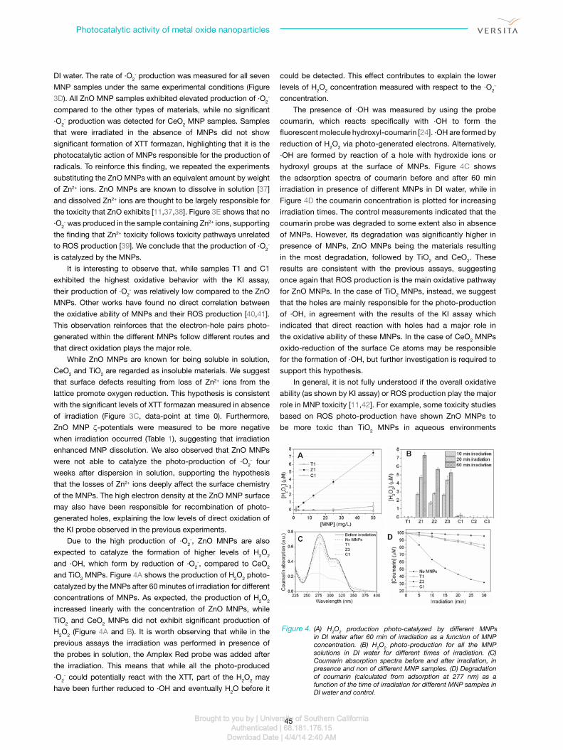
could be detected. This effect contributes to explain the lower
levels of H2O2 concentration measured with respect to the ·O2-
concentration.
The presence of ·OH was measured by using the probe
coumarin, which reacts specifically with ·OH to form the
fluorescent molecule hydroxyl-coumarin [24]. ·OH are formed by
reduction of H2O2 via photo-generated electrons. Alternatively,
·OH are formed by reaction of a hole with hydroxide ions or
hydroxyl groups at the surface of mnPs. Figure 4C shows
the adsorption spectra of coumarin before and after 60 min
irradiation in presence of different mnPs in DI water, while in
Figure 4D the coumarin concentration is plotted for increasing
irradiation times. The control measurements indicated that the
coumarin probe was degraded to some extent also in absence
of mnPs. However, its degradation was significantly higher in
presence of mnPs, ZnO mnPs being the materials resulting
in the most degradation, followed by TiO2 and CeO2. These
results are consistent with the previous assays, suggesting
once again that ROS production is the main oxidative pathway
for ZnO mnPs. In the case of TiO2 mnPs, instead, we suggest
that the holes are mainly responsible for the photo-production
of ·OH, in agreement with the results of the KI assay which
indicated that direct reaction with holes had a major role in
the oxidative ability of these mnPs. In the case of CeO2 mnPs
oxido-reduction of the surface Ce atoms may be responsible
for the formation of ·OH, but further investigation is required to
support this hypothesis.
In general, it is not fully understood if the overall oxidative
ability (as shown by KI assay) or ROS production play the major
role in mnP toxicity [11,42]. For example, some toxicity studies
based on ROS photo-production have shown ZnO mnPs to
be more toxic than TiO2 mnPs in aqueous environments
DI water. The rate of ·O2- production was measured for all seven
mnP samples under the same experimental conditions (Figure
3D). All ZnO mnP samples exhibited elevated production of ·O2-
compared to the other types of materials, while no significant
·O2- production was detected for CeO2 mnP samples. Samples
that were irradiated in the absence of mnPs did not show
significant formation of XTT formazan, highlighting that it is the
photocatalytic action of mnPs responsible for the production of
radicals. To reinforce this finding, we repeated the experiments
substituting the ZnO mnPs with an equivalent amount by weight
of Zn2+ ions. ZnO mnPs are known to dissolve in solution [37]
and dissolved Zn2+ ions are thought to be largely responsible for
the toxicity that ZnO exhibits [11,37,38]. Figure 3e shows that no
·O2- was produced in the sample containing Zn2+ ions, supporting
the finding that Zn2+ toxicity follows toxicity pathways unrelated
to ROS production [39]. We conclude that the production of ·O2-
is catalyzed by the mnPs.
It is interesting to observe that, while samples T1 and C1
exhibited the highest oxidative behavior with the KI assay,
their production of ·O2- was relatively low compared to the ZnO
mnPs. Other works have found no direct correlation between
the oxidative ability of mnPs and their ROS production [40,41].
This observation reinforces that the electron-hole pairs photo-
generated within the different mnPs follow different routes and
that direct oxidation plays the major role.
While ZnO mnPs are known for being soluble in solution,
CeO2 and TiO2 are regarded as insoluble materials. We suggest
that surface defects resulting from loss of Zn2+ ions from the
lattice promote oxygen reduction. This hypothesis is consistent
with the significant levels of XTT formazan measured in absence
of irradiation (Figure 3C, data-point at time 0). Furthermore,
ZnO mnP ζ-potentials were measured to be more negative
when irradiation occurred (Table 1), suggesting that irradiation
enhanced mnP dissolution. We also observed that ZnO mnPs
were not able to catalyze the photo-production of ·O2- four
weeks after dispersion in solution, supporting the hypothesis
that the losses of Zn2+ ions deeply affect the surface chemistry
of the mnPs. The high electron density at the ZnO mnP surface
may also have been responsible for recombination of photo-
generated holes, explaining the low levels of direct oxidation of
the KI probe observed in the previous experiments.
Due to the high production of ·O2-, ZnO mnPs are also
expected to catalyze the formation of higher levels of H2O2
and ·OH, which form by reduction of ·O2-, compared to CeO2
and TiO2 mnPs. Figure 4A shows the production of H2O2 photo-
catalyzed by the mnPs after 60 minutes of irradiation for different
concentrations of mnPs. As expected, the production of H2O2
increased linearly with the concentration of ZnO mnPs, while
TiO2 and CeO2 mnPs did not exhibit significant production of
H2O2 (Figure 4A and B). It is worth observing that while in the
previous assays the irradiation was performed in presence of
the probes in solution, the Amplex Red probe was added after
the irradiation. This means that while all the photo-produced
·O2- could potentially react with the XTT, part of the H2O2 may
have been further reduced to ·OH and eventually H2O before it
Figure 4. (A) H2O2 production photo-catalyzed by different MNPs in DI water after 60 min of irradiation as a function of MNP concentration. (B) H2O2 photo-production for all the MNP solutions in DI water for different times of irradiation. (C) Coumarin absorption spectra before and after irradiation, in presence and non of different MNP samples. (D) Degradation of coumarin (calculated from adsorption at 277 nm) as a function of the time of irradiation for different MNP samples in DI water and control.
Brought to you by | University of Southern CaliforniaAuthenticated | 68.181.176.15
Download Date | 4/4/14 2:40 AM

C. Minelli, R. Tantra
46
characteristics such as size, surface chemistry and propensity
to aggregate may influence their photo-catalytic properties.
The methodologies described here can be employed for
screening the photo-oxidative behavior of mnPs, in preparation
and support of expensive in vivo toxicological assessments.
Furthermore, these findings are important to achieve a deeper
understanding of the real biological damage that these materials
can cause in relation to their photo-oxidative ability and will
contribute to the formulation of effective solutions to contrast
their potential toxicity.
Acknowledgment
This work was conducted as part of PROSPecT, which is a
public-private partnership between DeFRA, ePSRC and TSB
and the nanotechnology Industries Association (nIA Ltd.)
and its members, and was administered by the DeFRA LInK
Programme. We thank Dr Tony Fry for the XRD measurements,
and Alex Shard and Andy Wain for the insight they offered in the
interpretation of the results.
[43-45], although the role of dissolved zinc needs to be
elucidated [45,46]. The development of relative simple protocols
for the assessment of the photo-catalytic properties of mnPs
in environmentally relevant media such those presented in
this work can support in vitro and in vivo investigation of mnP
toxicity and potentially offer further insights into mnP toxicity
mechanisms. Standardization of such protocols will facilitate
the comparison of the toxic effects of different mnPs in relation
to their photo-catalytic properties.
4. Conclusions
This study showed that mnPs have the ability to catalyze the
photo-oxidation of molecules in their surroundings. When
dispersed in the environment, these materials can therefore
potentially harm biomolecules and living organisms they may
interface. We have demonstrated that the mechanisms of
photo-oxidative behavior differ for TiO2, ZnO and CeO2 mnPs.
Furthermore, different behaviors were also observed within
the same family of mnPs, suggesting that specific particle
References
[1] A. Fujishima, X. Zhang, D.A. Tryk. TiO2 photocatalysis and
related surface phenomena. Surf. Sci. Rep. 63, 515-582
(2008).
[2] C.O. Robichaud, D. Tanzil, U. Weilenmann, M.R.
Wiesner. Relative Risk Analysis of Several Manufactured
Nanomaterials: An Insurance Industry Context. Environ. Sci.
Technol. 39, 8985-8994 (2005).
[3] N.C. Mueller, B. Nowack. Exposure Modeling of Engineered
Nanoparticles in the Environment. Environ. Sci. Technol. 42,
4447-4453 (2008).
[4] J.D. Judy, J.M. Unrine, P.M. Bertsch. Evidence for
Biomagnification of Gold Nanoparticles within a Terrestrial
Food Chain. Environ. Sci. Technol. 45, 776-781 (2010).
[5] K. Tiede, M. Hassellöv, E. Breitbarth, Q. Chaudhry, A.B.A. Boxall.
Considerations for environmental fate and ecotoxicity testing
to support environmental risk assessments for engineered
nanoparticles. J. Chromatogr. A 1216, 503-509 (2009).
[6] A. Kahru, H.-C. Dubourguier. From ecotoxicology to
nanoecotoxicology. Toxicology 269, 105-119 (2010).
[7] D. Darr, I. Fridovich. Free Radicals In Cutaneous Biology. J.
Invest. Dermatol. 102, 671-675 (1994).
[8] M.A. Maurer-Jones, J.R. Christenson, C.L. Haynes. TiO2
nanoparticle-induced ROS correlates with modulated
immune cell function. J. Nanopart. Res. 14, (2012).
[9] S.-R. Chae, E.M. Hotze, M.R. Wiesner. Evaluation of the
Oxidation of Organic Compounds by Aqueous Suspensions
of Photosensitized Hydroxylated-C60 Fullerene Aggregates.
Environ. Sci. Technol. 43, 6208-6213 (2009).
[10] S.J. Xiong, S.J. George, Z.X. Ji, S.J. Lin, H.Y. Yu, R.
Damoiseaux, et al. Size of TiO2 nanoparticles influences their
phototoxicity: an in vitro investigation. Arch. Toxicol. 87, 99-
109 (2013).
[11] H.B. Ma, P.L. Williams, S.A. Diamond. Ecotoxicity of
manufactured ZnO nanoparticles - A review. Environ. Pollut.
172, 76-85 (2013).
[12] A. Nel, T. Xia, L. Mädler, N. Li. Toxic Potential of Materials at
the Nanolevel. Science 311, 622-627 (2006).
[13] N.J. Rogers, N.M. Franklin, S.C. Apte, G.E. Batley, B.M.
Angel, J.R. Lead, et al. Physico-chemical behaviour and
algal toxicity of nanoparticulate CeO2 in freshwater. Environ.
Chem. 7, 50-60 (2009).
[14] M.M. Zou, Y.M. Kong, J. Wang, Q. Wang, Z.Q. Wang, B.X.
Wang, et al. Spectroscopic analyses on ROS generation
catalyzed by TiO2, CeO2/TiO2 and Fe2O3/TiO2 under
ultrasonic and visible-light irradiation. Spectroc. Acta Pt.
A-Molec. Biomolec. Spectr. 101, 82-90 (2013).
[15] H. Ma, A. Brennan, S.A. Diamond. Photocatalytic reactive
oxygen species production and phototoxicity of titanium
dioxide nanoparticles are dependent on the solar ultraviolet
radiation spectrum. Environmental Toxicology and Chemistry
31, 2099-2107 (2012).
[16] H. Ma, A. Brennan, S.A. Diamond. Phototoxicity of TiO2
nanoparticles under solar radiation to two aquatic species:
Daphnia magna and Japanese medaka. Environmental
Toxicology and Chemistry 31, 1621-1629 (2012).
[17] K.D. Pickering, M.R. Wiesner. Fullerol-Sensitized Production
of Reactive Oxygen Species in Aqueous Solution. Environ.
Sci. Technol. 39, 1359-1365 (2005).
[18] C.-Y. Chen, C.T. Jafvert. Photoreactivity of Carboxylated
Single-Walled Carbon Nanotubes in Sunlight: Reactive
Brought to you by | University of Southern CaliforniaAuthenticated | 68.181.176.15
Download Date | 4/4/14 2:40 AM

Photocatalytic activity of metal oxide nanoparticles
47
[33] A. Gomes, E. Fernandes, J.L.F.C. Lima. Fluorescence probes
used for detection of reactive oxygen species. J. Biochem.
Biophys. Methods 65, 45-80 (2005).
[34] G. Bartosz. Use of spectroscopic probes for detection of
reactive oxygen species. Clin. Chim. Acta 368, 53-76 (2006).
[35] N.W. Roehm, G.H. Rodgers, S.M. Hatfield, A.L. Glasebrook.
An improved colorimetric assay for cell-proliferation and
viability utilizing the tetrazolium salt XTT. J. Immunol. Methods
142, 257-265 (1991).
[36] U. Cernigoj, U.L. Stangar, P. Trebse, M. Sarakha.
Determination of catalytic properties of TiO2 coatings using
aqueous solution of coumarin: Standardization efforts. J.
Photochem. Photobiol. A 201, 142-150 (2009).
[37] N.M. Franklin, N.J. Rogers, S.C. Apte, G.E. Batley, G.E.
Gadd, P.S. Casey. Comparative toxicity of nanoparticulate
ZnO, bulk ZnO, and ZnCl2 to a freshwater microalga
(Pseudokirchneriella subcapitata): The importance of particle
solubility. Environ. Sci. Technol. 41, 8484-8490 (2007).
[38] W.H. Song, J.Y. Zhang, J. Guo, J.H. Zhang, F. Ding, L.Y. Li, et
al. Role of the dissolved zinc ion and reactive oxygen species
in cytotoxicity of ZnO nanoparticles. Toxicol. Lett. 199, 389-
397 (2010).
[39] T. Buerki-Thurnherr, L. Xiao, L. Diener, O. Arslan, C.
Hirsch, X. Maeder-Althaus, et al. In vitro mechanistic study
towards a better understanding of ZnO nanoparticle toxicity.
Nanotoxicology 2012 Mar 20, Epub ahead of print (2012).
[40] M.Y. Guo, A.M.C. Ng, F. Liu, A.B. Djurisic, W.K. Chan.
Photocatalytic activity of metal oxides-The role of holes and
OH center dot radicals. Applied Catalysis B-Environmental
107, 150-157 (2011).
[41] M.D. Hernandez-Alonso, A.B. Hungria, A. Martinez-Arias, M.
Fernandez-Garcia, J.M. Coronado, J.C. Conesa, et al. EPR
study of the photoassisted formation of radicals on CeO2
nanoparticles employed for toluene photooxidation. Applied
Catalysis B-Environmental 50, 167-175 (2004).
[42] W.J. Sun, A. Luna-Velasco, R. Sierra-Alvarez, J.A. Field.
Assessing protein oxidation by inorganic nanoparticles with
enzyme-linked immunosorbent assay (ELISA). Biotechnol.
Bioeng. 110, 694-701 (2013).
[43] L.K. Adams, D.Y. Lyon, P.J.J. Alvarez. Comparative eco-
toxicity of nanoscale TiO2, SiO2, and ZnO water suspensions.
Water Res. 40, 3527-3532 (2006).
[44] Q.L. Wu, A. Nouara, Y.P. Li, M. Zhang, W. Wang, M. Tang,
et al. Comparison of toxicities from three metal oxide
nanoparticles at environmental relevant concentrations in
nematode Caenorhabditis elegans. Chemosphere 90, 1123-
1131 (2013).
[45] D.W. Xiong, T. Fang, L.P. Yu, X.F. Sima, W.T. Zhu. Effects
of nano-scale TiO2, ZnO and their bulk counterparts on
zebrafish: Acute toxicity, oxidative stress and oxidative
damage. Sci. Total Environ. 409, 1444-1452 (2011).
[46] M. Li, D.H. Lin, L.Z. Zhu. Effects of water chemistry on
the dissolution of ZnO nanoparticles and their toxicity to
Escherichia coli. Environ. Pollut. 173, 97-102 (2013).
Oxygen Species Production in Water. Environ. Sci. Technol.
44, 6674-6679 (2010).
[19] J. Fabrega, R. Tantra, A. Amer, B. Stolpe, J. Tomkins, T. Fry,
et al. Sequestration of Zinc from Zinc Oxide Nanoparticles
and Life Cycle Effects in the Sediment Dweller Amphipod
Corophium volutator. Environ. Sci. Technol. 46, 1128-1135
(2012).
[20] R. Tantra. Evaluation and Assignment of Nanoparticle
Dispersion/Characterisation Methodologies, to be developed
under PROSPECT. NPL report AS 45 (2009).
[21] S. Merouani, O. Hamdaoui, F. Saoudi, M. Chiha. Influence of
experimental parameters on sonochemistry dosimetries: KI
oxidation, Fricke reaction and H2O2 production. J. Hazard.
Mater. 178, 1007-1014 (2010).
[22] M. Sutherland, B. Learmonth. The tetrazolium dyes MTS
and XTT provide new quantitative assays for superoxide
and superoxide dismutase. Free Radical Res. 27, 283-289
(1997).
[23] M.E. Bulina, D.M. Chudakov, O.V. Britanova, Y.G.
Yanushevich, D.B. Staroverov, T.V. Chepurnykh, et al. A
genetically encoded photosensitizer. Nature Biotech. 24, 95-
99 (2006).
[24] H. Czili, A. Horváth. Applicability of coumarin for
detecting and measuring hydroxyl radicals generated by
photoexcitation of TiO2 nanoparticles. App. Cat. B Env. 81,
295-302 (2008).
[25] J. Jiang, G. Oberdorster, A. Elder, R. Gelein, P. Mercer, P.
Biswas. Does nanoparticle activity depend upon size and
crystal phase? Nanotoxicology 2, 33-42 (2008).
[26] M.Y. Guo, A.M.C. Ng, F.Z. Liu, A.B. Djurisic, W.K. Chan, H.M.
Su, et al. Effect of Native Defects on Photocatalytic Properties
of ZnO. J. Phys. Chem. C 115, 11095-11101 (2011).
[27] Y.C. Liao, C.S. Xie, Y. Liu, Q.W. Huang. Enhancement of
photocatalytic property of ZnO for gaseous formaldehyde
degradation by modifying morphology and crystal defect. J.
Alloy. Compd. 550, 190-197 (2013).
[28] A. Asati, S. Santra, C. Kaittanis, S. Nath, J.M. Perez. Oxidase-
Like Activity of Polymer-Coated Cerium Oxide Nanoparticles.
Ang. Chem. Int. Ed. 48, 2308-2312 (2009).
[29] B.A. Rzigalinski, K. Meehan, R.M. Davis, Y. Xu, W.C. Miles,
C.A. Cohen. Radical nanomedicine. Nanomedicine 1, 399-
412 (2006).
[30] Y.C. Liao, C.S. Xie, Y. Liu, H. Chen, H.Y. Li, J. Wu. Comparison
on photocatalytic degradation of gaseous formaldehyde by
TiO2, ZnO and their composite. Ceram. Int. 38, 4437-4444
(2012).
[31] T. Berger, M. Sterrer, O. Diwald, E. Knozinger. Charge
trapping and photoadsorption of O-2 on dehydroxylated
TiO2 nanocrystals - An electron paramagnetic resonance
study. ChemPhysChem 6, 2104-2112 (2005).
[32] K.-i. Ishibashi, A. Fujishima, T. Watanabe, K. Hashimoto.
Quantum yields of active oxidative species formed on TiO2
photocatalyst. J. Photochem. Photobiol. A 134, 139-142
(2000).
Brought to you by | University of Southern CaliforniaAuthenticated | 68.181.176.15
Download Date | 4/4/14 2:40 AM