Metagenomic NGS for Pathogen Detection: Quality Control...
86
Metagenomic NGS for Pathogen Detection: Quality Control Issues and Strategies Steve Miller MD PhD Department of Laboratory Medicine University of California San Francisco
Transcript of Metagenomic NGS for Pathogen Detection: Quality Control...

Metagenomic NGS for
Pathogen Detection:
Quality Control Issues and
StrategiesSteve Miller MD PhD
Department of Laboratory Medicine
University of California San Francisco

Disclosures
• Major funding provided by:– CA Initiative to Advance Precision Medicine
– Sandler and Bowes Foundations
– Schwab Foundation
– Marcus Foundation

mNGS Pathogen DetectionClinical Sample
Nucleic Acid
RNA Virus
Report
Library Preparation
Bioinformatics Analysis
Sequencing
DNA Virus
Bacteria
Fungi
Parasite
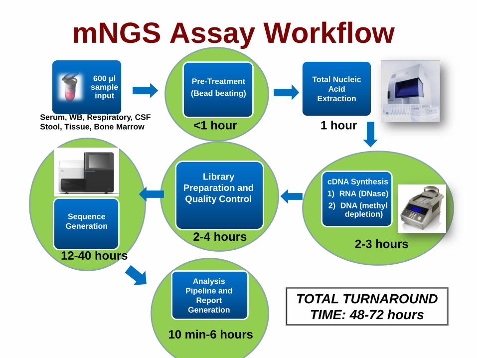
mNGS Assay Workflow
600 μlsample input
Total Nucleic
Acid
Extraction
<1 hour 1 hour
12-40 hours2-3 hours
2-4 hours
Serum, WB, Respiratory, CSF
Stool, Tissue, Bone Marrow
Analysis
Pipeline and
Report
Generation
10 min-6 hours
Pre-Treatment
(Bead beating)
Library
Preparation and
Quality Control
cDNA Synthesis
1) RNA (DNase)
2) DNA (methyl depletion)Sequence
Generation
TOTAL TURNAROUND
TIME: 48-72 hours
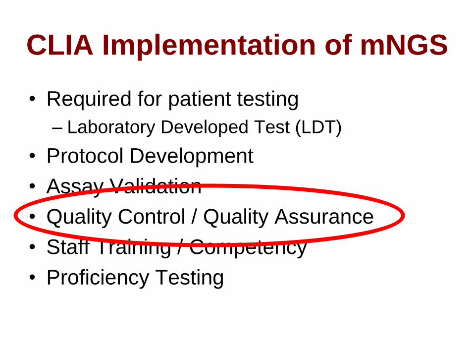
CLIA Implementation of mNGS
• Required for patient testing
– Laboratory Developed Test (LDT)
• Protocol Development
• Assay Validation
• Quality Control / Quality Assurance
• Staff Training / Competency
• Proficiency Testing
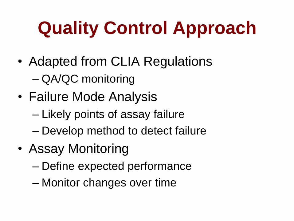
Quality Control Approach
• Adapted from CLIA Regulations
– QA/QC monitoring
• Failure Mode Analysis
– Likely points of assay failure
– Develop method to detect failure
• Assay Monitoring
– Define expected performance
– Monitor changes over time
Quality Control Methods
• Reagent Quality Control
• External Controls
• Internal Controls
• Process Controls / Checkpoints
• Contamination Control
Reagent Quality Control
• What reagents need QC?
• How to perform?
• What are acceptible criteria?
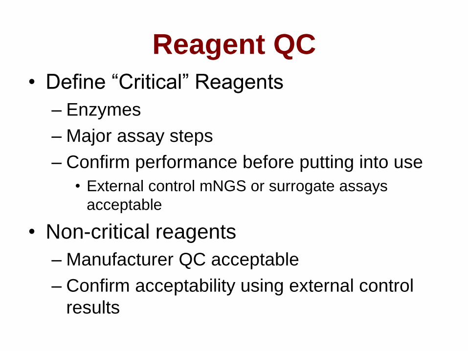
Reagent QC
• Define “Critical” Reagents
– Enzymes
– Major assay steps
– Confirm performance before putting into use
• External control mNGS or surrogate assays
acceptable
• Non-critical reagents
– Manufacturer QC acceptable
– Confirm acceptability using external control
results

Critical Reagent LogBaseline DNase Epicentre - IlluminaC.neoformans American Type Culture CollectionDNA Clean & Concentrator Kit -5 Capped Columns Zymo ResearchdNTPs Thermo Fisher - InvitrogenEZ1 Virus Mini Kit v.2.0 QIAGENHiSeq Rapid SR Cluster Kit v2 Flowcell IlluminaHiSeq Rapid SR Cluster Kit v2 Rapid SR Cluster Kit IlluminaHiSeq Rapid SR Cluster Kit v2 Flowcell IlluminaK.pneumoniae American Type Culture CollectionLinear acrylamide Thermo Fisher - Life TechnologiesLysing Matrix B Tube (2mL) MP BiomedicalsMS2 Phage American Type Culture CollectionNEBNext Microbiome DNA Enrichment Kit NEBNext (New England Biolabs)Nextera Index Kit v2 Set A (96 indexes, 384 samples) IlluminaPhusion High-Fidelity PCR Kit Thermo Fisher - Life TechnologiesSEQUENASE VERSION 2.0 DNA POLYMERASE - UNDILUTED AffymetrixSuperscript III enzyme Thermo Fisher - InvitrogenSynthetic CSF Matrix 100 mL Golden West BiologicalsT1 Phage American Type Culture CollectionTurbo Dnase Kit Thermo Fisher - AmbionWater, (DNASE, RNASE free), Fisher BioReagents Fisher ScientificBuffer EB QiagenWater, (DNASE, RNASE free), Fisher BioReagents Fisher Scientific
(Very partial list)
Include:
Enzyme kits
Control organisms
Sterile materials
QC metric:
Prior to use
Activity
Contamination

External Controls
• Positive Control
• Negative Control
– Elution buffer (negative extract)
– Also used as background normalizer
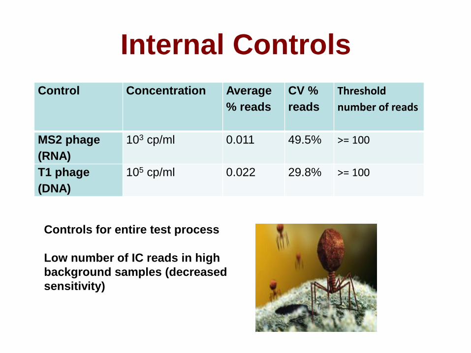
Internal Controls
Control Concentration Average
% reads
CV %
reads
Threshold
number of reads
MS2 phage
(RNA)
103 cp/ml 0.011 49.5% >= 100
T1 phage
(DNA)
105 cp/ml 0.022 29.8% >= 100
Controls for entire test process
Low number of IC reads in high
background samples (decreased
sensitivity)

DNA phage control
RNA phage control
Host DNA Interference
1
10
100
1000
10000
100000
1 10 100 1000
Lo
g o
f R
ea
ds
Pe
r M
illi
on
(M
S2
or
T1
p
hag
e)
cells/ul equivalents of spiked in DNA / RNA
DNA interference [DNA phage readout]
DNA interference [RNA phage readout]
RNA interference [DNA phage readout]
RNA interference [RNA phage readout]
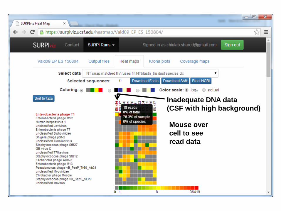
Mouse over
cell to see
read data
Inadequate DNA data
(CSF with high background)
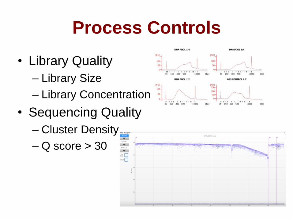
Process Controls
• Library Quality
– Library Size
– Library Concentration
• Sequencing Quality
– Cluster Density
– Q score > 30
Analyzer Controls
• Concentration controls (Qubit)
– Stock libraries
• Bioanalyzer controls
– Stock libraries
• Develop metrics for QC of library
analyzers
– Concentration range
– Size range

Periodic Monitoring
• Positive threshold verification
– Verify detection at 1:10 dilution of positive
control
• Software run log
– Document versions
• Instrument repair log
• Troubleshooting log

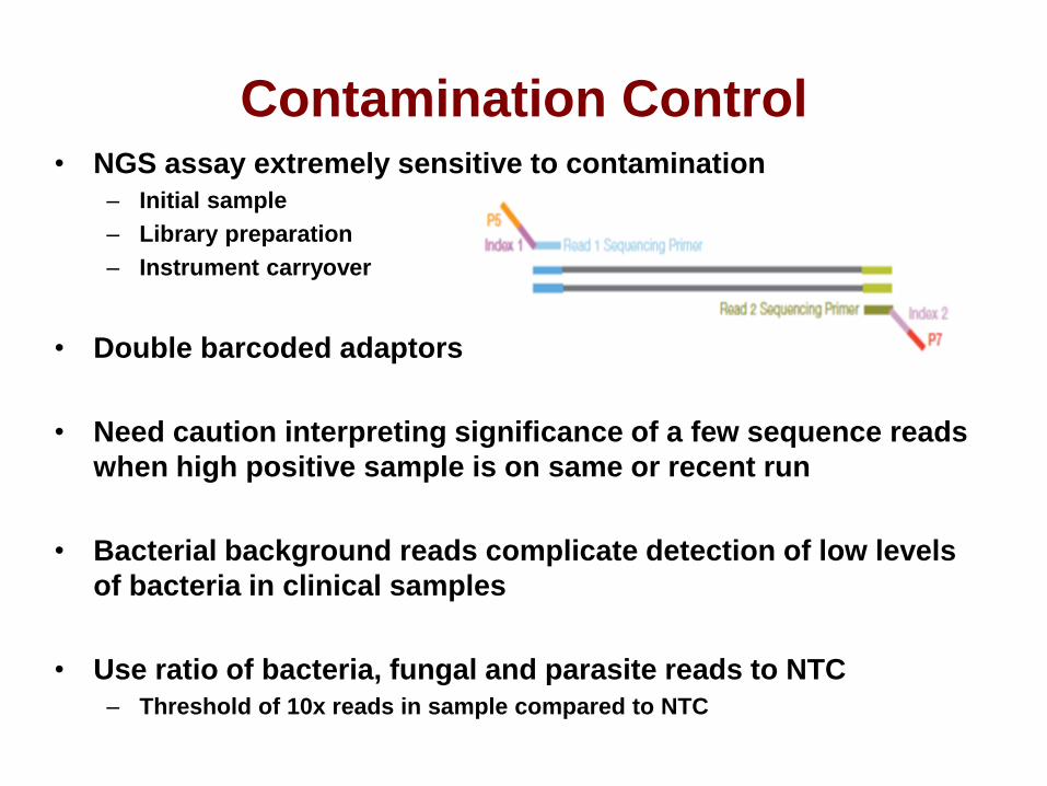
Contamination Control• NGS assay extremely sensitive to contamination
– Initial sample
– Library preparation
– Instrument carryover
• Double barcoded adaptors
• Need caution interpreting significance of a few sequence reads
when high positive sample is on same or recent run
• Bacterial background reads complicate detection of low levels of
bacteria in clinical samples
• Use ratio of bacteria, fungal and parasite reads to NTC
– Threshold of 10x reads in sample compared to NTC
Contamination Control
• Swipe Tests
– Background laboratory contamination
• Amplicon
• Organisms
• What is successful swipe test?
– Assess flora
– Unusual detections
– Pathogenic organisms

Swipe Test ResultsBacteria Fungi Parasite
DNA Virus RNA Virus
Environmental bacterial / fungal flora - acceptable
Few reads to potentially pathogenic viruses – clean and repeat
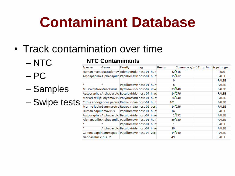
Contaminant Database
• Track contamination over time
– NTC
– PC
– Samples
– Swipe tests
NTC Contaminants
Troubleshooting Example
• Failure to achieve sufficient reads per
library
– 5 million required for adequate analysis
– Interpreted with comment that sensitivity may
be decreased
• Why are some libraries not providing
enough sequence?
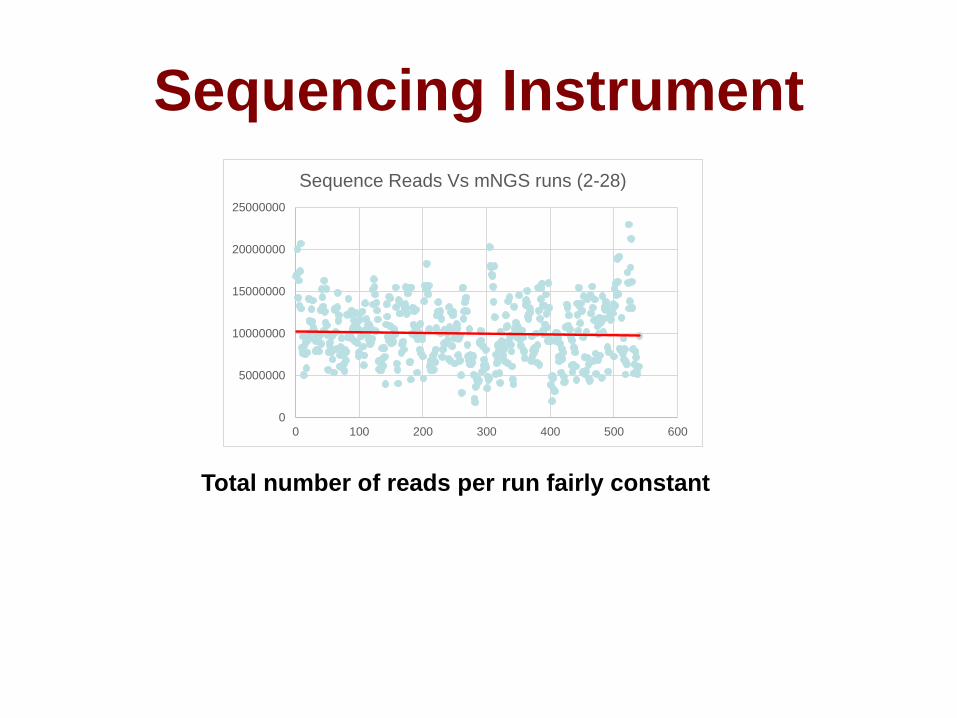
Sequencing Instrument
0
5000000
10000000
15000000
20000000
25000000
0 100 200 300 400 500 600
Sequence Reads Vs mNGS runs (2-28)
Total number of reads per run fairly constant
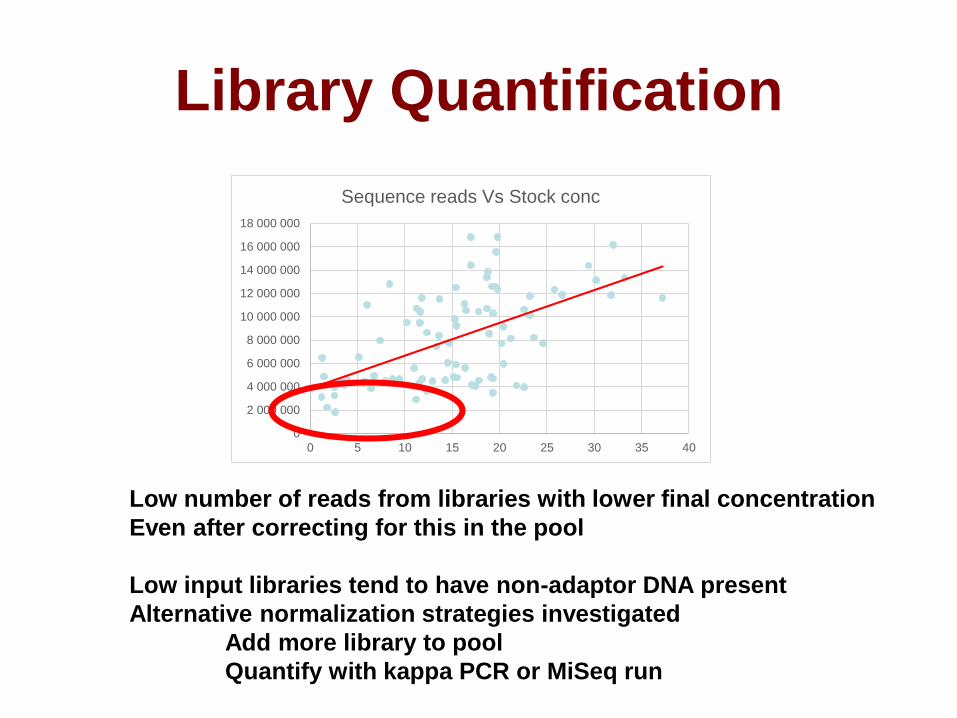
Library Quantification
0
2 000 000
4 000 000
6 000 000
8 000 000
10 000 000
12 000 000
14 000 000
16 000 000
18 000 000
0 5 10 15 20 25 30 35 40
Sequence reads Vs Stock conc
Low number of reads from libraries with lower final concentration
Even after correcting for this in the pool
Low input libraries tend to have non-adaptor DNA present
Alternative normalization strategies investigated
Add more library to pool
Quantify with kappa PCR or MiSeq run

Overview – mNGS QCClinical Sample
Nucleic Acid
RNA Virus
Report
Library Preparation
Bioinformatics Analysis
Sequencing
DNA Virus
Bacteria
Fungi
Parasite
Library QC Metrics
Library Quantification
Pooling
Sequence QC Metrics
Software Versioning
Process Controls
Reagent QC
Threshold Verification
Swipe Tests
Contaminant Database
Proficiency Testing
Sample Controls
Internal Controls
Clinical Review
Run Controls
External Controls
Contaminant Check
Stability Check
Sample Requirements
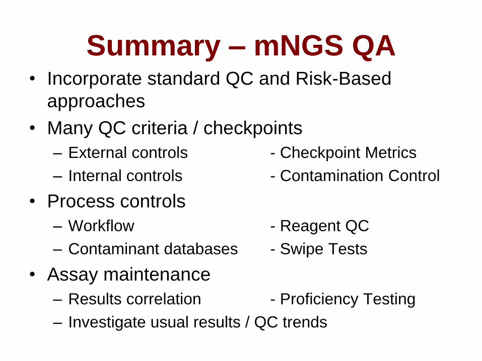
Summary – mNGS QA• Incorporate standard QC and Risk-Based
approaches
• Many QC criteria / checkpoints
– External controls - Checkpoint Metrics
– Internal controls - Contamination Control
• Process controls
– Workflow - Reagent QC
– Contaminant databases - Swipe Tests
• Assay maintenance
– Results correlation - Proficiency Testing
– Investigate usual results / QC trends
Acknowledgements
UCSF Chiu Lab and VDDC
Charles Chiu, MD/PhD
Erik Samayoa, CLS
Shaun Arevalo, CLS
Becky Fung, CLS
Hannah Sample, BS
Samia Naccache, PhD
Scot Federman, BS
Doug Stryke, BS
Joseph DeRisi, PhD
Michael Wilson, MD
Jeffrey Gelfand, MD
Michael Geschwind, MD
St. Jude’s Medical Center
Randall Hayden, MD/PhD
UCLA
Jeffrey Klausner, MD/MPH
Romney Humphries, PhD (ABMM)
Children’s Hospital Los Angeles
Jeffrey Bender, MD
Jennifer Dien-Bard, MDUniversity of California,
Berkeley
Brent Fulton, PhD/MBA
Children’s National Medical Center
Brittany Goldberg, MD
Joseph Campos, MD
UCDavis
Chris Polage, MD
Stuart Cohen, MD


Many Acute Infectious Diseases
Remain Undiagnosed
Pneumonia
15 – 25%
unknown cause
60-80%
unknown causeUp to 50%
unknown cause
Diarrheal Disease Meningitis / Encephalitis
Fever / Sepsis
~20% unknown
cause

Nearly All Agents that Cause
Infectious Diseases Contain
Nucleic Acid (DNA/RNA)
Bacteria Viruses
Fungi Parasites
Prions
NGS Pathogen Detection Allows For
• Diagnosis
– Treatment optimization
• Pathogen Discovery
• Public Health Investigation
– Unknown outbreak
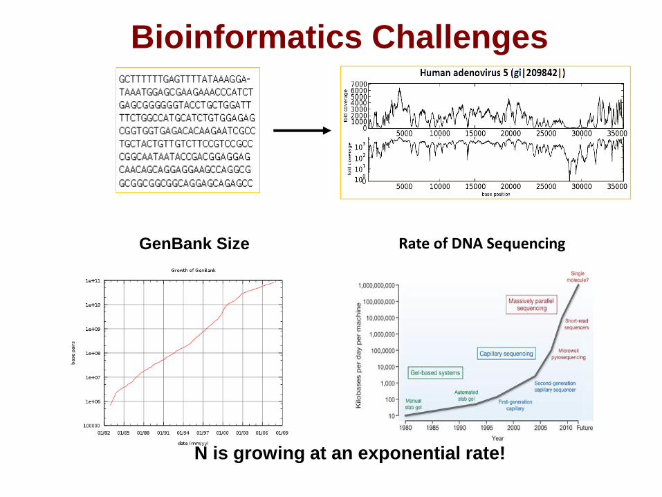
Rate of DNA SequencingGenBank Size
N is growing at an exponential rate!
Bioinformatics Challenges
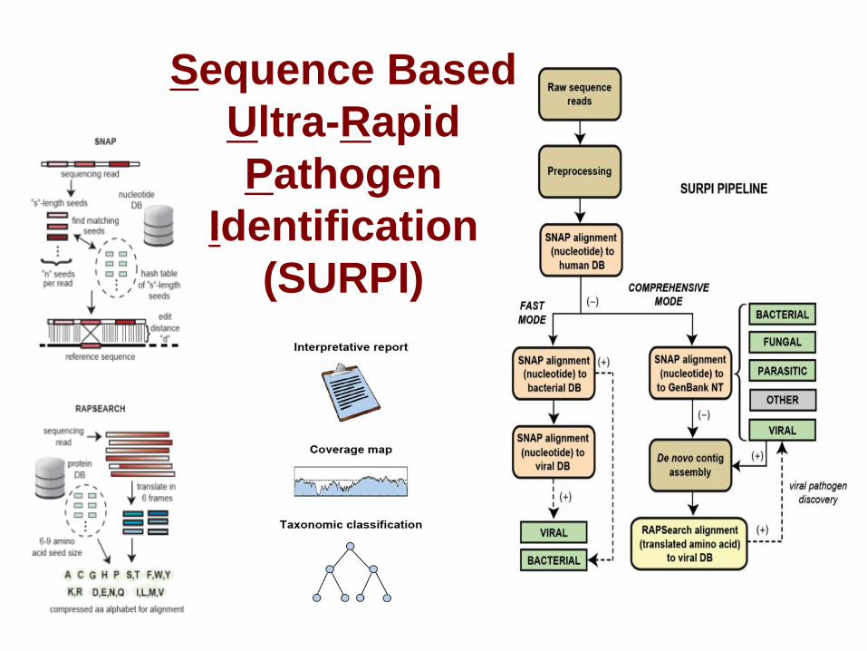
Sequence Based
Ultra-Rapid
Pathogen
Identification
(SURPI)
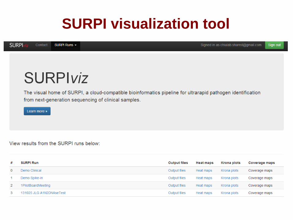
SURPI visualization tool
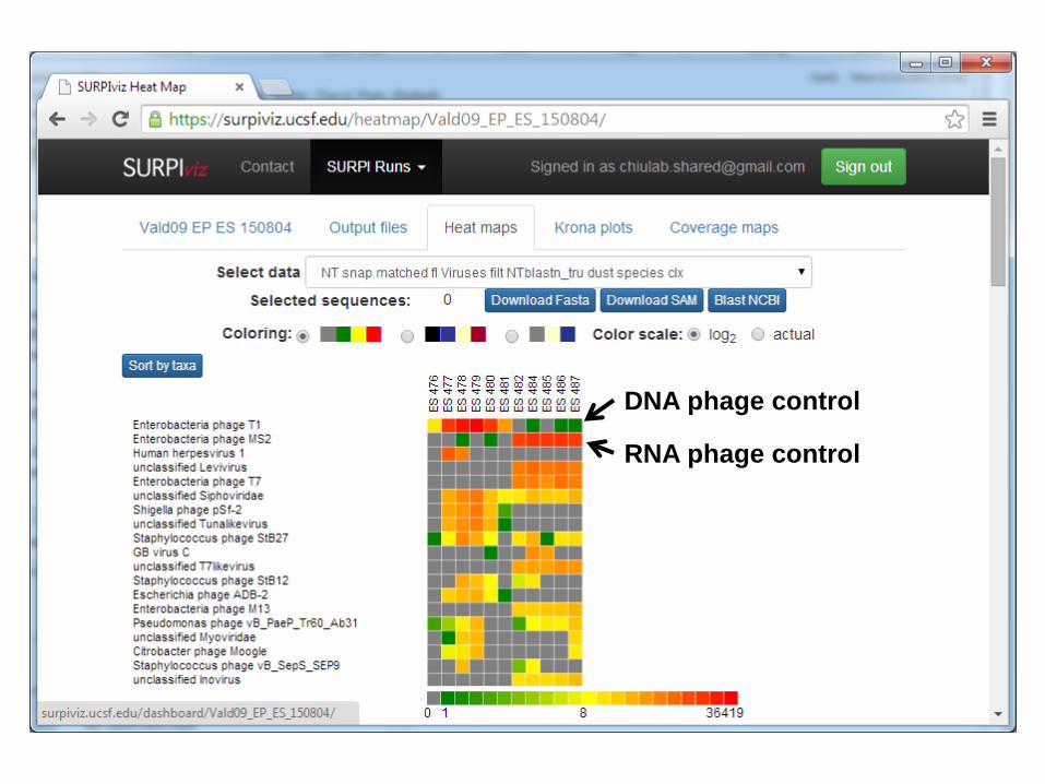
DNA phage control
RNA phage control
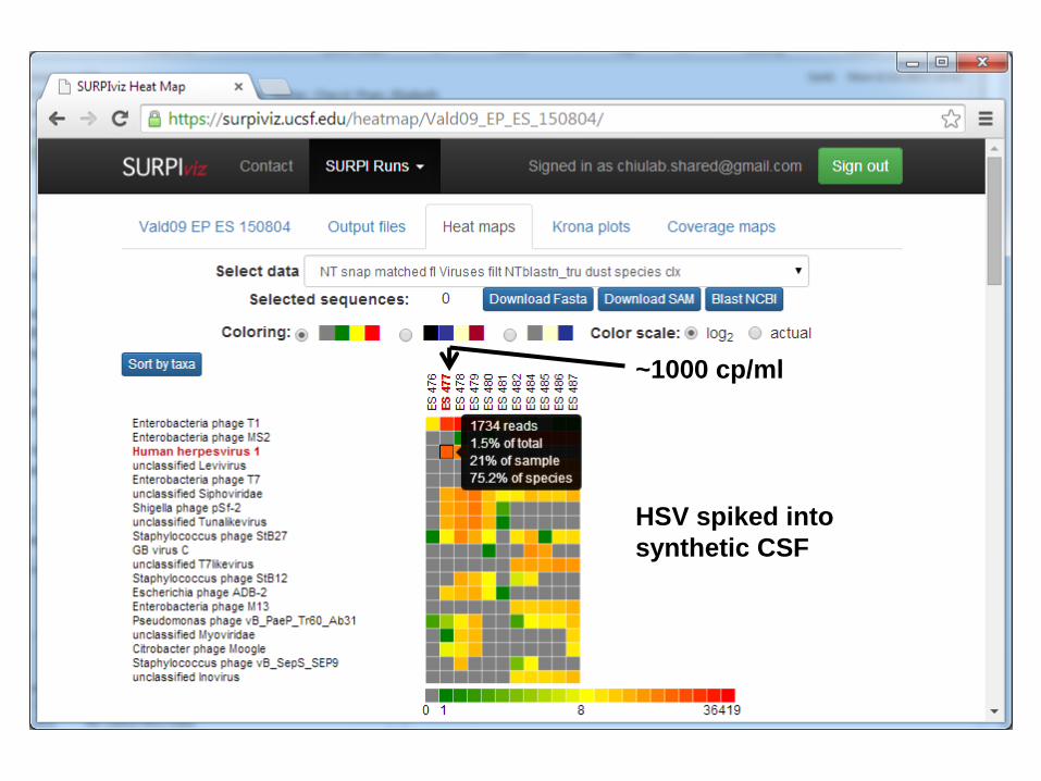
~1000 cp/ml
HSV spiked into
synthetic CSF
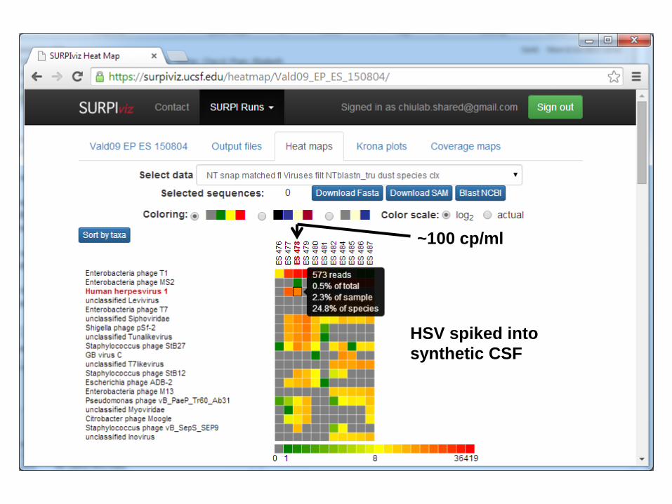
HSV spiked into
synthetic CSF
~100 cp/ml

HSV1 at 1000 cp/ml
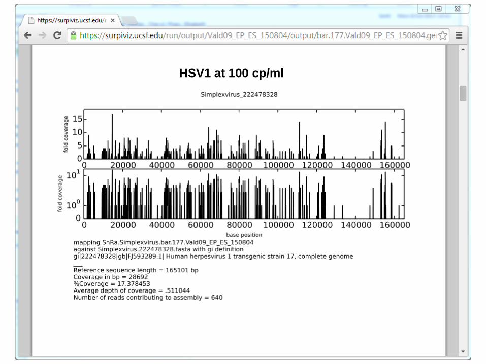
HSV1 at 100 cp/ml
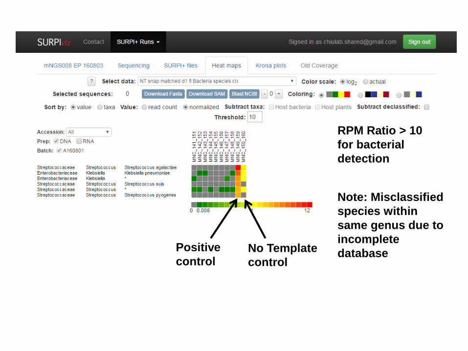
Positive
controlNo Template
control
RPM Ratio > 10
for bacterial
detection
Note: Misclassified
species within
same genus due to
incomplete
database

Validation of NGS
• Development of Controls
• Assay Performance Characteristics
• Contamination Control
• Clinical Study – Precision Diagnosis of
Acute Infectious Disease
– Interesting Case Reports & Discussion

Assay Sensitivity
RNA viruses
detected
to <100
copies/ml
DNA viruses
detected
to ~1000
copies/ml
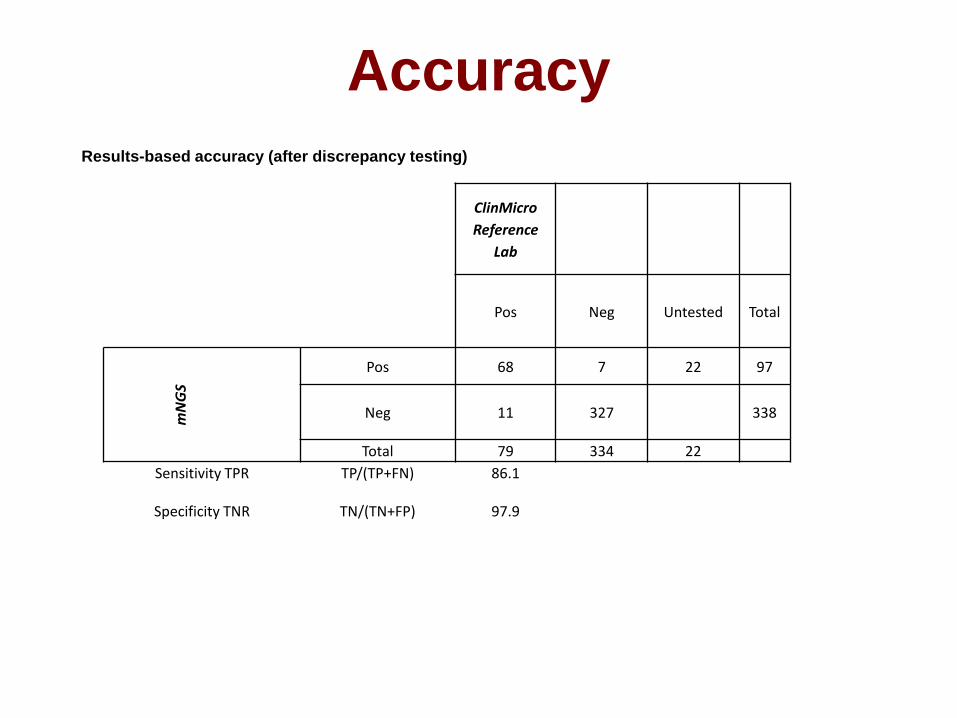
Accuracy
ClinMicro
Reference
Lab
Pos Neg Untested Total
mN
GS
Pos 68 7 22 97
Neg 11 327 338
Total 79 334 22
Sensitivity TPR TP/(TP+FN) 86.1
Specificity TNR TN/(TN+FP) 97.9
Results-based accuracy (after discrepancy testing)
Contamination Control• NGS assay extremely sensitive to contamination
– Initial sample
– Library preparation
– Instrument carryover
• Double barcoded adaptors
• Need caution interpreting significance of a few sequence reads
when high positive sample is on same or recent run
• Bacterial background reads complicate detection of low levels
of bacteria in clinical samples
• Use ratio of bacteria, fungal and parasite reads to NTC
– Threshold of 10x reads in sample compared to NTC

Precision Diagnosis of Acute
Infectious Disease
Diagnosis Review CSF Sample Review
mNGS test order Patient consent
Assay QCQC Checkpoints
External Positive Control
Internal Controls
Clinical Microbial
Sequencing Board
Patient Admitted to Hospital with M/E
mNGS Assay Performed
Review and Interpretation mNGS Report

mNGS Clinical Cases

An 11-year-old female
with HA and Back pain
• From Mexico
• 1 month PTA, she was admitted to a local hospital in
Mexico due to HA, nausea, abnormal jerking movements
of fingers, hands, and legs.
• CSF showed 183 WBC (100% mono), glucose 25,
protein 270
EBV and HHV-7 detected in CSF
• She was treated with acyclovir, then ganciclovir.
• 2 weeks after discharge, she developed back pain and
worsening HA.
An 11-year-old female
with HA and Back pain
• Repeat admission for worsening symptoms
• Brain MRI w/ + w/o contrast: normal
• CSF showed WBC 137 (L91%), glu 25, prot 200
Negative Gram-stain, bacterial culture
Negative HSV PCR, EBV PCR, enterovirus PCR
• Multiple ID tests (blood and CSF) were sent
• INH, rifampin, PZA, ETB + pyridoxine initiated for empiric
coverage of tuberculosis
An 11-year-old female
with HA and Back pain
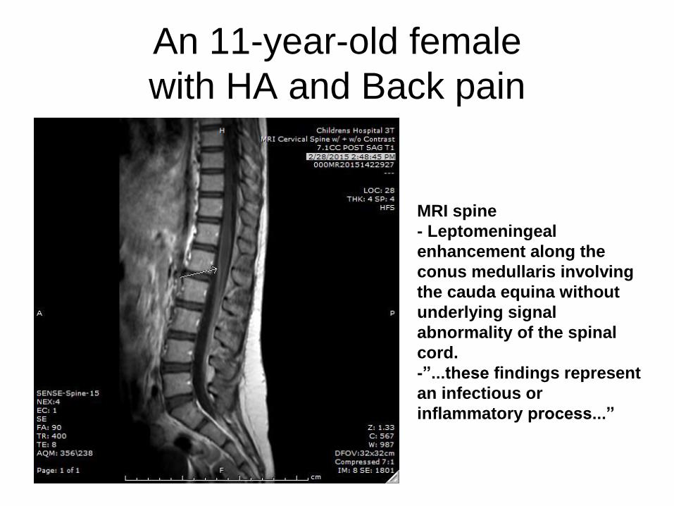
MRI spine
- Leptomeningeal
enhancement along the
conus medullaris involving
the cauda equina without
underlying signal
abnormality of the spinal
cord.
-”...these findings represent
an infectious or
inflammatory process...”
An 11-year-old female
with HA and Back pain
• Clonazepam added for polyminimyoclonus
• Negative CSF M. tb PCR
• No improvement after 8 days of therapy – repeat LP
• CSF (#2): WBC 176 (N53%, L38%, M9%), glucose 25, protein 131.
CSF#2 was positive for EBV PCR (Ct=36.44) and HHV-7 PCR– EBV serology: c/w remote infection
– Blood for EBV PCR was not detected
• Concern for drug-resistant TB:– ETB was changed to ethionamide
– Levofloxacin added to RIPE
• TB PCR CSF #2 negative
• she improved substantially and was discharged home
with 5 anti-TB medications
• At 1 week and 1 month follow-up after discharge, she
had near resolution of symptoms, but still reported
several episodes of shaking of her extremities
• Because of clinical improvement, she returned to Mexico
and continued her anti-TB therapy.
• CSF #2 submitted for mNGS analysis
An 11-year-old female
with HA and Back pain
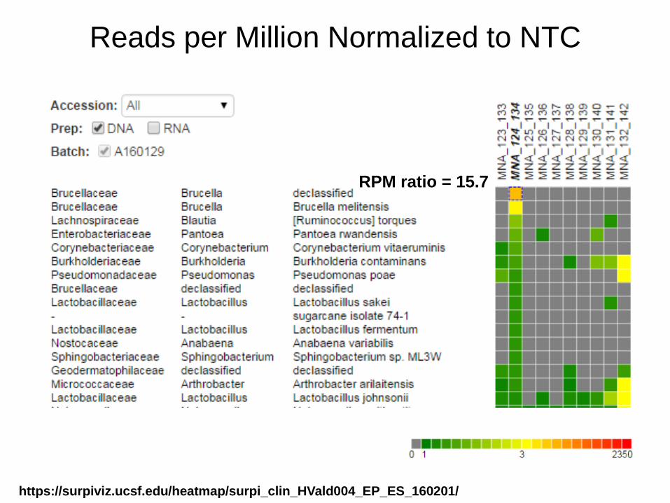
https://surpiviz.ucsf.edu/heatmap/surpi_clin_HVald004_EP_ES_160201/
Reads per Million Normalized to NTC
RPM ratio = 15.7
Followup
• Patient started on targeted Brucella therapy
– Doxycycline + Rifampin
• Symptoms fully resolved in 2 weeks
• Serum antibody agglutination positive 1:80 titer
• PCR performed confirming mNGS findings of
Brucella DNA
58 y/o woman with fever, headache, and nausea/vomiting
• History of idiopathic pulmonary fibrosis s/p bilateral lung transplant, migraines, hypercoagulability with DVT, and multiple sclerosis (MS) on chronic immunosuppression
• Admitted 8 days of fever, headache, refractory nausea/vomiting, neck stiffness, and photophobia
• Had been hospitalized prior complaining of “worst headache of her life” – treated with abortive migraine medications with only partial relief

58 y/o woman with fever, headache, and nausea/vomiting
• Resident of southern California
• Denied sick contacts, pets, insect bites, or eating shellfish or game meats
• Reported travel to mountains in Utah in Aug 2016, the Caribbean in 2010, and throughout Europe decades prior to admission
• Outpatient medications notable for immunosuppressive agents (tacrolimus, mycophenolate mofetil, prednisone, teriflunomide) and antimicrobial prophylaxis with trimethoprim/sulfamethoxazole and acyclovir; also on an intrathecal morphine pump
58 y/o woman with fever, headache, and nausea/vomiting
• On admission
– Fever to 38.3°C, otherwise vital signs normal
– Physical exam remarkable only for R leg tenderness attributed to known DVT
– Initial laboratories remarkable for stable pancytopenia (WBC 9, HgB 10.9, platelets 124,000), mildly elevated LFTs (AST 130 U/L, ALT 83 U/L, total bili 1.1 mg/dL, alkaline phosphatase 144 U/L) and borderline elevated prothrombin time of 14.3 (11-14 seconds)
– MRI – baseline periventricular and subcortical T2/FLAIR white matter intensities associated with her MS, no acute changes
– Started on empiric vancomycin, ceftazidime, acyclovir, voriconazole
• Lumbar puncture performed on day 3
– CSF pleocytosis (10 WBC/mm63; 88% lymphocytes; 12% monocytes; protein 29; glucose 48)
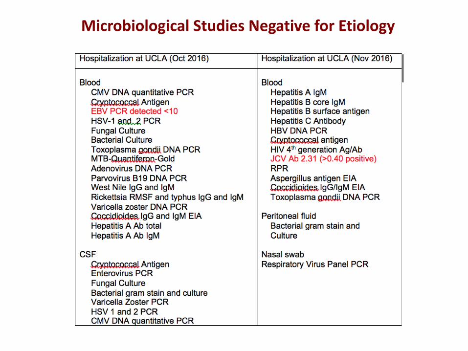
Microbiological Studies Negative for Etiology
58 y/o woman with fever, headache, and nausea/vomiting
• By day 6, patient clinically improved with resolution of all symptoms and fever, except for mild persistent headache
• Discharge diagnosis was meningoencephalitis due to viral infection or tacrolimus toxicity
• mNGS performed on CSF sample obtained on day 3
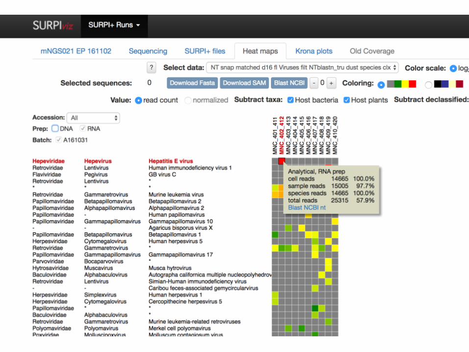
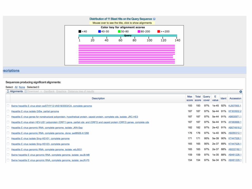
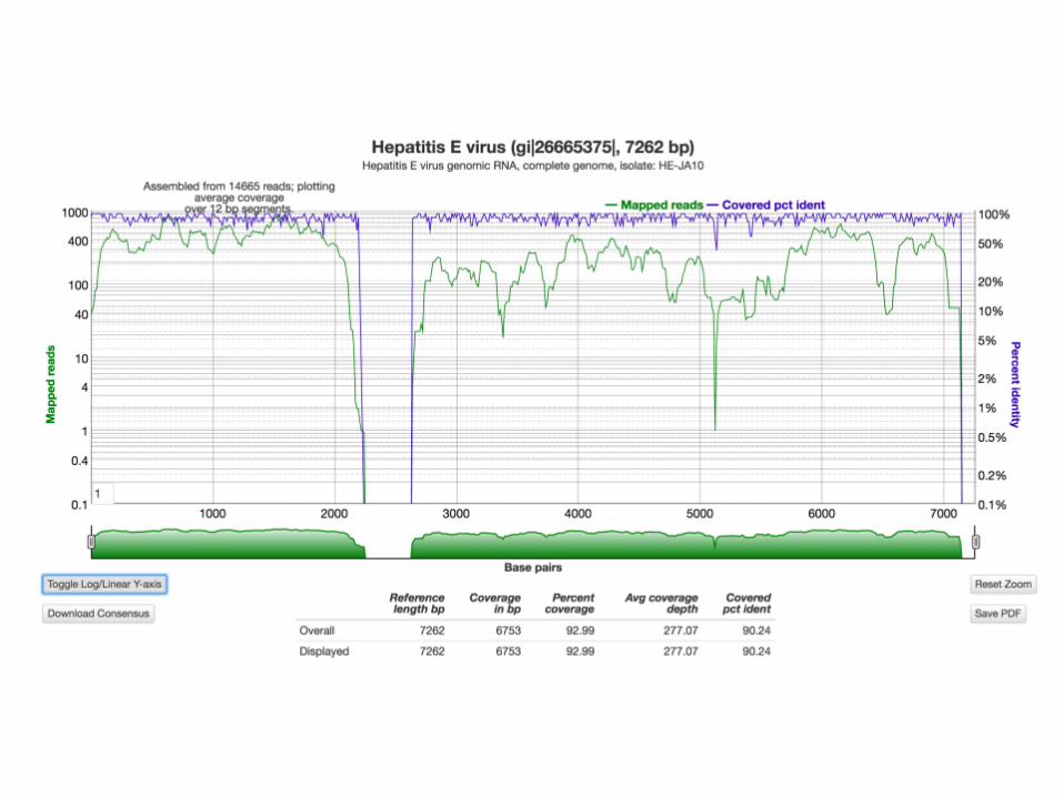

Hepatitis E phylogeny
Patient’s
HEV genome
Genotype 3

58 y/o woman with fever, headache, and nausea/vomiting
• Diagnosis of HEV-associated meningoencephalitis communicated to patient
• Retrospective review of EMR showed normal transaminase levels prior to lung transplantation, with persistent low-level transaminase elevations following transplantation in 2011
• Immediate follow up HEV testing showed serum IgM positivity, negative IgG, and plasma HEV viremia (5,960,000 IU/mL)
• Ribavirin monotherapy was started for this patient
• Case reported to United Network for Organ Sharing donor safety net
• HEV IgG/IgM donor serum was positive, but HEV RNA not detected
– Potential donor-derived infection
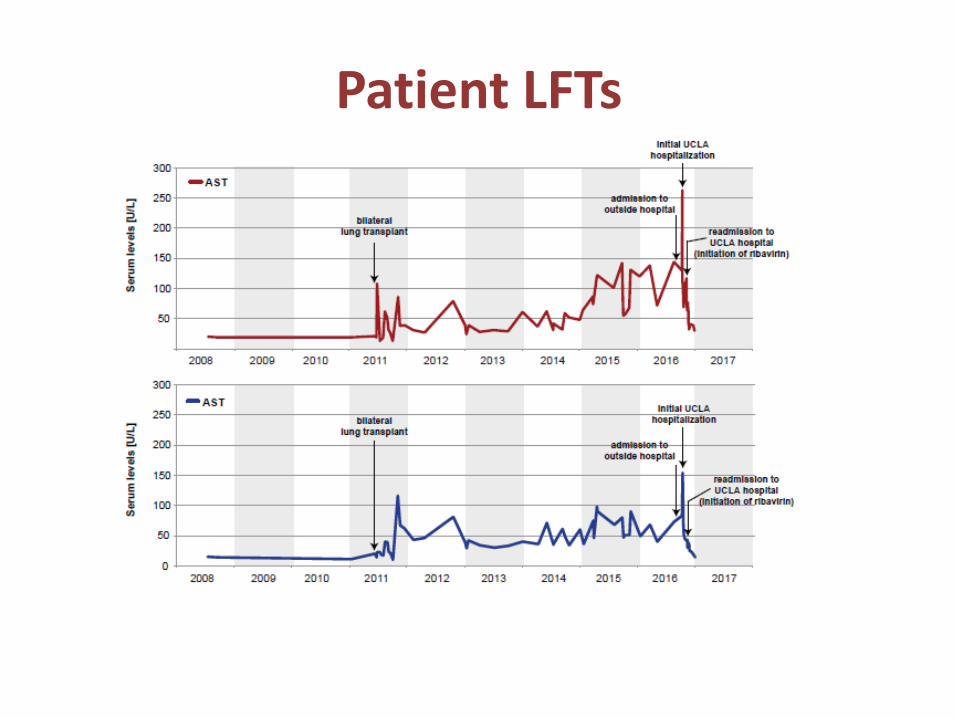
Patient LFTs

Hepatitis E – Neurologic Manifestations
• Commonest cause of Acute hepatitis worldwide
• Neurological disorders associated with HEV
– First case- 2000: GBS
– 2011: 5.5% of well characterized patients with acute and chronic Hepatitis E cases from Europe
– 100 cases reported to date

Case Discussion
• First report of potential HEV transmission via lung transplantation
• Evidence in support: positive anti-HEV IgG/IgM testing of donor serum
• Persistent low-level transaminase elevations post-transplant
• Prior HEV donor transmission reports include liver transplant recipient and 2 renal transplant recipients (from same donor)
– (Sclosser, et al., J hepatol 2012; Pourbaix, et al., Transplant Infect Dis 2016)
68 year old male with fever and AMS
• History of CAD, HTN, mantle cell lymphoma
• Developed fever, chills lethargy, fatigue, confusion
• Admitted and started on empirical vancomycin, meropenemand levofloxacin
• 3 days later became hypoxic with worsening AMS
– LP showed 18 WBC (35% monocytes, 33% lymphocytes, 32% neutrophils), normal glucose and protein
• Added acylovir for viral meningitis
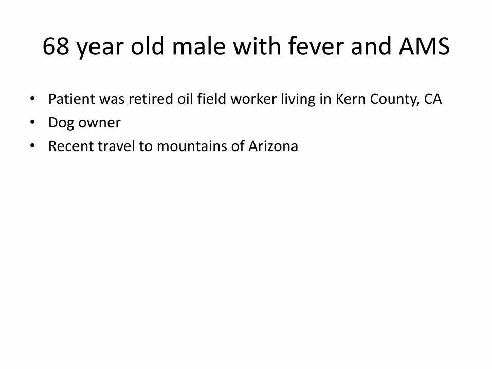
68 year old male with fever and AMS
• Patient was retired oil field worker living in Kern County, CA
• Dog owner
• Recent travel to mountains of Arizona

68 year old male with fever and AMS
• Patient continued to spike fevers
– Antifungal therapy added
• Repeat LP showed persistent pleocytosis
– Extensive infectious workup non-revealing
• CSF analyzed by mNGS

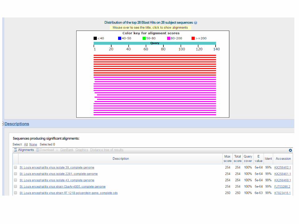
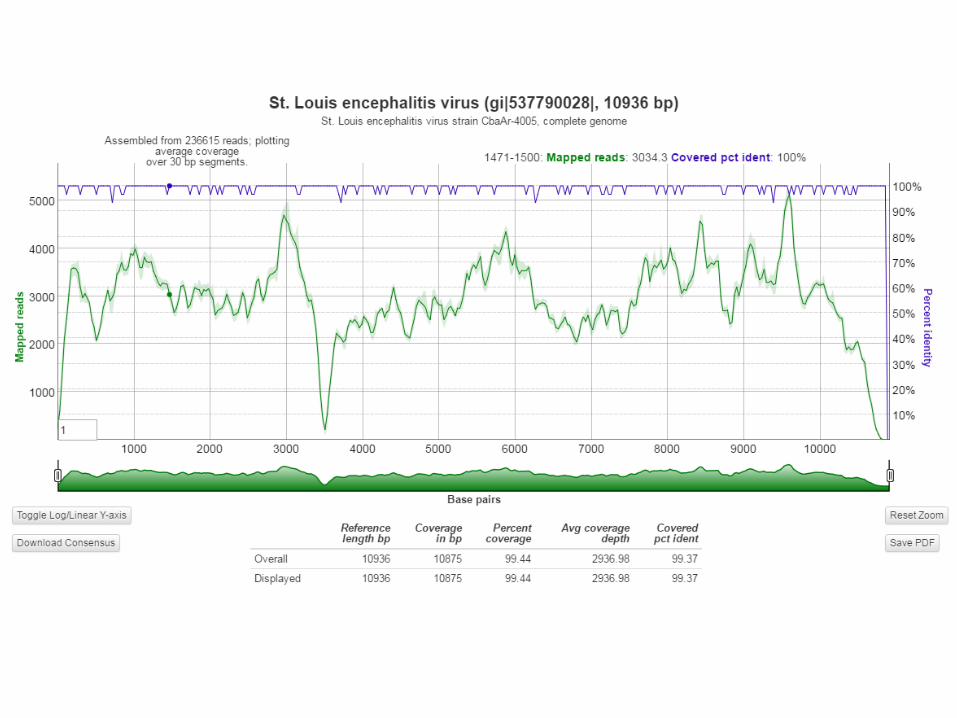
St. Louis Encephalitis Virus
• Normally self-limiting
• Can be prolonged / severe in immunocompromised patient
• Patient continued to deteriorate with supportive care
– Placed on comfort care and passed away
• Autopsy revealed mantle cell lymphoma and aspiration pneumonia
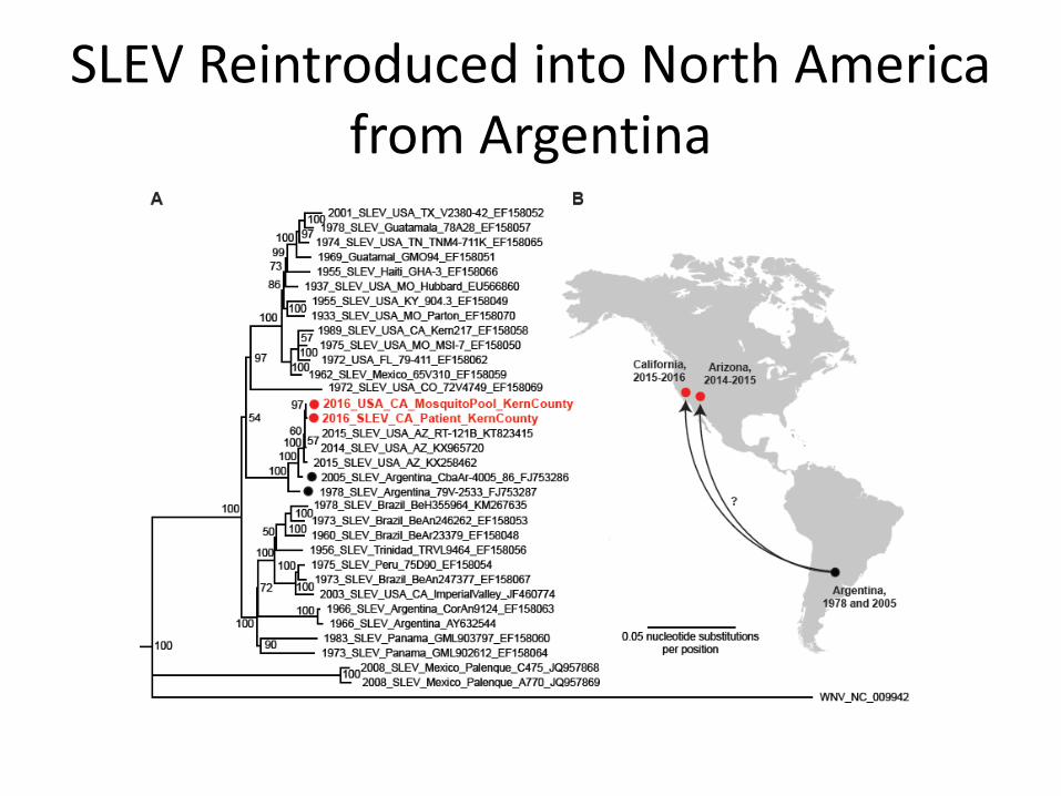
SLEV Reintroduced into North America from Argentina
Case Discussion
• SLEV re-introduced into Arizona in 2014 from Argentina
• Sequence analysis shows most similar to mosquito pool from Kern County, CA
– Patient likely acquired locally rather than from trip to Arizona
• Patient immunocompromised due to underlying disease leading to fatal infection
• 55 y/o male with bone marrow transplant for CLL. Prior immunosuppressive therapy.
In mid-October, developed rapidly progressive hearing loss over 2-3 weeks.
• CSF unremarkable; MRI negative; PCR for HSV and enterovirus negative; treated
empirically with high-dose valacyclovir, antibiotics, IVIg and steroids daily to no effect
• Over next few weeks, developed nausea, fatigue, ataxia, persistent hearing loss, then
depressed, irritable mood (unusual for patient per his wife).
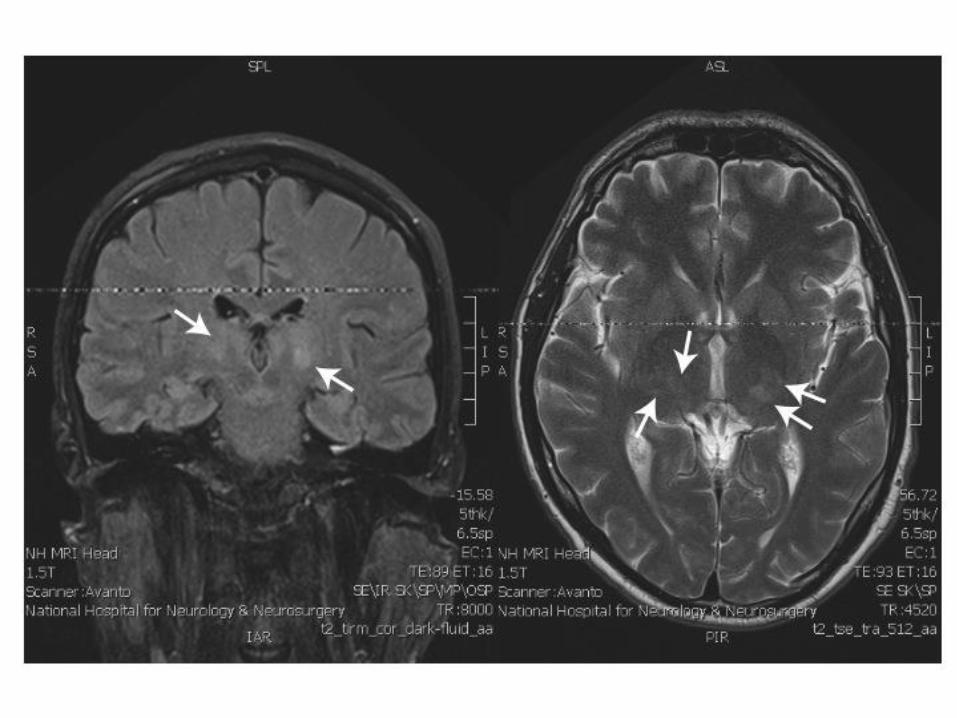
• Repeat MRI late December abnormal signal in thalamus and midbrain bilaterally;
frontal lobe biopsy performed (only accessible region)
55 y/o male with deafness and
behavioral change

Right frontal lobe
GFAP stain
Diffuse
reactive
gliosis
CD3 stain
T lymphocytes
CD8 stain
Microglial
activation
Cortex White matter
CD8 stain CD8 stain
Microglial
activation
Microglial
cell
processes
in proximity
to neurons


Genome Assembly of a
Novel Astrovirus

DNA In situ Hybridization of Brain Tissue
For Astrovirus
Neuronal
cytoplasmic
staining

“Encephalitic” Astrovirus Clade in Humans
• Patient started on ribavirin and IVIg
• Origin of virus unknown, but presumably community-
acquired
• Despite treatment, he continued to deterioriate, and passed
away
Case Discussion

Summary
• NGS assay shows acceptable performance for clinical testing
– Current study investigating utility of CSF testing
– Evaluation of other sample types (plasma for sepsis, BAL
for pneumonia)
• Allows for diagnosis of severe infections of unknown etiology
after extensive conventional testing
– Sensitive and specific but some limitations
– Pathogen DNA may be transient / serology used for
diagnosis
– Contamination control important
• Pathogen detection can guide optimal therapy

Acknowledgements
UCSF Chiu Lab and VDDC
Charles Chiu, MD/PhD
Erik Samayoa, CLS
Shaun Arevalo, CLS
Becky Fung, CLS
Hannah Sample, BS
Samia Naccache, PhD
Scot Federman, BS
Doug Stryke, BS
Joseph DeRisi, PhD
Michael Wilson, MD
Jeffrey Gelfand, MD
Michael Geschwind, MD
St. Jude’s Medical Center
Randall Hayden, MD/PhD
UCLA
Jeffrey Klausner, MD/MPH
Romney Humphries, PhD (ABMM)
Children’s Hospital Los Angeles
Jeffrey Bender, MD
Jennifer Dien-Bard, MDUniversity of California,
Berkeley
Brent Fulton, PhD/MBA
Children’s National Medical Center
Brittany Goldberg, MD
Joseph Campos, MD
UCDavis
Chris Polage, MD
Stuart Cohen, MD