Metabolism of Resorcinylic Compounds by Bacteria which forms is removed after 29min by...
13
THI JOURNAL OF B~o~oo~cn~ CHEMISTRY Vol. 250, No. lO,Iasue of May 25, pp. 3814-3825, 1975 Printed in U.S.A. Metabolism of Resorcinylic Compounds by Bacteria PURIFICATION AND PROPERTIES OF ORCINOL HYDROXYLASE FROM PSEUDOMONAS PUTIDA 01* (Received for publication, July 11, 1974) YOSHIYTJKI OHTA,$ IRVING J. HIGGINS~ AND DOUGLAS W. RIBBONS~ WITH THE TECHNICAL ASSISTANCE OF EDYE E. GROSECLOSE From the Department of Biochemistry, University of Miami School of Medicine, and Howard Hughes Medical Institute, Miami, Florida 33152 SUMMARY Orcinol hydroxylase (EC 1.14.13.6), which catalyzes the first reaction of orcinol catabolism in Pseudomonas putida 01, has been purified to homogeneity, and crystallized. Orcinol hydroxylase catalyzes the hydroxylation of orcinol with equimolar consumption of OZ and NADH (or NADPH) to 2,3 &trihydroxytoluene, which is nonenzymically oxi- dized to a quinone. The visible absorption spectrum of the enzyme shows maxima at 373 and 454 nm and a shoulder at 480 nm. FAD can be dissociated from the protein. Re- constitution of enzymic activity was achieved with FAD, and to a limited extent by FMN. The enzyme has a molecular weight of 63,000 to 68,000 and contains 1 mol of FAD per mol of protein. K, values for the three substrates orcinol, NADH, and 02 are 0.03,0.13, and 0.07 m, respectively. The molecular activity of the crystalline enzyme is 1560 min-l. In the absence of orcinol, NADH is only slowly oxidized with formation of H?Oz. Several analogsof orcinol also serve as substrates for hydroxylation, namely resorcinol, 4-methyl- resorcinol, and 4-bromoresorcinol. Other analogs, m-cresol, m-ethylphenol, 4-ethylresorcinol, and phloroglucinol, mimic orcinol as effecters, in that they (a) accelerate electron flow from NADH to the flavin and (b) decreasethe apparent K, for NADH but not to the same extent as the substrates that are hydroxylated. The latter compounds are not hydroxylated. Instead Hz02 accumulates as the only product of O2 reduc- tion. The enzyme therefore behaves either as a hydroxylase or an oxidase. The ratio of hydroxylase to oxidase activities of the enzyme is decreased by an increase in the tempera- ture of incubation; at 60” the reaction with orcinol is almost 50% uncoupled from hydroxylation. The apparent K, values for the effecters are in good * This work was supported in part by Grant 1 ROl GM 20172.01 from the National Institutes of Health. The preceding article of this work is Ref. 11. $ Howard Hughes Medical Institute Visiting Investigator. Present address, Faculty of Fisheries and Animal Husbandry, University of Hiroshima, Fukuyama-shi, Hiroshima-ken, Japan. S Howard Hughes Medical Institute Visit,ine Investigator. Present address,-Biological Labs, University of Kent at Canter- bury, Canterbury, Kent, England. lj Howard Hughes Medical Institute Investigator. agreement with the KD values obtained for orcinol, resorcinol, and m-cresol. KD values were obtained by measurement of the effector-induced perturbations of the visible absorption spectrum of the flavoprotein by difIerence absorption spec- troscopy. The circular dichroism spectrum of orcinol hy- droxylase is also altered in the presence of orcinol. The participation of the flavin in the over-all reaction is demonstrated by its rapid reduction under anaerobic condi- tions by NADH in the presence of orcinol, resorcinol, or m-cresol. Subsequent introduction of oxygen restores the oxidized form and yields H202 when m-cresol is the effector, but not when orcinol is the effector. Transfer of reducing equivalents from the reduced flavoprotein to free FAD may also occur. Reduction of orcinol hydroxylase by NADH in the absence of an effector is 104-fold slower than in the presence of an effector. The minimal structural requirements for effecters appear to be a 1,3-dihydroxy or 1-alkyl-3-hydroxybenzene, but only the former are substrates for hydroxylation. It has been proposed (1) that Pseudomonas putida 01 catabo- liaes orcinol by hydroxylation to give 2,3,5-trihydroxytoluene, which is the substrate of the ring cleavage enzyme of the orcinol pathway, a member of the m-pyrocatechase family of enzymes. Subsequent hydrolyses of the product of ring cleavage, 2,4,6- trioxoheptanoate, yield 2 mol of acetate and 1 mol of pyruvate, via the intermediate, acetylpyruvate (Scheme 1). The first enzyme of this inducible pathway, namely orcinol hydroxylase (EC 1.14.13.6) appears to be a typical monooxy- genase (mixed function oxidase) (2) which is relatively stable to usual enzyme fractionation techniques in the presence of 2-mer- captoethanol. It has been purified to homogeneity, crystallized, and shown to be a flavoprotein containing 1 mol of FAD per mol of protein (MW approximately 65,000) (3). The enzyme catalyzes the hydroxylation of orcinol to give 2,3,5-trihydroxytoluene in the presence of a reduced nicotinamide nucleotide and molecular oxygen, and thus represents another example of the flavoprotein 3814 by guest on February 1, 2019 http://www.jbc.org/ Downloaded from
Transcript of Metabolism of Resorcinylic Compounds by Bacteria which forms is removed after 29min by...

THI JOURNAL OF B~o~oo~cn~ CHEMISTRY Vol. 250, No. lO,Iasue of May 25, pp. 3814-3825, 1975
Printed in U.S.A.
Metabolism of Resorcinylic Compounds by Bacteria
PURIFICATION AND PROPERTIES OF ORCINOL HYDROXYLASE FROM PSEUDOMONAS PUTIDA 01*
(Received for publication, July 11, 1974)
YOSHIYTJKI OHTA,$ IRVING J. HIGGINS~ AND DOUGLAS W. RIBBONS~
WITH THE TECHNICAL ASSISTANCE OF EDYE E. GROSECLOSE
From the Department of Biochemistry, University of Miami School of Medicine, and Howard Hughes Medical Institute, Miami, Florida 33152
SUMMARY
Orcinol hydroxylase (EC 1.14.13.6), which catalyzes the first reaction of orcinol catabolism in Pseudomonas putida 01, has been purified to homogeneity, and crystallized. Orcinol hydroxylase catalyzes the hydroxylation of orcinol with equimolar consumption of OZ and NADH (or NADPH) to 2,3 &trihydroxytoluene, which is nonenzymically oxi- dized to a quinone. The visible absorption spectrum of the enzyme shows maxima at 373 and 454 nm and a shoulder at 480 nm. FAD can be dissociated from the protein. Re- constitution of enzymic activity was achieved with FAD, and to a limited extent by FMN. The enzyme has a molecular weight of 63,000 to 68,000 and contains 1 mol of FAD per mol of protein. K, values for the three substrates orcinol, NADH, and 02 are 0.03,0.13, and 0.07 m, respectively. The molecular activity of the crystalline enzyme is 1560 min-l.
In the absence of orcinol, NADH is only slowly oxidized with formation of H?Oz. Several analogs of orcinol also serve as substrates for hydroxylation, namely resorcinol, 4-methyl- resorcinol, and 4-bromoresorcinol. Other analogs, m-cresol, m-ethylphenol, 4-ethylresorcinol, and phloroglucinol, mimic orcinol as effecters, in that they (a) accelerate electron flow from NADH to the flavin and (b) decrease the apparent K, for NADH but not to the same extent as the substrates that are hydroxylated. The latter compounds are not hydroxylated. Instead Hz02 accumulates as the only product of O2 reduc- tion. The enzyme therefore behaves either as a hydroxylase or an oxidase. The ratio of hydroxylase to oxidase activities of the enzyme is decreased by an increase in the tempera- ture of incubation; at 60” the reaction with orcinol is almost 50% uncoupled from hydroxylation.
The apparent K, values for the effecters are in good
* This work was supported in part by Grant 1 ROl GM 20172.01 from the National Institutes of Health. The preceding article of this work is Ref. 11.
$ Howard Hughes Medical Institute Visiting Investigator. Present address, Faculty of Fisheries and Animal Husbandry, University of Hiroshima, Fukuyama-shi, Hiroshima-ken, Japan.
S Howard Hughes Medical Institute Visit,ine Investigator. Present address,-Biological Labs, University of Kent at Canter- bury, Canterbury, Kent, England.
lj Howard Hughes Medical Institute Investigator.
agreement with the KD values obtained for orcinol, resorcinol, and m-cresol. KD values were obtained by measurement of the effector-induced perturbations of the visible absorption spectrum of the flavoprotein by difIerence absorption spec- troscopy. The circular dichroism spectrum of orcinol hy- droxylase is also altered in the presence of orcinol.
The participation of the flavin in the over-all reaction is demonstrated by its rapid reduction under anaerobic condi- tions by NADH in the presence of orcinol, resorcinol, or m-cresol. Subsequent introduction of oxygen restores the oxidized form and yields H202 when m-cresol is the effector, but not when orcinol is the effector. Transfer of reducing equivalents from the reduced flavoprotein to free FAD may also occur. Reduction of orcinol hydroxylase by NADH in the absence of an effector is 104-fold slower than in the presence of an effector.
The minimal structural requirements for effecters appear to be a 1,3-dihydroxy or 1-alkyl-3-hydroxybenzene, but only the former are substrates for hydroxylation.
It has been proposed (1) that Pseudomonas putida 01 catabo- liaes orcinol by hydroxylation to give 2,3,5-trihydroxytoluene, which is the substrate of the ring cleavage enzyme of the orcinol pathway, a member of the m-pyrocatechase family of enzymes. Subsequent hydrolyses of the product of ring cleavage, 2,4,6- trioxoheptanoate, yield 2 mol of acetate and 1 mol of pyruvate, via the intermediate, acetylpyruvate (Scheme 1).
The first enzyme of this inducible pathway, namely orcinol hydroxylase (EC 1.14.13.6) appears to be a typical monooxy- genase (mixed function oxidase) (2) which is relatively stable to usual enzyme fractionation techniques in the presence of 2-mer- captoethanol. It has been purified to homogeneity, crystallized, and shown to be a flavoprotein containing 1 mol of FAD per mol of protein (MW approximately 65,000) (3). The enzyme catalyzes the hydroxylation of orcinol to give 2,3,5-trihydroxytoluene in the presence of a reduced nicotinamide nucleotide and molecular oxygen, and thus represents another example of the flavoprotein
3814
by guest on February 1, 2019http://w
ww
.jbc.org/D
ownloaded from
3815
Orcinol 2,3,5-Trihydroxy- 2,4,6-Trioxo- Akkylpyruvate toluene heptanoate
SCHEME 1. The catabolism of orcinol by Pseudomonas putidu: a, orcinol hydroxylase; b, 2,3,5-trihydroxytoluene 1,2-oxygenase; c, acetylpyruvate hydroxylase
hydroxylases, such as salicylate (4, 5), p-hydroxybenzoate (6-8), and melilotate hydroxylases (9, 10) isolated from pseudomonads.
This paper documents a modified purification procedure for orcinol hydroxylase as well as several of its physical and catalytic
properties. Some of the data have been presented earlier, in pre-
liminary form (3, 11).
EXPERIMENTAL PROCEDURE
Materials-Pseudomonas putida 01 was cultivated, and extracts prepared, as previously described (1). Sources of chemicals were given earlier (2, ll), except for acetylpyridine analog of NAD (Sigma Chemical Co.) and 4-bromoresorcinol (Aldrich Chemical Co.). 4-Methylresorcinol was a gift from Dr. P. J. Chapman and orsenillic acid was provided by Drs. F. Lynen, R. Light, and R. Bentley.
Microbiological Methods-Ps. putida 01 was isolated from pond mud by enrichment in a mineral salts medium containing orcinol (0.1%) as the sole source of carbon. The strain was maintained on nutrient agar slopes and induced cells were obtained by growth in mineral salts media of the following composition (grams/liter): KH,PO+ 5.4; NaOH, 1.8; (NH4)$04, 1.2; MgS04, 7 HZO, 0.4; and FeSOd, 7 H20, 0.01. Mg2+ and Fez+ in 5 mN HCl were sterilized sep- arately as a concentrated solution and 0.005 volume was added to the cooled sterile bulk of the medium. Sterile orcinol solutions (lOyO) were added (0.01 volume) to complete the medium. The sequence of cultivations used to obtain IO-liter batches of cells was as follows: nutrient broth (80 ml) was inoculated from stock cultures and allowed to grow for 16 to 24 hours; the entire culture was used to inoculate 1 liter of the minimal medium contained in a 4-liter Erlenmeyer flask which was incubated with shaking for 16 to 24 hours and added to 10 liters of the same medium in 14-liter New Brunswick fermentors (New Brunswick Scientific Co., New Brunswick, N. J.). Growth was usuallv comnlete within 6 to 8 hours at 36” due to exhaustion of orcinol from the media. Three further additions of orcinol were made at intervals determined by the extent of growth (measured turbidimetrically at 660 nm) the last addition being approximately 1 hour prior to harvesting at 60,000 X g in an air-turbine Sharples supercentrifuge at 26-33”. The cells were used directly or stored as a paste at 0” for up to 48 hours after assessing polarographically their capacity to oxidize orcinol (3).
Enzyme Assay-Hydroxylase activity was routinely assayed by following oxygen consumption polarographically with a Clark 0, electrode (Yellow Springs Instruments Co., Yellow Springs). The reaction mixtures usuallv contained 50 mM KH?POd-NaOH buffer. pH 6.8 (2.9 ml), enzymeUsolution (5 to 100 kl);-25 mu NADH (46 ~1); and 25 mM orcinol (20 ~1). The temperature was 30”. Air- saturated buffer was assumed to contain 232 nmol of 02 per ml at 30” (12). A unit of activity is that amount of enzyme that catalyzes the consumption of 1 rmol of 02 min-*.
PuriJication of Orcinol Hydrozylase-The purification procedure outlined here is now preferred to that described earlier (3) and is suitable for large quantities of crude extracts of cells. A paste of orcinol-grown Ps. putida (750 g wet weight) is suspended in 1500 ml of 20 mM KH2P04-NaOH buffer, pH 6.8, containing 0.3yc 2-mer- captoethanol. Batches (50 ml) are subjected three times to the highest output (150 to 200 watts) from a Branson ultrasonic dis- integrator for 20-s periods at 338”. The combined extracts are cen- trifuged at 27,000 X g for 20 min and the supernatants further clarified by ultracentrifugation at 55,000 X g for90min. Protamine
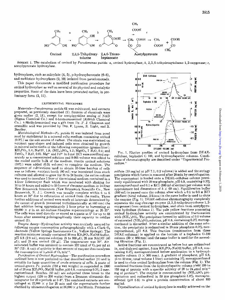
FIG. 1. Elution profiles of orcinol hydroxylase from DEAE- cellulose, Sephadex G-100, and hydroxylapatite columns. Condi- tions of chromatography are described under “Experimental Pro- cedure.”
sulfate (10 mg/ml at pH 7.1; 0.2 volume) is added and the stringy precipitate which forms is removed after 29min by centrifugation. The supernatant is loaded onto a DEAE-cellulose column previ- ously equilibrated with 20 rnM phosphate, pH 6.8, containing 0.370 mercaptoethanol and 0.1 M KC1 (500 ml of extract per column with approximate bed dimensions of 5 X 60 cm). Equilibration buffer (500 ml) is passed onto the column after which a 0.1 to 0.5 M KC1 gradient (total volume, 2 liters) in the same buffer is used to elute the enzyme (Fig. 1). DEAE-cellulose chromatography completely separates the ring cleavage enzyme (2,3,5-trihydroxytoluene 1,2- oxygenase) from orcinol hydroxylase, and often from acetylpyru- vate hydrolase (Scheme 1). The pale yellow fractions possessing orcinol hydroxylase activity are concentrated by fractionation with (NHI),SO,. The precipitate formed by addition of 0.6 volume of saturated (NHr&SO, solution, pH 6.8, containing 0.3y0 mercap- toethanol is discarded. After a similar addition of the same SO~U- tion. the nrecioitate is redissolved in 20 mM phosphate-0.3% mer- captoethanol, PH 6.8. This fraction (combination from three DEAE-columns) is applied to the bottom of a Sephadex G-100 column (50 X 860 mm) and the same buffer is used for the ascend- ing filtration (Fig. 1).
Active fractions are concentrated as before but are redissolved in, and dialyzed against, 5 mM KH2POI-NaOH buffer, pH 6.8, con- taining 0.3a/, mercaptoethanol before application to a hydroxyl- apatite column (3 X 300 mm). A gradient of phosphate, pH 6.8, (5 to 50 mM; total volume 1 liter) containing 1% mercaptoethanol is used to elute orcinol hydroxylase from the column (Fig. 1). The combined fractions from the hydroxylapatite column yield 100 to 150 mg of protein with a specific activity of 20 to 24 pmol mini mg of protein-i. The enzyme is concentrated by (NHI)ISCI pre- cipitation and redissolved in 50 mM phosphate-l.Oyo mercapto- ethanol (pH 6.8) to give a protein concentration of about IO0 mg/ml .
Crystallization of orcinol hydroxylase is readily achieved on the
by guest on February 1, 2019http://w
ww
.jbc.org/D
ownloaded from

3816
addition of (NHd)&O, solution until a faint turbidig persists. This solution is clarified by centrifugation and set aside for 2 to 7 days at 0” until crystallization occurs.
Chromatography-Flavins were chromatographed as described by Kilgour et al. (13). Phenols were checked for purity by chroma- tography on thin layer silica gel sheets in benzene-acetic acid- water (80:20:saturated), and detected by ultraviolet light or Gibbs reagent.
Gel Filtration-The molecular weight of orcinol hydroxylase was determined on Sephadex G-75 columns calibrated with pepsin, trypsin, cytochrome c, and alkaline phosphatase as described by Andrews (14).
Ultracentrifugation-Sedimentation velocity and equilibrium experiments, with schlieren and interference optics, respectively, were performed with a Spinco model E analytical ultracentrifuge. Sedimentation velocity runs, at 60,286 ‘pm were carried out with a wedge cell and a normal cell, allowing two samples (5.2 and 10.4 mg/ml) to be analyzed simultaneously. Equilibrium measurements were made by the meniscus depletion method (15) at three concen- trations, 0.3, 0.6, and 1.2 mg/ml at 26,522 rpm and analyzed as described by Small and Resnick (16). Molecular weight determina- tions were made assuming a partial specific volume of 0.73 for orcinol hydroxylase.
NHt-terminal Analysis-Dansylation of orcinol hydroxylase was carried out by the method of Gray (17) and the solution was chro- matographed as described by Hartley (18).
Disc Gel Electrophoresis-The procedures described by Cooksey (19) were used for electrophoresis at pH 8.6 and under denaturing conditions in the presence of urea andsodium dodecyl sulfate (19): Details of individual runs annear in the legends. Protein was de- tected by staining with Coomassie blue whych proved more sensi- tive than Amido schwarz for our preparations.
Orcinol hydroxylase activity was detected by immersing the un- fixed, unstained gels in an incubation mixture consisting of: 2-(p- iodophenyl)-3-p-iitrophenyl-5-phenyltetrazolium chl&ide, i0 me: 25 mM NADH (0.2 ml): 25 mM orcinol (0.1 ml). volume to 15 mr&ith 20 mM phosphate differ, pH 6.8. Incubatio& were carried out in the dark to minimize light-catalyzed dye reduction. Color developed in about 2Omin, and intensified over a period of about 18 hours. With crude preparations of the enzyme, several formazan precipitation bands appeared, due to diaphorase activities in the sample. These are easily distinguished from orcinol hydroxylase activity because they also appear when the detection mixtures lack orcinol, in which case the orcinol hydroxylase band is not observed.
The molecular weight of orcinol hydroxylase was determined by a modification of the methods of Dunker and Rueckert (20). En- zyme solution (6 mg/ml) was incubated for 90 min at 60” with an equal volume of 5 M urea containing 2 mM iodoacetamide and 1% sodium dodecyl sulfate. Samples and standards thus pretreated were mixed 1: 1 with 50yo glycerol and layered onto sodium dodecyl sulfate gels. After electrophoresis, gels were fixed overnight in 20yo sulfosalicylic acid, then stained for 3 hours with Coomassie blue. Cytochrome c, trypsin, pepsin, and ovalbumin were used as stand- ards.
Analytical Methods-Protein was determined by the method of Lowry et al. (21). Hydrogen peroxide was estimated by the release of 02 after addition of catalase to reaction mixtures, with a Clark electrode (22). These experiments require rigorous washing of the reaction cuvettes between separate determinations, particularly with aged cuvettes, electrode holders, and Teflon membranes on the electrode. We have observed that residual catalase activity is satisfactorily removed after several rinses in boiling water. Fail- ure to remove the adsorbed catalase results in irreproducible esti- mates of hydrogen peroxide and the extent of uncoupling. 2,3,5- Trihydroxytoluene and hydroxyquinol concentrations were meas- ured by the 02 consumed when a partially purified preparation of 2,3,5-trihydroxytoluene 1,2-oxygenase (3) catalyzed their oxida- tion. Absence of further 02 consumption on the addition of the ring cleavage enzyme for other potential hydroxylated products, e.g.~4-ethyliatechbl, was used as evidence that hydroxyiation (of 3-ethvlnhenol) had not occurred and this correlated well with the amount of Hz62 formed, and with the inability to detect the cate- chols by chromatography. Routine kinetic spectrophotometric measurements were made in a Unicam SP 800 ultraviolet visible spectrophotometer which could be adapted for simultaneous polarographic assays (23). Detailed spectral studies were per- formed with a Zeiss DMR 21 spectrophotometer. Circular di-
chroism measurements were made with a Gary model 60 spectro- polarimeter. Specific conditions used are given in the legends.
Spectral Assays of Flavin Reduction and Oxidation-Cuvettes (l-cm light path, l-ml volume), fitted with a Suba-seal rubber serum cap (William Freeman and Co., Ltd., Technical Sales Divi- sion, Staincross, Barnsley, Yorkshire, England) were charged with buffer and enzyme previously equilibrated with a nitrogen at- mosphere. Nz gas was bubbled through the reaction mixture for 3 to 5 min by means of two hypodermic syringes before substrate additions and spectral measurements were made. Substrate solu- tions were previously rendered 02 free before injection of micro- liter quantities with Hamilton syringes. Oxygen was delivered to the reaction mixtures by addition of aerated 20 mM phosphate buffer, pH 6.8. Measurements were made at room temperature, 24-27”.
Di$erence Spectra of Orcinol Hydroxylase-Enzyme-Effector Com- plex Minus Enzyme-Each pair of similar cuvettes in tandem contained orcinol hydroxylase and buffer separately. Difference spectra were recorded when addition of effecters to the enzyme (sample beam) and to the buffer (reference beam), and similar additions of water had been made to the remaining two cuvettes.
RESULTS
Puri$cation and Crystallization of Orcinol Hydroxylase-A sum- mary of the purification procedure outlined under “Experimental Procedure” is given in Table I and elution profiles are shown in
Fig. 1. The loss of enzymic activity from the DEAE-cellulose chromatography may appear large but this is due to the complete separation of the ring cleavage enzyme from orcinol hydroxylase. The specific activities recorded for crude and protamine sulfate- treated extracts are optimistic by a factor of 2, because the polarographic assay measures the two sequential reactions of the catabolic pathway catalyzed by orcinol hydroxylase and 2,3,5- trihydroxytoluene oxygenase, and the activity of the latter en- zyme is in excess. Each of these enzymes catalyzes the fixation of 1 mol of oxygen per mol of substrate (Scheme 1).
Orcinol hydroxylase readily crystallizes from (NH&S04 solu- tions as yellow plates.
Stability of Orcinol Hydroxylase and pH Optimum-Orcinol hy- droxylase activity is stable in crude extracts for days at 4”, but thiol reagents, such as mercaptoethanol, are required to stabilize purified preparations. Orcinol, EDTA, and FAD singly or in combination were not as effective as stabilizers. The enzyme was most stable at pH 7.
This pH optimum for stability is broadened by the presence of 0.3% mercaptoethanol. The pH optimum for activity is difficult to evaluate because the values do not take into account the non- enzymic oxidation rate of the product of reaction, which increases lo-fold between pH values of 6 and 8. At pH 6.8 in the presence of 0.3% mercaptoethanol suspensions of crystals and solutions at 5 to 10 mg of protein per ml of orcinol hydroxylase lose about
TABLE I Summary of purification of orcinol hydroxylase
step Volume Fyot& Specific act1wty Yield
Crude extract. .._............... 55,000 X g Supernatant after pro-
1710
tamine sulfate. 1500 DEAE-cellulose eluate.. 822 (NHa)&Od and Sephadex G-100. 124 (NH&Son and hydroxylapatite. 150
??&I
- fit?&
m/ml tnin- m of
prolcin-’
35.7 0.28
30.3 0.34 8.3 1.26 2.9 12.7 1.05 24
%
100
90 49 26 22
by guest on February 1, 2019http://w
ww
.jbc.org/D
ownloaded from

FIG. 2. Schlieren patterns from a sedimentation velocity study of orcinol hydroxylase. The lower cell contained 10.4 mg of orcinol hydroxylase and the upper cell contained 5.2 mg of orcinol hydroxylase dissolved in 20 mM KHZPOI-NaOH buffer, pH 6.8, and 0.3% mercaptoethanol. Frames recorded are for 2, 14,37, and 68 min (left to riyht). Rotor speed, 60,286 rpm. Temperature, 4.2”.
45 I I
43.3 43.5 I
43.1 43.9 44.1 44.3 RADIUS* (cm.‘]
FIG. 3. Plot of log c (c = fringe displacement) against the square of the distance from the axis of rotation (r2) for orcinol hydroxylase in 20 mM KH2POa-NaOH buffer, pH 6.8, containingO.l% 2-mercap- toethanol. Fringe patterns were obtained 20 hours after attaining a speed of 26,522 rpm. The initial protein concentration was 0.6 mg per ml. Temperature, 10”.
50% activity in 8 to 10 weeks, at 4”. Comparable measurements in the absence of mercaptoethanol were not made because large losses of enzymic activity occurred during purification without this supplementation.
Homogeneity-Orcinol hydroxylase obtained from hydroxyl- apatite columns yields two diffuse bands after polyacrylamide disc gel electrophoresis at pH 8.6 in Tris-HCl buffer. In sodium dodecyl sulfate gels a single band appears. The enzyme appears homogeneous upon ultracentrifugation giving a single symmetri- cal schlieren pattern (Fig. 2) and linear relationships between log C versus r* from the sedimentation equilibrium data (Fig. 3). The latter measurements were made between 0.3 and 1.2 mg of pro- tein per ml and gave a molecular weight value of approximately 65,000. Analysis of the NHz-terminal amino acid of this prepara- tion showed that isoleucine was the only NH*-terminal amino acid present in the sample.
From the purification data it can be calculated that the orcinol hydroxylase content of Pseudomonas puticla accounts for about 0.67” of the total protein of cells grown on orcinol as the sole car- bon source, bearing in mind that the sensitivity of the assay is reduced 2-fold when the ring cleavage enzyme has been removed.
I I I I
540 500 460 420 380 340
WAVELENGTH [nm] FIG. 4. Absorption spectra of orcinol hydroxylase and FAD in
20 mM KHzPOr-NaOH buffer, pH 6.9. -, orcinol hydroxylase (0.073 mM). - - -, FAD (0.073 mM).
Molecular Weight Determination-The molecular weight of or- cinol hydroxylase was estimated as 68,000 by dodecyl sulfate disc gel electrophoresis and as 63,000 by gel filtration chromatography, respectively, values in reasonable agreement with those obtained from sedimentation equilibrium measurements.
Flavin Content of Orcinol Hydroxylase-The visible absorption spectrum of orcinol hydroxylase is shown in Fig. 4 and compared with that of FAD. The yellow color of the enzyme is abolished by dithionite but reappeared on aeration. The identification of the enzyme-bound flavin as FAD was achieved by a combination of chromatography and absorption spectroscopy of the chromophore dissociated from the enzyme. The cofactor co-chromatographed with FAD in Solvents 1 and 3 of Ref. 6, although faint spots were usually observed with Rp values similar to FMN; other minor components were also observed; the absorption maxima and minima of the protein-free cofactor coincided with those of FAD. A molar extinction at 450 nm (assuming a molecular weight of 65,000) of orcinol hydroxylase was estimated as 10,500, which in- dicated that 1 mol of FAD is bound per mol of orcinol hydroxylase (a value of 0.94 is obtained using the molar extinction of 11,300 for FAD). Reconstitution of enzymic activity by the addition of flavin nucleotides to the apoenzyme is shown in Table II. Under these conditions most of the activity of the apoenzyme was recon-
by guest on February 1, 2019http://w
ww
.jbc.org/D
ownloaded from

3818
Reconstitution of apo-otcinol hydroxytase with FAD Orcinol hydroxylase (5 ml, 5.25 mg of protein) was mixed with
saturated (NHa)2SOd solution (6 ml) adjusted to pH 2.8 with 0.5 N HCl and kept at 0” for 16 hours. The supernatant was used for the identification of the flavin. The precipitate was sedimented and resuspended in 20 mM phosphate, pH 6.8 (1.5 ml). Recentrifu- g&ion yielded a colorless apoenzyme solution (33 rg/ml) free from denatured protein. Activities were assayed polarographically after incubation with the flavin nucleotides (0.04 mM) for 3 hours
TABLE II TABLE III
Stoichiometry of orcinol hydroxylase reactions
Reaction mixtures contained: 20 mM KHzP04-NaOH buffer, pH 6.8, (2.9 ml); orcinol hydroxylase (10 ~1, 20 pg of protein); volumes of 25 mM orcinol and 25 mM NADH to give the quantities indicated. Experiments 3,4, 5, and 6 contained in addition 2,3,5- trihydroxytoluene 1,2-oxygenase (50 ~1, 81 lg of protein). Final volume, 3.0 to 3.1 ml. Temperature. 30”. All units for the table are
at 0”.
Enzyme Flavin addition
I
Holoenzyme Apoenzyme Apoenzyme Apoenzyme
FMN FAD
Specific activity
2.8 3.5
14.7
FIG. 5. Simultaneous assay of orcinol hydroxylation by oxygen consumption and quinone formation at pH 6.8 and 8.0 by orcinol hydroxylase. The reaction mixture contained: 100 mM KH,POa- NaOH buffer, pH 6.8 or 8.0 (as indicated, 2.9 ml) and orcinol hy- droxylase (10 ~1). Additions of 25 mM NADH (20 ~1) and 25 mM orcinol (20 ~1) were made as indicated. The rapid pH-dependent nonenzymic formation of the quinone of 2,3,5-trihydroxytoluene is shown by the absorbance traces at 490 nm. The final addition of NADH shows the reduction of the quinone formed and further consumption of oxygen, until the reaction mixtures become an- aerobic. (This nonenzymic oxidation of NADH by 02 in the presence of 2,4,5-trihydroxytoluene has been demonstrated in the absence of orcinol hydroxylase.) Temperature, 30”.
stituted by the addition of FAD; FMN was a poor substitute for FAD.
Stoichiometry-Initial attempts to determine the stoichiometric relationships of the hydroxylation reaction were thwarted by the rapid nonenzymic oxidation of the product of orcinol hydroxyla- tion, 2,3,5-trihydroxytoluene, to a quinone (3, 11). This oc- curred too rapidly even at pH 6.8, as shown in Fig. 5, which com- pares the time course of the reaction at pH 6.8 and at pH 8.0. The amount of oxygen consumed is in excess of that required for a monooxygenase (mixed function oxidase) reaction, due to the subsequent nonenzymic oxidation of the product. Formation of the quinone is shown by the increase in absorbance at 490 nm.
We have obtained indirect evidence that orcinol hydroxylase catalyzes a reaction in which equimolar consumption of orcinol, 02, and NADH occurs. In the presence of limiting quantities of NADH, excess orcinol, and large quantities of enzyme, approxi- mately 1.5 pmol of 02 were consumed per pmol of NADH sup- plied and the reaction mixtures typically turned brick-red (X,,, 485 nm) indicating that the quinone accumulated. By incorporat-
Experiment Orcinol NADH 02 NADH supplied supplied consumed consumed
1 1.0 0.2 2 1.0 0.3 3 1.0 0.2 4 1.0 0.3 5 0.1 0.5 6 0.2 0.5
o ND, not determined. * ---, irrelevant.
0.285 ND5 0.42 ND 0.38 0.18 0.58 0.26 0.18 ND 0.38 ND
9 _-
Theoretical 02 consumption
For For sub- uinone torma-
sequent rmg
tion cleavage
0.3 0.45
-
- -
-b
0.4 0.6 0.2 0.4
ing large quantities of 2,3,5trihydroxytoluene 1,2-oxygenase, purified to the DEAE-cellulose stage (3)) into these reaction mix- tures, quinone formation was not detected by its visible absorp- tion spectrum; the amount of 02 consumed however increased to approximately 2 mol of O2 per mol of NADH supplied (Table III), a result expected for the two sequential reactions of the orcinol pathway (Scheme 1). As expected, approximately 2 mol of 02 are similarly consumed per mol of orcinol supplied (excess NADH) when the ring cleavage enzyme is present.
Aromatic Substrate-Effector Specificity of Orcinol Hydroxylase- The difficulties encountered in directly determining the stoichiom- etry of orcinol hydroxylation led to an examination of the reac- tions with analogs of orcinol. It was known that resorcinol and m-cresol aIso stimulate NADH oxidation by orcinol hydroxylase and that one of the presumed analogous products, 3-methyleate- chol (from m-cresol) was more stable to nonenzymic oxidation by oxygen than 2,3,5-trihydroxytoluene (3). An attempt to establish the stoichiometry with m-cresol revealed that 02 and NADH were consumed in equimolar quantities, but in excess of that required for a simple hydroxylation. An equimolar amount of hydrogen peroxide was shown to be formed (11). 3-Methyl- catechol, the expected product from m-cresol hydroxylation, was not detected chromatographically, nor by the dioxygenase assay, for which it is known to be a substrate (1,3) and hydrogen perox- ide was formed in amounts equivalent to the NADH and oxygen consumed (Table IV). Hydroxyquinol was detected as a product of the reaction with resorcinol as effector by (a) chromatography, (b) formation of its quinone, and (c) oxidation by the ring cleav- age enzyme, 2,3,5-trihydroxytoluene oxygenase (and hydroxy- quinol 1,2-oxygenase purified from Ps. putida ORC) .I Fig. 6 shows the course of reaction when resorcinol is the aromatic sub- strate; during the initial stages of the reaction both 02 concen- tration and AsdO nm rapidly decreased, but after 4 min the Asdon,,, values began to rise again, probably due to the nonen- zymic formation of hydroxybenzoquinone from hydroxyquinol, the product of resorcinol oxidation. Sequential additions of catalase and 2,3,5-trihydroxytoluene 1,2-oxygenase increased
1 P. J. Chapman and D. W. Ribbons, unpublished results
by guest on February 1, 2019http://w
ww
.jbc.org/D
ownloaded from

3819
2 65 I I nmoles Al340
02 0.1
FIG. 6 (left). Partial uncoupling of electron flow from NADH to oxygen catalyzed by orcinol hydroxylase in the presence of the “partial substrate” resorcinol. The reaction mixture contained: 100 mM KHZPOI-NaOH buffer, pH 6.8 (2.9 ml) and additions of 25 mM NADH (20 J), orcinol hydroxylase (50 ~1) and 25 mM resor- cinol (20 ~1) were made as indicated. When the reaction had pro- ceeded nearly to completion as judged by the O2 consumption, catalase (50~1) was added, resulting in a burst of oxygen evolution. When this had ceased 2,3,5-trihydroxytoluene oxygenase (100 ~1) was added and a rapid consumption of oxygen occurred. Tempera- ture, 30’.
FIG. 7 (right). Uncoupling of electron flow from hydroxylation in orcinol hydroxylase with the effector m-ethylphenol. The reac- tion mixture for the simultaneous assay of 02 consumption and NADH utilization contained: 100 mM KHzPOd-NaOH buffer, pH 6.8 (2.9 ml) and additions of 25 mM NADH (20 ~1); orcinol hydrox- ylase (50 ~1); and 25 mM m-ethylphenol (20~1) were made as indi- cated. When the reaction had proceeded nearly to completion, catalase (50~1) was added and after the rapid evolution of oxygen had ceased, 2,3,5-trihydroxytoluene oxygenase was added (100 ~1). Temperature, 30’.
TABLE IV TABLE V
Kinetic constants with different eflectors of orcinol hydroxylase
The values given were derived from Lineweaver-Burk plots of
Role of aromatic compounds as substrates and efectors
Reaction mixtures contained: 20 mM KH2POr-NaOH buffer, pH 6.8 (2.8 ml) ; 25 mM solution of aromatic compound (10 ~1) ; 25 mM NADH (20 ~1) ; and orcinol hydroxylase (5 to 100 ~1 of a 6 mg/ ml solution). Oxygen consumption and NADH consumption were measured simultaneously as described in Figs. 1 and 2. Hydroxyl- ated products were estimated by the addition of 2,3,5-trihydroxy- toluene 1,2-oxygenase, and Hz02 by the addition of catalase. The experiments with orcinol, resorcinol, 4-methylresorcinol, and 4- bromoresorcinol as the effecters were the only ones that were d- lowed to progress to completion before the addition of ring cleav- Aromatic substrate
initial velocities of oxygen consumption versus substrate concen- tration. Apparent K, values for the effecters were determined iu the presence of 417 FM NADH. Apparent K,,, values for NADH were determined in the presence of 417 FM of effector in (A), and 208 PM in (B). The specific activities of the orcinol hydroxylase preparations used in (A) and (B) were 14.8 and 11.4 units/mg, respectively.
I- Apparent Km for
substrate age enzyme and catalase.
Aromatic compound supplied
A. Orcinol. Orcinol. . . Resorcinol Resorcinol Phloroglucinol. m-Ethylphenol. . 4-Ethylresorcinol .
B. Orcinol. Resorcinol . m-Cresol. 4-Bromoresorcinol , . 4-Methylresorcinol.
a N.D., not determined.
PI 5 5
20 20 50 30 50 10
25 25
100
100
Hydroxylated noduct formed Hzoz formed
%
A. Orcinol. ................. Resorcinol. .............. 4-Ethvlresorcinol . ........
N.D.a 94
N.D. 32
0 0 0
89 43 0
N.D. N.D.
l-5 N.D.
63 N.D.
88 96 97
N.D. 49 95
m-Ethylphenol. Phloroglucinol m-Cresol . .
B. Orcinol. . m-Cresol 4-Methylresorcinol. . 4-Bromoresorcinol. L
a N.D., Not determined.
Appm&,, for
?nM
0.03 0.19 0.03 0.01
3.2 0.2 N.D.” N.D. 0.04 0.025
0.13 2.5 2.1 6.7 1.3 3.3 0.07 2.0 0.53 0.83
5-10 5-10 due to the nonenzymic formation of the quinone in the reaction
mixtures. A survey of a number of analogs of orcinol for substrate and
effector function with orcinol hydroxylase is shown in Tables IV
and depleted the 0% concentration, respectively, indicating that and V. m-Cresol, m-ethylphenol, 4-ethylresorcinol, and phloro-
both Hz02 and hydroxyquinol had been formed (Fig. 6). The glucinol completely uncouple electron flow from hydroxylation
combined measurements of Hz02 and hydroxyquinol formed how- with consequent formation of hydrogen peroxide. Fig. 7 shows a
ever did not account for all of the O2 initially consumed, probably reaction with m-ethylphenol as effector, yielding only hydrogen
by guest on February 1, 2019http://w
ww
.jbc.org/D
ownloaded from

3820
TABLE VI TABLE VII Kinetic constants for electron donors in orcinol hydroxylation The values were derived as described in Table V, except that the
apparent K,,, for oxygen was obtained from tangents drawn from a progress curve. This reaction mixture differed by containing initially only 23 PM OZ.
Substrate analogs as inhibitors of orcinol hydrozylase Reaction mixtures contained the standard assay components
and the analog at 1.7 mM. Inhibitors used were recrystallized or sublimed when necessary.
Inhibitor at 1.7 rng Inhibition
Coeneyme Apparent K,
Nucleotide Or&d Oxygen
?nM
NADH . 0.13 0.034 0.07 NADPH...................... 1.1 3-Acetylpyridine NADH.. . 0.65
I I
0.085 N.D.a 0.05 N.D.
0 N.D., Not determined.
3-Hydroxybenzaldehyde. ............... 3,4-Dimethylphenol. .................... 3,5-Dihydroxybenzoate. ................. 2-Methylresorcinol, ..................... 1,3-Dimethoxybenzene. ................ 3,5-Dimethylphenol. .................... 2-Hydroxy-4-methoxybenzoate ........... 2-Nitroorcinol.. .................. ......
peroxide as a product of oxygen reduction. Only orcinol, 4-meth- ylresorcinol, 4-bromoresorcinol, and to a lesser extent resorcinol have been found as effecters that also undergo hydroxylation (Table IV). Compounds which did not stimulate oxygen reduc- tion by NADH included: orsellinic acid, pyrogallol, 3,5-dihy- droxybenzoate, 2,3-dihydroxybenzoate, 2,4-dihydroxybenzoate, and those compounds listed in Table VII.
E$ector and Nucleotide SpeciJicity, and their Kinetic Constants- Apparent K, values for the aromatic substrates (effecters), elec- tron donors, and oxygen, for orcinol hydroxylase are given in Tables V and VI. Although these values were not obtained in the presence of saturating concentrations of the other substrates, other experiments have shown that the apparent K, values for orcinol are not appreciably affected between NADH concentra- tions of 50 and 830 PM; similarly, the apparent K, for NADH is not altered between orcinol concentrations of 200 and 500 PM.
V mall values obtained are not recorded because they have only comparative value within a particular group of experiments. The most notable feature of these results is that in the presence of orcinol, 4-methylresorcinol, and 4-bromoresorcinol, i.e. the sub- strates that do not show extensive uncoupling of electron flow from hydroxylation, the apparent K, values for NADH are rela- tively low. With other aromatic effecters, apparent Km values for NADH are much greater. The KD values (see later, Figs. 14 and 15) for the enzyme-effector complexes compare favorably with these kinetic values obtained for orcinol, resorcinol, and m-cresol (Table V). The apparent K, value for orcinol is not significantly altered by the use of different electron donors (Table VI) and NADH is the preferred nucleotide for the enzyme.
1 IO 20 30 40
TIME [mini
I I 560 520 480 440 400
WAVELENGTH [nm]
Inhibitors of Orcinol Hydroxylase-A preliminary survey of possible inhibitors of orcinol hydroxylase is given in Table VII. These results show that several analogs of orcinol which are not effecters significantly inhibit the reaction, as do high concentra- tions of orcinol and phloroglucinol. Effecters with only two sub- stituents in the benzene ring do not show “substrate” inhibition at high concentrations.
FIG. 8. Anaerobic reduction of orcinol hydroxylase by NADH in the absence of substrate. Orcinol hydroxylase (1 ml containing 3 mg of protein in 20 mM KH2POa-NaOH buffer, pH 6.8) was added to the cuvette and flushed with N, (Spectrum 0). Then, 25 mM NADH (lOk1) was added and the spectrum was retraced after 2,5, 10, 15, 20, 30, and 40 min. Temperature, 26”. The inset shows the time course of FAD reduction at 454 nm.
Reduction of Orcinol Hydroxylase by NADH-The preparation of large amounts of pure orcinol hydroxylase has allowed us to examine the reduction of flavin in the enzyme by NADH. In the absence of an effector and oxygen, the rate of reduction of the FAD of orcinol hydroxylase by NADH is low (Fig. 8) and ap- pears biphasic; the final spectrum obtained is similar to that for the enzyme reduced by dithionite. The molecular activity calcu- lated for the initial rate of reduction by NADH is approximately 0.05 min-i as compared with the value of 1560 mini for the over-all hydroxylation reaction when orcinol is the effector. Addition of oxygen to the reaction mixtures restores the oxidized
flavin spectrum. Similar anaerobic experiments in the presence of orcinol, resorcinol, m-cresol, or m-ethylphenol gave instantane- ous bleaching of the flavin spectrum. Fig. 9 shows experiments where the flavin is titrated to its reduced form by NADH in the presence of (a) orcinol, (b) resorcinol, and (c) m-cresol.
Reoxidation of Reduced Orcinol Hydroxylase-Fig. 10 shows the spectral changes that occur when aerated buffer is introduced to orcinol hydroxylase that had been reduced previously by NADH, in the presence of orcinol or m-cresol as described in Fig. 9. With m-cresol, addition of successive increments of 02 to the reduced enzyme produced increases of A 451 of only one-half the magnitude (Curves 1, 2, and S) produced by similar quantities of 02 with orcinol as substrate. After the addition of catalase the AM in- creased (Curves .9 to 6) by approximately those values observed when orcinol was present. Therefore, in confirmation of the data
7%
23 54 59 27 45 68 68 64
by guest on February 1, 2019http://w
ww
.jbc.org/D
ownloaded from

3821
’ bi RtSOkClNOL’ /,,
1 1 1 1 I
520 440 360 WAVELEN6TH (nm)
FIG. 9. Anaerobic titration of orcinol hydroxylase by NADH in the presence of (a) orcinol, (5) resorcinol, and (c) m-cresol. Each cuvette contained: orcinol hydroxylase (0.8 ml containing 32 pM of enzyme in 20 rnM KH2POa-NaOH buffer, pH 6.8). Curve 0 shows the spectra of the enzyme after flushing with Nz in the presence of effector (0.32 mM). (a) orcinol, Curves 1, 2, S, 4, and 6 show the spectral changes that occurred after the sequential additions of 2 ~1 of 7 mM NADH to the cuvette. (5) resorcinol, Curves 1, 2, 3, 4, and 6 were drawn after sequential additions of 3, 2, 2, 2, and 2 ~1 of 7 mM NADH. (c) m-Cresol, Curves 1, 2, 5, 4, and 5 were ob- tained after sequential additions of 3, 2, 2, 3, and 3 ~1 of 7 mM NADH. Temperature, 26”.
of Table IV, it appears that Hz02 is formed by addition of 02 to reduced orcinol hydroxylase in the presence of m:cresol, but not orcinol. The increment seen upon the addition of catalase sup- ports this notion (Curues S and 4, Fig. 10, right).
Reoxidation of Reduced Orcinol Hydroxylase by Other Electron Acceptors-Reduced orcinol hydroxylase is able to transfer re- ducing equivalents to a variety of electron acceptors, other than oxygen. These include free FAD, ferricyanide, cytochrome c, acetylpyridine-NAD, and tetrazolium salts. Fig. 11 shows the
r (t ORCINOL) (+ m-CRESOL)
- 520 480 440 400 360 520 480 440 400 360 Wavelength (nm]
FIG. 10. Reoxidation of flavin in orcinol hydroxylase by Cc,. Titration of the reduced form of orcinol hydroxylase by molecular 02, in the presence of the aromatic substrate, orcinol (left) a.td m-cresol (right). The effect of the addition of catalase on the spec- tral changes is also documented. Temperature, 26”. Left, orcrnol hydroxylase (1 ml containing 2.8 mg of protein in 20 mr,r KHIPqa- NaOH buffer, pH 6.8) was flushed under NZ and titrated anaero- bically, after the addition of 25 mM orcinol (10 rl), and the addi- tion of 25 mM NADH (4~1) which gave Curve 1. Air-saturated buffer was then introduced sequentially in l-p1 quantities giving Curves 2 and 8. Addition of 1~1 of catalase yielded Curve 4. Further se- quential additions of air-saturated buffer gave Curves 6 (1 pl), 6 (2 J), and 7 (1 ~1). Right, conditions of this experiment are the same except that m-cresol was used as the aromatic substrate and reduction of the flavin spectrum was achieved with 5 pl of 25 mM NADH to give Curve 1. Reoxidation of the reduced flavin was ob- tained by additions of buffer. Two sequential additions (1 ~1) of the buffer yielded Curves R and 9. Addition of catalase (1~1) gave Curve 4, and subsequent additions (1 ~1) of the aerated buffer yielded Curves 6,6, and 7.
rapid reduction of free FAD by NADH in the presence of orcinol hydroxylase and orcinol, which also compares the slow nonen- zymic rate of FAD reduction by NADH.
Perturbation of Flavin Spectrum of Orcinol Hydroxylase by Ef- fectors-The absorption spectra of flavoproteins are often per- turbed in the presence of their natural substrates or analogs of them. Figs. 12 and 13 show the perturbations of the flavin spec- trum of orcinol hydroxylase by orcinol, and by resorcinol and m-cresol. The difference spectra (enzyme-effector complex minus enzyme) are qualitatively similar to each other with the exception of a broad band and shift in X,,, at lower wavelengths for the m-cresol-enzyme complex (Fig. 13). The extent of the spectral changes as a function of effector concentrations are shown in Figs. 14 and 15. The insets for Figs. 14 and 15 are double recipro- cal plots for the absorption changes induced by the effecters versus the effector concentration and allow estimates for the KD of the enzyme-effector complexes to be made. They are 0.026, 0.3, and 0.28 mM for orcinol, resorcinol (not shown), and m-cresol respectively. These values are in reasonable agreement with the apparent K, values obtained earlier, i.e. 0.03, 0.19, and 0.2 mM, respectively (Table V).
Circular Dichroism of Orcinol Hydroxylase-The circular di- chroism spectrum of orcinol hydroxylase is shown in Fig. 16. In the presence of the substrate orcinol the circular dichroism spectrum is altered by a slight shift and decrease in positive values to longer wavelengths (368 - 371 nm), whereas the nega- tive band became less intense at 455 nm (Fig. 16).
by guest on February 1, 2019http://w
ww
.jbc.org/D
ownloaded from
3822
I I
520 480 440 400 360 WAVELEIIGTH (nm]
L I 1 I
520 410 440 400 360 WAVELEWHH [nml
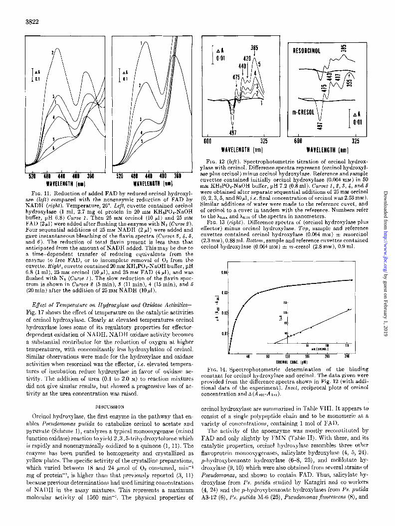
FIG. 11. Reduction of added FAD by reduced orcinol hydroxyl- ase (Zejt) compared with the nonenaymic reduction of FAD by NADH (right). Temperature, 26”. Left, cuvette contained orcinol hydroxylase (1 ml, 2.7 mg of protein in 20 mM KH2P04-NaOH buffer, pH 6.8) Curve 1. Then 25 mM orcinol (10 ~1) and 25 mM FAD (2~1) were added after flushing the enzyme with N2 (Curve 2). Four sequential additions of 25 rnM NADH (2 pl) were added and gave instantaneous bleaching of the flavin spectra (Curves S, 4, 6, and 6). The reduction of total flavin present is less than that anticipated from the amount of NADH added. This may be due to a time-dependent transfer of reducing equivalents from the enzyme to free FAD, or to incomplete removal of O2 from the cuvette. Right, cuvet.te contained 20m~ KH2POa-NaOH buffer, pH 6.8 (1 ml), 25 mM orcinol (10 pl), and 25 mM FAD (4 rl), and was flushed with Nz (Curve 1). The slow reduction of the flavin spec- trum is shown in Curves 2 (5 min), 5 (11 min), 4 (15 min), and 6 (20 min) after the addition of 25 mM NADH (10,~l).
Effect of Temperature on Hydroxylase and Oxidase Activities- Fig. 17 shows the effect of temperature on the catalytic activities of orcinol hydroxylase. Clearly at elevated temperatures orcinol hydroxylase loses some of its regulatory properties for effector- dependent oxidation of NADH. NADH oxidase activity becomes a substantial contributor for the reduction of oxygen at higher temperatures, with concomitantly less hydroxylation of orcinol. Similar observations were made for the hydroxylase and oxidase activities when resorcinol was the effector, i.e. elevated tempera- tures of incubation reduce hydroxylase in favor of oxidase ac- tivity. The addition of urea (0.1 to 2.0 M) to reaction mixtures did not give similar results, but showed a progressive loss of ac- tivity as the urea concentration was raised.
DISCUSSION
Orcinol hydroxylase, the first enzyme in the pathway that en- ables Pseudomonas putida to catabolize orcinol to acetate and pyruvate (Scheme 1), catalyzes a typical monooxygenase (mixed function oxidase) reaction to yield 2,3,5-trihydroxytoluene which is rapidly and nonenzymically oxidized to a quinone (1, 11). The enzyme has been purified to homogeneity and crystallized as yellow plates. The specific activity of the crystalline preparations, which varied between 18 and 24 pmol of 02 consumed, min-1 mg of protein-i, is higher than that previously reported (3, 11) because previous determinations had used limiting concentrations of NADH in the assay mixtures. This represents a maximum molecular activity of 1560 min-i. The physical properties of
6 I 4h I 00 325
WAVELENGTH [nm]
RESORCINOL
A
I I 600 325
WAVELENGTH [nm]
FIG. 12 (left). Spectrophotometric titration of orcinol hydrox- ylase with orcinol. Difference spectra represent (orcinol hydroxyl- ase plus orcinol) minus orcinol hydroxylase. Reference and sample cuvettes contained initially orcinol hydroxylase (0.064 mM) in 50 mM KHtPOI-NaOH buffer, pH 7.2 (0.8 ml). Curves 1, 2, S,4, and 6 were obtained after separate sequential additions of 25 mM orcinol (0, 2,3,5, and 80~1, i.e. final concentration of orcinol was 2.53m~). Similar additions of water were made to the reference cuvet, and of orcinol to a cuvet in tandem with the reference. Numbers refer to the X max and Amin of the spectra in nanometers.
FIG. 13 (right). Difference spectra of (orcinol hydroxylase plus effector) minus orcinol hydroxylase. Top, sample and reference cuvettes contained orcinol hydroxylase (0.064 mM) =t resorcinol (2.3 mM), 0.88 ml. Bottom, sample and reference cuvettes contained orcinol hydroxylase (0.064 mM) & m-cresol (2.8 mM), 0.9 ml.
FIG. 14. Spectrophotometric determination of the binding constant for orcinol hydroxylase and orcinol. The data given were provided from the difference spectra shown in Fig. 12 (with addi- tional data of the experiment). Inset, reciprocal plots of orcinol concentration and A(Aa~A385).
orcinol hydroxylase are summarized in Table VIII. It appears to consist of a single poiypeptide chain and to be monomeric at a variety of concentrations, containing 1 mol of FAD.
The activity of the apoenzyme was mostly reconstituted by FAD and only slightly by FMN (Table II). With these, and its catalytic properties, orcinol hydroxylase resembles three other flavoprotein monooxygenases, salicylate hydroxylase (4, 5, 24). p-hydroxybenzoate hydroxylase (6-8, 25), and melilotate hy- droxylase (9, 10) which were also obtained from several strains of Pseudomonas, and shown to contain FAD. Thus, salicylate hy- droxylase from Ps. putida studied by Katagiri and co-workers (4, 24) and the p-hydroxybenzoate hydroxylases from Ps. putida A3-12 (6), Ps. putida M-6 (25), Pseudomonas fluorescens (8), and
by guest on February 1, 2019http://w
ww
.jbc.org/D
ownloaded from
oprn
oprn
oprn

3823
FIG. 15. Spectrophotometric determination of the binding constant for orcinol hydroxylase and m-cresol. The data given were provided from difference spectra obtained under similar con- ditions to those described in Fig. 13 (bottom). Inset, reciprocal plots of m-cresol concentration and A(A197-A41,).
300 350 400 450 500 WAYtltNGlH (nmj
FIG. 16. Circular dichroism spectra of orcinol hydroxylase alone and in the presence of orcinol. Enzyme (0.114 mM) in 20 mm KHzPOd-NaOH buffer, pH 6.8; path length, 1 cm; temperature, 0”. Enzyme alone (-); enzyme plus 0.42 mM orcinol (- - -).
50 -
s 40- -
Z g 30-
Z = 20.
14
IEMPfRATURE ["C]
FIG. 17. Effect of temperature on uncoupling of electron flow from hydroxylation with orcinol hydroxylase. Enzyme activity was measured polarographically at various temperatures. Reac- tion mixtures contained: 190 mM KHZPOI-NaOH buffer, pH 6.8; 25 mM orcinol or resorcinol (20 ~1); 25 rnM NADH (40 ~1); orcinol hydroxylase (100 ~1, 100 pg of protein). The amount of hydrogen peroxide formed was determined by adding 50 ~1 of twice crystal- lized beef liver catalase (Sigma Chemical Co.) and measuring the amount of oxygen released, polarographically.
Pseudomonas desmolytica (7) (typed as Pseudomonas acidovorans by Stanier et al. (26)) all contain 1 mol of FAD per molecular weight of protein of 83,500, 93,600, 58,000, and 68,000, respec- tively, and show no evidence of subunit structure. However,
TABLE VIII
Summary of properties of orcinol hydroxylase
Molecular weight....... Sedimentation equilibrium. . Dodecyl sulfate gel electrophoresis Sephadex G-75.....
Sedimentation s2,,,,,,. FAD content NHz-terminal analysis.. Conclusion..
65,000 68,000 63,000
4.2 0.94 mo1/65,000 daltons Isoleucine Single polypeptide chain
of MW N 65,000 con- taining 1 mol of FAD
salicylate hydroxylase from an unidentified soil bacterium (5) is a dimer of identical subunits each possessing 1 mol of FAD per molecular weight subunit of about 45,000 and melilotate hy- droxylase (10) has recently been shown to be a tetramer of similar subunits of molecular weight 65,000, each containing 1 mol of FAD. Neujahr and Gaal (27) have also described a flavoprotein hydroxylase from the yeast, Trichosporon cufaneum. It, like the enzymes from pseudomonads possesses 1 mol of FAD, although it has a much higher molecular weight (146,000). Additionally, it shares with orcinol hydroxylase the ability to hydroxylate res- orcinol, one of several substrates the yeast uses for growth. All five enzymes, and also the flavoproteins 3-hydroxybenzoate 6-hy- droxylase (28) and 3-hydroxybenzoate 4-hydroxylase (29) cata- lyze the introduction of a hydroxyl group into the benzenoid ring, o- or p- to an existing hydroxyl substituent. This occurs with a concomitant decarboxylation for salicylate hydroxylase.
Direct determination of the stoichiometry of the orcinol hy- droxylase reaction has been particularly t.roublesome due to the rapid nonenzymic oxidation of the product, 2,3,5-trihydroxy- toluene by oxygen, and reduction of the quinone so formed by NADH. In addition, 2,3,5-trihydroxytoluene may serve as an effector of NADH oxidation by the enzyme in a manner observed for p-hydroxybenzoate hydroxylase where the product, proto- catechuate is an effector of electron flow, as is gentisate for 3-hy- droxybenzoate-6-hydroxylase (28). Indirect evidence of the ex- pected quantitative relationships of oxygen and NADH con- sumption is, however, available. Thus, when reactions are limited by NADH, the molar ratio of substrate consumption is oreinol- NADH-02 (1:1:1.5). When 2,3,5-trihydroxytoluene 1,2-oxy- genase is present such that the ring fission rate is in excess of the nonenzymic rate of oxidation of 2,3,5-trihydroxytoluene, these ratios change to I : 1:2; the 2nd mol of 02 being consumed by the dioxygenative reaction, and the quinone is not formed.
The catalytic properties of orcinol hydroxylase closely resemble those of salicylat,e, p-hydroxybenzoate, and melilotate hydroxyl- ases. The rate of enzyme-catalyzed oxidation of NAD(P)H by molecular oxygen (or other electron acceptors) is elevated by at least 4 orders of magnitude in the presence of orcinol or some of its structural analogs. The enzyme probably catalyzes the quan- titative hydroxylation of orcinol in vivo, although small and varia- ble amounts of hydrogen peroxide are formed in vitro, possibly due to the product accumulating in reaction mixtures and acting as an effector. Orcinol hydroxylase does not hydroxylate m-cresol, phloroglucinol, m-ethylphenol, or 4-ethylresorcinol. Instead the only detected product of oxygen reduction for the last four ef- fectors is hydrogen peroxide. 4-Methylresorcinol and 4-bromo- resorcinol are good substrates for the hydroxylation reaction but resorcinol is intermediate in its role as a substrate or as an effector, in that substantial quantities of both hydroxyquinol and hy-
by guest on February 1, 2019http://w
ww
.jbc.org/D
ownloaded from

3824
drogen peroxide are formed as products of oxygen reduction. These latter three substrates, and orcinol, have dual roles of act- ing as (a) substrates for hydroxylation, and (b) as effecters for the facilitated oxidation of reduced nicotinamide nucleotides by the FAD in the enzyme. In addition, orcinol also regulates the synthesis of this inducible enzyme, a property not shown for the other effectors.2
It is not possible from these studies to determine if the elevated rate of flavin reduction observed is due only to a change in the apparent K,,, for pyridine nucleotide or also to a change in the kred (of flavin reduction), or both. However, more detailed studies of the reaction mechanisms of p-hydroxybenzoate hydroxylase (6-8,25, 30, 31)) salicylate hydroxylase (4, 5, 32,33), and melilo- tate hydroxylase (9, 10, 34), by steady state kinetic analyses, and stopped flow techniques indicate that the kred is an altered parameter. The apparent K,,, values for the nucleotide in the presence of different analogs are also considerably different, as we have observed for orcinol hydroxylase (Table V)
The absorption spectrum of orcinol hydroxylase is perturbed by the presence of orcinol (Fig. 12), resorcinol, and m-cresol (Fig. 13) ; the effector-induced changes in the flavin spectrum are re- markably similar to those observed for p-hydroxybenzoate (8, 25, 30) and salicylate hydroxylases (5) and provide estimates of KD values which are in good agreement with the apparent K, values (Table V and Figs. 14 and 15). Orcinol has also been shown to substantially change the circular dichroism spectrum of orcinol hydroxylase (Fig. 16). The fluorescence spectrum of orcinol hy- droxylase is also quenched in the presence of orcinol.3 The flavo- protein hydroxylases possess a built-in regulatory property, that prevents indiscriminate oxidation of NAD(P)H and transfer of reducing equivalents to the flavin, a property shown also by pteridine (35), P-450 (36-38), and dioxygenase hydroxylases (39). Once the flavin in the enzyme has been reduced, then alternative routes exist for the reduction of molecular oxygen to either hy- droxylated product and water, or hydrogen peroxide, and these are presumably determined by the orientation and reactivity of the reduced enzyme-effector- complex, when reoxidation of the flavin occurs. The reduced species of the flavin hydroxylases all appear to be readily oxidized by molecular oxygen, irrespective of the method used to reduce the enzyme, e.g. by reduced nucleo- tides, EDTA, and light or dithionite.
Many of the flavoprotein hydroxylases are particularly versa- tile in the catalytic activities they possess, and those “isofunc- tional” enzymes isolated from different strains of related bacteria possess quantitatively different but often overlapping specificities for both the substrates (effecters) and the products of reaction (hydroxylation or hydrogen peroxide). Thus p-hydroxybenzoate hydroxylases isolated from the fluorescent pseudomonads, P.S. juorescens (8) and Ps. putida (25) and the nonfluorescent species Ps. de.smoZyfica (acidovoruns) (7, 30) exhibit common capabilities for the transformation of p-hydroxybenzoate, 2,4-dihydroxy- benzoate to their 3-hydroxylated products, but this is not shared by them for the transformation of benzoate and 2,3,4-trihy- droxybenzoate. Similar differences in the substrate or effector specificities have been shown for the orcinol hydroxylase that another strain of Ps. pufidu (not that used in this study) elab- orates during growth on orcinol.3 Further versatility of substrate hydroxylation is shown by the 3-hydroxybenzoate 6- and 4- hy- droxylases from Pseudomonas aeruginosu and Pseudomonas leslosteroni respectively (28, 29). These enzymes hydroxylate sev- eral 3-hydroxybenzoates substituted in the 2,4,5- and 6- posi-
2 Unpublished observations. 3 Unpublished data.
tions of the benzene nucleus, albeit with different efficiencies. Likewise the phenol hydroxylase from Trichosporon cutuneum catalyzes the sequential hydroxylation of phenol, and the prod- uct, catechol, as well as the three cresols, and all of the isomeric fluoro- and chlorophenols (27). Re sorcinol, a partial substrate of orcinol hydroxylase is equally as good a substrate for phenol hy- droxylase as phenol. Phenol hydroxylase however is restricted to the use of NADPH as reductant (27). This enzyme then is possi- bly used during growth of the yeast on resorcinol, as is the orcinol hydroxylase of the mutant of Ps. putidu 01 (strain OlOC), being recruited by mutation to constitutivity.2 Substrate specificity and analog inhibitor studies with orcinol hydroxylase indicate that a necessity for competent binding to orcinol hydroxylase is a 1,3-substitution of either (a) two hydroxyl groups, or (b) an alkyl and a hydroxyl group. However, for hydroxylation to occur, case (a) is a prerequisite, and limited substitution in the 4- and 5-positions is allowed, e.g. 4- or 5-methylresorcinols, but not 4-ethylresorcinol are hydroxylated, the latter is an effector only. Those compounds that possess 1,3,5-substitution patterns, e.g. orcinol and phloroglucinol, also show “substrate” inhibition at high concentrations.
The effect of elevated temperatures on the catalytic activity of orcinol hydroxylase is to reduce its efficiency for hydroxylation of substrates, such that reoxidation of reduced enzyme-effector complexes by oxygen is altered in that different ratios of products are formed (Fig. 17). The increase in oxidase activity over hy- droxylase activity is paralleled by increases in the apparent K, values for NADH (0.13 mM at 30” to 1.7 mM at 50”), and reduc- tion of tritide transfer from 4R-4-[aH]NADH (Ref. 11 and foot- note 3), but the significance of these observations is not clear, and the phenomena may be unrelated. However, the hydroxylase and oxidase activities are related in the proposals made by Palmer and Massey (40) for the alternative routes of flavoprotein oxida- tion. Thus, the evolutionary origin of flavoprotein hydroxylases from the oxidases is suggested, because physical (temperature) or chemical (41) modification of these proteins results in different products of catalysis.
Acknowledgmenis--We are grateful to Dr. W. M. Awad for help with the NHz-terminal analysis, Dr. J. F. Woessner for the ultracentrifugation experiments, and Dr. A. H. Brady who kindly recorded the CD spectra. Mr. J. L. Michalover expertly as- sisted us.
REFERENCES
RIBBONS, D. W., AND CHAPMAN, P. J. (1968) Biochem. J. 106, 44P-45P
HAYAISHI, 0. (1966) Bacterial. Rev. 30, 720-731 OHTA, Y., AND RIBBONS, D. W. (1970) FEBS (Fed. Eur. Bio-
them. Sot.) Lett. 11. 189-192
5.
6.
7.
TAKEMOXI, S., NAKAMURA,M., KATAGIRI, M., AND NAKAMURA, T. (1971) in Flavins and Flavoproteins, Proceedings of the Third International Symposium dn FL&s and Flavoproieins, Durham. N.C.. 1969 (KAMIN. H.. ed) DD. 463-473 Universitv Park P&s, B&t&&e ’ ’ A&
WHITE-STEVENS, R. H., AND KAMIN, H. (1972) J. Biol. Chem. 247,2358-2370
HOSOKAWA, K., AND STANIER, R. Y. (1966) J. Biol. Chem. 241, 2453-2460
8.
NAKBMURA, S., OGURA, Y., YANO, K., HIGASHI, N., AND ARIMA, K. (1971) in Flavins and Flavoproteins, Proceedings of the Third International Symposium on Flavins and Flavo- proteins, Durham, N.C., 1969 (KAMIN, H., ed) pp. 475-497, University Park Press, Baltimore
HOWELL, L. G., SPECTOR, T., AND MASSEY, V. (1972) J. Biol. Chem. 247, 434@4350
9. LEVY, C. C. (1967) J. Biol. Chem. 242,747-753
by guest on February 1, 2019http://w
ww
.jbc.org/D
ownloaded from

3825
10. STRICKLAND. S.. AND MASSEY. V. (1973) J. Biol. Chem. 248. 26. STANIER. R. Y.. PALLERONI. N. J.. AND DOUDOROFF. M. (1966) 2944-2952 ’ ’
I \
11. RIBBONS, D. W., OHTA, Y., AND HIGGINS, I. J. (1972) in Molecu- lar Basis of Electron Transport. llfiami Winter Symposia Series (SCHULTZ, J., AND CAMERON, B. F., eds) Vol. 4, 251- 274, Academic Press, New York
12. CHAPPELL, J. B. (19G4) Biochem. J. 90, 225-237 13. KILGOUR, G. L., FELTON, S. P., AND HUENNEKENS, F. M.
(1957) J. Am. Chem. Sot. 79, 2254-2256 14. ANDREWS, P. (1964) Biochem. J. 91, 222-232 15. YPHANTIS, D. A. (1964) Biochemistry 3, 297-317 16. SMALL, P. A., AND RESNICK, R. A. (1965) Fortran program for
the meniscus depletion method of molecular weight deter- mination. National Institutes of Health, Bethesda, Mary- land.
17. GRAY, W. It. (1967) Methods Enzymol. 11, 139-151 18. HARTLEY, B. 8. (1970) Biochem. J. 119, 805-822 19. COOKSEY, K. E. (1971) in Methods in Microbiology (NORRIS,
J. R., AND R~nuoxs, D. W., eds) Vol. 5B, pp. 573-594 Aca- demic Press, London
20. DUNKER, A. K., AND RUECKERT, 1~. It. (1969) J. Biol. Chem. 244, 5074-5080
21. LOWRY, 0. H., ROSEHROUGH, N. J., FARR, A. L., AND RANDALL, R. J. (19511 j. Biol. Chem. 193. 265-275
22. HOWELL, L. G., SPECTOIL, T., AND MASSEY, V. (1972) J. Biol. Chem. 247, 4340-4350
23. RIBBONS, D. W., HEWITT, A. J. W., AND SMITH, F. A. (1968) Biotechnol. Bioeng. 10, 238-242
24. YAMAMOTO, S., KATAGIRI, M., MAENO, H., AND HAYAISHI, 0. (1965) J. Biol. Chem. 240, 3408-3413
25. HESP, B., CALVIN, M., AND HOSOKAWA, K. (1969) J. Biol. Chem. 244, 5644-5655
27.
28.
29.
30.
31.
32.
33.
34.
35. 36.
37.
38.
39. 40.
41.
J. Gen: Micrdbiot. 43, 1591271 ‘ , ~ ,
NEUJAHR, H. Y., AND GAAL, A. (1973) Eur. J. Biochem. 36, 386-400
GROSECLOSE, E. E., AND RIBBONS, D. W. (1973) Biochem. Bio- phys. Res. Commun. 66, 897-903
MICHALOVER, J. L., AND RIBBONS, D. W. (1973) Biochem. Bio- phys. Res. Commun. 66, 1102-1110
NAKAMURA, S., OGURA; Y., YANO, K., HIGASHI, N., AND ARIMA, K. (1970) Biochemistry 9, 3235-3242
SPECTOR, T., AND MASSEII, V. (1972) J. Biol. Chem. 247, 4679- 4687
Suzu~cr, K., TAICEMORI, S., AND KATAGIRI, M. (1969) Biochim. Biophys. Acta 191, 77-85
WHITE-STEVENS, Ii. H., KAMIN, H., AND GIBSON, Q. H. (1972) J. Biol. Chem. 247, 237@2381
STRICKLAND, S., AED MASSEY, V. (1973) J. Biol. Chem. 248, 2953-2962
K.IUFMAN, S. (1961) Biochim. Biophys. Acta 61, 619-623 GUNSALUS, I. C., AND MARSHALL, J. J. (1972) Crit. Rev. Micro-
biol. 1, 291-310 BERNHARDT, F. H., ERDIN, N., STAUDINGER, H., AND ULLRICH,
V. (1973) Eur. J. Biochem. 36, 126-134 PETERSON, J. A., Basu, D., AND COON, M. J. (1966) J. Biol.
Chem. 241, 5162-5164 GIBSON, D. T. (1972) Crit. Rev. kficrobiol. 1, 199-223 PALMER, G., AND MASSEY, V. (1968) in Biological Ozidations
(Singer, T. P., ed) pp. 263-299, Interscience, John Wiley and Sons, New York
Y~MAUCHI, T., YAMAMOTO, S., AND HAYAISHI, 0. (1973) J. Biol. Chem. 248.375@3752
by guest on February 1, 2019http://w
ww
.jbc.org/D
ownloaded from

Y Ohta, I Higgins and D W Ribbonsorcinol hydroxylase from Pseudomonas putida 01.
Metabolism of resorcinylic compounds by bacteria. Purification and properties of
1975, 250:3814-3825.J. Biol. Chem.
http://www.jbc.org/content/250/10/3814Access the most updated version of this article at
Alerts:
When a correction for this article is posted•
When this article is cited•
to choose from all of JBC's e-mail alertsClick here
http://www.jbc.org/content/250/10/3814.full.html#ref-list-1
This article cites 0 references, 0 of which can be accessed free at
by guest on February 1, 2019http://w
ww
.jbc.org/D
ownloaded from


![DNAof Vegetative Bacteriophage Lambda, VI.* Electron ...authors.library.caltech.edu/7480/1/KIGpnas71.pdf · *-Lambda [3H]DNAeluted frombenzoylated-naphthoylated DEAE-cellulose bycaffeine.](https://static.fdocuments.in/doc/165x107/60351ddb194f3d11bd364382/dnaof-vegetative-bacteriophage-lambda-vi-electron-lambda-3hdnaeluted.jpg)