


DR.SOBAN SADIQ. Pharmacokinetics Absorption Distribution Metabolism(Biotransformation) Excretion.

1
1
Metabolism, Excretion, and Mass Balance of the HIV-1 Integrase Inhibitor, 2
Dolutegravir, in Humans 3
4
Stephen Castellino1, Lee Moss1, David Wagner1, Julie Borland1, Ivy Song1, Shuguang 5
Chen1, Yu Lou1, Sherene S Min1, Igor Goljer2, Amanda Culp1, Stephen C. Piscitelli1, and 6
Paul M. Savina1# 7
8
GlaxoSmithKline, Research Triangle Park, North Carolina1, GlaxoSmithKline, King of 9
Prussia, Pennsylvannia2 10
11 Running title: Disposition of Dolutegravir in Humans 12
Correspondence (and reprint requests) to: 13
Paul Savina 14
Address: PTS Drug Metabolism and Pharmacokinetics 15
GlaxoSmithKline 16
Five Moore Drive 17
Research Triangle Park, NC 27709 18
Phone: 919-483-9910 19
Fax: 919-483-0443 20
E-mail: [email protected] 21
Keywords: Dolutegravir, human, metabolism, excretion, HIV-1 integrase 22
23
24
Copyright © 2013, American Society for Microbiology. All Rights Reserved.Antimicrob. Agents Chemother. doi:10.1128/AAC.00292-13 AAC Accepts, published online ahead of print on 13 May 2013
on May 11, 2018 by guest
http://aac.asm.org/
Dow
nloaded from

2
Abstract 25
The pharmacokinetics, metabolism, and excretion of dolutegravir, an unboosted, once-daily 26
human immunodeficiency virus type-1 integrase inhibitor, were studied in healthy male subjects 27
following single oral administration of [14C]dolutegravir at a dose of 20 mg (80 μCi). 28
Dolutegravir was well tolerated, and absorption of dolutegravir from the suspension formulation 29
was rapid (median time to peak concentration of 0.5 hours), declining in a biphasic fashion. Both 30
dolutegravir and the radioactivity had similar terminal plasma half-lives (t½; 15.6 vs 15.7 hours), 31
indicating metabolism was formation-rate limited with no long-lived metabolites. Only minimal 32
association with blood cellular components was noted with systemic radioactivity. Recovery was 33
essentially complete (mean of 95.6%) with 64.0% and 31.6% of the dose recovered in feces and 34
urine, respectively. Unchanged dolutegravir was the predominant circulating radioactive 35
component in plasma and was consistent with minimal presystemic clearance. Dolutegravir was 36
extensively metabolized. An inactive ether glucuronide, formed primarily via UGT1A1, was the 37
principal biotransformation product at 18.9% of the dose excreted in urine and the principal 38
metabolite in plasma. Two minor biotransformation pathways included oxidation by CYP3A4 39
(7.9% of the dose), and an oxidative defluorination and glutathione substitution (1.8% of the 40
dose). No disproportionate human metabolites were observed. 41
42
on May 11, 2018 by guest
http://aac.asm.org/
Dow
nloaded from

3
Introduction 43
Since zidovudine was first approved in the US in 1987 for the treatment of human 44
immunodeficiency virus type-1 (HIV-1) infection, the incorporation of multiple agents into 45
combination antiretroviral therapy that target the different phases of the HIV replication cycle 46
has enhanced the management of HIV infection. Inhibitors of HIV-1 integrase have represented 47
the last of the three virally encoded enzymes (reverse transcriptase, integrase, and protease) for 48
which therapeutic agents have been developed, with raltegravir in 2007 being the first licensed 49
HIV-1 integrase inhibitor. The HIV integrase enzyme coordinates the insertion of viral DNA into 50
the host chromosome (1). The 2-metal binding class of integrase inhibitors target the binding of 51
the divalent metal ions, Mg2+ or Mn2+ (primarily Mg2+ at physiological conditions), to prevent 52
the strand transfer step in the integration process, thereby inhibiting viral replication (2, 3, 4). 53
Dolutegravir (S/GSK1349572, DTG) is a potent tricyclic carbamoyl pyridone integrase inhibitor 54
(5) possessing subnanomolar antiviral EC50 values in vitro and a distinct resistance profile 55
designed to retain activity against raltegravir- and elvitegravir-resistant strains (6). Furthermore, 56
there is a potential for a higher barrier to resistance based on serial passage experiments, 57
dolutegravir activity against HIV-1 with single and multiple integrase mutation combinations (7), 58
and prolonged binding to mutant integrase proteins in integrase/Mg2+/DNA complexes (8). 59
Dolutegravir has been generally well tolerated with in vivo efficacy demonstrated in a phase IIa, 60
10-day monotherapy study (9), at 48 weeks of a phase IIb antiretroviral-naïve adult study (10), 61
and at 24 weeks of a phase IIb treatment experienced subject pilot study (11). Dolutegravir 62
exhibits rapid oral absorption and dose-proportional kinetics (2 to 100 mg) from a suspension 63
formulation, and its low apparent clearance and oral terminal half-life (t½) of approximately 64
on May 11, 2018 by guest
http://aac.asm.org/
Dow
nloaded from

4
15 hours supports once-daily dosing without the need for a boosting agent (12). Dolutegravir has 65
demonstrated low to moderate pharmacokinetic variability with a predictable exposure-response 66
relationship (9). In vitro, dolutegravir is primarily metabolized by UDP-glucuronosyltransferase 67
(UGT) 1A1 and cytochrome P450 (CYP) 3A4, and at clinically relevant concentrations is not an 68
inhibitor of CYP or UGT enzymes (13). DTG has demonstrated a limited number of clinically 69
significant drug-drug interactions without dose adjustment for most antiretroviral (ART) and 70
other commonly co-administered drugs in integrase-naïve subjects (14). 71
This mass balance study identifies the major metabolic profile in humans for the evaluation of 72
cross-species comparisons to assess the systemic exposure to dolutegravir and its metabolites. 73
This comparison provides support for the selection of animal species used in the nonclinical 74
safety assessment. Data from human mass balance studies also provides information on 75
excretory routes that can be used to guide studies in special patient populations, such as those 76
individuals with hepatic or renal impairment. In addition, the exposure assessment to total drug-77
related material can aid in the design of pharmacodynamic studies, such as a cardiac 78
repolarization study by helping to identify time points for collecting electrocardiogram data 79
relative to anticipated exposures of parent and key metabolites (15). 80
The objectives of this investigation were to determine the pharmacokinetics, metabolism, and 81
excretion of dolutegravir following single oral administration of [14C]dolutegravir as a 82
suspension formulation to healthy human subjects. 83
84
on May 11, 2018 by guest
http://aac.asm.org/
Dow
nloaded from

5
Materials and Methods 85
Chemicals: [14C]Dolutegravir sodium salt (3.63 µCi/mg; radiochemical purity 99.0%) was 86
provided by the GlaxoSmithKline (GSK) radiochemistry group (Stevenage, Hertfordshire, UK) 87
as a bulk powder to be reconstituted as a suspension in a vehicle of hypromellose, sodium lauryl 88
sulfate, and sterile water for injection. The reference standard dolutegravir, 15N2H7-dolutegravir 89
stable isotopic label, and dolutegravir glucuronide (GSK2832500; M2) reference standard 90
material were synthesized and supplied by Shionogi and Co, Ltd (Osaka, Japan). The chemical 91
structure of dolutegravir with the radiocarbon position is depicted in Figure 1. Chemicals, 92
solvents of reagent or high-performance liquid chromatography (HPLC) grade, and the 93
scintillation cocktails Ultima Flo M, Ultima Gold XR, Perma Fluor, and Carbosorb were 94
obtained from commercial sources. 95
Subjects: Healthy, nonsmoking adult male subjects aged 30 to 55 years and weighing greater 96
than 50 kg with a body mass index of 18.5 to 31 kg/m2 were eligible to participate in this study. 97
Women were excluded from participation. Additional exclusion criteria were similar to those 98
outlined previously (16), including prohibition of the use of multivitamins, iron supplements, 99
antacids, any prescription drugs, herbal and dietary supplements, and grapefruit-containing 100
products within 14 days before the start of the dosing through discharge from the clinical 101
research unit. Subjects were housed in the clinical research unit from the day before dosing until 102
discharge. Discharge criteria were met when less than or equal to 1% of the administered 103
radioactive dose was excreted in 2 consecutive 24-hour collection periods for both urine and 104
feces. 105
on May 11, 2018 by guest
http://aac.asm.org/
Dow
nloaded from

6
Study Design, Dosing: This study was an open-label, phase I single-dose mass balance study 106
conducted at the Covance Clinical Research Unit, Inc (Madison, WI). The study was conducted 107
in accordance with Good Clinical Practice, applicable regulatory requirements and conformed to 108
the principles of the Declaration of Helsinki. The recommendations from the 1992 International 109
Commission on Radiological Protection (ICRP) for radiological protection in biomedical 110
research were used to assist the Institutional Review Board in their evaluation of the protocol. 111
Following Institutional Review Board approval and collection of written informed consent, all 112
subjects underwent an initial screening assessment within 30 days of the first dose. Safety was 113
assessed by vital signs, physical assessment, adverse event assessments, laboratory tests 114
(chemistry, hematology, and urinalysis), and a 12-lead electrocardiogram. Follow-up visits 115
occurred 7 to 14 days after the last inpatient study assessment. 116
After an overnight fast of at least 10 hours, subjects received a single oral dose of 117
[14C]dolutegravir at 20.9 ± 0.1 mg containing 80 ± 0.4 μCi (0.96 mSv) of radioactivity as a 118
suspension in hypromellose, sodium laural sulphate, and sterile water for injection immediately 119
followed by water rinses for a total administration volume of 240 mL. Any radioactivity 120
recovered from the dosing vial was subtracted from the calculated dose. Criteria for selecting the 121
radioactive dose was based on balancing the analytical requirements for meeting the study 122
objectives, minimizing the radioactivity exposure of the volunteers, and a radioactivity exposure 123
not to exceed approximately one-third of the annual average background effective dose to the 124
whole body (including radon) of ~3 mSv. The effective radioactive dose of 0.96 mSv was 125
selected following review of the effective dose and effective-dose equivalent calculations 126
performed independently of GSK using the Medical Internal Radiation Dose system (17) and the 127
MIRDOSE 3.1 software package obtained from the Radiation Internal Dose Information Center 128
on May 11, 2018 by guest
http://aac.asm.org/
Dow
nloaded from

7
at Oak Ridge, TN. The 20-mg dose was selected to be within the linear range established from a 129
previous study (12) to provide sufficient mass and a characteristic ratio of labeled to unlabeled 130
material to aid in metabolite identification efforts. Except for an hour before and an hour 131
immediately after dosing, subjects were allowed water ad libitum and were provided a breakfast 132
following the 4-hour postdose sample collection. 133
As part of a separate drug interaction study with efavirenz, informed consent was received from 134
a group of 12 nonsmoking fasted adult male subjects who received 50 mg of unlabeled 135
dolutegravir alone as two 25-mg tablets once daily for five days. After five days of dosing, 136
residual urine collected over 24 hours was used to isolate the target metabolite, M4, for further 137
characterization. 138
Sample Collection: Following single dose [14C]dolutegravir administration, blood and plasma 139
samples were collected (EDTA anticoagulant) at pre-dose, 0.5, 1, 1.5, 2, 3, 4, 5, 6, 8, 10, 12, 24, 140
48, and 72 hours postdose with sample collection continuing until radioactivity in 2 successive 141
samples was less than or equal to twice the background radiation. Blood was stored at 2 to 8°C 142
before analysis, and plasma was stored at -20°C or colder. 143
Urine was collected predose and at 0 to 6, 6 to 12, and 12 to 24 hours and at 24-hour intervals 144
until discharge criteria were met. Urine was stored at 2 to 8°C during collection, pooled over the 145
collection interval, then portions were subsequently frozen at -20°C or colder. Feces were 146
collected at predose and pooled at 24-hour intervals until the subject was discharged. Fecal 147
samples were homogenized with 20% ethanol in deionized water (approximately 3 times sample 148
weight) by using a probe-type homogenizer, and then portions of the homogenate were stored 149
frozen at -20°C or colder. Scintillation counting data (counts per minute) were automatically 150
on May 11, 2018 by guest
http://aac.asm.org/
Dow
nloaded from

8
corrected for counting efficiency using the external standardization technique and an instrument-151
stored quench curve generated from a series of sealed quenched standards. 152
Measurement of Radioactivity: Plasma (in duplicate) and urine (in triplicate) sample 153
radioactivity was determined by liquid scintillation counting (LSC). Portions of each sample 154
were mixed with Ultima Gold XR scintillation cocktail and analyzed for radioactivity using a 155
Model 2900TR liquid scintillation counter (Packard Instrument Co; Meriden, CT) for at least 156
5 minutes or 100,000 counts. 157
Whole blood (in duplicate) and fecal homogenate (in triplicate) samples were combusted in a 158
Model 307 Sample Oxidizer (Packard Instrument Co), and the resulting 14CO2 was trapped in a 159
mixture of Perma Fluor and Carbo-Sorb and then analyzed by LSC. Oxidation efficiency was 160
evaluated on each day of sample combustion by analyzing a commercial radiolabeled standard 161
both directly in scintillation cocktail and by oxidation. Acceptance criteria included combustion 162
recoveries of between 95% and 105%. 163
Quantification of Dolutegravir in Plasma: Plasma samples were analyzed for dolutegravir 164
using a validated liquid chromatography and tandem mass spectrometry (LC-MS/MS) method. 165
Plasma samples were extracted by protein precipitation with acetonitrile containing 166
[15N2H7]dolutegravir as the internal standard. The extract was injected onto an AcquityTM HPLC 167
system (Waters Associates, Milford, MA), and a mobile phase of 39% acetonitrile in aqueous 168
0.1% formic acid was used to elute components from a 2.1 x 50 mm 3.5-micron XBridgeTM C18 169
column (Waters Associates). The eluate was detected by using a Sciex API-4000 (AB Sciex, 170
Framingham, MA) equipped with a TurboIonspray™ ionization source using positive ion mode 171
and multiple reaction monitoring (dolutegravir m/z 420>277; internal standard m/z 428>277). 172
on May 11, 2018 by guest
http://aac.asm.org/
Dow
nloaded from

9
Data acquisition and processing were performed with Analyst 1.4.1 software (AB Sciex). The 173
calibration range for dolutegravir was 5 to 5000 ng/mL. Quality control samples, prepared at 174
three different analyte concentrations and stored with study samples, were analyzed with each 175
batch of samples against separately prepared calibration standards. For the analysis to be 176
acceptable, no more than one-third of the total quality control results and no more than one-half 177
of the results from each concentration level were to deviate from the nominal concentration by 178
more than 15%. The applicable analytical runs met all predefined run acceptance criteria. 179
Pharmacokinetic Analysis of Dolutegravir: All pharmacokinetic parameters were calculated 180
by using standard noncompartmental analysis and WinNonlin Pro 5.2 software (Pharsight Corp, 181
Mountain View, CA) based on actual sampling times. After single dose administration, the 182
following pharmacokinetic parameters were calculated for total radioactivity in whole blood and 183
plasma and for dolutegravir in plasma: area under the concentration-time curve from time zero to 184
the last quantifiable time point (AUC[0-t]), area under the concentration-time curve from time 185
zero to infinity (AUC[0-∞]), maximum observed concentration (Cmax), time to Cmax, 186
absorption lag time, and terminal phase t½. The additional parameters, apparent oral clearance, 187
and apparent volume of distribution were calculated for plasma dolutegravir. Total radioactivity 188
to dolutegravir ratios were calculated for the terminal phase t½ values and for plasma AUC(0-∞) 189
values. The blood to plasma ratio of total radioactivity (Cb/Cp) was calculated at each time point, 190
and the percent association with blood cellular components was calculated as follows: %Assoc = 191
100 – [(Cp*(1-Hct))/(Cb)]*100, where Hct is the hematocrit expressed as a fraction, Cb is the 192
concentration of radioactivity in blood, and Cp is the concentration of radioactivity in plasma. 193
Plasma concentration-time profiles for dolutegravir were compared with those for total 194
on May 11, 2018 by guest
http://aac.asm.org/
Dow
nloaded from

10
radioactivity to estimate how much of the total measured radioactivity was due to metabolites. 195
Descriptive statistics were provided for pharmacokinetic parameters. 196
Sample Preparation for Radiochemical Profiling: 197
Sample pooling: Three individual pools of plasma were created from the 6-, 24-, and 48-hour 198
postdose samples by combining equal volumes of plasma from each subject at each time point. 199
Representative samples of urine and fecal homogenates from each subject were pooled 200
proportionally by sample weight to obtain a pool containing 85% or greater of the radioactivity 201
excreted via that route. 202
Sample pretreatment: To enhance extraction of radioactivity, each plasma and fecal sample 203
portion was mixed with a half portion of EDTA disodium salt solution (10 mg/mL in water) 204
before initial extraction. 205
Plasma and fecal homogenate extraction: A portion of each pooled plasma or fecal homogenate 206
sample was extracted by adding one sample volume of methanol and vortex mixing followed by 207
the addition of 3three sample volumes of acetonitrile. The extract was centrifuged, and the 208
supernatant was transferred into a fresh tube. The extraction procedure was repeated twice on the 209
residual pellet, and the resulting supernatants were combined with the supernatant from the first 210
extraction. The fecal pellet was further extracted twice with acetonitrile:water:formic acid 211
(50:50:0.1 by volume), and the resulting supernatants were combined with the previous 212
extractions. The total weight of each combined extract was determined, and weighed portions 213
were removed for LSC to determine the efficiency of extraction of radioactivity. The combined 214
supernatants were dried under a stream of nitrogen and reconstituted by adding methanol 215
(200 μL) and water containing EDTA (2 mg/mL, 1000 μL). The sample extract was centrifuged, 216
on May 11, 2018 by guest
http://aac.asm.org/
Dow
nloaded from

11
and then a portion of each supernatant was removed to determine the recovery of radioactivity 217
upon reconstitution. A portion was analyzed for the metabolic profile by using HPLC with 218
radiochemical detection. 219
Urine extraction: A pooled urine sample from each subject was centrifuged, and a portion was 220
analyzed by using HPLC with radiochemical detection. 221
Conditions for HPLC Radiochemical Profiling: Radiochemical profiles of plasma, urine, and 222
feces were generated by using an Agilent 1200 HPLC system (Palo Alto, CA). 223
Radiochromatographic peaks were separated following injection onto a Symmetry C18 column 224
(100 x 4.6 mm, 3.5 um; Waters Associates) at 40°C and eluted under gradient conditions with a 225
mobile phase consisting of 2 solvents: solvent A, 0.1% ammonium acetate in water; and solvent 226
B, 0.1% ammonium acetate in acetonitrile. The gradient conditions at a flow rate of 1.5 mL/min 227
were as follows: solvent B started at 5% and increased linearly to 25% from 3.3 to 40 min, then 228
to 35% from 40 to 43.3 min, and to 95% from 43.3 to 46.7 min. The gradient was held at 95% B 229
for 1.3 min and returned to initial conditions. The eluate was analyzed with a model 625TR 230
series radiochemical flow detector (Perkin Elmer, Waltham, MA) and Ultima Flo M scintillation 231
fluid (3 mL/min). For samples requiring greater sensitivity, radiochemical profiles were 232
generated offline by collecting column effluent fractions into Deepwell LumaPlate 96-well plates 233
(Perkin Elmer), drying under a stream of nitrogen, and then analyzing with a TopCount NXT 234
microplate scintillation counter (Perkin Elmer). The limit of detection and quantification for 235
offline analysis was set to 2 times background. 236
Metabolite Isolation and Identification: The HPLC method described for radiochemical 237
profiling was also applied for metabolite analyses. An Agilent 1100 HPLC System was 238
on May 11, 2018 by guest
http://aac.asm.org/
Dow
nloaded from

12
interfaced with a LTQ-Orbitrap hybrid mass spectrometer (ThermoFisher Scientific, San Jose, 239
CA), and LC-MS/MS spectra were acquired by electrospray ionization in the positive ion mode 240
using data-dependent scanning from a mass list which consisted of all known and likely 241
metabolites. During the liquid chromatography (LC) separation, a postcolumn split was used to 242
direct 20% of the sample to the mass spectrometer. A full scan mass spectrum (resolution 243
30,000) was collected and the data were interrogated in real-time to identify mass peaks (+/- 5 244
ppm). If present, the metabolite mass peaks were selected as target peaks for subsequent MS/MS 245
scans using either high or low resolution settings. 246
The remaining 80% of LC effluent from the postcolumn split was fractionated by a Collect Pal 247
fraction collector (LEAP Technologies, Carrboro, NC) at 20 seconds per well. A portion (25 μL) 248
from each fractionated well was transferred into a deep well LumaPlate™-96 solid scintillant 249
microplate (Perkin Elmer) and dried under a stream of nitrogen. The radioactivity in each well 250
was measured by using a Topcount NXT microplate scintillation counter (Perkin Elmer). The 251
data from the Topcount NXT was transferred to Excel (Microsoft, Redmond, WA) and used to 252
generate reconstructed radiochemical profiles. The reconstructed radiochemical profiles were 253
compared with the LC-MS/MS data and the definitive radiochemical profiles to ensure proper 254
peak assignment. 255
Urine from the repeat-dose administration was used to isolate adequate amounts of M4 for 256
nuclear magnetic resonance (NMR) characterization. Urine was pooled by volume across all 257
subjects to create a pool of 2.3L then was filtered using 0.45 µM nylon filter(s) before being 258
freeze-dried in an AdvantageTM Lyophilizer (Virtis, Gardiner, NY). The dried urine sample was 259
reconstituted in 0.1% ammonium acetate in water: 0.1% ammonium acetate in acetonitrile, 260
(90:10 v/v, 230 mL) and portions were centrifuged at ambient temperature before metabolite 261
on May 11, 2018 by guest
http://aac.asm.org/
Dow
nloaded from

13
isolation. To ensure adequate purity for NMR analysis, the isolation of M4 was performed using 262
three separate LC methods. During each LC separation, a postcolumn split was used to direct 263
10% of the sample to an LTQ mass spectrometer (ThermoFisher Scientific). Data-dependent 264
scanning with a mass list was used to initiate time-based fraction collection for the remaining 265
90% of the LC effluent. Fractions were combined and reduced to dryness using a rotary 266
concentrator (Genevac Inc, Gardiner, NY). Before each clean up, LC/MS was performed to 267
provide mass confirmation of M4. Liquid chromatography-method 1 employed an Agilent 1100 268
HPLC System with a Symmetry Prep C18 column (7.8 x 150 mm, 7 µm; Waters Associates) at 269
40°C and eluted under gradient conditions with a mobile phase consisting of two solvents: 0.1% 270
ammonium acetate in water; and solvent B, 0.1% ammonium acetate in acetonitrile. The gradient 271
conditions at a flow rate of 5.0 mL/min were as follows: solvent B started at 10% and increased 272
linearly to 22% from 0 to 15 min, then to 95% from 15 to 15.1 min. The gradient was held at 273
95% B for 3 min and returned to initial conditions. The fractions corresponding to M4 were then 274
subject to a second LC method (LC-method 2), which employed an Agilent 1200 HPLC System 275
with a Luna Phenyl Hexyl column (4.6 x 150 mm, 7 µm; Phenomenex, Torrance, CA) at 40°C 276
and eluted under gradient conditions with a mobile phase consisting of 2 solvents: water with 277
0.1% formic; and solvent B, acetonitrile with 0.1% formic acid. The gradient conditions at a flow 278
rate of 1.0 mL/min were as follows: solvent B started at 15% and increased linearly to 27% from 279
0 to 15 min, then to 95% from 15 to 15.1 min. The gradient was held at 95% B for 3 min and 280
returned to initial conditions. The final purification (LC-method 3) was on the same LC system 281
as method 2 but used a Zorbax SB-C8 column (4.6 x 150 mm, 5 µm; Agilent, Walnut Creek, 282
CA) and the following linear gradient: solvent B started at 5% and increased linearly to 30% 283
on May 11, 2018 by guest
http://aac.asm.org/
Dow
nloaded from

14
from 0 to 25 min, then to 95% from 25 to 25.1 min. The gradient was held at 95% B for three 284
min and returned to initial conditions. 285
Purified metabolite M4 was dissolved in 180 μL of d4-methanol (D4 99.8% CIL; Cambridge 286
Isotope Laboratories, Andover, MA), sonicated and placed in 3 mm OD NMR tube (Norell S-3-287
HT-7; Landisville, NJ) before NMR analysis. The NMR experiments were conducted on either 288
an Avance II 700 MHz (5 mm TCI cryoprobe) or 600 MHz (5 mm TFI cryoprobe) NMR 289
spectrometer (Bruker, Billerica, MA). Experiments conducted included: 1H with and without 19F 290
decoupling, 19F, 2D-TOCSY (total correlation spectroscopy), 2D-ROESY (rotating frame 291
Overhauser effect spectroscopy), 1H-13C gHSQC (gradient-selected heteronuclear single quantum 292
coherence), 1H-13C gHMBC (gradient-selected heteronuclear multiple bond coherence), and 1H-19F 293
gHSQC as implemented in TopSpin 2.0.4 software (Bruker). 294
Density functional theory calculations of 13C-chemical shifts for possible regioisomers of M4 295
were completed using Spartan’10 software (Wavefunction, Irvine, CA). Both geometry 296
optimization and 13C-chemical shifts were determined using the B3LYP/6-31G* level of theory 297
and basis sets. 298
299
on May 11, 2018 by guest
http://aac.asm.org/
Dow
nloaded from

15
Results 300
Subjects: Six healthy, nonsmoking white males aged between 32 and 46 years (mean 37.5 years) 301
with body mass indices between 23.7 and 28.4 (mean 26.6) participated in the study. 302
Safety and Tolerability: All subjects completed the study portion as planned and received the 303
correct treatment in the fasting state. The treatments were well tolerated, and no deaths, nonfatal 304
serious adverse events, pregnancies in female partners of male subjects, or withdrawals due to 305
adverse events (AEs) were reported. Five out of six subjects (83%) reported at least one AE 306
during the study with diarrhea (2 subjects; 33%) being the most commonly reported drug-related 307
AE. All AEs were mild (grade 1) to moderate (1 subject with grade 2 vomiting) in intensity. No 308
grade 4 or drug-related grade 3 laboratory abnormalities were reported. No clinically significant 309
trends in postdose clinical laboratory values, vital signs, or electrocardiograms were observed. 310
Excretion and Recovery of Radioactivity: After oral administration of [14C]dolutegravir, the 311
recovered radioactivity, which was principally detected in feces, accounted for a mean of 64.0% 312
of the administered dose (range of 59.4%-72.0%; Table 1). Urinary excretion accounted for a 313
mean of 31.6% of the administered dose (range of 24.0%-36.7%). The mean total recovery of 314
radioactivity (Figure 2) was 95.6% of the dose (range of 93.2%-97.6%) by 216 hours postdose. 315
Most of the dose (94.5%) was recovered in feces and urine by 144 hours postdose. Variability in 316
recovery of radioactivity was very low with coefficient of variation values of 1.5% (total 317
recovery), 7% (feces), and 15% (urine). The radioactivity recovery measurements were based on 318
the actual radioactive dose administered to each volunteer. 319
Pharmacokinetics of Radioactivity and Dolutegravir: The recovery of radioactive material 320
from the 6-, 24-, and 48-hour pooled plasma samples following solvent extraction ranged from 321
on May 11, 2018 by guest
http://aac.asm.org/
Dow
nloaded from

16
99.8% to 104%, indicating no noticeable irreversible binding to or sequestration by plasma 322
components. 323
The mean concentration-time profiles of total radioactivity in blood and plasma and for 324
dolutegravir in plasma are presented in Figure 3. A summary of select pharmacokinetic 325
parameters are presented in Table 2. Absorption of radioactivity and dolutegravir from the 326
suspension formulation was rapid with median time to peak concentration ranging from 327
0.5 hours (plasma dolutegravir and radioactivity) to 1.25 hours (blood radioactivity). Plasma 328
concentrations of radioactivity and dolutegravir declined biphasically, falling below the limit of 329
quantification by 168 hours postdose. Dolutegravir represented the predominant drug-related 330
component in the plasma, essentially accounting for all the plasma radioactivity through 4 hours 331
postdose and plasma dolutegravir AUC(0-∞) accounting for greater than 97% of the total plasma 332
radioactivity AUC(0-∞). The mean blood:plasma radioactivity concentration ratios through 333
72 hours postdose ranged from 0.441 to 0.535, and the association with blood cellular 334
components (hematocrit range = 43.1 to 46.6) was less than 5% at all time points in all subjects, 335
indicating minimal association of dolutegravir or metabolites with blood cells. The estimated 336
terminal t½ values were similar among plasma dolutegravir (15.6 hours), plasma radioactivity 337
(15.7 hours), and blood radioactivity (14.6 hours). The geometric mean t½ ratio of dolutegravir 338
to blood radioactivity and of dolutegravir to plasma radioactivity were 1.07 and 0.99, 339
respectively. 340
Metabolite Profiling: Representative radiochromatograms from pooled 24-hour plasma 341
samples, the 0- to 72-hour urine collection pool, and the 0- to 96-hour fecal homogenate pool are 342
presented in Figure 4. A summary of the mean relative abundance of the principal quantifiable 343
on May 11, 2018 by guest
http://aac.asm.org/
Dow
nloaded from
17
radiochromatographic peaks is presented in Table 3. Recovery of radioactivity from each excreta 344
sample following the extraction procedures ranged from 95% to 101%. 345
Plasma: Dolutegravir accounted for 95.2%, 96.8%, and 99.8% of the radioactivity in the 6-, 24-, 346
and 48-hour pooled plasma radiochromatograms, respectively. The glucuronide, M2, was a 347
minor component corresponding to 2.4% of the 6-hour, and 1.5% of the 24-hour plasma pool 348
radiochromatograms and was not quantifiable in the 48-hour plasma pool radiochromatogram. 349
Combined, dolutegravir and M2 accounted for approximately 98.6% of the total radioactivity in 350
each of the three plasma pools. 351
Urine: The principal quantifiable radiochromatographic peaks identified (Table 3) were 352
unchanged dolutegravir, its glucuronide conjugate (M2), and a product of oxidation (M3) with its 353
hydrolysis product (M1). The predominant component in urine was M2, which represented a 354
mean of 62.5% of the radioactivity (18.9% of the dose). The renal excretion of unchanged 355
dolutegravir was low (0.7% of the dose). Small radiochromatographic peaks present, that 356
collectively accounted for <5% of the administered dose, were not fully characterized. 357
Feces: Three quantifiable radiochromatographic peaks were detected, with dolutegravir 358
accounting for 89.1% of the radioactivity (53.1% of the dose). The two minor radiocarbon peaks 359
corresponded to M1 and M4. Together these three drug-related components accounted for 94.4% 360
of the total radioactivity in the fecal homogenates (Table 3). 361
Metabolite Identification: The metabolites observed in plasma or excreta were structurally 362
characterized by high-resolution mass spectrometry. In addition, M3 and M4 required NMR 363
methods for full characterization (Table 4). The primary MS/MS fragmentation of dolutegravir 364
on May 11, 2018 by guest
http://aac.asm.org/
Dow
nloaded from

18
differentiates the difluorobenzyl group from the tricyclic moiety and, therefore, is useful in 365
characterizing the biotransformations. The most prominent metabolite in plasma and urine, M2, 366
was characterized as an ether glucuronide conjugate of dolutegravir and matched 367
chromatographic and mass spectral data of the synthetic standard. Mass spectral data for the two 368
diastereomers of M3 indicated the addition of an oxygen atom on the difluorobenzyl portion of 369
the molecule. Further, characterization by 1H-NMR (unpublished data) indicated that the 370
prochiral benzylic methylene group was oxidized to form a hemiaminal. The N-dealkylated 371
metabolite M1, which is related to M3, was readily characterized by MS analysis. 372
The most challenging metabolite structure elucidation was M4. The decision to initiate a detailed 373
characterization of M4, even though in urine it was below the limits of quantification and 374
represented on average 1.8% of the dose in feces, was based on the preliminary observation that 375
it was derived from a glutathione pathway and linked to an initial a bioactivation step. Based on 376
accurate mass data, M4 formation consisted of formal loss of a fluorine atom and the additions of 377
an oxygen atom and a cysteine residue. A total of 12 possible regioisomers would be consistent 378
with these data if the biotransformations occurred exclusively on the aromatic ring. An additional 379
two isomers must be considered if oxidation of the benzylic carbon is included. Our strategy 380
relied on the isolation of sufficient quantities for NMR interrogation using dolutegravir chemical 381
shift and coupling constants as a reference. Even though the concentration of M4 in human urine 382
from a repeat-dose study was lower than that observed in feces from the [14C]dolutegravir single-383
dose study, the repeat-dose urine samples were deemed an easier biological matrix to isolate and 384
purify metabolites. We estimate that <10 µg of M4 were isolated for NMR analysis. The 385
1H-NMR spectrum acquired with 19F-decoupling displayed two singlet aromatic resonances at 386
6.65 and 7.54 ppm. The absence of observable 1H-1H coupling is consistent with these two 387
on May 11, 2018 by guest
http://aac.asm.org/
Dow
nloaded from
19
protons having a 1,4-relationship on the aromatic ring. Both the total correlation spectroscopy 388
(TOCSY) and rotating frame Overhauser effect spectroscopy (ROESY) spectra indicate that the 389
proton at 7.54 ppm has a correlation to the benzyl protons at 4.51 ppm, establishing the benzyl 390
group adjacent or ortho to this proton. These data exclude all but four possible structural isomers 391
(Figure 5). Carbon chemical shift data for protonated carbons were acquired from a gradient-392
selected heteronuclear multiple quantum coherence (gHSQC) experiment. Unfortunately, too 393
little material was present to collect quanternary carbon chemical shift data using the gradient-394
selected heteronuclear multiple bond coherence (gHMBC) pulse sequence. Carbon chemical 395
shifts were calculated using ab initio methods for the four remaining isomers (Figure 5). The 396
observed carbon chemical shift data are most consistent with both isomers I and III based on the 397
comparison with the calculated values. 398
The single fluorine atom of M4 has a chemical shift of -117.1 ppm, which is similar to the ortho-399
fluorine chemical shift of dolutegravir that was observed at -116.4 ppm (para-fluorine chemical 400
shift of DTG was -113.4 ppm). However, 19F-chemical shifts vary with concentration, solvent, 401
and endogenous components; therefore, the observed 19F-chemical shift for M4 could not 402
definitively be used to identify a specific regioisomer. In dolutegravir, a large H-F coupling 403
constant was observed between the ortho-fluorine and the benzylic protons. This was not 404
observed in the case of M4, but this negative evidence was not compelling enough to make a 405
definitive assignment between isomers. Consequently, the experimental ambiguity associated 406
with both the 19F-chemical shift and the coupling constant to the benzylic protons did not permit 407
us to determine if M4 contains an ortho- or para-fluorine and, thus, differentiate between 408
isomers I and III (Figure 5). 409
410
on May 11, 2018 by guest
http://aac.asm.org/
Dow
nloaded from
20
Discussion 411
The pharmacokinetics, metabolism, and excretion of dolutegravir after a single oral suspension 412
dose (20 mg/80 μCi) of [14C]dolutegravir were investigated in this study. Dolutegravir was well 413
tolerated in these healthy male subjects with a safety profile consistent with other Phase I 414
dolutegravir studies and which have been previously reported in patients who have received a 415
dose of 50 mg that resulted in a significant drop in plasma HIV-1 RNA (9,10,11). Mass balance 416
was achieved with most of the radioactivity recovered in urine and feces within six days after 417
oral administration, consistent with the observed oral t½ and indicative of the expected metabolic 418
stability of the radiolabel. By its mode of action, dolutegravir binds multivalent cations, and the 419
mass balance that was attained in addition to the complete extraction from sample matrices 420
indicates no notable sequestration or covalent binding of dolutegravir or metabolites. During an 421
initial rising dose pharmacokinetic study, the AUC of dolutegravir increased proportionally with 422
single oral suspension doses of 2 to 100 mg and showed time-independent pharmacokinetics 423
during administration of repeat oral suspension doses of 10 to 50 mg (12). The results from this 424
initial pharmacokinetic study combined with the pharmacokinetics from our current study 425
indicate that a 20-mg dose can be extrapolated directly to the anticipated clinical dose of 50 mg. 426
Recovery from excreta and radiochromatograms showed low intersubject variability with only 427
small quantitative differences across each of the six subjects complementing the low 428
pharmacokinetic variability of dolutegravir. Fecal excretion was the primary route of elimination 429
and reflects elimination both of unabsorbed material and biliary secretion of dolutegravir or its 430
metabolic products. 431
on May 11, 2018 by guest
http://aac.asm.org/
Dow
nloaded from

21
Absorption of dolutegravir from the suspension formulation was rapid, consistent with earlier 432
observations (12). Based on the fraction of the dose renally eliminated (31.6%) and excreted in 433
the feces as oxidative metabolic products (3.1%), at least 34% of the administered dose was 434
absorbed. Expected biliary secretion of dolutegravir glucuronide, the principal metabolite, 435
suggests that the absorption of dolutegravir is higher. Although bile was not collected as part of 436
this study, biliary secretion of dolutegravir and dolutegravir conjugates (i.e., M2) in these 437
subjects is inferred from the analysis of bile samples collected from rats and monkeys 438
(unpublished data) and from molecular weight hypotheses of biliary secretion (18). Mean whole 439
gut (colonic) transit time (time from ingestion to passage) in humans has been reported to be 440
approximately 30 to 40 hours with an upper limit of normal of 70 and a lower value of 14 hours 441
(19). Therefore, if the mean percentage of the dose at 48 and 72 hours post dose recovered in the 442
feces of 16% and 31%, respectively, represents unabsorbed material then the 33% to 48% 443
remaining to be excreted after this time would be indicative of dolutegravir conjugates that 444
undergo biliary secretion and are excreted as parent dolutegravir. Thus, absorption of 445
dolutegravir is expected to be higher than reflected by fecal elimination and total systemic 446
burden of absorbed material would be approximately 70% to 80%. Food was noted to modestly 447
increase systemic exposure to dolutegravir but the effect is not clinically significant and 448
dolutegravir can be taken without regard to meals (12, 20). Therefore the results from this study 449
are applicable to the nonfasted or fasted state as food is not expected to alter the metabolic 450
profile of dolutegravir. 451
The similarity of the dolutegravir concentration and the radioactivity concentration plasma 452
profiles indicates that unchanged dolutegravir was the predominant drug-related component in 453
the plasma accounting for more than 97% of the total plasma radiocarbon AUC(0-∞). This was 454
on May 11, 2018 by guest
http://aac.asm.org/
Dow
nloaded from

22
confirmed by LC-MS/MS analysis where unchanged dolutegravir represented the entire 455
radioactivity content at Cmax. Metabolic products appeared at later time points, consistent with 456
low to negligible presystemic clearance. The parallel terminal phase t½ values of radioactivity 457
and of dolutegravir indicated that the products that were present in the systemic circulation were 458
formation-rate limited and did not persist. This observation supported the electrocardiographic 459
sampling scheme for a cardiac repolarization study, which primarily focused on the anticipated 460
maximum concentration and duration in plasma of parent compound (16). 461
The pharmacokinetic parameters from the suspension dose indicated low variability and were 462
consistent with the parameter estimates previously reported for doses 2 to 100 mg when 463
administered as a suspension (12). The mean concentration, 0.502 μg/mL, of dolutegravir at 464
24 hours after a single 20-mg dose was 8-fold higher than the protein-adjusted 90% inhibitory 465
concentration (0.064 μg/mL) and, together with the observed oral dolutegravir t½ (15.6 hours), 466
further supports a once-daily regimen. The low exposure to total circulating metabolites 467
representing less than 5% of the dolutegravir-related components would be expected to not 468
contribute significantly to either the pharmacological activity of or intolerance to dolutegravir. 469
Dolutegravir was extensively metabolized with observed biotransformations following three 470
primary pathways: glucuronidation of dolutegravir principally by UGT1A1, carbon oxidation via 471
CYP3A4 (13), and what appears to be a sequential oxidative defluorination and glutathione 472
conjugation (Figure 6). The ether glucuronide metabolite, M2, was the only metabolite 473
characterized in plasma and represented the predominant metabolism pathway based on mean 474
percent of the dose recovered (18.9% in urine). The formation of the ether glucuronide disrupts 475
the ability of the molecule to bind to metal ions, and thus, this metabolite is inactive. 476
on May 11, 2018 by guest
http://aac.asm.org/
Dow
nloaded from

23
The oxidation of the benzylic methylene generated two diastereomeric hemiaminal intermediates 477
(M3) detected in urine. The reduced basicity of the amide nitrogen prevents complete chemical 478
decomposition to the corresponding amide, M1, and the corresponding difluorobenzaldehyde. 479
Consequently, the significance of this biotransformation is best reflected in the sum of the M3 480
diastereomers and M1 as a mean percent of dose (6.6%). It was noted that during the course of 481
sample manipulation and repeated chromatographic runs that the radio-peak areas corresponding 482
to the two diastereomers changed, suggesting that some decomposition to M1 could occur during 483
sample processing. 484
Refinement beyond isomers I or III to a definitive structure could not be achieved for M4 due to 485
the limited amount of sample and ambiguous 19F-NMR data. From a mechanism perspective, 486
isomer I can be rationalized as the result of an initial enzyme-mediated addition of an oxygen 487
atom to generate either a fluoro-hydroxyl cyclohexyl diene or the corresponding epoxide 488
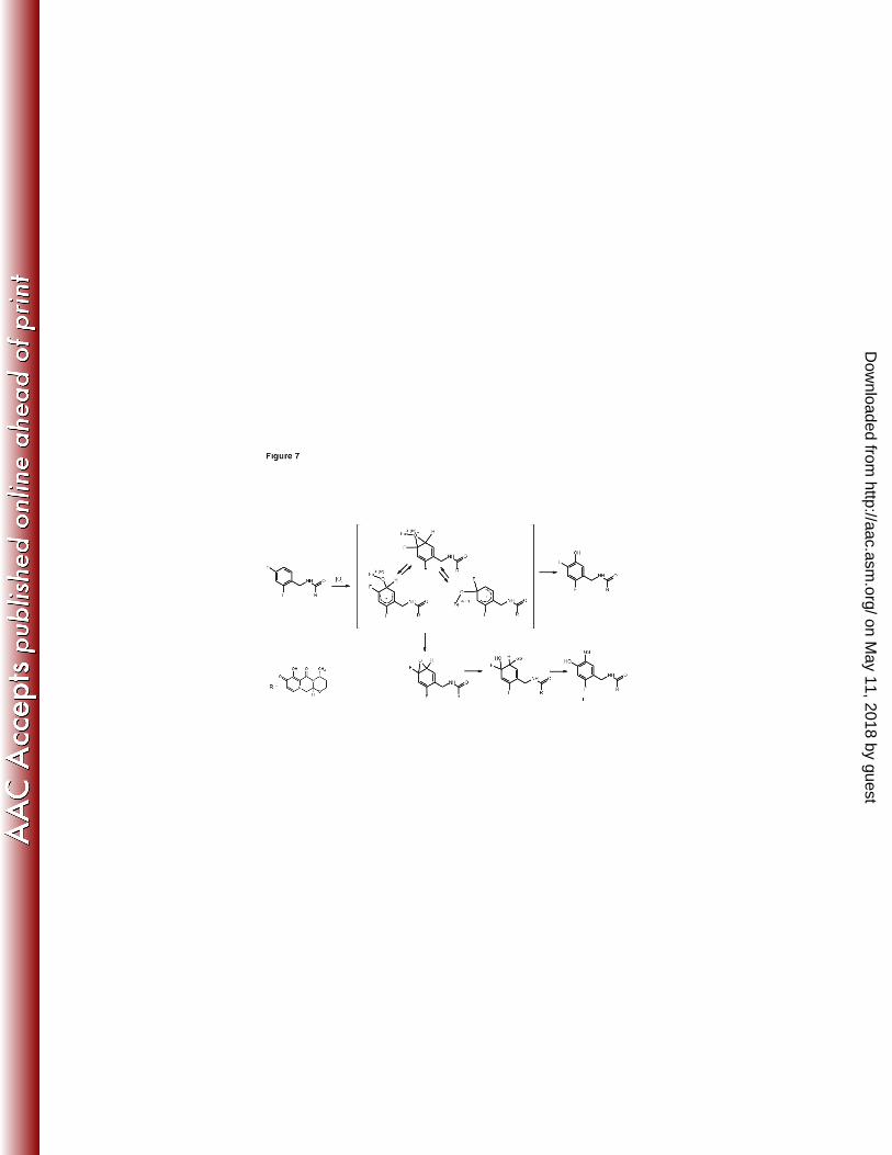
intermediate followed by glutathione addition and HF elimination (Figure 7). In contrast, 489
formation of isomer III would require two separate biotransformations: oxidative defluorination 490
to generate a stable 2-hydroxy-4-fluorobenzyl group and glutathione opening of an epoxide 491
intermediate where water rather than HF is eliminated. Both biotransformations in the formation 492
of isomer III would appear to involve epoxide intermediates. The mechanism leading to isomer 493
III is more difficult to rationalize based on the biotransformation literature (21,22,23). Thus, 494
although there are not enough spectroscopic data from this study to definitively characterize M4 495
as isomer I, mechanistic considerations favor this structure. Although the proposed mechanism 496
of formation for M4 proceeds through an arene oxide, the low fractional clearance through this 497
pathway, lack of evidence for covalent binding, and the low to moderate dose administered 498
suggest the risk for drug-induced toxicities via this potential bioreactive species is low. This 499
on May 11, 2018 by guest
http://aac.asm.org/
Dow
nloaded from

24
analysis indicating the low body burden of systemic metabolites is consistent with the strategic 500
analysis recently reviewed (24). 501
In conclusion, following oral administration, the recovery of dolutegravir and dolutegravir-502
related components from urine and feces was nearly complete, with feces being the principal 503
route of excretion (Figure 8). Dolutegravir was the predominant circulating compound in plasma 504
and was consistent with minimal presystemic clearance. Radioactivity was minimally associated 505
with the blood cellular components. At least 34% of the dose was absorbed with an additional 506
33% to 48% fraction undergoing enterohepatic recirculation. Dolutegravir was extensively 507
metabolized, and the disposition kinetics of the metabolites was formation-rate limited, 508
indicating no long-lived metabolites. An inactive ether glucuronide, formed primarily via 509
UGT1A1, was the principal biotransformation pathway (at least 18.9% of the dose with 510
additional amount secreted in bile and deconjugated), and minor biotransformation pathways 511
included oxidation by CYP3A4 (7.9% of the dose) and a correlated oxidative defluorination and 512
glutathione substitution (1.8% of the dose). These biotransformations also were observed in 513
nonclinical studies, thereby supporting selection of the nonclinical species for safety coverage in 514
humans. Thus, this study provides an understanding of the clearance pathways via the routes of 515
metabolism and excretion of dolutegravir underpinned by the nonclinical safety assessment and 516
the low risk of idiosyncratic reactions by bioreactive metabolites. The metabolic profile and 517
fractional clearances obtained from this study in conjunction with the in vitro investigations (16) 518
provide the mechanistic basis for understanding potential effects when dolutegravir is co-519
administered with other therapeutic products. 520
521
on May 11, 2018 by guest
http://aac.asm.org/
Dow
nloaded from

25
Acknowledgments 522
All listed authors meet the criteria for authorship set forth by the International Committee for 523
Medical Journal Editors and are employees of GlaxoSmithKline. We are grateful to the 524
following study contributors for their support of this investigation: Stephen D. Flach, MD, 525
principal investigator, and the staff at the Covance Clinical Research Unit; Nate Ensrud and 526
Vikash Patel for radioanalysis at Covance Laboratories; and the subject participants without 527
whom this study would not be possible. We also acknowledge Glenn Tabolt for quantitative MS 528
assistance, Ernest Schubert and Andy Roberts for helpful NMR discussions, and Eri Kanaoka of 529
Shionogi & Co., Ltd, for review of the experimental protocols and reports. 530
This study was supported by ViiV Healthcare. Editorial assistance was provided under the 531
direction of the authors by Chris Lawrence with funding from ViiV Healthcare. 532
Conference where work was previously presented: 13th Annual International Workshop on 533
Clinical Pharmacology of HIV Therapy; April 16-18, 2012; Barcelona, Spain. 534
535
on May 11, 2018 by guest
http://aac.asm.org/
Dow
nloaded from

26
Authorship Contributions 536
Participated in study design: Borland, Piscitelli, Song, Chen, Lou, Min, Savina 537
Conducted experiments: Wagner, Moss, Goljer, Culp 538
Performed data analysis: Borland, Piscitelli, Song, Chen, Min, Lou, Castellino, Savina 539
Wrote or contributed to the writing of the manuscript: Borland, Piscitelli, Song, Chen, Lou, Min, 540
Savina, Castellino, Wagner, Moss, Goljer, Culp 541
542 543
on May 11, 2018 by guest
http://aac.asm.org/
Dow
nloaded from

27
References 544
1. Engelman A, Craigie R. 1992. Identification of conserved amino acid residues critical for 545
human immunodeficiency virus type 1 integrase function in vitro. J. Virol. 66:6361–6369. 546
2. Semenova EA, Johnson AA, Marchand C, Davis DA, Yarchoan R, Pommier Y. 2006. 547
Preferential inhibition of the magnesium-dependent strand transfer reaction of HIV-1 548
integrase by alpha-hydroxytropolones. Mol. Pharmacol. 69:1454–1460. 549
3. Hare S, Gupta SS, Valkov E, Engelman A, Cherepanov P. 2010. Retroviral intasome 550
assembly and inhibition of DNA strand transfer. Nature 464:232–236. 551
4. Hare S, Vos AM, Clayton RF, Thuring JW, Cummings MD, Cherepanov P. 2010. 552
Molecular mechanisms of retroviral integrase inhibition and the evolution of viral resistance. 553
Proc. Natl. Acad. Sci. U. S. A. 107:20057–20062. 554
5. Hare S, Smith SJ, Métifiot M, Jaxa-Chamiec A, Pommier Y, Hughes SH, Cherepanov 555
P. 2011. Structural and functional analyses of the second-generation integrase strand transfer 556
inhibitor dolutegravir (S/GSK1349572). Mol. Pharmacol. 80:565–572. 557
6. Sato A, Kobayashi M, Seki T, Morimoto CW, Yoshinaga T, Fujiwara T, Johns BA, 558
Underwood M. 2010. S/GSK1349572: a next generation integrase inhibitor with limited or 559
no cross resistance to first generation INIs or other classes of anti-virals. 8th European HIV 560
Drug Resistance Workshop, Sorrento, Italy. 561
7. Kobayashi M, Yoshinaga T, Seki T, Wakasa-Morimoto C, Brown KW, Ferris R, Foster 562
SA, Hazen RJ, Miki S, Suyama-Kagitani A, Kawauchi-Miki S, Taishi T, Kawasuji T, 563
on May 11, 2018 by guest
http://aac.asm.org/
Dow
nloaded from

28
Johns BA, Underwood MR, Garvey EP, Sato A, Fujiwara T. 2011. In vitro antiretroviral 564
properties of S/GSK1349572, a next-generation HIV integrase inhibitor. Antimicrob. Agents 565
Chemother. 55:813–821. 566
8. Hightower KE, Wang R, Deanda F, Johns BA, Weaver K, Shen Y, Tomberlin GH, 567
Carter HL III, Broderick T, Sigethy S, Seki T, Kobayashi M, Underwood MR. 2011. 568
Dolutegravir (S/GSK1349572) exhibits significantly slower dissociation than raltegravir and 569
elvitegravir from wild-type and integrase inhibitor-resistant HIV-1 integrase-DNA 570
complexes. Antimicrob. Agents Chemother. 55:4552–4559. 571
9. Min S, Sloan L, DeJesus E, Hawkins T, McCurdy L, Song I, Stroder R, Chen S, 572
Underwood M, Fujiwara T, Piscitelli S, Lalezari J. 2011. Antiviral activity, safety, and 573
pharmacokinetics/pharmacodynamics of dolutegravir as 10-day monotherapy in HIV-1-574
infected adults. AIDS 25:1737–1745. 575
10. van Lunzen J, Maggiolo F, Arribas JR, Rakhmanova A, Yeni P, Young B, Rockstroh 576
JK, Almond S, Song I, Brothers C, Min S. 2012. Once daily dolutegravir (S/GSK1349572) 577
in combination therapy in antiretroviral-naïve adults with HIV: planned interim 48 week 578
results from SPRING-1, a dose-ranging, randomised, phase 2b trial. Lancet Infect. Dis. 579
12:111–118. 580
11. Eron JJ, Clotet B, Durant J, Katlama C, Kumar P, Lazzarin A, Poizot-Martin I, 581
Richmond G, Soriano V, Ait-Khaled M, Fugiwara T, Huang J, Min S, Vavro C, Yeo J. 582
(2013). Safety and efficacy of dolutegravir in treatment-experienced subjects with 583
raltegravir-resistant HIV Type 1 infection:24-week results of the VIKING study. J Inf Dis 584
DOI: 10.1093/infdis/jis750. 585
on May 11, 2018 by guest
http://aac.asm.org/
Dow
nloaded from

29
12. Min S, Song I, Borland J, Chen S, Lou Y, Fujiwara T, Piscitelli SC. 2010. 586
Pharmacokinetics and safety of S/GSK1349572, a next-generation HIV integrase inhibitor, in 587
healthy volunteers. Antimicrob. Agents Chemother. 54:254–258. 588
13. Reese MJ, Savina PM, Generaux GT, Tracey H, Humphreys JE, Kanaoka E, Webster 589
LO, Harmon KA, Clarke JD Polli JW. 2013. In vitro investigations into the roles of drug 590
transporters and metabolizing enzymes in the disposition and drug interactions of 591
dolutegravir, a HIV integrase inhibitor. Drug Metab. Dispos. 41:353–361. 592
14. Song I, Borland J, Chen S, Peppercorn A, Savina P, Wajima T, Min S, Nichols G, 593
Stephen Piscitelli S. (2012).Metabolism and Drug-Drug Interaction Profile of 594
Dolutegravir(DTG, S/GSK1349572). 13th International Workshop on clinical Pharmacology 595
of HIV Therapy, Barcelona, Spain. Abstract O-07. 596
15. US Department of Health and Human Services, Food and Drug Administration, Center 597
for Drug Evaluation and Research, Center for Biologics Evaluation and Research. 2005. 598
Guidance for Industry. E14 clinical evaluation of QT/QTc interval prolongation and 599
proarrhythmic potential for non-antiarrhythmic drugs. US Department of Health and Human 600
Services, Rockville, MD. 601
16. Chen S, Min SS, Peppercorn A, Borland J, Lou Y, Song I, Fujiwara T, Piscitelli SC. 602
2012. Effect of a single supratherapeutic dose of dolutegravir on cardiac repolarization. 603
Pharmacotherapy 32:333–339. 604
17. Loevinger RL, Budinger TF, Watson EE. 1999. MIRD primer for absorbed dose 605
calculations, revised ed. Society of Nuclear Medicine, New York, NY. 606
on May 11, 2018 by guest
http://aac.asm.org/
Dow
nloaded from

30
18. Yang X, Gandhi YA, Duignan DB, Morris ME. 2009. Prediction of biliary excretion in 607
rats and humans using molecular weight and quantitative structure-pharmacokinetic 608
relationships. AAPS J. 11:511–525. 609
19. Rao SSC, Camilleri M, Hasler WL, Maurer AH, Parkman HP, Saad R, Scott MS, 610
Simren M, Soffer E, Szarka L. 2011. Evaluation of gastrointestinal transit in clinical 611
practice: postion paper of the American and European Neurogastroenterology and Motility 612
Societies. Neurogastroenterol Motil 23:8-23. 613
20. Song I, Borland J, Chen S, Patel P, Wajima T, Peppercorn A, Piscitelli S. 2012. Effect of 614
food on the pharmacokinetics of the integrase inhibitor dolutegravir. Antimicrob. Agents 615
Chemother. 56:1627-1629. 616
21. Dear GJ, Ismail IM, Mutch PJ, Plumb RS, Davies LH, Sweatman BC. 2000. Urinary 617
metabolites of a novel quinoxaline non-nucleoside reverse transcriptase inhibitor in rabbit, 618
mouse and human: identification of fluorine NIH shift metabolites using NMR and tandem 619
MS. Xenobiotica 30:407–426. 620
22. Yergey JA, Trimble LA, Silva J, Chauret N, Li C, Therien M, Grimm E, Nicoll-Griffith 621
DA. 2001. In vitro metabolism of the COX-2 inhibitor DFU, including a novel glutathione 622
adduct rearomatization. Drug Metab. Dispos. 29:638–644. 623
23. Samuel K, Yin W, Stearns RA, Tang YS, Chaudhary AG, Jewell JP, Lanza T Jr, Lin 624
LS, Hagmann WK, Evans DC, Kumar S. 2003. Addressing the metabolic activation 625
potential of new leads in drug discovery: a case study using ion trap mass spectrometry and 626
tritium labeling techniques. J. Mass Spectrom. 38:211–221. 627
on May 11, 2018 by guest
http://aac.asm.org/
Dow
nloaded from

31
24. Smith DA, Obach RS. 2009. Metabolites in safety testing (MIST): considerations of 628
mechanisms of toxicity with dose, abundance, and duration of treatment. Chem. Res. 629
Toxicol. 22:267–279. 630
631
on May 11, 2018 by guest
http://aac.asm.org/
Dow
nloaded from

32
Tables 632
633
Table 1. Summary of Percent Radioactivity Recovered in Urine and Feces From Healthy 634 Male Human Subjects After a Single Oral 20-mg (80 μCi) Suspension Dose of 635 [14C]Dolutegravir 636
Percentage of administered dose Subject Feces Urine Total 531001 72.0 24.0 96.1 531002 65.2 30.6 95.8 531003 61.0 34.8 95.8 531004 59.4 33.8 93.2 531005 65.7 29.4 95.1 531006a 60.8 36.7 97.6 Mean 64.0 31.6 95.6 SD 4.7 4.6 1.4 aSubject 531006 withdrew from the study after 144 hours postdose. The subject was excluded from descriptive statistics after this time point.
637
638
on May 11, 2018 by guest
http://aac.asm.org/
Dow
nloaded from

33
Table 2. Summary of Selected Pharmacokinetic Parameters for Plasma Dolutegravir, 639 Plasma Radioactivity, and Blood Radioactivity in Healthy Male Human Subjects 640 After a Single Oral 20-mg (80 μCi) Dose of [14C]Dolutegravir as a Suspension 641
Analyte n Cmaxa
(μg/mL) Tmaxb
(h) AUC(0-t)a
(μg.h/mL)AUC(0-∞)a (μg.h/mL)
CL/Fa (L/hr)
Vz/Fa (L)
t½a (h)
Plasma dolutegravir
6 2.57 (24)
0.50 (0.50-2.00)
35.7 (12)
35.9 (12)
0.56 (12)
12.5 (9)
15.6 (16)
Plasma radioactivity
6 2.46 (24)
0.50 (0.50-1.50)
35.9 (11)
36.1 (11)
NR NR 15.7 (14)
Blood radioactivity
6 1.13 (25)
1.25 (0.50-2.00)
17.7 (13)
18.4 (13)
NR NR 14.6 (12)
AUC, area under the concentration-time curve; CL/F, apparent oral clearance; Cmax, maximum observed concentration; NR, not reported; t½, terminal phase half-life; Tmax, time to Cmax; Vz/F, apparent volume of distribution. a Geometric mean (CV%). b Median (range).
642
on May 11, 2018 by guest
http://aac.asm.org/
Dow
nloaded from

34
Table 3. Radiochromatographic Analyses of Urine and Feces Following a Single Oral 643 Administration of [14C]Dolutegravir to Healthy Male Human Subjects at a 644 Target Dose of 20 mg (80 μCi) 645
646
Peak ID
Retention time
(minutes)
Meana ± sd % matrix radioactivity; [mean ± sd % dose]
Urine Feces
M1 12.2-12.8
11.8±2.0; [3.6±0.9]
2.2±0.7; [1.3±0.4]
M2 24.4-24.8 62.5±3.9; [18.9±3.0]
ND
M3b 37.2 + 40.2 10.1±2.8; [3.0±0.9]
ND
M4 24.0-24.4 NQ 3.1±1.3; [1.8±1.3]
Dolutegravir 44.4 2.2±0.2; [0.7±0.1]
89.1±3.3; [53.1±4.4]
Total radioactive material assigned
86.6±2.3; [26.2±3.9]
94.4±3.5; [56.3±3.9]
% Dose in matrix pool analyzed 30.2±4.3 59.7±5.5
Total % dose excreted in matrix (all time points)
31.6±4.6 64.0±4.7
ND Not Detected (below lower limit of detection of 3 times background). NQ Not Quantifiable (radioactivity was present at this retention time but concentration too low for complete structural identification). a. n=6 b. The reported values for M3 are the sum of both diastereomers.
on May 11, 2018 by guest
http://aac.asm.org/
Dow
nloaded from

35
Table 4. Structural Characterization Data for Dolutegravir and its Metabolites from High-Resolution Mass Spectrometric 647 and NMR Analysis. 648
649
ID RT
(min) Proposed structure
Parent ion [M+H]+ (error; ppm)
Fragment ions 1H-NMR
DTG a 44
1
6
2
5
3
4
7
F29
9
O28
NH8
F30
11
10
15
N26
1312 N
2418
16
17
20
19
21
O23
CH322O25
OH14
O27
H
127 Da
292 Da
142 Da
277 Da
420.1377 (2.7)
295 277
1H{19F} NMR (600 MHz, DMSO-d6) δ ppm 1.33
(d, J=6.88 Hz, 3 H) 1.54 (d, J=13.75 Hz, 1 H) 1.97 -
2.04 (m, 1 H) 3.16 (d, J=5.23 Hz, 1 H) 3.89 (dd,
J=11.69, 2.89 Hz, 1 H) 4.00 - 4.06 (m, 1 H) 4.35
(dd, J=13.76, 5.78 Hz, 1 H) 4.54 (d, J=6.05 Hz, 2
H) 4.55 - 4.58 (m, 1 H) 4.79 (t, J=6.60 Hz, 1 H)
5.45 (t, J=4.81 Hz, 1 H) 7.06 (dd, J=8.53, 2.48 Hz,
1 H) 7.25 (d, J=2.48 Hz, 1 H) 7.38 (d, J=8.80 Hz, 1
H) 8.50 (s, 1 H) 10.36 (t, J=6.05 Hz, 1 H)
F29= -116.4 ppm, F30= -113.4 ppm
M1 14 9
O28
NH2 8 1110
15
N26
1312 N
2418
16
17
20
19
21
O23
CH322O25
OH14
O27
H
294.1087 (0.8)
277
1H NMR (600 MHz, DMSO-d6) δ ppm 1.26 (d, J=6.59 Hz, 3 H) 1.49 (d, J=13.17 Hz, 1 H) 1.93(m, 1 H) 3.79 - 3.88 (m, 1 H) 3.90 - 3.95 (m, 250 H) 4.23 (dd, J=14, 4 Hz, 1 H) 4.41(dd, J=14, 2 Hz, 1 H) 4.74 (m, 1 H) 5.34 (br m, 1 H) 8.29 (s, 1 H)
on May 11, 2018 by guest
http://aac.asm.org/
Dow
nloaded from

36
ID RT
(min) Proposed structure
Parent ion [M+H]+ (error; ppm)
Fragment ions 1H-NMR
M2 26.5
3836
34
32
31O42
39OH35
OH33
OH37
OH41
O40
1
6
2
5
3
4
7
F29
9
O28
NH8
F30
11
10
15
N26
1312 N
2418
16
17
20
19
21
O23
CH322O25
O14
O27
H
596.1674 (-2.2)
420
1H NMR (600 MHz, DMSO-d6) δ ppm 1.16 (d, J=6.96 Hz, 3 H) 1.48 (d, J=13.18 Hz, 1 H) 1.92 (td, J=12.45, 6.22 Hz, 1 H) 3.10 - 3.19 (m, 1 H) 3.21 - 3.28 (m, 1 H) 3.29 - 3.35 (m, 1 H) 3.40 (d, J=9.88 Hz, 1 H) 3.76 - 3.89 (m, 1 H) 4.17 (dd, J=13.36, 7.51 Hz, 1 H) 4.48 (d, J=6.59 Hz, 2 H) 4.51 (d, J=3.30 Hz, 1 H) 4.61 - 4.70 (m, 1 H) 5.14 (d, J=7.69 Hz, 1 H) 5.27 (dd, J=7.32, 3.66 Hz, 1 H) 7.00 (td, J=8.42, 2.20 Hz, 1 H) 7.15 (td, J=9.79, 2.38 Hz, 1 H) 7.35 (dd, J=15.38, 8.79 Hz, 1 H) 8.46 (s, 1 H) 10.22 (t, J=5.86 Hz, 1 H)
M3 39 &
42
1
6
2
5
3
47
F29
9
O28
NH8
F30
1110
15
N26
1312 N
2418
16
17
20
19
21
O23
CH322O25
OH14
O27
HOH
436.1304 (-2.4) 418
294
1H NMR (600 MHz, DMSO-d6) δ ppm 1.27 (d, J=7.03 Hz, 1 H) 1.46 - 1.54 (m, 1 H) 1.95 (none, 1 H) 3.80 - 3.88 (m, 1 H) 3.91 - 4.02 (m, 1 H) 4.23 - 4.34 (m, 1 H) 4.47 (td, J=13.40, 3.95 Hz, 1 H) 4.74 (dd, J=12.30, 6.15 Hz, 1 H) 5.37 (q, J=4.83 Hz, 1 H) 6.54 (d, J=8.35 Hz, 1 H) 7.07 (t, J=8.35 Hz, 1 H) 7.15 (t, J=9.45 Hz, 1 H) 7.56 (dd, J=15.38, 8.35 Hz, 1 H) 8.38 (s, 1 H) 10.67 (d, J=8.35 Hz, 1 H)
M4b 26
32
33NH2 34a 34
O35
OH36
S31
54N3
2 O1
6
13 1412
10N8
11
9
16
7
NH21
O20
O18
OH19
O17
22
26
23
27
25
28
24
CH315
F29
OH30
537.1470 (3.8)
294 277 244
1H{19F} (600 MHz, METHANOL-d4) δ ppm 1.40 (d, J=7 Hz, 3 H), 4.29 (m, 1 H), 4.51 (m, 1 H),4.49 (m., 1 H) 6.65 (s, 1 H) 7.54 (s, 1 H) 8.38 (s, 1 H); H5-H6, cysteine resonances not assigned. 13C (HSQC) δ ppm C2-75.8, C7-51.6, C9-139.9, C15-13.4, C22-35.7, C25-102.9, C28-136.8 F29= -117.1 ppm
a DTG, dolutegravir; NMR, nuclear magnetic resonance; RT, retention time. b Putative structural isomer shown. 650
on May 11, 2018 by guest
http://aac.asm.org/
Dow
nloaded from

37
Figure Captions 651
Figure 1: Structure of [14C]dolutegravir (sodium salt). 652
Figure 2: Overall mean cumulative elimination of radioactivity by six healthy male subjects after a single 20 mg (80 μCi) oral 653
dose of [14C]dolutegravir. 654
Figure 3: Mean concentration-time profiles of plasma dolutegravir and total radioactivity in blood and plasma after a single 655
20 mg (80 μCi) oral dose of [14C]dolutegravir to six healthy male subjects. Inset figure shows data using log scale on 656
y-axis. 657
Figure 4: Representative radiochromatograms of plasma, urine, and feces after a single oral 20 mg (80 μCi) dose of 658
[14C]dolutegravir to an individual healthy male subject. (A) Plasma at 24 hours. (B) Urine at 0-72 hours. (C) Feces at 659
0-96 hours. 660
Figure 5: Nuclear magnetic resonance characterization of M4 regioisomers. 661
Figure 6: Metabolic scheme for dolutegravir after a single oral 20 mg (80 μCi) dose of [14C]dolutegravir to six healthy male 662
subjects. 663
Figure 7: Proposed mechanism for the formation of M4 isomer I. 664
Figure 8: Summary of the mean excretion and mass balance after a single oral administration of [14C]dolutegravir to six 665
healthy male subjects at a target dose of 20 mg (80 μCi). 666
on May 11, 2018 by guest
http://aac.asm.org/
Dow
nloaded from


![Absorption, metabolism and excretion of [ C]pomalidomide in humans following oral ... · Absorption, metabolism and excretion of [14C]pomalidomide in humans following oral administration](https://static.fdocuments.in/doc/165x107/5ad218a67f8b9afa798c5160/absorption-metabolism-and-excretion-of-cpomalidomide-in-humans-following-oral.jpg)


