Metabolic profiling of alcohol consumption in 9778 young ...

RESEARCH ARTICLE Open Access
Metabolic profiling of pregnancy: cross-sectional and longitudinal evidenceQin Wang1,2, Peter Würtz1, Kirsi Auro3,4,5, Ville-Petteri Mäkinen1,6,7, Antti J. Kangas1, Pasi Soininen1,2, Mika Tiainen1,2,Tuulia Tynkkynen1,2, Jari Jokelainen8,9, Kristiina Santalahti10, Marko Salmi10, Stefan Blankenberg11,12, Tanja Zeller11,12,Jorma Viikari13,14, Mika Kähönen15, Terho Lehtimäki16, Veikko Salomaa3, Markus Perola3,4,17, Sirpa Jalkanen10,Marjo-Riitta Järvelin8,9,18, Olli T. Raitakari19,20, Johannes Kettunen1,2,3, Debbie A. Lawlor21,22
and Mika Ala-Korpela1,2,21,22*
Abstract
Background: Pregnancy triggers well-known alterations in maternal glucose and lipid balance but its overall effectson systemic metabolism remain incompletely understood.
Methods: Detailed molecular profiles (87 metabolic measures and 37 cytokines) were measured for up to 4260women (24–49 years, 322 pregnant) from three population-based cohorts in Finland. Circulating molecularconcentrations in pregnant women were compared to those in non-pregnant women. Metabolic profiles were alsoreassessed for 583 women 6 years later to uncover the longitudinal metabolic changes in response to change inthe pregnancy status.
Results: Compared to non-pregnant women, all lipoprotein subclasses and lipids were markedly increased inpregnant women. The most pronounced differences were observed for the intermediate-density, low-density andhigh-density lipoprotein triglyceride concentrations. Large differences were also seen for many fatty acids andamino acids. Pregnant women also had higher concentrations of low-grade inflammatory marker glycoproteinacetyls, higher concentrations of interleukin-18 and lower concentrations of interleukin-12p70. The changes inmetabolic concentrations for women who were not pregnant at baseline but pregnant 6 years later (or vice versa)matched (or were mirror-images of) the cross-sectional association pattern. Cross-sectional results wereconsistent across the three cohorts and similar longitudinal changes were seen for 653 women in 4-year and497 women in 10-year follow-up. For multiple metabolic measures, the changes increased in magnitude acrossthe three trimesters.
Conclusions: Pregnancy initiates substantial metabolic and inflammatory changes in the mothers.Comprehensive characterisation of normal pregnancy is important for gaining understanding of the keynutrients for fetal growth and development. These findings also provide a valuable molecular reference inrelation to studies of adverse pregnancy outcomes.
Keywords: Pregnancy, Trimesters, Postpartum, Metabolomics, Cytokines, Lipoprotein lipids, Fatty acids, Aminoacids, Hormones, Inflammation, Metabolic networks
* Correspondence: [email protected] A. Lawlor and Mika Ala-Korpela are shared senior authors of thiswork.1Computational Medicine, Faculty of Medicine, University of Oulu andBiocenter Oulu, Oulu, Finland2NMR Metabolomics Laboratory, School of Pharmacy, University of EasternFinland, Kuopio, FinlandFull list of author information is available at the end of the article
© The Author(s). 2016 Open Access This article is distributed under the terms of the Creative Commons Attribution 4.0International License (http://creativecommons.org/licenses/by/4.0/), which permits unrestricted use, distribution, andreproduction in any medium, provided you give appropriate credit to the original author(s) and the source, provide a link tothe Creative Commons license, and indicate if changes were made. The Creative Commons Public Domain Dedication waiver(http://creativecommons.org/publicdomain/zero/1.0/) applies to the data made available in this article, unless otherwise stated.
Wang et al. BMC Medicine (2016) 14:205 DOI 10.1186/s12916-016-0733-0

BackgroundPregnancy induces remarkable changes in maternal me-tabolism to support fetal demands [1, 2]. Maternal circu-lating glucose is an essential nutrient for the developingfetus [3] and during pregnancy women become increas-ingly insulin resistant, particularly from the second tri-mester onwards [4]. In the third trimester, maternalfasting insulin levels increase by over 30%, while fastingglucose concentrations decrease by about 10%, despiteincreased insulin resistance [1, 5]. In addition to glucose,there is evidence that circulating lipids and amino acidsare also important nutrients for the fetus [1, 2]. Duringthe third trimester, maternal circulating lipid concentra-tions dramatically increase, with triglycerides beingelevated by approximately twofold, and total and low-density lipoprotein (LDL) cholesterol by some 30–50%[1, 5–7]. Maternal circulating amino acid concentrationsare also suggested to be altered largely in response to in-creased protein synthesis for placental and fetal growth[8]. In addition, pregnancy triggers considerable changesin the maternal immune system, with circulating cyto-kines being reprogrammed to sustain the integrity of theallograft fetus [9].Most of the changes in maternal metabolism and
inflammatory status are considered to be normalphysiological responses to support fetal growth anddevelopment. These changes typically return to pre-pregnancy states soon after delivery [2]. However, insome women the changes may be harmful and re-lated to adverse pregnancy outcomes, for example,gestational diabetes, hypertensive disorders, and pre-term birth [2, 10, 11]. These adverse pregnancy out-comes are associated with increased future risk ofdiabetes and cardiovascular disease both in mothersand offspring, though the extent to which these arecausal or reflect pre-existing maternal risk is unclear[2]. Characterising the normal metabolic and inflam-matory changes in pregnancy is crucial for gainingunderstanding of the key nutrients for normal fetalgrowth and development, and for identifying meta-bolic and inflammatory changes in pregnancy thatmight herald the risk of adverse outcomes. However,so far, most epidemiological studies on the metaboliceffects of pregnancy have had very small numbers ofindividuals (typically only a few dozen pregnantwomen) and lack replication. The metabolic informa-tion is generally limited to standard lipids and glu-cose [1, 5], and studies on the effects of pregnancyon mothers’ systemic metabolism are lacking. Theaim of this study is to comprehensively characterisethe maternal systemic metabolism across a widerange of metabolic and inflammatory measures inboth cross-sectional and longitudinal settings coupledwith replication.
MethodsStudy populationSerum metabolic profiles were quantified in three inde-pendent population-based cohorts from Finland. Sam-ples for profiling were collected in 1997 for participantsin the Northern Finland Birth Cohort 1966 (NFBC1966,n = 2963 women aged 31) [12, 13], in 2001 for those inthe Cardiovascular Risk in Young Finns Study (YFS, n =1239 women aged 24–39) [14], and in 1997 for those inthe FINRISK1997 study (FINRISK1997, n = 2101 womenaged 24–49) [15]. Overnight fasting samples were ob-tained for NFBC1966 and YFS, whereas semi-fastingsamples (median fasting time 5 hours) were obtainedfor FINRISK1997. Further details of the study popula-tions are provided in Additional file 1: SupplementaryMethods. We excluded women whose information onpregnancy status at the time of metabolic profilingwas missing (n = 136); women using oral contraception(n = 1279); and women with a fasting glucose ≥7 mmol/L(n = 33, of which two were pregnant), systolic blood pres-sure ≥140 mmHg, or diastolic blood pressure ≥90 mmHg(n = 595; 13 pregnant). After these exclusions, 4260women, of whom 322 were pregnant (Table 1), were in-cluded in the study. A subset of 583 women from YFSattended a 6-year follow-up (baseline in 2001; follow-upin 2007) at which their clinical and pregnancy status wereassessed and the metabolic profiling performed again. Inthese women prospective changes in their metabolic pro-files, with respect to the change in their pregnancy status,were examined. Cytokine profiles were analysed for 2321women from FINRISK1997 (1415 women, 69 pregnant)and YFS in 2007 (906 women, 35 pregnant).We were not able to exclude breastfeeding women or
those women who were pregnant with multiple babiesbecause such information was not available in all the co-horts. However, in NFBC1966 where the information onthe number of babies per pregnancy was available, therewere only two pregnant women with a twin pregnancyand none with higher multiples. We therefore assumethat the vast majority of the data in these population-based cohorts are on singleton pregnancies. Informationon breastfeeding was available in YFS; 6.5% of non-pregnant women were breastfeeding. The metabolic as-sociations with pregnancy were almost identical whetherthe data for breastfeeding women were excluded fromthose for non-pregnant women or not (Additional file 1:Figure S1).
Information on pregnancy and covariatesPregnancy status, parity, current smoking, and alcoholconsumption were assessed by questionnaires. In YFS,the participants reported the gestational age (in weeks)at the time of blood sampling. In NFBC1966, the gesta-tional age (in weeks) at the time of blood sampling was
Wang et al. BMC Medicine (2016) 14:205 Page 2 of 14

calculated based on a birth registry. Information on thegestational age was not available for the participants inFINRISK1997. Weight, height and blood pressure wereassessed in clinics using established protocols.In the primary analyses, we compared pregnant (n =
322) to non-pregnant women (n = 3938). In the second-ary analyses, we compared pregnant women in differenttrimesters to non-pregnant women using data fromNFBC1966 and YFS. Profiles for women in their first tri-mester (pregnancy length ≤12 weeks, n = 48), second tri-mester (pregnancy length >12 weeks and ≤28 weeks, n =116), and third trimester (pregnancy length >28 weeks,n = 67) were compared to those of the non-pregnantwomen (n = 2588). Information on gestational age wasmissing for 20 pregnant women who were therefore ex-cluded from these analyses.
Molecular profilingWe analysed 124 biomarkers, including 87 metabolicmeasures and 37 cytokines. Eighty out of the 87 meta-bolic measures were quantified by a high-throughputserum nuclear magnetic resonance (NMR) metabolo-mics platform [16, 17]. These measures represent abroad molecular signature of systemic metabolism andcover multiple metabolic pathways, including lipoproteinlipids and subclasses, fatty acids, amino acids, andglycolysis-related metabolites. The NMR-based meta-bolic profiling has previously been used in multiplelarge-scale epidemiological and genetic studies [18–25]and the experimentation is described elsewhere [16, 17,26]. The seven other metabolic measures assessed werehigh-sensitivity C-reactive protein (CRP); vitamin D; sexhormone-binding globulin (SHBG); and the hormones in-sulin, leptin, adiponectin and testosterone. Additionally,
37 cytokines were analysed using Bio-Rad’s Bio-Plex As-says. Details of these measurements are given inAdditional file 1: Supplementary Methods.
Statistical analysesThe metabolic and cytokine measures were log-transformed because their distributions were generallyskewed (Additional file 1: Figure S2) and likely to resultin non-normal residuals in the regression analyses. Themeasures were then scaled to standard deviations (SD)in each cohort. Due to the correlated nature of the data,we used principal component analysis to evaluate theappropriate number of independent tests for multipletesting correction [22, 24]. Sixty-one principal compo-nents explained over 99% of the variation in the mo-lecular measures across all three cohorts (Additionalfile 1, Table S1). Therefore, P < 0.0008 (0.05/61) wasused to infer statistical significance [22, 24].For the cross-sectional analyses, a linear regression
model was fitted for each outcome measure (concentra-tion of a metabolic or a cytokine measure) using preg-nancy status as the explanatory variable. Non-pregnantwomen were used as the reference group, so the associ-ation magnitudes denote the difference in each outcomemeasure between pregnant and non-pregnant women.Association magnitudes are reported in SD units toease the comparison across multiple measures. Differ-ences in the original units of the metabolic and cyto-kine measures and the percentage differences are givenin Additional file 1: Supplementary Material. All threecohorts were analysed separately and the results com-bined using fixed-effect inverse variance-weightedmeta-analysis after confirming the consistency acrossthe cohorts. In the main analyses we adjusted for age.
Table 1 Characteristics of the study participants
Characteristics NFBC1966 YFS FINRISK1997
Non-pregnant Pregnant Non-pregnant Pregnant Non-pregnant Pregnant
Number 1782 191 806 60 1350 71
Gestational age at blood sampling (week) – 22 (13–29) – 23 (15–30) – –
Age (year) 31 (0) 31 (0) 32 (5) 31 (5) 37 (7) 32 (5)
Weight (kg) 65 (13) 67 (11) 67 (13) 71 (12) 66 (12) 70 (13)
Waist (cm) 79 (11) 82 (12) 80 (11) 85 (10) 77 (10) 83 (13)
Hip (cm) 97 (8) 97 (8) 100 (9) 102 (7) 99 (8) 101 (9)
BMI (kg/m2) 24 (4) 24 (4) 24 (5) 26 (4) 25 (4) 26 (4)
Systolic blood pressure (mmHg) 117 (10) 116 (9) 111 (10) 109 (11) 119 (10) 114 (11)
Diastolic blood pressure (mmHg) 73 (8) 66 (10) 68 (8) 61 (9) 74 (8) 68 (9)
Smoking prevalence (%) 38 25 20 10 23 8
Alcohol usage (g/day) 2.1 (0.6–5.7) 1.0 (0.0–3.5) 3.3 (0.0–8.2) 0.0 (0.0–0.0) 1.8 (0.0–7.0) 0.0 (0.0–0.0)
Parity (n) 2 (1–2) 1 (0–2) 1 (0–2) 1 (0–2) – –
Values are mean (SD) for normally distributed and median (interquartile range) for skewed variables. Information on whether women have given birth before wasnot available for FINRISK1997. BMI: body mass index
Wang et al. BMC Medicine (2016) 14:205 Page 3 of 14

In the second set of models we additionally adjusted forbody mass index (BMI), parity, mean arterial pressure(MAP), current smoking and alcohol consumption.Similar linear models were used to determine the meta-bolic differences between trimesters and non-pregnantwomen using data from NFBC1966 and YFS.Those 583 women from the YFS cohort who had both
baseline and 6-year follow-up data were classified aspregnant at follow-up (non-pregnant at baseline butpregnant at follow-up, n = 18), pregnant at baseline(pregnant at baseline but non-pregnant at follow-up,n = 44) and those non-pregnant at both time points(n = 519). Two women who were pregnant at bothbaseline and follow-up were excluded from the ana-lysis. The 6-year changes in metabolic concentrationswere compared between (1) women who were preg-nant at follow-up, and (2) women who were pregnantat baseline, and women who were non-pregnant atboth time points. The longitudinal models were ad-justed for baseline age and further for the baselineparity and 6-year change in BMI, MAP, smoking andalcohol use. The longitudinal analyses were addition-ally replicated for 653 women who had 4-year follow-up data (from year 2007 to 2011 in YFS) and 497women who had 10-year follow-up data (from year2001 to 2011 in YFS). Details of these data are de-scribed in Additional file 1: Supplementary Methods.
ResultsThe characteristics of the study participants are given inTable 1. The pregnant women, in comparison to thosenon-pregnant, tended to have lower diastolic blood pres-sure, smoke less and consume less alcohol.
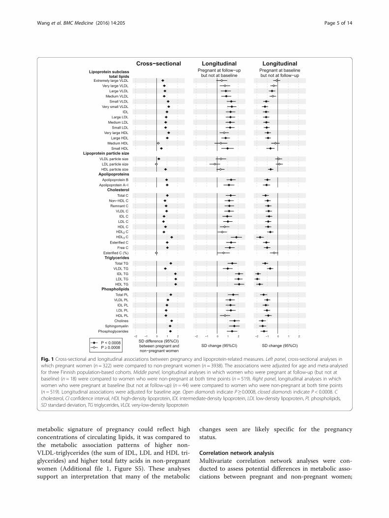
Lipoprotein-related measures in pregnancyFigure 1 (left panel) illustrates the cross-sectional associ-ations between pregnancy and 44 lipoprotein-relatedmeasures. The metabolic differences between pregnantand non-pregnant women are given in SD units inAdditional file 1: Table S2, in absolute physiologicalunits in Additional file 1: Table S3 and as a percentagedifference relative to non-pregnant women in Additionalfile 1: Table S4. Pregnancy was associated with increasedlevels of almost all lipoprotein-related measures (P < 0.0008for 41 out of 44 measures meta-analysed). The associ-ation magnitudes were substantial, with a median of0.9 SD unit increments (interquartile range 0.8–1.1SDs), corresponding to a median of a 33% (interquar-tile range 25–51%) higher concentration in pregnantthan in non-pregnant women. The increase in totallipids was similar across the smaller very-low-density(VLDL), intermediate-density (IDL) and low-density(LDL), and the larger high-density (HDL) lipoproteinsubclasses. Pregnancy was positively associated with all
cholesterol, triglyceride and phospholipid concentra-tions as well as with both apolipoprotein B and A-I.The differences for IDL, LDL and HDL triglycerides wereapproximately twofold in comparison to VLDL triglycer-ides. In contrast, differences in cholesterol and phospho-lipid concentrations in all these main lipoprotein fractionswere broadly similar. The percentage of esterified choles-terol (of total cholesterol) did not differ between pregnantand non-pregnant women. Pregnancy was positively asso-ciated with the average size of VLDL and HDL particlesbut not with the average size of LDL particles.
Fatty acids in pregnancyFigure 2 (left panel) displays the cross-sectional associa-tions between pregnancy and multiple fatty acid mea-sures. The circulating concentrations of all fatty acidswere considerably increased during pregnancy, rangingapproximately between 1.0 and 1.5 SDs, being equivalentto 24–56% higher concentrations relative to non-pregnant women. However, the proportions of individualfatty acids (relative to total fatty acids) exhibited a di-verse association pattern. The proportions of saturatedand monounsaturated fatty acids as well as the propor-tion of docosahexaenoic acid (DHA) were increased. Incontrast, the proportion of omega-6 fatty acids, includ-ing linoleic acid, were decreased.
Metabolites, inflammatory markers and hormones inpregnancyFigure 3 (left panel) shows the concentration differencesin multiple circulating biomarkers between pregnant andnon-pregnant women. The amino acids displayed a het-erogeneous association pattern: strong positive associa-tions for alanine, phenylalanine and histidine; negativeassociations for glutamine, glycine, valine and tyrosine;and null associations for isoleucine and leucine. Glucose,glycerol and acetoacetate concentrations were modestlydecreased but lactate and pyruvate increased in thepregnant women. Creatinine and albumin concentra-tions were decreased and those of the low-grade in-flammatory markers, glycoprotein acetyls (GlycA) [27]and CRP, increased. Of the hormones, testosterone andleptin concentrations were increased, as was that ofSHBG, which displayed the strongest association acrossthe molecular profile (2.5 SDs; equivalent to 483%higher concentration in pregnant women than in thenon-pregnant women).The associations with lipoprotein-related measures,
fatty acids, low-molecular-weight metabolites, inflamma-tory markers and hormones were highly consistentacross the three cohorts (Additional file 1: Figure S3).The results remained very similar with further adjust-ments for parity, BMI, MAP, smoking and alcohol(Additional file 1: Figure S4). To assess how much the
Wang et al. BMC Medicine (2016) 14:205 Page 4 of 14
metabolic signature of pregnancy could reflect highconcentrations of circulating lipids, it was compared tothe metabolic association patterns of higher non-VLDL-triglycerides (the sum of IDL, LDL and HDL tri-glycerides) and higher total fatty acids in non-pregnantwomen (Additional file 1, Figure S5). These analysessupport an interpretation that many of the metabolic
changes seen are likely specific for the pregnancystatus.
Correlation network analysisMultivariate correlation network analyses were con-ducted to assess potential differences in metabolic asso-ciations between pregnant and non-pregnant women;
Fig. 1 Cross-sectional and longitudinal associations between pregnancy and lipoprotein-related measures. Left panel, cross-sectional analyses inwhich pregnant women (n = 322) were compared to non-pregnant women (n = 3938). The associations were adjusted for age and meta-analysedfor three Finnish population-based cohorts. Middle panel, longitudinal analyses in which women who were pregnant at follow-up (but not atbaseline) (n = 18) were compared to women who were non-pregnant at both time points (n = 519). Right panel, longitudinal analyses in whichwomen who were pregnant at baseline (but not at follow-up) (n = 44) were compared to women who were non-pregnant at both time points(n = 519). Longitudinal associations were adjusted for baseline age. Open diamonds indicate P ≥ 0.0008, closed diamonds indicate P < 0.0008. Ccholesterol, CI confidence interval, HDL high-density lipoprotein, IDL intermediate-density lipoprotein, LDL low-density lipoprotein, PL phospholipids,SD standard deviation, TG triglycerides, VLDL very-low-density lipoprotein
Wang et al. BMC Medicine (2016) 14:205 Page 5 of 14

Fig. 2 Cross-sectional and longitudinal associations between pregnancy and fatty acids. The study design is as explained in the legend for Fig. 1.The percentage sign (%) refers to the proportion of an individual measure of the total fatty acids. Open diamonds indicate P ≥ 0.0008, closeddiamonds indicate P < 0.0008. CI confidence interval, FA fatty acids, MUFA monounsaturated fatty acids, PUFA polyunsaturated fatty acids, SDstandard deviation
Fig. 3 Cross-sectional and longitudinal associations between pregnancy and metabolic, inflammatory and hormonal measures. The study designis as explained in the legend for Fig. 1. Open diamonds indicate P ≥ 0.0008, closed diamonds indicate P < 0.0008. CI confidence interval, SDstandard deviation, SHBG sex hormone-binding globulin
Wang et al. BMC Medicine (2016) 14:205 Page 6 of 14

these analyses and the results are described in detail inAdditional file 2. Briefly, a higher degree of pair-wisecorrelations was observed in pregnant (mean Spearman|r| = 0.39) compared to non-pregnant women (mean |r| =0.29). A pronounced node difference was noted for SHBG,together with triglycerides in multiple lipoprotein categor-ies (‘Triglyceride enrichment’ community), alanine andphenylalanine (‘Amino acid’) and various fatty acids (‘Fattyacid composition’). These nodes shared a similar patternof higher inter-community connectivity for the preg-nant than for the non-pregnant women. In general, thecorrelation network analyses suggest that pregnancy isacting as a metabolic stressor that amplifies intrinsicpatterns of metabolism.
Metabolic changes in response to change in pregnancystatusFigures 1, 2 and 3 (middle and right panels) illustratesix-year changes in 84 metabolic measures in responseto change in pregnancy status (follow-up data weremissing for leptin, SHBG and testosterone). In the mid-dle panels, the metabolic changes in women who werepregnant at follow-up but not at baseline were compared
to those of women who were non-pregnant at both timepoints. In the right panels, the metabolic changes inwomen who were pregnant at baseline but not at follow-up were compared to those of women who were non-pregnant at both time points. Substantial metabolicchanges were observed for women who were pregnantat follow-up (middle panel); the association magnitudeswere highly similar to those observed in the cross-sectional setting (left panel). Pronounced metabolicchanges were also observed for women who were preg-nant at baseline (right panel); the absolute associationmagnitudes again matched the cross-sectional setting,but they were in the opposite direction. The overallconsistency between the longitudinal metabolic changesin women pregnant at follow-up and the cross-sectionalmetabolic differences followed a straight line with aslope of 1.04 ± 0.04 (R2 = 0.90; Fig. 4, left panel). Simi-larly, the magnitudes of the metabolic associations forwomen pregnant at baseline and those of cross-sectionalassociations also fell in a straight line with R2 = 0.93 butwith a negative slope of −1.02 ± 0.03 (Fig. 4, right panel).Differences in the point estimates were noted for DHA
(%) in the longitudinal analysis of women who were
Fig. 4 Correlations between cross-sectional and longitudinal metabolic associations of pregnancy. Left panel, linear fit to summarise thecorrespondence between cross-sectional (pregnant women compared to non-pregnant women) and longitudinal associations for womenpregnant at follow-up but not at baseline (in comparison to women non-pregnant at both time points). Right panel, linear fit to summarisethe correspondence between cross-sectional and longitudinal associations for women pregnant at baseline but not at follow-up (in comparison towomen non-pregnant at both time points). Each point represents a single metabolic measure. Horizontal and vertical grey lines denote 95% CIs for thecross-sectional and longitudinal associations, respectively. The grey shaded areas serve to guide the eye for the slope. A linear fit for the overallcorrespondence summarises the match between cross-sectional and longitudinal associations, with R2 denoting the goodness of fit. A slopeof ±1 and R2 = 1 would strongly support the causal effects of pregnancy on the metabolic measures. C cholesterol, CI confidence interval, DHAdocosahexaenoic acid, FA fatty acids, HDL high-density lipoprotein, IDL intermediate-density lipoprotein, LDL low-density lipoprotein, PL phospholipids,PUFA polyunsaturated fatty acids, SD standard deviation, TG triglycerides, VLDL very-low-density lipoprotein
Wang et al. BMC Medicine (2016) 14:205 Page 7 of 14

pregnant at follow-up but not at baseline, in comparisonwith the other longitudinal analysis and cross-sectionalanalysis. However, the confidence intervals of these ana-lyses overlapped, suggesting consistency of the results.Larger longitudinal studies would be required to ascertainthe relevance of these findings on DHA% in pregnancy.The longitudinal associations remained similar when fur-ther adjusted for baseline parity and 6-year change inBMI, MAP, smoking and alcohol consumption (Additionalfile 1: Figures S6 and S7). To assess the consistency of
these longitudinal associations in the YFS cohort, we fur-ther compared the results from the 6-year follow-up tothose from 4-year (from year 2007 to 2011) and 10-yearfollow-up (from year 2001 to 2011). The longitudinal asso-ciations were highly consistent across the 4-year, 6-yearand 10-year follow-up (Additional file 1: Figure S3).
Metabolic profile during trimestersFigure 5 shows cross-sectional concentration differencesin 87 metabolic measures between pregnant women in
Fig. 5 Cross-sectional associations between pregnancy trimesters and metabolic measures. Pregnant women in their first (n = 48), second (n = 116)and third trimester (n = 67) were compared to non-pregnant women (n = 2588). Associations were adjusted for age and meta-analysed for NFBC1966and YFS. Open circles indicate P ≥ 0.0008, closed circles indicate P < 0.0008. C cholesterol, CI confidence interval, FA fatty acids, HDL high-densitylipoprotein, IDL intermediate-density lipoprotein, LDL low-density lipoprotein, PL phospholipids, MUFA monounsaturated fatty acids, PUFApolyunsaturated fatty acids, SD standard deviation, SHBG sex hormone-binding globulin, TG triglycerides, VLDL very-low-density lipoprotein
Wang et al. BMC Medicine (2016) 14:205 Page 8 of 14

different trimesters and non-pregnant women (Additionalfile 1: Table S5). The differences in SD units, in absolutephysiological units and as percentages relative to non-pregnant women are given in Additional file 1: Tables S2,S3 and S4, respectively. During the first trimester manycirculating metabolic concentrations were similar to thosein non-pregnant women, although some clear differenceswere already observable: for example, glutamine, creatin-ine, SHBG and testosterone displayed over 1 SD metabolicdifferences. Women in their second or third trimesters ofpregnancy had large metabolic aberrations in comparisonto non-pregnant women, with similar association patternsas those described earlier (Figs. 1, 2 and 3) when all tri-mesters were combined. There was a clear trend of in-creasing metabolic differences in most lipoprotein-relatedmeasures across the trimesters, with median percentagedifference −9% (interquartile range −13% to 0%) for thefirst trimester, 34% (interquartile range 28–51%) for thesecond trimester, and 59% (interquartile range 39–100%)for the third trimester. However, HDL-related measureswere similar during the second and third trimesters. Theassociation pattern across the three trimesters for most ofthe absolute concentrations of fatty acids followed that ofthe apolipoprotein B-containing lipoprotein subclasses.However, exceptions were observed for omega-3 fattyacids and DHA, for which the association pattern acrossthe three trimesters resembled that of the large HDL sub-classes and apolipoprotein A-I. The differences in the ma-jority of the proportions of the fatty acids also had thetendency to be largest during the third trimester. Thecirculating concentrations of multiple low-molecular-weight metabolites, albumin, inflammatory markers andhormone-related measures were broadly similar duringthe second and third trimesters. The metabolic associa-tions during the trimesters were highly consistent inNFBC1966 and YFS (Additional file 1: Figure S8).
Metabolic retention of pregnancyThe metabolic retention of pregnancy was evaluated atvarious times postpartum (calculated as the durationfrom the delivery to the blood sampling). These analyseswere done in NFBC1966 (serum samples collected at1997) utilising the birth registry data from 1995 to 1997.Women postpartum up to 2 months (n = 28), over 2 andup to 4 months (n = 46), over 4 and up to 6 months(n = 42) and over 6 and up to 12 months (n = 123) werecompared to those 1–3 years postpartum (n = 389).There were clear trends of metabolic normalisationafter the delivery. Within 3–6 months postpartum,most of the metabolic disturbances were back to levelssimilar to those in women 1–3 years postpartum(Additional file 1: Figure S9). We only had the birthregistry data for NFBC1966 so we could not specify thedelivery dates for all the non-pregnant women in this
study. However, according to the NFBC1966 data, only6.5% of women had given birth within 6 months priorto blood sampling. When these women were excludedfrom the non-pregnant women in the analyses, themetabolic associations with pregnancy were almostidentical (Additional file 1: Figure S10).
Cytokine profile in pregnancyThe cross-sectional associations of pregnancy with 37cytokines are illustrated in Additional file 1: Figure S9.Among interleukins, positive associations were foundwith IL-1ra and IL-18 and negative associations withIL-12p70. In addition, pregnant women had lower levelsof TRAIL, MCP-1, eotaxin and VEGF and higher levels ofHGF and βNGF than non-pregnant women. The abbrevi-ations of the cytokines are explained in the caption forAdditional file 1: Figure S11.
DiscussionHere we have comprehensively characterised the effectsof pregnancy on maternal systemic metabolism across awide range of metabolic and inflammatory measures inlarge numbers of women. These findings are importantfor a broader metabolic understanding of the physio-logical changes caused by pregnancy per se that arelikely to be key to normal fetal growth and development.They also provide a valuable molecular reference in rela-tion to studies of adverse pregnancy outcomes [2]. Thecross-sectional associations in three population-basedstudies consistently showed that pregnancy was associ-ated with a wide range of metabolic pathways, includingthose for lipoproteins, fatty acids, amino acids, inflam-matory markers and hormones. The metabolic associa-tions were pronounced, with magnitudes during thethird trimester being generally 1–3 SDs different fromthe non-pregnant women. The exceptionally large meta-bolic effects, together with the consistent results fromlongitudinal analyses, indicate that the metabolic aberra-tions arise as a direct result of pregnancy [28]. Themetabolic changes increased in magnitudes for manymetabolic measures across the trimesters, and thechanges caused by pregnancy generally normalisedwithin 3–6 months postpartum.Metabolic adaptation to pregnancy has been widely
studied with respect to glucose metabolism and standardlipids [1, 5–7]. Observational studies have reported thatdecreased circulating glucose and increased triglycerides,LDL and HDL cholesterol as well as insulin are associ-ated with pregnancy [1, 5–7]. Our results here confirmthese metabolic changes in pregnant women and theirchange patterns and effect sizes with respect to the tri-mesters. These findings, together with the results forleptin [29, 30], SHBG and testosterone [31], serve as
Wang et al. BMC Medicine (2016) 14:205 Page 9 of 14

good positive controls for the novel metabolomics ap-proach taken in this work.The most pronounced metabolic changes during preg-
nancy were observed in the circulating concentrations ofIDL, LDL and HDL triglycerides. In these lipoproteinfractions, the increase in triglycerides was more pro-nounced than the increase in cholesterol and phos-pholipids, suggesting triglyceride-enrichment of thesecirculating lipoprotein particles in pregnancy [32], par-ticularly during the second and third trimesters. Incontrast, in the VLDL fraction, all these key lipid constit-uents increased almost in parallel with each other.Similar results have been reported previously in smalllongitudinal studies [32, 33], with a further suggestionthat the triglyceride-enrichment in various lipoproteinfractions would be related to oestrogen-induced en-hanced VLDL production and decreased activity oflipoprotein lipase and hepatic lipase during pregnancy.Maternal triglycerides are widely considered to be animportant nutrient for fetal growth. Various observa-tional studies have shown that higher maternal triglycer-ide concentrations are associated with larger neonatal fatmass and higher birth weight [34–37]. However, a recentMendelian randomisation analysis challenged the notionthat maternal triglycerides would be causally related tobirth weight [3]. The triglyceride-enrichment in IDL,LDL and HDL observed here suggests a specific role ofmaternal triglycerides in different lipoprotein fractions.This points to a potential value of further research to as-sess the causal relations between maternal triglyceridescarried in different lipoprotein particles and birth weight.Fatty acids are required by the developing fetus to sup-
port rapid cellular growth and activity [38]. Our resultsshowed that total circulating maternal fatty acid concen-tration gradually increased during pregnancy, in linewith the overall increase in lipoprotein lipids. The fattyacid composition, relative to total fatty acids, was alsolargely modified during pregnancy. Similar findings formaternal fatty acids and their proportions in plasmaand plasma phospholipids have been reported previ-ously [39, 40]. DHA may be of particular importance tofetal brain and retinal development [38]. Omega-3 fattyacid supplementation during pregnancy appears to in-crease maternal and fetal DHA concentration [38], butmeta-analysis of randomised controlled trials suggeststhat omega-3 fatty acid supplementation, in addition toan adequate omnivore intake, can only cause a modestincrease in birth weight and seems to have no effectson birth length or head circumference [41, 42]. The po-tential effects of maternal omega-3 supplementationduring pregnancy on cognitive and visual developmentin early childhood remain inconclusive [43].Maternal amino acids are considered to be key deter-
minants for fetal growth [1]. Nevertheless, information
on maternal amino acids during pregnancy is sparse andcomes from studies with only small numbers of partici-pants [8, 44, 45]. A small cross-sectional study (withsome 10 pregnant women in each trimester) found thatmost amino acid concentrations decreased during preg-nancy [44]. However, our cross-sectional and longitu-dinal findings here consistently indicate that amino acidsactually display a complex association pattern, withsome being markedly elevated (alanine, phenylalanineand histidine), some depleted (glutamine, glycine, valineand tyrosine) and some (isoleucine and leucine) notresponding to pregnancy. The association of pregnancywith branched-chain and aromatic amino acid concen-trations did not follow the pattern of consistent eleva-tions previously seen with obesity and insulin resistance[18, 19, 46]. Thus, it is unlikely that the weight gain orthe development of insulin resistance during pregnancyunderlie these changes in maternal amino acids. Thecomplex associations of pregnancy with amino acids arelikely to reflect the intricate molecular-specific interplaybetween maternal, placental and fetal metabolism [8].For example, there is evidence that the concentrations ofsome amino acids are generally higher in umbilical cordplasma than in maternal circulation at delivery [8].Amino acids cross the placenta via a combination ofmultiple active transport systems [47]. Dietary interven-tions during pregnancy have shown that an increase inmaternal circulating amino acid concentrations does notappear to affect their circulating concentrations in theumbilical cord or in the neonate [8]. These findings sug-gest a robust placental transporting system in adequatelynourished mothers. However, in the present study weare unable to explore these maternal–fetal interactions.The maternal immune system is challenged by the
conflicting demands of maintaining robust immune re-activity to protect both the mother and the fetus whileat the same time tolerating highly immunogenic alloanti-gens to sustain fetal integrity [48]. We found that con-centrations of the low-grade inflammatory markers CRPand GlycA were clearly increased during pregnancy, sug-gesting the up-regulation of systemic maternal inflam-mation. The new inflammatory marker, GlycA, which isquantified via the serum NMR metabolomics platform,has recently been shown to be associated with bothacute-phase and chronic inflammation, as well as beingrelated to increased neutrophil activities [27, 49]. Eleva-tion of GlycA during pregnancy may thereby suggest anenhancement of maternal innate immunity. With respectto adaptive immunity, previous studies have suggestedthat normal pregnancy polarises the immune responsetowards the T helper 2 (Th2) response [9, 48, 50]. Thisnotion is consistent with our findings of increased circu-lating IL-18 but decreased levels of IL12p70, which areimportant regulators of the Th2/Th1 balance [9, 50].
Wang et al. BMC Medicine (2016) 14:205 Page 10 of 14
Metabolic and immunological changes in the motherspredominantly occur to the benefit of the fetus. How-ever, some of these changes may be unfavourable to themothers. Hypertriglyceridaemia, elevated LDL andremnant cholesterol, insulin resistance, and up-regulatedinflammation during pregnancy are all features relatedto the increased risk of cardiometabolic diseases. Inaddition, our results here indicate that multiple new bio-markers for cardiovascular diseases [17, 20] show un-favourable changes during pregnancy; these includeelevated concentrations of phenylalanine and the in-creased proportion of monounsaturated and decreasedproportions of omega-6 fatty acids. Although we foundthat pregnancy-related metabolic alterations generallynormalise soon after delivery (Additional file 1: Figure S9),there is some evidence that repeated pregnancies mightpredispose women to higher cardiovascular risk at olderage [51, 52]. It has been reported that the association be-tween parity and cardiovascular risk is J-shaped, with twobirths representing the lowest risk [51, 52]. Compared towomen with two births, women with five or more birthshad a 66% higher risk of cardiovascular disease, the associ-ation remaining robust when adjusting for socioeconomicstatus and pregnancy complications [52]. However, it re-mains unclear whether the association between parity andcardiovascular risk indicates a causal relationship [2].Oral contraceptives are used to prevent pregnancy by
mimicking the reproductive hormone status of naturalpregnancy through the supplementation of a combin-ation of oestrogen and progestin (combined oral contra-ceptive pills; COCPs) or progestin-only contraceptives(POCs). Understandably, this simple mimic cannot repli-cate the complex physiological effects of natural preg-nancy and the multilevel interactions between themother, the placenta and the fetus. This is in line withour findings that there is little correspondence in thepattern of cytokine changes in pregnant women andthose using COCPs [53]. Interestingly, however, the pat-tern of metabolic changes for lipoprotein measures, fattyacids, amino acids and low-grade inflammatory markersduring pregnancy has a strong resemblance to the onecaused by COCPs. Though the absolute metabolicchanges are stronger in the case of pregnancy, the direc-tion of change is generally consistent with that inwomen who use COCPs. On the other hand, the meta-bolic effects of POCs are weak or negligible [53]. Thesefindings imply that oestrogen plays a key role in theregulation of maternal metabolism.Owing to the population-based design of our cohorts,
we were not able to look at how the circulating meta-bolic measures relate to potential pregnancy complica-tions or perinatal outcomes. However, it is an importantstart to examine the metabolic consequences of normalpregnancy across a wide range of metabolic and
inflammatory measures. We excluded women (whetherpregnant or not) who had high fasting glucose or highblood pressure, at levels indicative of diabetes or hyper-tension, respectively. While we cannot be certain thatwe have excluded all cases of gestational diabetes orhypertensive disorders of pregnancy, the nature of gen-eral population studies suggests that our results reflectchanges in normal pregnancy. The strengths of thisstudy include extensive molecular profiling of systemicmetabolism with replication across three independentpopulation-based cohorts. The highly consistent meta-bolic findings from multiple longitudinal analyses (4, 6and 10 years of follow-up) provide compelling additionalevidence that the metabolic aberrations are a direct re-sult of pregnancy. We also acknowledge that the trimes-ter analyses are cross-sectional and the pregnancy statusis assessed via questionnaires. This may slightly affectthe metabolic associations during the first trimester,since some women may not be aware of being pregnantin their early pregnancy and thus report themselves asnon-pregnant. Nevertheless, this information bias wouldbe minimal in the case of middle and late pregnancy.Importantly, the extraordinarily large association mag-nitudes and gradual increases in effect sizes across thetrimesters, together with the replication in individualcohorts, strongly suggest that the effect of confoundingis minimal.
ConclusionsThis work characterises the effects of pregnancy on ma-ternal metabolism across a wide range of metabolic andinflammatory measures. The findings are important for abroader metabolic understanding of the physiologicalchanges caused by pregnancy per se and for assessingthe metabolic milieu related to normal fetal growth anddevelopment. The metabolic effects of pregnancy are ex-ceptionally large, gradually increase across the trimesters,and generally normalise within 3–6 months postpartum.These findings provide a comprehensive foundation to thesystemic molecular understanding of maternal metabol-ism during pregnancy.
Open Peer Review ReportsThe authors’ response to reviewers is available asAdditional file 3.
Additional files
Additional file 1: Supplementary materials including detailed informationon study populations, generalised data supporting our findings, and resultsfrom sensitivity analyses. (DOCX 2656 kb)
Additional file 2: Correlation network analysis. (PDF 7311 kb)
Additional file 3: Authors’ response to reviewers. (PDF 7434 kb)
Wang et al. BMC Medicine (2016) 14:205 Page 11 of 14
AbbreviationsBMI: Body mass index; COCP: Combined oral contraceptive pill; CRP: C-reactive protein; DHA: Docosahexaenoic acid; GlycA: Glycoprotein acetyls;HDL: High-density lipoprotein; IDL: Intermediate-density lipoprotein;LDL: Low-density lipoprotein; MAP: Mean arterial pressure;NFBC1966: Northern Finland Birth Cohort 1966; NMR: Nuclear magneticresonance; POC: Progestin-only contraceptives; SD: Standard deviation;SHBG: Sex hormone-binding globulin; Th2: T helper 2.; VLDL: Very-low-density lipoprotein; YFS: Cardiovascular Risk in Young Finns StudyThe abbreviations of the cytokines are available in the caption of Figure S12(Additional file 1).
FundingThe quantitative serum NMR metabolomics platform and its developmenthas been supported by the Academy of Finland, TEKES (the Finnish FundingAgency for Technology and Innovation), Sigrid Juselius Foundation, NovoNordisk Foundation, Finnish Diabetes Research Foundation, Paavo NurmiFoundation, and strategic and infrastructural research funding from theUniversity of Oulu, Finland, as well as by British Heart Foundation, WelcomeTrust, and Medical Research Council, UK. The Young Finns Study has beenfinancially supported by the Academy of Finland: grant numbers 134309(Eye), 126925, 121584, 124282, 129378 (Salve), 117797 (Gendi), 41071 (Skidi),and 286284; the Social Insurance Institution of Finland; Kuopio, Tampere andTurku University Hospital Medical Funds; Juho Vainio Foundation; SigridJuselius Foundation; Yrjö Jahnsson Foundation; Paavo Nurmi Foundation;Finnish Foundation for Cardiovascular Research and Finnish CulturalFoundation; Tampere Tuberculosis Foundation; and Emil AaltonenFoundation. MS and SJ have been supported through funds from theAcademy of Finland (grant number 141136). NFBC1966 has received financialsupport from the Academy of Finland (project grants 104781, 120315,129269, 1114194, and SALVE); University Hospital Oulu, Biocenter, Universityof Oulu, Finland (75617); European Commission (EURO-BLCS, Framework 5award QLG1-CT-2000-01643; EU H2020-PHC-2014-grant no. 633595); MedicalResearch Council, UK (G0500539, G0600705, PrevMetSyn/SALVE, PS0476); andWellcome Trust (project grant GR069224). KA has received funding fromFinnish Medical Association. SB and TZ have been supported by European UnionSeventh Framework Programme (FP7/2007-2013) under grant agreement No.HEALTH-F2-2011-278913 (BiomarCaRE). VS has received funds from the Academyof Finland (grant number 139635) and from Finnish Foundation for Cardiovascu-lar Research. JK was supported through funds from the Academy of Finland(grant number 283045). The contribution of DAL to this study was supported bygrants from the US National Institute of Health (R01 DK10324), EuropeanResearch Council (ObesityDevelop; Grant no. 669545) and UK National Institutefor Health Research (NF-SI-0166-10196). DAL and MAK work in a unit that issupported by the University of Bristol and UK Medical Research Council(MC_UU_1201/1 and MC_UU_1201/5). The funders had no role in study design,data collection and analysis, decision to publish, or preparation of themanuscript.
Availability of data and materialsMetabolic and clinical data are available according to the rules of eachindividual cohort and can be requested from the Institutional Data AccessCommittees of the Northern Finland Birth Cohort Studies (University of Oulu,Finland), the Cardiovascular Risk in Young Finns Study (University of Turku,Finland), and the FINRISK study committee at the National Institute forHealth and Welfare (Helsinki, Finland).
Authors’ contributionsConception and design: QW, PW, JK, DAL, MAK. Data interpretation: QW,VPM, DAL, MAK. Statistical analysis: QW, VPM. Contribution to first draft of themanuscript: QW, DAL, MAK. Contribution to reagents/materials/analysis tools:AJK, PS, MT, TT, JJ, KS, MS, SB, TZ, SJ, MAK. Contribution to data collection:KA, JV, MK, TL, VS, MP, MRJ, OTR. All authors have read and approved thefinal version of the manuscript.
Competing interestsPW, AJK, PS and MAK are shareholders of Brainshake Ltd. (www.brainshake.fi),a company offering NMR-based metabolic profiling. PW, AJK, PS, MT, TT andJK report employment for Brainshake Ltd. KA is currently employed by GSK.SB has received research funding from Boehringer Ingelheim, Bayer, AbbottDiagnostics, SIEMENS, Thermo Fisher and Roche Diagnostics and received
honoraria for lectures or consulting from Boehringer Ingelheim, Bayer, Roche,Astra Zeneca, SIEMENS, ThermoFisher and Abbott Diagnostics. DAL hasreceived research support from Medtronic and Roche Diagnostics. All otherauthors declare that they have no competing interests.
Consent for publicationNot applicable.
Ethics approval and consent to participateThe research protocols were approved by the ethics committee of NorthernOstrobothnia Hospital District, Finland; the ethics committees of UniversityHospitals of Turku, Helsinki, Tampere, Kuopio, and Oulu; and theCoordinating Ethical Committee of the Helsinki and Uusimaa HospitalDistrict, Finland. All participants gave written informed consent.
Author details1Computational Medicine, Faculty of Medicine, University of Oulu andBiocenter Oulu, Oulu, Finland. 2NMR Metabolomics Laboratory, School ofPharmacy, University of Eastern Finland, Kuopio, Finland. 3National Institutefor Health and Welfare, Helsinki, Finland. 4Institute for Molecular Medicine(FIMM), University of Helsinki, Helsinki, Finland. 5Department of Obstetricsand Gynecology, Helsinki University Central Hospital and University ofHelsinki, Helsinki, Finland. 6Heart Health Theme, South Australian Health andMedical Research Institute, Adelaide, Australia. 7School of Biological Sciences,University of Adelaide, Adelaide, Australia. 8Center for Life Course HealthResearch and Biocenter Oulu, University of Oulu, Oulu, Finland. 9Unit ofPrimary Care, Oulu University Hospital, Oulu, Finland. 10Department ofMedical Microbiology and Immunology, and MediCity Research Laboratory,University of Turku, Turku, Finland. 11Clinic for General and InterventionalCardiology, University Heart Center Hamburg, Hamburg, Germany. 12GermanCenter for Cardiovascular Research (DZHK e.V.), partner site Hamburg,Lübeck, Kiel, Germany. 13Department of Medicine, University of Turku, Turku,Finland. 14Division of Medicine, Turku University Hospital, Turku, Finland.15Department of Clinical Physiology, University of Tampere and TampereUniversity Hospital, Tampere, Finland. 16Department of Clinical Chemistry,Fimlab Laboratories, School of Medicine, University of Tampere, Tampere,Finland. 17Estonian Genome Center, University of Tartu, Tartu, Estonia.18Department of Epidemiology and Biostatistics, MRC-PHE Centre forEnvironment and Health, School of Public Health, Imperial College London,London, UK. 19Research Centre of Applied and Preventive CardiovascularMedicine, University of Turku, Turku, Finland. 20Department of ClinicalPhysiology and Nuclear Medicine, Turku University Hospital, Turku, Finland.21Medical Research Council Integrative Epidemiology Unit at the Universityof Bristol, Bristol, UK. 22School of Social and Community Medicine, Universityof Bristol, Bristol, UK.
Received: 12 June 2016 Accepted: 31 October 2016
References1. Liu LX, Arany Z. Maternal cardiac metabolism in pregnancy. Cardiovasc Res.
2014;101:545–53.2. Rich-Edwards JW, Fraser A, Lawlor DA, Catov JM. Pregnancy characteristics
and women’s future cardiovascular health: an underused opportunity toimprove women’s health? Epidemiol Rev. 2014;36:57–70.
3. Tyrrell J, Richmond RC, Palmer TM, Feenstra B, Rangarajan J, Metrustry S, etal. Genetic evidence for causal relationships between maternalobesity-related traits and birth weight. JAMA. 2016;315:1129–40.
4. Stanley K, Fraser R, Bruce C. Physiological changes in insulin resistance inhuman pregnancy: longitudinal study with the hyperinsulinaemiceuglycaemic clamp technique. BJOG. 1998;105:756–9.
5. Lain KY, Catalano PM. Metabolic changes in pregnancy. Clin Obstet Gynecol.2007;50:938–48.
6. Lippi G, Albiero A, Montagnana M, Salvagno GL, Scevarolli S, Franchi M, etal. Lipid and lipoprotein profile in physiological pregnancy. Clin Lab. 2007;53:173–7.
7. Diareme M, Karkalousos P, Theodoropoulos G, Strouzas S, Lazanas N. Lipidprofile of healthy women during normal pregnancy. J Med Biochem.2009;28:152–60.
8. Rossary A, Farges M-C, Lamas B, Miles EA, Noakes PS, Kremmyda L-S, et al.Increased consumption of salmon during pregnancy partly prevents the
Wang et al. BMC Medicine (2016) 14:205 Page 12 of 14

decline of some plasma essential amino acid concentrations in pregnantwomen. Clin Nutr. 2014;33:267–73.
9. Szarka A, Rigó J, Lázár L, Beko G, Molvarec A. Circulating cytokines,chemokines and adhesion molecules in normal pregnancy andpreeclampsia determined by multiplex suspension array. BMC Immunol.2010;11:59.
10. Lawlor DA, Relton C, Sattar N, Nelson SM. Maternal adiposity–a determinant ofperinatal and offspring outcomes? Nat Rev Endocrinol. 2012;8:679–88.
11. Lawlor DA. The Society for Social Medicine John Pemberton Lecture 2011.Developmental overnutrition—an old hypothesis with new importance?Int J Epidemiol. 2013;42:7–29.
12. Järvelin M-R, Sovio U, King V, Lauren L, Xu B, McCarthy MI, et al. Early lifefactors and blood pressure at age 31 years in the 1966 northern Finlandbirth cohort. Hypertension. 2004;44:838–46.
13. Männistö T, Mendola P, Vääräsmäki M, Järvelin M-R, Hartikainen A-L, PoutaA, et al. Elevated blood pressure in pregnancy and subsequent chronicdisease risk. Circulation. 2013;127:681–90.
14. Raitakari OT, Juonala M, Rönnemaa T, Keltikangas-Järvinen L, Räsänen L,Pietikäinen M, et al. Cohort profile: the cardiovascular risk in Young FinnsStudy. Int J Epidemiol. 2008;37:1220–6.
15. Jousilahti P, Laatikainen T, Peltonen M, Borodulin K, Männistö S, Jula A, et al.Primary prevention and risk factor reduction in coronary heart diseasemortality among working aged men and women in eastern Finland over40 years: population based observational study. BMJ. 2016;352:i721.
16. Soininen P, Kangas AJ, Würtz P, Tukiainen T, Tynkkynen T, Laatikainen R, etal. High-throughput serum NMR metabonomics for cost-effective holisticstudies on systemic metabolism. Analyst. 2009;134:1781–5.
17. Soininen P, Kangas AJ, Würtz P, Suna T, Ala-Korpela M. Quantitative serumnuclear magnetic resonance metabolomics in cardiovascular epidemiologyand genetics. Circ Cardiovasc Genet. 2015;8:192–206.
18. Würtz P, Mäkinen V-P, Soininen P, Kangas AJ, Tukiainen T, Kettunen J, et al.Metabolic signatures of insulin resistance in 7,098 young adults. Diabetes.2012;61:1372–80.
19. Würtz P, Wang Q, Kangas AJ, Richmond RC, Skarp J, Tiainen M, et al.Metabolic signatures of adiposity in young adults: Mendelianrandomization analysis and effects of weight change. PLoS Med.2014;11:e1001765.
20. Würtz P, Havulinna AS, Soininen P, Tynkkynen T, Prieto-Merino D, Tillin T, etal. Metabolite profiling and cardiovascular event risk: a prospective study ofthree population-based cohorts. Circulation. 2015;131:774–85.
21. Kettunen J, Tukiainen T, Sarin A-P, Ortega-Alonso A, Tikkanen E, Lyytikäinen L-P,et al. Genome-wide association study identifies multiple loci influencinghuman serum metabolite levels. Nat Genet. 2012;44:269–76.
22. Kujala UM, Mäkinen V-P, Heinonen I, Soininen P, Kangas AJ, Leskinen TH, etal. Long-term leisure-time physical activity and serum metabolome.Circulation. 2013;127:340–8.
23. Fischer K, Kettunen J, Würtz P, Haller T, Havulinna AS, Kangas AJ, et al.Biomarker profiling by nuclear magnetic resonance spectroscopy for theprediction of all-cause mortality: an observational study of 17,345 persons.PLoS Med. 2014;11:e1001606.
24. Wang Q, Kangas AJ, Soininen P, Tiainen M, Tynkkynen T, Puukka K, et al. Sexhormone-binding globulin associations with circulating lipids andmetabolites and the risk for type 2 diabetes: observational and causal effectestimates. Int J Epidemiol. 2015;44:623–37.
25. Kettunen J, Demirkan A, Würtz P, Draisma HHM, Haller T, Rawal R, et al.Genome-wide study for circulating metabolites identifies 62 loci and revealsnovel systemic effects of LPA. Nat Commun. 2016;7:11122.
26. Inouye M, Kettunen J, Soininen P, Silander K, Ripatti S, Kumpula LS, et al.Metabonomic, transcriptomic, and genomic variation of a populationcohort. Mol Syst Biol. 2010;6:441.
27. Ritchie SC, Würtz P, Nath AP, Abraham G, Havulinna AS, Fearnley LG, et al.The biomarker GlycA is associated with chronic inflammation and predictslong-term risk of severe infection. Cell Systems. 2015;1:293–301.
28. Grimes DA, Schulz KF. Bias and causal associations in observational research.Lancet. 2002;359:248–52.
29. Sivan E, Whittaker PG, Sinha D, Homko CJ, Lin M, Reece EA, et al. Leptin inhuman pregnancy: the relationship with gestational hormones. Am J ObstetGynecol. 1998;179:1128–32.
30. Sattar N, Greer IA, Pirwani I, Gibson J, Wallace AM. Leptin levels inpregnancy: marker for fat accumulation and mobilization? Acta ObstetGynecol Scand. 1998;77:278–83.
31. O’Leary P, Boyne P, Flett P, Beilby J, James I. Longitudinal assessment ofchanges in reproductive hormones during normal pregnancy. Clin Chem.1991;37:667–72.
32. Montelongo A, Lasunción MA, Pallardo LF, Herrera E. Longitudinal study ofplasma lipoproteins and hormones during pregnancy in normal anddiabetic women. Diabetes. 1992;41:1651–9.
33. Alvarez JJ, Montelongo A, Iglesias A, Lasunción MA, Herrera E. Longitudinalstudy on lipoprotein profile, high density lipoprotein subclass, and postheparinlipases during gestation in women. J Lipid Res. 1996;37:299–308.
34. Misra VK, Trudeau S, Perni U. Maternal serum lipids during pregnancyand infant birth weight: the influence of prepregnancy BMI. Obesity(Silver Spring). 2011;19:1476–81.
35. Whyte K, Kelly H, O’Dwyer V, Gibbs M, O’Higgins A, Turner MJ. Offspringbirth weight and maternal fasting lipids in women screened for gestationaldiabetes mellitus (GDM). Eur J Obstet Gynecol Reprod Biol. 2013;170:67–70.
36. Hwang J-Y, Choi HI, Kim H, Jang W, Ha E-H, Park C, et al. Relationship ofmaternal grain intake and serum triglyceride levels with infant birth weight:Mothers and Children’s Environmental Health (MOCEH) study. Eur J ClinNutr. 2015;69:676–80.
37. Vrijkotte TGM, Algera SJ, Brouwer IA, van Eijsden M, Twickler MB. Maternaltriglyceride levels during early pregnancy are associated with birth weightand postnatal growth. J Pediatr. 2011;159:736–42. e1.
38. Haggarty P. Fatty acid supply to the human fetus. Annu Rev Nutr.2010;30:237–55.
39. Al MDM, van Houwelingen AC, Kester ADM, Hasaart THM, De Jong AEP,Hornstra G. Maternal essential fatty acid patterns during normal pregnancyand their relationship to the neonatal essential fatty acid status. Br J Nutr.1995;74:55–68.
40. Wadhwani NS, Pisal HR, Mehendale SS, Joshi SR. A prospective study ofmaternal fatty acids, micronutrients and homocysteine and their associationwith birth outcome. Matern Child Nutr. 2015;11:559–73.
41. Imhoff Kunsch B, Briggs V, Goldenberg T, Ramakrishnan U. Effect of n-3long-chain polyunsaturated fatty acid intake during pregnancy on maternal,infant, and child health outcomes: a systematic review. Paediatr PerinatEpidemiol. 2012;26:91–107.
42. Makrides M, Duley L, Olsen SF. Marine oil, and other prostaglandinprecursor, supplementation for pregnancy uncomplicated bypre-eclampsia or intrauterine growth restriction. Cochrane DatabaseSyst Rev. 2006;3:CD003402.
43. Gould JF, Smithers LG, Makrides M. The effect of maternal omega-3 (n − 3)LCPUFA supplementation during pregnancy on early childhood cognitiveand visual development: a systematic review and meta-analysis ofrandomized controlled trials. Am J Clin Nutr. 2013;97:531–44.
44. Cetin I, Ronzoni S, Marconi AM, Perugino G, Corbetta C, Battaglia FC, et al.Maternal concentrations and fetal-maternal concentration differences ofplasma amino acids in normal and intrauterine growth-restrictedpregnancies. Am J Obstet Gynecol. 1996;174:1575–83.
45. Cetin I, de Santis MSN, Taricco E, Radaelli T, Teng C, Ronzoni S, et al.Maternal and fetal amino acid concentrations in normal pregnancies and inpregnancies with gestational diabetes mellitus. Am J Obstet Gynecol.2005;192:610–7.
46. Stancáková A, Civelek M, Saleem NK, Soininen P, Kangas AJ, Cederberg H, et al.Hyperglycemia and a common variant of GCKR are associated with the levelsof eight amino acids in 9,369 Finnish men. Diabetes. 2012;61:1895–902.
47. Lager S, Powell TL. Regulation of nutrient transport across the placenta.J Pregnancy. 2012;2012:179827.
48. Konstantinov SR, van der Woude CJ, Peppelenbosch MP. Do pregnancy-related changes in the microbiome stimulate innate immunity? Trends MolMed. 2013;19:454–9.
49. Ala-Korpela M. Serum nuclear magnetic resonance spectroscopy: one morestep toward clinical utility. Clin Chem. 2015;61:681–3.
50. Sakai M, Shiozaki A, Sasaki Y, Yoneda S, Saito S. The ratio of interleukin (IL)-18 toIL-12 secreted by peripheral blood mononuclear cells is increased in normalpregnant subjects and decreased in pre-eclamptic patients. J Reprod Immunol.2004;61:133–43.
51. Lawlor DA, Emberson JR, Ebrahim S, Whincup PH, Wannamethee SG,Walker M, et al. Is the association between parity and coronary heartdisease due to biological effects of pregnancy or adverse lifestyle riskfactors associated with child-rearing? Findings from the British Women'sHeart and Health Study and the British Regional Heart Study. Circulation.2003;107:1260–4.
Wang et al. BMC Medicine (2016) 14:205 Page 13 of 14

52. Parikh NI, Cnattingius S, Dickman PW, Mittleman MA, Ludvigsson JF,Ingelsson E. Parity and risk of later-life maternal cardiovascular disease.Am Heart J. 2010;159:215–6.
53. Wang Q, Würtz P, Auro K, Morin Papunen L, Kangas AJ, Soininen P, et al.Effects of hormonal contraception on systemic metabolism: cross-sectionaland longitudinal evidence. Int J Epidemiol. 2016;45:1445–1457.
• We accept pre-submission inquiries
• Our selector tool helps you to find the most relevant journal
• We provide round the clock customer support
• Convenient online submission
• Thorough peer review
• Inclusion in PubMed and all major indexing services
• Maximum visibility for your research
Submit your manuscript atwww.biomedcentral.com/submit
Submit your next manuscript to BioMed Central and we will help you at every step:
Wang et al. BMC Medicine (2016) 14:205 Page 14 of 14