Metabolic bone diseases by Dr. Bharat Kumar Goud. C
80
Metabolic Bone Diseases Metabolic Bone Diseases
-
Upload
chukkalabharat -
Category
Health & Medicine
-
view
249 -
download
5
Transcript of Metabolic bone diseases by Dr. Bharat Kumar Goud. C

Metabolic Bone DiseasesMetabolic Bone Diseases

BONE STRUCTURE AND METABOLISMBONE STRUCTURE AND METABOLISM
Specialised connective tissue. Specialised connective tissue. Mineralised organic matrix in which living cells are dispersed.Mineralised organic matrix in which living cells are dispersed.
Bone
Mineralised bone matrix
Bone matrix or organic Bone matrix or organic fraction (30 per cent)fraction (30 per cent)
Collagen and Collagen and ground substanceground substance
Inorganic portion Inorganic portion (70 per cent)(70 per cent)
Cells of mesenchymal Cells of mesenchymal originorigin
crystalline hydroxyapatite crystalline hydroxyapatite (Ca(Ca1010 (PO (PO44))66OHOH22))

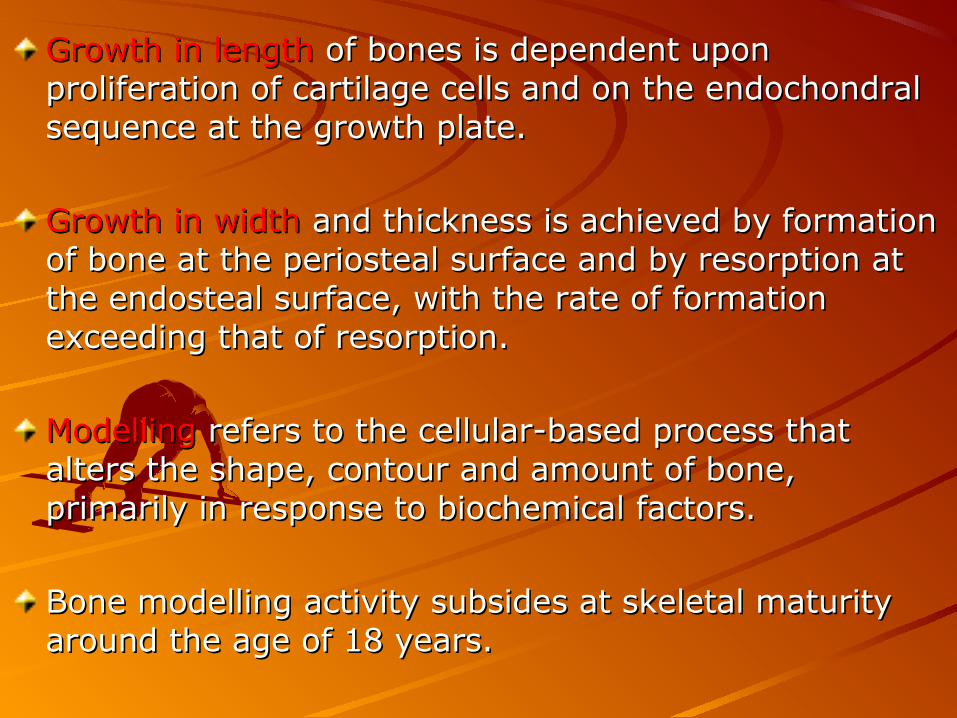
Growth in length Growth in length of bones is dependent upon of bones is dependent upon proliferation of cartilage cells and on the endochondral proliferation of cartilage cells and on the endochondral sequence at the growth plate. sequence at the growth plate.
Growth in widthGrowth in width and thickness is achieved by formation and thickness is achieved by formation of bone at the periosteal surface and by resorption at of bone at the periosteal surface and by resorption at the endosteal surface, with the rate of formation the endosteal surface, with the rate of formation exceeding that of resorption.exceeding that of resorption.
ModellingModelling refers to the cellular-based process that refers to the cellular-based process that alters the shape, contour and amount of bone, alters the shape, contour and amount of bone, primarily in response to biochemical factors. primarily in response to biochemical factors.
Bone modelling activity subsides at skeletal maturity Bone modelling activity subsides at skeletal maturity around the age of 18 years.around the age of 18 years.
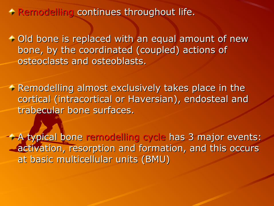
RemodellingRemodelling continues throughout life. continues throughout life.
Old bone is replaced with an equal amount of new Old bone is replaced with an equal amount of new bone, by the coordinated (coupled) actions of bone, by the coordinated (coupled) actions of osteoclasts and osteoblasts. osteoclasts and osteoblasts.
Remodelling almost exclusively takes place in the Remodelling almost exclusively takes place in the cortical (intracortical or Haversian), endosteal and cortical (intracortical or Haversian), endosteal and trabecular bone surfaces. trabecular bone surfaces.
A typical bone A typical bone remodelling cycleremodelling cycle has 3 major events: has 3 major events: activation, resorption and formation, and this occurs activation, resorption and formation, and this occurs at basic multicellular units (BMU) at basic multicellular units (BMU)


OsteoclastsOsteoclasts are giant multinucleated cells derived are giant multinucleated cells derived from a haematopoietic cell related to the from a haematopoietic cell related to the mononuclear-phagocyte series. They remove bone.mononuclear-phagocyte series. They remove bone.
OsteoblastsOsteoblasts synthesise and secrete the organic synthesise and secrete the organic matrix and regulate its mineralisation. The matrix is matrix and regulate its mineralisation. The matrix is composed of type I collagen and non collagenous composed of type I collagen and non collagenous proteins.proteins.
OsteocytesOsteocytes are the osteoblasts which secrete matrix. are the osteoblasts which secrete matrix. This is then mineralised.This is then mineralised.
Lining cellsLining cells separate bone from bone marrow and separate bone from bone marrow and from systemic ECF.from systemic ECF.
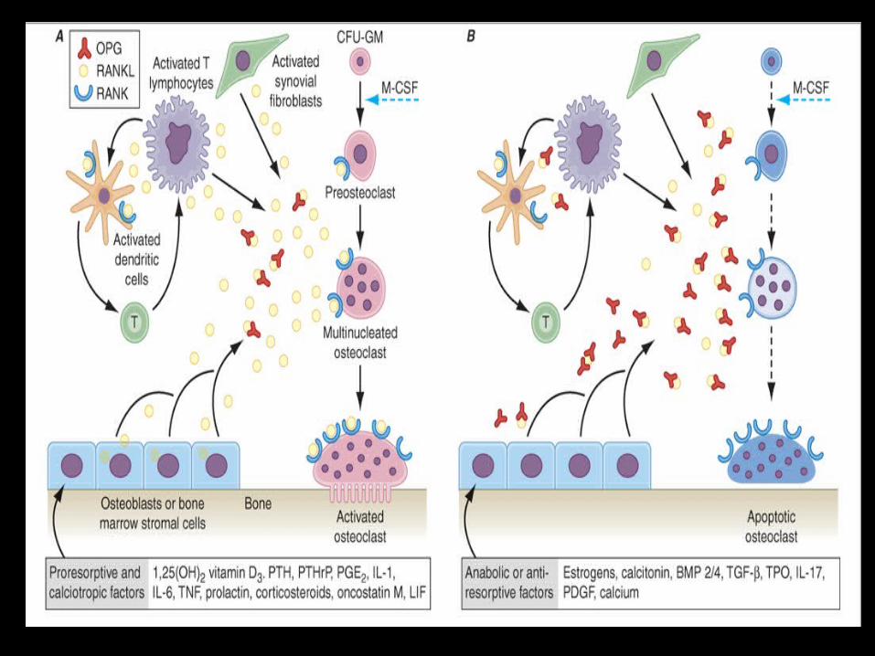

RANKL is secreted by osteoblasts, synovial fibroblasts, stromal cells, T cells.
The osteoclast receptor for this protein is referred to as RANK.
OPG binds and neutralizes RANKL, leading to a block in osteoclastogenesis and decreased survival of preexisting osteoclasts.

Bone remodeling also is regulated by several circulating hormones, including estrogens, androgens, vitamin D, and parathyroid hormone (PTH), as well as locally produced growth factors such as IGF-I and immunoreactive growth hormone II (IGH-II), transforming growth factor β (TGF-β), parathyroid hormone–related peptide (PTHrP), interleukins (ILs), prostaglandins, and members of the tumor necrosis factor (TNF) superfamily.

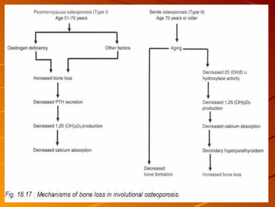
OsteoporosisOsteoporosisOsteoporosis is defined as a reduction in the strength of bone
that leads to an increased risk of fractures.
Normal homeostatic bone remodeling is altered – the rate of bone resorption is greater than the rate of bone formation.

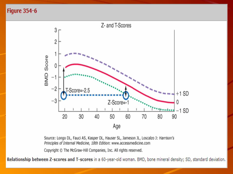
The World Health Organization (WHO) operationally defines osteoporosis as a bone density that falls 2.5 standard deviations (SD) below the mean for young healthy adults of the same sex—also referred to as a T-score of –2.5.


Measurement of Bone MassDual-energy x-ray absorptiometry (DXA), Single-energy x-ray absorptiometry (SXA), Quantitative CT, Ultrasound (US).

DXA Highly accurate x-ray technique
Standard for measuring bone density
Clinical determinations usually are made of the lumbar spine and hip.
Two x-ray energies are used to estimate the area of mineralized tissue, and the mineral content is divided by the area, which partially corrects for body size.
Cannot estimate the depth or posteroanterior length of the bone.
Small people tend to have lower than average bone mineral density (bmd).
Bone spurs, which are common in osteoarthritis, tend to falsely increase bone density of the spine.
Because DXA instrumentation is provided by several different manufacturers, the output varies in absolute terms.
Consequently, it has become standard practice to relate the results to "normal" values by using T-scores, which compare individual results to those in a young population that is matched for race and sex. Z-scores compare individual results to those of an age-matched population that also is matched for race and sex.


CT
Primarily to measure the spine and, more recently, the hip.
Peripheral CT is used to measure bone in the forearm or tibia.
Technique is three-dimensional and can provide a true density (mass of bone tissue per unit volume).
CT also can specifically analyze trabecular bone and cortical bone content and volume separately.
CT remains expensive, involves greater radiation exposure, and is less reproducible than DXA.
A new technique employing high-resolution CT scanning called Xtreme CT also can provide information on skeletal architecture, including cancellous connectivity

When to Measure Bone Mass

When to Treat Based on Bone Mass Results
BMD is >2.5 SD below the mean value for young adults (T-score ≤–2.5).
Postmenopausal women with fracture risk factors even if BMD is not in the osteoporosis range.
Risk factors (age, prior fracture, family history of hip fracture, low body weight, cigarette consumption, excessive alcohol use, steroid use, and rheumatoid arthritis) can be combined with BMD to assess the likelihood of a fracture over a 5- or 10-year period.

Elements of osteoporosis preventionElements of osteoporosis prevention

Treatment OsteoporosisHistory and physical examination should be performed to identify risk factors for osteoporosis.
A low Z-score increases the suspicion of a secondary disease.
Height loss >2.5–3.8 cm (>1–1.5 in.) is an indication for radiography or vertebral fracture assessment by DXA to rule out asymptomatic vertebral fractures, as is the presence of significant kyphosis or back pain, particularly if it began after menopause.
For patients who present with fractures, it is important to ensure that the fractures are not caused by an underlying malignancy.

OP fracturesOP fractures


Routine Laboratory EvaluationComplete blood countSerum and 24-h urine calciumRenal and hepatic function tests
serum calcium level hyperparathyroidism or malignancy serum calcium level malnutrition and osteomalacia.
Serum PTH level differentiates between hyperparathyroidism (PTH ) ↑and malignancy (PTH ), and a high PTHrP level can help document the ↓presence of humoral hypercalcemia of malignancy.
Urine calcium (<50 mg/24 h) suggests osteomalacia, malnutrition, or malabsorption; Urine calcium (>300 mg/24 h) is indicative of hypercalciuria Hypercalciuria in three situations: (1) a renal calcium leak (2) absorptive hypercalciuria, which can be idiopathic or associated with increased 1,25(OH)2D in granulomatous disease; or (3) hematologic malignancies or conditions associated with excessive bone turnover such as Paget's disease, hyperparathyroidism, and hyperthyroidism.

Measurement of serum 25(OH)D level.
Hyperthyroidism should be evaluated by measuring thyroid-stimulating hormone (TSH).
Urinary free cortisol levels or a fasting serum cortisol should be measured after overnight dexamethasone.
When bowel disease, malabsorption, or malnutrition is suspected, serum albumin, cholesterol, and a complete blood count should be checked.
Myeloma commonly presents with bone pain and characteristic "punched-out" lesions on radiography. Serum and urine electrophoresis and evaluation for light chains in urine are required to exclude this diagnosis.


Nutritional RecommendationsCalciumVitamin DSalt High animal protein intakes Vitamin K
Pharmacologic TherapiesEstrogensProgestinsSERMsBisphosphonatesCalcitoninDenosumabParathyroid HormoneFluoride


EstrogensFor oral estrogens,0.3 mg/d esterified estrogens, 0.625 mg/d for conjugated equine estrogens, 5 μg/d for ethinyl estradiol. For transdermal estrogen, 50 μg estradiol per day.
Hormone therapy reduces the risk of hip and clinical spine fracture by 34% and that of all clinical fractures by 24%.
The majority of estrogen (and androgen) effects on bone resorption are mediated indirectly through paracrine factors produced by osteoblasts.
These actions include (1) increasing IGF-I and TGF-β and (2) suppressing IL-1 (α and β), IL-6, TNF-α, and osteocalcin synthesis. The indirect estrogen actions primarily decrease bone resorption.
Combined estrogen-progestin treatment increased risk of fatal and nonfatal myocardial infarction by 29%,40% increase in stroke, a 100% increase in venous thromboembolic disease, and a 26% increase in risk of breast cancer.
SERMsTwo SERMs are used currently in postmenopausal women:Raloxifene for the prevention and treatment of osteoporosisTamoxifen for the prevention and treatment of breast cancer.
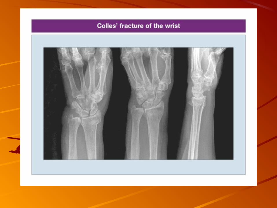
Tamoxifen reduces bone turnover and bone loss in postmenopausal women.Tamoxifen acts as an estrogenic agent in bone. Breast Cancer Prevention study indicated a possible reduction in clinical vertebral, hip, and Colles' fractures.
Raloxifene (60 mg/d) has effects on bone turnover and bone mass that are very similar to those of tamoxifen, indicating that this agent is also estrogenic on the skeleton.
The effect of raloxifene on bone density (+1.4–2.8% versus placebo in the spine, hip, and total body) is somewhat less than that seen with standard doses of estrogens.
Raloxifene reduces the occurrence of vertebral fracture by 30–50%, depending on the population; however, there are no data confirming that raloxifene can reduce the risk of nonvertebral fractures over 8 years of observation.

Bisphosphonates
Alendronate, risedronate, and ibandronate are approved for the prevention and treatment of postmenopausal osteoporosis. Risedronate and alendronate are approved for the treatment of steroid-induced osteoporosis. Risedronate also is approved for prevention of steroid-induced osteoporosis. Both alendronate and risedronate are approved for treatment of osteoporosis in men.Alendronate treatment (5 mg/d for 2 years and 10 mg/d for 9 months afterward) reduces vertebral fracture risk by about 50%, multiple vertebral fractures by up to 90%, and hip fractures by up to 50%.
Once-weekly therapy generally is preferred because of the low incidence of gastrointestinal side effects and ease of administration.
Alendronate should be given with a full glass of water before breakfast.
Patients remain upright for at least 30 min after taking the medication to avoid esophageal irritation.

Risedronate demonstrated 40–50% reduction in vertebral fracture risk over 3 years, accompanied by a 40% reduction in clinical nonspine fractures.
Etidronate was the first bisphosphonate to be approved, initially for use in Paget's disease and hypercalcemia. Its not approved by the FDA for treatment of osteoporosis.
Ibandronate doses of 150 mg/month PO or 3 mg every 3 months IV had greater effects on turnover and bone mass than did 2.5 mg/d.
Zoledronic acid (5 mg as a single IV infusion annually) reduced the risk of vertebral fractures by 70%, nonvertebral fractures by 25%, and hip fractures by 40%.
Bisphosphonates specifically impair osteoclast function and reduce osteoclast number, in part by inducing apoptosis.

Calcitonin
Calcitonin preparations are approved by the FDA for Paget's disease, hypercalcemia, and osteoporosis in women >5 years past menopause.
A nasal spray containing calcitonin (200 IU/d) is available for treatment of osteoporosis in postmenopausal women.
Calcitonin suppresses osteoclast activity by direct action on the osteoclast calcitonin receptor
Denosumab
A novel agent that was given twice yearly by SC administration in a randomized controlled trial in postmenopausal women with osteoporosis has been shown to increase BMD in the spine, hip, and forearm and reduce vertebral, hip, and nonvertebral fractures over a 3-year period by 70, 40, and 20%, respectively.
Denosumab was approved by the FDA in 2010 for the treatment of postmenopausal women who have a high risk for osteoporotic fractures, including those with a history of fracture or multiple risk factors for fracture, and those who have failed or are intolerant to other osteoporosis therapy.
Denosumab is a fully human monoclonal antibody to RANKL.
Denosumab binds to RANKL, inhibiting its ability to initiate formation of mature osteoclasts from osteoclast precursors and to bring mature osteoclasts to the bone surface and initiate bone resorption. Denosumab also plays a role in reducing the survival of the osteoclast.
Teriparatide
An exogenous PTH analogue (1-34hPTH; teriparatide) that has been approved for the treatment of established osteoporosis in both men and women.
The first randomized controlled trial in postmenopausal women showed that PTH, when superimposed on ongoing estrogen therapy, produced substantial increments in bone mass (13% over a 3-year period compared with estrogen alone) and reduced the risk of vertebral compression deformity.
20 μg PTH(1–34) daily by SC injection reduced vertebral fractures by 65% and nonvertebral fractures by 45%.
Treatment is administered as a single daily injection given for a maximum of 2 years.
Strontium Ranelate
Strontium ranelate is approved for the treatment of osteoporosis. It increases bone mass throughout the skeleton; in clinical trials, the drug reduced the risk of vertebral fractures by 37% and that of nonvertebral fractures by 14%.

OsteoporosisOsteoporosisSurgical intervention for vertebral Surgical intervention for vertebral fracturesfractures–VertebroplastyVertebroplasty
High pressure injection of bone High pressure injection of bone cement through pedicles to cement through pedicles to vertebral bodyvertebral bodyContraindicated in severe Contraindicated in severe vertebral body collapsevertebral body collapse

OsteoporosisOsteoporosisSurgical intervention for vertebral Surgical intervention for vertebral fracturesfractures–KyphoplastyKyphoplasty
Bone tamp through cortical Bone tamp through cortical window window Inflation of bladder in vertebral Inflation of bladder in vertebral bodybodyInjection of bone cement under Injection of bone cement under LOW PRESSURELOW PRESSURE

Rickets and Osteomalacia Rickets and Osteomalacia
Rickets and osteomalacia describe disorders in which there is failure of or Rickets and osteomalacia describe disorders in which there is failure of or defective mineralisation of newly formed organic matrix of the skeleton. defective mineralisation of newly formed organic matrix of the skeleton.
In rickets, the growing skeleton is involved and defective mineralisation In rickets, the growing skeleton is involved and defective mineralisation occurs not only in bone but also in the cartilaginous matrix of the growth occurs not only in bone but also in the cartilaginous matrix of the growth plate. plate.
Osteomalacia is the adult counterpart of rickets. Osteomalacia is the adult counterpart of rickets.
Deficiency of vitamin D, or ineffective formation or action of one of its active Deficiency of vitamin D, or ineffective formation or action of one of its active metabolites, leads to decreased concentrations of circulating calcium and metabolites, leads to decreased concentrations of circulating calcium and phosphate and to impaired mineralisation of bone.phosphate and to impaired mineralisation of bone.


Vitamin D deficiencyVitamin D deficiency
Deficiency of vitamin D is the commonest cause of rickets and osteomalacia in Deficiency of vitamin D is the commonest cause of rickets and osteomalacia in our country. our country.
Inadequate exposure to ultraviolet light is the critical factor in the causation Inadequate exposure to ultraviolet light is the critical factor in the causation of rickets and osteomalacia. of rickets and osteomalacia.
Dietary calcium deficiency alone does not cause rickets or osteomalacia. Dietary calcium deficiency alone does not cause rickets or osteomalacia.
Repeated pregnancies and lactation impose a considerable drain on the bone Repeated pregnancies and lactation impose a considerable drain on the bone mineral reserves and vitamin D stores of the mother, causing osteomalacia. mineral reserves and vitamin D stores of the mother, causing osteomalacia.
Age-related impaired intestinal absorption of calcium and deficient synthesis Age-related impaired intestinal absorption of calcium and deficient synthesis and supply of vitamin D precipitate osteomalacia in elderly women. and supply of vitamin D precipitate osteomalacia in elderly women.
Congenital (neonatal) rickets may occur in babies born to osteomalacic Congenital (neonatal) rickets may occur in babies born to osteomalacic mothers who had completely exhausted their bone mineral and vitamin D mothers who had completely exhausted their bone mineral and vitamin D reserves.reserves.

Muscular hypotoniaMuscular hypotonia
Skeletal deformities such as widening of the epiphyses at the Skeletal deformities such as widening of the epiphyses at the wrists and ankles and of the costochondral junctions wrists and ankles and of the costochondral junctions (rickety (rickety rosary) rosary) are characteristic of rickets.are characteristic of rickets.
Between 6 and 12 months, softening of the skull Between 6 and 12 months, softening of the skull (craniotabes, (craniotabes, frontal bossing)frontal bossing) and later deformities of the thorax and spine and later deformities of the thorax and spine (kyphosis or lordosis)(kyphosis or lordosis), , knock-kneesknock-knees, , bowing of legs bowing of legs and and waddling waddling gaitgait develop and result in develop and result in short stature short stature . .
TeethTeeth show irregular pits and grooves. Enamel hypoplasia and show irregular pits and grooves. Enamel hypoplasia and caries are frequent and severe in rachitic children. caries are frequent and severe in rachitic children.
The softened lower ribs may be pulled in at the site of attachment The softened lower ribs may be pulled in at the site of attachment of the diaphragm, forming of the diaphragm, forming Harrison’s grooveHarrison’s groove. .
The abdomen, due to hypotonia of the muscles, is markedly The abdomen, due to hypotonia of the muscles, is markedly protuberant protuberant (rachitic potbelly)(rachitic potbelly). .
Spontaneous carpopedal spasmsSpontaneous carpopedal spasms, laryngeal stridor and even , laryngeal stridor and even convulsions suggestive of convulsions suggestive of tetanytetany (positive Chvostek’s and (positive Chvostek’s and Trousseau’s signs) may occur if the calcium level drops below 6 Trousseau’s signs) may occur if the calcium level drops below 6 mg/dl.mg/dl.

Muscular weakness, bone pains and tenderness, backache, waddling Muscular weakness, bone pains and tenderness, backache, waddling gait, difficulty in walking or climbing, and deformities of the spine gait, difficulty in walking or climbing, and deformities of the spine and pelvis are the most common features of osteomalacia. and pelvis are the most common features of osteomalacia.
Inability to stand unaided or the tendency to prop on self while rising Inability to stand unaided or the tendency to prop on self while rising up from the floor is an important clue to the diagnosis of up from the floor is an important clue to the diagnosis of osteomalacia. osteomalacia.
Severe and long-standing cases may develop ankylosis and Severe and long-standing cases may develop ankylosis and generalised forward flexion at the spine generalised forward flexion at the spine

The most diagnostic biochemical feature of rickets and osteomalacia is The most diagnostic biochemical feature of rickets and osteomalacia is low low 25 (OH)D concentration in plasma 25 (OH)D concentration in plasma in the presence of secondary in the presence of secondary hyperparathyroidism. hyperparathyroidism.
Treatment of vitamin D-deficiency rickets consists of intake of Treatment of vitamin D-deficiency rickets consists of intake of 1-2 g of 1-2 g of elemental calcium per dayelemental calcium per day. This can be obtained by adding . This can be obtained by adding 1 litre milk1 litre milk to to the diet or 1 g as calcium supplements. the diet or 1 g as calcium supplements.
Vitamin D in a dose of Vitamin D in a dose of 60,000 IU (1.5 mg of D60,000 IU (1.5 mg of D33) ) is given is given orallyorally and and repeated at repeated at 2-42-4 week intervalsweek intervals. In mild cases 1 to 3 doses, in moderate . In mild cases 1 to 3 doses, in moderate cases 3 to 6 doses, and in severe cases 6 to 10 such doses may be cases 3 to 6 doses, and in severe cases 6 to 10 such doses may be necessary for raising the circulating concentration of 25 (OH)Dnecessary for raising the circulating concentration of 25 (OH)D 33 into the into the normal range and for complete healing of bone disease. normal range and for complete healing of bone disease.
Vitamin D administered to patients with secondary hyperparathyroidism is Vitamin D administered to patients with secondary hyperparathyroidism is quickly converted to 1,25 (OH)quickly converted to 1,25 (OH)22DD33. The . The response to treatment usually response to treatment usually appears after 2-3 weeks and bone lesions heal completely in 8 to 12 appears after 2-3 weeks and bone lesions heal completely in 8 to 12 weeks.weeks.
Vitamin D can be replaced by use of vitamin D metabolites such as 25 Vitamin D can be replaced by use of vitamin D metabolites such as 25 (OH)D(OH)D33 (calcifediol) (calcifediol) 50µg daily or 1,25 (OH)50µg daily or 1,25 (OH)22DD33 (calcitriol)(calcitriol) or its analogue or its analogue (1-aD) 0.25 to 0.5 µg daily for 4-8 weeks. (1-aD) 0.25 to 0.5 µg daily for 4-8 weeks.

Vitamin D-dependent rickets (Pseudo vitamin D Vitamin D-dependent rickets (Pseudo vitamin D deficiency)deficiency)
Type I Type I Autosomal recessive disorder.Autosomal recessive disorder.Deficiency of or presence of a biologically inactive form of renal Deficiency of or presence of a biologically inactive form of renal 25 (OH) D-la-25 (OH) D-la-hydroxylase. hydroxylase. Affected persons develop early onset (less than one year of age) vitamin D deficiency Affected persons develop early onset (less than one year of age) vitamin D deficiency (low serum 1,25 (OH)(low serum 1,25 (OH)22DD33). ).
The resultant rickets is unresponsive to usual doses of calciferol and 25-hydroxy D. The resultant rickets is unresponsive to usual doses of calciferol and 25-hydroxy D. Complete cure is obtained on continuous treatment with 1,25 (OH)Complete cure is obtained on continuous treatment with 1,25 (OH)22DD33 or high doses of or high doses of vitamin D. vitamin D. Rickets recurs if therapy is withdrawn.Rickets recurs if therapy is withdrawn.
Type II Type II Vitamin D-dependency rickets.Vitamin D-dependency rickets.Serum 1,25 (OH)Serum 1,25 (OH)22DD33 levels are normal. levels are normal.
There is deficiency in There is deficiency in the target tissue cytosol receptor the target tissue cytosol receptor for 1,25 (OH)for 1,25 (OH)22DD3.3.
10-25 µg of 1,25 (OH)10-25 µg of 1,25 (OH)22DD33 or 1-a-OHD or 1-a-OHD33 per day may be required for cure. per day may be required for cure.

Anticonvulsant - induced osteomalaciaAnticonvulsant - induced osteomalaciaAnticonvulsant drugs, especially phenobarbitone and phenytoin, induce liver Anticonvulsant drugs, especially phenobarbitone and phenytoin, induce liver enzymes, resulting in diminished 25 (OH)Denzymes, resulting in diminished 25 (OH)D33 that leads to rickets and that leads to rickets and osteomalacia. osteomalacia. Calcium supplements of 1-2 g per day and calciferol in doses varying from 0.1 Calcium supplements of 1-2 g per day and calciferol in doses varying from 0.1 to 1.0 mg correct and prevent the biochemical and radiographic abnormalities to 1.0 mg correct and prevent the biochemical and radiographic abnormalities of osteomalacia.of osteomalacia.
Renal tubular disordersRenal tubular disordersIncreased renal clearance of inorganic phosphorus and hypophosphataemia Increased renal clearance of inorganic phosphorus and hypophosphataemia with normal glomerular filtration rate. with normal glomerular filtration rate. X-linked familial hypophosphataemia (phosphate diabetes) is the commonest X-linked familial hypophosphataemia (phosphate diabetes) is the commonest form of form of hereditary vitamin D-resistant rickets (VDRR) hereditary vitamin D-resistant rickets (VDRR) and is characterised by and is characterised by X-linked dominant inheritance, progressively severe skeletal deformities and X-linked dominant inheritance, progressively severe skeletal deformities and dwarfism. dwarfism. The bone disease is more severe in males, and females often exhibit only The bone disease is more severe in males, and females often exhibit only hypophosphataemia. hypophosphataemia. In other patients, the tubular dysfunction may be part of Fanconi’s syndrome. In other patients, the tubular dysfunction may be part of Fanconi’s syndrome. Rickets may also occur in proximal renal tubular acidosis.Rickets may also occur in proximal renal tubular acidosis.Treated with large doses ofTreated with large doses of calciferol (2.5 to 10 mg per day) and oral calciferol (2.5 to 10 mg per day) and oral supplements of inorganic phosphate (1.4 g per day). supplements of inorganic phosphate (1.4 g per day). In renal tubular acidosis, In renal tubular acidosis, sodium bicarbonate (5 to 15 mEq/kg per day) reverses the acidosis, improves sodium bicarbonate (5 to 15 mEq/kg per day) reverses the acidosis, improves serum phosphate levels, heals the bone disease and with maintenance use of serum phosphate levels, heals the bone disease and with maintenance use of alkali, recurrence is prevented.alkali, recurrence is prevented.

In children with rickets, failure of cartilage calcification is radiologically manifested by frayed or irregularly widened and "cupped" ends of growing metaphyses at the wrists, knees and ankles Characteristic shaft deformities of the legs (genu valgum, genu varum, coxa vara) may be present. Secondary hyperparathyroidism usually manifests as coarse cystic trabeculations and irregular lacy subperiosteal erosions, especially about the metaphyses of long bones at the wrists, knees and ankles . Vitamin D deficiency rickets around puberty often shows a characteristic radiolucent band in the submetaphyseal region, especially at the wrists.

The pathognomonic radiological feature of osteomalacia is the presence of pseudofracture, a straight, transverse, ribbon-like band of rarefaction representing unmineralised bone. Pseudofractures, also known as Looser’s zones or Milkman’s fractures, are often bilateral and symmetrical (Fig. 18.7). Common sites are the concave side of the femoral necks and shafts of long bones, ischio-pubic rami, clavicles, ribs, scapulae, metacarpals and metatarsal bones. In severe cases, the vertebrae become biconcave and the spaces at the symphysis pubis and sacroiliac joints are widened. Narrowing of the pelvic cavity from inward pressure of the hips may lead to a triradiate pelvis

HyperparathyroidismHyperparathyroidismPrimary hyperparathyroidism:Primary hyperparathyroidism:– Often an incidental findingOften an incidental finding
– May be part of MEN I, MEN IIMay be part of MEN I, MEN II
Secondary hyperparathyroidismSecondary hyperparathyroidism– Compensates for chronic low Ca eg. Renal failure or Compensates for chronic low Ca eg. Renal failure or
malabsorptionmalabsorption
– [Ca[Ca2+2+] and [PO] and [PO442-2-] normal PTH high] normal PTH high
Tertiary hyperparathyroidismTertiary hyperparathyroidism– Hyperplasia in longstanding secondary diseaseHyperplasia in longstanding secondary disease

Solitary AdenomasA single abnormal gland is the cause in 80% of patients.∼In 15% of patients, all glands are hyperfunctioning; ∼ chief cell parathyroid hyperplasia is usually hereditary and frequently associated with other endocrine abnormalities.
Hereditary Syndromes and Multiple Parathyroid Tumors
MEN 1 (Wermer's syndrome) consists of hyperparathyroidism and tumors of the pituitary and pancreas, often associated with gastric hypersecretion and peptic ulcer disease (Zollinger-Ellison syndrome).
MEN 2A is characterized by pheochromocytoma and medullary carcinoma of the thyroid, as well as hyperparathyroidism.
MEN 2B has additional associated features such as multiple neuromas but usually lacks hyperparathyroidism.
Each of these MEN syndromes is transmitted in an apparent autosomal dominant manner.
The hyperparathyroidism jaw tumor (HPT-JT) syndrome occurs in families with parathyroid tumors (sometimes carcinomas) in association with benign jaw tumors.

One-half or more of patients with hyperparathyroidism are asymptomatic.
Manifestations of hyperparathyroidism involve primarily the kidneys and the skeletal system. Kidney involvement, due either to deposition of calcium in the renal parenchyma or to recurrent nephrolithiasis, was present in 60–70%. Nephrocalcinosis may also cause decreased renal function and phosphate retention.
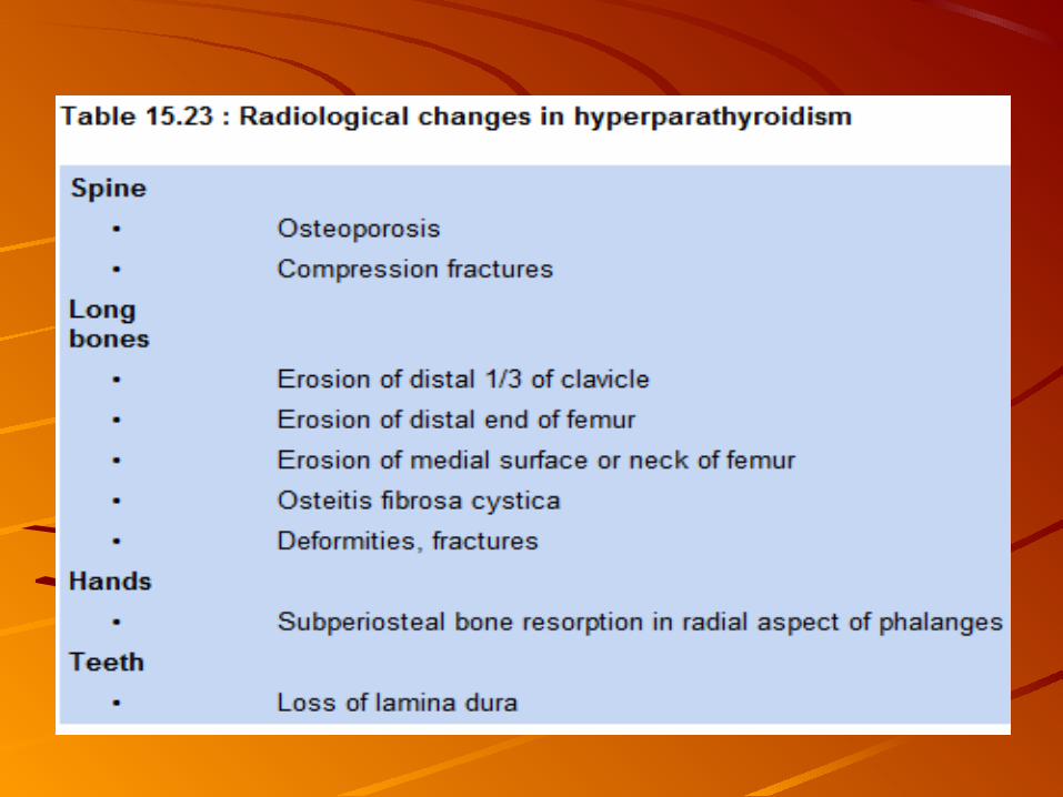
The distinctive bone manifestation of hyperparathyroidism is osteitis fibrosa cystica, which occurred in 10–25% of patients.Histologically, the pathognomonic features are an increase in the giant multinucleated osteoclasts in scalloped areas on the surface of the bone (Howship's lacunae) and a replacement of the normal cellular and marrow elements by fibrous tissue.
X-ray changes include resorption of the phalangeal tufts and replacement of the usually sharp cortical outline of the bone in the digits by an irregular outline (subperiosteal resorption). In recent years, osteitis fibrosa cystica is very rare in primary hyperparathyroidism, probably due to the earlier detection of the disease.


Renal OsteodystrophyRenal OsteodystrophyEffect on bone of disordered calcium Effect on bone of disordered calcium homeostasishomeostasisMay be osteomalacia, hyperparathyroidismMay be osteomalacia, hyperparathyroidismLeads toLeads to– Bone painBone pain– Skeletal deformitySkeletal deformity– Muscular weaknessMuscular weakness– Ectopic calcificationEctopic calcification– Growth retardationGrowth retardation


The clinical manifestations include bone pains and tenderness, fractures, growth The clinical manifestations include bone pains and tenderness, fractures, growth retardation, joint disease and abnormal calcification of cartilage. retardation, joint disease and abnormal calcification of cartilage.
Both osteomalacia and osteitis fibrosa cystica (OFC) may cause bone pain, Both osteomalacia and osteitis fibrosa cystica (OFC) may cause bone pain, tenderness and muscle weakness. tenderness and muscle weakness.
Pain in the lower limbs, pelvis and back is particularly common and may worsen Pain in the lower limbs, pelvis and back is particularly common and may worsen on exercise. on exercise.
Proximal muscle weakness is common. Fractures are uncommon with OFC, but Proximal muscle weakness is common. Fractures are uncommon with OFC, but are a feature of aluminium-induced bone diseaseare a feature of aluminium-induced bone disease
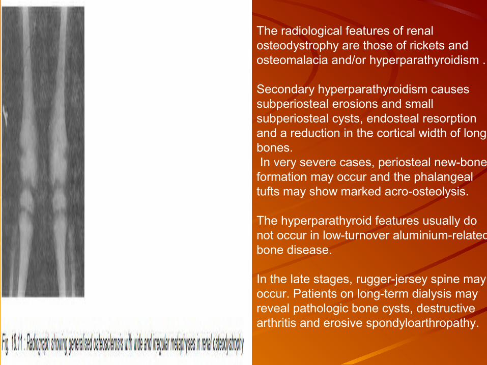
The radiological features of renal osteodystrophy are those of rickets and osteomalacia and/or hyperparathyroidism .
Secondary hyperparathyroidism causes subperiosteal erosions and small subperiosteal cysts, endosteal resorption and a reduction in the cortical width of long bones. In very severe cases, periosteal new-bone formation may occur and the phalangeal tufts may show marked acro-osteolysis.
The hyperparathyroid features usually do not occur in low-turnover aluminium-related bone disease.
In the late stages, rugger-jersey spine may occur. Patients on long-term dialysis may reveal pathologic bone cysts, destructive arthritis and erosive spondyloarthropathy.
Control of hyperphosphataemia and acidosis Control of hyperphosphataemia and acidosis : :
Dietary phosphate is restricted to 700-900 mg per day with protein restriction to 40 Dietary phosphate is restricted to 700-900 mg per day with protein restriction to 40 g/day and avoidance of dairy products.g/day and avoidance of dairy products.
Additionally, administration of calcium and aluminium compounds to bind intestinal Additionally, administration of calcium and aluminium compounds to bind intestinal phosphate is necessary. phosphate is necessary.
Aluminium compounds to bind phosphate in the intestine are almost always necessary; Aluminium compounds to bind phosphate in the intestine are almost always necessary; these include aluminium hydroxide either as tablet/powder or in gel form. These these include aluminium hydroxide either as tablet/powder or in gel form. These preparations may lead to aluminium toxicity. The use of an anion exchange resin may preparations may lead to aluminium toxicity. The use of an anion exchange resin may be a better alternative.be a better alternative.
Oral vitamin DOral vitamin D22 and D and D33 dose of 0.5 - 12 mg/day. dose of 0.5 - 12 mg/day.
Di hydro tachysterol (DHT) Di hydro tachysterol (DHT) is a synthetic compound which does not require prior renal is a synthetic compound which does not require prior renal 1-alpha hydroxylation for biological activity. A daily dose of 250-370 µg is sufficient to 1-alpha hydroxylation for biological activity. A daily dose of 250-370 µg is sufficient to cure osteitis fibrosa and osteomalacia in a majority of cases. cure osteitis fibrosa and osteomalacia in a majority of cases.
25(OH)D25(OH)D33 (dose 40-125 µg/day) (dose 40-125 µg/day) bypasses hepatic hydroxylation and is less likely to be bypasses hepatic hydroxylation and is less likely to be stored in body fat or muscles. stored in body fat or muscles.

Paget’s DiseasePaget’s DiseaseOsteitis DeformasOsteitis Deformas↑↑ Bone resorption Bone resorption →→ ↑↑ bone bone formation formation →→develop large irregularly shaped develop large irregularly shaped
bones with poor mineralization bones with poor mineralization →→ thick brittle bonesthick brittle bones
EtiologyEtiology–Slow progressing diseaseSlow progressing disease–Often occurs between 50-70 yearsOften occurs between 50-70 years–Familial tendency in malesFamilial tendency in males–Usually asymptomaticUsually asymptomatic

Paget’s DiseasePaget’s DiseaseClinical PresentationClinical Presentation– Deep aching sensation Deep aching sensation ↑↑ with weight with weight
bearingbearing– Pain - mild to severe unrelated to Pain - mild to severe unrelated to
activityactivity– May have bony deformities – skullMay have bony deformities – skull– Loss of heightLoss of height
Physical ExaminationPhysical Examination– Kyphosis / Bowing of long bonesKyphosis / Bowing of long bones– Conductive hearing lossConductive hearing loss– Fracture healing is impairedFracture healing is impaired– Complications – CHF / Paget’s Complications – CHF / Paget’s
sarcomasarcoma

Paget’s DiseasePaget’s DiseaseDiagnosticsDiagnostics– Serum alkaline phosphatase -Serum alkaline phosphatase -↑↑– Urinary hydroxyproline - Urinary hydroxyproline - ↑↑– Serum/ urinary citrate – Serum/ urinary citrate – ↑↑– Serum uric acid – Serum uric acid – ↑↑in < 50%in < 50%– X-RaysX-Rays
Early localized demineralizationEarly localized demineralizationLater bony overgrowth – irregularLater bony overgrowth – irregularMosaic patternMosaic pattern
– Bone scanBone scanMetabolic activity- Metabolic activity- ↑↑
Paget’s DiseasePaget’s DiseaseClinical ManagementClinical Management– AsymptomaticAsymptomatic
Monitor patientMonitor patient
– SymptomaticSymptomaticNSAIDsNSAIDsCalcitonin – relieve bone painCalcitonin – relieve bone painBisphosphonatesBisphosphonates
– Ambulation with assistive devicesAmbulation with assistive devices
Surgical InterventionSurgical Intervention– Correction of malalignment / Correction of malalignment /
fracturesfractures


OSTEOGENESIS IMPERFECTAOSTEOGENESIS IMPERFECTA
Osteogenesis imperfecta is a heritable disorder of connective Osteogenesis imperfecta is a heritable disorder of connective tissue due to defect in tissue due to defect in synthesis of type I collagen synthesis of type I collagen interfering interfering with bone function.with bone function.
The usual clinical features include multiple fractures, blue The usual clinical features include multiple fractures, blue sclerae, deafness, high-pitched voice, deformities of long sclerae, deafness, high-pitched voice, deformities of long bones, joint hyperextensibility, pectus deformity, dental bones, joint hyperextensibility, pectus deformity, dental abnormalities, thin skin and generally normal intelligence. abnormalities, thin skin and generally normal intelligence. Many types have been identified; the congenital (newborn Many types have been identified; the congenital (newborn type) is the most severe. type) is the most severe.
Routine biochemical investigations are typically normal; Routine biochemical investigations are typically normal; elevations in serum alkaline phosphatase and urine elevations in serum alkaline phosphatase and urine hydroxyproline levels occur with fractures.hydroxyproline levels occur with fractures.

Characteristic radiological findings include generalised osteopenia, deformities of long bones from recurrent fractures, collapsed vertebrae, defective periosteal bone formation, thin cortices and disordered maturation of growing plates (popcorn calcification).

OSTEOPETROSISOSTEOPETROSIS
Osteopetrosis is rare developmental disorder of bone characterised by Osteopetrosis is rare developmental disorder of bone characterised by osteosclerosis and fractures. It results from insufficient resorption of osteosclerosis and fractures. It results from insufficient resorption of primary bone trabeculae. primary bone trabeculae.
Two distinct forms of the disease:Two distinct forms of the disease:
Malignant osteopetrosisMalignant osteopetrosis, of autosomal recessive inheritance, , of autosomal recessive inheritance, presenting at birth or early infancy, is severe and death due to presenting at birth or early infancy, is severe and death due to pancytopenia or obliteration of marrow cavity occurs in the first pancytopenia or obliteration of marrow cavity occurs in the first decade of life. decade of life.
Anaemia, hepatosplenomegaly, lymph node enlargement, cranial Anaemia, hepatosplenomegaly, lymph node enlargement, cranial nerve palsies, hydrocephalus and death due to infection or nerve palsies, hydrocephalus and death due to infection or haemorrhage are common. haemorrhage are common.
Flask-shaped swelling of bone ends, particularly around long bones, Flask-shaped swelling of bone ends, particularly around long bones, and fractures may cause angulation and deformity. and fractures may cause angulation and deformity.
Mild form, is inherited as autosomal dominant, and is variable in age Mild form, is inherited as autosomal dominant, and is variable in age of onset and severity. This is often discovered incidentally.of onset and severity. This is often discovered incidentally.

Plasma biochemistry is normal except for rise in Plasma biochemistry is normal except for rise in alkaline phosphatase with fractures. Secondary alkaline phosphatase with fractures. Secondary hyperparathyroidism with elevated PTH and calcitriol hyperparathyroidism with elevated PTH and calcitriol levels is often seen. Acid phosphatase is often levels is often seen. Acid phosphatase is often increased. In severe cases there is an excessive increased. In severe cases there is an excessive positive calcium balance.positive calcium balance.
Radiologically, long bones are uniformly dense . Radiologically, long bones are uniformly dense . Within the bone, particularly the spine and long Within the bone, particularly the spine and long bones, sclerotic foci are seen. Around this there is a bones, sclerotic foci are seen. Around this there is a more lucent region and peripheral to this is more more lucent region and peripheral to this is more sclerotic bone (bone within bone appearance).sclerotic bone (bone within bone appearance).
CARBONIC ANHYDRASE II DEFICIENCYCARBONIC ANHYDRASE II DEFICIENCY
Carbonic anhydrase II deficiency is characterised by osteopetrosis, Carbonic anhydrase II deficiency is characterised by osteopetrosis, renal tubular acidosis and cerebral calcification.renal tubular acidosis and cerebral calcification.
Failure to thrive, developmental delay, short stature, frequent Failure to thrive, developmental delay, short stature, frequent fractures, impaired vision from compression atrophy of optic nerve, fractures, impaired vision from compression atrophy of optic nerve, and dental malocclusion are frequent problems. and dental malocclusion are frequent problems.
There may be hypotonia, apathy and muscle weakness. Life There may be hypotonia, apathy and muscle weakness. Life expectancy appears to be normal.expectancy appears to be normal.
The radiologic findings are similar to those in osteopetrosis. The radiologic findings are similar to those in osteopetrosis.
Cerebral calcification appears around 2-5 years of age and Cerebral calcification appears around 2-5 years of age and complicates complicates onlyonly this type of osteopetrosis. Metabolic acidosis this type of osteopetrosis. Metabolic acidosis (proximal or distal renal tubular acidosis) may be present.(proximal or distal renal tubular acidosis) may be present.
The impact of correcting acidosis is not known since treatment could The impact of correcting acidosis is not known since treatment could conceivably block the protective effect of acidosis on bone density.conceivably block the protective effect of acidosis on bone density.
Bone marrow transplantation is under evaluationBone marrow transplantation is under evaluation

HYPOPHOSPHATASIAHYPOPHOSPHATASIA
Hypophosphatasia is an inherited metabolic bone disease (rickets and Hypophosphatasia is an inherited metabolic bone disease (rickets and osteomalacia) characterised by generalised decrease in tissue nonspecific osteomalacia) characterised by generalised decrease in tissue nonspecific (liver, bone, kidney) alkaline phosphatase (ALP); placental and intestinal (liver, bone, kidney) alkaline phosphatase (ALP); placental and intestinal ALP activity is normal. ALP activity is normal.
Hypophosphatasia affects importantly only the skeleton and dentition. Hypophosphatasia affects importantly only the skeleton and dentition.
Four clinical types - perinatal (lethal), infantile, childhood and adult - are Four clinical types - perinatal (lethal), infantile, childhood and adult - are known. known.
Rickets occurs in the infantile and childhood forms. Rickets occurs in the infantile and childhood forms.
In adults the disorder may be recognised in middle age. Osteomalacia and In adults the disorder may be recognised in middle age. Osteomalacia and calcium pyrophosphate disease may present in the adult form.calcium pyrophosphate disease may present in the adult form.
In the infantile form skeletal radiographs may reveal progressive skeletal In the infantile form skeletal radiographs may reveal progressive skeletal demineralisation and rickets. Radiolucent tongues may extend from demineralisation and rickets. Radiolucent tongues may extend from growth plate cartilage into the metaphyses in the childhood form, and growth plate cartilage into the metaphyses in the childhood form, and osteomalacia with pseudofractures may be present in adults.osteomalacia with pseudofractures may be present in adults.

Fibrodysplasia Ossificans Progressiva
Also called myositis ossificans progressiva;
Rare autosomal-dominant disorder.
Congenital deformities of the hands and feet and episodic soft tissue swellings that ossify.
Ectopic bone formation occurs in fascia, tendons, ligaments, and connective tissue within voluntary muscles. Tender, rubbery induration, sometimes precipitated by trauma, develops in the soft tissue and gradually calcifies.
Heterotopic bone forms at these sites of soft tissue trauma.
Mortality is usually related to restrictive lung disease caused by an inability of the chest to expand. Laboratory tests are unremarkable.
Bisphosphonates, glucocorticoids, and a low-calcium diet have largely been ineffective in halting progression of the ossification.
Surgical removal of ectopic bone is not recommended.