Metabolic Abnormalities in Patients with Chronic...
28
Metabolic Abnormalities in Patients with Chronic Candidiasis The Acetaldehyde Hypothesis C. Orian Truss, M.D. 1 Introduction Mold sensitivity is common among allergy patients, occurring alone or in association with reactions to foods and to other inhalants, such as house dust, animal danders, and the pollens of weeds, grasses, and trees. Symptoms of allergy result from an altered reaction to substances encountered in the environment. Contact with food is primarily intestinal, while that with pollen and house dust is principally respiratory. Mold exposure, however, is abundant by both routes. Mold spores are prevalent in inhaled air, especially in damp weather and in environments that are "musty". Patients commonly develop symptoms from hay, leaves, basement air, feather pillows, and house plants. In addition to these inhaled spores, much exposure occurs in the intestine from molds that are in or on foods; fruits, aged cheeses, breads and pastries, and vinegar and alcoholic beverages are prominent among the foods that have a high yeast content. The most effective treatment of allergies is avoidance. When contact with the allergen can be minimized to a sufficient degree, other treatment is unnecessary. Allergies that may be completely controlled in this way include those to foods, drugs, animal dander, and poison ivy. When complete avoidance is not possible, minimizing exposure to the allergen aids greatly in controlling symptoms; better house dust control is an example. Mold avoidance includes the elimination of house plants, wet basements and other sources of excess humidity, and foods with a high yeast content. This is an essential part of treatment. In fact, without this, results from the remainder of the treatment program often will be disappointing. When contact cannot be reduced sufficiently to adequately control symptoms, standard allergy treatment includes the injection of extracts of the allergen, usually over an extended period. Often called "hyposensitization," this may effectively control symptoms that remain despite maximum avoidance. Such extracts are used in treating pollen, house dust, and mold allergies, since contact is inevitable with these allergens that are in the air we must breathe, and often only minimal benefit is realized from the use of antihistamines. Some think that this ther- apy relieves the symptoms of allergy by 1. 2614 Highland Ave., Birmingham, Alabama 35205 Presented at the Yeast-Human Interaction Conference, December 10,1983, Birmingham, Alabama. 66
-
Upload
nguyennhan -
Category
Documents
-
view
215 -
download
0
Transcript of Metabolic Abnormalities in Patients with Chronic...

Metabolic Abnormalities in Patients with Chronic Candidiasis The Acetaldehyde Hypothesis
C. Orian Truss, M.D.1
Introduction Mold sensitivity is common among allergy
patients, occurring alone or in association with reactions to foods and to other inhalants, such as house dust, animal danders, and the pollens of weeds, grasses, and trees.
Symptoms of allergy result from an altered reaction to substances encountered in the environment. Contact with food is primarily intestinal, while that with pollen and house dust is principally respiratory.
Mold exposure, however, is abundant by both routes. Mold spores are prevalent in inhaled air, especially in damp weather and in environments that are "musty". Patients commonly develop symptoms from hay, leaves, basement air, feather pillows, and house plants. In addition to these inhaled spores, much exposure occurs in the intestine from molds that are in or on foods; fruits, aged cheeses, breads and pastries, and vinegar and alcoholic beverages are prominent among the foods that have a high yeast content.
The most effective treatment of allergies is avoidance. When contact with the allergen can be minimized to a sufficient degree, other treatment is unnecessary. Allergies that may be completely controlled in this way include those to foods, drugs, animal dander, and poison ivy.
When complete avoidance is not possible, minimizing exposure to the allergen aids greatly in controlling symptoms; better house dust control is an example.
Mold avoidance includes the elimination of house plants, wet basements and other sources of excess humidity, and foods with a high yeast content. This is an essential part of treatment. In fact, without this, results from the remainder of the treatment program often will be disappointing.
When contact cannot be reduced sufficiently to adequately control symptoms, standard allergy treatment includes the injection of extracts of the allergen, usually over an extended period. Often called "hyposensitization," this may effectively control symptoms that remain despite maximum avoidance. Such extracts are used in treating pollen, house dust, and mold allergies, since contact is inevitable with these allergens that are in the air we must breathe, and often only minimal benefit is realized from the use of antihistamines. Some think that this ther-apy relieves the symptoms of allergy by
1. 2614 Highland Ave., Birmingham, Alabama 35205 Presented at the Yeast-Human Interaction Conference, December 10,1983, Birmingham, Alabama.
66

METABOLIC ABNORMALITIES
stimulating the formation of IgG or "blocking" antibodies, or perhaps by an effect on suppressor T-cells.
Thus, to summarize, total avoidance of the allergen is the perfect and only treatment necessary, as for food allergies. When this is not possible, as with allergens in the air, immunotherapy with extracts of the allergens is used to alleviate symptoms. The principle of trying to control immunologically those symptoms that cannot be eliminated by avoidance is common to the methods of most allergists, although there may be differences in their methods of administering the extracts.
The purpose of this brief discussion has been to consider the principles that apply to allergy treatment in general, so that we might point out a third element that is uniquely characteristic of mold sensitivity.
In addition to mold allergens that are derived from air and food, others are produced by fungi living on the surface-coverings of the human body. Skin and mucous membranes normally are colonized by living organisms that are the source of antigens to which we may become allergic. When these organisms are fungi, mold allergens actively produced in the body are added to the mold in food and in inhaled air, and quantitatively may be of great importance in the total mold load.
Since the amount of fungus colonizing tissues will have much to do with its contri-bution to the total mold load, an important addition to treatment will be made by measures that reduce the growth of mold in the mucous membranes and skin.
The "id" reaction to trichophyton was perhaps the first reported example of an allergic response distant from the site of growth of the fungus producing the allergen. However, of the various fungi living in the human body, Candida albicans is quantitatively the most important, since it colonizes the entire intestinal tract and vagina, lives on skin, and may infect nails. Patients with mold sensitivity often describe great difficulty in controlling "athlete's foot," as well as fungous infections of the nails, vagina, and intestines. Frequently these cease to be a problem with successful therapy of the patients' mold sensitivity. Allergenic extracts of trichophyton, Candida albicans, aspergillus, and
other fungi that so colonize the human body are used widely in the treatment of mold sensitivity. They are used singly or as part of mixtures of mold allergens. One particular combination used widely is TOE, containing extracts of Trichophyton, Candida albicans, and Epidermophyton.
A second reason that Candida albicans has become of great importance in this clinical problem is that its growth is strongly stimulated by antibiotics, contraceptive hormones, and immunosuppressant drugs. For the past 35 years the increasing use of these substances in medical practice has adversely affected the growth of this yeast in many individuals. It has become a major factor in mold sensitivity, and indeed, in many cases may eventually become the predominant problem.
In the overall treatment program for mold sensitivity, measures of avoidance should include an attempt to minimize the impact of these factors that increase the growth of this yeast in the human body. This includes greater care in determining the indication for the use of antibiotics and steroid hormones, and the selection of alternate methods of birth control in patients with mold sensitivity.
Yeast growth may also be suppressed by the use of standard anti-fungal medication. Several are available, nystatin being the most widely used. It has been used for over 30 years to treat "thrush" in infants, vaginitis in women and girls, and intestinal symptoms due to yeast in both sexes and at all ages. Finally, we may add standard Candida albicans extract to those of the other molds used in immunotherapy.
Although Candida albicans cannot be avoided completely, its contribution to the total mold exposure can be minimized by thus applying to it the principles of avoidance and immunotherapy common to the treatment of other allergies.
Treatment of the chronic yeast infection, besides contributing to the overall control of mold sensitivity, is important in itself. This infection may become so severe that it dominates the clinical picture. The original symptoms of inhalant mold allergy may have become less important than those of the "chronic candidiasis" that has evolved as a result of the yeast susceptibility that is part of
67

JOURNAL OF ORTHOMOLECULAR PSYCHIATRY, VOLUME 13, NUMBER 2
the problem of mold sensitivity. The contribution of this fungus to the total
clinical picture in patients susceptible to its overgrowth led to the metabolic studies presented in this paper. Clinical observations on this subject were first presented in 1977 (Truss, 1977). Three subsequent papers (Truss, 1978; 1980; 1981) and a book (Truss, 1983) about illness caused by this yeast described its manifestations, the factors that precipitate and aggravate it, and its treatment.
It was stressed throughout that, because of its universal presence in the human body, commonly used diagnostic techniques reveal little about this condition, and that the diagnosis must be suspected from the clinical picture and confirmed by the response to treatment of its various manifestations.
Because of the desirability of establishing a laboratory basis for the diagnosis of this condition, studies were initiated in 24 patients considered classic cases of mold sensitivity and chronic yeast susceptibility. Patients with this condition usually exhibit symptoms that are severe and of long duration. It seemed likely that such symptoms reflect underlying disturbances in basic metabolic pathways. If so, laboratory evidence of such disturbances would aid both in the diagnosis of this condition, and in the assessment of its status during treatment.
The design of this study was to carry out a general evaluation of protein, fat, and carbo-hydrate metabolism, with the addition of more specific studies later if indicated by abnormalities brought out in the study's initial phase. Accordingly, pre-treatment determinations included amino acids in the urine, fatty acids in plasma and erythrocyte membranes, and intermediates of the glycolytic pathway.
In addition, special studies were incorporated to test the hypothesis that acetaldehyde, produced in the intestine by the anaerobic fermentation of sugars by Candida albicans, is the principal mediator of metabolic distur-bances surfacing clinically as the symptoms of this condition.
Reported in this paper are the results of measurements before treatment in 24 patients, together with results in seven of the 24 who were restudied after varying periods of treatment, and following substantial clinical
improvement. The toxicity of acetaldehyde is reviewed, and the hypothesis presented that this substance is the major toxin responsible for the metabolic abnormalities found in these patients. The concluding discussion considers the laboratory findings in these patients in terms of acetaldehyde toxicity, and presents facts pertinent to the concept that fermentation of sugars by Candida albicans underlies the physiologic disturbances manifested by the many symptoms characteristic of this condition.
The Acetaldehyde Hypothesis
Patients with an overgrowth of yeast in the intestine exhibit much gas formation following the ingestion of sugar or digestible carbo-hydrates. This is evidenced clinically by abdominal distention, excess flatus, and belching. The metabolism of sugar by yeasts releases carbon dioxide (C02), whether this be under anaerobic (fermentation) or aerobic conditions. Formation of Acetaldehyde in Vitro
In yeasts, pyruvate is produced from sugar by glycolysis, just as in human cells. If oxygen is not available, many species of yeast de-carboxylate the pyruvate to acetaldehyde, which is then reduced to ethanol. If oxygen is present, pyruvate is oxidatively decarboxy-lated to acetyl Co-enzyme A (acetyl CoA), and acetaldehyde and ethanol are not formed. Therefore, in many types of yeast the fate of pyruvate (and therefore of sugar) is determined primarily by the availability, or lack thereof, of oxygen. Most strains of Candida albicans, however, are not able to convert acetaldehyde to ethanol to any significant degree, but can under anaerobic conditions convert pyruvate to acetaldehyde. We will return later to the potential significance of this in the human body.
Yeasts also are able to metabolize ethanol to acetaldehyde, if oxygen is available. The ethanol may be supplied exogenously, or produced by the yeast itself. Thus ethanol that has resulted from anaerobic fermentation will be quickly oxidized to acetaldehyde as soon as oxygen is available.
Candida albicans is one species of yeast that is able to accomplish in vitro this conversion of ethanol to acetaldehyde when oxygen is available. (If it can do so also in the
68
METABOLIC ABNORMALITIES
intestine, this pathway would provide an additional source of acetaldehyde, since small amounts of ethanol are produced normally by intestinal bacteria.)
Thus there are two pathways for the generation of acetaldehyde by Candida albi-cans: (1) the conversion of pyruvate to acetaldehyde when oxygen is not available and (2) the conversion of ethanol (exogenous or endogenous) to acetaldehyde when oxygen is available.
It is possible that most strains of Candida albicans produce ethanol in the human in-testine, but in an amount too small to be detectable in the peripheral blood. That the intestinal environment is sufficiently anaerobic for this to occur is indicated by the fact that, if adequate sugar is available, certain strains of Candida albicans can produce ethanol in quantities sufficient to induce a rising blood-alcohol level, and clinical drunkenness (Iwata, 1972). Thus such strains can efficiently accomplish the final step in ethanol production, i.e., the conversion of acetaldehyde to ethyl alcohol. Therefore, these special strains of Candida albicans are capable of accomplishing in the human intestine the two reactions by which pyruvate is anaerobi-cally converted to ethanol: (1) Pyruvate+H+ --------► acetaldehyde+CO2 (2) Acetaldehyde+NADH+H+ -----------►
ethanol + NAD+ Common strains of Candida albicans,
although largely incapable of accomplishing reaction (2), are able under anaerobic condi-tions in vitro to convert pyruvate to acetalde-hyde and C02. The production of ethanol is minimal or absent (Van Niel and Anderson, 1941; Van Niel and Cohen, 1942).
If the same reactions prevail in vivo, the large amount of gas generated in the intestinal tract when patients with yeast overgrowth eat carbohydrate would be the C02 liberated by the decarboxylation of pyruvate, as shown in equation (1). Acetaldehyde would be the remaining product.
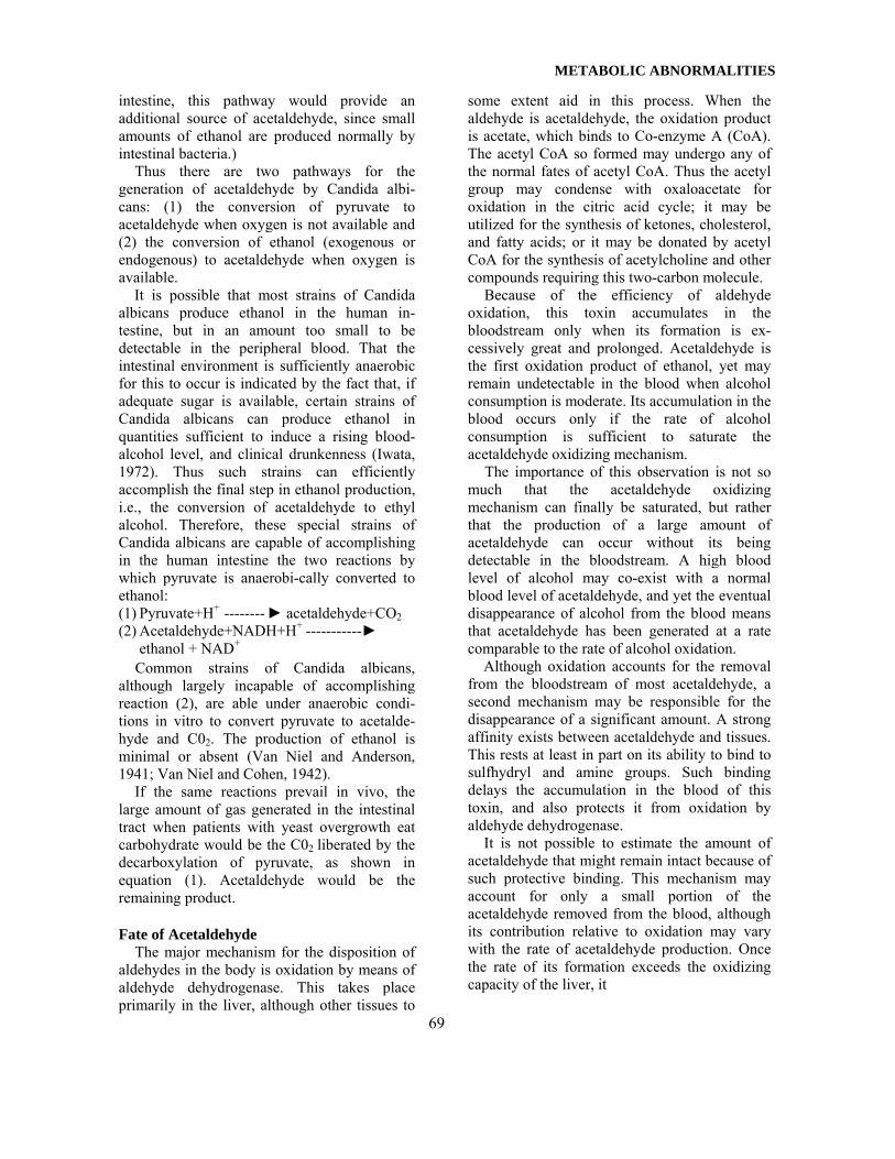
Fate of Acetaldehyde The major mechanism for the disposition of
aldehydes in the body is oxidation by means of aldehyde dehydrogenase. This takes place primarily in the liver, although other tissues to
some extent aid in this process. When the aldehyde is acetaldehyde, the oxidation product is acetate, which binds to Co-enzyme A (CoA). The acetyl CoA so formed may undergo any of the normal fates of acetyl CoA. Thus the acetyl group may condense with oxaloacetate for oxidation in the citric acid cycle; it may be utilized for the synthesis of ketones, cholesterol, and fatty acids; or it may be donated by acetyl CoA for the synthesis of acetylcholine and other compounds requiring this two-carbon molecule.
Because of the efficiency of aldehyde oxidation, this toxin accumulates in the bloodstream only when its formation is ex-cessively great and prolonged. Acetaldehyde is the first oxidation product of ethanol, yet may remain undetectable in the blood when alcohol consumption is moderate. Its accumulation in the blood occurs only if the rate of alcohol consumption is sufficient to saturate the acetaldehyde oxidizing mechanism.
The importance of this observation is not so much that the acetaldehyde oxidizing mechanism can finally be saturated, but rather that the production of a large amount of acetaldehyde can occur without its being detectable in the bloodstream. A high blood level of alcohol may co-exist with a normal blood level of acetaldehyde, and yet the eventual disappearance of alcohol from the blood means that acetaldehyde has been generated at a rate comparable to the rate of alcohol oxidation.
Although oxidation accounts for the removal from the bloodstream of most acetaldehyde, a second mechanism may be responsible for the disappearance of a significant amount. A strong affinity exists between acetaldehyde and tissues. This rests at least in part on its ability to bind to sulfhydryl and amine groups. Such binding delays the accumulation in the blood of this toxin, and also protects it from oxidation by aldehyde dehydrogenase.
It is not possible to estimate the amount of acetaldehyde that might remain intact because of such protective binding. This mechanism may account for only a small portion of the acetaldehyde removed from the blood, although its contribution relative to oxidation may vary with the rate of acetaldehyde production. Once the rate of its formation exceeds the oxidizing capacity of the liver, it
69

JOURNAL OF ORTHOMOLECULAR PSYCHIATRY, VOLUME 13, NUMBER 2
will appear in the peripheral blood, making possible its binding to tissues throughout the body. It seems likely, also, that its binding to amine and sulfhydryl groups will be cumulative when it is generated chronically, even though in small amount. Formation of Acetaldehyde in Vivo by Candida Albicans
If, as proposed in this hypothesis, Candida albicans is capable of fermenting sugars to acetaldehyde in the human intestine, the amount formed should be influenced by the following: 1. The availability of sugar for conversion into pyruvate by glycolysis. 2. The degree of anaerobiasis. 3. The quantity of viable yeast cells in the intestine, and their fermenting efficiency. 4. Other intestinal environmental factors that might be capable of influencing glycolysis or fermentation by Candida albicans, e.g., pH, bacterial flora. Fate of Acetaldehyde Formed in Vivo by Candida Albicans
Acetaldehyde formed in this way would enter the portal circulation and, in the liver, undergo oxidization to acetate. It is unlikely that a quantity of acetaldehyde sufficient to saturate liver aldehyde dehydrogenase would be generated by most strains of Candida albicans. (That it could occur, however, is suggested by experience with the ethanol-producing strains.) Therefore, it would be unlikely that acetaldehyde formation in the intestine could be detected by measurement of its level in the peripheral blood.
Thus, in this situation, opportunity for acetaldehyde to bind to sulfhydryl and amine groups would be limited to the time between its formation in the intestine and its oxidation in the liver. Although brief, this interval would allow contact with enzymes, and with constituents of the intestinal wall, liver cells, and portal blood. The latter would include nutrients as well as serum proteins, the latter having been found to bind acetaldehyde. Also exposed to acetaldehyde in the portal blood would be platelets, leukocytes, and erythrocytes. The release of acetaldehyde from erythrocytes was reported in 1941 (Barker, 1941).
Therefore, the presence of acetaldehyde in the intestine, the intestinal wall, and in the
portal blood would afford many opportunities for its binding to such substances as nutrients, enzymes, vitamins, and polypeptides. If formed high in the intestinal tract, acetaldehyde could react with digestive enzymes in the small intestine. The possiblities are many.
Although the time available for binding is brief, presumably the same erythrocytes, leukocytes, and serum proteins come around repeatedly, each passage through the portal vessels exposing them to the cumulative effect of newly formed acetaldehyde, just as would occur in the fixed cells of the intestinal wall and liver. The ultimate effect of this very toxic substance could include disruption of intestinal absorptive processes, as well as impairment of function in erythrocytes, leukocytes, and other cells in which it accumulates.
As we will see in the next section, the known toxic effects of acetaldehyde are more than adequate to account for the metabolic disturbances found in the patients who were the subjects of this study. We know that common strains of Candida albicans produce acetaldehyde when they ferment sugars anaerobically in vitro. That they can do so in the human body remains to be demonstrated.
Toxicity of Acetaldehyde Because acetaldehyde is produced by the intial
step in the oxidation of ethyl alcohol, its toxic effects have received much attention in studies of alcohol addiction and toxicity. Many of the metabolic disturbances caused by acetaldehyde have been attributed to its strong affinity for sulfhydryl (-SH) and amine groups, and to the increase in the ratio of NADH to NAD that results from the oxidation of acetaldehyde by aldehyde dehydrogenase. This discussion will focus on the physiologic consequences of such acetaldehyde binding and oxidation. Schiff Base Formation
The condensation of an aldehyde and an amine results in the formation of a Schiff base. An example of Schiff base formation in normal metabolism is the reaction of pyri-doxal phosphate (PLP) with the amine group of an amino acid, as this co-enzyme participates in transamination reactions. PLP is normally protected from degradation
70

METABOLIC ABNORMALITIES
by its binding to the amine group of lysine residues of proteins, including serum proteins and hemoglobin. Acetaldehyde may, by binding preferentially to these residues, displace PLP and result in its increased destruction, and in abnormally low blood levels of this co-enzyme (Lumeng and Ting-Kai Li, 1974; Veitch et al., 1975).
Acetaldehyde may also bind to the amine group of neurotransmitters to form complex compounds that may function as "false neurotransmitters" (Cohen and Collins, 1970; Davis and Walsh, 1970). These have been demonstrated in minute amount in human urine (Sandler et al., 1973). It has been proposed that these substances are related in some way to alcohol addiction. Little is known about their effect on normal physiology, but it might be anticipated that it would depend upon the particular neurotransmitter involved in the condensation with acetaldehyde, just as does the name of the condensation product. For example, the tetrahydroisoquinoline that results from the condensation of dopamine and acetaldehyde is salsolinol. Condensation of acetaldehyde with indolealkylamines leads to the formation of tetrahydro-B-carbolines.
Other opportunities for Schiff-base formation undoubtedly exist where amine groups are available to participate in this reaction with acetaldehyde. The examples cited illustrate two areas of physiology that could be profoundly affected by this mechanism. Sulfhydryl Binding
Aldehydes react with sulfhydryl groups to form hemiacetals. The -SH group is active in many metabolic steps. For example, it is involved in the first oxidative step of glycolysis, being essential to the activity of glycer-aldehyde dehydrogenase, the enzyme that catalyzes the conversion of glyceraldehyde 3-phosphate to 1,3 diphosphoglycerate. The sulfhydryl group is also the active site of Coenzyme A (CoA). It is the -SH site of CoA that binds the acetyl group to form acetyl CoA.
CoA serves as carrier of the acetyl groups that are derived from carbohydrates, fats, and certain amino acids. Thus acetyl CoA is the point of convergence in the metabolism of the three classes of food, for their subsequent entry into
and further oxidation by the citric acid cycle. Acetyl CoA also provides the two carbon units from which are synthesized fatty acids, ketones, and cholesterol and steroid hormones, and it donates the acetyl group required for the synthesis of acetylcholine and other substances.
There is possibly no single point in metabolic pathways more pivotal than acetyl CoA, nor one at which interference would have more far-reaching consequences in all organs of the body.
Ammon has shown in both brain and liver homogenates that acetaldehyde induces a dose-dependent suppression of acetyl CoA activity (Ammon et al., 1969; Ammon et al., 1971). He further showed that this was due to blocking of the active -SH group of CoA, which he believed to be due to the binding of acetaldehyde at this site. Finally, he demonstrated decreased respiration in these tissues, oxygen consumption falling in parallel with the declining level of acetyl CoA as the concentration of acetaldehyde increased.
Glyceraldehyde dehydrogenase and acetyl CoA are two examples of sites at which the integrity of -SH groups is essential in normal metabolic pathways. As is true for amine groups and Schiff-base formation, sulfhydryl groups are important at many additional sites in metabolic pathways, each offering the opportunity for acetaldehyde to exert its toxic effect by this mechanism. Redox Potential
When the ratio of NADH/NAD increases, many secondary abnormalities occur. An uninterrupted supply of NAD is necessary in many areas of metabolism. NAD is converted to NADH when this nucleotide serves as hydrogen receptor in glycolysis, in the citric acid cycle, and elsewhere in metabolic reactions.
The amount of NAD in the body is quite limited. It is therefore imperative that it be regenerated continually for these processes to continue. Normally, by means of oxidative phosphorylation and the electron-transport chain, molecular oxygen eventually accepts the hydrogen from NADH, yielding ATP and NAD. The latter thus is again available for pathways that utilize it, such as glycolysis and the citric acid cycle.
When either ethanol or acetaldehyde is oxidized by its respective dehydrogenase, NAD is converted to NADH. When the necessity exists for the removal of these
71

JOURNAL OF ORTHOMOLECULAR PSYCHIATRY, VOLUME 13, NUMBER 2
toxins in large amount over an extended period of time, the resulting shift to NADH decreases the amount of available NAD. The many metabolic transformations that utilize NAD as hydrogen receptor cannot proceed normally under these conditions.
An increased NADH/NAD ratio leads to many metabolic abnormalities. Some of the more important are listed. Lieber has recently reviewed the biochemical abnormalities that may occur in this situation (Lieber, 1980).
1. Increase in the ratio of lactate to pyruvate. 2. Increase in the ratio of plasma hydroxy-
steroids to ketosteroids. 3. Decreased galactose tolerance. The
conversion of galactose to glucose is inhibited by NADH.
4. Alterations in amine metabolism. Where alternate pathways exist, reduced metabolites may be excreted at the expense of oxidized metabolites. One example is the altered metabolism of serotonin. The 5-HIAA (oxidative) pathway is inhibited by NADH. An increase in the reductive pathway results in the formation of more tryptophol. Thus the tryptophol /5-HIAA ratio is increased when NADH is present in excess relative to NAD.
5. The citric acid cycle is inhibited by NADH. Among the effects are: a.) Inhibition of fatty acid oxidation, which necessitates the disposal of excess fatty acids by a second mechanism involving ketone-body formation, and lipoprotein synthesis. Elevation of the B-hydroxybutyrate/acetoacetate ratio results from a high NADH/NAD ratio. b.) Inhibition of gluconeogenesis. Citric acid cycle intermediates are the entry points of many amino acids for their subsequent conversion to glycogen. Simultaneous inhibition of glycolysis by NADH, leading to decreased pyruvate production, increases the deficit of the citric acid cycle intermediate, oxaloace-tate, with alteration of the malate/ oxaloacetate ratio.
6. Inhibition of glycolysis by NADH at the first oxidative step (conversion of glyceralde- hyde 3-P04 to 1,3 diphosphogly cerate). This inhibits energy release (ATP formation) as well as pyruvate formation. The deficiency of
pyruvate reduces available oxaloacetate (derived from pyruvate) with which acetyl CoA must condense for acetate to enter the citric acid cycle.
7. Elevation of blood uric acid secondary to the elevated lactate levels caused by NADH excess. Lactate and uric acid compete for excretion in the kidney tubule.
8. Abnormalities in porphyrin metabolism. NAD is used in the conversion of copro-porphyrin to protoporphyrin. Excess excretion of coproporphyrin results.
9. Oxidative phosphorylation is inhibited by acetaldehyde. This is reversed by NAD.
10. Decreased protein synthesis and in creased proteolysis.
In addition to the effects of the high NADH/NAD ratio, Lieber lists the following toxic effects of acetaldehyde:
1. Reduction in the number of microtubules in the liver.
2. Decreased protein secretion, with protein retention in the liver.
3. Engorgement of the Golgi apparatus with VLDL particles.
4. Accumulation of lipid and protein, causing increased size of hepatocytes.
5. Depression of glutathione in the liver, possibly as a result of the binding of acetaldehyde to its cysteine component. Glutathione is important in the removal of toxic free-radicals; a reduction in glutathione may damage membranes. Lipid peroxidation and decreased GSH (reduced glutathione) occurred in animals following chronic ethanol intake.
6. Stimulation of hepatic smooth endoplasmic reticulum, leading to an increase in the rate of metabolism of alcohol and of drugs and, less desirably, to an increased rate of conversion of various substances to more toxic compounds.
7. The mitochondrial ethanol oxidizing system (MEOS) produces acetaldehyde in the endoplasmic reticulum, in contrast to that produced by alcohol dehydrogenase in the cytosol. This leads to greater acetaldehyde concentration at this site (i.e., endoplasmic reticulum), possibly with induction of enzymes that are important in carcinogenesis and in lipid peroxidation.
8. Increased collagen deposition in the liver (detectable only chemically in its early
72

METABOLIC ABNORMALITIES
stages, but later by light microscopy). This is thought to be due in part to the increased lactate associated with the high NADH/NAD ratio. This leads to an increase in peptidyl proline hydroxylase activity, favoring collagen deposition in other tissues as well as in the liver.
This brief summary lists only some of the suspected toxic effects of acetaldehyde. It indicates the profound and varied effects to be expected from prolonged exposure to this substance. The excellent review by Lieber provides a more detailed discussion of this subject, as well as a valuable bibliography of many of the papers pertaining to the metabolism of alcohol and acetaldehyde, and their effects in animals and humans. Metabolic Studies in Patients With Chronic
Candidiasis Twenty-four patients with symptoms typical of
mold sensitivity and yeast susceptibility were the subjects of a study designed to demonstrate, if possible, interference in pathways involved in the metabolism of proteins, fats, and carbohydrates. If present, abnormalities in the levels of enzymes, co-factors, or metabolic intermediates should be helpful in diagnosing this condition and, more im-portantly, in fostering an understanding of the patho-physiologic mechanisms that account for symptoms that bridge anatomical boundaries between different organ systems of the body. Findings to date in this continuing study are presented in this paper.
Additional studies have been selected spe-cifically to test the hypothesis that acetaldehyde produced chronically in the body by Candida albicans is exerting significant toxic effects in these patients. The results of one of these studies are also presented.
In the initial screening studies, protein metabolism was evaluated by measurement of the 24-hour excretion of amino acids in the urine (Beckman 119 Amino Acid Analyzer). Specimens were collected the second 24 hours of a 48-hour period during which the patients were on low-protein diets.
Fat metabolism was evaluated by measuring fatty acids in plasma and in erythrocyte membranes. We are indebted to Dr. David
Horrobin for these determinations. Erythrocyte filtration was incorporated into the
design of the study because acetaldehyde is known to cause increased rigidity of Rbc membranes, (Gaines, 1977; Palmer and Betts, 1975; Vassar et al., 1972) and because acetaldehyde has been demonstrated in red blood cells. (Barker, 1941).
In addition to the pre-treatment results in these twenty-four patients, repeat studies are presented in several following significant clinical improvement, the periods of treatment ranging from two to nine months. Special emphasis in the interpretation of all abnormalities is given to areas of metabolism thought to be affected by acetaldehyde. 24-Hour Amino-Acid Analyses Of the 41 amino acids analyzed by the Beckman Amino Acid Analyzer, normal ranges have been established for 25 (Bio-Science Laboratories - Los Angeles, CA). In these 24 patients, many values outside the normal range were present before treatment (Table I). For certain important amino acids the abnormalities were severe, and were present either in all, or in a high percentage of these patients.
Caution is needed in interpreting the results of 24-hour amino acid excretion. This test is useful for the most part as a general evaluation of protein metabolism. Diet, of course, has a profound effect on the level of excretion of the essential amino acids. Also, it is probable that the normal ranges given will require modification as knowledge in-creases of these levels in healthy subjects.
Table II shows for each amino acid the percentage of these 24 patients with values that were normal, low, or elevated. For several amino acids exhibiting marked abnormality, bar graphs are used to show the values for each patient in relation to the normal range. The following trends may be seen in these findings:
1. Although occasional elevations are seen, the general tendency is one of values lower than normal.
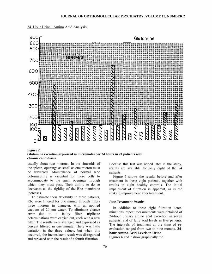
2. Strikingly low are certain non-essential amino acids that derive their carbon skeletons from intermediates in the citric acid cycle: a.) glutamate (Figure 1) and glutamine (Figure 2), from alpha ketoglutarate b.) asparagine (Figure 3), from oxalo-acetate 3. Relatively normal levels are present of non-essential amino acids derived from
73
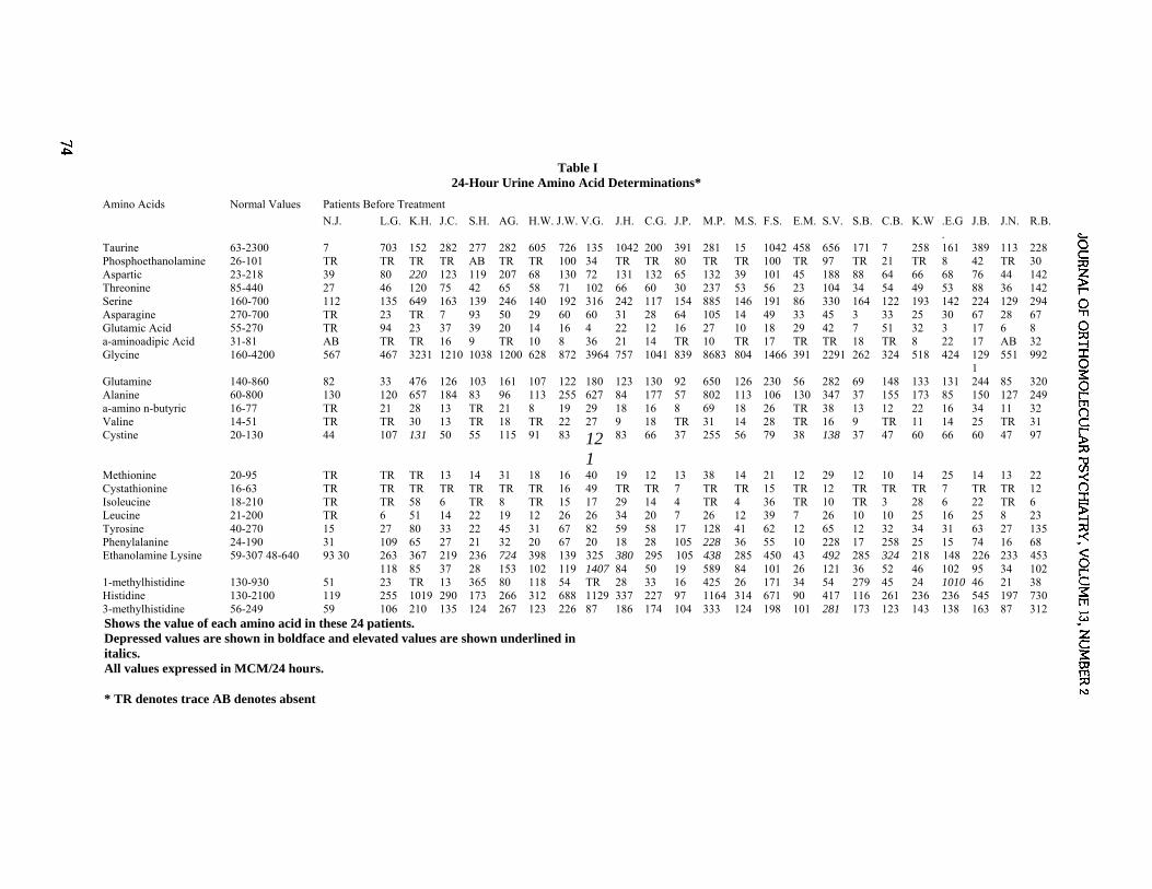
Table I
24-Hour Urine Amino Acid Determinations*
Amino Acids Normal Values Patients Before Treatment N.J. L.G. K.H. J.C. S.H. AG. H.W. J.W. V.G. J.H. C.G. J.P. M.P. M.S. F.S. E.M. S.V. S.B. C.B. K.W .E.G
. J.B. J.N. R.B.
Taurine 63-2300 7 703 152 282 277 282 605 726 135 1042 200 391 281 15 1042 458 656 171 7 258 161 389 113 228 Phosphoethanolamine 26-101 TR TR TR TR AB TR TR 100 34 TR TR 80 TR TR 100 TR 97 TR 21 TR 8 42 TR 30 Aspartic 23-218 39 80 220 123 119 207 68 130 72 131 132 65 132 39 101 45 188 88 64 66 68 76 44 142 Threonine 85-440 27 46 120 75 42 65 58 71 102 66 60 30 237 53 56 23 104 34 54 49 53 88 36 142 Serine 160-700 112 135 649 163 139 246 140 192 316 242 117 154 885 146 191 86 330 164 122 193 142 224 129 294 Asparagine 270-700 TR 23 TR 7 93 50 29 60 60 31 28 64 105 14 49 33 45 3 33 25 30 67 28 67 Glutamic Acid 55-270 TR 94 23 37 39 20 14 16 4 22 12 16 27 10 18 29 42 7 51 32 3 17 6 8 a-aminoadipic Acid 31-81 AB TR TR 16 9 TR 10 8 36 21 14 TR 10 TR 17 TR TR 18 TR 8 22 17 AB 32 Glycine 160-4200 567 467 3231 1210 1038 1200 628 872 3964 757 1041 839 8683 804 1466 391 2291 262 324 518 424 129
1 551 992
Glutamine 140-860 82 33 476 126 103 161 107 122 180 123 130 92 650 126 230 56 282 69 148 133 131 244 85 320 Alanine 60-800 130 120 657 184 83 96 113 255 627 84 177 57 802 113 106 130 347 37 155 173 85 150 127 249 a-amino n-butyric 16-77 TR 21 28 13 TR 21 8 19 29 18 16 8 69 18 26 TR 38 13 12 22 16 34 11 32 Valine 14-51 TR TR 30 13 TR 18 TR 22 27 9 18 TR 31 14 28 TR 16 9 TR 11 14 25 TR 31 Cystine 20-130 44 107 131 50 55 115 91 83 12
1
83 66 37 255 56 79 38 138 37 47 60 66 60 47 97
Methionine 20-95 TR TR TR 13 14 31 18 16 40 19 12 13 38 14 21 12 29 12 10 14 25 14 13 22 Cystathionine 16-63 TR TR TR TR TR TR TR 16 49 TR TR 7 TR TR 15 TR 12 TR TR TR 7 TR TR 12 Isoleucine 18-210 TR TR 58 6 TR 8 TR 15 17 29 14 4 TR 4 36 TR 10 TR 3 28 6 22 TR 6 Leucine 21-200 TR 6 51 14 22 19 12 26 26 34 20 7 26 12 39 7 26 10 10 25 16 25 8 23 Tyrosine 40-270 15 27 80 33 22 45 31 67 82 59 58 17 128 41 62 12 65 12 32 34 31 63 27 135 Phenylalanine 24-190 31 109 65 27 21 32 20 67 20 18 28 105 228 36 55 10 228 17 258 25 15 74 16 68 Ethanolamine Lysine 59-307 48-640 93 30 263
118 367 85
219 37
236 28
724 153
398 102
139 119
325 1407
380 84
295 50
105 19
438 589
285 84
450 101
43 26
492 121
285 36
324 52
218 46
148 102
226 95
233 34
453 102
1-methylhistidine 130-930 51 23 TR 13 365 80 118 54 TR 28 33 16 425 26 171 34 54 279 45 24 1010 46 21 38 Histidine 130-2100 119 255 1019 290 173 266 312 688 1129 337 227 97 1164 314 671 90 417 116 261 236 236 545 197 730 3-methylhistidine 56-249 59 106 210 135 124 267 123 226 87 186 174 104 333 124 198 101 281 173 123 143 138 163 87 312 Shows the value of each amino acid in these 24 patients. Depressed values are shown in boldface and elevated values are shown underlined in italics. All values expressed in MCM/24 hours.
* TR denotes trace AB denotes absent

METABOLIC ABNORMALITIES 24 Hour Urine Amino Acid Analysis
Glutamic acid excretion expressed in micromoles per 24 hours in 24 patients with chronic candidiasis. intermediates of the glycolytic pathway: a.) alanine (from pyruvate) b.) glycine (from 3 phosphoglycerate) 4. Ethanolamine levels generally are elevated, while those for phosphoethanolamine tend to be low. Since ethanolamine normally is converted to phosphoethanolamine, these results suggest a block in this pathway.
Taken as a whole, these findings suggest impairment in the synthesis of non-essential amino acids, particularly those synthesized from citric acid cycle intermediates. Such interconversions as ethanolamine to phos-phoethanolamine, and aspartate to aspara-gine, appear inhibited. Attention is called to the very low levels of glutamate and gluta-mine, the donors of amine groups in transamination reactions. Fatty Acid Levels in Plasma and in Eryth-rocyte Membranes
Plasma: Table III shows for these 24 patients
the average levels of fatty acids of both the omega six and omega three series, together with the normal value for each fatty acid. The p values for the abnormalities found are shown.
Rbc membranes: Table IV shows similar studies and comparisons for fatty acids in the membranes of erythrocytes, with p values.
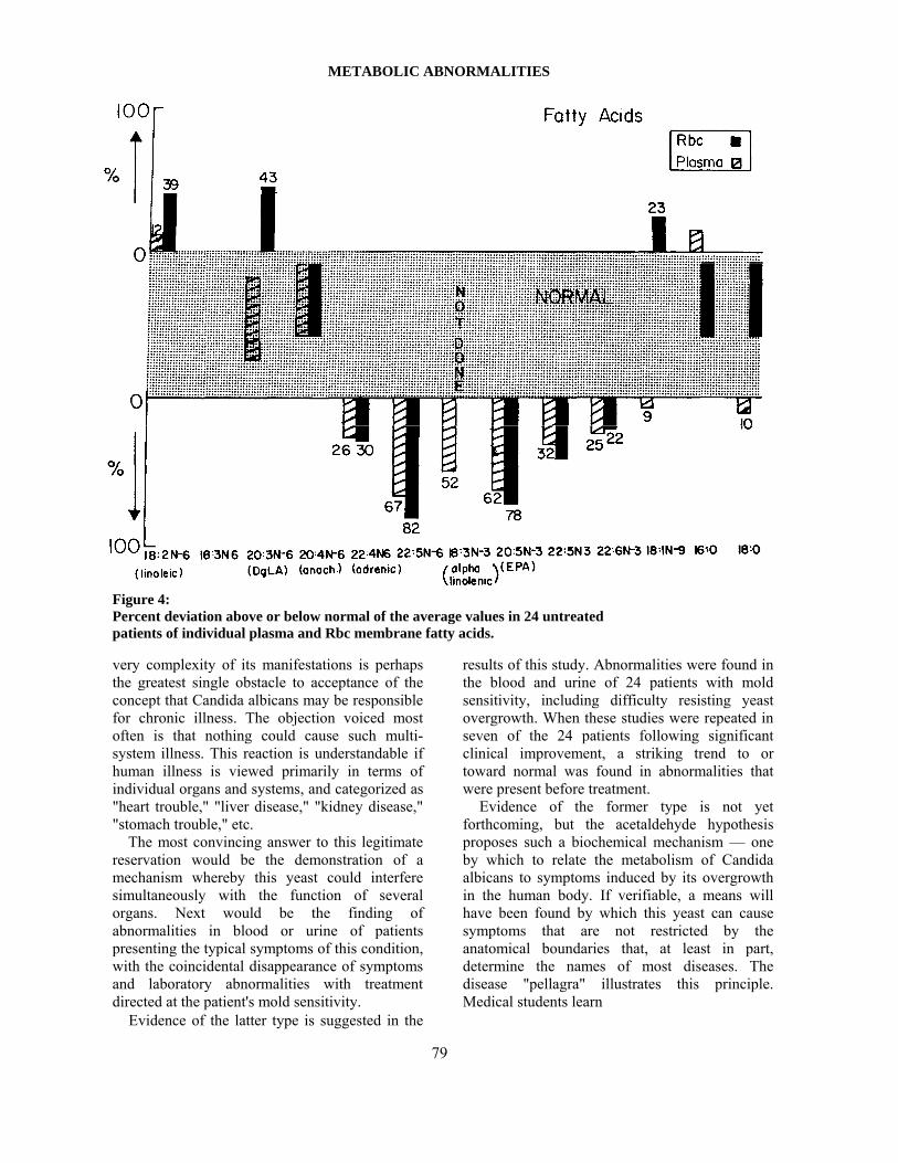
This same data is depicted graphically in Figure 4, which shows for each fatty acid in plasma and in Rbc membranes, the percent above or below normal of the average of the individual values before treatment. It will be noted that abnormalities were consistent throughout. In the omega six series the levels of the shorter chain fatty acids were elevated, while the levels of the longer chain fatty acids into which these are normally converted were depressed. Particularly striking were the very low levels of 22:5 omega six fatty acids. Erythrocyte Filtration
The diameter of the human Rbc is approximately seven microns. These cells pass through capillaries of much smaller diameter,
75
JOURNAL OF ORTHOMOLECULAR PSYCHIATRY, VOLUME 13, NUMBER 2
24 Hour Urine Amino Acid Analysis
Figure 2: Glutamine excretion expressed in micromoles per 24 hours in 24 patients with chronic candidiasis. usually about two microns. In the sinusoids of the spleen, openings as small as one micron must be traversed. Maintenance of normal Rbc deformability is essential for these cells to accommodate to the small openings through which they must pass. Their ability to do so decreases as the rigidity of the Rbc membrane increases.
To estimate their flexibility in these patients, Rbc were filtered for one minute through filters three microns in diameter, with an applied vacuum of 20 cm water. To eliminate chance error due to a faulty filter, triplicate determinations were carried out, each with a new filter. The results were averaged and expressed as percent filtered in one minute. There was little variation in the three values, but when this occurred, the inconsistent result was disregarded and replaced with the result of a fourth filtration.
Because this test was added later in the study, results are available for only eight of the 24 patients.
Figure 5 shows the results before and after treatment in these eight patients, together with results in eight healthy controls. The initial impairment of filtration is apparent, as is the striking improvement after treatment.
Post-Treatment Results
In addition to these eight filtration deter-minations, repeat measurements were obtained of 24-hour urinary amino acid excretion in seven patients, and of fatty acid levels in five patients. The intervals of treatment at the time of re-evaluation ranged from two to nine months. 24-hour Amino-Acid Levels in Urine Figures 6 and 7 show graphically the
76

METABOLIC ABNORMALITIES
Table II
24-Hour Urine Amino Acid Determinations (Percent of 24 Patients)
Amino Acids Normal Below Normal Above Normal Taurine 87.5 12.5 0 Phosphoethanolamine 29.2 70.8 0 Aspartic 95.8 0 4.2 Threonine 25.0 75.0 0 Serine 50.0 45.8 4.2 Asparagine 0 100.0 0 Glutamic 4.2 95.8 0 d-amino adipic 8.3 91.7 0 Glycine 95.8 0 4.2 Glutamine 37.5 62.5 0 Alanine 91.7 4.2 4.2 a-amino n-butyric 62.5 37.5 0 Valine 50.0 50.0 0 Cystine 83.3 0 16.7 Methionine 29.2 70.8 0 Cystathionine 8.3 91.7 0 Isoleucine 20.8 79.2 0 Tyrosine 50.0 50.0 0 Phenylalanine 54.2 33.3 12.5 Ethanolamine 54.2 4.2 41.7 Lysine 62.5 33.3 4.2 1-methylhistidine 20.8 75.0 4.2 Histidine 83.3 16.7 0 3-methylhistidine 83.3 0 16.7 Leucine 45.8 54.2 0
24-hour urinary amino acid levels before treatment. These are expressed for each amino acid as the percent of these twenty-four patients with values that were normal, low, or elevated.
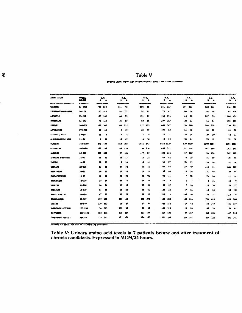
findings on repeat measurement of four of the amino acids that had shown the greatest deviation from normal before treatment. Table V shows for each patient comparative values before and after treatment of the individual amino acids.
Particularly noteworthy is the change to normal of very low glutamine values (Figure 6). Four of the seven were low before treatment. Three returned to normal, and the fourth showed a sharp increase that
brought it almost to normal. Also of interest is the suggestion of improved conversion of ethanolamine to phosphoethanolamine (depicted in Figure 7), the general trend being one of decreasing ethanolamine and increasing phosphoethanolamine values.
Future studies in these, and in the remaining 24 patients, will determine the ultimate significance of these changes that coincided with relief of long-standing symptoms, as a result of yeast suppression.
77

24 Hour Urine Amino Acid Analysis
Figure 3: Asparagine excretion expressed in micromoles per 24 hours in 24 patients with chronic candidiasis.
Fatty Acid Levels in Five Patients In plasma (Table VI) and in Rbc membranes
(Table VII) the changes in fatty acid values suggest that, at this point in treatment, a definite trend toward normal has been established. This is particularly true in the omega six series, in which the elevated levels of short-chain acids have fallen to (or toward) normal, as the levels of longer chain acids into which they are converted have risen (e.g. 22:5 omega six). Changes in the omega three series are less pronounced, although in both plasma and Rbc membranes the 22:6 omega 3 fatty acids have come into the normal range from their initial depressed values. Also, the 20:5 omega 3 fatty acids in Rbc membranes showed a substantial increase from 0.20 to 0.28, although the level remains well below the normal range. Future
measurements will be necessary to determine whether this early trend continues. Rbc Filtration in Eight Patients
The change in Rbc filterability has already been noted. These eight patients included the seven in whom amino acid excretion was restudied, and the five whose membrane and plasma fatty acid levels were re-measured.
Discussion The most striking characteristic of the clinical
picture of chronic candidiasis is its complexity. Erratic function in many organs is evidenced by appropriate symptoms. Particularly common are those originating in the central nervous system, G.I. and G.U. tracts, endocrine glands, skin, muscles and joints, and respiratory system. To those hearing of it for the first time, this
78
METABOLIC ABNORMALITIES
Figure 4: Percent deviation above or below normal of the average values in 24 untreated patients of individual plasma and Rbc membrane fatty acids.
very complexity of its manifestations is perhaps the greatest single obstacle to acceptance of the concept that Candida albicans may be responsible for chronic illness. The objection voiced most often is that nothing could cause such multi-system illness. This reaction is understandable if human illness is viewed primarily in terms of individual organs and systems, and categorized as "heart trouble," "liver disease," "kidney disease," "stomach trouble," etc.
The most convincing answer to this legitimate reservation would be the demonstration of a mechanism whereby this yeast could interfere simultaneously with the function of several organs. Next would be the finding of abnormalities in blood or urine of patients presenting the typical symptoms of this condition, with the coincidental disappearance of symptoms and laboratory abnormalities with treatment directed at the patient's mold sensitivity.
Evidence of the latter type is suggested in the
results of this study. Abnormalities were found in the blood and urine of 24 patients with mold sensitivity, including difficulty resisting yeast overgrowth. When these studies were repeated in seven of the 24 patients following significant clinical improvement, a striking trend to or toward normal was found in abnormalities that were present before treatment.
Evidence of the former type is not yet forthcoming, but the acetaldehyde hypothesis proposes such a biochemical mechanism — one by which to relate the metabolism of Candida albicans to symptoms induced by its overgrowth in the human body. If verifiable, a means will have been found by which this yeast can cause symptoms that are not restricted by the anatomical boundaries that, at least in part, determine the names of most diseases. The disease "pellagra" illustrates this principle. Medical students learn
79

JOURNAL OF ORTHOMOLECULAR PSYCHIATRY, VOLUME 13, NUMBER 2
Figure 5: Red blood cell filtration values of 8 healthy adults as compared with the filtration rate of 8 patients with candidiasis. The solid bars represent filtration values of controls and of patients before treatment. Hatched bars represent values of patients after treatment. The figures under each bar show the number of months of treatment for each patient.
"the three D's" (diarrhea, dermatitis, dementia) of this illness caused by Vitamin B3 deficiency. The intestine, brain, and skin are manifesting abnormal function because, even though widely separated anatomically, they share metabolic pathways that require adequate B3.
At the beginning of these studies, only amino acid and fatty acid determinations were available. Rbc filtration was added somewhat later. Specific studies of carbohydrate metabolism were the last to be incorporated into the study and will be reported separately. Indirect but suggestive evidence of interference in carbohydrate pathways will be discussed in connection with the amino acid abnormalities found in these 24 patients.
The finding of abnormalities in all three areas studied increases the likelihood of their
significance. It also greatly lessens the chance of these findings being invalid because of errors in the normal control values. While regional differences in such factors as diet and climate could conceivably affect "normal" values, it is quite improbable that this would occur in all three aspects of this investigation, particularly in view of the wide geographic separation of the three laboratories participating in the study.
Among these abnormal results are certain findings worthy of special comment. These will be discussed briefly, with emphasis on changes consistent with those that have been reported among the toxic effects of acetaldehyde. Fatty Acids
First, in the omega 6 (W-6) series of fatty acids in Rbc membranes, the levels of the shorter chain 18:2W-6 (linoleic acid) and
80
METABOLIC ABNORMALITIES
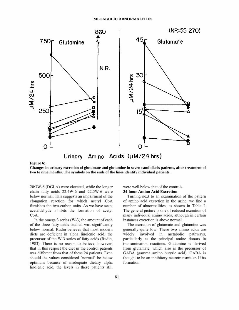
Figure 6: Changes in urinary excretion of glutamate and glutamine in seven candidiasis patients, after treatment of two to nine months. The symbols on the ends of the lines identify individual patients.
20:3W-6 (DGLA) were elevated, while the longer chain fatty acids 22:4W-6 and 22:5W-6 were below normal. This suggests an impairment of the elongation reaction for which acetyl CoA furnishes the two-carbon units. As we have seen, acetaldehyde inhibits the formation of acetyl CoA.
In the omega 3 series (W-3) the amount of each of the three fatty acids studied was significantly below normal. Rudin believes that most modern diets are deficient in alpha linolenic acid, the precursor of the W-3 series of fatty acids (Rudin, 1983). There is no reason to believe, however, that in this respect the diet in the control patients was different from that of these 24 patients. Even should the values considered "normal" be below optimum because of inadequate dietary alpha linolenic acid, the levels in these patients still
were well below that of the controls. 24-hour Amino Acid Excretion
Turning next to an examination of the pattern of amino acid excretion in the urine, we find a number of abnormalities, as shown in Table I. The general picture is one of reduced excretion of many individual amino acids, although in certain instances excretion is above normal.
The excretion of glutamate and glutamine was generally quite low. These two amino acids are widely involved in metabolic pathways, particularly as the principal amine donors in transamination reactions. Glutamine is derived from glutamate, which also is the precursor of GABA (gamma amino butyric acid). GABA is thought to be an inhibitory neurotransmitter. If its formation
81
JOURNAL OF ORTHOMOLECULAR PSYCHIATRY, VOLUME 13, NUMBER 2
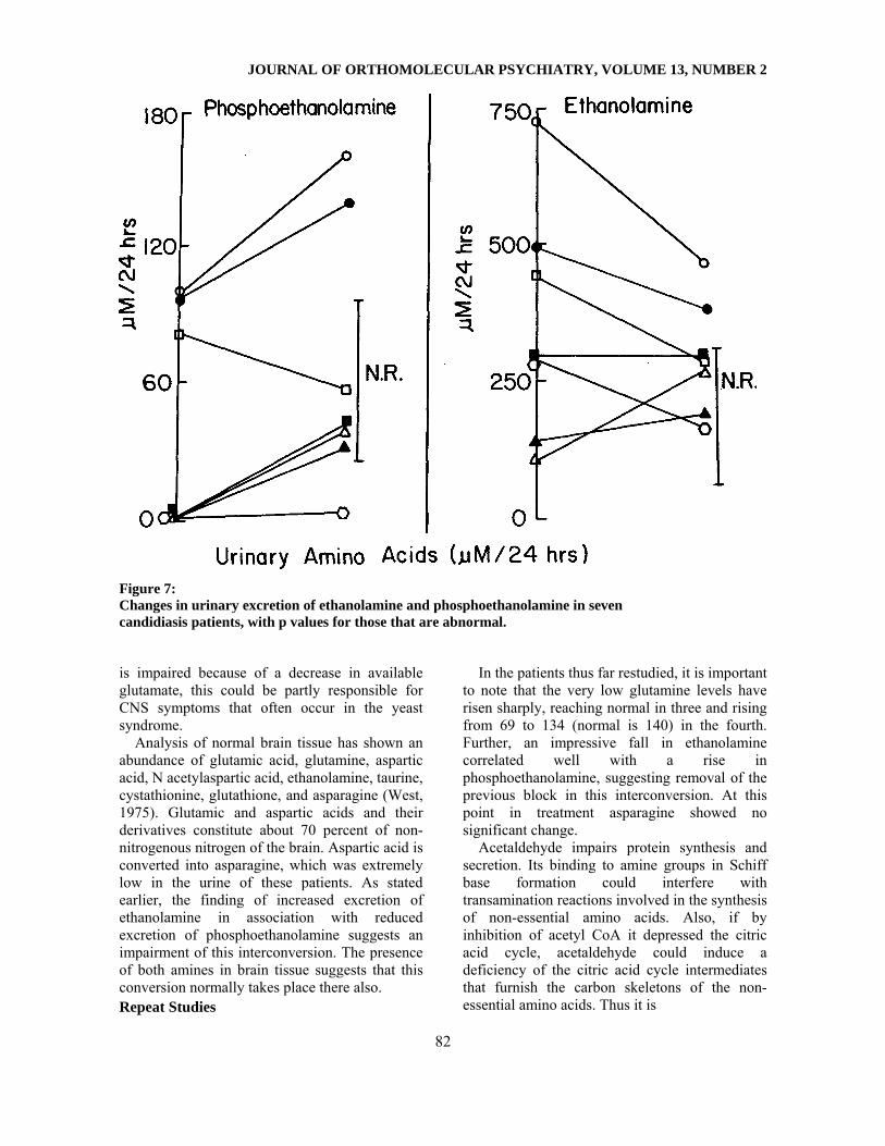
Figure 7: Changes in urinary excretion of ethanolamine and phosphoethanolamine in seven candidiasis patients, with p values for those that are abnormal.
is impaired because of a decrease in available glutamate, this could be partly responsible for CNS symptoms that often occur in the yeast syndrome.
Analysis of normal brain tissue has shown an abundance of glutamic acid, glutamine, aspartic acid, N acetylaspartic acid, ethanolamine, taurine, cystathionine, glutathione, and asparagine (West, 1975). Glutamic and aspartic acids and their derivatives constitute about 70 percent of non-nitrogenous nitrogen of the brain. Aspartic acid is converted into asparagine, which was extremely low in the urine of these patients. As stated earlier, the finding of increased excretion of ethanolamine in association with reduced excretion of phosphoethanolamine suggests an impairment of this interconversion. The presence of both amines in brain tissue suggests that this conversion normally takes place there also. Repeat Studies
In the patients thus far restudied, it is important to note that the very low glutamine levels have risen sharply, reaching normal in three and rising from 69 to 134 (normal is 140) in the fourth. Further, an impressive fall in ethanolamine correlated well with a rise in phosphoethanolamine, suggesting removal of the previous block in this interconversion. At this point in treatment asparagine showed no significant change.
Acetaldehyde impairs protein synthesis and secretion. Its binding to amine groups in Schiff base formation could interfere with transamination reactions involved in the synthesis of non-essential amino acids. Also, if by inhibition of acetyl CoA it depressed the citric acid cycle, acetaldehyde could induce a deficiency of the citric acid cycle intermediates that furnish the carbon skeletons of the non-essential amino acids. Thus it is
82

METABOLIC ABNORMALITIES
PLASMA PHOSPHOLIPIDS
FATTY ACID NORMAL 21.45 +/- 2.81
CANDIDA
18:2n-6
24.96 +/- 4.5 *
18:3n-6 0.16 +/- 0.12 ---- 20:3n-6 3.06 +/- 0.60 3.04 +/- 0.91 20:4n-6 11.36 +/- 1.67 11.57 +/- 2.27 22:4n-6 0.73 +/- 0.26 0.54 +/- 0.24 * 22:5n-6 1.12 +/- 0.67 0.36 +/- 0.21 ** 18:3n-3 0.27 +/- 0.53 0.13 +/- 0.15 * 20:5n-3 1.01 +/- 0.36 0.38 +/- 0.37 ** 22:5n-3 0.93 +/- 0.27 0.02 +/- 0.28 ** 22:6n-3 3.54 +/- 0.89 2.67 +/- 1.01 ** 18:ln-9 13.50 +/- 2.20 12.29 +/- 1.50 * 16:0 25.81 +/- 1.69 29.41 +/- 2.19 ** 18:0 11.61 +/- 1.32 10.43 +/- 1.67 * * - p < 0.05 ** = p < 0.01 or less
Table III: Average plasma phospholipid values in normal healthy adults and in 24 patients with candidiasis, with p values for those that are abnormal. Expressed as area percent of total phospholipids.
possible, theoretically, to relate acetaldehyde to the amino acid abnormalities found in these patients.
The trend toward normal of the pattern of amino acid excretion was associated with a similar trend in fatty acid balance both in plasma and in Rbc membranes. The very low levels of 22:5W6 fatty acids became normal as their shorter-chain precursors fell from elevated pre-treatment levels, suggesting a correction of the previous impairment of elongation.
Changes in fatty acids of the W-3 series were mixed, some decreasing and others increasing. A 40 percent increase occurred in the average of the value of 22:6W3. Whether this rise will continue must await later determinations.
As stated earlier, acetaldehyde will increase the
rigidity of erythrocyte membranes, and has been used for that purpose by physiologists studying membrane composition and function. In patients with chronic candidiasis, diminished flexibility of Rbc membranes resulted in a decrease in filter-ability of intact red blood cells. This is an important finding, both as a potential diagnostic test, and for its implications in the patho-physiology of this illness.
The return to normal of Rbc filtration was striking in patients whose symptoms simul-taneously showed substantial improvement. Improved oxygenation of tissues suggests itself as an important factor in this clinical improvement, particularly of those symptoms related to brain function.
83

JOURNAL OF ORTHOMOLECULAR PSYCHIATRY, VOLUME 13, NUMBER 2
RED BLOOD CELL TOTAL PHOSPHOLIPIDS
FATTY ACID NORMAL + SD 9.78 +/- 1.64
CANDIDA Linoleic
18:2n-6
13.63 +/- 4.78 **
20:3n-6 1.37 +/- 0.37 1.96 +/- 0.52 DGLA ** 20:4n-6 15.13 +/- 1.98 15.73 +/- 3.35 Arachidonic 22:4n-6 5.54 +/- 1.37 3.89 +/- 1.02 Adrenic ** 22:5n-6 3.99 +/- 1.85 0.73 +/- 0.45 ** 20:5n-3 0.65 +/- 0.24 0.14 +/- 0.18 EPA ** 22:5n-3 2.53 +/- 0.90 1.45 +/- 0.68 ** 22:6n-3 4.20 +/- 1.03 3.26 +/- 0.89 ** 18:ln-9 14.83 +/- 1 .69 18.25 +/- 2.46 Oleic ** 16:0 20.68 +/- 1 .54 21.31 +/- 2 .18 Palmitic 18:0 14.71 +/- 1 .52 16.04 +/- 2.86 Stearic * = p < 0.05 ** = p < 0.01 or less
Table IV: Average erythrocyte phospholipids in normal healthy adults and in 24 patients with candidiasis, with p values for those that are abnormal. Expressed as area percent of total phospholipids.
Speculation about Membranes and Chronic Candidiasis
Membrane physiology has attracted much attention in recent years. The great importance of membranes in normal physiologic processes is generally accepted. In addition to demarcating its boundaries, membranes compartmentalize the different functions of the cell and its subdivisions, and control the passage of substances involved in its various metabolic processes. Molecules pass in both directions through membranes that also de-fine such anatomical limits as those of the cell nucleus, the mitochondrion, and the cell itself. Passage may be passive or active. Changes in the chemical and physical properties of membranes may alter such transport, resulting in abnormal function of the cell, and ultimately of its parent organ.
Numerous membrane pumps and shunts function in metabolic pathways. Several examples illustrate their importance in normal
cellular function. (1) NADH formed in the cytoplasm during glycolysis cannot cross the mitochondrial membrane for reconversion to NAD, without which glycolysis cannot proceed. Cytoplasmic NAD is regenerated by the transfer of hydrogen from NADH to glycerol 3-phosphate or to malate, both of which are able to cross the mitochondrial membrane. Thus, in addition to assuring a continuous supply of NAD in the cytoplasm, these "shuttles" accomplish the necessary transfer of hydrogen into the mitochondria, where, by oxidative phosphorylation and the electron transport chain, it is ultimately accepted by molecular oxygen as ATP is formed. (2) The reciprocal transport of ADP and ATP across the inner mitochondrial membrane requires the specific carrier, "ATP-ADP translocase." (3) In fatty acid oxidation, carnitine carries acyl groups across the inner mitochondrial membrane to
84
METABOLIC ABNORMALITIES
the mitochondrial matrix, in which oxidation takes place. (4) Gluconeogenesis takes place in the cytoplasm, where are located the glycolytic enzymes that catalyze the formation of glycogen from pyruvate, and from amino acids that have first been converted to oxaloacetate. However, the formation of oxaloacete from amino acids occurs in the mitochondria, and oxaloacete is unable to cross the mitochondrial membrane into the cytoplasm; to do so it must be converted to malate, which, once in the cytoplasm, is reconverted to oxalacetate. Thus gluconeogenesis is dependent on the proper passage of malate through the mitochondrial membrane. (5) Glycolysis takes place in the cytoplasm, but the further oxidation of carbohydrate is by the citric acid cycle and oxidative phosphorylation, both of which occur in the mitochondrion. Therefore pyruvate must be transported from the cytoplasm into the mitochondria by the "pyruvate carrier," (6) Steroid hormones, after passing through cell walls from serum, bind to appropriate receptor proteins in the cytoplasm and are carried into the nucleus, where they exert their normal modulating effect on cell function. (7) Electrical conductance and normal function in nerve and muscle tissue depend upon the movement across membranes of such ions as sodium, potassium, and calcium. Such transmembrane proteins as Na+-K+ ATPase and Ca++ ATPase, as they are alternately phosphorylated and dephosphor-ylated, undergo configurational changes that are vital to their function. The importance of these two pumps needs no emphasis. Membrane abnormalities sufficient to impair their efficient operation would have far-reaching effects on normal physiologic processes. For example, the movement into the cell of such metabolic fuels as glucose and amino acids is often tied to the movement of Na+ , which is driven by the N+-K+ pump. (8) Other transport systems of the mitochondrial membrane include the dicarboxylate and tricarboxylate carriers for transporting citric acid cycle intermediates, the glutamate carrier, and a transport system for calcium ions. These few examples illustrate the importance of properly regulated passage of metabolic intermediates, minerals, and electrolytes into and out of the cell and its various compartments. It is
difficult to conceive of pathways that would remain unaffected by major membrane abnormalities.
In these candidiasis patients abnormalities of fatty acid balance in serum and Rbc membranes occurred consistently, in association with impaired filterability of these cells. Although at this time we do not know whether these cell-wall abnormalities extend to cells other than erythrocytes, it would seem more probable than not, in view of the fact that the same imbalance of fatty acids was found in serum as in red cell membranes. This is a matter of great importance.
Acetaldehyde may affect Rbc membranes directly, or it may do so by metabolic inter-ference that leads to imbalances of fatty acids and other membrane constituents. Either effect of this toxin could be exerted on membranes elsewhere in the body, affecting some perhaps more than others. It is interesting, as part of the acetaldehyde hypothesis, to speculate about the possible relationship of such membrane changes to some of the clinical manifestations of chronic candidiasis in patients with mold sensitivity and the associated susceptibility of yeast infections. Membranes and Gastro-Intestinal Function
The absorption of nutrients would hardly proceed normally if the walls of the cells of the intestinal mucosa were abnormal with respect to permeability and transport. Indeed, even the digestion that must precede the absorption of many nutrients might well be affected if enzyme formation and release were impaired as a result of changes in the cells responsible for these functions.
Acetaldehyde has been shown to have a different effect on intestinal microvillus mem-brane vesicles than on Rbc membranes (Tillotson et al., 1981). These studies indicated an increase in the fluidity of these membranes, with an associated dissipation of the Na+ gradient, and an inhibition of carrier function. These studies indicated that the permeability of these membranes increased upon exposure to acetaldehyde. Thus, at least in this one instance, acetaldehyde affected membranes by increasing rather than decreasing their fluidity, but in so doing definitely altered vital membrane func-tions.
85

METABOLIC ABNORMALITIES
Table VI Plasma Fatty Acids
Average values of five patients Normal range18:1N9 before after 11.82
16.27 11.3 -15.7
18:2N6 before after 25.83 22.47
18.6 - 24.3
20:3N6 before after 3.06 3.23
2.5 - 3.7
20:4N6 before after 10.77 12.08
9.7 -13.0
22:4N6 before after 0.46 0.63
0.47 - 0.99
22:5N6 before after 0.36 0.51
0.45 -1.8
20:5N3 before after 0.47 0.36
0.65 - 1.4
22:5N3 before after 0.72 0.63
0.66 - 1.2
22:6N3 before after 2.52 2.68
2.6 - 4.4
Table VI: Average of individual values in five patients before and after treatment for each of nine fatty acids. Expressed as area percent of total phospholipids.
87

JOURNAL OF ORTHOMOLECULAR PSYCHIATRY, VOLUME 13, NUMBER 2
Table VII Red Blood Cell Membrane Fatty Acids
Average patient values Normal range18.1N9 before after 17.65
16.83 13.14 - 16.52
18:2N6 before after 16.16 10.93
8.14 - 11.42
20:3N6 before after 2.05 1.83
1.0 - 1.74
20:4N6 before after 16.4 16.9
13.15 - 17.11
22:4N6 before after 4.11 4.19
4.17-6.91
22:5N6 before after 0.% 1.13
2.14-5.84
20:5N3 before after 0.20 0.28
0.41 - 0.89
22:5N3 before after 1.78 1.69
1.63 - 3.43
22:6N3 before after 3.11 3.81
3.17-5.23
Table VII: Average of nine individual fatty acids in the Rbc membranes of five patients before and after treatment. Expressed as area percent of total phospholipids.
88

METABOLIC ABNORMALITIES
Membranes and Lymphocyte Function Membranes are uniquely important in the function of lymphocytes. The many complex events involved in their function follow the initial recognition of markers on the surface of target cells by appropriate sites on the membranes of the lymphocyte itself. This is important both in triggering the normal response to "foreign" antigens, and in preventing the abnormal attacks of "self antigens that lead to "auto-immune" disease. Abnormalities of the membranes of lymphocytes should have an effect on both functions. An impaired response to foreign antigens should weaken the attack on Candida albicans itself, such a condition of "immunologic tolerance" being borne out by the chronic, resistant nature of infections with this yeast. It could also weaken our immunologic defense against other microorganisms, and perhaps even against malignant cells. On the other hand, failure to properly recognize "self antigens could result in ineffective suppression, with resulting immunologic responses to normal cells, immune-complex deposition, and autoimmune disease. There is another way that membrane changes could result in autoimmune disease. Should such changes in cells other than lymphocytes lead to exposure of antigenic determinants that are normally hidden (e.g., the hydrophobic portion of a membrane protein), lymphocytes might then falsely recognize as "foreign" these "new" markers. Thus autoimmune attacks could result (1) when lymphocyte function is normal, but hidden antigens become exposed or, (2) when lymphocyte function is abnormal and suppressor function fails, allowing attacks on antigens that are normally exposed. Certain characteristics of autoimmune reactions are more easily explained by the second hypothesis, i.e., that antigenic determinants normally hidden are exposed as a result of membrane damage. First, in autoimmune illness the attacks are directed at different organs, so that different "diseases" occur among such patients. Second, often more than one autoimmune "disease" is present simultaneously in the same patient, even though one usually predominates. This variability in the intensity of autoimmune attacks is readily explained by the hypothesis that
membranes of different organs vary in their degree of abnormality, and hence of exposed "hidden" determinants. Suppressor cell weakness alone, in the absence of any "strange" antigens, should lead to attacks more nearly equal in intensity in the various organs.
As stated earlier, it would be likely that both mechanisms would be involved, since membrane abnormality, if widespread, would involve suppressor lymphocytes, as well as the cells of the organs under attack. Membranes and Erythrocyte Function
Turning now to erythrocytes, we can anticipate that the increased rigidity of their membranes would render these cells less able to accomplish the changes in shape and fluidity necessary for passage through capillaries one-third their own diameter. If this should affect oxygenation of an organ, it would impair its function. The brain, for example, is highly susceptible to even slight reductions in oxygen tension, this being manifested particularly as an impairment of short-term memory, a symptom characteristically seen in these patients (Davis and Berger, 1979).
The "anion channel" of the Rbc membrane is another example of the importance of membrane integrity in regulating the movement of ions and molecules across membranes. The anion channel is a glycoprotein that traverses the Rbc membrane, all molecules pointing in the same direction. It accounts for about 20-30 percent of Rbc membrane protein. This channel allows the passage of C1—and HCO3
- through the Rbc membrane as erythrocytes transport C02. A decrease in the normal fluidity of the Rbc membrane, as found in these patients, could affect the conformational changes necessary for the proper function of such a protein pump.
Special Problems in Patients with Chronic Candidiasis
In patients with mold sensitivity and yeast susceptibility, several problems occur at a frequency greater than expected in the general population. Mitral Valve Prolapse
The first of these is mitral valve prolapse with dysautonomia. The medical history
89
JOURNAL OF ORTHOMOLECULAR PSYCHIATRY, VOLUME 13, NUMBER 2
usually reveals that the symptoms on which these diagnoses were based had their beginning after typical symptoms of mold sensitivity had been present for several years. If we assume that these conditions are being diagnosed with reasonable accuracy, there has been a sharp increase in their incidence, and it has paralleled the similar increase that has occurred in chronic yeast infections since the advent of broad-spectrum antibiotics, birth-control pills, and steroid hormones. Carpal Tunnel Syndrome
Another condition that has been seen with some frequency in this syndrome is carpal tunnel syndrome. The effects of acetalde-hyde, especially that on collagen metabolism, could be related to this and to the mitral valve prolapse problem. Formaldehyde Sensitivity
Many of these patients exhibit extreme intolerance to formaldehyde. Since it also binds to sulfhydryl and amine groups, and in general exhibits toxicity similar to that of acetaldehyde, the effects of these two aldehydes should be additive. A patient whose tissues already contained a significant a-mount of acetaldehyde might well exhibit symptoms from an amount of formaldehyde that otherwise would be easily tolerated. In addition, aldehyde dehydrogenase oxidizes both toxins. The requirement for the con-tinuous removal of acetaldehyde should to some degree diminish the capacity of the liver to cope with formaldehyde. Thus the hypothesis of acetaldehyde toxicity could account for what has been one of the most puzzling aspects of this illness.
Occasionally patients date the onset of this illness to heavy formaldehyde exposure. Such an event could be superimposed on mild acetaldehyde toxicity (i.e., mild candidiasis), or indeed, might theoretically initiate the problem, which would then be perpetuated by acetaldehyde despite the elimination of further exposure to formaldehyde. In fact, once the membrane (and metabolic) abnormalities have been initiated by either aldehyde, according to this hypothesis the resulting impairment of lymphocyte function would allow further growth of Candida albicans. As this occurred, acetaldehyde would be produced in increasing quantities, to perpetuate and advance the illness.
Careful histories frequently relate the onset of yeast susceptibility to such yeast-stimulating factors as antibiotics and contraceptive hormones, indicating that in these cases acetaldehyde initiated the aldehyde toxicity. In theory, however, heavy and prolonged formaldehyde exposure could start the process. The practical fact is that formaldehyde exposure in the usual amount tolerated by healthy individuals would almost always be present as an additional factor in patients, even as the process was being initiated by acetaldehyde. End-Organ Response to Hormones
Another important category of symptoms in these patients includes manifestations suggestive of impaired end-organ responsiveness to hormones, especially the steroid hormones. For example, severe menstrual bleeding and poor vaginal cornification may occur despite normal blood levels of ovarian hormones.
The functional state of membranes could influence the cell's response to hormones. In order to exert their proper physiologic effect, steroid hormones must pass through the cell wall, and then through the nuclear membrane. Insulin and similar membrane-active hormones bind to receptors on cell walls. Should membrane changes affect the entry of steroid hormones into the cytoplasm or nucleus, or alter the insulin receptor so as to affect its binding, these hormones could be hindered in exerting their normal effect on the cell. It is just such an impairment of the cellular response to hormones that is suggested by many of the symptoms in this syndrome. Autonomic Imbalance
Other frequently occurring symptoms are those characteristic of imbalance of the autonomic nervous system. Acetylcholine is the neurotransmitter at both sympathetic and parasympathetic synapses of the autonomic nervous system, and also mediates the action of post-ganglion parasympathetic neurons. Acetaldehyde is a potent synaptic blocking agent and could be responsible for the autonomic imbalance in these patients, perhaps in part by binding to the sulfhydryl group of CoA, thus affecting the availablility of the acetyl group required for the synthesis of acetylcholine.
90

METABOLIC ABNORMALITIES
Central Nervous System Symptoms Defective short-term memory, also common in
patients with this condition, could be another effect of impaired acetylcholine function, brought on either by such poor trans-acetylation, or by membrane abnormality that affected its release from synaptic vesicles.
Impairment of the ability to concentrate usually accompanies the memory deficit, most often in association with depression and anxiety. The many neurotransmitters that are amines may, by Schiff base formation with acetaldehyde, form "false neurotransmitters." These compounds should have an adverse effect on neuronal function that utilizes these amines as neurotransmitters.
Thus, among the mechanisms by which acetaldehyde theoretically could cause the common symptoms of depression, anxiety, and difficulty with memory and concentration, we have the following: 1. Metabolic disturbances in neurons (fatty acid, protein, carbohydrate) 2. Membrane abnormalities 3. Impairment of acetylcholine synthesis or release 4. Defective neurotransmitter function a.) defective synthesis, release, or binding b.) false neurotransmitter formation 5. Diminished oxygen delivery due to loss of flexibility of erythrocyte membranes Allergic Symptoms
Allergic reactions to products of Candida albicans occur frequently. Allergic rhinitis and asthma are not uncommon. Chronic "idiopathic" urticaria is frequently due to the antigens of this yeast.
Whether, or how, the toxic effects of acetaldehyde play a part in the allergic response is not clear. Its effect on membranes could alter the reactivity of cells of the mucous membranes and skin, but this seems less likely than an acetaldehyde-induced change in leukocytes.
At this time too little is known of the changes that occur in leukocytes when they begin to mediate the symptoms of allergy. Theoretically there might have occurred a decrease in a normal function that suppresses this type reaction. Or it could be an entirely new reaction — one that does not occur normally, for which therefore no sup- pressor mechanisms exist, or are needed. If it
affected their metabolism or membranes (or both), theoretically acetaldehyde could alter leukocytes in such a way that they begin to function in this abnormal way.
Two observations are of interest in this regard. Certain patients previously free of allergies have experienced their initial allergic reactions to pollen and other inhalants while receiving long-term antibiotic therapy for acne. The timing of the initial reaction (to pollen, for example) often coincides with the onset of diarrhea, vaginitis, and other manifestations of the yeast problem. Then, in rapid succession, other inhalants and foods begin to trigger the allergic response.
The second observation dates from ap-proximately three years ago. For many years prior to that time there had been no way to test the idea that the effect of Candida albicans on the cells of the immune system might include those changes in lymphocytes (whatever they are) that account for the onset of allergic responses. Withholding standard pollen extract therapy, for example, could not be justified in patients whose history and skin tests clearly indicated Spring and Fall pollen hay fever.
When chronic yeast infections are treated in patients who also have major autoimmune illness, vaccines are not used, since such stimuli to the immune system theoretically might also stimulate its autoimmune component. Thus, even in the presence of severe Spring or Fall pollen hay fever, the usual allergy injections are not used; symptomatic treatment (e.g. antihistamines) is recommended.
Some patients began reporting complete disappearance of these typical pollen symptoms. In some cases, total relief was experienced from both Spring and Fall pollen hay fever symptoms, even though symptoms in both seasons had been severe for many consecutive years. This simultaneous remission (i.e., in the same year) of tree and/or grass hay fever in the Spring, and ragweed hay fever in the Fall, virtually insures that the responsible change was in the patient rather than in the environment. This occurrence suggests a relationship between Candida albicans and the functional abnormality of the immune system that is associated with this type of allergy response.
91

JOURNAL OF ORTHOMOLECULAR PSYCHIATRY, VOLUME 13, NUMBER 2
Thus both observations — that allergies to pollen, other inhalants, and foods may appear in quick succession soon after the onset of chronic yeast infections, and on occasion may disappear abruptly with no therapy other than yeast suppression — suggest a relationship between Candida albicans and the unknown changes in the immune system that allow (or cause) allergic reactions to occur. How often this might be true remains to be determined.
It has also proved true that in some cases of intractable asthma, the addition of yeast suppression has led to sudden improvement beyond that achieved by extract therapy. This may mean only that certain yeast substances were among the antigens to which the patient was allergic. However, considered in conjunction with the two observations just discussed, it suggests that the benefit may possibly have resulted from an effect of nystatin on some factor more basic to the allergic mechanism.
Summary The results of metabolic studies in 24 patients
with chronic candidiasis have been presented. An attempt has been made to consider the principal symptoms of this condition, and these metabolic findings, in relation to various toxic effects of acetal-dehyde. Though speculative, these thoughts as to how this toxin could cause the clinical and laboratory characteristics of this illness are a vital part of this hypothesis. The extensive literature on the subject of acetal-dehyde provides a number of mechanisms by which its toxicity could lead to this illness, and to the abnormal laboratory results re-ported in this paper.
The appeal of the acetaldehyde hypothesis is that it defines the symptoms of chronic yeast infection in terms of a toxin that is readily produced in vitro by common strains of Candida albicans. It provides the needed chemical link between normal yeast fermentation and metabolic abnormalities in patients with yeast susceptibility.
Indirect evidence indicates that, in vivo, Candida albicans ferments sugars to acetaldehyde. Final proof of this hypothesis will require direct evidence to that effect. References AMMON, H.P.T. et al.: Effect of Alcohol and Acetaldehyde on Coenzyme A. In: The Biological Aspects of Alcohol . Roach, Mary K. (ed), University of Texas Press, 185-
211,1971. AMMON, H.P.T. et al.: Inactivation of Coenzyme A by Ethanol: Acetaldehyde as Mediator of the Inactivation of Coenzyme A Following the Administration of Ethanol In Vivo. Biochem. Pharmacol. 18, 29-33, 1969. BARKER, S.B.: Acetaldehyde in Mammalian Red Blood Cells. J. Biol. Chem. 137, 783-784,1941. COHEN, G. and COLLINS. M.A.: Alkaloids from Catecholamines in Adrenal Tissue: Possible Role in Alcoholism. Science 167,1749-1751,1970. DAVIS, K.L. and BERGER, P.A. (eds): Brain Acetyl choline and Neuropsychiatric Disease. Plenum Press, 1979. DAVIS, W.E. and WALSH, M. J.: Alcohol, Amines, and Alkaloids: A Possible Biochemical Basis for Alcohol Addiction. Science 167, 1005-1007,1970. GAINES, K.C. et al.: Reaction of Acetaldehyde with Human Erythrocyte Membrane Proteins. FEBS Letters 75, 115-121,1977. IWATA, K.: Proceedings of Second International Spe cialized Symposium on Yeasts, Tokyo, 1972. LIEBER, C.S.: Metabolism and Metabolic Effects of Alcohol. Seminars in Hematology 17, 85-99,1980. LUMENG, L. and TING-KAI LI: Vitamin B6 in Chronic Alcohol Abuse: Pyridoxal Phosphate Levels in Plasma and the Effects of Acetaldehyde on Pyridoxal Phosphate Synthesis and Degradation in Human Erythrocytes. J. of Clin. Invest. 53, 693-704,1974. PALMER, A.A. and BETTS, W.H.: The Axial Drift of Fresh and Acetaldehyde-Hardened Erythrocytes in 25 mm Capillary Slits of Various Lengths. Biorheo-logy 12, 283-292,1975. RUDIN, D.O.: The Three Pellagras. Journal of Ortho molecular Psychiatry 12, 91-110, 1983. SANDLER, M. et al.: Tetrahydroisoquinoline Alkaloids: In Vivo Metabolites of L-DOPA in Man. Nature 241, 439-443, 1973. TILLOTSON, C.G. et al.: Inhibition of Na+-Stimulated Glucose Transport Function and Perturbation of Intestinal Microvillus Membrane Vesicles by Ethanol and Acetaldehyde. Arch. Biochem. and Biophysics 207, 360-370, 1981. TRUSS, CO.: Clinical Observations on Candida Albicans. Paper presented at the Eighth Scientific Symposium, Academy of Orthomolecular Psychiatry, Toronto, 1977. TRUSS, CO.: Restoration of Immunologic Compe tence to Candida Albicans. Journal of Orthomole cular Psychiatry 9, 287-301,1980. TRUSS, CO.: Tissue Injury Induced by Candida Albicans: Mental and Neurologic Manifestations. Journal of Orthomolecular Psychiatry 7,17-37,1978. TRUSS, CO.: The Missing Diagnosis. P.O. Box 26508, Birmingham, AL 35226,1983. TRUSS, CO.: The Role of Candida Albicans in Human Illness. Journal of Orthomolecular Psychiatry 10, 228- 238, 1981. VAN NIEL, C.B. and ANDERSON, E.H.: On the Occurrence of Fermentative Assimilation. J. Cell and Comp. Physiol 17, 49-56,1941.
92

METABOLIC ABNORMALITIES
VAN NIEL, C.B. and COHEN, A.L.: On the Metabolism of Candida Albicans. J. Cell. Comp. Physiol. 20, 95-112,1942.
VASSAR, P.S. et al.: Physicochemical Effects of Aldehydes on the Human Erythrocyte. J. Cell Bio. 53, 809-818,1972.
VEITCH, R. et al.: Vitamin B6 Metabolism in Chronic Alcohol Abuse: Effect of Ethanol Oxidation on Hepatic Pyridoxal 5'-Phosphate Metabolism. J. Clin. Invest. 55, 1026-1032,1975.
WEST, E.: Textbook of Biochemistry. 4th ed., Mac-millan Co. p. 1317-1319, 1975.
93