Mesenchymal stem cells secretome-induced axonal outgrowth ...
27
Mesenchymal stem cells secretome-induced axonal outgrowth is mediated by BDNF Martins LF, Costa RO, Pedro JR, Aguiar P, Serra SC, Teixeira FG, Sousa N, Salgado AJ, Almeida RD. Scientific Reports | 7: 4153 | DOI:10.1038/s41598- 017-03592-1 Secretomes of apoptotic mononuclear cells ameliorate neurological damage in rats with focal ischemia. Altmann P, Mildner M, Haider T, Traxler D, Beer L, Ristl R, Golabi B, Gabriel C, Leutmezer F, Ankersmit HJ. 19 Jun 2014, 3:131 | DOI: 10.12688/f1000research.4219.1
Transcript of Mesenchymal stem cells secretome-induced axonal outgrowth ...

Mesenchymal stem cells secretome-induced axonal outgrowth is mediated by BDNF Martins LF, Costa RO, Pedro JR, Aguiar P, Serra SC, Teixeira FG, Sousa N, Salgado AJ, Almeida RD. Scientific Reports | 7: 4153 | DOI:10.1038/s41598-017-03592-1
Secretomes of apoptotic mononuclear cells ameliorate neurological damage in rats with focal ischemia. Altmann P, Mildner M, Haider T, Traxler D, Beer L, Ristl R, Golabi B, Gabriel C, Leutmezer F, Ankersmit HJ. 19 Jun 2014, 3:131 | DOI: 10.12688/f1000research.4219.1

30.10.2017
Introduction Stroke
Vera Vorstandlechner
2
• Pathophysiology of stroke:
• Loss of blood supply Ischemia failure of ATP-production
failure of ion pumps reduction of transmembrane
gradient release of glutamate calcium influx
enzyme/signal activation, failure of mitochondria further
energy depletion Apoptosis
• Ischemia production of oxygen free radicals, reactive
oxygen species

30.10.2017
Introduction Stroke
• Etiology: Ischemic vs
hemorrhagic
• Ischemic: thrombosis,
embolism, systemic
hypoperfusion, cerebral
venous thrombosis
• Hemorrhagic: cerebral or
subarachnoidal
hemorrhage
Vera Vorstandlechner
3
https://en.wikipedia.org/wiki/Stroke#/ media/File:StrokeMCA_overlay.png

30.10.2017
Introduction Neurons
• = electrically excitable cell,
receives, processes and
transmits information
• Sensory vs motor neurons
• Neurite = dendrite or Axon
• Dendrite = multiple,
receiving information
• Axon = only one; signal
transduction from axon to
dendrite of another neuron
Vera Vorstandlechner
4
https://en.wikipedia.org/wiki/Neuron#/media/File:Complete_neuron_cell_diagram_en.svg

30.10.2017
Introduction Neurotrophins
Vera Vorstandlechner
5
• Neurotrophins = family of proteins that induce survival,
development, function of neurons
• NGF = Nerve growth factor
• Neurotrophin 3&4
• BDNF= brain deriverd neurotrophic factor
• support the survival of existing neurons,
• growth and differentiation of new neurons and synapses
• active in the hippocampus, cortex, and basal forebrain
• Receptors:
• TrkB
• LNGFR (low affinity nerve growth receptor)
.wikipedia.org/wiki/ Brain-derived_neurotrophic_factor

30.10.2017
Mesenchymal stem cells secretome-induced axonal outgrowth is mediated by BDNF Martins LF, Costa RO, Pedro JR, Aguiar P, Serra SC, Teixeira FG, Sousa N, Salgado AJ, Almeida RD. Scientific Reports | 7: 4153 | DOI:10.1038/s41598-017-03592-1
Vera Vorstandlechner
6
• Abbreviations
• MSC = mesenchymal stem calls
• HUCPVC = human umbilical cord perivascular cells
• NBM = neurobasal medium
• BDNF = brain derived neurotrophic factor
• CM = conditioned medium
• DIV = days in vitro
• EGFP = enhanced-green fluorescence protein

30.10.2017
Methods
Vera Vorstandlechner
7
• Neuronal culture: 17 Wistar-han rats, embryonic day 17-18;
hippocampi and cortices gained
• Microfluidic devices described as follows
• Conditioned medium:
• HUCPVCs isolated; resuspended in alpha-MEM medium + 10%
FBS;
• For CM, HUCPVCs in Neurobasal-A medium; 4000cells/cm2
grown for 3 days, medium renewed, cultivated for 24h,
100x concentrated
• BDNF-depletion: human recombinant TrkB Fc chimera protein
30.10.2017
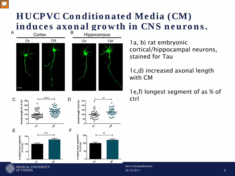
HUCPVC Conditionated Media (CM) induces axonal growth in CNS neurons.
Vera Vorstandlechner
8
1a, b) rat embryonic cortical/hippocampal neurons, stained for Tau 1c,d) increased axonal length with CM 1e,f) longest segment of as % of ctrl
Vorführender
Präsentationsnotizen
Figure 1: HUCPVC Conditioned Media (CM) induces axonal growth I n CNS neurons. (A,B) Effect of CM in axonal outgrowth on cortical and hippocampal neurons. At DIV3 neurons were stimulated for 14 hours with CM. Axonal outgrowth was assessed by immunocytochemistry using an antibody against Tau, an axonal specific marker. Images were taken from random neurons using an AxioObserver Z1 fluorescent microscope with a PlanApochromat 20× objective. (C–F) Quantification of axonal length and axonal longest segment. Results show that axonal network increase after 14 h of CM stimulation (C,D), and in addition neurons stimulated with CM have the longest axonal segments (E,F), demonstrating that global application of CM to both hippocampal and cortical neurons induce an increase in axonal outgrowth. Axonal length and longest axonal segments analysis was performed with Image J 1.45e software. Bars and plots represent the mean ± SEM of approximately 45 neurons randomly selected of 3 independent experiments. (C) ****Represents p < 0.0001; (D) *Represents p = 0.0025; (E) ***Represents p = 0.0010; (F) *Represents p = 0.0083 by Mann Whitney unpaired t-test when compared to Ctr. The scale bar is 25 µm. Tau-Protein = Zytoskelett-Protein; Tauopathien etc….

30.10.2017
Microfluidic chambers for culturing CNS neurons
Vera Vorstandlechner 9
2a,b) composition of microfluidic chambers allow separation of axons from soma and dendrites 2c) tubulin-staining of neurons in microfluiic chamber DIV 5-6
Vorführender
Präsentationsnotizen
Figure 2: Microfluidic chambers for culturing CNS neurons. (A,B) Representative model of a microfluidic chamber. These small systems (20 mm × 25 mm) consist of a molded PDMS chamber placed against a glass coverslip. The microfluidic device consist of a somal compartment (red) and an axonal compartment (blue), each 1.5 mm wide, 7 mm long, which are separated by microgrooves (450 μm long, 10 μm wide). Neurons are plated in the somal compartment and between days 4–5 the axons pass through the microgrooves into the axonal compartment. The height difference between microgrooves (3 μm) and compartments (100 μm) combined with a minimal volume difference between the two sides (~25 μl) leads to a fluidic isolation between the two compartments. (C) Representative image of cortical neurons cultured in microfluidic chambers. At DIV5-6, cortical neurons were immunostained for tubulin (red) and stained for DNA (blue). The image shows that cell bodies are restricted to the somal compartment while in the opposite compartment only axons are observed. Thus microfluidic chambers allow axonal isolation and specific manipulation of distal axons without soma contribution (for further details see the Material and methods section and ref. 17). Contiguous images were taken from a random area of the microfluidic chamber using an AxioObserver Z1 fluorescent microscope with a PlanApochromat 20× objective and assembled into a single image using the ZEN 2011 software. The scale bar is 100 µm.
30.10.2017
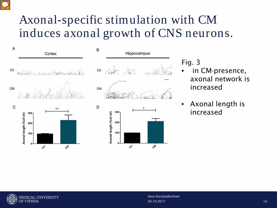
Axonal-specific stimulation with CM induces axonal growth of CNS neurons.
Vera Vorstandlechner
10
Fig. 3 • in CM-presence,
axonal network is increased
• Axonal length is increased
Vorführender
Präsentationsnotizen
Figure 3: Axonal-specific stimulation with CM induces axonal growth of CNS neurons. (A,B) Effect of local application of CM on axonal outgrowth. Cortical (A) and hippocampal (B) axons present in the axonal compartment were stimulated with CM for 24 h at DIV5-6. Axonal outgrowth was evaluated by immunocytochemistry using anti-tubulin βIII. The area selected comprises 3 mm of chamber length which comprises the area between the first and the last microgrooves. Contiguous images were taken using an AxioObserver Z1 fluorescent microscope with a PlanApochromat 20× objective and assembled into a single image using the ZEN 2011 software. (C,D) Quantification of axonal length. Results show that in the presence of CM the axonal network significantly increases comparatively to control, demonstrating that specific local application of CM to cortical and hippocampal axons promotes axonal outgrowth. Axonal network was measured using Neurolucida software. Bars represent the mean ± SEM of 3 independent experiments. *Represents p = 0.0286 by Mann Whitney unpaired t-test when compared to Ctr. (C) **Represents p = 0.0079 by Mann Whitney unpaired t-test when compared to Ctr. The scale bar is 250 µm.

30.10.2017
BDNF is an important molecule for CM-induced axonal outgrowth in cortical neurons.
Vera Vorstandlechner
11
Fig 4) TrkB Fc neutralizes BDNF in CM
TrkB Fc = BDNF binding/neutralizing molecule
Vorführender
Präsentationsnotizen
Fig 4) TrkB Fc reduces the levels of BDNF present in CM. (A) HUCPVC conditioned media was incubated with TrkB Fc and the levels of BDNF were determined by the Bioplex-Luminex assay. Our results show that TrkB Fc is able to cause a reduction of approximately 25% in the levels of BDNF in CM (74.37 ± 1.86), comparatively to CM (100.0 ± 3.35) proving that TrkB Fc is able to significantly deplete BDNF from CM. (B) BDNF concentration in CM is 37 pg/ml. On the other hand, the presence of TrkB Fc, reduces the levels of BDNF present in CM to approximately 27 pg/ml. BDNF levels were measured using a MILLIPLEX Kit. *Represents p = 0.0216 by Mann Whitney unpaired t-test when compared to CM. TrkB Fc binds and neutralizes BDNF

30.10.2017
BDNF is the main component of CM-induced axonal outgrowth in cortical neurons
Vera Vorstandlechner
12
5a) BDNF-depletion from CM reduced CM-mediated axonal outgrowth 5b) axon outgrowth with TrkB Fc treated CM was similar to basal levels
Vorführender
Präsentationsnotizen
Figure 5: BDNF is the main component of CM-induced axonal outgrowth in cortical neurons. (A) Depletion of BDNF prevented CM-induced axon outgrowth. Cortical neurons were globally stimulated with CM for 14 h at DIV5-6. Axonal outgrowth was assessed by immunocytochemistry using an antibody against Tau. Images were taken from random neurons using an AxioObserver Z1 fluorescent microscope with a PlanApochromat 20× objective. Global application of CM induced axon outgrowth, as previously described. However, when BDNF is depleted from the conditioned media using a specific antibody this effect is abolished. In the absence of BDNF, the axonal length and the range of the longest axons are similar to control values, indicating that BDNF is one the major molecules involved in the CM-induced axonal outgrowth effect. (B,C) Quantification of axonal length (B) and axonal longest segment (C). Axonal length and longest axonal segments measuring was performed with Image J 1.45e software. Results show that CM stimulation increases the axonal network, and in addition neurons stimulated with CM have the longest axonal segments. However, when BDNF is depleted, with the addition of 1 µg/mL of TrkB Fc, a molecule that binds specifically to BDNF molecules neutralizing them, there is no increase in axonal length nor in the axonal longest segments. Bars and plots represent the mean ± SEM of approximately 60 neurons randomly selected of 4 independent experiments. (B) ***Represents p = 0.0004 by one-way ANOVA analysis of variance using Tukey post test when Ctr is compared to CM; #represents p = 0,0307 by one-way ANOVA analysis of variance using Tukey post test when CM is compared to CM + TrkB Fc. (C) **Represents p = 0,0014 by one-way ANOVA analysis of variance using Tukey post test when Ctr is compared to CM; #represents p = 0.0341 by one-way ANOVA analysis of variance using Tukey post test when CM + TrkB Fc compared to CM. The scale bar is 25 µm.

30.10.2017
BDNF is the molecule responsible for CM-induced axonal elongation in distal cortical axons
Vera Vorstandlechner
13
Fig. 6: Local effect of CM/CM+TrkB Fc/control • on axonal length • Before and after 24h of
stimulation
• BDNF acts locally witout contribution from the cell body
Vorführender
Präsentationsnotizen
Figure 6: BDNF is the molecule responsible for CM-induced axonal elongation in distal cortical axons. (A–C) Quantification of axonal outgrowth with the MATLAB script AxoFluidic. Profile along the xx axis of the microfluidic device showing the area occupied by the axons at 0 hours (clear red area) and at 24 hours (clear green area). Red line represents axonal projection at 0 hours and the green line represents axonal projection at 24 hours. (D) Quantification of axonal projections outgrowth between 0 and 24 hours. Distal axons treated with CM for 24 hours present more pronounced elongation than control or CM-lacking BDNF axons. Bars represent the mean ± SEM of 6 microfluidic chambers randomly selected of 3 independent experiments. *Represents p = 0.0346 by one-way ANOVA analysis of variance using Tukey post test when Ctr is compared to CM; #represents p = 0.0145 by one-way ANOVA analysis of variance using Tukey post test when CM is compared to CM + TrkB Fc.

30.10.2017
BDNF works as a localized signal in CM-induced axonal outgrowth
Vera Vorstandlechner
14
Fig. 7 Calculation of growth rate • CM-treated axons had
4-fold outgrowth rate • TrkB Fc-mediated
BDNF-depletion attenuates outgrowth rate
Vorführender
Präsentationsnotizen
Figure 7: BDNF works as a localized signal in CM-induced axonal outgrowth. (A) Depletion of BDNF from CM in distal cortical axons abolished CM-induced axonal outgrowth. The axonal outgrowth of individual axons was assessed by live-cell imaging at the moment of CM addition to the axonal compartment of microfluidic chambers and 24 h later. The results show that BDNF depletion results in the abolishment of CM-induced axonal outgrowth in isolated cortical axons, in agreement with the results observed in Fig. 6, demonstrating that BDNF is a key component of CM-induced axonal elongation. Contiguous images were taken using an AxioObserver Z1 fluorescent microscope with a PlanApochromat 20× objective and assembled into a single image using the ZEN 2011 software. (B) Quantification of axonal growth rate. Fresh NBM, CM and CM + Trkb-Fc were added to the axonal compartment at DIV4 and axons allowed to develop for 24 hours. After this period, new images were acquired at the exact same position in the axonal compartment of microfluidic chambers, and the growth rate of individual axons were analyzed with Image J 1.45e software (see Material and methods for details). Results show an increase in axonal growth rate during CM stimulation, however depletion of BDNF blocks the observed axonal growth rate after CM stimulation. Plots represent the mean ± SEM of approximately 30 axons selected of 3 independent experiments. ****Represents p < 0.0001 by one-way ANOVA analysis of variance using Tukey post test when Ctr is compared to CM; ##represents p = 0.0080 by one-way ANOVA analysis of variance using Tukey post test when CM is compared to CM + TrkB Fc. The scale bar is 50 µm.

30.10.2017
Proposed model for secretome-induced axonal outgrowth
Vera Vorstandlechner
15
Fig. 8: BDNF (from MSC-secretome) binds to TrkB in the membrane of growth cones, activates signalling pathways responsible for axonal outgrowth
Vorführender
Präsentationsnotizen
Figure 8: Proposed model for secretome-induced axonal outgrowth. MSCs secretome is composed of a diverse set of molecules, including trophic factors, cytokines, microvesicles and exosomes (1). When the secretome is applied to distal axons (2), BDNF, one of the molecules present, will bind to TrkB present in the membrane of growth cones (3). This interaction leads to the activation of the receptor and downstream signaling pathways, resulting in the activation of the machinery necessary for axonal outgrowth (4). The secretome can act locally, at sites distant from the cell body, engaging intra-axonal mechanisms effectively promoting axonal elongation.

30.10.2017
Discussion
Vera Vorstandlechner
16
• Pros:
• Easily understandable paper, conclusive figures
• Microfuidic-chamber model
• Cons:
• TrkB Fc relevance in vitro? only inconclusive in vivo data
• Exact amount of BDNF in their CM? easy, just do an ELISA!
• What about other neurotrophic/growth factors?
• Valid control?
Compare BDNF only (e.g. recombinant) to CM
• Signalling cascade/downstream molecules of BDNF after CM-/vs. BDNF treatment?
• Relevance for human use/translational science?

30.10.2017
Secretomes of apoptotic mononuclear cells ameliorate neurological damage in rats with focal ischemia. Altmann P, Mildner M, Haider T, Traxler D, Beer L, Ristl R, Golabi B, Gabriel C, Leutmezer F, Ankersmit HJ. 19 Jun 2014, 3:131 | DOI: 10.12688/f1000research.4219.1
Vera Vorstandlechner
17
Abbreviations
• rMNC apo sec/hMNC apo sec = rat/human apoptotic mononuclear
cells
• MCAO = middle cerebral artery occlusion
• HLV = hemispheric lesion volume

30.10.2017
Methods
Vera Vorstandlechner
18
• Animals: 84 adult male Wistar rats
• Production of rat MNC-secretome: harvesting of spleens, lysing of red blood cells,
irradiation (45 Gy), resuspension in serum-free medium, cultivated for 18h, cells
removed (centrifugation), lyophilisation
• Production of human MNC-secretome: GMP-according; venous blood samples; Ficoll-
separation, irradiation, concentration: 25x10^6 cells/ml; methylene blue and light
treatment, gamma irradiation for pathogen removal
• Verification of apoptosis via flow cytometry
• Animal experiment for focal ischemia: MCAO via suture model as prescribed
• Postoperative MNC-administration as descriebed in Fig.1)
• Neurological evaluation (blinded investigator): 7 point-scale: left forepaw extension,
instability to lateral push from right, tail hanging, walking on ground, whisker
movement on the left, hearing, and vision
• Determination of BDNF in rat plasma: injection intraperitoneally; Euthanization;
measurement of BDNF with ELISA in rat plasma

30.10.2017
Experimental study setting
Vera Vorstandlechner
19
Fig 1: two study settings using different time points for rMNC apo sec/hMNC apo sec administration
Vorführender
Präsentationsnotizen
Figure 1: Experimental study setting.For setting 1, rMNC apo sec (apoptotic MNC-secretomes from rats) were injected 40 minutes after MCAO (blue arrow). In setting 2, hMNC apo sec (apoptotic MNC-secretomes from humans) were administered twice at 40 minutes (0.7 hours) and 24 hours after MCAO (red arrows). In both settings, neurological evaluations were performed at 0 hours (before surgery) as well as 6, 24, and 48 hours after surgery (boxes). Both treatment and control animals were euthanized 48 hours after surgery and brain slices were treated with TTC (2,3,5-triphenyltetrazolium chloride) to stain ischemic areas in the brain.

30.10.2017
Apoptotic MNC-secretomes reduce the infarction volume in an experimental MCAO model
Vera Vorstandlechner
20
Figure 2 Represantative brain slices of rats with MCAO, treated with hMNC and control (48h after MCAO)
Vorführender
Präsentationsnotizen
Figure 2: Brains were stained with a 2% solution of TTC forty-eight hours after MCAO. Animals received either treatment (in this representative scan: hMNC apo sec) or control medium, in this case, 40 minutes 24 hours after surgery. White areas indicate ischemic tissue while red areas stain for non-ischemic tissue. Animals treated with control medium (left image) had larger ischemic (=white) areas than animals treated with hMNC apo sec (right image).

30.10.2017
Apoptotic MNC-secretomes improve neurological outcome in an experimental MCAO model
Vera Vorstandlechner
21
Fig. 3: Quantification of viable cells in irradiated vs non irradiated cultured MNCs
Fig. 4: rMNC apo sec/hMNC apo sec decreased HLV after MCAO in two experiment settings
Vorführender
Präsentationsnotizen
Figure 3: Apoptosis rates in irradiated and non-irradiated cultured rMNCs.Mean percentage values of viable (white) and apoptotic (red) rMNCs are given in this bar graph. The percentage of cells in either state are shown for non-irradiated rMNCs (control) and irradiated rMNCs after 18 hours in cell culture. Irradiated rMNCs correspond to the compound we used throughout the study. Figure 4: Infarct volumes in control animals and animals treated with apoptotic MNC-secretomes.The percentage of hemispheric lesion volumes (%HLV) are represented as box and whiskers plots, wherein the boxes indicate the 1 st and the 2 nd quartile and the whiskers the minimum and maximum within 1.5 times the interquartile range from the box. Figure 4a shows lesion volumes as the extend of ischemia in setting 1, where apoptotic MNC-secretomes derived from rats were administered 40 minutes after MCAO compared to controls. Figure 4b corresponds to setting 2, where apoptotic MNC-secretomes derived from humans were administered 40 minutes and 24 hours after MCAO. In both settings, MNC apo sec (red boxes) caused a significant decrease in infarct volumes (* p=0.0006 for setting 1, Figure 4a, and * p=0.0041 for setting 2, Figure 4b) compared to the control group (white boxes) that received only cell culture medium.

30.10.2017
Apoptotic MNC-secretomes improve neurological outcome in an experimental MCAO model
Vera Vorstandlechner
22
Fig 5: Neurological outcome of rats after MCAO with rMNC apo
sec/hMNC apo sec or control in two experiment settings • Neurological
eximanation on 4 time points
• Significant neuroscore decrease over time in treatment group
Vorführender
Präsentationsnotizen
Figure 5. Neurological outcome score in control animals and animals treated with apoptotic MNC-secretomes. Mean neuroscores (±SD) are plotted over time. Treated animals (red triangles for setting 1, , and red squares for setting 2, ) improved over time compared to controls (black/white triangles for setting 1, , and black/white squares for setting 2, ). Error bars correspond to +/- one standard deviation.

30.10.2017
Apoptotic MNC-secretomes activate signaling cascades involved in cytoprotection in glia cells
Vera Vorstandlechner
23
Fig. 6: hMNC apo sec-administration activates/phosphorylates signaling molecules in human Schwann cells (SC) and astrocytes (AC) • Increased phosphorylation of CREB,
Erk1/2, c-Jun, Akt
Vorführender
Präsentationsnotizen
Figure 6. Expression of cytoprotective proteins in human Astrocytes (AC) and Schwann Cells (SCs). Cell extracts were prepared after stimulation with hMNC apo sec or cell culture Medium as control. Western blot analysis revealed an activation of CREB, ERK1/2, c-Jun, Akt, and HSP27. Proteins were normalized to the respective non-phosphorylated proteins.

30.10.2017
Apoptotic MNC-secretomes induce CREB phosphorylation and neuronal sprouting in human primary neurons and contain BDNF
Vera Vorstandlechner
24
Fig. 8: 8a) BDNF is the only neurotophic factor in hMNC apo sec 8b) BDNF-levels are higher in hMNC apo sec –treated rats after
Fig. 7 a) dose-dependent activation of CREB in hMNC apo sec–treated human primary neuron cultures b, c) hMNC apo sec –treatment leads to increase in neuron length
Vorführender
Präsentationsnotizen
Figure 7: Enhanced CREB phosphorylation and neurite length in neurons treated with apoptotic MNC-secretomes.( a) Cell extracts were prepared after stimulation with hMNC apo sec or cell culture medium as control. Western blot analysis for phospho-CREB revealed a dose dependent activation of CREB in astrocytes and in neurons. ( b) Neuron cultures treated with hMNC apo sec or control medium for five days were stained with methylene-blue. One representative picture of ten is shown. Bar=10 µm ( c) Lengths of neurons treated with hMNC apo sec or cell culture medium as control were calculated using ImageJ software. Bars represent the mean of five different cultures. Figure 8. Profile of neurotrophic factors in hMNC apo sec and animals treated with hMNC apo sec. ( a) ELISA for BDNF, GDNF, and NGF detected only levels of BDNF (356±14pg/mL, mean±SEM) in hMNC apo sec. ( b) Six animals received an intraperitoneal injection with hMNC apo sec (n=3, red bar) or control medium (n=3, black/white bar) and BDNF plasma levels were determined 24 hours after administration using ELISA.

30.10.2017
Discussion
Vera Vorstandlechner
25
• „(i) hMNC apo sec activate several mechanisms ultimately
leading to the expression of protective proteins in cultured
primary human glial cells, such as astrocytes, Schwann cells
and human neurons, and
• (ii) induce notable sprouting of neurites in primary neuron
cultures”
• “ … Apoptotic MNCsecretomes derived from human blood
can aid in the development of new treatment strategies in
ischemic stroke.”
• Rat and human secretome hard to compare but similar
effects
30.10.2017
Discussion
Vera Vorstandlechner
26
• Pros
• Conclusive, thoroughly argued study
• Easliy understandable
• Two experiment settings, precisely described
• Large number of animals for significant results
• Effective and easy neurological examination
• Relevance for human use (GMP-according hMNC apo sec)
• Cons
• Limitations of a small animal study
• Exact anti-inflammatory action of hMNC apo sec to be studied

Vera Vorstandlechner
27
Danke! Noch Fragen?