Mesenchymal stem cell responses to mechanical stimulieprints.bice.rm.cnr.it/8135/1/article.pdfwill...
12
Muscles, Ligaments and Tendons Journal 2012; 2 (3): 169-180 169 Review article Robin M. Delaine-Smith, Gwendolen C. Reilly The Kroto Research Institute, Department of Materials Science and Engineering, University of Sheffield, UK Corresponding author: Gwendolen C. Reilly Kroto Research Institute, North Campus University of Sheffield Broad Lane, Sheffield. S3 7HQ e-mail: [email protected] Summary Mesenchymal stem cells (MSCs) have the potential to replace or restore the function of damaged tissues and offer much promise in the successful application of tissue engineering and regenerative medicine strategies. Optimising culture conditions for the pre- differentiation of MSCs is a key goal for the research community, and this has included a number of differ- ent approaches, one of which is the use of mechan- ical stimuli. Mesenchymal tissues are subjected to mechanical stimuli in vivo and terminally differenti- ated cells from the mesenchymal lineage respond to mechanical stimulation in vivo and in vitro. MSCs have also been shown to be highly mechanosensitive and this may present an ideal method for controlling MSC differentiation. Here we present an overview of the response of MSCs to various mechanical stimuli, focusing on their differentiation towards the mes- enchymal tissue lineages including bone, cartilage, tendon/ligament, muscle and adipose tissue. More re- search is needed to elucidate the complex interac- tions between biochemically and mechanically stim- ulated differentiation pathways. Key words: mechanical stimuli, mesenchymal stem cell, osteogenesis, tenogenesis. Introduction Mesenchymal stem cells (MSCs) are a promising cell source for tissue engineering and regenerative medicine strategies and offer an alternative to fully differentiated cells that are often in limited supply due to tissue damage or disease. MSCs have multipotent differentiation poten- tial, self-renewing ability, and apparent immunosuppres- sive properties 1 . Typically MSCs are isolated from the stroma of adult bone marrow (BMSCs), but cells with similar phenotypic characteristics and differentiation ca- pabilities have also been isolated from a range of other mesenchymal tissues including adipose 2 (ADMSCs), ten- don, muscle and skin. MSCs cultured in vitro can be chemically-induced to differentiate into cell types of the mesoderm, including bone, cartilage, tendon/ligament and fat (for a recent review see Vater et al. 3 ). Common biochemical agents and growth factors include: dexam- ethansone (dex) and bone morphogenetic proteins (BMPs) for osteogenesis, serum free medium and trans- forming growth factor ß (TGF-ß) for chondrogenesis, dex, insulin, and 3-isobutyl-1-methylxanthine (IBMX) for adipo- genesis 4 (Fig. 1). It has also been claimed that MSCs are able to differentiate into other tissue types such as smooth muscle, endothelial and nervous tissue 5 . A key task for tissue engineers is to identify the appropri- ate culture conditions for development of a tissue engi- neered construct in vitro ready for implantation in vivo that forms the target tissue and reduces subsequent healing time. It is well known that biochemical cues, such as cy- tokines, growth factors and signalling events 6 , can control the function of stem cells, as well as environmental fac- tors (e.g. surface chemistry and topography) 7,8 but it is also becoming clear that mechanical forces can greatly influ- ence stem cell behaviour. Tissues and cells in the human body are exposed to a range of different external forces, which influence their de- velopment and maintenance 9 . For example, it is well doc- umented that excercise increases bone and muscle mass 10 and inadequate loading as occurs during space flight, prolonged periods of bed rest or spinal cord injury 11 results in decreased muscle and bone mass. MSCs in vivo reside in the stem cell niche, which contains many biochemical factors that function to regulate their behav- iour. In the bone marrow, mechanical forces in the form of tension, compression, and fluid-induced shear are all present 12 but the nature of these forces is not well under- stood, neither is what effect they have on stem cell mo- bilization and function. Many different cell types have been demonstrated to be highly mechanosensitive in vitro 13 and recent TE strate- gies for MSC differentiation have included modifying in- trinsic (via substrate stiffness 8,14 , and external stresses to simulate the physiologically relevant mechanical envi- ronment. Mechanical stimulation of MSCs in vitro has shown that tensile strain enhances osteogenesis and tenogenesis but inhibits adipogenesis 15,16 , hydrostatic pressure and compressive loading induces chondrogen- esis 17 , and fluid flow induced shear stress upregulates genes associated with osteogenesis 18,19 . In this review we will describe some of the outcomes seen when these Mesenchymal stem cell responses to mechanical stimuli
Transcript of Mesenchymal stem cell responses to mechanical stimulieprints.bice.rm.cnr.it/8135/1/article.pdfwill...

Muscles, Ligaments and Tendons Journal 2012; 2 (3): 169-180 169
Review article
Robin M. Delaine-Smith, Gwendolen C. Reilly
The Kroto Research Institute, Department of MaterialsScience and Engineering, University of Sheffield, UK
Corresponding author:Gwendolen C. ReillyKroto Research Institute, North Campus University ofSheffieldBroad Lane, Sheffield. S3 7HQe-mail: [email protected]
Summary
Mesenchymal stem cells (MSCs) have the potential toreplace or restore the function of damaged tissuesand offer much promise in the successful applicationof tissue engineering and regenerative medicinestrategies. Optimising culture conditions for the pre-differentiation of MSCs is a key goal for the researchcommunity, and this has included a number of differ-ent approaches, one of which is the use of mechan-ical stimuli. Mesenchymal tissues are subjected tomechanical stimuli in vivo and terminally differenti-ated cells from the mesenchymal lineage respond tomechanical stimulation in vivo and in vitro. MSCshave also been shown to be highly mechanosensitiveand this may present an ideal method for controllingMSC differentiation. Here we present an overview ofthe response of MSCs to various mechanical stimuli,focusing on their differentiation towards the mes-enchymal tissue lineages including bone, cartilage,tendon/ligament, muscle and adipose tissue. More re-search is needed to elucidate the complex interac-tions between biochemically and mechanically stim-ulated differentiation pathways.
Key words: mechanical stimuli, mesenchymal stem cell,osteogenesis, tenogenesis.
Introduction
Mesenchymal stem cells (MSCs) are a promising cellsource for tissue engineering and regenerative medicinestrategies and offer an alternative to fully differentiatedcells that are often in limited supply due to tissue damageor disease. MSCs have multipotent differentiation poten-tial, self-renewing ability, and apparent immunosuppres-
sive properties1. Typically MSCs are isolated from thestroma of adult bone marrow (BMSCs), but cells withsimilar phenotypic characteristics and differentiation ca-pabilities have also been isolated from a range of othermesenchymal tissues including adipose2 (ADMSCs), ten-don, muscle and skin. MSCs cultured in vitro can bechemically-induced to differentiate into cell types of themesoderm, including bone, cartilage, tendon/ligamentand fat (for a recent review see Vater et al.3). Commonbiochemical agents and growth factors include: dexam-ethansone (dex) and bone morphogenetic proteins(BMPs) for osteogenesis, serum free medium and trans-forming growth factor ß (TGF-ß) for chondrogenesis, dex,insulin, and 3-isobutyl-1-methylxanthine (IBMX) for adipo-genesis4(Fig. 1). It has also been claimed that MSCs areable to differentiate into other tissue types such as smoothmuscle, endothelial and nervous tissue5. A key task for tissue engineers is to identify the appropri-ate culture conditions for development of a tissue engi-neered construct in vitro ready for implantation in vivo thatforms the target tissue and reduces subsequent healingtime. It is well known that biochemical cues, such as cy-tokines, growth factors and signalling events6, can controlthe function of stem cells, as well as environmental fac-tors (e.g. surface chemistry and topography)7,8 but it is alsobecoming clear that mechanical forces can greatly influ-ence stem cell behaviour.Tissues and cells in the human body are exposed to arange of different external forces, which influence their de-velopment and maintenance9. For example, it is well doc-umented that excercise increases bone and musclemass10 and inadequate loading as occurs during spaceflight, prolonged periods of bed rest or spinal cord injury11
results in decreased muscle and bone mass. MSCs invivo reside in the stem cell niche, which contains manybiochemical factors that function to regulate their behav-iour. In the bone marrow, mechanical forces in the formof tension, compression, and fluid-induced shear are allpresent12 but the nature of these forces is not well under-stood, neither is what effect they have on stem cell mo-bilization and function.Many different cell types have been demonstrated to behighly mechanosensitive in vitro13 and recent TE strate-gies for MSC differentiation have included modifying in-trinsic (via substrate stiffness8,14, and external stresses tosimulate the physiologically relevant mechanical envi-ronment. Mechanical stimulation of MSCs in vitro hasshown that tensile strain enhances osteogenesis andtenogenesis but inhibits adipogenesis15,16
, hydrostaticpressure and compressive loading induces chondrogen-esis17, and fluid flow induced shear stress upregulatesgenes associated with osteogenesis18,19. In this review wewill describe some of the outcomes seen when these
Mesenchymal stem cell responses to mechanical stimuli

stimuli have been applied to MSCs. However, due to thewide variety of mechanical stimuli available, the enormousarray of possible loading conditions, and the addition ofdifferent chemical stimulants, the optimum conditions forcontrolling lineage specific MSC differentiation remainunspecified.
Mechanical Regulation of MSCs
The most extensively studied differentiation pathways ofMSCs are osteoblastic, chondrogenic, and adipogenic,other pathways such as tenogenesis and myogenesishave also been investigated to a lesser extent. The opti-mum durations, magnitudes and frequencies of mechan-ical loading for lineage specific differentiation of MSCs isnot known due to the difficulty of undertaking multipleloading regimens within one set of experiments. The re-sponse of MSCs to loading are likely to be age-specific,may be specific to site of origin, and appear to depend onhow differentiated the cells are at the time of loading, aswell as whether loading is performed in conjunction withbiochemical supplements20,21. There are many ways in which researchers have stimu-lated cells with mechanical forces in vitro which can gen-
erally be categorized into the primary type of stress theyinduce22
. These include stretching (tensile stress)23 hydro-static pressure or platen abutment (compressive stress),fluid flow (shear stress)24,25, ultrasound26,27, high frequency,low magnitude displacement (vibration)28,29, and directcell membrane magnetic stimuli30. For each stimulationmode the stimulus can be applied in 2D (monolayer cul-ture) or 3D (multilayer culture) and differences betweencells cultured in 2D and 3D have been observed in termsof cellular morphology and migration strategies, matrix ad-hesion, gene and protein expression and responses tofluid flow31. It is important to note that in many loading systems therewill be secondary effects along with the main mechanicalstimulus. For example, in 3D tensile and compressionloading systems, there will be fluid drawn in and out of thescaffold causing shear stress at the cell membrane andimproved nutrient transfer to the cells32. Also, the cells willmost likely be subjected to additional compressive ortensile forces caused by substrate bending or the Pois-son’s effect. Scaffold architecture will also regulate howmuch of the applied force is received by the cells, for ex-ample in a cell-seeded gel the loading received will be rel-atively homogeneous throughout the scaffold whereas aporous scaffold will have an uneven strain transfer. In-
R. M. Delaine - Smith et al.
170 Muscles, Ligaments and Tendons Journal 2012; 2 (3): 169-180
Figure 1. Diagram summarising the lineage potential of adult human MSC. The figure depicts the in vitro culture conditions (boxed) usedto promote the differentiation into the lineage indicated and some of the signalling pathways and transcription factors involved in theprocess (italics). Reprinted from Arthritis Research and Therapy (4), BioMed Central, with kind permission of Professor Tuan.

creased nutrient transfer from fluid flow can result in bet-ter cell infiltration and matrix distribution, as well as en-hancement of cell differentiation due to mass transport ef-fects33. Therefore, in order to optimise stimulationregimens it is important to identify the effects of individ-ual stimuli.
Tensile Loading
Tenogenesis: when targeting tenogenesis, MSCs are of-ten seeded on collagen-based or collagen coated scaf-folds and then cultured in standard media as there is cur-rently no defined medium for inducing tenogenesis ofMSCs. Chen et al.34 subjected hBMSCs to 3% and 10%global strain and observed an increase in Collagen typeI (Col I), Col III, and tenascin-C mRNA at 10%, whereas3% strain favoured osteogenic differentiation. Farng etal.35 subjected mouse BMSC-seeded poly(caprolactone)scaffolds to 10% strain, which also increased tenogenicgene production of Col I, Col III, and the tendon transcrip-tion factor scleraxis. Zhang et al.36 observed the effects ofvarying the time period (3-36 h) rat BMSCs were sub-jected to 10% cyclic strain and saw that Col I, Col III, andtenascin-C mRNA were upregulated after 24 h. 10%strain at a low frequency (0.0167 Hz) on human andbovine BMSCs in collagen gels also resulted in an upreg-ulation of Col I, Col III, and tenascin-C mRNA, but thistook 14 days of culture37. Strains lower than 10% havealso been used in an attempt to induce tenogenesis. 1%strain was applied to human BMSC-seeded collagen gelsresulting in an upregulation of Col III and maintaining thelevel of scleraxis mRNA, whereas in static controls the ex-pression reduced over time38. The effect of a 6.7N staticstrain was observed by van Eijk et al.39 on goat BMSC-seeded PLGA scaffolds. Initially, collagen content washighest in scaffolds strained during seeding, but after 21days, unloaded scaffolds had the highest collagen contentsuggesting that a constant strain inhibits differentiation.
Myogenesis: the proportion of MSCs that have exhibit amyogenic phenotype is low using defined or conditionedculture medium40. Some studies have suggested thatmyogenic differentiation of MSCs can be influenced bymechanical tension. Increased gene and protein expres-sion associated with myogenesis have been seen whenbovine MSCs were subjected to stretching including thecalcium binding protein calponin41, the calcponin relatedprotein SM22α42 and α smooth muscle actin (SMA)43,44.However, Park et al.42 found that while uniaxial strain in-creased SM22α expression, equiaxial strain reduced itsexpression highlighting the different regulatory roles ofthese two stimuli. Ku et al.45 subjected human BMSCs tostrains of 7-20% over 4 days and observed the highestcollagen production at 14% strain and an increase in ly-syl oxidase (Lox), however, there were no increases in ColII mRNA (the collagen found in cartilage) or AlkalinePhosphatase (ALP) activity. ALP is involved in bone min-eralisation and high ALP activity is used to indicate os-teogenic differentiation. Colazzo et al.46 subjected hu-man BMSCs and ADMCSs to 14% strain for 3 dayscausing increases in collagen production and Col IV
mRNA, and upregulation of Col III and elastin cross-link-ing in ADMSCs. Huang et al.40 tested a range a loadingregimens and showed that 10% stretch at 1 Hz for 24hours was optimal for inducing cardimyocyte gene upreg-ulation in rat MSCs compared to lower or higher strainsand frequencies, interestingly the stretch stimulus wasmuch more effective than a 1 Pa unidirectional shearstress stimulus. In agreement, Maul et al.47 compared theeffects of cyclic tension, compression, and fluid flow in-duced shear stress on the expression of smooth musclerelated proteins and only stretch (1-10% at 1or 2.75 Hz)upregulated SMA and calponin.
Osteogenesis: application of cyclic tensile loading toMSCs has resulted in increased expression of early os-teogenic markers as well as increased mineralised matrixdeposition, both in the presence and absence of os-teogenic media. Bone morphogenic protein 2 (BMP-2) ex-pression was upregulated following cyclic loading of hu-man BMSC-seeded gels48,49 and rat ADMSCs on 2Dsubstrates50. Alkaline phosphatase activity (ALP) in-creased with 1 week of cyclic loading in human BMSCs.It appears that dexamethasone can have a synergistic ef-fect or an inhibitory effect on mechanically induced osteo-genesis, depending on the concentration used and themarker investigated. For example Jagodzinski et al.51 ap-plied tensile strain to human BMSCs for 6 hours/day, onthe first 3 days of culture, at two different strain rates (2%and 8%). Cyclic tensile strain upregulated COLI mRNAand ALP activity, but only at the 8% strain rate. Stretch-ing alone was seen to be as effective as dex treatmentalone and there was a synergistic effect of the combina-tion of dex and cyclic tensile strain. Muaney et al.52 inves-tigated the effect of the concentration of dex (0, 10 or100nM) on the osteogenic enhancing potential of loading(bending). Without dex, loading alone was able to upreg-ulate ALP activity and expression, but it had no effect onthe bone matrix proteins osteopontin (OPN) and oscteo-clacin (OCN). The addition of 10nM dex seemed to causea synergistic response but at higher dex levels (100nM)the effect of loading was suppressed.
Summary of tensile stimuli: the literature agrees in gen-eral that osteogenesis of MSCs tends to occur at strainmagnitudes lower than that for tenogenesis and that car-diomyogenesis is stimulated by using larger strains. Inthe absence of osteogenic media, upregulation of early(ALP activity) and late (mineralized matrix deposition)osteogenic markers have been observed53. Tenogene-sis is also induced in the absence of any chemical induc-ers, but as for osteogenesis, it appears that staticstretching or long term continuous loading has a nega-tive effect on matrix production39,54. Stretching inhibitsadipogenesis even in the presence of adipogenic me-dia55 and stretching does not appear to be favourable forchondrogenesis, although tensile strains induced in amore biomimetic loading regimen, that of sliding contactloading does slightly enhance chondrogenesis ofMSCs56. These findings indicate that intermittent, cyclicstretching of MSCs is beneficial for the osteogenic,tenogenenic, and myogenic lineages and the productionof a fibrous matrix.
Mesenchymal stem cell responses to mechanical stimuli
Muscles, Ligaments and Tendons Journal 2012; 2 (3): 169-180 171

R. M. Delaine - Smith et al.
172 Muscles, Ligaments and Tendons Journal 2012; 2 (3): 169-180
Compressive Loading
Compressive loading of MSCs has mainly been investi-gated for its potential in promoting chondrogeneic differ-entiation. The chondrogenic response of MSCs to load-ing is highly complex and the various loading regimensused and the effects that the time at which loading is ap-plied to the cultures has on the outcome is reviewed inmore detail elsewhere21.
Osteogenesis: a small number of studies have investi-gated the effect that global compressive loading20,57 or hy-drostatic compression58 of cell-seeded scaffolds mayhave on osteogenic induction. Hydrostatic compression atloads lower than those usually used for chondrogenesisupregulated ALP activity and the bone transcription fac-tor RUNX2/cbfa1 in MSCs58. Interestingly early markersof either osteogenesis or chondrogenesis can be inducedby dynamic compression in the same batch of humanMSCs under the same conditions, in an alginate gel-filled collagen sponge, just by varying the strain magni-tude (10% strain induced osteogenic genes 15% inducedboth osteo and chondrogenic genes) but it is not clearwhat type of tissue would be formed by these cells59. In
our studies20 scaffold compression of a polymer scaffoldseeded with human MSCs upregulated genes associ-ated with bone matrix formation (Fig. 2) and enhanced theformation of mineralised bone-like matrix. Continuousloading was not necessary to induce MSC differentiationin that study, just 2 hours of loading every 5 days upreg-ulated calcium deposition by more than 50%. However asdiscussed previously in this type of porous scaffold the in-dividual cells are unlikely to be subjected to compressionbut to secondary effects such as tension and bending ofthe scaffolds struts and fluid flow of media into and out ofthe scaffold20,21. Overall, true compressive loading ofMSCs seems to be beneficial for the production of a non-fibrous, cartilage-like matrix, in contrast to tensile loading.
Fluid Flow Induced Shear Stress
The most commonly used method for inducing shearstresses over a cell monolayer is the parallel-plate flowchamber60,61 although simple lab equipment such as rock-ers and orbital shakers can also be used to apply a char-acterised, though less homogenous flow stimulus tocells62.64 (Brennan 2012). In 3D cultures flow is applied us-
Figure 2. Compression loading of human MSCs in polyurethane foam scaffolds. A: Fluorescent micrograph of a pore of the scaffoldwith MSCs attached (blue = cell nucleus stained with DAPI, red = cell cytoskeleton stained with TRITC-phaloidin). B: PCR analysis ofmRNA for RUNX2, OPN and ALP showed that these genes were only slightly upregulated by the short (2 hour) loading period and notas much as by continuous dex treatment. However Col 1 was upregulated by loading and inhibited by dex which was also reflected incollagen analysis by Sirius red at a later time-point (data not shown). ALP activity was stimulated by loading to levels seen in dex treatedcells as was calcium secretion which was highest with a combination of dex and loading. Adapted from (20) reproduced with kind per-mission from eCM journal (www.ecmjournal.org).

Mesenchymal stem cell responses to mechanical stimuli
Muscles, Ligaments and Tendons Journal 2012; 2 (3): 169-180 173
ing perfusion bioreactors65,66 with a steady, pulsatile anduni-directional flow having all been investigated. The ma-jority of studies have focused on osteogenic differentiationof MSCs. Given that osteoblasts (bone forming cells)and osteocytes (the terminally differentiated cell that re-side in bone matrix) have been repeatedly shown to re-spond to fluid forces in vitro67-69, there is an natural as-sumption that fluid shear stress will influence osteogensisof MSCs. MSCs seeded on 2D substrates are usuallysubjected to levels of shear stress around (0.1-2 Pa)whereas the shear stresses experienced in 3D constructsare often much lower than in 2D experiments (0.1 mPa-0.03 Pa) as summarised in McCoy and O’Brien70. Overall, fluid flow appears to either induce or enhance os-teogenesis in MSCs. Osteogenic human MSCs weresubjected to 1.2 Pa shear forces for either 30 or 90 minsand an increase in ALP activity was seen, with 30mins ofstimulation showing the highest levels71. However, therewas no increase in cbfa1expression and interestingly ColI gene expression was lower after flow. This enhancementof ALP activity was also seen in other 2D systems61 includ-ing our own62. In our laboratory only 1 hour per day of os-cillating flow at 1Hz, beginning on day 5 of culture was suf-ficient to upregulate ALP, collagen and calcium production(Fig. 3). However, some studies have noticed an inhibitionof ALP activity by flow72. Interestingly, Yourek et al.19
noted that although cellular ALP in human MSCs waslower after exposure to 24 hours of continuous fluid flow
compared to static controls there was more ALP releaseinto the media in the cells exposed to flow, which is notusually measured in most experimental set-ups, sug-gesting that flow causes ALP mobilisation, rather than in-hibiting osteogenic differentiation. In those experimentsthere was little additional effect of fluid flow induced searstress when dex was already present but shear stress up-regulated the bone matrix proteins OPN and bone sialo-protein (BSP) and the growth factor BMP-2 to induce os-teogenesis when dex was not present. Steady perfusion of 3D cell-seeded constructs is almostalways reported to stimulate ALP activity, with the great-est effects occurring at the earlier time points of 4-8days65,66,73 and then often levelling off, although in somestudies, significant increases have been seen up to 14-16days of culture74,75. Increasing the flow rate can increasecalcium production up to a point suggesting that increas-ing the fluid flow induced shear stress affects later stagesof differentiation more than earlier stages or is more im-portant for matrix formation than stimulation of differenti-ation66,74. The aim of continuous fluid flow through a scaf-fold is usually to improve nutrient perfusion with anymechanobiological effects of fluid flow induced shearstresses being a positive side-effect. We have shownthat continuous perfusion is not necessary to upregulateALP activity or mineralisation of human MSCs both ofwhich can be upregulated by short bouts of oscillatoryfluid flow in a variety of scaffold types including the
Figure 3. Osteogenic progenitorcells of the hES-MP line weresubjected to oscillatory fluidflow induced shear stress(FFSS) using a simple rockingplatform. ALP activity (A) wassignificantly increased at day14 with FSS for cells cultured inosteogenic media containingdex (OM). Matrix deposition atday 21 (B) was highest in bothFFSS groups for Sirius Red(collagen) and in FSS + OMgroup for Alizarin Red (calcium).Adapted from (62) reproducedwith kind permission from eCMjournal (www.ecmjournal.org).

R. M. Delaine - Smith et al.
174 Muscles, Ligaments and Tendons Journal 2012; 2 (3): 169-180
polyurethane foam described in figure 2 and a nondegradable borosilicate glass scaffold76,77.
Low Magnitude High Frequency Loading
Other methods of mechanically stimulating MSCs in vitrohave included direct straining of cell-bound integrins bymagnetic force for osteochondrogenesis78,79 and low-in-tensity pulsed ultrasound (LIPUS) for promoting osteoge-nesis80-82 or chondrogenesis81,83,84 (reviewed in 21). Whilethese techniques have so far had limited use in the me-chanical stimulation of MSCs, the studies performed sug-gest that they may be useful tools for non-invasive stim-ulation of MSC differentiation. An intriguing recently advocated stimulus for MSC differ-entiation is low magnitude, high frequency (LMHF) load-ing (or vibration) for osteogenesis29,85. This is based on thefindings that whole body vibration in animals enhancesbone formation86 and decreases the formation of adiposetissue87,88. It was also shown by Luu et al.87 that moreMSCs within a population showed commitment towardsthe osteogenic lineage compared to the adipogenic line-age after the animals were subjected to vibration. LMHFvibration could be a way to stimulate cells in 3D scaffoldswithout needing a specific bioreactor tailored to the shapeof the construct and making it much easier to maintainsterile conditions. In our laboratory whole plate vibration(15-60 Hz) was performed on a human MSC cell line(hES-MP from Cellartis) resulting in increased ALP activ-ity in the presence of dex at 60 Hz after 45 min of stimu-lation29. However, we found that the outcome was highlydependent on the precise combination of accelerationrate, frequency and even the serum lot that the cellswere cultured in, ALP was not upregulated at any otherfrequency tested and we did not find effects on extracel-lular matrix deposition. Similarly Lau et al.89 found no ef-fects of a 1h per day 60Hz LMHF loading regimen on ratMSCs. In contrast Sen et al.85 performed LMHF loadingon a mouse MSC cell line in multipotential media and ob-served a down regulation in adiponectin and PPARα (adi-pogenic) gene expression, whereby the mRNA for thebone matrix protein osteocalcin was upregulated. Prelim-inary experiments in our laboratory subjecting MSCs toLMHF loading in a range of 3D scaffolds have also not yetprovided any evidence of a positive effect of LMHF on os-teogenesis. However Zhou et al.90 seeded MSC on dem-ineralised bone scaffolds and found upregulation for themRNA of a cbfa1 and range of osteogenic matrix proteins.As very little is understood about how cells sense theseLMHF movements these different results could be ex-plained by different orientation and arrangements of cellsrelative to the substrate movement causing the vibrationto initiate very different mechanosensory effects.
Summary of interactions between loading and bio-chemical supplements
There are a number of studies that have shown mechan-ical loading alone is able to induce expression of os-teogenic genes (Runx2, osteopontin, osteocalcin)19,61 and
a few that report calcium deposition by MSCs can be stim-ulated as a result of mechanical stimulus alone91. In mostcases, the addition of dex appears to enhance the sen-sitivity of MSCs to shear stress or strain including enablingmechanoregulated-increased calcium deposition62. How-ever collagen production by MSCs can be inhibited bydex, including in our studies20,62 and tenogenesis in termsof improved Col1 production seems to occur best with noadditional supplements36,92. Smooth muscle differentiationstimulated either by fluid flow induced shear stress orcyclic tensile strain was enhanced synergistically withthe addition of 5-aza40,60. It may be that biochemical fac-tors and mechanical forces need to work synergisticallyto stimulate specific pathways or that MSCs need to beat a certain level of maturity (along the specific differen-tiation pathway) before they sense the load.
Mechanical regulation of MSC proliferation and mi-gration
When Li et al.72 subjected hMSCs to oscillatory fluid flow,they noticed an increase in intracellular calcium mobiliza-tion as well as cell proliferation and in another study byRiddle et al.93, fluid shear stresses were seen to enhancehMSC proliferation in part due to calcium signalling. Cyclictensile strain can also increase MSC proliferation asdemonstrated by Ghazanfari et al.94. In contrast, there aremany studies that have shown mechanical stimulation tohave no effect on cell proliferation95-97 as well as reducingMSC proliferation16,43,98. These mixed findings can mostlikely be explained by the diversity of conditions includingmechanical stimulation, MSC species, and culture mediaused, as well as the wide range of loading parametersused. However, high strains and flow rates can be alsodetrimental to cell viability for instance Kearney et al.99
subjected rat MSCs to uniaxial cyclic strain and observedthat 7.5% strain or greater lead to cell apoptosis. In MSC-seeded tissue engineered constructs, flow perfu-sion, even for short bouts, appears to cause cells tospread evenly through the entirety of the construct, com-pared with poor spreading under static conditions, indicat-ing that flow improves cell mobility100. However Ode etal.101 showed that MSCs in a fibrin clot subjected to highstains of 20% (aimed at mimicking a fracture healing en-vironment) had a lower ability to migrate compared to nonstrained cells, an effect mediated by the surface proteinsCD73 and integrin ß 1. Mechanical loading has been shown to affect cell orien-tation and spreading, and in particular substrate strain cancause elongation and alignment of MSCs41,43,102
. Differ-ences in direction of alignment relative to the load direc-tion have been observed with MSCs seeded on 2D sub-strates orientating perpendicular41,43 and MSCs seeded in3D gels orientating parallel102. It is thought that on 2D sub-strates, cells re-orientate to minimize the stretch forces feltby the cell body42 while in 3D matrices, elongation ofscaffold pores and struts caused by the strain may dictatecellular orientation. Fluid flow in a parallel plate flowchamber can also induce cell morphological changesand orientation usually in parallel to the flow direction,when it is unidirectional. Interestingly even when rat BM-
SCs from the same batch, grown in the same bioreactorare subjected to fluid flow or tensile strain they align par-allel to the flow direction but perpendicular to the tensilestrain direction47.
Mechanotransduction mechanisms
Targeting the mechanisms responsible for the conversionof extracellular mechanical stimuli into biochemical signalswill aid with future stem cell strategies. There have beena number of possible cell membrane mechanoreceptorsidentified including integrins (transmembrane proteins),stretch activated ion channels and g-protein coupled re-ceptors, the pericellular glycocalyx, and the non-motile pri-mary cilia. Integrins couple the cytoskeleton to the ECM and clusterat focal adhesion points on the cell surface forming an in-tegrin-ligand bond with the ECM. Application of an exter-nal force pulls on the integrin-ligand bond, which transfersacross the cell membrane and can result in cytoskeletaldeformation. Another mechanism involves deformation ofthe plasma membrane causing ion flux into/out of the cellvia stretch activated ion channels or g-protein coupled re-ceptors103. The third proposed mechanism, the glycoca-lyx, is a pericelllular GAG-proteoglycan rich layer sur-rounding the cell membrane that creates a drag forcewhen fluid flow passes over causing plasma membranedeformation104-106. More recently, the primary cilium, an im-motile microtubule-based organelle, that protrudes like anantenna from the apical cell surface, has been implicatedas a mechanosensor in a variety of cell types includingMSCs107. Primary cilia have been shown to bend underfluid flow107, adjust their length in response to load in ten-
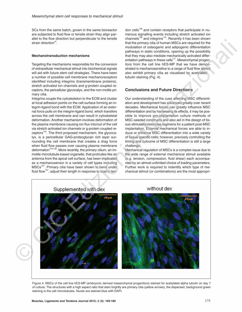
don cells108 and contain receptors that participate in nu-merous signalling events including stretch activated ionchannels109 and integrins110. Recently it has been shownthat the primary cilia of human MSCs are required for themodulation of osteogenic and adipogenic differentiationpathways in static conditions, opening up the possibilitythat they may also mediate mechanically activated differ-entiation pathways in these cells111. Mesenchymal progen-itors from the cell line hES-MP that we have demon-strated is mechanosensitive to a range of fluid flow stimulialso exhibit primary cilia as visualised by acetylated αtubulin staining (Fig. 4).
Conclusions and Future Directions
Our understanding of the cues affecting MSC differenti-ation and development has advanced greatly over recentdecades. Mechanical forces can greatly influence MSCdifferentiation and by harnessing its effects, it may be pos-sible to improve pre-implantation culture methods ofMSC-seeded constructs and also aid in the design of tis-sue stimulation/exercise regimens for a patient post-MSCimplantation. External mechanical forces are able to in-duce or enhance MSC differentiation into a wide varietyof tissue specific cells; however, precisely controlling thetiming and outcome of MSC differentiation is still a largechallenge. Mechanical regulation of MSCs is a complex issue due tothe wide range of external mechanical stimuli available(e.g. tension, compression, fluid shear) each accompa-nied by an almost unlimited choice of loading parameters.Further work is required to indentify which type of me-chanical stimuli (or combinations) are the most appropri-
Mesenchymal stem cell responses to mechanical stimuli
Muscles, Ligaments and Tendons Journal 2012; 2 (3): 169-180 175
Figure 4. MSCs of the cell line hES-MP (embryonic derived mesenchymal progenitors) stained for acetylated alpha tubulin on day 7of culture. The structures with a high aspect ratio that stain brightly are primary cilia (yellow arrows), the dispersed, background greenstaining is the cell microtubules. Nuclei are stained blue with DAPI.

ate as well as the load magnitude, duration and fre-quency, and when to initiate the loading during culture, inorder to pinpoint the optimal strategies. Characterisationof the exact forces MSCs experience in loading systemsis important to know what mechanisms are actually induc-ing the observed responses and to help simplify the de-sign of future loading systems. While most bioreactorsystems employ a common force type (e.g. tension orcompression), there are also likely to be other mecha-nisms at work causing significant secondary effects whichcan cause a misinterpretation in the reason for the ob-tained results. Mathematical and computer modelling isimportant for characterising the forces that are being ex-perienced by the cells and will subsequently provide a bet-ter understanding of what the cell is responding to112.
Acknowledgements
RMD-S is sponsored by the Engineering and PhysicalSciences Research Council.
References
1. Sioud M, Mobergslien A, Boudabous A, Floisand Y.Mesenchymal stem cell-mediated T cell suppres-sion occurs through secreted galectins. Int J Oncol2011;38(2):385-390.
2. Zuk PA, Zhu M, Mizuno H, Huang J, Futrell JW, KatzAJ et al. Multilineage cells from human adipose tis-sue: Implications for cell-based therapies. TissueEng 2001;7(2):211-228.
3. Vater C, Kasten P, Stiehler M. Culture media for thedifferentiation of mesenchymal stromal cells. ActaBiomater 2011;7(2):463-477.
4. Tuan RS, Boland G, Tuli R. Adult mesenchymal stemcells and cell-based tissue engineering. Arthrit ResTher 2003;5(1):32-45.
5. Nombela-Arrieta C, Ritz J, Silberstein LE. The elusivenature and function of mesenchymal stem cells. NatRev Mol Cell Biol 2011;12:126-131.
6. Augello A, De Bari C. The Regulation of Differentia-tion in Mesenchymal Stem Cells. Hum Gene Ther2010;21(10):1226-1238.
7. Dalby MJ, Gadegaard N, Tare R, Andar A, RiehleMO, Herzyk P et al. The control of human mes-enchymal cell differentiation using nanoscale symme-try and disorder. Nature Materials 2007;6(12):997-1003.
8. Reilly GC, Engler AJ. Intrinsic extracellular matrixproperties regulate stem cell differentiation. J. Bio-mech 2010;43(1):55-62.
9. Vogel V. Mechanotransduction involving multimodu-lar proteins: Converting force into biochemical signals. Annu Rev Biophys Biomol Struct2006;35:459-488.
10. Bonnet N, Ferrari SL. Exercise and the Skeleton:How It Works and What It Really does. InternationalBone and Mineral Society 2010;7:235-248.
11. Dudley-Javoroski S, Shields RK. Muscle and boneplasticity after spinal cord injury: Review of adapta-
tions to disuse and to electrical muscle stimulation.J Rehabil Res Dev 2008;45(2):283-296.
12. Gurkan UA, Akkus O. The Mechanical Environmentof Bone Marrow: A Review. Ann Biomed Eng2008;36(12):1978-1991.
13. Orr AW, Helmke BP, Blackman BR, Schwartz MA.Mechanisms of mechanotransduction. Dev Cell2006;10(1):11-20.
14. De Santis G, Lennon AB, Boschetti F, Verhegghe B,Verdonck P, Prendergast PJ. How can cells sensethe elasticity of a substrate? An analysis using a celltensegrity model. European Cells and Materials2011;22:202-213.
15. Sen B, Xie ZH, Case N, Ma MY, Rubin C, Rubin J. Me-chanical Strain Inhibits Adipogenesis in MesenchymalStem Cells by Stimulating a Durable beta-CateninSignal. Endocrinology. 2008;149(12):6065-6075.
16. Simmons CA, Matlis S, Thornton AJ, Chen SQ,Wang CY, Mooney DJ. Cyclic strain enhances ma-trix mineralization by adult human mesenchymalstem cells via the extracellular signal-regulated ki-nase (ERK1/2) signaling pathway. J Biomech2003;36(8):1087-1096.
17. Luo ZJ, Seedhom BB. Light and low-frequency pul-satile hydrostatic pressure enhances extracellularmatrix formation by bone marrow mesenchymal cells:an in-vitro study with special reference to cartilage re-pair. Proceedings of the Institution of MechanicalEngineers Part H-Journal of Engineering in Medicine2007;221(H5):499-507.
18. Arnsdorf EJ, Tummala P, Kwon RY, Jacobs CR. Me-chanically induced osteogenic differentiation - therole of RhoA, ROCKII and cytoskeletal dynamics. J.Cell Sci. 2009;122(4):546-553.
19. Yourek G, McCormick SM, Mao JJ, Reilly GC. Shearstress induces osteogenic differentiation of humanmesenchymal stem cells. Regenerative Medicine2010;5(5):713-724.
20. Sittichokechaiwut A, Edwards JH, Scutt AM, ReillyGC. Short bouts of mechanical loading are as effec-tive as dexamethasone at inducing matrix productionby human bone marrow mesenchymal stem cellsEuropean Cells & Materials 2010;20:45-57.
21. Delaine-Smith RM, Reilly GC. The effects of me-chanical loading on mesenchymal stem cell differen-tiation and matrix production. Vitam Horm2011;87:417-480.
22. Brown TD. Techniques for mechanical stimulation ofcells in vitro: a review. J. Biomech. 2000;33(1):3-14.
23. Li S, Jia XL, Duance VC, Blain EJ. The effects ofcyclic tensile strain on the organisation and expres-sion of cytoskeletal elements in bovine intervertebraldisc cells: an in vitro study. European Cells and Ma-terials 2011;21:508-522.
24. El Haj AJ, Cartmell SH. Bioreactors for bone tissueengineering. Proceedings of the Institution of Me-chanical Engineers Part H-Journal of Engineering inMedicine 2010;224(H12):1523-1532.
25. Jacobs CR, Yellowley CE, Davis BR, Zhou Z, Cim-bala JM, Donahue HJ. Differential effect of steadyversus oscillating flow on bone cells. J Biomech1998;31(11):969-976.
R. M. Delaine - Smith et al.
176 Muscles, Ligaments and Tendons Journal 2012; 2 (3): 169-180

26. Kobayashi Y, Sakai D, Iwashina T, Iwabuchi S,Mochida J. Low-intensity pulsed ultrasound stimu-lates cell proliferation, proteoglycans synthesis andexpression of growth factor-related genes in a humannucleus pulposus cell line. European Cells & Mate-rials. 2009;17:15-22.
27. Korstjens CM, van der Rijt RHH, Albers GHR, Se-meins CM, Klein-Nulend J. Low-intensity pulsed ul-trasound affects human articular chondrocytes invitro. Med Biol Eng Comput 2008;46(12):1263-1270.
28. Dumas V, Ducharne B, Perrier A, Fournier C, Guig-nandon A, Thomas M et al. Extracellular Matrix Pro-duced by Osteoblasts Cultured Under Low-Magni-tude, High-Frequency Stimulation is Favourable toOsteogenic Differentiation of Mesenchymal StemCells. Calcif. Tissue Int 2010;87(4):351-364.
29. Edwards JH, Reilly GC. Low magnitude, high fre-quency vibration modulates mesenchymal progeni-tor differentiation. Trans Annu Meet Orthop Res Soc2011;57.
30. Hughes S, El Haj AJ, Dobson J. Magnetic micro- andnanoparticle mediated activation of mechanosensi-tive ion channels. Med Eng Phys 2005;27(9):754-762.
31. Pedersen JA, Swartz MA. Mechanobiology in thethird dimension. Ann Biomed Eng 2005;33(11): 1469-1490.
32. Tanaka SM, Sun HB, Roeder RK, Burr DB, TurnerCH, Yokota H. Osteoblast responses one hour afterload-induced fluid flow in a three-dimensional porousmatrix. Calcif Tissue Int 2005;76:261-271.
33. Donahue TLH, Haut TR, Yellowley CE, Donahue HJ,Jacobs CR. Mechanosensitivity of bone cells to os-cillating fluid flow induced shear stress may be mod-ulated by chemotransport. J Biomech 2003;36(9):1363-1371.
34. Chen YJ, Huang CH, Lee IC, Lee YT, Chen MH,Young TH. Effects of cyclic mechanical stretchingon the mRNA expression of tendon/ligament-re-lated and osteoblast-specific genes in human mes-enchymal stem cells. Connect Tissue Res2008;49(1):7-14.
35. Farng E, Urdaneta AR, Barba D, Esmende S, McAl-lister DR. The effects of GDF-5 and uniaxial strain onmesenchymal stem cells in 3-D culture. Clin Orthop2008;466(8):1930-1937.
36. Zhang L, Tran N, Chen HQ, Kahn CJF, Marchal S,Groubatch F et al. Time-related changes in expres-sion of collagen types I and III and of tenascin-C inrat bone mesenchymal stem cells under co-culturewith ligament fibroblasts or uniaxial stretching. CellTissue Res 2008;332(1):101-109.
37. Altman GH, Horan RL, Martin I, Farhadi J, StarkPRH, Volloch V et al. Cell differentiation by mechan-ical stress. FASEB J. 2002;16:270-272.
38. Kuo CK, Tuan RS. Mechanoactive Tenogenic Differ-entiation of Human Mesenchymal Stem Cells. TissueEng. Part A. 2008;14(10):1615-1627.
39. Van Eijk F, Saris DBF, Creemers LB, Riesle J,Willems WJ, Van Blitterswijk CA et al. The effect oftiming of mechanical stimulation on proliferation anddifferentiation of goat bone marrow stem cells cul-
tured on braided PLGA scaffolds. Tissue Eng Part A2008;14(8):1425-1433.
40. Huang Y, Zheng L, Gong X, Jia X, Song W, Liu M etal. Effect of cyclic strain on cardiomyogenic differen-tiation of rat bone marrow derived mesenchymalstem cells. PloS one 2012 (Epub 2012;7(4):e34960.
41. Kurpinski K, Chu J, Hashi C, Li S. Anisotropicmechanosensing by mesenchymal stem cells. Proc.Natl. Acad. Sci. U. S. A. 2006;103(44):16095-16100.
42. Park JS, Chu JS, Cheng C, Chen F, Chen D, Li S.Differential effects of equiaxial and uniaxial strainon mesenchymal stem cells. Biotechnol Bioeng2004;88:359-368.
43. Hamilton DW, Maul TM, Vorp DA. Characterization ofthe response of bone marrow-derived progenitor cellsto cyclic strain: implications for vascular tissue-engi-neering applications. Tissue Eng 2004;10:361-369.
44. Kobayashi N, Yasu T, Ueba H, Sata M, Hashimoto S,Kuroki M et al. Mechanical stress promotes the ex-pression of smooth muscle-like properties in marrowstromal cells Exp Hematol 2004;32:1238-1245.
45. Ku CH, Johnson PH, Batten P, Sarathchandra P,Chambers RC, Taylor PM et al. Collagen synthesisby mesenchymal stem cells and aortic valve intersti-tial cells in response to mechanical stretch. Cardio-vasc Res 2006;71(3):548-556.
46. Colazzo F, Sarathchandra P, Smolenski RT, ChesterAH, Tseng YT, Czernuszka JT et al. Extracellularmatrix production by adipose-derived stem cells: Im-plications for heart valve tissue engineering. Bioma-terials. 2010;32(1):119-127.
47. Maul TM, Chew DW, Nieponice A, Vorp DA. Me-chanical stimuli differentially control stem cell be-havior: morphology, proliferation, and differentiation.Biomech. Model. Mechanobiol. 2011;10(6):939-953.
48. Haudenschild AK, Hsieh AH, Kapila S, Lotz JC. Pres-sure and Distortion Regulate Human MesenchymalStem Cell Gene Expression. Ann Biomed Eng2009;37(3):492-502.
49. Sumanasinghe RD, Bernacki SH, Loboa EG. Os-teogenic differentiation of human mesenchymal stemcells in collagen matrices: Effect of uniaxial cyclic ten-sile strain on bone morphogenetic protein (BMP-2)mRNA expression. Tissue Eng 2006;12(12):3459-3465.
50. Yang XM, Gong P, Lin YF, Zhang LR, Li XY, Yuan QAet al. Cyclic tensile stretch modulates osteogenicdifferentiation of adipose-derived stem cells via theBMP-2 pathway. Archives of Medical Science.2010;6(2):152-159.
51. Jagodzinski M, Drescher M, Zeichen J, HankemeierS, Krettek C, Bosch U et al. Effects of cyclic longitu-dinal mechanical strain and dexamethasone on os-teogenic differentiation of human bone marrow stro-mal cells. Eur Cell Mater 2004 2004;7:35-41;discussion 41.
52. Mauney JR, Sjostorm S, Blumberg J, Horan R,O’Leary JP, Vunjak-Novakovic G et al. Mechanicalstimulation promotes osteogenic differentiation ofhuman bone marrow stromal cells on 3-D partiallydemineralized bone scaffolds in vitro. Calcif Tissue Int2004 May;74(5):458-468.
Mesenchymal stem cell responses to mechanical stimuli
Muscles, Ligaments and Tendons Journal 2012; 2 (3): 169-180 177

53. Huang CH, Chen MH, Young TH, Jeng JH, Chen YJ.Interactive Effects of Mechanical Stretching and Ex-tracellular Matrix Proteins on Initiating OsteogenicDifferentiation of Human Mesenchymal Stem Cells JCell Biochem 2009;108(6):1263-1273.
54. Ngiam M, Liao S, Jie TOJ, Sui XD, Dong YX, Ra-makrishna S et al. Effects of mechanical stimulationin osteogenic differentiation of bone marrow-derivedmesenchymal stem cells on aligned nanofibrousscaffolds. J Bioact Compatible Polym 2011;26(1):56-70.
55. Lee JS, Ha L, Park JH, Lim JY. Mechanical stretchsuppresses BMP4 induction of stem cell adipogen-esis via upregulating ERK but not through downreg-ulating Smad or p38. Biochem Biophys Res Commun418(2):278-283.
56. Huang AH, Baker BM, Ateshian GA, Mauck RL. Slid-ing contact loading enhances the tensile propertiesof mesenchymal stem cell-seeded hydrogels. Euro-pean cells & materials 2012;24:29-45.
57. Wagner DR, Lindsey DP, Li KW, Tummala P, Chan-dran SE, Smith RL et al. Hydrostatic pressure en-hances chondrogenic differentiation of human bonemarrow stromal cells in osteochondrogenic medium.Ann Biomed Eng 2008;36(5):813-820.
58. Liu J, Zhao ZH, Li J, Zou L, Shuler C, Zou YW et al.Hydrostatic Pressures Promote Initial Osteodiffer-entiation With ERK1/2 Not p38 MAPK Signaling In-volved. J Cell Biochem 2009;107(2):224-232.
59. Michalopoulos E, Knight RL, Korossis S, Kearney JN,Fisher J, Ingham E. Development of Methods forStudying the Differentiation of Human Mesenchy-mal Stem Cells Under Cyclic Compressive Strain.Tissue Eng Part C-Methods. 18(4):252-262.
60. Huang Y, Jia XL, Bai K, Gong XH, Fan YB. Effect ofFluid Shear Stress on Cardiomyogenic Differentiationof Rat Bone Marrow Mesenchymal Stem Cells. ArchMed Res 2010;41(7):497-505.
61. Kreke MR, Huckle WR, Goldstein AS. Fluid flowstimulates expression of osteopontin and bone sialo-protein by bone marrow stromal cells in a temporallydependent manner. Bone 2005;36(6):1047-1055.
62. Delaine-Smith RM, Macneil S, Reilly GC. Matrix pro-duction and collagen structure are enhanced in twotypes of osteogenic progenitor cells by a simple fluidshear stress stimulus. European cells & materials2012;24:162-174.
63. Hoey DA, Kelly DJ, Jacobs CR. A role for the primarycilium in paracrine signaling between mechanicallystimulated osteocytes and mesenchymal stem cells.Biochem. Biophys. Res Commun 2011;412(1):182-187.
64. Brennan MA. Close to the bone; investigations intobone tissue mineralisation and mechanobiology ofosteoporosis. Thesis (Ph.D.) University of Southamp-ton, Bioengineering Research Group. 2012.
65. Holtorf HL, Jansen JA, Mikos AG. Flow perfusion cul-ture induces the osteoblastic differentiation of marrowstromal cell-scaffold constructs in the absence ofdexamethasone. Journal of Biomedical MaterialsResearch Part A. 2005;72A(3):326-334.
66. Sikavitsas VI, Bancroft GN, Holtorf HL, Jansen JA,
Mikos AG. Mineralized matrix deposition by marrowstromal osteoblasts in 3D perfusion culture increaseswith increasing fluid shear forces. Proc Natl Acad SciU. S. A. 2003;100(25):14683-14688.
67. Bonewald LF, Johnson ML. Osteocytes, mechanosens-ing and Wnt signaling. Bone. 2008;42(4):606-615.
68. Klein-Nulend J, Bacabac RG, Mullender MG.Mechanobiology of bone tissue. Pathol Biol (Paris)2005;53(10):576-580.
69. You J, Reilly GC, Zhen XC, Yellowley CE, Chen Q,Donahue HJ et al. Osteopontin gene regulation byoscillatory fluid flow via intracellular calcium mobiliza-tion and activation of mitogen-activated protein ki-nase in MC3T3-E1 osteoblasts. J Biol Chem2001;276(16):13365-13371.
70. McCoy RJ, O’Brien FJ. Influence of Shear Stress inPerfusion Bioreactor Cultures for the Development ofThree-Dimensional Bone Tissue Constructs: A Re-view. Tissue Engineering Part B-Reviews.2010;16(6):587-601.
71. Grellier M, Bareille R, Bourget C, Amedee J. Respon-siveness of human bone marrow stromal cells toshear stress. Journal of Tissue Engineering and Re-generative Medicine. 2009;3(4):302-309.
72. Li YJ, Batra NN, You LD, Meier SC, Coe IA, Yellow-ley CE et al. Oscillatory fluid flow affects human mar-row stromal cell proliferation and differentiation. J Or-thop Res 2004;22(6):1283-1289.
73. VanGordon SB, Voronov RS, Blue TB, ShambaughRL, Papavassiliou DV, Sikavitsas VI. Effects of Scaf-fold Architecture on Preosteoblastic Cultures underContinuous Fluid Shear. Industrial & EngineeringChemistry Research. 2011;50(2):620-629.
74. Bancroft GN, Sikavitsast VI, van den Dolder J,Sheffield TL, Ambrose CG, Jansen JA et al. Fluid flowincreases mineralized matrix deposition in 3D perfu-sion culture of marrow stromal osteloblasts in a dose-dependent manner. Proc Natl Acad Sci U. S. A.2002;99(20):12600-12605.
75. Gomes ME, Sikavitsas VI, Behravesh E, Reis RL,Mikos AG. Effect of flow perfusion on the osteogenicdifferentiation of bone marrow stromal cells culturedon starch-based three-dimensional scaffolds. Journalof Biomedical Materials Research Part A.2003;67A(1):87-95.
76. Shaeri M, Phillips S, Athev D, Chong CK, Reilly GC.Effects of oscillatory and unidirectional flows on mes-enchymal stem cells in 3D glass scaffold. J Biomech2012;45 S655.
77. Matsiko A, Edwards J, Reilly GC. Human mesenchy-mal stem cell responses to steady and oscillatoryfluid flow in a porous scaffold. Regenerative Medi-cine. 2009;4:S159.
78. Kanczler JM, Sura HS, Magnay J, Green D, OreffoROC, Dobson JP et al. Controlled Differentiation ofHuman Bone Marrow Stromal Cells Using MagneticNanoparticle Technology. Tissue Eng Part A2009;16(10):3241-3250.
79. Kasten A, Muller P, Bulnheim U, Groll J, Bruellhoff K,Beck U, et al. Mechanical Integrin Stress and Mag-netic Forces Induce Biological Responses in Mes-enchymal Stem Cells Which Depend on Environ-
R. M. Delaine - Smith et al.
178 Muscles, Ligaments and Tendons Journal 2012; 2 (3): 169-180

mental Factors. J Cell Biochem 2010;111(6):1586-1597.
80. Angle SR, Sena K, Sumner DR, Virdi AS. Osteogenicdifferentiation of rat bone marrow stromal cells byvarious intensities of low-intensity pulsed ultrasound.Ultrasonics. 2011;51(3):281-288.
81. Ikeda K, Takayama T, Suzuki N, Shimada K, OtsukaK, Ito K. Effects of low-intensity pulsed ultrasound onthe differentiation of C2C12 cells. Life Sci.2006;79(20):1936-1943.
82. Marvel S, Okrasinski S, Bernacki SH, Loboa E, Day-ton PA. The Development and Validation of a LIPUSSystem With Preliminary Observations of UltrasonicEffects on Human Adult Stem Cells. Ieee Transac-tions on Ultrasonics Ferroelectrics and FrequencyControl. 2010;57(9):1977-1984.
83. Lai CH, Chen SC, Chiu LH, Yang CB, Tsai YH, Zuo CSet al. Effects of low-intensity pulsed ultrasound dexam-ethasone/TGF-beta1 and/or BMP-2 on the transcrip-tionsal expression of genes in human mesenchymalstem cells: chondrogenic vs. Osteogenic differentia-tion. Ultrasound Med Biol 2010;36(6):1022-1033.
84. Lee HJ, Choi BH, Min BH, Son YS, Park SR. Low-in-tensity ultrasound stimulation enhances chondro-genic differentiation in alginate culture of mesenchy-mal stem cells. Artif Organs 2006;30(9):707-715.
85. Sen B, Xie ZH, Case N, Styner M, Rubin CT, RubinJ. Mechanical signal influence on mesenchymal stemcell fate is enhanced by incorporation of refractoryperiods into the loading regimen. J Biomech2011;44(4):593-599.
86. Ozcivici E, Luu YK, Rubin CT, Judex S. Low-Level Vi-brations Retain Bone Marrow’s Osteogenic Potentialand Augment Recovery of Trabecular Bone duringReambulation. Plos One. 2010;5(6).
87. Luu YK, Capilla E, Rosen CJ, Gilsanz V, Pessin JE,Judex S et al. Mechanical Stimulation of MesenchymalStem Cell Proliferation and Differentiation PromotesOsteogenesis While Preventing Dietary-Induced Obe-sity. J Bone Miner Res. 2009;24(1):50-61.
88. Rubin CT, Capilla E, Luu YK, Busa B, Crawford H,Nolan DJ et al. Adipogenesis is inhibited by brief,daily exposure to high-frequency, extremely low-magnitude mechanical signals. Proc Natl Acad Sci U.S. A. 2007;104(45):17879-17884.
89. Lau E, Lee WD, Li J, Xiao A, Davies JE, Wu Q et al.Effect of Low-Magnitude, High-Frequency Vibrationon Osteogenic Differentiation of Rat MesenchymalStromal cells. J Orthop Res. 2011;29:1075-1080.
90. Zhou Y, Guan X, Zhu Z, Gao S, Zhang C, Li C et al.Osteogenci differentiation of bone marrow-drivedmesenchymal stromal cells on bone-derived scaf-folds: effect of microvibration and role of ERK1/2 ac-tivation. European cells & materials. 2011;22:12-25.
91. Ward DF, Salasznyk RM, Klees RF, Backiel J, AgiusP, Bennett K et al. Mechanical strain enhances extra-cellular matrix-induced gene focusing and promotesosteogenic differentiation of human mesenchymalstem cells through an extracellular-related kinase-de-pendent pathway. Stem Cells and Development.2007;16(3):467-479.
92. Nirmalanandhan VS, Juncosa-Melvin N, Shearn JT,
Boivin GP, Galloway MT, Gooch C et al. CombinedEffects of Scaffold Stiffening and Mechanical Precon-ditioning Cycles on Construct Biomechanics, GeneExpression, and Tendon Repair Biomechanics. Tis-sue Eng Part A 2009;15(8):2103-2111.
93. Riddle RC, Taylor AF, Genetos DC, Donahue HJ.MAP kinase and calcium signaling mediate fluid flow-induced human mesenchymal stem cell prolifera-tion. Am J Physiol 2006;290:C776-C84.
94. Ghazanfari S, Tafazzoli-Shadpour M, ShokrgozarMA. Effects of cyclic stretch on proliferation of mes-enchymal stem cells and their differentiation intosmooth muscle cells. Biochem. Biophys Res Com-mun 2009;388:601-605.
95. Angele P, Yoo JU, Smith C, Mansour J, Jepsen KJ,Nerlich M et al. Cyclic hydrostatic pressure enhancesthe chondrogenic phenotype of human mesenchymalprogenitor cells differentiated in vitro. J Orthop Res2003;21(3):451-457.
96. Terraciano V, Hwang N, Moroni L, Park HB, Zhang Z,Mizrahi J et al. Differential response of adult and embry-onic mesenchymal progenitor cells to mechanical com-pression in hydrogels. Stem Cells. 2007;25(11):2730-2738.
97. Thomas GP, el Haj AJ. Bone marrow stromal cellsare load responsive in vitro. Calcif Tissue Int1996;58:101-108.
98. Zhao F, Chella R, Ma T. Effects of shear stress on 3-D human mesenchymal stem cell construct develop-ment in a perfusion bioreactor system: Experimentsand hydrodynamic modeling. Biotechnol. Bioeng.2007;96(3):584-595.
99. Kearney EM, Prendergast PJ, Campbell VA. Mech-anisms of strain-mediated mesenchymal stem cellapoptosis. J Biomech Eng 2008;130:061004.
100.Bjerre L, Bunger CE, Kassem M, Mygind T. Flow per-fusion culture of human mesenchymal stem cells onsilicate-substituted tricalcium phospahte scaffolds.Biomaterials. 2008;229:2616-2627.
101.Ode A, Kopf J, Kurtz A, Schmidt-Bleek K, Schrade P,Kolar P et al. CD73 and CD29 concurrently mediatethe mechanically induced decrease of migratory ca-pacity of mesenchymal stromal cells. European cells& materials. 2011;22:26-42.
102.Nieponice A, Maul TM, Cumer JM, Soletti L, Vorp DA.Mechanical stimulation induces morphological andphenotypic changes in bone marrow-derived pro-genitor cells within a three-dimensional fibrin ma-trix. Journal of Biomedical Materials Research PartA. 2007;81:523-530.
103.Liedert A, Claes L, Ignatius A. Signal transductionpathways involved in mechanotransduction in osteoblastic and mesenchymal stem cells.Mechanosensitivity in Cells and Tissues; 2008. p.253-265.
104.Morris HL, Reed CI, Haycock JW, Reilly GC. Mech-anisms of fluid-flow-induced matrix production inbone tissue engineering. Proceedings of the Institu-tion of Mechanical Engineers Part H-Journal of En-gineering in Medicine. 2010;224(H12):1509-1521.
105.Reilly GC, Haut TR, Yellowley CE, Donahue HJ, Ja-cobs CR. Fluid flow induced PGE(2) release by
Mesenchymal stem cell responses to mechanical stimuli
Muscles, Ligaments and Tendons Journal 2012; 2 (3): 169-180 179

R. M. Delaine - Smith et al.
180 Muscles, Ligaments and Tendons Journal 2012; 2 (3): 169-180
bone cells is reduced by glycocalyx degradationwhereas calcium signals are not. Biorheology.2003;40(6):591-603.
106.Weinbaum S, Tarbell JM, Damiano ER. The structureand function of the endothelial glycocalyx layer. Annu.Rev Biomed Eng 2007;9:121-167.
107.Malone AMD, Anderson CT, Tummala P, Kwon RY,Johnston TR, Stearns T et al. Primary cilia mediatemechanosensing in bone cells by a calcium-inde-pendent mechanism. Proc Natl Acad Sci. U. S. A.2007;104(33):13325-13330.
108.Gardner K, Arnoczky SP, Lavagnino M. Effect of Invitro Stress-Deprevation and Cyclic Loading on theLength of Tendon Cell Cilia in situ. J Orthop Res2010;10:582-587.
109.Nauli SM, Alenghat FJ, Luo Y, Williams E, Vassilev P,
Li X et al. Polycystins 1 and 2 mediate mechanosen-sation in the primary cilium of kidney cells. Nat.Genet. 2003;33:129-137.
110. McGlashan SR, Jensen CG, Poole CA. Localization ofextracellular matrix receptors on the chondrocyte pri-mary cilium. J Histochem Cytochem 2006;54:1005-1014.
111. Tummala P, Arnsdorf EJ, Jacobs CR. The role of theprimary cilia in mesenchymal stem cell differentiation:A pivotal switch in guiding lineage commitment. Celland Molecular Bioengineering. 2010;3:207-212.
112. Thompson MS, Epari DR, Bieler F, Duda GN. In vitromodels for bone mechanobiology: applications in boneregeneration and tissue engineering. Proceedings ofthe Institution of Mechanical Engineers Part H-Journalof Engineering in Medicine. 2010;224(H12):1533-1541.