Mental health and asthma control during pregnancy ... › dspace › bitstream › ...1 Mental...
98
1 Mental health and asthma control during pregnancy: Investigating underlying immune mechanisms Isabella-Rose Sibly Meredith BSc, B Health Science (Hons) Submitted in fulfilment of the requirements for the degree of M. Philosophy (Medical Science) School of Paediatrics and Reproductive Health, Discipline of Obstetrics and Gynaecology, University of Adelaide February 2015 Supervisors: Associate Professor Vicki Clifton, Dr Luke Grzeskowiak, Dr Annette Osei-Kumah
Transcript of Mental health and asthma control during pregnancy ... › dspace › bitstream › ...1 Mental...
1
Mental health and asthma control during
pregnancy: Investigating underlying
immune mechanisms
Isabella-Rose Sibly Meredith
BSc, B Health Science (Hons)
Submitted in fulfilment of the requirements for the degree of
M. Philosophy (Medical Science)
School of Paediatrics and Reproductive Health,
Discipline of Obstetrics and Gynaecology,
University of Adelaide
February 2015
Supervisors: Associate Professor Vicki Clifton, Dr Luke Grzeskowiak,
Dr Annette Osei-Kumah

2
Declaration
I certify that this work contains no material which has been accepted for the award of any other degree or diploma
in my name, in any university or other tertiary institution and, to the best of my knowledge and belief, contains no
material previously published or written by another person, except where due reference has been made in the
text. In addition, I certify that no part of this work will, in the future, be used in a submission in my name, for any
other degree or diploma in any university or other tertiary institution without the prior approval of the University of
Adelaide and where applicable, any partner institution responsible for the joint-award of this degree.
I give consent to this copy of my thesis, when deposited in the University Library, being made available for loan
and photocopying, subject to the provisions of the Copyright Act 1968.
I also give permission for the digital version of my thesis to be made available on the web, via the University’s
digital research repository, the Library Search and also through web search engines, unless permission has been
granted by the University to restrict access for a period of time.
________________________ ________________
Isabella-Rose Meredith Date

3
Table of Contents
List of Figures ...................................................................................................................................................... 6
List of Tables ....................................................................................................................................................... 7
Acknowledgements .............................................................................................................................................. 8
Abbreviations ....................................................................................................................................................... 9
Abstract .................................................................................................................................................................. 12
Chapter 1: Literature Review .................................................................................................................................. 13
1.1. Asthma and Exacerbations ................................................................................................................... 13
1.2. Asthma and Pregnancy ........................................................................................................................ 15
1.3. Mental Health and Asthma ................................................................................................................... 15
1.4. Immune Mechanisms of Asthma and Pregnancy ................................................................................. 16
1.4.1. Immune Cells and Asthma ........................................................................................................... 16
1.4.1.1. T lymphocytes ..................................................................................................................... 17
1.4.1.2. Monocytes ........................................................................................................................... 20
1.4.2. Inflammation Subtypes in Asthma ................................................................................................ 23
1.4.2.1. Eosinophilic Inflammation .................................................................................................... 23
1.4.2.2. Neutrophilic Inflammation .................................................................................................... 25
1.4.3. Lung Function in Pregnancies Complicated by Asthma ............................................................... 27
1.5. Immune Mechanisms of Mental Health ................................................................................................. 28
1.5.1. Asthma and Mental Health ........................................................................................................... 30
1.5.2. Anxiety ......................................................................................................................................... 30
1.5.3. Depression ................................................................................................................................... 32
1.5.4. Dopamine and Serotonin ............................................................................................................. 32
1.5.5. Asthma, Pregnancy and Mental Health ........................................................................................ 33
1.6. Conclusion ............................................................................................................................................ 37
1.7. Research .............................................................................................................................................. 37
1.7.1. Knowledge Gap ........................................................................................................................... 37
1.7.2. Research Questions..................................................................................................................... 38
1.7.3. Hypotheses .................................................................................................................................. 38
1.7.4. Aims ............................................................................................................................................. 38
Chapter 2: Methodology ......................................................................................................................................... 40
2.1. Part A: Subject Recruitment and Assessment ...................................................................................... 40
2.1.1. Subjects ............................................................................................................................................... 40
2.1.2. Maternal Data Collection .............................................................................................................. 41
2.1.3. Assessment of Maternal Asthma ................................................................................................. 42
2.1.4. Assessment of Maternal Depression/Anxiety ............................................................................... 42
2.1.5. Sample Collection ........................................................................................................................ 42

4
2.2. Part B: Epidemiology ............................................................................................................................ 43
2.2.1. Statistics ....................................................................................................................................... 43
2.3. Part C: Immune Cell Experiments ......................................................................................................... 44
2.3.1. FACS Analysis of Cell Surface Molecules.................................................................................... 44
2.3.1.1. Staining of Cell Surface Molecules .......................................................................................... 44
2.3.1.2. Flow Cytometry Analysis ......................................................................................................... 45
2.3.1.3. Statistics .................................................................................................................................. 45
2.3.2. Chemotaxis .................................................................................................................................. 46
2.3.2.1. Optimisation ............................................................................................................................. 46
2.3.2.1.1. Cell number and Incubation Time........................................................................................ 46
2.3.2.1.2. nfMLP vs. MCP-1 ................................................................................................................ 46
2.3.2.1.3. FBS Concentration .............................................................................................................. 47
2.3.2.2. Final Protocol ........................................................................................................................... 47
2.3.2.3. Statistics .................................................................................................................................. 48
Chapter 3: Results - Epidemiology ......................................................................................................................... 49
3.1. Part A: Maternal Demographics ............................................................................................................ 49
3.2. Part B: Epidemiology ............................................................................................................................ 50
3.2.1. Asthma Exacerbations ................................................................................................................. 51
3.2.2. Uncontrolled asthma .................................................................................................................... 54
3.3. Discussion ............................................................................................................................................ 56
Chapter 4: Results – Immune Cell Experiments..................................................................................................... 59
4.1. Part A: Flow cytometric analysis of cell surface molecules (FACS) ............................................................ 59
4.1.1. Maternal Demographics ...................................................................................................................... 59
4.1.2. Percentage of Total Monocytes ........................................................................................................... 60
4.1.3. CD14Bright, CD16Bright, CD14+CD16+ and CD14-CD16- Monocytes ............................................... 62
4.1.4. Adhesion Receptor and HLA-DR Expression ...................................................................................... 62
4.1.5. Effect of Uncontrolled Asthma and Asthma Exacerbations ................................................................. 63
4.2. Part B: PBMC Chemotaxis .......................................................................................................................... 65
4.2.1. Maternal Demographics ...................................................................................................................... 65
4.2.2. PBMC Chemotaxis .............................................................................................................................. 67
4.3. Discussion .................................................................................................................................................. 67
4.3.1. FACS Analysis ..................................................................................................................................... 67
4.3.2. PBMC Chemotaxis .............................................................................................................................. 69
Chapter 5: Discussion ............................................................................................................................................ 71
5.1. Discussion .................................................................................................................................................. 71
5.2. Strengths and Limitations ........................................................................................................................... 73
5.3. Further work ................................................................................................................................................ 75
5.4. Conclusion .................................................................................................................................................. 76

5
References ............................................................................................................................................................. 77
Appendix ................................................................................................................................................................ 85

6
List of Figures
Figure 1: Factors influencing asthma control………………………………………………………………………….….14
Figure 2: The interactions of the major immune cells involved in the pathophysiology of asthma ………………...17
Figure 3: The interactions of inflammatory cells and cytokines involved in asthma, depression/anxiety and
pregnancy……………………………………………………………………………………………………………….……36
Figure 4: Visiting schedule of participants and experimental immune cell analysis………………………………....41
Figure 5: The FlowJo gating strategy for sorting total monocytes from Tc cells (A) and distinguishing CD14 Bright,
CD16 Bright and intermediate (CD14 Bright + CD16 Bright) monocytes from each other………………………..…45
Figure 6: Diagram of chemotaxis assay using a Transwell® insert……………………………………………………47
Figure 7: No effect of depression/anxiety in pregnancies complicated by asthma on the percentage of women
experiencing an exacerbation throughout gestation (A; p=0.362). Frequency of exacerbations was also
unchanged with the presence of depression/anxiety in pregnancies complicated by asthma (B; p=0.589)……....53
Figure 8: The presence of depression/anxiety in pregnancies complicated by asthma significantly increased the
percentage of women experiencing uncontrolled asthma throughout gestation (A; p=0.017). The number of
uncontrolled asthma events was also significantly increased with the presence of depression/anxiety in
pregnancies complicated by asthma (B; p=0.009)……………………………………………………….………………55
Figure 9: Percentage total monocytes in the peripheral blood mononuclear cells of pregnant women with and
without asthma and with and without depression/anxiety at 18 (A) and 30 weeks gestation (B)…………………..61
Figure 10: Percentage CD11a expression on CD14 Bright monocytes in pregnant women with and without
asthma and with and without depression/anxiety during pregnancy at 30 weeks gestation………………...………63
Figure 11: Effect of uncontrolled asthma on percentage total monocytes at 18 weeks gestation………………….64

7
List of Tables
Table 1: Experimental numbers at 18 weeks and 30 weeks gestation for each group for the flow cytometric
analysis of cell surface molecules (FACS) performed on monocytes………………………………………………….44
Table 2: Experimental numbers at 18 and 30 weeks gestation for each group for the chemotaxis assays
performed on the peripheral blood mononuclear cells (PBMCs)………………………………………………...……..47
Table 3: Participant data at booking visit (12 or 18 weeks gestation)………………………………………………….49
Table 4: Participant data of asthmatic women at booking visit (12 or 18 weeks gestation)…………………………51
Table 5: Asthma control data of women throughout pregnancy………………………………….…….………………52
Table 6: Monocyte flow cytometric analysis subgroup data at booking visit (12 or 18 weeks gestation) and asthma
control throughout pregnancy…………………………………………………………….…………………………….…..59
Table 7: Percentages of total, CD14Bright, CD16Bright, CD14+CD16+ and CD14-CD16- monocytes in pregnant
women with and without asthma and with and without depression/anxiety at 18 and 30 weeks gestation…….….62
Table 8: Peripheral blood mononuclear cell chemotaxis subgroup data at booking visit (12 or 18 weeks gestation)
and asthma control throughout pregnancy……………………………………………………………………….……….65
Table 9: Migration index of peripheral blood mononuclear cells throughout pregnancy…………………………….67

8
Acknowledgements
I would like to acknowledge the support of my supervisors Vicki Clifton, Luke Grzeskowiak and Annette
Osei-Kumah. Vicki, you have been a great support and inspiration over the past two years, thanks for all
the time and energy you put into me, I could not have done this without you. Luke thanks for your help
and willingness to supervise me late in the day. Your insights and expertise has given me a have really
enhanced my research experience. Annette, your help in the laboratory has been crucial to my
development as a researcher and so I want to take this opportunity to thank you for all your help and
support.
Also to my family and friends who supported, encouraged and calmed me down when things were
crazy. I could not have completed this without you all.

9
Abbreviations
ACQ: Asthma Control Questionnaire
ACTH: Adrenocorticotrophic hormone
ANRQ: Antenatal Risk Questionnaire
AQLQ: Juniper Asthma Quality of Life Questionnaire
AVP: Vasopressin
BMI: Body mass index
CD: Cluster of differentiation i.e. CD14
CD14Bright: ‘Classical’ monocytes
CD16Bright: ‘non-classical’ monocytes
CD14+CD16+: ‘Intermediate monocytes
CCR2: Monocyte chemoattractant protein-1 receptor
CeA: Central nuclei of the amygdala
CI: Confidence interval
CRH: Corticotrophin-releasing hormone
DC: Dendritic cell
DPBS: Dulbecco’s phosphate buffed saline
EGF: Epidermal growth factor
EMT: Epithelial-mesenchymal transition
EPDS: Edinburgh Postnatal Depression Score
FACS: Flow cytometric analysis of cell surface molecules
FBS: Foetal bovine serum
FCV: Force vital capacity
FENO: Fractional exhaled nitric oxide
FEV1: Forced expiratory volume in one second
GR: Glucocorticoid receptor
HADS: Hospital Anxiety Depression Scale
HLA: Human leukocyte antigen

10
HPA axis: Hypothalamic-pituitary-adrenal axis
ICAM: Intercellular Adhesion Molecule
ICS: Inhaled corticosteroids
IFN: Interferon
IgE: Immunoglobulin E
IL: Interleukin
IQR: Interquartile range
IRR: Incidence rate ratio
LABA: Long acting β2 agonists
LMH: Lyell McEwin Hospital
LPS: Lipopolysaccharide
M1: Classically activated macrophage
M2: Alternatively activated macrophage
MCP: Monocyte chemoattractant protein
MeA: Medial nuclei of the amygdala
nfMLP: N-formly-met-leu-phe
NK: Natural killer cells
OCS: Oral corticosteroids
OVA: Ovalbumin
PBMC: Peripheral blood mononuclear cell
RANTES: Regulated And Normal T cell Expressed and Secreted
RR: Relative risk
SGA: Small for gestational age
Tc cells: T cytoxic cells
TGF: Transforming growth factor
Th cells: T helper cells
TNF: Tumor Necrosis Factor
T reg: Regulatory T cells

11
VEGF: Vascular endothelial growth factor

12
Abstract
Background: Asthma during pregnancy has been associated with poor pregnancy outcomes such
as pre-eclampsia, small for gestational age babies and preterm birth. Depression and anxiety are
associated with reduced asthma control in non-pregnant individuals. This study investigated
whether depression/anxiety in combination with pregnancies complicated by asthma has a
negative effect on asthma control. Potential immune mechanisms that may drive worsening
asthma were also investigated.
Methods: One hundred and eighty-nine asthmatic women with and without depression/anxiety
were followed throughout their pregnancies. Incidences of uncontrolled asthma and exacerbations
were measured throughout gestation. At 18 and 30 weeks of gestation, monocyte inflammatory
profile was examined using flow cytometric analysis of cell surface molecules (FACS) and
peripheral blood mononuclear cell (PBMC) chemotaxis was also examined.
Results: The incidence of uncontrolled asthma increased in women with depression/anxiety
compared to women without depression/anxiety during pregnancy (unadjusted incidence rate ratio
(IRR) 1.739, adjusted IRR 1.633, CI 1.092-2.442, p=0.017). Relative risk of experiencing
uncontrolled asthma during pregnancy was also increased with depression/anxiety (unadjusted RR
1.619; adjusted RR 1.538, CI 1.114-2.122, p=0.009). There was no increase in the incidence rate
ratio (unadjusted IRR 0.770; adjusted IRR 0.755, CI 0.412-1.382, p=0.362) or relative risk
(unadjusted RR 0.867; adjusted RR 0.859, CI 0.496-1.489, p=0.589) of asthma exacerbations
during pregnancies complicated by depression/anxiety. Asthma without depression/anxiety was
associated with an increase in peripheral blood total monocyte percentage at 18 but not 30 weeks
gestation when compared to asthmatic women with depression/anxiety (p=0.027). There were no
changes in PBMC chemotaxis at 18 or 30 weeks gestation in pregnant women regardless of the
presence of asthma or depression/anxiety.
Conclusion: The presence of asthma and depression/anxiety during pregnancy is associated with
an increase in uncontrolled asthma, but not a change in exacerbation risk. This increase in
uncontrolled asthma in women with depression/anxiety was not a result of alterations in monocyte
inflammatory profile or PBMC chemotaxis.

13
Chapter 1: Literature Review
Asthma is one of the most common chronic diseases to complicate pregnancy with a prevalence of 12%
in Australia1 and 3.7-8.4% in the USA2. Asthma during pregnancy has been associated with a range of
adverse pregnancy outcomes including pre-eclampsia and gestational diabetes1, small for gestational
age (SGA) babies3-6, preterm delivery4-6 and stillbirth4,5. Reducing these adverse pregnancy outcomes is
central to research investigating asthma and its control during pregnancy.
1.1. Asthma and Exacerbations
Asthma is an inflammatory disease characterised by reversible airway obstruction, airway
hyperresponsiveness and the recruitment, adhesion and migration of inflammatory cells to the sub-
mucosa. Asthma control can be worsened by several factors including, non-adherence to medication,
smoking7, gastric reflux8, obesity8, viral infection9, poor mental health10-12 and pregnancy3,13. Figure 1
demonstrates the ways in which these factors can interact to worsen asthma control. This thesis will
focus on the changes in immune function that can result from the interactions of the various factors, and
impact negatively on asthma control.

14
Figure 1: Asthma control is influenced by a number of factors. Pregnancy influences external triggers and
self-management/ adherence/ health literacy in positive and negative ways i.e. women may quit smoking
or not adhere to their asthma medication during pregnancy when trying to provide the best pregnancy
environment for their baby. Pregnancy also affects mental health and immune function. A woman’s
mental health will impact self-management/ adherence/ health literacy, external triggers and immune
function. Both socioeconomic status and obesity influence external triggers, self-management/
adherence/ health literacy and immune function. Through these various interactions, asthma control is
influenced by pregnancy and mental health.
An asthma exacerbation is an acute inflammatory event which results in bronchoconstriction,
inflammation and a decrease in lung function. The American Thoracic Society and European
Respiratory Society have jointly defined asthma control as the degree of reduction or removal through
treatment of the various asthma symptoms and defined asthma exacerbations as events resulting from
a change in the previous status of the patient14. These exacerbations can be severe (requiring urgent
action to prevent a serious outcome, i.e. hospitalisation or death) or moderate (distressing occasions
that require a change in treatment, but are not considered severe)14. An asthma exacerbation can be
triggered by a number of factors, including allergens, infection, exercise, cold air, cigarette smoke and
pollution.
Asthma control can be monitored using the Juniper Asthma Control Questionnaire (ACQ). This
questionnaire is a seven question survey of the frequency of the patient’s asthma symptoms and β2-
agonist use in the previous week. Asthma symptoms deemed most important by clinicians for inclusion
in the questionnaire were night time symptoms, limitation of normal activities, daytime symptoms,

15
breathing difficulties and wheezing15. Juniper et al. has validated the tool in an asthmatic adult
population15. The ACQ is a highly regarded tool for measuring ongoing asthma control in adults in both
the clinical and the laboratory setting.
In response to allergic triggers in the lung, allergen specific IgE binds to FcεRI on the surface of mast
cells16. This causes a release of histamine and the subsequent recruitment of leukocytes from the blood
stream to the airways16. Interleukin (IL)-4 and IL-13 are responsible for enhancing IgE production; IL-9
and IL-13 play a role in mucus secretion; and IL-4, IL-5 and IL-13 activate eosinophils, mast cells and
basophils16. An enhanced IgE production leads to a more frequent response to allergens and results in
increased hyperresponsiveness of smooth-muscle cells to contractile agents; the adhesion, migration
and activation of inflammatory cells to the sub mucosa; and the secretion of mucus. This manifests as
acute exacerbations and airway remodelling, leading to the exacerbation symptoms of increasing
breathlessness, wheezing, coughing and the sensation of tightness in the chest.
The control of asthma is important as uncontrolled asthma leads to permanent tissue damage, tissue
remodelling and a loss of lung function. There is a range of asthma medications used to prevent and
treat asthma and asthma exacerbations. These range in strength from short-acting β2 agonists, inhaled
corticosteroids (ICS), combination ICS and long-acting β2 agonists to oral corticosteroids.
1.2. Asthma and Pregnancy
The increased risk of adverse outcomes in pregnancies complicated by asthma, could be related to
reduced asthma control or increased asthma exacerbations during pregnancy. Both of these have been
observed to be more frequent during pregnancy3. Reduced control or increased exacerbations could be
due to smoking, obesity or an increased risk of viral infection. Medication non-adherence due to
perceived harm to the foetus, and other co-morbidities including poor mental health could also play a
role in reducing asthma control. Pregnancy-induced changes in immune function to enable tolerance of
the genetically disparate foetus may influence the underlying inflammatory pathways associated with
asthma. This thesis will focus on the changes in immune function as just one mechanism of many that
affects asthma control during pregnancy.
1.3. Mental Health and Asthma
Mental health can be considered as one’s psychological and emotional wellbeing. This can be
evaluated in many ways and is influenced by a number of factors, including stress and social
circumstances such as socioeconomic status, exposure to community or family violence, air pollution,
and isolation or loneliness. It is also affected by disease states such as anxiety, depression, bipolar
disorder and schizophrenia.

16
Mental illness can be a common comorbidity in individuals with asthma. Previous studies have
demonstrated that anxiety, depression and panic disorders are more common among people with
asthma than in the general population10-12. While mental illness may be either a consequence of or a
contributor to asthma, previous studies demonstrate a strong correlation between poor mental health
and poor asthma control10,11,17,18. There are several factors which could potentially influence asthma
control when the individual also suffers from depression and/or anxiety. These include social factors
such as high cost of medications (for both depression/anxiety and asthma), the social stigma associated
with depression/anxiety and a lack of understanding of how to self-manage asthma and maintain control
(poor health literacy). These could all result in medication non-adherence or poor health seeking
behaviours which would naturally reduce asthma control. Diet, obesity19 and increased rates of
smoking7 could also have a negative influence on asthma control in individuals with depression and/or
anxiety. There is also the possibility of an immune mechanism, involved in the association of increased
rates of depression and/or anxiety with asthma, which may influence asthma control and exacerbations.
1.4. Immune Mechanisms of Asthma and Pregnancy
1.4.1. Immune Cells and Asthma
The variable airway obstruction and hyperresponsiveness typical in asthma are a result of an increase
in the number of inflammatory cells which are either recruited to, or activated at, the airways in response
to the release of histamine. The major immune cells associated with asthma as an inflammatory disease
are: eosinophils, neutrophils, monocytes, T lymphocytes, dendritic cells, mast cells, basophils,
macrophages and epithelial cells (Figure 2). Previous work has demonstrated changes in peripheral
blood T lymphocytes and monocytes during pregancy, therefore this study will focus on the role of these
cells during pregnancies complicated by asthma.

17
Figure 2: The interactions of the major immune cells involved in the pathophysiology of asthma (from 20
). Allergens enter the airways and initiate an allergic response via epithelial cells, macrophages and dendritic cells (derived from monocytes) and mast cells. These cells stimulate the release of various chemokines which recruit and activate Th2 and Th17 cells, eosinophils and neutrophils. These inflammatory cells release cytokines which result in the exacerbation symptoms of mucus secretion, bronchoconstriction and airway remodelling.
1.4.1.1. T lymphocytes
T lymphocytes can be grouped into T cytoxic cells (Tc cells), T helper cells (Th cells), and regulatory T
cells (T reg cells). Tc cells destroy foreign bodies and secrete chemokines and cytokines to enhance
immune defence. Th cells secrete chemokines and cytokines (specific to the various subsets) which
activate Tc cells. T reg cells regulate inflammation and immune responses by suppressing the various
Th subset responses16. Both Tc cells and Th cells are increased in the lungs of asthmatics and an
increase in Th2 cell-derived cytokines was traditionally viewed as essential to the development of
asthma. This view then lead to the understanding of asthmatic disease as a skewing of the Th1/Th2 cell
balance21. The recent discovery of a new Th subset, Th17, which produce IL-17, led to a change in the
understanding of Th1/Th2 cell balance in asthma and pregnancy. The concept has been expanded and
is now considered a balance of Th1/Th2/Th17/T reg.
Differences in the Th cell balance in the presence of asthma have been observed in the circulation.
Peripheral blood T lymphocyte profile was examined in asthmatic and non-asthmatic adults using flow
cytometry21,22. After antigen stimulation in vitro, Th2 cells were increased in female atopic (allergy

18
driven) asthmatics compared to male atopic asthmatics and controls of both sexes22. This increase in
Th2 cells in asthmatics compared to healthy controls was also observed after PMA/ionomycin
stimulation (simulating bacterial infection)21. Percentages of Th17 cells as well as Th2 cells were higher
in peripheral blood of uncontrolled, mostly moderate to severe persistent, allergic, asthmatics, when
compared to healthy controls23. Therefore there appears to be a skewing towards Th2 and Th17 in the
blood of asthmatics that was not apparent in non-asthmatics.
Differences in immune cell recruitment to the lungs in the presence of asthma have also been observed.
T reg cell numbers were quantified in the bronchoalveolar lavage of moderate to severe asthmatics and
then compared with those of mild asthmatics24. The use of flow cytometry showed a significant increase
in T reg cell number in moderate to severe asthmatics compared to mild asthmatics and controls24. In
light of the fact that T reg cells can suppress the action of the Th2 subset, the association of increased T
reg cell numbers with more severe disease may seem counterintuitive. This is possibly the result of a
need to regulate the increases in the many different inflammatory cells associated with asthma25. A
weaker expression of anti-inflammatory cytokine IL-10 by T reg cells in asthmatics has been observed in
sputum26 and serum27. This may help explain the increase in pro-inflammatory cytokines that occurs
with asthma.
The immune response is a dynamic process, with the cytokines produced by the various Th subsets
affecting the differentiation and cytokine production of themselves or other Th subsets. This effect
means that the immune response can be impacted in a positive or negative manner. The Th1 cytokines
(mainly IFN-γ) inhibit differentiation of Th2 and Th17 cells28-30. IL-4 (a Th2 cytokine) inhibits
development of Th1 and Th17 cells28-30. IL-23 is produced by Th17 and helps in maintaining Th17
immune cell pathology31. The result of these interactions is that an excess of one cytokine or a
dominance of one Th subset can skew the Th1/Th2/Th17/T reg cell balance and contribute to disease
or allergy.
Th2 and Th17 cells express interleukins which regulate the recruitment and activation of other
inflammatory cells to the lung,s as well as enhance the allergy response. Expressed by Th2 cells, IL-4,
IL-5 and IL-13 are responsible for the activation of eosinophils, mast cells and basophils16. IL-17 and IL-
8, expressed by Th17 cells, are potent activators of neutrophils16. IL-4 and IL-13 also enhance IgE
production, while IL-9 and IL-13 enhance mucus production16. IL-17 also increases survival and
proliferation of airway smooth muscle mass32. This is a marker of airway remodelling and a good
surrogate of asthma progression.
High concentrations of IL-17 were observed in the blood of asthmatics, but it was suggested that this
was due to expression by Th2 cells rather than Th17 cells33. These IL-17 producing Th2 cells were
unique as they still produced IL-4, a Th2 cytokine33. It is possible there may be differences in the

19
asthmatic response due to the ways in which IL-17 is produced. In mice, after allergen challenge there
was an influx of Th17 cells to the lungs within the first three hours33. Three days later the majority of IL-
17 producing cells also expressed IL-433. Histological analysis of mice lungs demonstrated there was an
increase in the recruitment of eosinophils, neutrophils, macrophages and lymphocytes after the transfer
of antigen-specific IL-17 producing Th2 cells, compared to a transfer of classical Th2 cells, Th17 cells or
saline alone33. The IL-17 producing Th2 cells resulted in greater peribronchial inflammation, mucin
production and goblet cell hyperplasia33. This may indicate a two-pronged process of inflammation
during an asthma exacerbation:
1. the initial influx of Th17 cells resulting in the acute response and
2. the secondary influx of Th2 cells resulting in a more prolonged inflammation.
Therefore IL-17 (whether expressed by Th17 or Th2) is a cytokine of interest in the pathogenesis of
asthma.
Studies have used both peripheral blood immune cells and sputum immune cells to characterise the
inflammatory cells associated with asthma. The proportion of peripheral blood IL-4 producing CD4+
(Th2) cells in asthmatics correlated positively with sputum eosinophil counts and exhaled nitric oxide21,
both of which are important markers of airway inflammation. This validates the use of peripheral blood
immune cells to examine immune function in asthmatics as well as induced sputum, bronchial biopsies
and other lung measurements. Although these latter techniques may be the gold standard, they are
often more expensive, invasive and of greater risk than collecting a peripheral blood sample. Decreased
lung function and a hypoxic insult to the foetus can result from obtaining induced sputum during
pregnancy. Therefore the peripheral blood sample is especially important when investigating asthma
during pregnancy as this is the only safe technique for use in a pregnant population.
Pregnancy induces a very complex immunological situation. Maternal circulating leukocytes experience
modifications in cell concentrations, phenotype and function throughout gestation. The pregnancy
information in this thesis does not refer to the implantation stage of pregnancy, due to the ways in which
inflammation is a necessary and beneficial immune response.
Post-implantation, pregnancy involves a shift away from a pro-inflammatory immune response to an
anti-inflammatory immune response25,34,35 in order to suppress the rejection of the genetically disparate
foetus. Th1, Th2 and Th17 cells are mediators of inflammation, while T reg cells suppress
inflammation25,36. Circulating T reg cells increased throughout pregnancy25 and suppressed immune
responses to the foetus37. In newly pregnant women a lack of this increase in T reg cell level was
associated with an increased risk of miscarriage38. In cases of unexplained recurrent spontaneous
abortion, increased Th17 cells were observed in the peripheral blood and decidua33,39. This was
inversely related to the number of T reg cells, resulting in a pro-inflammatory state33. Progesterone,
20
produced early in pregnancy by the corpus luteum and then later by the placenta, reduces inflammatory
cytokines and blocks allogeneic responses40,41. In these ways the immune status of the mother is
modified to reduce maternal response to the foetus.
In pregnancies complicated by asthma, studies have demonstrated there are Th1/Th2/Th17/T reg cell
imbalances25,34. Th2 polarization increased throughout pregnancy and greater Th2 cytokine production
was associated with worsening of asthma42,43. Th17 producing cells increased in both pregnant and
non-pregnant asthmatics25; however the pregnancy-induced increase in T reg cells was not observed in
pregnant asthmatic subjects25,34. A higher Th17/T reg cell ratio was also reported in pregnancies with
poor outcomes such as preeclampsia44, preterm labour45 and spontaneous abortion33. These studies
suggest that the Th1/Th2/Th17/T reg cell imbalances inherent in asthma could adversely influence the
immune balance during pregnancy, leading to an increased risk of poor pregnancy outcomes with
asthma. It could also be that this interaction could worsen asthma control during pregnancy through an
additive effect of the pregnancy-induced immune changes on T lymphocyte balance.
This shifting of the immune profile, with the addition of asthma during pregnancy, suggests a reduced
immune tolerance to asthmatic allergens. Combined with the increased chemotactic response of
peripheral blood mononuclear cells (PBMC; T lymphocytes approximately 45-70%) during pregnancy46,
this increase in Th2 and Th17 could be a mechanism for the reduction in asthma control observed with
pregnancy.
1.4.1.2. Monocytes
Monocytes are circulating inflammatory cells which, via various chemokines, are released in response to
inflammation. They quickly migrate to sites of inflammation (in asthma, the inflammatory site of interest
is the lungs) where they then differentiate. Monocytes can differentiate into dendritic cells (DC), as well
as classically activated (M1) macrophages and alternatively activated (M2) macrophages. DC play a
role in antigen presentation and initiate the immune response in atopic asthma. Both M1 and M2
macrophages play a role in airway inflammation (reviewed by 47, 48).
Monocytes consist of three different subsets characterised by varying levels of CD14 and CD16
expression. The ‘classical’ monocytes are highly positive for CD14 and negative for CD16
(CD14++CD16-) and are the majority of monocytes in healthy individuals. CD16+ monocytes can be
either the strongly CD16 positive ‘non-classical’ monocytes (CD14+CD16++) or the ‘intermediate’
(CD14+CD16+) monocytes.
While the CD16 positive monocytes are only 5-15% of the monocyte population, they significantly
increase in number in inflammatory conditions49. In non-pregnant asthmatics, co-expression of CD14
and CD16 by the monocytes (intermediate monocytes) was increased compared to non-asthmatics, but

21
there was no difference in other monocyte membrane markers50. There were also no differences
associated with the type of asthma (atopic or non-atopic)50. Obesity is associated with inadequate
asthma control8 and a BMI greater than 30 was associated with an increased percentage of
CD14+CD16+ monocytes51. In chronically stressed caregivers to a terminally ill family member, there
was a significant increase in CD16 positive monocytes compared to non-stressed members of the
community52. In the stressed individuals these monocytes were a major source of pro-inflammatory
gene expression52. These data demonstrated that monocytes expressing CD16 produced more
inflammatory cytokines than those that did not express CD1653,54. The increase in pro-inflammatory
markers as a result of increased CD16 positive monocytes, could result in greater uncontrolled asthma
or increased exacerbations in stressed or obese asthmatics.
Monocytes are the main producers of IL-10. IL-10 is an anti-inflammatory cytokine which down-
regulates the production of pro-inflammatory cytokines, including those also produced by monocytes55.
Thus it is desirable to have adequate IL-10 levels. However, in a mouse model of lung fibrosis, up
regulation of IL-10 increased the Th2 response and significantly contributed to silica induced lung
fibrosis56. This apparent contradiction in the generally recognised anti-inflammatory effects of IL-10 and
the inflammatory effects observed in mouse models of allergic lung inflammation and atopic diseases,
led Prasse et al. to analyse monocyte subsets in patients with atopic disease. Monocytes were sorted
on the basis of IL-10 secretion using flow cytometry. After an in vitro lipopolysaccharide (LPS)
challenge, atopic patients had almost twice as many IL10+CD14+ monocytes in their circulation as
healthy controls (13.6 ±1.6% vs. 7.3 ±1.0%, p=0.004)57. It appears that the increase in IL-10 producing
monocyte subset may come at the expense of the IL-12 producing monocytes. The IL-12 producing
monocytes were almost halved in atopic patients compared to healthy controls (5.4 ±0.9% vs. 11.4
±1.8%, p=0.003)57. IL-12 expression results in differentiation of naïve T lymphocytes into Th1 cells and
enhances the activity of Tc cells58. A reduction in IL-12 enables the further suppression of the Th1
subset by IL-1058. This reduction in IL-12 expression in atopic patients could result in an increase in the
Th2 cell population enhancing atopy. There were further changes in differentiation and influence on T
cell cytokine production by IL-10 secreting monocytes when compared with IL-12 secreting monocytes.
Instead of DC, IL-10 secreting monocytes were more likely to differentiate into M2 macrophages (74.8
±10.1% vs. 50.8 ±7.6% IL12), and in co-culture with Tc cells more IL-13 was produced (103 ±37 vs. 82
±38.3 pg/mol, p=0.039)57. These data demonstrate the pro-inflammatory effects of overexpression of IL-
10. These pro-inflammatory effects may be due to IL-10 secreting monocytes’ role in preferentially
producing M2 macrophages. M2 macrophages are a major source of IL-13, an interleukin involved in
increased IgE production, mucus secretion and eosinophil, mast cell and basophil activation. Through
their role in cytokine production and macrophage differentiation, monocytes are a key player in the lung
inflammation associated with asthma.

22
A key chemoattractant of monocytes to sites of inflammation is Monocyte Chemoattractant Protein-1
(MCP-1). Expression levels of MCP-1 and its receptor CCR2 change in response to various influences.
Obesity (BMI ≥30) is associated with increased monocyte expression of CCR2 and migration in vitro51.
An acute exercise stress test increased MCP-1 and cortisol expression59. Interestingly, exercise stress
had no effect on monocyte CCR2 expression in vivo, but in vitro the addition of post-exercise serum to
the monocytes for 24 hours was associated with increased CCR2 expression59. It was observed that
MCP-1 expression increased in 45 women exposed to prolonged psychosocial stress60. In contrast, in a
similar population of 42 women exposed to prolonged psychosocial stress, there was no difference in
MCP-1 expression61. Factors that influence monocyte chemotaxis in individuals under chronic stress
(caregivers to terminally ill cancer patients) were examined and ICAM-1 (adhesion molecule) and IL-6
(chemoattractant) were over-expressed in these people52. Increased MCP-1, CCR2 and ICAM-1
expression suggests that stress may enhance the chemotactic ability of monocytes, though there are
some inconsistencies with these findings. This may present a problem for asthmatics as stress could
then reduce asthma control.
During normal pregnancy, a progressive up-regulation of CD11a, CD54 and CD64 expression by
monocytes was observed without an increase in total monocyte number62. This suggests increased
monocyte activation throughout pregnancy, rather than increases in the monocyte population. However,
this contradicted the first description of leucocyte blood counts during pregnancy. Monocyte numbers
increased in early gestation (3-4 months) before gradually decreasing and by late pregnancy (9th month)
were at the levels of non-pregnant women63.
In pregnancies complicated by asthma, total monocyte number increased significantly as pregnancy
progressed, with a trend of increasing monocytes in pregnancies without asthma46. Total monocyte
number increased from the first trimester (mean 0.5 (0.4–0.7)) to the third trimester (mean 0.6 (0.6–0.9);
p=0.03) in women using inhaled glucocorticoids. There was a similar progressive increase in asthmatics
not using inhaled glucocorticoids but no significant progressive increase in non-asthmatic pregnant
women46. Another study observed these increases in monocyte numbers only in asthmatic women
pregnant with a female foetus64. The increase in monocytes may be one of the mechanisms behind the
worsening of asthma control during pregnancy. Further work would be needed to clarify these
inconsistencies in monocyte number.
A pregnancy-induced suppression of peripheral blood mononuclear cell (PBMC; monocytes
approximately 10-30%) chemotaxis has been observed46. A reduced expression of CCR2 (MCP1
receptor) on monocytes during pregnancy65 supports this idea of pregnancy induced chemotaxis
suppression. Reduced CCR2 expression was considered to be a pregnancy response rather than an
effect of systemic inflammation, as pregnant women with systemic lupus had a similar response65. In
pregnancies complicated by asthma, this pregnancy-induced suppression of PBMC chemotaxis was not

23
maintained46. Using plasma from pregnant asthmatics as the chemoattractant resulted in migration
levels similar to the plasma of non-pregnant asthmatics46. Using non-asthmatic pregnant plasma
resulted in a significant reduction of PBMC chemotaxis46. The migration of PBMCs in response to non-
asthmatic pregnant plasma was half the migration of PBMCs in response to asthmatic pregnant
plasma46. It appears that asthma overrides the pregnancy-induced suppression of PBMC chemotaxis.
This up-regulated immune pathway may play a key role in increased uncontrolled asthma during
pregnancy.
Asthma is associated with increases in CD14+CD16+ monocytes and subsequent increases in pro-
inflammatory cytokines. Exposure to acute and chronic stress increased chemoattractant and adhesion
molecule expression, and led to increased monocyte migration. Pregnancy appears to be associated
with a progressive up-regulation of monocytes, while conflicting data suggests an early increase in
monocyte number. When asthma complicates pregnancy, circulating monocytes are increased and
there is no pregnancy-induce suppression of PBMC chemotaxis. These alterations in monocyte profile
and function with asthma, stress and pregnancy, suggest that monocytes may be an immune
mechanism of worsening asthma control during pregnancy and will be examined in the current thesis.
1.4.2. Inflammation Subtypes in Asthma
The heterogeneity of asthma is a result of the differing mechanisms of inflammation in the lung. As
previously stated, asthma is characterised by increases in eosinophils, neutrophils and other
lymphocytes in the lungs, sputum and mucus. Asthma can be driven by atopic or non-atopic
mechanisms; manifest in childhood or adulthood; and can be induced by exercise, occupation, infection
or environmental triggers. Eosinophils and neutrophils are the major inflammatory cells involved in the
airway inflammation associated with asthma. Tc cells and monocytes are also involved in this
inflammatory process as previously discussed.
1.4.2.1. Eosinophilic Inflammation
The most common type of inflammation in asthma is eosinophilic inflammation. Early research linked
eosinophils to the pathogenesis of asthma by demonstrating a correlation between the levels of
peripheral blood eosinophils and patients’ asthma severity and pulmonary function, in asthmatic patients
not using inhaled corticosteroids (ICS)66. Increased levels of sputum eosinophils were associated with
atopic asthma67. High peripheral blood eosinophil count has been associated with increased IgE and a
lower FEV168. Asthmatics with a high eosinophil count also presented with a more active asthma than
those with a lower eosinophil count68.
Eosinophilic inflammation involves the Th2 and Th17 cells. A mouse model revealed (through enforced
expression of IL-23 and adoptive transfer of Th17 cells) that eosinophilic airway inflammation, although

24
still mediated by Th2 cells, was up-regulated by IL-23 and Th17 cells69. This enhancement of
eosinophilic inflammation seemed to be specific to the Th17 chemokine, IL-23, as it was observed even
without IL-17A69. Therefore imbalances in Th1/Th2/Th17/T reg cells which result in increased Th2 and
Th17 cells, would increase eosinophilic inflammation. This could adversely impact on asthma control.
Expression of eotaxin, the specific chemoattractant for eosinophils70, is increased in the bronchial
mucosa of asthmatics not using ICS, and corresponds to asthma severity71. Eotaxin is expressed by
many cells including the epithelial cells, Tc cells, macrophages and eosinophils71. Exotoxin appears to
play a key role in the recruitment of eosinophils to the lungs and the subsequent eosinophil-induced
tissue damage and loss of lung function.
Eosinophil-induced tissue damage was examined by measuring epithelial-mesenchymal transition
(EMT) of the bronchial epithelial cells. An intra-tracheal transplant of eosinophils derived from mouse
bone marrow was administered to mice72. After the eosinophil transplant E-cadherin levels decreased
suggesting increased EMT72. In vitro, eosinophils from eosinophilic leukaemia were cultured with the
human bronchial epithelial cell line BEAS-2B and also induced EMT72. It appears that eosinophilic
inflammation in the lungs promotes airway remodelling through bronchial epithelial cell EMT.
Eosinophilic inflammation responds well to ICS through the induction of apoptosis of the eosinophils by
glucocorticoids73. Using FKBP51 as a surrogate measure of the glucocorticoid receptor (GR), high blood
eosinophil proportions in steroid-naïve asthmatics were associated with lower FKBP51 expression74.
Reduced FKBP51 implies that expression of GR is decreased in these patients. It is unknown at present
whether this reduced GR expression is causative, or is a result of worsening eosinophilic inflammation.
However in mice, injection of a GR antagonist reduced asthma-induced eosinophilic inflammation in the
lungs75. Eosinophilic inflammation is a complex process and it could be that GR sensitivity and
glucocorticoids are key players in its control.
In the first trimester of pregnancy, peripheral blood eosinophil counts significantly increased in
pregnancies complicated by asthma compared to normal pregnancy64,46. Throughout pregnancy, a
progressive reduction in eosinophil count was observed in pregnancies complicated by asthma
compared to pregnancies without asthma46. This reduction in eosinophils would be beneficial for asthma
control and it may be the reason why some women report an improvement of their asthma during
pregnancy. Reduced asthma control observed in a large percentage of women occurs in the late
second trimester3, around the time when eosinophil counts are decreasing. This suggests that there
could be a change in inflammation type during pregnancy that could result in reduced asthma control.
Given there has been an observed increase in monocytes and decrease in eosinophils as pregnancy
progressed in women in asthma, it is possible that worsening asthma is driven by a non-eosinophilic
pathway which may include monocytes. For this reason examining changes in the profile and function of
25
an alternate immune cell could be beneficial in revealing an immune mechanism that may drive
worsening asthma control during pregnancy.
1.4.2.2. Neutrophilic Inflammation
The major inflammatory cell responsible for non-eosinophilic inflammation observed in up to 50% of
asthmatics is the neutrophils76,77. Neutrophilic inflammation is associated with an increased prevalence
of chest infections, rhinosinusitis and symptoms of gastroesophageal reflux disease when compared to
eosinophilic inflammation78. These are potential triggers of increased neutrophilic inflammation, so it is
not surprising that asthmatics with neutrophilic inflammation report increased unscheduled doctor visits
for asthma exacerbations compared to asthmatics with eosinophilic inflammation78.
In non-atopic asthmatics, neutrophils play a large part in asthma exacerbations68 with these asthmatics
having increased numbers of neutrophils in their circulation79. Atopic asthmatics have reduced
neutrophilic inflammation and this neutrophil inflammatory response was impaired after an LPS
challenge, when compared with healthy controls67. This suggests that in atopic asthma there may be a
reduced activation of the neutrophils which may cause increased susceptibility to infections and
subsequent asthma exacerbations.
IL-17 and IL-8 (expressed by Th17 cells) play a role in chronic neutrophilic inflammation16. Early work
demonstrated that IL-8 was essential for the recruitment and activation of neutrophils80. Peripheral blood
neutrophils of non-eosinophilic asthmatics secreted increased levels of IL-8 at rest than neutrophils of
eosinophilic asthmatics81. This suggests that not only is IL-8 needed for the activation of neutrophils, but
that neutrophilic inflammation has a positive feedback effect. IL-8 recruits and activates neutrophils
which then, in the absence of eosinophilic inflammation, secrete greater amounts of IL-8 which further
recruits and activats neutrophils. IL-17 also activates and attracts neutrophils as well as having a role in
the production of neutrophils31. IL-17 is expressed by both Th17 cells and a specific subset of Th2 cells,
at different stages of the asthmatic response to an inhaled allergen33. These two distinct pathways of IL-
17 secretion and subsequent action may help to explain the heterogeneity of the inflammation response
in severe asthma. Along with Th17 cells, IL-23 induces neutrophilic inflammation in the airways of
mice69. These interleukins indicate several possible mechanisms for an increase in neutrophilic
inflammation in asthmatics.
Not only are neutrophils less responsive to corticosteroids, the most widely used asthma medication, but
these medications also contribute to neutrophilic inflammation. Although glucocorticoids induce
apoptosis in eosinophils, they have the opposite effect in neutrophils82. The administration of four
different glucocorticoids to isolated neutrophils inhibited apoptosis and enhanced their survival rate in a
dose-dependent manner82. Prolonged neutrophil survival with glucocorticoid treatment is one possible
mechanism for an increase in neutrophilic inflammation in asthmatics.
26
Impaired macrophage phagocytosis may also play a role in chronic inflammation. Sputum-derived
macrophages from asthmatics with neutrophilic inflammation demonstrated reduced phagocytosis of
apoptotic bronchial epithelial cells compared to those from eosinophilic inflammation76. Reduced
macrophage phagocytosis may explain the increased inflammation in these individuals as the uncleared
apoptotic cells may be cleared via a secondary pro-inflammatory necrosis. Although only bronchial
epithelial cells were examined, the reduced macrophage phagocytosis may also affect the clearance of
apoptotic neutrophil, resulting in increased neutrophilic inflammation in the lungs.
Other factors can also influence levels of neutrophilic inflammation. Cigarette smoking is associated with
increased neutrophilic inflammation83. Percentages of neutrophils in the induced sputum of current
smokers increased compared to healthy age matched non-smokers83. This increase in neutrophils was
negatively correlated with fractional exhaled nitric oxide (FENO)83. IL-8 secretion by monocytes
increased after stimulation with cigarette smoke84. This could be the mechanism responsible for
increased neutrophil recruitment and activation with cigarette smoking.
Obesity is also associated with increased neutrophilic inflammation85,86. This may be due to increased
systemic inflammation in response to adiposity86. Sputum neutrophil percentage was positively
associated with total plasma saturated fatty acids and negatively associated with monounsaturated fatty
acids in obese asthmatics85. This was only observed with neutrophil percentage, as there was no effect
of obesity on the level of eosinophilic inflammation observed in the disease of asthma85. To examine
this further, Fu et al. investigated the spirometry and sputum gene expression of asthmatics with
systemic inflammation, as they hypothesised that these patients could have a group of differentially
expressed genes compared to asthmatics without systemic inflammation86. IL-8 and IL-8 receptor gene
expression significantly increased with systemic inflammation86, providing a possible pathway by which
neutrophilic inflammation is increased in obese asthmatics.
In normal pregnancies neutrophils numbers are increased87,88, possibly due to the delay in apoptosis89.
Reduced neutrophil activation was observed in the second and third trimesters of normal pregnancy87.
Placental factors activate the neutrophils and greater activation was observed in pre-eclamptic
pregnancies90. Neutrophils from healthy non-pregnant females were cultured in conditioned medium
from either normal or pre-eclamptic placental villious culture. Neutrophil adhesion and CD62L and
CD11a expression increased when exposed to pre-eclamptic medium compared to normal pregnancy
medium90. Generation of superoxide radicals increased and there was no difference in elastase activity
when neutrophils were exposed to either media90. Therefore neutrophilic inflammation during pregnancy
appears to enhance neutrophil activation and this is further increased in pregnancies already
complicated by inflammation.

27
Concentrations of peripheral blood neutrophils increased throughout gestation in pregnancies
complicated by asthma, along with an increase in monocytes and a decrease in eosinophils46. This
suggests that worsening asthma during pregnancy may be a non-eosinophilic form driven by neutrophils
and monocytes. An increase in neutrophilic inflammation during pregnancy could further worsen asthma
control in obese women or those who smoke, as obesity and smoking also increase neutrophilic
inflammation.
Based on current findings, there are several pregnancy related immune changes that may worsen
asthma control. Th2 polarisation increases throughout pregnancy in all women regardless of asthma
status. In combination with this, Th17 cells are increased in pregnant asthmatics with no pregnancy-
induced expansion of the T reg cells to regulate this pro-inflammatory pathway. This results in a higher
Th17/T reg cell ratio which could worsen asthma control during pregnancy. Other mechanisms are also
disrupted which could contribute to a greater pro-inflammatory environment in asthmatic women. In
pregnancies complicated by asthma, monocyte and T lymphocyte chemotaxis is not suppressed as it is
in normal pregnancy. Monocytes are progressively activated throughout pregnancy and, in pregnancies
complicated by asthma, monocyte and neutrophil numbers are increased throughout gestation.
Therefore Th17 cells, monocytes and neutrophils may drive a non-eosinophilic form of asthma during
pregnancy contributing to worsening asthma during pregnancy.
1.4.3. Lung Function in Pregnancies Complicated by Asthma
Although there are physiological changes during pregnancy that result in a compression on the lungs by
the diaphragm, adaptions of the thoracic cage and muscles result in no decrease in total lung capacity
with normal pregnancy91. In pregnancies complicated by asthma, lung function, measured by
spirometry, decreases compared to pregnancies without asthma64. This decrease in lung function
worsens with a female foetus64, as gestation progresses and with asthma severity5. Decreased lung
function is associated with an increased risk of exacerbations5. While cells of the innate immune
system, such as monocytes and neutrophils, are up-regulated during pregnancy which may exacerbate
asthma; changes in cell function in the lungs may also affect asthma control. Airway remodelling
including increased epithelial hypertrophy, subepithelial fibrosis and hyperplasia have been observed in
the lungs of non-pregnant, mice with a previous infection, during allergen challenge92. Airway
remodelling in response to allergens after respiratory infection could be one mechanism which results in
worsening lung function observed during human pregnancy, especially as pregnant women are more
susceptible to viral infection9,93,94.
There could be a direct influence of pregnancy on lung function via inflammatory changes in the
bronchial smooth muscle and airway epithelium. Inflammatory mediator release in bronchial smooth
muscle cells has been examined in vitro through the addition of plasma from pregnant women with and

28
without asthma, and from non-pregnant women to a cultured smooth muscle cell line43. IL-6, soluble
Intercellular Adhesion Molecule (sICAM)-1 and Regulated And Normal T cell Expressed and Secreted
(RANTES, a recruiter of leukocytes to sites of inflammation) production by the bronchial smooth muscle
were increased with the addition of plasma from pregnant women compared to non-pregnant women43.
These increases in IL-6, sICAM-1 and RANTES production by the bronchial smooth muscle cells were
also observed when maternal plasma was used from pregnancies complicated by asthma compared to
non-pregnant asthmatic plasma43. This suggests that these changes were independent of asthma
status. Therefore pregnancy-related factors may activate asthma-associated mediators in bronchial
smooth muscle cells43. These increases in production of chemokines and adhesion molecules by the
bronchial smooth muscle in response to pregnancy, could enhance immune cell migration to the lungs.
In this way, pregnancy-induced changes in inflammation could directly contribute to the reduction in lung
function experienced by pregnant asthmatics.
Airway epithelial cells also express different levels of chemokines and adhesion receptors when
exposed to maternal plasma. In the presence of plasma from pregnant asthmatics, airway epithelial cell
production of ICAM-1, IL-6, IL-8 and RANTES increased when compared to non-asthmatic pregnant
women46. This suggests that it is the additional complication of asthma which is affecting airway
epithelial cell chemokine and adhesion receptor production, rather than it being a pregnancy-induced
effect. The increased production of ICAM-1, IL-6, IL-8 and RANTES by the airway epithelium was in
addition to the pregnancy-induced changes in the underlying bronchial smooth muscle. These increases
in chemokines and adhesion receptor expression in the lungs would influence immune cell chemotaxis.
This may explain the lack of suppression of PBMC chemotaxis in asthmatic pregnant women compared
to non-asthmatic women43. Increased monocyte and neutrophil chemotaxis in response to the increased
secretion of pro-inflammatory chemokines and adhesion receptors in the lungs may be one mechanism
by which asthma can worsen during pregnancy.
1.5. Immune Mechanisms of Mental Health
In the disease state of depression there is increased activity of the hypothalamic–pituitary–adrenal
(HPA) axis (reviewed by 95). HPA axis activity is governed by corticotrophin-releasing hormone (CRH)
and vasopressin (AVP) release from the hypothalamus. These then cause the secretion of
adrenocorticotrophic hormone (ACTH) from the pituitary which in turn stimulates the secretion of cortisol
from the adrenal cortex. As well as interacting with its receptors in various tissues, cortisol also interacts
with the HPA axis resulting in the negative feedback inhibition of CRH, AVP and ACTH secretion95,96.
However cortisol also enhances activity of the amygdala. The medial (MeA) and central (CeA) nuclei of
the amygdala contribute to activation of the HPA axis in different ways (reviewed in 97). Neurons in the
MeA respond to ‘emotional’ stressors such as predators, restraint stress and social interaction97. CeA

29
neurons respond to ‘physiological’ stressors including haemorrhage and immune challenge97. Cortisol
increases expression of CRH in the CeA and in this way helps to potentiate autonomic responses to
chronic stress97. Anxiety, stress and depression influence immune function, although there are
conflicting data on the mechanisms involved. However alterations in cortisol and the subsequent
disruption of normal HPA axis function could be one mechanism by which immune function is affected.
Many depressed patients have increased cortisol levels in their plasma, saliva and urine98. These
increased levels of cortisol are thought to be a result of the reduction in glucocorticoid receptor (GR)
sensitivity observed during depression95,98. The GR is the main cortisol receptor and so helps to
regulate HPA axis activity. Reduced GR sensitivity in the peripheral blood mononuclear cells (PBMC)
has been observed in depressed patients96. However a reduction in GR sensitivity has not been
observed in stressed individuals. Monocyte function was examined in individuals with chronic stress
(carers of a terminally ill family member) compared to individuals without major stressors in their lives52.
The chronically stressed had reduced gene expression of the glucocorticoid response elements,
although there was no functional difference in glucocorticoid sensitivity of the monocytes52, suggesting
that stress may not have as great an effect on GR sensitivity as depression does.
Reduced GR sensitivity reduced feedback inhibition and resulted in higher levels of cortisol. Increased
cortisol in depressed patients resulted in a greater stimulation of cytokines and chemokines and their
receptors, including RANTES, MCP-1, IFNγ, THFα, IL-1β, IL-6 and IL-1295,96,99, suggesting reduced
sensitivity to the anti-inflammatory effects of cortisol. In depressed patients this reduction in cortisol
sensitivity may result in alterations in immune function towards a pro-inflammatory phenotype. Evidence
for this interaction with stress is inconclusive. In otherwise healthy women exposed to chronic
psychosocial stress, increased levels of circulating MCP-1, VEGF and EGF were observed when
compared to women without chronic stress60. Conversely, another study in a similar group of stressed
women observed no differences in these markers61. There may be a different stress-related pathway
that affects immune function.
Increased levels of circulating IL-6, TNFα and IL-1β with depression and anxiety also influence T
lymphocyte and monocyte function. IL-6 prolongs T cell survival in vitro, expands the T lymphocyte
population, shifts the Th1/Th2 balance towards Th2 and promotes Th17 differentiation (reviewed in 100).
In monocytes, IL-6 switches differentiation from dendritic cells to macrophages101 and increases
monocyte migration102. TNFα and IL-6 synergistically induce T lymphocyte growth and activation103.
TNFα impairs T reg cell function and upsets the Th1/Th2/Th17/T reg cell balance104,105. Early
experiments demonstrated that IL-1 is a co-stimulator for Th2, but not Th1, proliferation106. Later work
has shown that IL-1β changes the T lymphocyte balance by increasing the proportion of IL-4 producing
Th2 cells and also the IL-17 producing Th17 cells107. IL-1β and IL-6 together also block the suppressive
effect of the T reg cells on T lymphocytes108. These effects of IL-6, TNFα and IL-1β on the inflammatory

30
cells suggest that changes in expression of these cytokines with depression, anxiety and stress could
result in a more pro-inflammatory state.
The increased IL-6, TNFα and IL-1β in depression and anxiety, and the subsequent effects on the T
lymphocytes and monocytes, could lead to more frequent uncontrolled asthma in asthmatics with
depression or anxiety. The increased Th2 and Th17 cell populations with increased IL-6 and IL-1β and
the increased monocyte migration due to increases in IL-6, mirror inflammatory pathways associated
with asthma. The increase in TNFα with depression, along with anxiety reduced expression of IL-10,
may increase pro-inflammatory cytokines and ultimately affect asthma control.
1.5.1. Asthma and Mental Health
Asthmatics have a greater incidence of anxiety and depression than non-asthmatics18,109. Mental health
status can influence an asthmatic’s asthma control, as suffering from anxiety or depression led to an
increased risk of poor asthma control110. However experiencing an asthma exacerbation increases the
asthmatic’s risk of developing depression111 and the subsequent further increased risk of poor control19.
To further add to the complexity, it is unclear at this stage which is present first, anxiety and depression
or asthma. While some studies show asthmatics developing anxiety and/or depression, other studies
observe that a prior mental illness can lead to the development of asthma112. Ruel et al. collected
longitudinal data within the South Australian adult population and observed that after eight years, those
with initial mood and anxiety disorders had an increased risk of developing asthma (OR 1.62; 1.05-
2.49)113. Regardless of whether it is asthma or poor mental health which develops initially, it is clear that
this comorbidity leads to an increased risk of poor asthma control and increased exacerbations
1.5.2. Anxiety
The illness of anxiety and the experience of stress both have many similar and far reaching effects on
the immune system. Women who were sexually or physically abused in childhood had enhanced
activation of the HPA axis in adulthood96. Repeated stress sensitized the HPA axis response to IL-1β,
increasing its activation and subsequent cortisol release114. An increase in cortisol caused a shift away
from Th1 towards Th2 through inhibition of IL-12, INFγ, IFNα and TNFα and up-regulation of IL-4, IL-10
and IL-13 to avoid prolonged Th1 exposure and possible tissue damage115. Th2 polarisation is also
observed in asthma and this may have implications for maintaining asthma control.
The shift towards Th2 and increases in IL-4 and IL-13 in times of stress affects allergy. Skin prick test
responses in adults with allergic rhinitis were examined before and after a public speaking stressor116.
Immediately after the stress test, wheal sizes for allergens that previously tested positive had increased
and many allergens which previously tested as negative were positive116. Subjects who reported a

31
higher baseline anxiety had a greater number of these previously negative tests appear positive after
stress116. Therefore it appears that the disease of anxiety and the experience of stress have an
immediate effect on the immune pathways, resulting in a more acute experience of allergy.
Stressful experiences, both acute events and chronically stressful situations, are risk factors for
subsequent asthma exacerbations in children117,118. Low social support and socioeconomic status are
associated with elevated levels of IL-6 and cortisol119-123. Although cortisol has anti-inflammatory
properties, in chronically stressed individuals with high levels of cortisol, there was reduced expression
of genes encoding the response elements of the glucocorticoid receptor (GR)124. This suggests that
cortical signalling was not recognised and instead there was over-expression of pro-inflammatory
genes124. Asthmatic children with high levels of acute and chronic stress also exhibit reduced gene
expression of GR, suggesting increased susceptibility to inflammation and reduced responsiveness to
medication124,125.
In prestressed mice, though there was no difference in the circulating corticosterone level of mice after
ovalbumin (OVA) challenge75, changes were observed in the lungs. In the prestressed mice, a greater
number of eosinophils and a higher percentage of mucus-containing goblet cells were observed in the
lungs, compared to non-stressed mice75. Furthermore, this increase disappeared with the injection of
RU-486, a glucocorticoid receptor antagonist75. This suggests that the influx of eosinophils to the lungs
and the increase in mucus containing goblet cells in stressed mice after OVA challenge, are most likely
mediated by glucocorticoids. A similar pathway in humans might account for the increased risk of
asthma exacerbation after a stressful event.
Chronic stress can also influence monocyte behaviour. As mentioned previously, perceived stress levels
and depressive symptoms were higher in carers with family members suffering from terminal brain
cancer than healthy controls recruited from the community and undergoing no life stressor52.
Interestingly, in the monocytes of the chronically stressed carers, genes for pro-inflammatory cytokines
IL-1β and IL-6, adhesion molecule ICAM1, and transcription factors involved in monocyte/ macrophage
activation were overexpressed52. There were also increases in the non-classical and intermediate
monocyte subsets52. These changes result in a more pro-inflammatory state with a greater activation of
monocytes, which could potentially, in an individual with asthma, lead to an increased frequency of
uncontrolled asthma.
Immune function is affected by both anxiety and the experience of stress. During stress and anxiety
cortisol increases, there is a shift towards Th2 and IL-4 and IL-13 increase in allergic individuals.
Monocyte behaviour is affected by stress, with increased non-classical and intermediate monocyte
subsets and increased gene expression of pro-inflammatory cytokines, adhesion molecules and
monocyte activation transcription factors52. This results in a more pro-inflammatory state with a greater
32
activation of monocytes. Together these data suggest that the anxiety-driven effects on immune
function, especially monocytes, could result in increases in uncontrolled asthma.
1.5.3. Depression
Depression sufferers have an increased risk of developing asthma112,113. Asthmatics are at a greater
risk of suffering from depression than the general population109, with hospitalisations and asthma
severity being key risk factors17,111. In a Swedish study, the Hospital Anxiety Depression Scale (HADS)
score was the most useful determinant of night-time symptoms and attacks of breathlessness in a group
of adult asthmatics10. Indeed, participants with anxiety and depression had more respiratory symptoms
than the asthmatics without anxiety and depression10. The authors did note that, due to the cross-
sectional nature of the study, the cause and effect of the relationship was unable to be determined.
Another study in Poland also observed that patients with uncontrolled asthma were more likely to suffer
from depression and that as the severity of depression increased, the patient’s asthma control
declined11. Again, this study was cross-sectional and cannot conclusively say which factor is driving the
relationship.
To investigate a possible immune mechanism that could link depression and allergic diseases, Jiang et
al. conducted a meta-analysis focusing on the profile of allergy cytokines in subjects with and without
depression109. Compared to healthy controls, depression sufferers had an increased concentration of IL-
1, IL-4 and IL-6 with similar levels of IL-10109. The previously mentioned cortisol-induced predominance
of Th2 cells with increased Th17 cells, macrophage differentiation and monocyte migration and reduced
T reg cell suppressive effects with increased IL-6 and IL-1β, provide a possible mechanism for the
reduction in asthma control with the additional complication of depression or anxiety.
1.5.4. Dopamine and Serotonin
Dopamine and serotonin are released by the hypothalamus and brain stem respectively. They function
as neurotransmitters; their effect is widespread and includes mood, reward-based learning, motivation,
memory processing and sleep. Dopamine is involved in several aspects of normal brain function, being
released upon nerve stimulation. Dopamine levels can be influenced by drugs, mood, food and alcohol
(reviewed in 126). IL-6, TNFα and IL-1β, secreted by monocytes and Tc cells, reduce available serotonin
by increasing serotonin reuptake and the metabolism of tryptophan (a serotonin precursor)127,128.
Dopamine and serotonin stimulate the HPA axis and affect immune function. Both dopamine and
serotonin secrection is reduced in depression and anxiety.
The release of dopamine and serotonin effects immune function, via different mechanisms. Serotonin
was observed to increase IL-1β and IL-10 production and to inhibit TNFα and IL-12 in PBMCs129,130.

33
Serotonin also reduced the stimulatory activity of monocyte derived dendritic cells towards allogeneic Tc
cells131. Therefore a reduction in serotonin could result in a more pro-inflammatory state with decreased
IL-10 and increased THFα and IL-12 secretion by the PBMCs. Dopamine influences the growth and
proliferation of lymphocytes. At levels commensurate with uncoping stress, dopamine significantly
inhibited IL-2-induced proliferation and cytotoxicity of Tc and Th cells132. Depression and anxiety
adversely affect the immune system through the disruption of dopamine and serotonin secretion.
Some of the effects of reduced dopamine and serotonin on the immune system are similar to those
observed in asthma. This could have an additive affect in an asthmatic with the additional complication
of anxiety or depression. A decrease in IL-10 secretion could result in the production of pro-
inflammatory cytokines. This, along with a reduction in IL-2-induced Th cells may upset the
Th1/Th2/Th17/T reg inflammatory balance, and so lead to a reduction in asthma control.
1.5.5. Asthma, Pregnancy and Mental Health
There are few studies investigating the influence of depression/anxiety in pregnant asthmatics. Asthma-
related quality of life during pregnancy and subsequent asthma control was examined using a
standardized version of the Juniper Asthma Quality of Life Questionnaire (AQLQ) and measuring
asthma exacerbations133. Women who experienced subsequent asthma exacerbations during
pregnancy initially reported a lower quality of life133. For every 1-point increase in AQLQ score for
emotion domain, symptoms domain and overall score, there was more than a 25% decrease in the
chance of a future exacerbation133. The relationship between a women’s perceived control of asthma
and psychological state, specific to asthma, and the incidence of future exacerbations was
examined134,135. Future exacerbation risk was associated with perceived control and anxiety135,136.
Quality of life was reduced by higher levels of anxiety and more negative beliefs about the
consequences of asthma134. A reduction in quality of life was associated with an increased risk of future
asthma exacerbations 133. Many pregnant women also considered asthma medications, in particular oral
corticosteroids, to be harmful to the foetus134. This may have implications for the continued adherence
to medications as well as their psychological state during pregnancy. In order for women to achieve
better asthma control while they are pregnant, it would be beneficial to consider a holistic approach
such as asthma education, specifically about the risks to their baby of asthma exacerbations versus
asthma medications, as well as considering ways to alleviate social stresses and anxiety about asthma.
As mentioned previously, both asthma and pregnancy change immune function. Monocyte and
neutrophil numbers are increased while eosinophils are reduced in pregnancies complicated by asthma.
Th cells in asthma are skewed towards Th2 and Th17 and this also occurs in depression and anxiety.
This suggests a possible immune mechanism behind the reduction in asthma control experienced
during pregnancy by many women. Research so far has not focused on how the additional complication

34
of depression/anxiety may influence the immune mechanisms behind a worsening of asthma control
during pregnancy. An increase in cortisol level occurs in response to stress114,137, in the diseases of
anxiety138 and depression137, and during pregnancy139. A shift towards Th2 with the cortisol increased
may worsen asthma control in these situations. Increases in IL-6, TNFα and IL-1β with
depression/anxiety also increase Th cell survival, macrophage differentiation and monocyte migration.
This may contribute to an exacerbation during pregnancies complicated by asthma. Figure 3
demonstrates the complexity of the immune changes associated with asthma, pregnancy and
depression/anxiety and the possible interactions that might contribute to uncontrolled asthma during
pregnancy. In the airway, several chemokines (RANTES, IL-6, eotaxin, IL-8, MCP-1) are upregulated
and the majority of these are common to both asthma and pregnancy. In the circulation, there are
increases in immune cells (monocytes and neutrophils) in both asthma and pregnancy. Circulatory
cytokines (IL-1β, IL-17, IL-6, IL-4, and MCP-1) are increased in both asthma and depression/anxiety.
Cortisol increases in asthma, pregnancy and depression/anxiety, shifting immune function towards a
Th2 phenotype and increasing expression of pro-inflammatory cytokines. It is thought that these
increases in inflammatory cytokines and immune cells with asthma and depression/anxiety may interact
during pregnancy to worsen asthma control.

35
Figure 3: During pregnancy IL-6, RANTES and ICM-1 expression is increased in the airways with increased cortisol and neutrophils in the circulation (B). Eosinophils, IL-17, IL-10 and IL-1β are increased in the circulation and MCP-1 is increased in the airways of asthmatics (A). When asthma complicates pregnancy there are additional increases in IL-6, RANTES and ICAM-1 in the airways as well as increases in IL-8 and eotaxin. Monocytes, neutrophils and cortisol are also increased in the circulation of pregnant asthmatics. In sufferers of depression/anxiety levels of IL-1β, TNFα, IL-6, IL-4, MCP-1 and cortisol are increased in the circulation and GR sensitivity declines (D). All these increases in pro-inflammatory cytokines and inflammatory cells in asthma, pregnancy and depression/anxiety may interact in pregnancies complicated by asthma and depression/anxiety (C) and result in worsening asthma. Adapted from
43,46,60,95,109,140,141 RANTES= Regulated And Normal T cell Expressed and Secreted, MCP-1= monocyte chemoattractant protein-1,
ICAM= intercellular adhesion molecule, IL= interleukin, TNF= tumor necrosis factor, GR= glucocorticoid receptor

36
(D) Depression/Anxiety (B) Pregnancy (A) Asthma (C) Asthma, Pregnancy & Depression/Anxiety
airway
circulation
RANTES
IL-6
IL-6
IL-6
IL-6 IL-6
IL-1β
IL-6
RANTES
RANTES
RANTES
RANTES
cortisol
cortisol
cortisol
cortisol
GR sensitivity
GR sensitivity
circulation
IL-6
IL-1β IL-4
IL-4
ICAM-1 ICAM-1 ICAM-1 ICAM-1 ICAM-1
monocytes
neutrophils
eosinophils
eotaxin
eotaxin
IL-8
IL-8
MCP-1
MCP-1
MCP-1
IL-17
IL-10
IL-1β
TNFα
TNFα
37
1.6. Conclusion
Pregnancy induces many changes in maternal physiology. In the immune system, this involves
increased production of inflammatory chemokines by the bronchial smooth muscle and airway
epithelium which are enhanced in the presence of asthma. There are also increases in circulating
monocytes, neutrophils and pro-inflammatory cytokines and a decrease in eosinophils. Anxiety and
depression also change immune function with increases in circulating cortisol and pro-inflammatory
cytokines, increases in macrophage differentiation and monocyte migration and a decrease in
glucocorticoid receptor sensitivity. In both asthma and depression/anxiety there is a skewing of the
Th1/Th2/Th17/T reg cell balance towards Th2 and Th17. The individual immune changes induced by
asthma and depression/anxiety may be exacerbated when these conditions are combined as
complications of pregnancy, contributing to worsening asthma control during pregnancy.
Current depression/anxiety and asthma research focuses on adult non-pregnant asthmatics. Asthma
control during pregnancy is vital in achieving the best possible maternal and foetal outcomes. Asthmatic
women with the additional burden of anxiety and/or depression may be at a greater risk of these poor
outcomes. Finding a mechanism for this worsening of asthma would be beneficial in the maintenance
and treatment of asthma during pregnancy. There are increases in monocytes during pregnancies
complicated by asthma, as well as increases in CD16 positive monocytes, MCP-1 and IL-6 with asthma
and with depression/anxiety. This specifically suggests that the monocytes are a target to investigate
immune mechanisms of uncontrolled asthma in pregnancies complicated by asthma and
depression/anxiety.
1.7. Research
1.7.1. Knowledge Gap
Poor asthma control during pregnancy is associated with poor maternal and foetal outcomes. Research
should investigate any influence of depression/anxiety on asthma control during pregnancy. Asthma is
driven by alterations in immune function; both pregnancy and depression/anxiety change immune
function in different ways. There is a lack of research examining how immune changes resulting from
asthma, pregnancy and depression/anxiety interact and impact on asthma control. Therefore there is a
need to investigate if there are any significant alterations in immune function during pregnancies
complicated by both asthma and depression/anxiety.
Monocyte number increases throughout gestation in pregnancies complicated by asthma and pro-
inflammatory monocyte subsets are increased in asthma and with stress. Monocyte chemokines MCP-1
and IL-6 are increased at the lungs in pregnancies complicated by asthma and in the circulation of

38
individuals with depression/anxiety. Increased monocyte migration is associated with
depression/anxiety. Together these findings suggest that the monocyte population is an obvious target
for investigating a possible immune mechanism that may be driving worsening asthma control during
pregnancies complicated by depression/anxiety.
1.7.2. Research Questions
Does the presence of depression/anxiety in pregnancies complicated by asthma have a negative
influence on asthma control?
Does the presence of depression/anxiety in pregnancies complicated by asthma alter the inflammatory
profile of the monocytes?
Does the presence of depression/anxiety in pregnancies complicated by asthma induce changes in
monocyte and T lymphocyte function?
1.7.3. Hypotheses
Pregnancies complicated by depression/anxiety as well as asthma will be associated with a reduction in
asthma control (uncontrolled asthma events = Asthma Control Questionnaire score > 1.5) and/or an
increase in exacerbations (an unscheduled visit to a doctor, a course of oral corticosteroids, an
emergency department presentation or a hospital admission).
In pregnancies complicated by depression/anxiety as well as asthma, there will be an increase in total
peripheral blood monocyte percentage, the percentages of the ‘non-classical’ and ‘intermediate’
monocyte (CD16Bright and CD14+CD16+) subsets will increase and the percentage of monocytes
expressing adhesion receptors will increase.
Monocytes and T lymphocytes (as peripheral blood mononuclear cells) from pregnancies complicated
by both asthma and depression/anxiety will have a greater chemotactic ability than the monocytes and
T lymphocytes from pregnancies not complicated by both asthma and depression/anxiety.
1.7.4. Aims
The aims of this study were the following:
1. To investigate the effect of depression/anxiety and asthma during pregnancy on asthma control.
2. To characterise the peripheral blood monocyte profile of pregnancies complicated by asthma
and explore the effect that mental health status may have on the inflammatory profile of
monocytes

39
3. To examine the chemotactic ability of pheriperal blood monocytes and T lymphocytes of
pregnancies complicated by asthma and depression/anxiety.

40
Chapter 2: Methodology
The purpose of this study was to determine if there were any significant alterations in asthma control in
a prospective cohort of pregnant women with and without depression/anxiety. The study also
investigated whether the presence of maternal depression/anxiety during pregnancies complicated by
asthma altered the inflammatory profile of the monocytes and the chemotactic ability of the peripheral
blood mononuclear cells (PBMC).
Experiments were designed to answer the following questions:
Does the presence of depression/anxiety in pregnancies complicated by asthma have a negative
influence on asthma control?
Does the presence of depression/anxiety in pregnancies complicated by asthma alter the inflammatory
profile of the monocytes?
Does the presence of depression/anxiety in pregnancies complicated by asthma induce changes in
monocyte and T lymphocyte function?
Methods will be described as follows:
A) Subject recruitment and assessment
B) Epidemiology
C) Immune cell experiments
2.1. Part A: Subject Recruitment and Assessment
2.1.1. Subjects
This study was part of a larger prospective cohort study undertaken at the Lyell McEwin Hospital (LMH)
that assessed the effects of asthma throughout pregnancy on the mother, placenta and baby. The LMH
is a tertiary teaching hospital in a disadvantaged area of Adelaide, South Australia. Recruitment and
data collection for all mothers and babies began in May 2009 and ended in July 2013. Of the 347
women who were recruited at their first booking visit (either 12 or 18 weeks gestation), 189 were
asthmatics, 85 of whom suffered from depression/anxiety and 104 who did not. Forty-one of the 158
non-asthmatics suffered from depression/anxiety and 117 did not.
The project was approved by The Queen Elizabeth Hospital and Lyell McEwin Hospital Human
Research Ethics Committee and The University of Adelaide Human Research Ethics Committee.
Participants gave informed, written consent.
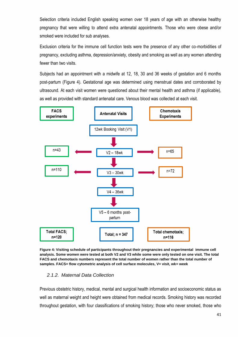
41
Selection criteria included English speaking women over 18 years of age with an otherwise healthy
pregnancy that were willing to attend extra antenatal appointments. Those who were obese and/or
smoked were included for sub analyses.
Exclusion criteria for the immune cell function tests were the presence of any other co-morbidities of
pregnancy, excluding asthma, depression/anxiety, obesity and smoking as well as any women attending
fewer than two visits.
Subjects had an appointment with a midwife at 12, 18, 30 and 36 weeks of gestation and 6 months
post-partum (Figure 4). Gestational age was determined using menstrual dates and corroborated by
ultrasound. At each visit women were questioned about their mental health and asthma (if applicable),
as well as provided with standard antenatal care. Venous blood was collected at each visit.
Figure 4: Visiting schedule of participants throughout their pregnancies and experimental immune cell
analysis. Some women were tested at both V2 and V3 while some were only tested on one visit. The total
FACS and chemotaxis numbers represent the total number of women rather than the total number of
samples. FACS= flow cytometric analysis of cell surface molecules, V= visit, wk= week
2.1.2. Maternal Data Collection
Previous obstetric history, medical, mental and surgical health information and socioeconomic status as
well as maternal weight and height were obtained from medical records. Smoking history was recorded
throughout gestation, with four classifications of smoking history; those who never smoked, those who

42
quit before pregnancy (median 3 years previously), those who quit during pregnancy (median at 5.5
weeks of gestation) and those who were current smokers.
2.1.3. Assessment of Maternal Asthma
Asthma (yes/no) was determined by the midwife asking the questions “Have you ever been told by a
doctor that you have asthma?” and “Have you used any asthma medications in the past year like
Ventolin or a preventer?”. Maternal asthma control at each visit was assessed with a combination of
self-reporting of symptoms, lung function and medication history. Spirometry was used to assess lung
function in both asthmatic and non-asthmatic pregnant women. FEV1 and FVC were measured with the
percent predicted FEV1 calculated based on reference values and corrected for sex, age and height.
Medication use and compliance was assessed, and asthmatics received education about asthma
control and an asthma action plan. Ongoing asthma control was assessed using the Asthma Control
Questionnaire (ACQ) which assesses self-reported frequency of morning symptoms, night-time
symptoms, the degree of limitation of physical activity and the usage of reliever medications in the
previous week (Appendix)
2.1.4. Assessment of Maternal Depression/Anxiety
Maternal depression/anxiety was assessed at the booking visit, with women answering yes/no to the
question “Do you have depression/anxiety?”. At the same visit women also answered the Antenatal Risk
Questionnaire (ANRQ; Appendix) and the Edinburgh Postnatal Depression Score Questionnaire (EPDS;
Appendix) as part of their routine antenatal care.
2.1.5. Sample Collection
Blood was collected in 9 ml Lithium Heparin coated tubes. Peripheral blood mononuclear cells (PBMCs)
were isolated according to the following procedure. Blood was diluted in 10 ml Dulbecco’s modified
Phosphate Buffered Saline (DPBS; Sigma-Aldrich Co, St Louis, MO, USA), layered onto 7 ml
Lymphoprep™ (Axis Shield, Oslo, Norway) and centrifuged at 850 x g for 20 minutes at room
temperature. After centrifugation, the PBMC band was removed using a Pasteur pipette and
resuspended in 20 ml of DPBS. This was centrifuged at 360 x g for 5 minutes at room temperature, the
supernatant decanted and the cell pellet resuspended in another 20 ml DPBS. After further
centrifugation (360 x g, 5 minutes, room temperature), the cell pellet was resuspended in 1 ml 25%
foetal bovine serum (FBS; Sigma-Aldrich Co, St Louis, MO, USA) and 15% dimethyl sulfoxide (DMSO;
Sigma-Aldrich Co, St Louis, MO, USA) in RPMI-1640 media (Life Technologies Australia, Mulgrave, Vic,
Aus.). Uncounted PBMCs were stored in 2 x 500 μl aliquots at -80˚C until required. Cell number was

43
only recorded before the experimental tests as previous data in the lab had shown that cell number
decreased slightly due to the freezing and thawing process. Samples had to be frozen in order to store
them until there were sufficient samples to undertake the chemotaxis work. Recording cell number after
thawing ensued in a more accurate live cell count for each sample and resulted in a more precise
dilution in order to seed all samples at the correct cell density. The viability of pre-frozen PBMC samples
had been previously determined in the laboratory through cell culture and LPS stimulation. Although
there was some cell death due to the freezing process (30% live to 20% live after a freeze and thaw
cycle in a control sample) there were still adequate numbers to perform the experiments. The viability of
all PBMCs used in the experiments was obtained using a TC10 Automated Cell Counter which gave a
cell count as a percentage of live and dead cells in each sample. The range for all samples was 20 –
30% live cells.
2.2. Part B: Epidemiology
One hundred and eighty-nine asthmatic pregnant women were included in the analysis. One hundred
and four women did not experience depression/anxiety during their pregnancy and 85 did experience
depression/anxiety. The primary exposure was the presence of depression/anxiety with the sub-analysis
exposures being a high EPDS score, high ANRQ score, antidepressant use and the various
psychosocial stressors identified at the booking visit. The primary study outcomes were experiencing an
asthma exacerbation during pregnancy and presenting with uncontrolled asthma (ACQ > 1.5) at any
visit during pregnancy. An asthma exacerbation was considered one or more of the following: an
unscheduled visit to a general practitioner (GP), a course of oral corticosteroids, an emergency
department presentation or a hospital admission.
Smoking status was defined as smokers (current smokers plus those who quit during pregnancy) and
non-smokers (those who never smoked and those who quit before pregnancy). BMI was calculated
based on maternal height and weight recorded at antenatal booking visit. Parity was defined as either
the participant’s first pregnancy (parity=0) or at least the participant’s second pregnancy (parity= 1+).
2.2.1. Statistics
Incidences of asthma exacerbations and uncontrolled asthma were compared between the two groups
using Poisson regression with robust variance estimates, with resulting Incidence Rate Ratios (IRR) and
95% confidence intervals (CIs).
The prevalence of asthma exacerbations and uncontrolled asthma was compared between the two
groups using a generalised linear model (Poisson distribution) with robust variance estimates, with
resulting relative risks (RR) and 95% confidence intervals (CIs).

44
Each of the models was adjusted for potential confounders including maternal smoking, BMI, age,
parity, and baseline ICS use. Statistical analyses were performed using statistical package IBM SPSS
Statistics 21 (IBM, Armonk, NY, USA)
2.3. Part C: Immune Cell Experiments
In the following experiments, the PBMCs of a random sample from the initial cohort comprising of 48
non-asthmatic and 76 asthmatic pregnant women with and without depression/anxiety were used in the
flow cytometric analysis of cell surface molecules (FACS) experiments. A random sample comprising 35
non-asthmatic and 81 asthmatic pregnant women were used in the chemotaxis experiments. Seventeen
non-asthmatic and 45 asthmatic pregnant women were common to both experiments. Some women
had samples tested at both 18 and 30 weeks gestation, while others were only tested on one occasion.
2.3.1. FACS Analysis of Cell Surface Molecules
Gestation
Study Group
Non-asthmatic Asthmatic
Depression/
Anxiety - No
Depression/
Anxiety - Yes
Depression/
Anxiety - No
Depression/
Anxiety - Yes
18 wk n=11 n=5 n=14 n=13
30 wk n=36 n=8 n=45 n=21
Table 1: Experimental samples at 18 weeks and 30 weeks gestation for each group for the flow cytometric
analysis of cell surface molecules (FACS) performed on monocytes from pregnant women with and
without asthma and depression/anxiety.
2.3.1.1. Staining of Cell Surface Molecules
For flow cytometry analysis, monocytes from isolated PBMCs were stained with monoclonal antibodies
against CD14, CD16, CD11a, CD11b, CD62L and HLADR (BD Biosciences, San Diego, USA; Co-
operative Research Centre for Biomarker Translation, Australia). Antibodies were titrated to determine
the optimal staining concentration. All incubations were performed in the dark. Staining was performed
according to standard surface molecule staining protocol for flow cytometry. Data was acquired using
FACS Canto (BD Biosciences) and analysed using FlowJo (FlowJo LLC, Ashland, OR, USA).
Cryopreserved PBMCs (1 x 10⁶) were thawed at 37ºC in a water bath. 500 μl of RF10 media (RPMI
1640 media with 10% FBS, 1% penicillin-streptomycin and 2% glutamine) was slowly added to the cells
and centrifuged at 1300 rpm for 5 minutes. Cells were resuspended in 100 μl of Dulbecco PBS (DPBS)
(Sigma Aldrich Co, St Louis, MO, USA). 50 μl of cells was added to 96 well round bottom plates and
mixed with 12.5 μl of premixed anti-human labelled CD14-APCCy7, CD16-FITC, HLADR-PerCP,

45
CD11a-APC, CD11b-PECy7, CD62L-PE (BD Biosciences, San Jose, CA, USA). After incubation on ice
for 30 minutes, the cells were washed in DPBS and centrifuged at 1300 rpm for 5 minutes. Labelled
cells were detected using FACS Canto in which 30,000 cells were analysed in each gated event using
Flow Jo Software. Cells only and individual antibody alone for compensation were included as controls.
2.3.1.2. Flow Cytometry Analysis
A fluorescence compensation method was applied by using unstained cells and single-fluorochrome
stained anti-mouse Ig, k CompBeads (cat# 552843, BD Biosciences) for every analysis, to correct the
spectral overlap. Acquisition of flow cytometry data was done in FACS Canto. Further data analysis was
done using FlowJo software. A FACS Aria (BD Biosciences) was used to sort CD14Bright, CD16Bright
and intermediate monocytes. The gating strategy for sorting is shown in figures 5A and 5B.
Figure 5: The FlowJo gating strategy for sorting total monocytes from Tc cells (A) and distinguishing
CD14 Bright, CD16 Bright and intermediate (CD14 Bright + CD16 Bright) monocytes from each other.
2.3.1.3. Statistics
The median and interquartile range (IOR) of the percentages of total monocytes, monocyte subtype
(CD14Bright, CD16Bright and Intermediates) and adhesion receptor expression were compared
between the four groups. The normalities of the data were tested using frequency histograms and
observed to be skewed. Therefore the non-parametric Kruskal-Wallis test was used to analyse the data.
A B

46
Results were adjusted for potential confounders including maternal smoking, BMI, age, parity, and
baseline ICS use. Statistical tests were performed using SPSS with p values < 0.05 considered
statistically significant.
2.3.2. Chemotaxis
Frozen 500 μl aliquots of PBMCs were thawed at 37˚C and diluted with 1 ml RPMI-1640 (1% FBS, 1%
Penicillin & Streptomycin (Sigma-Aldrich)). After spinning at 4˚C the cell pellet was resuspended in 1 ml
media and the cell number counted using a TC10 Automated Cell Counter (Bio-Rad, Hercules, CA,
USA). Cells were diluted to 2 x 10⁵ cells/ml for assessment of chemotaxis using a Transwell®
Chemotaxis Assay (Corning Inc., NY, USA). Assays consisted of a 24 well plate with 12 Transwell®
inserts that separated the upper and lower chambers of the well with a 5 μm porous membrane.
2.3.2.1. Optimisation
In the following optimisation experiments, PBMCs were used which had been isolated from two non-
pregnant women, one non-asthmatic and one asthmatic.
2.3.2.1.1. Cell number and Incubation Time
After trialling 5 x 10⁴ cells/well and 1 x 10⁵ cells/well, a greater migratory response was observed using
1 x 10⁵ cell/well (2 x 10⁵ cells/ml). 1 x 10⁵ cells/well were used in all subsequent experiments. Previous
work in our laboratory suggested that a 3 hour incubation was most appropriate in assessing PBMC
migration. This was confirmed in two separate experiments using 2, 3 and 4 hour and 1.5, 2 and 3 hour
incubation times. It was important that the cell migration for these optimisations was in the middle of the
migration range. This occurred at 3 hours and would enable the enhanced migration of more active
PBMCs to be detected.
2.3.2.1.2. nfMLP vs. MCP-1
N-Formyl-Met-Leu-Phe (nfMLP) (Sigma-Aldrich Co, St Louis, MO, USA) is a ubiquitous
chemoattractant. Monocyte Chemoattractant Protein-1 (MCP-1) (Sigma-Aldrich Co, St Louis, MO, USA)
is a more specific chemoattractant. The later preferentially attracts monocytes and T lymphocytes,
which make up the majority of PBMCs. Various concentrations of nfMLP and MCP-1 were compared to
optimise the most appropriate chemoattractant and concentration. It was important that the
concentration used was in the middle of the migration range, so that migration at the upper limits of the
scale could still be detected in those individuals with more active PBMCs. Concentrations of 1, 5 and 10
ng/ml nfMLP and 10, 50 and 100 ng/ml MCP-1 were trialled. After a 3 hour incubation, 10 ng/ml MCP-1
resulted in cell migration in the middle of the PBMC migration range. This was further confirmed in
another experiment using 1, 5 and 10 ng/ml MCP-1.

47
2.3.2.1.3. FBS Concentration
The addition of 1% foetal bovine serum (FBS) to the media was beneficial in keeping the cells alive and
active (compared to no FBS), without changing the morphology of the cells (compared to 10% FBS).
2.3.2.2. Final Protocol
The treatment (in duplicate) was 10 ng/ml MCP-1 and control was media alone (RPMI-1640, 1% FBS +
1% P&S). 600 μl control or treatment was placed into the lower chamber, membrane inserted and 500 μl
(1 x 10⁵ cells) loaded into the upper chamber (Figure 6).
Figure 6: Diagram of chemotaxis assay using a Transwell® insert. The lower chamber contained 10 ng/ml
MCP-1 and the upper chamber contained 1 x 10⁵ PBMCs. Chambers were separated by a 5 μm pore
microporous membrane.
After a 3 hour incubation (37˚C, 5% CO2), migrated cells were counted using the TC10 Automated Cell
Counter. Every plate included a quality control sample of PBMCs isolated from a non-asthmatic, non-
pregnant female adult.
Gestation
Study Group
Non-asthmatic Asthmatic
Depression/
Anxiety - No
Depression/
Anxiety - Yes
Depression/
Anxiety - No
Depression/
Anxiety - Yes
18 wk n=14 n=8 n=24 n=19
30 wk n=14 n=9 n=22 n=17
Table 2: Experimental samples at 18 and 30 weeks gestation for each group for the chemotaxis assays
performed on the peripheral blood mononuclear cells (PBMCs) from pregnant women with and without
asthma and depression/anxiety.
Transwell® insert
Upper chamber; contains PBMCs
Microporous membrane
Lower chamber; contains chemoattractant

48
2.3.2.3. Statistics
Chemotactic response was expressed as a migration index, relative to cells with media alone. The
resulting median and interquartile range (IQR) was compared between the different groups using the
non-parametric Kruskal-Wallis test.
Results were adjusted for potential confounders including maternal smoking, BMI, age, parity, and
baseline ICS use. Statistical tests were performed using SPSS with p values <0.05 considered
statistically significant.

49
Chapter 3: Results - Epidemiology
3.1. Part A: Maternal Demographics
One hundred and fifty-eight non-asthmatic and 189 asthmatic pregnant women were included in the
analysis. One hundred and seventeen non-asthmatic and 104 asthmatic women did not have
depression/anxiety during their pregnancy. Forty-one non-asthmatic and 85 asthmatic women did
experience depression/anxiety during their pregnancy. Table 3 describes the maternal demographics of
the groups used in this study. Large percentages of the women in all groups (85.5%, 82.9%, 81.8% and
89.4%) had a socioeconomic status ranking of ≤ 1 on a scale of 0 – 4 where 0 is the lowest and
denotes considerable social disadvantage.
Women with depression/anxiety had higher total EPDS and ANRQ scores regardless of asthma status
than their counterparts without depression/anxiety (p=0.00 for all comparisons). High EPDS and ANRQ
scores can be considered a surrogate for symptomatic depression and anxiety. Over three times as
many women with depression/anxiety had a high EPDS or ANRQ as those women without
depression/anxiety, confirming that our method for assigning depression/anxiety status was valid.
Non-Asthmatic Asthmatic
p value Without
Depression/ Anxiety
(n=117)
With Depression/ Anxiety
(n=41)
Without Depression/
Anxiety
(n=104)
With Depression/
Anxiety
(n=85)
Maternal Age: median
(25% - 75%)
26.0 (23.0 – 31.0)a
26.0 (23.0 – 29.0)a
25.0 (22.0 – 29.0)a
25.0 (22.0 – 30.0)a
0.632
BMI: median
(25% - 75%)
25.35 (22.2 – 30.7)a
30.48 (24.0 – 34.7)b,c
27.53 (23.1 – 32.8)b,c
27.72 (23.2 – 32.1)a,c
0.048
Parity: n (%):
0
1+
47 (40.2)a
70 (59.8)a
16 (39.0)a
25 (61.0)a
45 (43.3)a
59 (56.7)a
33 (38.8)a
52 (61.2)a
0.926
Foetal Sex: n (%)
Male
Female
57 (48.7)a
60 (51.3)a
16 (39.0)a
25 (61.0)a
60 (57.7)a
44 (42.3)a
37 (43.5)a
48 (56.5)a
0.122
Ethnicity: n (%): Caucasian
Other
108 (92.3)a
9 (7.7)a
36 (87.8)a
5 (12.2)a
93 (89.4)a
11 (10.6)a
83 (97.6)a
2 (2.4)a
0.125

50
Smoking Status:
n (%):
Non-smoker
Former smoker
Quit during pregnancy
Current smoker
55 (47.0)a
24 (20.5)a
10 (8.5)a,b
28 (23.9)a
13 (31.7)b
9 (22.0)a
10 (24.4)a
9 (22.0)a
58 (55.8)a
18 (17.3)a
8 (7.7)b
20 (19.2)a
34 (40.0)a
18 (21.2)a
15 (17.6)a,b
18 (21.2)a
0.074
FEV1 L:
mean (SD) 3.1 (0.4)a,b 3.1 (0.6)a,b 2.9 (0.4)a 3.2 (0.5)b 0.050
FEV1 %:
mean (SD) 0.9 (0.1)a 0.9 (0.2)a 0.9 (0.1)a 0.9 (0.1)a 0.685
FVC L
mean (SD) 3.7 (0.5)a,b 3.7 (0.8)a,b 3.6 (0.5)a 3.9 (0.6)b 0.015
FEV/FVC %: mean (SD)
0.8 (0.1)a 0.8 (0.6)a 0.8 (0.1)a 0.8 (0.1)a 0.236
ICS use before pregnancy n (%):
None
ICS
ICS + LABA
117 (100)
0 (0)
0 (0)
41 (100)
0 (0)
0 (0)
72 (69.2)a
9 (8.7)a
23 (22.1)a
56 (65.9)a
4 (4.7)a
25 (29.4)a
0.172
Total ANRQ: median (25% -
75%) 9 (7 – 17)a 29 (21- 37)b 11 (7 – 18)a 29 (23 – 39)b 0.001
High ANRQ: n (%) 13 (11.1)a 23 (56.1)b 17 (17.2)a 62 (78.5)b 0.001
Total EPDS: median (25% -
75%) 3 (1 – 6)a 6 (3- 9)b 4 (2 – 6)a 7 (3 – 10)b 0.001
High EPDS: n (%) 3 (2.6)a 4 (9.8)a,b 4 (4.3)a 15 (18.8)b 0.001
Antidepressant use: n (%)
0 (0)a 10 (24.4)b 0 (0)a 18 (21.2)b 0.001
Table 3: Participant data at booking visit (12 or 18 weeks gestation). BMI= body mass index, FEV1L=
forced expiratory volume in 1 second, FVC= forced vital capacity, ICS= inhaled corticosteroids, LABA=
long acting β2 agonists, ANRQ= antenatal risk questionnaire, EPDS= Edinburgh Postnatal Depression
Score. Statistical tests: Pearson Chi-Square Test or Kruskal-Wallis Test. Significance: p <0.05; a,
b denote
significantly different values
3.2. Part B: Epidemiology
Maternal demographics were similar in both asthmatic groups, except in measures of mental health
status (Table 4). Women from lower socioeconomic backgrounds were highly represented, with 85
(81.8%) of those asthmatics without depression/anxiety and 76 (89.4%) of those with

51
depression/anxiety having a socioeconomic status ranking of ≤ 1 on a scale of 0 – 4 (where 0 is the
lowest).
Asthmatics (n= 189)
p values Without
Depression/Anxiety
(n=104)
With
Depression/Anxiety
(n=85)
Maternal Age: median (25% - 75%) 25.0 (22.0 – 29.0) 25.0 (22.0 – 30.0) 0.821
BMI: median (25% - 75%) 27.5 (23.1 – 32.8) 27.7 (23.2 – 32.1) 0.938
Parity: n (%): 0
1+
45 (43.3)
59 (56.7)
33 (38.8)
52 (61.2) 0.537
Foetal Sex: n (%)
Male
Female
60 (57.7)
44 (42.3)
37 (43.5)
48 (56.5)
0.053
Ethnicity: n (%): Caucasian
Other
93 (89.4)
11 (10.6)
83 (97.6)
2 (2.4) 0.094
Smoking Status: n (%)
Non-smoker
Former smoker
Quit during pregnancy
Current smoker
58 (55.8)
18 (17.3)
8 (7.7)
20 (19.2)
34 (40.0)
18 (21.2)
15 (17.6)
18 (21.2)
0.084
FEV1 L: mean (SD) 2.9 (0.4) 3.2 (0.5) 0.004
FEV1 %: mean (SD) 0.9 (0.1) 0.9 (0.1) 0.210
FVC L: mean (SD) 3.6 (0.5) 3.9 (0.6) 0.001
FEV/FVC %: mean (SD) 0.8 (0.1) 0.8 (0.1) 0.307
ICS use before pregnancy n (%):
None
ICS
ICS + LABA
72 (69.2)
9 (8.7)
23 (22.1)
56 (65.9)
4 (4.7)
25 (29.4)
0.172
Total ANRQ: median (25% - 75%) 11 (7 – 18) 29 (23 – 39) 0.000
High ANRQ: n (%) 17 (17.2) 62 (78.5) 0.000
Total EPDS: median (25% - 75%) 4 (2 – 6) 7 (3 – 10) 0.000
High EPDS: n (%) 4 (4.3) 15 (18.8) 0.002
Antidepressant use: n (%) 0 (0) 18 (21.2) 0.000
Table 4: Participant data of asthmatic women at booking visit (12 or 18 weeks gestation). BMI= body mass
index, FEV1L= forced expiratory volume in 1 second, FVC= forced vital capacity, ICS= inhaled
corticosteroids, LABA= long acting β2 agonists, ANRQ= antenatal risk questionnaire, EPDS= Edinburgh
Postnatal Depression Score. Statistical tests: Pearson Chi-Square Test or Mann-Whitney U Test.
Significance: p <0.05
3.2.1. Asthma Exacerbations
The incidence of overall asthma exacerbations (p=0.377) and individual exacerbation types: GP visits
(p=0.443), OCS prescriptions (p=0.694), ED presentations (p=0.328) and hospital admissions
(p=0.886); were similar in both groups of women (Table 5).

52
There was no increase in the incidence rate ratio for asthma exacerbations when the mother had
depression/anxiety (unadjusted IRR 0.770; adjusted IRR 0.755, CI 0.412- 1.382, p=0.362; Figure 7A).
There was no increase in the relative risk of an asthma exacerbation during pregnancy with the added
complication of depression/anxiety (unadjusted RR 0.867; adjusted RR 0.859, CI 0.496- 1.489, p=0.589;
Figure 7B).
Table 5: Asthma control data of women throughout pregnancy. GP= general practitioner, OCS= oral
corticosteroids, ED= emergency department, ICS= inhaled corticosteroids, LABA= long acting β2
agonists. Statistical tests: Pearson Chi-Square Test or Mann-Whitney U Test. Significance: p <0.05
Asthmatics (n= 189)
p values Without
Depression/Anxiety
(n=104)
With
Depression/Anxiety
(n=85)
Uncontrolled Asthma: n
(%) 34 (32.7) 45 (52.9) 0.005
No. of Uncontrolled
Asthma Events: median
(25%- 75%)
0 (0- 1) 1 (0- 2) 0.005
Asthma Exacerbation: n
(%) 24 (23.1) 17 (20.0) 0.610
No. of Asthma
Exacerbations: median
(25%- 75%)
0 (0- 0) 0 (0- 0) 0.377
GP visit: n (%) 19 (18.3) 12 (14.1) 0.443
OCS use: n (%) 7 (6.7) 7 (8.2) 0.694
ED presentation: n (%) 7 (6.7) 3 (3.5) 0.328
Hospital Admission: n (%) 1 (1.0) 1 (1.2) 0.886
ICS prescribed during
pregnancy, n (%): none
ICS
ICS + LABA
Already on ICS
56 (53.8)
8 (7.7)
8 (7.7)
32 (30.8)
46 (54.1)
4 (4.7)
6 (7.1)
29 (34.1)
0.839
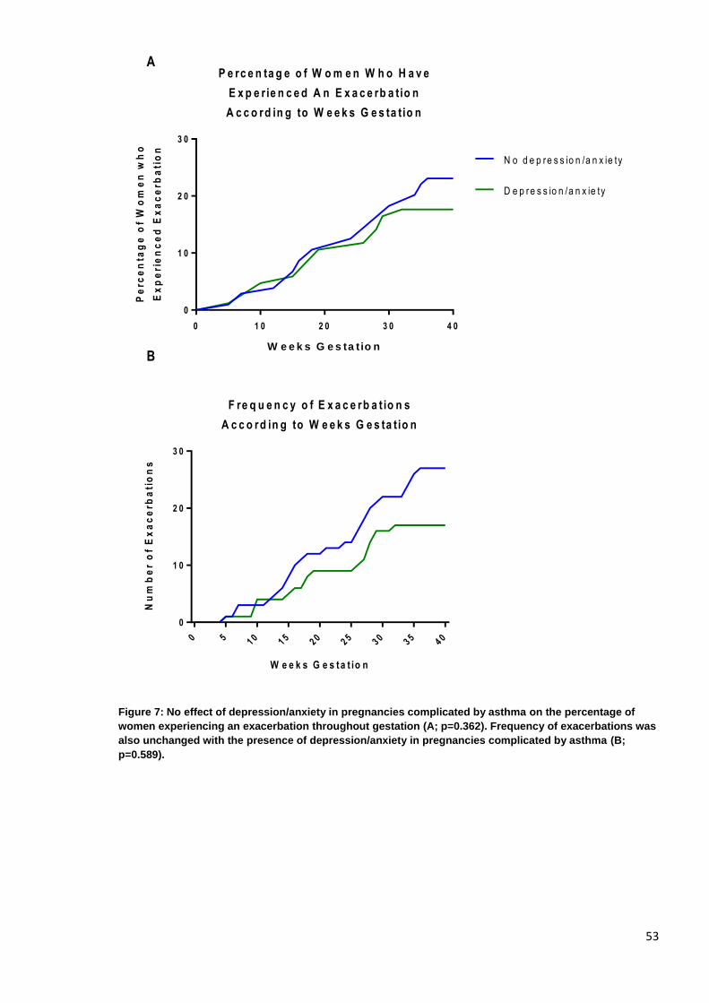
53
Figure 7: No effect of depression/anxiety in pregnancies complicated by asthma on the percentage of
women experiencing an exacerbation throughout gestation (A; p=0.362). Frequency of exacerbations was
also unchanged with the presence of depression/anxiety in pregnancies complicated by asthma (B;
p=0.589).
P e rc e n ta g e o f W o m e n W h o H a v e
E x p e r ie n c e d A n E x a c e rb a tio n
A c c o rd in g to W e e k s G e s ta tio n
W e e k s G e s ta t io n
Pe
rce
nta
ge
of
Wo
me
n w
ho
Ex
pe
rie
nc
ed
Ex
ac
erb
ati
on
0 1 0 2 0 3 0 4 0
0
1 0
2 0
3 0
N o d e p re s s io n /a n x ie ty
D e p re s s io n /a n x ie ty
F re q u e n c y o f E x a c e rb a t io n s
A c c o rd in g to W e e k s G e s ta tio n
W e e k s G e s ta t io n
Nu
mb
er
of
Ex
ac
erb
ati
on
s
0 5 1 0 1 5 2 0 2 5 3 0 3 5 4 0
0
1 0
2 0
3 0
N o d e p re s s io n /a n x ie ty
D e p re s s io n /a n x ie ty
A
B

54
3.2.2. Uncontrolled asthma
The incidence of uncontrolled asthma increased in women with depression/anxiety (52.9%) compared to
women without depression/anxiety (32.7%; p=0.005; Table 5). This resulted in an increased incidence
rate ratio of over 60% for these women (unadjusted IRR 1.739, adjusted IRR 1.633, CI 1.092- 2.442,
p=0.017; Figure 8A).
Relative risk of experiencing uncontrolled asthma was also increased over 50% when the woman also
had depression/anxiety. (unadjusted RR 1.619; adjusted RR 1.538, CI 1.114- 2.122, p=0.009; Figure
8B).

55
Figure 8: The presence of depression/anxiety in pregnancies complicated by asthma significantly
increased the percentage of women experiencing uncontrolled asthma throughout gestation (A; p=0.017).
The number of uncontrolled asthma events was also significantly increased with the presence of
depression/anxiety in pregnancies complicated by asthma (B; p=0.009).
P e rc e n ta g e o f W o m e n W h o H a v e
E x p e r ie n c e d U n c o n tro lle d A s th m a
A c c o rd in g to W e e k s G e s ta tio n
W e e k s G e s ta t io n
Pe
rce
nta
ge
of
Wo
me
n w
ho
Ex
pe
rie
nc
ed
Un
co
ntr
oll
ed
As
thm
a
0 1 0 2 0 3 0 4 0
0
2 0
4 0
6 0
F re q u e n c y o f U n c o n tro lle d A s th m a
A c c o rd in g to W e e k s G e s ta tio n
W e e k s G e s ta t io n
Nu
mb
er
of
Un
co
ntr
oll
ed
Ev
en
ts
0 5 1 0 1 5 2 0 2 5 3 0 3 5 4 0
0
2 0
4 0
6 0
8 0
1 0 0
N o d e p re s s io n /a n x ie ty
D e p re s s io n /a n x ie ty
F re q u e n c y o f U n c o n tro lle d A s th m a
A c c o rd in g to W e e k s G e s ta tio n
W e e k s G e s ta t io n
Nu
mb
er
of
Un
co
ntr
oll
ed
Ev
en
ts
0 5 1 0 1 5 2 0 2 5 3 0 3 5 4 0
0
2 0
4 0
6 0
8 0
1 0 0
N o d e p re s s io n /a n x ie ty
D e p re s s io n /a n x ie ty
A
B
56
3.3. Discussion
In this study of 189 pregnant asthmatics, we have observed for the first time in pregnancy that asthma
control worsens in women with depression/anxiety compared to those without. There is no increased
risk or rate of asthma exacerbations regardless of the presence or absence of depression/anxiety. The
outcomes of uncontrolled asthma and asthma exacerbations were analysed separately as both have
been observed to be more common during pregnancy3,5.
This study has built on previous work by examining asthma control in a population with
depression/anxiety. Quality of life, anxiety, perceived control of asthma and perceived risks of asthma
medications were examined in pregnant, asthmatic women134. Overall the women had a good quality of
life and perceived control of their asthma as well as having low levels of anxiety134. However, the more a
woman was affected emotionally by her asthma, the more frequently she experienced uncontrolled
asthma134. Women overestimated the risk of harm to the foetus of asthma medications, though this did
not appear to lead to greater anxiety134. Asthma-specific quality of life has also been used previously to
examine asthma exacerbations during pregnancy133. An association was observed between an increase
in asthma-specific quality of life in early pregnancy and a decrease in the risk of a subsequent asthma
exacerbation133. The current study differs from previous work in pregnant women, by comparing asthma
control during pregnancy in women with depression or anxiety to women without these complications,
rather than the woman’s emotional response or perceived quality of life. This work does support a link
between mental health status (be it depression/anxiety, an increased emotional response to asthma or
poor quality of life) and uncontrolled asthma during pregnancy. We could not confirm a link between
mental health status and asthma exacerbations during pregnancy.
The observation of increased risk of uncontrolled asthma with depression/anxiety during pregnancy was
unsurprising as similar observations have been reported in a non-pregnant population. Our study in
pregnant asthmatics follows on from previous work in non-pregnant asthmatics demonstrating
associations between depression and anxiety and asthma control. Asthmatics with uncontrolled asthma
are more likely to suffer from depression than those who are controlled11. As depression severity
increases, asthma control declines11. Respiratory symptoms were increased in asthmatics with anxiety
and depression compared to those without, with Hospital Anxiety Depression Scale (HADS) score being
the most useful determinant of night-time symptoms and attacks of breathlessness10. Similar findings
were observed in our study with increased levels of depression/anxiety in asthmatics compared to non-
asthmatics (45.0% and 25.9% respectively; p=0.000) and a higher incidence of uncontrolled asthma in
women with depression/anxiety compared to women without this added complication. This work
contributes to the current understanding of mental health status and asthma control by broadening the
research to include pregnant women. The fact that the association between depression/ anxiety and

57
poor asthma control still holds in pregnancy suggests that a woman’s mental health status can have an
overreaching effect on her health during pregnancy through interactions with other complications such
as asthma. This will have implications for the provision of appropriate antenatal care to these women at
higher risk of uncontrolled asthma.
Several societal factors could be contributing to this increase in uncontrolled asthma. Medication
adherence and appropriate self-management are of great importance to good asthma control. Non-
adherence to asthma medications during pregnancy can result because women do not understand
when to use medications or what a loss of asthma control is (poor health literacy) or due to fears of
harming their baby. While not specifically investigated in this study, previous work has demonstrated
that women overestimate the risks of asthma medications134. In future, it would be interesting to
examine if women with depression/anxiety are more likely to overestimate the risks of either their
asthma or their depression/anxiety medication. If this were the case, it may help explain the observed
increase in uncontrolled asthma in pregnant women with depression/anxiety due to a worsening of their
asthma or their depression/anxiety.
Both uncontrolled asthma and exacerbations have been observed to be more common during
pregnancy3,5. Appropriate asthma control throughout pregnancy and not just the avoidance of
exacerbations could be important for maternal and foetal health. Future work should assess other
maternal and foetal outcomes (such as pre-eclampsia, preterm birth and small for gestational age
babies) to examine whether there are any associations with the increase in uncontrolled asthma with
depression/anxiety and adverse pregnancy outcomes.
While an association between smoking and asthma control during pregnancy has been demonstrated7,
our cohort had similar rates of smoking regardless of the presence of depression/anxiety. No additional
effect of smoking on uncontrolled asthma or exacerbations was observed in pregnant asthmatics with
depression/anxiety, suggesting that a woman’s mental health is more important in determining her risk
of uncontrolled asthma during pregnancy. This was somewhat surprising, however it could have been
due to the way in which smoking status was analysed. The number of cigarettes per day was not taken
into account due to the lack of appropriate power. With greater numbers it would be interesting to see if
the quantity of cigarettes per day might have an additional adverse effect on asthma control or increase
the risk of exacerbation.
There is an association between low socioeconomic status and an increase in social stress, and an
increased risk of asthma exacerbations117,118. Our study recruited from a population of high social
disadvantage. Both women with and without depression/anxiety predominantly lived in a disadvantaged
area and were consequently more exposed to social stressors during their pregnancies such as
insecure housing, financial difficulties, relationship breakdown and unemployment. This increased social

58
stress may have increased the emotional burden on already vulnerable women and contributed to their
increased risk of uncontrolled asthma. This was difficult to examine in the current study as there was
not appropriate power for these sub-analyses. It would be interesting in future work to examine the
effect of depression/anxiety on asthma control during pregnancy in women from a less disadvantaged
area to determine if there is an additional effect of socioeconomic status.
Obesity is associated with poor asthma control8 and may contribute to the increased risk of uncontrolled
asthma with depression/anxiety during pregnancy. However, BMI was similar in asthmatics with and
without depression/anxiety and we observed no additional effect of BMI in our analysis. This finding is
similar to recent findings in obese (BMI >30) adults observing that although both obesity and depression
were associated with worse asthma control, their contributions were independent of each other19.
There may also be an immune mechanism behind this worsening of asthma control as a result of the
influences of depression/anxiety, asthma and pregnancy itself on immune function. The second part of
this study examined whether a possible change in monocyte profile or differences in monocyte and T
lymphocyte chemotaxis, may help to explain this reduction in asthma control.

59
Chapter 4: Results – Immune Cell Experiments
4.1. Part A: Flow cytometric analysis of cell surface molecules (FACS)
4.1.1. Maternal Demographics
The individuals randomly selected for the FACS analysis were representative of the entire cohort (Table
6).
Non-asthmatic Asthmatic
p
value
Without
Depression/
Anxiety
(n=37)
With
Depression/
Anxiety
(n=10)
Without
Depression/
Anxiety
(n=48)
With
Depression/
Anxiety
(n=22)
Maternal Age: median
(25% - 75%)
27.0
(23.0- 30.0)a
29.5
(25.0- 32.0)a
27.0
(23.0- 29.5)a
25.0
(22.0- 30.0)a 0.534
BMI: median
(25% - 75%)
23.9
(22.5- 28.5)a
32.0
(29.1- 35.0)b
28.2
(23.0- 33.3)a,c
24.9
(22.8- 30.9)a,c 0.016
Parity: n (%): 0
1+
18 (48.6)a
19 (51.4)a
3 (30.0)a
7 (70.0)a
21 (43.8)a
27 (56.2)a
13 (59.1)a
9 (40.9)a 0.441
Foetal Sex: n (%)
Male
Female
18 (48.6)a
19 (51.4)a
4 (40.0)a
6 (60.0)a
26 (54.2)a
22 (45.8)a
12 (54.5)a
10 (45.5)a
0.834
Ethnicity: n (%):
Caucasian
Other
36 (97.3)a
1 (2.7)a
9 (90.0)a
1 (10.0)a
41 (85.4)a
7 (14.6)a
22 (100.0)a
0 (0)a
0.093
Smoking Status n (%):
Non-smoker
Former smoker
Quit during pregnancy
Current smoker
20 (54.1)a
7 (18.9)a
3 (8.1)a
7 (18.9)a
5 (50.0)b
1 (10.0)a
2 (20.0)a
2 (20.0)a
30 (62.5)a
6 (12.5)a
3 (6.3)a
9 (18.8)a
8 (36.4)a
4 (18.2)a
6 (27.3)a
4 (18.2)a
0.398
FEV1L:
mean (SD) 3.0 (0.4)a,b 3.3 (0.7)a,b 2.9 (0.4)a 3.4 (0.6)b 0.018

60
FEV1 %:
mean (SD) 0.9 (0.1)a 1.0 (0.2)a 0.9 (0.1)a 1.0 (0.2)a 0.060
FVCL:
mean (SD) 3.7 (0.4)a,b 3.9 (0.8)a,b 3.5 (0.5)a 4.1 (0.8)b 0.028
FEV/FVC %: mean
(SD) 0.8 (0.1)a 0.8 (0.1)a 0.8 (0.1)a 0.8 (0.0)a 0.869
ICS use before
pregnancy n (%):
None
ICS
ICS + LABA
N/A N/A
Missing=7
24 (50.0)a
5 (20.8)a
12 (25.0)a
Missing=4
11 (50.0)a
3 (1.4)a
4 (18.2)a
0.808
High ANRQ: n (%) 5 (15.8)a 7 (70.0)b 7 (14.6)a 16 (72.0)b 0.000
High EPDS: n (%) 1 (2.6)a 1 (10.0)a 1 (2.1)a 2 (12.0)a 0.386
Antidepressant use: n
(%) 0 (0) 5 (50.0)a 0 (0) 7 (28.0)a 0.000
Uncontrolled Asthma:
n (%) N/A N/A 17 (35.4)a 9 (40.0)a 0.659
Asthma Exacerbation:
n (%) N/A N/A 12 (25.0)a 5 (20.0)a 0.837
Table 6: Monocyte flow cytometric analysis subgroup data at booking visit (12 or 18 weeks gestation) and
asthma control throughout pregnancy.
Abbreviations: BMI= body mass index, FEV1L= forced expiratory volume in 1 second, FVC= forced vital
capacity, ICS= inhaled corticosteroids, LABA= long acting β2 agonists, ANRQ= antenatal risk
questionnaire, EPDS= Edinburgh Postnatal Depression Score. Statistical tests: Pearson Chi-Square Test
or Kruskal-Wallis Test. Significance: p <0.05; a,
b denote significantly different values
4.1.2. Percentage of Total Monocytes
At 18 weeks gestation, total monocyte percentage was increased in asthmatic pregnant women without
depression/anxiety compared to asthmatic pregnant women with depression/anxiety (p=0.027) and non-
asthmatic pregnant women without depression/anxiety (p=0.005; figure 9A). By 30 weeks gestation,
these increases were no longer apparent (p=0.788 and 0.328 respectively; figure 9B). There was no
difference in total monocyte percentage due to asthma status in those women with depression/anxiety
at 18 or 30 weeks gestation (p=0.60 and 0.126 respectively).

61
A
B
Figure 9: Percentage total monocytes in the peripheral blood mononuclear cells of pregnant women with
(triangles) and without (circles and squares) asthma and with (red and purple) and without (blue and
green) depression/anxiety. Percentage total monocytes is significantly increased at 18 weeks gestation
(A) in asthmatic pregnant women without depression/anxiety compared. There were no changes in
percentage total monocytes at 30 weeks gestation (B). Significance: p <0.05.
P e rc e n t T o ta l M o n o c y te s a t 1 8 w e e k s G e s ta t io n
% T
ota
l M
on
oc
yte
s
No
n-a
s thm
a tic
- d
e pre
s s ion
/an
x iety
No
n-a
s thm
a tic
+ de p
res s io
n/a
nx ie
ty
As th
ma ti
c
- d
e pre
s s ion
/an
x iety
As th
ma ti
c
+ de p
res s io
n/a
nx ie
ty
0
5
1 0
1 5
2 0
P e rc e n t T o ta l M o n o c y te s a t 3 0 w e e k s G e s ta t io n
% T
ota
l M
on
oc
yte
s
No
n-a
s thm
a tic
- d
e pre
s s ion
/an
x iety
No
n-a
s thm
a tic
+ de p
res s io
n/a
nx ie
ty
As th
ma ti
c
- d
e pre
s s ion
/an
x iety
As th
ma ti
c
+ de p
res s io
n/a
nx ie
ty
0
1 0
2 0
3 0
P=0.027 P=0.005
Non-asthmatic
- depression/anxiety
Non-asthmatic
+ depression/anxiety
Asthmatic
- depression/anxiety
Asthmatic
+ depression/anxiety
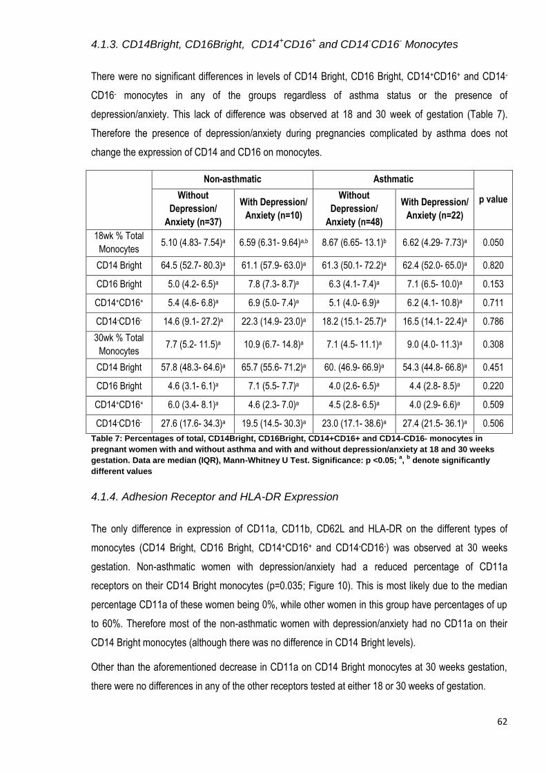
62
4.1.3. CD14Bright, CD16Bright, CD14+CD16+ and CD14-CD16- Monocytes
There were no significant differences in levels of CD14 Bright, CD16 Bright, CD14+CD16+ and CD14-
CD16- monocytes in any of the groups regardless of asthma status or the presence of
depression/anxiety. This lack of difference was observed at 18 and 30 week of gestation (Table 7).
Therefore the presence of depression/anxiety during pregnancies complicated by asthma does not
change the expression of CD14 and CD16 on monocytes.
Non-asthmatic Asthmatic
p value Without
Depression/
Anxiety (n=37)
With Depression/
Anxiety (n=10)
Without
Depression/
Anxiety (n=48)
With Depression/
Anxiety (n=22)
18wk % Total
Monocytes 5.10 (4.83- 7.54)a 6.59 (6.31- 9.64)a,b 8.67 (6.65- 13.1)b 6.62 (4.29- 7.73)a 0.050
CD14 Bright 64.5 (52.7- 80.3)a 61.1 (57.9- 63.0)a 61.3 (50.1- 72.2)a 62.4 (52.0- 65.0)a 0.820
CD16 Bright 5.0 (4.2- 6.5)a 7.8 (7.3- 8.7)a 6.3 (4.1- 7.4)a 7.1 (6.5- 10.0)a 0.153
CD14+CD16+ 5.4 (4.6- 6.8)a 6.9 (5.0- 7.4)a 5.1 (4.0- 6.9)a 6.2 (4.1- 10.8)a 0.711
CD14-CD16- 14.6 (9.1- 27.2)a 22.3 (14.9- 23.0)a 18.2 (15.1- 25.7)a 16.5 (14.1- 22.4)a 0.786
30wk % Total
Monocytes 7.7 (5.2- 11.5)a 10.9 (6.7- 14.8)a 7.1 (4.5- 11.1)a 9.0 (4.0- 11.3)a 0.308
CD14 Bright 57.8 (48.3- 64.6)a 65.7 (55.6- 71.2)a 60. (46.9- 66.9)a 54.3 (44.8- 66.8)a 0.451
CD16 Bright 4.6 (3.1- 6.1)a 7.1 (5.5- 7.7)a 4.0 (2.6- 6.5)a 4.4 (2.8- 8.5)a 0.220
CD14+CD16+ 6.0 (3.4- 8.1)a 4.6 (2.3- 7.0)a 4.5 (2.8- 6.5)a 4.0 (2.9- 6.6)a 0.509
CD14-CD16- 27.6 (17.6- 34.3)a 19.5 (14.5- 30.3)a 23.0 (17.1- 38.6)a 27.4 (21.5- 36.1)a 0.506
Table 7: Percentages of total, CD14Bright, CD16Bright, CD14+CD16+ and CD14-CD16- monocytes in
pregnant women with and without asthma and with and without depression/anxiety at 18 and 30 weeks
gestation. Data are median (IQR), Mann-Whitney U Test. Significance: p <0.05; a,
b denote significantly
different values
4.1.4. Adhesion Receptor and HLA-DR Expression
The only difference in expression of CD11a, CD11b, CD62L and HLA-DR on the different types of
monocytes (CD14 Bright, CD16 Bright, CD14+CD16+ and CD14-CD16-) was observed at 30 weeks
gestation. Non-asthmatic women with depression/anxiety had a reduced percentage of CD11a
receptors on their CD14 Bright monocytes (p=0.035; Figure 10). This is most likely due to the median
percentage CD11a of these women being 0%, while other women in this group have percentages of up
to 60%. Therefore most of the non-asthmatic women with depression/anxiety had no CD11a on their
CD14 Bright monocytes (although there was no difference in CD14 Bright levels).
Other than the aforementioned decrease in CD11a on CD14 Bright monocytes at 30 weeks gestation,
there were no differences in any of the other receptors tested at either 18 or 30 weeks of gestation.

63
Figure 10: Percentage CD11a expression on CD14 Bright monocytes in pregnant women with (triangles)
and without (circles and squares) asthma and with (red and purple) and without (blue and green)
depression/anxiety during pregnancy at 30 weeks gestation.
There were no effects of smoking, BMI, foetal sex, maternal age or parity on monocyte percentages or
receptor expression (data not shown).
4.1.5. Effect of Uncontrolled Asthma and Asthma Exacerbations
In women with depression/anxiety an increase in total monocyte percentage at 18 weeks gestation was
observed in those with uncontrolled asthma compared to those with good asthma control (p= 0.019,
Figure 11). It is the presence of depression/anxiety which appears to reduce total monocyte percentage
rather than uncontrolled asthma increasing monocytes. In women with controlled asthma, those with
depression/anxiety had a significant reduction in total monocyte percentage compared to those without
depression/anxiety (p=0.001). In asthmatics without depression/anxiety there is no difference in total
monocyte percentage at 18 weeks regardless of asthma control status. When the total monocyte
percentage of controlled asthmatics without depression/anxiety was compared to non-asthmatics, also
without depression/anxiety, they were observed to be significantly increased (p=0.004).
E x p re s s io n o f C D 1 1 a re c e p to r
o n C D 1 4 B r ig h t M o n o c y te s a t 3 0 w e e k s G e s ta t io n
% C
D1
1a
ex
pre
ss
ing
CD
14
Bri
gh
t M
on
oc
yte
s
No
n-a
s thm
a tic
- d
e pre
s s ion
/an
x iety
No
n-a
s thm
a tic
+ de p
res s io
n/a
nx ie
ty
As th
ma ti
c
- d
e pre
s s ion
/an
x iety
As th
ma ti
c
+ de p
res s io
n/a
nx ie
ty
0
2 0
4 0
6 0
8 0
1 0 0
1 2 0 P=0.005 P=0.006
Non-asthmatic -
depression/anxiety
Non-asthmatic +
depression/anxiety
Asthmatic -
depression/anxiety
Asthmatic +
depression/anxiety

64
Figure 11: Effect of uncontrolled asthma on percentage total monocytes at 18 weeks gestation in non-
asthmatics without (blue) or with (red) depression/anxiety compared to controlled asthmatics without
(green) of with (purple) depression/anxiety and uncontrolled asthmatics without (orange) or with (black)
depression/anxiety. Statistical significance: p <0.05.
There were no other differences in monocyte markers at 18 or 30 weeks gestation.
There were no differences in total monocyte percentage, the percentages of the various monocytes
types (CD14 Bright, CD16 Bright, CD14+CD16+ and CD14-CD16-) and the monocyte adhesion receptors
at 18 or 30 weeks gestation when data was analysed according to the presence or absence of asthma
exacerbations during pregnancy.
There was no confounding effect of smoking, BMI, foetal sex, maternal age or parity on monocyte
receptor expression when data was analysed in light of uncontrolled asthma or asthma exacerbations
(data not shown).
P e rc e n t T o ta l M o n o c y te s a t 1 8 w e e k s G e s ta t io n in W o m e n
w ith a n d w ith o u t C o n tro l le d A s th m a a n d D e p re s s io n /A n x ie ty
% T
ota
l M
on
oc
yte
s
No
n-a
s thm
a tic
- d
e pre
s s ion
/an
x iety
No
n-a
s thm
a tic
+ de p
res s io
n/a
nx ie
ty
Co
ntr
olle
d a
s thm
a tic
- d
e pre
s s ion
/an
x iety
Co
ntr
olle
d a
s thm
a tic
+ de p
res s io
n/a
nx ie
ty
Un
c on
tro
lled
as th
ma ti
c
- d
e pre
s s ion
/an
x iety
Un
c on
tro
lled
as th
ma ti
c
+ de p
res s io
n/a
nx ie
ty
0
5
1 0
1 5
2 0
p=0.019
p=0.001 p=0.004 Non-asthmatic -
depression/anxiety
Non-asthmatic +
depression/anxiety
Controlled asthmatic
- depression/anxiety
Controlled asthmatic
+ depression/anxiety
Uncontrolled asthmatic
- depression/anxiety
Uncontrolled asthmatic
+ depression/anxiety

65
4.2. Part B: PBMC Chemotaxis
4.2.1. Maternal Demographics
The individuals chosen for the chemotaxis experiments were representative of the entire cohort (Table
8).
Non-asthmatic Asthmatic
p
value Without
Depression/
Anxiety (n=21)
With
Depression/
Anxiety (n=13)
Without
Depression/
Anxiety (n=47)
With
Depression/
Anxiety (n=32)
Maternal Age:
median
(25% - 75%)
24.0
(21.0- 27.0)a
25.0
(22.0- 29.0)a
25.0
(22.0- 29.0)a
25.0
(21.0- 30.0)a 0.861
BMI: median
(25% - 75%)
25.3
(21.7- 29.6)a
32.3
(26.2- 40.0)b
28.6
(23.2- 33.6)a,b
25.0
(22.7- 30.7)a,c 0.027
Parity: n (%): 0
1+
9 (42.9)a
12 (57.1)a
5 (38.5)a
8 (61.5)a
17 (36.2)a
30 (63.8)a
16 (50.0)a
16 (50.0)a 0.667
Foetal Sex: n
(%)
Male
Female
10 (47.6)a
11 (52.4)a
5 (38.5)a
8 (61.5)a
24 (51.1)a
23 (48.9)a
15 (46.9)a
17 (53.1)a
0.881
Ethnicity: n (%):
Caucasian
Other
19 (90.5)a
2 (9.5)a
11 (84.6)a
2 (15.4)a
42 (89.4)a
5 (10.6)a
31 (96.9)a
1 (3.1)a
0.535
Smoking Status:
n (%):
Non-smoker
Former smoker
Quit during
pregnancy
Current smoker
13 (61.9)a
2 (9.5)a
2 (9.5)a
4 (19.0)a
3 (23.1)a
2 (15.4)a
4 (30.8)a
4 (30.8)a
26 (55.3)a
8 (17.0)a
5 (10.6)a
8 (17.0)a
12 (37.5)a
6 (18.8)a
10 (31.3)a
4 (12.5)a
0.179
FEV1L:
mean (SD) 3.0 (0.5)a 3.2 (0.4)a 2.9 (0.4)a 3.2 (0.6)a 0.097
FEV1 %:
mean (SD) 0.9 (0.1)a 1.0 (0.1)a 0.9 (0.1)a 0.9 (0.1)a 0.323

66
FVCL:
mean (SD) 3.6 (0.6)a,b 3.8 (0.3)a,b 3.5 (0.4)a 3.9 (0.5)b 0.018
FEV/FVC %:
mean (SD) 0.8 (0.1)a 0.9 (0.1)a 0.8 (0.1)a 0.8 (0.1)a 0.386
ICS use before
pregnancy n
(%):
None
ICS
ICS + LABA
N/A N/A
25 (53.2)a
6 (12.8)a
8 (17.0)a
[Missing n=5]
19 (59.4)a
1 (3.1)a
7 (21.9)a
0.308
High ANRQ: n
(%) 3 (15.8)a 10 (70.0)b 12 (25.5)a 25 (78.1)b 0.000
High EPDS: n
(%) 1 (2.6)a 0 (0.0) 1 (2.1)a 3 (9.4)a 0.357
Antidepressant
use: n (%) 0 (0) 6 (50.0)a 0 (0) 8 (25.0)a 0.000
Uncontrolled
Asthma: n (%) N/A N/A 15 (31.9)a 16 (50.0)a 0.106
Asthma
Exacerbation:
n (%)
N/A N/A 12 (25.5)a 7 (21.9)a 0.709
Table 8: Peripheral blood mononuclear cell chemotaxis subgroup data at booking visit (12 or 18 weeks
gestation) and asthma control throughout pregnancy. BMI= body mass index, FEV1L= forced expiratory
volume in 1 second, FVC= forced vital capacity, ICS= inhaled corticosteroids, LABA= long acting β2
agonists, ANRQ= antenatal risk questionnaire, EPDS= Edinburgh Postnatal Depression Score. Statistical
tests: Pearson Chi-Square Test or Kruskal-Wallis Test. Significance: p <0.05; a,
b denote significantly
different values

67
4.2.2. PBMC Chemotaxis
There were no differences in migration index due to asthma status or the presence or absence of
depression/anxiety at each individual time point (Table 9).
Non-Asthmatic Asthmatic
p
value Without
Depression/ Anxiety
(n= 21)
With Depression/
Anxiety (n= 13)
Without
Depression/
Anxiety (n= 47)
With Depression/
Anxiety (n= 32)
18
weeks 0.9 (0.7- 1.1) 1.2 (1.1- 1.5) 1.0 (0.8- 1.4) 0.9 (0.6- 1.4) 0.171
30
weeks 1.1 (0.8- 1.4) 1.4 (1.2- 1.7) 1.2 (0.8- 1.4) 0.9 (0.8- 1.2) 0.066
Table 9: Migration index of peripheral blood mononuclear cells throughout pregnancy. Data are median
(IQR), Kruskal-Wallis Test. Significance: p <0.05.
No changes in migration index were observed when women with uncontrolled asthma were compared to
those with good asthma control at 18 (p=0.169) and 30 (p=0.179) weeks gestation. Migration index was
unchanged when women who experienced an asthma exacerbation during pregnancy were compared
to those who did not at 18 (p=0.196) and 30 (p=0.160) weeks gestation (data not shown).
No effect was observed due to smoking, BMI, initial ICS use, high ANRQ score or foetal sex (data not
shown).
4.3. Discussion
4.3.1. FACS Analysis
Asthma without depression/anxiety was associated with a small increase in monocyte percentage at 18
weeks but not 30 weeks of pregnancy. This contradicted previous research that observed increased
monocytes throughout gestation in pregnancies complicated by asthma46. This was observed in women
who were and were not using inhaled glucocorticoids46. When the current asthmatic group was
reanalysed according to their use of inhaled corticosteroids (ICS), there was no significant differences in
total monocyte percentages at 18 or 30 weeks of pregnancy (p= 0.215 and 0.742) or any increase in
monocyte number throughout pregnancy. One limitation of this study was that daily glucocorticoid dose
was not calculated as was undertaken by Osei-Kumah et al.. Instead, as the focus was on the effect of
depression/anxiety on monocyte characteristics, we used the pre-pregnancy ICS use as a medication
approximation. A more complete study might consider the effect of the ICS dose. This was unable to be
performed in this study as the stratification for depression/anxiety and ICS dose resulted in inadequate
power.

68
In asthmatic women, not having depression/anxiety during pregnancy was associated with an increase
in total monocyte percentage at 18 weeks gestation. This was no longer apparent at 30 weeks
gestation. This increase in monocyte percentage might be expected to enhance inflammatory pathways
associated with asthma and lead to a loss of asthma control in women without depression/anxiety.
However, uncontrolled asthma increased in women with depression/anxiety compared to women
without depression/anxiety. It was thought that this contradiction might be a result of the heterogeneity
of asthma. Increased monocyte percentage at 18 weeks gestation might worsen asthma control in
asthmatic women without depression/anxiety, but when depression/anxiety further complicates
pregnancy, a different mechanism could be activated leading to a greater loss of control.
The presence of depression/anxiety during pregnancies complicated by asthma did not change
monocyte expression of CD14 and CD16. Although early work had demonstrated an increase in
CD14+CD16+ monocytes in asthmatics compared to non-asthmatics50, this was not observed in our
study even when discounting any influence of depression/anxiety. However there were a couple of key
differences between this study and the earlier work. Some women in our study were using ICS, whereas
in River et al.’s study, the asthmatics had not been using ICS in the previous month. ICS use reduces
inflammation and may cause the monocytes in these asthmatics to express CD14 and CD16 in a similar
manner to monocytes of non-asthmatics. The major difference was that our study involved pregnant
women, specifically 48 non-asthmatic and 73 asthmatic pregnant women compared to the 9 non-
asthmatic and 11 asthmatic adults used previously50. A number of studies have observed differences in
monocyte activation62 and number 46,63,64 with pregnancy so it does not seem unreasonable to expect
monocytes in pregnant women to behave differently to non-pregnant adults.
A decrease in CD11a on CD14 Bright monocytes at 30 weeks gestation was observed in non-asthmatic
women with depression/anxiety (n=10). There were no changes at 18 weeks. This is in contrast to
another study that observed a progressive up-regulation of CD11a on CD14+ monocytes throughout
pregnancy62. This raises the possibility that depression/anxiety in the pregnant individual down-
regulates CD11a expression on CD14+ monocytes. This conclusion may be a little premature, as there
were no changes in CD11a in our non-asthmatic women without depression/anxiety from 18 to 30
weeks, so we were unable to replicate Luppi et al.’s initial finding in a non-asthmatic pregnant
population.
In addition, the reduced CD11a expression on CD14 Bright monocytes in non-asthmatics with
depression/anxiety, although initially exciting, is due to individuals with 0 or 0.1% CD11a expression.
This result should be interpreted with caution as 0 and 0.1% CD11a expression occurs in some
individuals in all groups. Non-asthmatics with depression/anxiety are a much smaller group (11.8% of
the total cohort; depression/anxiety being less common in our controls than asthmatics) therefore this
significant reduction in expression could be due to random selection bias rather than true result. The

69
spread in these non-asthmatics does appear similar to asthmatics with depression/anxiety. Perhaps with
greater numbers the median would be more similar to that of the asthmatics.
Other factors may influence monocyte number, type and receptor expression. Although the body of
work is small, increases in monocyte numbers have been observed in women pregnant with a female64
Obesity also appears to increase the percentage of CD14+CD16+ monocytes51. In our study these
findings were unable to be replicated. Both foetal sex and BMI had no effect on monocyte number or the
various receptors expressed on the monocytes. The effects of smoking, maternal age and parity were
also examined and there were no effects on monocyte count or receptor expression.
The changes in monocyte number at 18 weeks gestation when women were assessed according to
uncontrolled asthma during pregnancy, appear somewhat conflicting. We observed increased
monocytes with depression/anxiety in uncontrolled asthma, but decreased monocytes with
depression/anxiety in controlled asthma. Total monocyte percentages were similar in asthmatics without
depression/anxiety regardless of asthma control. However, we observed increased monocytes in
controlled asthmatics compared to non-asthmatics when both had no depression/anxiety. From this it
appears that asthma control does not affect monocyte numbers but the addition of depression/anxiety to
asthma has opposing effects on monocytes number according to asthma control status. At this stage it
is unclear as to what effect these differences may have on asthma control, considering that by week 30
of pregnancy, these changes were no longer observed. It would be interesting to investigate this further,
perhaps with more follow up around the timing of asthma events and the changes in monocyte profile.
In keeping with our first finding that depression/anxiety did not affect the incidence of asthma
exacerbations, there was no difference in monocyte number or markers at either 18 or 30 weeks
gestation when women were assessed according to the presence or absence of asthma exacerbations
during pregnancy.
4.3.2. PBMC Chemotaxis
No increase in T lymphocyte and monocyte (the PBMCs) migration was observed with the complication
of asthma during pregnancy. Previous work had observed a suppression of PBMC chemotaxis with
pregnancy46,65 and a lack of this suppression in asthmatic pregnant women46. Instead plasma from
asthmatic pregnant women resulted in a PBMC chemotaxis response similar to that in response to non-
pregnant plasma46. We were unable to replicate a lack of suppression of chemotaxis in the PBMCs from
pregnant asthmatics. A major difference in the current study and Osei-Kumah et al.’s work was the
chemoattractant used in the assay. As has been used previously65, we used monocyte chemoattractant
protein-1 (MCP-1) while Osei-Kumah et al. used maternal plasma46, thereby more completely recreating
the maternal environment. Our work examined the chemotactic response of monocytes and T

70
lymphocytes from pregnancies with different complications to a specific chemoattractant (MCP-1) and
this may not fully replicate the in vivo experience of the cells. Increases in chemokines (due to stress,
depression, anxiety, obesity, smoking and the like) may change the chemokine milieu in individuals and
this is not controlled for in our study. Future work using maternal plasma from pregnancies with the
complications of asthma and depression/anxiety would control for any changes in the chemokine milieu
due the presence of asthma or depression/anxiety.
There was also no increase in chemotactic ability of the PBMCs with the addition of depression/anxiety
to pregnancies complicated by asthma as originally hypothesised. This could also be because using
MCP-1 at the one concentration for all groups, may not adequately replicate the chemokine milieu in
vivo. Other work has demonstrated increases in ICAM-1 and IL-6 expression in non-asthmatic, non-
pregnant individuals under stress52. MCP-1 levels were increased in women exposed to prolonged
psychosocial stress60 although this should be interpreted with caution as, in a similar study, no
differences in MCP-1 levels were observed61. It could be that these in vivo increases in MCP-1 and
ICM-1 result in increased PBMC migration and subsequent uncontrolled asthma, however our
experimental design did not examine in vivo levels of MCP-1 or ICAM-1.
Obesity affected 34.8% of women in this study and there was some expectation that obesity might have
an effect on PBMC chemotaxis, as a previously increased monocyte expression of CCR2 (MCP-1
receptor) and in vitro migration in obese women was observed51. This did not occur in our study. The
previous work involved non-pregnant women and although their obese measurement was the same as
ours (BMI ≥ 30.0) our participants were pregnant. This may have resulted in some participants being
inappropriately classed as obese due to their pregnancy weight gain and may have affected our results.
Alternatively, it may be that pregnancy has a greater effect on PBMC chemotaxis than obesity and
therefore masked any possible effects due to obesity.
Both the FACS and chemotaxis work in these experiments show that monocytes and T lymphocytes are
not associated with increased uncontrolled asthma during pregnancies complicated by asthma and
depression/anxiety. This suggests that these cells may not be central to mechanisms associated with
uncontrolled asthma or exacerbations during pregnancy. Further studies need to be conducted with
greater numbers of non-asthmatics with depression/anxiety and possibly using a monocyte enriched
PBMC population to investigate the chemotactic capabilities of monocytes alone.

71
Chapter 5: Discussion
5.1. Discussion
The presence of depression/anxiety during pregnancies complicated by asthma is associated with an
increase in uncontrolled asthma, but not a change in exacerbation risk. This increase in loss of control
in women with depression/anxiety was not a result of alterations in monocyte percentage, type or
adhesion receptor expression or changes in PBMC chemotaxis.
Our work rules out a change in monocyte receptor expression, an expansion of the monocyte
population or an increase in T lymphocyte and monocyte chemotaxis as an explanation of the increase
in uncontrolled asthma observed in pregnancies already complicated by asthma and
depression/anxiety. As mentioned earlier, there could be a range of societal factors (medication non-
adherence, smoking, poor health literacy, obesity, socioeconomic status) that could account for our
result. Changes in other immune cells and inflammatory processes could also play a role in the
increased risk of uncontrolled asthma with depression/anxiety during pregnancy.
An increase in susceptibility to viral infection with stress and anxiety could also trigger uncontrolled
asthma during pregnancy. When influenza virus was administered to adults, those who tested higher on
a perceived stress scale reported more severe flu symptoms142. The greater symptom severity was not
just a product of the subjects’ perception of illness, the underlying immune response was also changed.
Mucus weights were increased along with IL-6 concentrations in nasal lavage142. Therefore highly
stressed individuals are more susceptible to infection and it could be this increased susceptibility which
may have triggered the increased uncontrolled asthma in our women with depression/anxiety.
This study used peripheral blood immune cells. This is the safest immune cell sample collection in a
pregnant population. It could be that the increases in monocyte percentages that we expected to
observe were not measurable in circulating immune cells but changes occurred locally in immune cells
present in the lung. In mice, cell changes in the lungs have been associated with increased allergic
airway disease. IL-4 induced development of allergic inflammation and hyperplasia in the lungs during
allergen challenge has been demonstrated after early-life viral infection92. In the same model IL-25
induced subepithelial fibrosis and epithelial hypertrophy92. Early-life Chlamydia respiratory infection in
mice, increased Tc cells, macrophages, neutrophils and dendritic cells in the lungs compared to sham
infection143. This influx of inflammatory cells in the lungs promoted airway hyperresponsiveness and
increased the severity of allergic airway disease in later life143. A link between respiratory infections
early in life and subsequent respiratory disease may be tumor necrosis factor-related apoptosis-inducing
ligand (TRAIL). TRAIL is expressed by most inflammatory cells and regulates both inflammation and

72
apoptosis. In mice, it was demonstrated to be vital in promoting infection-induced inflammation and
mucus hypersecretion as well as the subsequent lung function impairment144. Allergic inflammation and
airway remodelling contribute to worsening lung function and could result in an increase in uncontrolled
asthma in humans via a similar pathway.
Induced sputum or a lung biopsy would clarify the level of inflammatory cell migration to the lungs.
These procedures are not ethically possible in pregnant women. Induced sputum can result in a
decrease in lung function which in turn could result in a hypoxic insult to the foetus. Therefore these
procedures are unethical in pregnant women and so only peripheral blood immune cells are accessible
in this population. Alternatively, the lack of changes in circulating monocyte inflammatory profile could
mean that a different cell population is responsible for the increased uncontrolled asthma we observed.
Worsening asthma during pregnancies complicated by asthma and depression/anxiety could be driven
by the neutrophil population. Neutrophils increase during pregnancies complicated by asthma and this
could result in an increase in pro-inflammatory cytokine production or increased neutrophil chemotaxis.
IL-6, which is increased during depression/anxiety as well as asthma and pregnancy, increases
neutrophil migration towards IL-8145 (which itself is increased in depression146). Neutrophils are also less
responsive to corticosteroids and corticosteroids are protective against neutrophil apoptosis. If the
underlying inflammation type in women with asthma and depression/anxiety is changed from
eosinophilic to neutrophilic inflammation during pregnancy, this could contribute to the increase in
uncontrolled asthma through a reduction in medication efficacy.
Differences in glucocorticoid receptor (GR) sensitivity could also be a plausible immune mechanism of
worsening asthma during pregnancies complicated by asthma and depression/anxiety. Glucocorticoids
bind to the GR to supress the expression of various pro-inflammatory cytokines, inhibit the recruitment
of neutrophils and monocytes147 and induce apoptosis in lymphocytes148. Although there is only one
gene encoding the GR, several different isoforms exist with differing sensitivities. There are two main
isoforms, GRα and GRβ149 (GRα being more active than GRβ) with an additional eight isoforms150. The
GR-C3 isoform has an enhanced response and greater anti-inflammatory action compared to the other
isoforms, while GR-D is the least sensitive151.
The ratio of GRα/GRβ in PBMCs of stable and unstable asthmatics on high and low doses of
glucocorticoids is similar152. GRβ, the isoform associated with glucocorticoid resistance, is up-regulated
by IL-17 and -23 in PBMCs153. In adults with depression, monocyte GRα gene expression was down-
regulated in those with a higher depression score146. In contrast, serum levels of IL-6, IL-8, MCP-1 and
VEGF were significantly increased in individuals with a higher depression score146. In fact, hyperactivity
of the HPA axis as occurs in depression and anxiety, is a marker of glucocorticoid resistance96. The GR
ratio has not yet been examined in pregnant asthmatics with the additional complication of

73
depression/anxiety. It may be that the combination of pregnancy and depression/anxiety increases the
level of GRβ and/or GR-D in asthmatics. This would increase glucocorticoid resistance and could lead
to a loss of asthma control during pregnancy, even with appropriate asthma medication. More research
is needed to determine if these are possible mechanisms by which uncontrolled asthma is increased in
pregnant asthmatics with depression/anxiety.
Our research will have implications for the antenatal care of asthmatic women with depression/anxiety
as a more holistic approach may need to be taken. This would need to incorporate the traditional
aspects of asthma management: an asthma action plan and asthma education (encouraging medication
adherence, smoking cessation and correct inhaler technique), with depression and anxiety specific
treatment. This might include counselling and medication reviews as well as more frequent
appointments with a respiratory physician.
5.2. Strengths and Limitations
This study is the first to specifically examine the effect of the presence of depression/anxiety on asthma
control in pregnancies complicated by asthma. We observed a 63.3% increased incidence of
uncontrolled asthma with the additional complication of depression/anxiety. This resulted in women with
asthma and depression/anxiety having a 53.8% increased relative risk of experiencing uncontrolled
asthma. Furthermore, we investigated a possible immune mechanism for this worsening of asthma,
being the first to examine changes in monocyte characteristics and changes in PBMC chemotactic
capabilities during pregnancies complicated by asthma and depression/anxiety. As such, this study
suggests that there are is an increase in total monocyte percentage at 18 weeks gestation in asthmatic
women compared to non-asthmatic women, that is not observed when asthmatic women also suffer
from depression/anxiety. This increase is observed whether the asthma is controlled or uncontrolled. No
changes in PBMC chemotaxis were observed with the additional complication of depression/anxiety
during pregnancies already complicated by asthma.
This study was limited by the way in which depression and anxiety were defined. There was no doctor
diagnosis, instead it was self-reported with participants who answered yes to the question “Do you have
depression/anxiety?” being considered to have depression/anxiety. As there is still stigma associated
with mental health issues there is the slight possibility that some women did not disclose
depression/anxiety and have been misclassified. However, as this question was asked by a nurse in the
clinical setting of the patient’s first antenatal visit we have confidence that women did disclose
appropriately. Case notes were also reviewed for verification of the report from the patient. The
Edinburgh Postnatal Depression Score (EPDS) and the Antenatal Risk Questionnaire (ANRQ), while not
being diagnostic tools, did give a good indication of the depression and anxiety risk profiles of the

74
women, and corresponded to their depression/anxiety profile. Of the women with depression/anxiety,
18.8% had a high EPDS and 78.5% had a high ANRQ compared to 4.3% and 17.2% respectively, of the
women without depression/anxiety. An interesting addition to the study which could be carried out in
future work, would be to collect salivary cortisol measurements at each visit. This would enable an
objective measurement of stress at the same time as asthma control was assessed and blood collected.
A major limitation of this study was the way in which the outcomes were defined. An asthma
exacerbation was considered a self-reported, unscheduled doctor’s visit, a course of oral
corticosteroids, an emergency department presentation or a hospital admission. By its very nature, for
an exacerbation to occur and be reported as such, the patient needed to take some action. This can be
subject to bias if for some reason the patient does not take the appropriate action for example through a
lack of understanding of asthma control (i.e. poor health literacy), lack of money to pay for medications
or a lack of access to a doctor. Therefore the actual number of exacerbations may be under-reported
and this may be affecting the results, such that a woman who is not considered to have exacerbated
during pregnancy may have actually had an exacerbation. However these measurements for an
exacerbation are internationally recognised and considered appropriate.
Uncontrolled asthma was defined as an Asthma Control Questionnaire score of greater than 1.5. This
was administered at every visit but also relied on self-reported symptoms as well as spirometry. The
timing of the visit may introduce some bias into our results as the women were assessed at 12, 18, 30
and 36 weeks of gestation. There is the possibility that women were experiencing uncontrolled asthma
in between visits and this would not have been considered unless they visited their doctor and reported
it at the next visit. One way to improve the accuracy of these outcomes, might be to include telephone
follow ups fortnightly. This would have the two-fold benefit of increasing the accuracy of exacerbation
reporting and help to improve the women’s asthma and pregnancy health literacy, thereby increasing
their inclination to visit their doctor when their asthma is uncontrolled. Of course while this would remove
any doubt about the exacerbations and uncontrolled asthma that women are experiencing during
pregnancy, it does have to be weighed against the costs to this scenario, both in terms of time and
monetary costs for the researcher and the inconvenience cost to the participant.
The timing of uncontrolled asthma and asthma exacerbations did affect the immune cell experiments.
Some individuals experienced these events earlier in their pregnancy and some had later events. As a
result, the immune cell experiments had a mixture of pre- and post-exacerbation blood samples. As the
focus of the current study was investigating the effect of depression/anxiety on asthma control during
pregnancy and any changes in monocyte profile and PBMC chemotaxis, when wanting to examine
further the possible effect of uncontrolled asthma or exacerbations, there were insufficient experimental
numbers to properly stratify to this level. Analysis of monocyte markers and PBMC chemotaxis at 18

75
and 30 weeks gestation in pre- and post-exacerbation individuals resulted in no differences in markers
or migration index. However, this could not adjust for depression/anxiety.
One limitation of the study is that there was no characterisation of the type of asthma (atopic/ non-
atopic) or inflammation subtype (eosinophilic/ neutrophilic) at the first visit. Both the asthma and
inflammation type will affect medication responsiveness and asthma control. Unfortunately these effects
cannot be adjusted for in the current analysis. It would be interesting to investigate this further to
observe if the increase in uncontrolled asthma in pregnant women with depression/anxiety is due to a
particular inflammation subtype. This would also help to clarify which immune pathways may be
involved in this uncontrolled asthma, and may help with maintaining control during pregnancy through
the identification of a marker of uncontrolled asthma or result in a treatment opportunity.
5.3. Further work
The immune cell function part of this study was cross-sectional in nature and as such can only give
insights into what is happening at the immune cell level at one time point. To give a more complete
picture about what is happening to the immune cells throughout pregnancy and what influence an early
exacerbation may have on later asthma control, it would be useful to undertake a longitudinal study on
these women. This may also include monocyte cytokine expression in response to inflammatory stimuli
such as bacterial and viral infection. It may be that there is increased production of monocyte cytokines
in pregnancies complicated by asthma and depression/anxiety. This may then lead to uncontrolled
asthma through subsequent enhanced chemotaxis of inflammatory cells after infection.
As we did not observe major changes in monocyte profile or chemotaxis that would be an adequate
immune mechanism of worsening asthma during pregnancy, another area of interest might be the
neutrophils. Neutrophilic inflammation is one of the major inflammation subtypes in the disease of
asthma and previous work has demonstrated an increase in neutrophils during pregnancies complicated
by asthma46. Although it was beyond the scope of this study, it would be interesting to examine if there
are any changes in neutrophil number, chemotaxis and cytokine production during pregnancies
complicated by asthma and depression/anxiety.
Glucocorticoid receptor (GR) sensitivity plays a large role in asthma control, especially as the increased
cortisol observed in depression and anxiety can lead to GR insensitivity95. This work did not investigate
the various GR isoforms and examine which are more prevalent in pregnant asthmatics with
depression/anxiety. It would be useful to investigate this in another study as the increased uncontrolled
asthma with depression/anxiety observed in these pregnant women could be due to reduced mediation
efficacy rather than non-adherence. At this stage it is impossible to determine which is the leading
cause. While it is always important to educate pregnant women about medication use and adherence,
76
identifying the major GR isoforms in women with uncontrolled asthma would help to provide some
clarity.
5.4. Conclusion
Women with depression/anxiety during pregnancies complicated by asthma are at an increased risk of
uncontrolled asthma. This uncontrolled asthma could lead to an increased risk of poor pregnancy
outcomes such as pre-eclampsia, preterm birth or small for gestational age babies. Asthma control
during pregnancy may be improved by asthma education, administration of the influenza vaccine, the
encouragement of smoking cessation, the encouragement of medication adherence (both asthma and
depression/anxiety) and potentially professional counselling.
There could also be an underlying immune mechanism resulting in uncontrolled asthma in women who
already take all appropriate precautions. This study suggests that a change in the monocyte
inflammatory profile or an increase in peripheral blood mononuclear cell chemotaxis is not the potential
cause of uncontrolled asthma in these women. Future work should focus on the expression patterns of
the various glucocorticoid receptor isoforms or the function of the neutrophil population.

77
References 1. Clifton VL, Engel P, Smith R, Gibson P, Brinsmead M, Giles WB. Maternal and neonatal outcomes of pregnancies complicated by asthma in an Australian population. The Australian & New Zealand journal of obstetrics & gynaecology 2009; 49(6): 619-26. 2. Kwon HL, Belanger K, Bracken MB. Asthma prevalence among pregnant and childbearing-aged women in the United States: estimates from national health surveys. Annals of epidemiology 2003; 13(5): 317-24. 3. Murphy VE, Clifton VL, Gibson PG. Asthma exacerbations during pregnancy: incidence and association with adverse pregnancy outcomes. Thorax 2006; 61(2): 169-76. 4. Clifton V. Maternal asthma during pregnancy and fetal outcomes: potential mechanisms and possible solutions. Current opinion in allergy and clinical immunology 2006; 6(5): 307-11. 5. Murphy VE, Gibson PG, Talbot PI, Clifton VL. Severe asthma exacerbations during pregnancy. Obstetrics and gynecology 2005; 106: 1046-54. 6. Bracken M. Asthma symptoms, severity, and drug therapy: a prospective study of effects on 2205 pregnancies. Obstetrics & Gynecology 2003; 102(4): 739-52. 7. Murphy VE, Clifton VL, Gibson PG. The effect of cigarette smoking on asthma control during exacerbations in pregnant women. Thorax 2010; 65(8): 739-44. 8. Novosad S, Khan S, Wolfe B, Khan A. Role of obesity in asthma control, the obesity-asthma phenotype. Journal of allergy 2013; 2013: 538642. 9. Nicholson KG, Kent J, Ireland DC. Respiratory viruses and exacerbations of asthma in adults. BMJ (Clinical research ed) 1993; 307(6910): 982-6. 10. Leander M, Lampa E, Rask-Andersen A, et al. Impact of anxiety and depression on respiratory symptoms. Respiratory medicine 2014. 11. Trzcinska H, Zwierzchowska B, Kozlowski B, Derdowski S, Przybylski G. Analysis of the role of selected demographic and psychological variables (anxiety and depression) as risk factors of inadequate control of bronchial asthma. Annals of Agricultural and Environmental Medicine 2013; 20(3): 504-8. 12. Urrutia I, Aguirre U, Pascual S, et al. Impact of anxiety and depression on disease control and quality of life in asthma patients. The Journal of asthma : official journal of the Association for the Care of Asthma 2012; 49(2): 201-8. 13. Murphy VE, Gibson PG, Smith R, Clifton VL. Asthma during pregnancy: mechanisms and treatment implications. The European respiratory journal 2005; 25(4): 731-50. 14. Reddel HK, Taylor DR, Bateman ED, et al. An official American Thoracic Society/European Respiratory Society statement: asthma control and exacerbations: standardizing endpoints for clinical asthma trials and clinical practice. American journal of respiratory and critical care medicine 2009; 180(1): 59-99. 15. Juniper EF, O'Byrne PM, Guyatt GH, Ferrie PJ, King DR. Development and validation of a questionnaire to measure asthma control. The European respiratory journal 1999; 14(4): 902-7. 16. Palomares O, Yaman G, Azkur AK, Akkoc T, Akdis M, Akdis CA. Role of Treg in immune regulation of allergic diseases. European journal of immunology 2010; 40(5): 1232-40. 17. Amelink M, Hashimoto S, Spinhoven P, et al. Anxiety, depression and personality traits in severe, prednisone-dependent asthma. Respiratory medicine 2014; 108(3): 438-44. 18. Centanni S, Di Marco F, Castagna F, Boveri B, Casanove F, Piazzini A. Psychological issues in the treatment of asthmatic patients. Respiratory medicine 2000; 94: 742-9. 19. Kapadia SG, Wei C, Bartlett SJ, et al. Obesity and symptoms of depression contribute independently to the poor asthma control of obesity. Respiratory medicine 2014; 108(8): 1100-7. 20. Barnes PJ. New drugs for asthma. Nature reviews Drug discovery 2004; 3(10): 831-44. 21. Shirai T, Inui N, Suda T, Chida K. Correlation between peripheral blood T-cell profiles and airway inflammation in atopic asthma. The Journal of allergy and clinical immunology 2006; 118(3): 622-6.

78
22. Loza MJ, Foster S, Bleecker ER, Peters SP, Penn RB. Asthma and gender impact accumulation of T cell subtypes. Respiratory research 2010; 11: 103. 23. Zhao Y, Yang J, Gao YD, Guo W. Th17 immunity in patients with allergic asthma. International archives of allergy and immunology 2010; 151(4): 297-307. 24. Smyth LJ, Eustace A, Kolsum U, Blaikely J, Singh D. Increased airway T regulatory cells in asthmatic subjects. Chest 2010; 138(4): 905-12. 25. Toldi G, Molvarec A, Stenczer B, et al. Peripheral T(h)1/T(h)2/T(h)17/regulatory T-cell balance in asthmatic pregnancy. International immunology 2011; 23(11): 669-77. 26. Takanashi S, Hasegawa Y, Kanehira Y, et al. Interleukin-10 level in sputum is reduced in bronchial asthma, COPD and in smokers. The European respiratory journal 1999; 14(2): 309-14. 27. Yalcin AD, Bisgin A, Gorczynski RM. IL-8, IL-10, TGF-beta, and GCSF levels were increased in severe persistent allergic asthma patients with the anti-IgE treatment. Mediators of inflammation 2012; 2012: 720976. 28. Weaver CT, Harrington LE, Mangan PR, Gavrieli M, Murphy KM. Th17: an effector CD4 T cell lineage with regulatory T cell ties. Immunity 2006; 24(6): 677-88. 29. Chen Z, O'Shea JJ. Regulation of IL-17 production in human lymphocytes. Cytokine 2008; 41(2): 71-8. 30. Steinman L. A brief history of T(H)17, the first major revision in the T(H)1/T(H)2 hypothesis of T cell-mediated tissue damage. Nature medicine 2007; 13(2): 139-45. 31. Holt PG, Strickland DH, Wikstrom ME, Jahnsen FL. Regulation of immunological homeostasis in the respiratory tract. Nature reviews Immunology 2008; 8(2): 142-52. 32. Chang Y, Al-Alwan L, Risse PA, et al. Th17-associated cytokines promote human airway smooth muscle cell proliferation. FASEB journal : official publication of the Federation of American Societies for Experimental Biology 2012; 26(12): 5152-60. 33. Wang YH, Voo KS, Liu B, et al. A novel subset of CD4(+) T(H)2 memory/effector cells that produce inflammatory IL-17 cytokine and promote the exacerbation of chronic allergic asthma. The Journal of experimental medicine 2010; 207(11): 2479-91. 34. Bohacs A, Cseh A, Stenczer B, et al. Effector and regulatory lymphocytes in asthmatic pregnant women. American journal of reproductive immunology 2010; 64(6): 393-401. 35. Bohacs A, Pallinger E, Tamasi L, et al. Surface markers of lymphocyte activation in pregnant asthmatics. Inflammation research : official journal of the European Histamine Research Society [et al] 2010; 59(1): 63-70. 36. Saito S, Nakashima A, Shima T, Ito M. Th1/Th2/Th17 and regulatory T-cell paradigm in pregnancy. American journal of reproductive immunology 2010; 63(6): 601-10. 37. Aluvihare VR, Kallikourdis M, Betz AG. Regulatory T cells mediate maternal tolerance to the fetus. Nature immunology 2004; 5(3): 266-71. 38. Winger EE, Reed JL. Low circulating CD4(+) CD25(+) Foxp3(+) T regulatory cell levels predict miscarriage risk in newly pregnant women with a history of failure. American journal of reproductive immunology 2011; 66(4): 320-8. 39. Nakashima A, Ito M, Shima T, Bac ND, Hidaka T, Saito S. Accumulation of IL-17-positive cells in decidua of inevitable abortion cases. American journal of reproductive immunology 2010; 64(1): 4-11. 40. Beagley KW, Gockel CM. Regulation of innate and adaptive immunity by the female sex hormones oestradiol and progesterone. FEMS Immunology & Medical Microbiology 2003; 38(1): 13-22. 41. Miyaura H, Iwata M. Direct and indirect inhibition of Th1 development by progesterone and glucocorticoids. J Immunol 2002; 168(3): 1087-94. 42. Rastogi D, Wang C, Lendor C, Rothman PB, Miller RL. T-helper type 2 ploarization among asthmatics during and following pregnancy. Clinical and experimental allergy : journal of the British Society for Allergy and Clinical Immunology 2006; 36(7): 892-8. 43. Osei-Kumah A, Ammit AJ, Smith R, Ge Q, Clifton VL. Inflammatory mediator release in normal bronchial smooth muscle cells is altered by pregnant maternal and fetal plasma independent of asthma. Placenta 2006; 27(8): 847-52. 44. Toldi G, Rigo J, Jr., Stenczer B, Vasarhelyi B, Molvarec A. Increased prevalence of IL-17-producing peripheral blood lymphocytes in pre-eclampsia. American journal of reproductive immunology 2011; 66(3): 223-9.

79
45. Ito M, Nakashima A, Hidaka T, et al. A role for IL-17 in induction of an inflammation at the fetomaternal interface in preterm labour. Journal of reproductive immunology 2010; 84(1): 75-85. 46. Osei-Kumah A, Wark PA, Smith R, Clifton VL. Asthma during pregnancy alters immune cell profile and airway epithelial chemokine release. Inflammation research : official journal of the European Histamine Research Society [et al] 2010; 59(5): 349-58. 47. Byers DE, Holtzman MJ. Alternatively activated macrophages and airway disease. Chest 2011; 140(3): 768-74. 48. Mazzarella G, Grella E, D'Auria D, Paciocco G. Phenotypic features of aveolar monocytes/macrophages and Il-18 gene activation by Il-1 and TNF-α in asthmatic patients. Allergy 2000; 55: 36-41. 49. Ziegler-Heitbrock L. The CD14+ CD16+ blood monocytes: their role in infection and inflammation. Journal of leukocyte biology 2007; 81(3): 584-92. 50. River A, Pene J, Rabesandratana H, Chanez P, Bousqurt J, Campbell AM. Blood monocytes of untreated asthmatics exhibit some features of tissue macrophages. Clin Exp Immunol 1995; 100: 314-8. 51. Krinninger P, Ensenauer R, Ehlers K, et al. Peripheral monocytes of obese women display increased chemokine receptor expression and migration capacity. The Journal of clinical endocrinology and metabolism 2014; 99(7): 2500-9. 52. Miller GE, Murphy ML, Cashman R, et al. Greater inflammatory activity and blunted glucocorticoid signaling in monocytes of chronically stressed caregivers. Brain, behavior, and immunity 2014; 41: 191-9. 53. Belge K-U, Dayyani F, Horelt A, et al. The proinflammatory CD14+CD16+DR++ monocytes are a major source of TNF. J Immunol 2002; 168: 3536-42. 54. Chapuis F, Rosenzwajg M, Yagello M, Ekman M, Biberfeld P, Gluckman JC. Differentiation of human dendritic cells from monocytes in vitro. European journal of immunology 1997; 27(2): 431-41. 55. de Waal Malefyt R, Abrams J, Bennett B, Figdor CG, de Vries JE. Interleukin 10(IL-10) inhibits cytokine synthesis by human monocytes: an autoregulatory role of IL-10 produced by monocytes. The Journal of experimental medicine 1991; 174(5): 1209-20. 56. Barbarin V, Xing Z, Delos M, Lison D, Huaux F. Pulmonary overexpression of IL-10 augments lung fibrosis and Th2 responses induced by silica particles. American journal of physiology Lung cellular and molecular physiology 2005; 288(5): L841-8. 57. Prasse A, Germann M, Pechkovsky DV, et al. IL-10-producing monocytes differentiate to alternatively activated macrophages and are increased in atopic patients. The Journal of allergy and clinical immunology 2007; 119(2): 464-71. 58. Hsieh CS, Macatonia SE, Tripp CS, Wolf SF, O'Garra A, Murphy KM. Development of TH1 CD4+ T cells through IL-12 produced by Listeria-induced macrophages. Science (New York, NY) 1993; 260(5107): 547-9. 59. Okutsu M, Suzuki K, Ishijima T, Peake J, Higuchi M. The effects of acute exercise-induced cortisol on CCR2 expression on human monocytes. Brain, behavior, and immunity 2008; 22(7): 1066-71. 60. Asberg M, Nygren A, Leopardi R, et al. Novel biochemical markers of psychosocial stress in women. PloS one 2009; 4(1): e3590. 61. Jonsdottir IH, Hagg DA, Glise K, Ekman R. Monocyte chemotactic protein-1 (MCP-1) and growth factors called into question as markers of prolonged psychosocial stress. PloS one 2009; 4(11): e7659. 62. Luppi P, Haluszczak C, Betters D, Richard CAH, Trucco M, DeLoia JA. Monocytes are progressively activated in the circultaion of pregnant women. Journal of leukocyte biology 2002; 72: 874-84. 63. Valdimarsson H, Mulholland C, Fridriksdottir V, Coleman DV. A longitudinal study of leucocyte blood counts and lymphocyte responses in pregnancy: a marked early increase of monocyte-lymphocyte ratio. Clin Exp Immunol 1983; 53(2): 437-43. 64. Murphy VE, Gibson PG, Giles WB, et al. Maternal asthma is associated with reduced female fetal growth. American journal of respiratory and critical care medicine 2003; 168(11): 1317-23. 65. Bjorkander S, Heidari-Hamedani G, Bremme K, Gunnarsson I, Holmlund U. Peripheral monocyte expression of the chemokine receptors CCR2, CCR5 and CXCR3 is

80
altered at parturition in healthy women and in women with systemic lupus erythematosus. Scandinavian journal of immunology 2013; 77(3): 200-12. 66. Bousquet J, Chanez P, Lacoste JY, et al. Eosinophilic inflammation in asthma. The New England journal of medicine 1990; 323(15): 1033-9. 67. Hernandez ML, Herbst M, Lay JC, et al. Atopic asthmatic patients have reduced airway inflammatory cell recruitment after inhaled endotoxin challenge compared with healthy volunteers. The Journal of allergy and clinical immunology 2012; 130(4): 869-76 e2. 68. Nadif R, Siroux V, Oryszczyn MP, et al. Heterogeneity of asthma according to blood inflammatory patterns. Thorax 2009; 64(5): 374-80. 69. Wakashin H, Hirose K, Maezawa Y, et al. IL-23 and Th17 cells enhance Th2-cell-mediated eosinophilic airway inflammation in mice. American journal of respiratory and critical care medicine 2008; 178(10): 1023-32. 70. Garcia-Zepeda EA, Rothenberg ME, Ownbey RT, Celestin J, Leder P, Luster AD. Human eotaxin is a specific chemoattractant for eosinophil cells and provides a new mechanism to explain tissue eosinophilia. Nature medicine 1996; 2(4): 449-56. 71. Mattoli S, Stacey MA, Sun G, Bellini A, Marini M. Eotaxin expression and eosinophilic inflammation in asthma. Biochemical and Biophysical Research Communications 1997; 236: 299-301. 72. Yasukawa A, Hosoki K, Toda M, et al. Eosinophils promote epithelial to mesenchymal transition of bronchial epithelial cells. PloS one 2013; 8(5): 1-12. 73. Druilhe A, Letuve S, Pretolani M. Glucocorticoid-induced apoptosis in human eosinophils: mechanisms of action. Apoptosis : an international journal on programmed cell death 2003; 8(5): 481-95. 74. Tajiri T, Matsumoto H, Niimi A, et al. Association of eosinophilic inflammation with FKBP51 expression in sputum cells in asthma. PloS one 2013; 8(6): 1-9. 75. Okuyama K, Dobashi K, Miyasaka T, et al. The involvement of glucocorticoids in psychological stress-induced exacerbations of experimental allergic asthma. International archives of allergy and immunology 2014; 163(4): 297-306. 76. Simpson JL, Gibson PG, Yang IA, et al. Impaired macrophage phagocytosis in non-eosinophilic asthma. Clinical and experimental allergy : journal of the British Society for Allergy and Clinical Immunology 2013; 43(1): 29-35. 77. Simpson JL, Scott R, Boyle MJ, Gibson PG. Inflammatory subtypes in asthma: assessment and identification using induced sputum. Respirology 2006; 11(1): 54-61. 78. Simpson JL, Baines KJ, Ryan N, Gibson PG. Neutrophilic asthma is characterised by increased rhinosinusitis with sleep disturbance and GERD. Asian Pacific journal of allergy and immunology / launched by the Allergy and Immunology Society of Thailand 2014; 32(1): 66-74. 79. Lewis SA, Pavord ID, Stringer JR, Knox AJ, Weiss ST, Britton JR. The relationship between peripheral blood leukocyte counts and respiratory symptoms, atopy, lung function and airway responsiveness in adults. Chest 2001; 119: 105-14. 80. Harada A, Sekido N, Akahoshi T, Wada T, Mukaida N, Matsushima K. Essential involvement of interleukin-8 (IL-8) in acute inflammation. Journal of leukocyte biology 1994; 56(5): 559-64. 81. Baines KJ, Simpson JL, Bowden NA, Scott RJ, Gibson PG. Differential gene expression and cytokine production from neutrophils in asthma phenotypes. The European respiratory journal 2010; 35(3): 522-31. 82. Cox G. Glucocorticoid treatment inhibits apoptosis in human neutrophils. Separation of survival and activation outcomes. J Immunol 1995; 154(9): 4719-25. 83. Rytila P, Rehn T, Ilumets H, et al. Increased oxidative stress in asymptomatic current chronic smokers and GOLD stage 0 COPD. Respiratory research 2006; 7: 69. 84. Karimi K, Sarir H, Mortaz E, et al. Toll-like receptor-4 mediates cigarette smoke-induced cytokine production by human macrophages. Respiratory research 2006; 7: 66. 85. Scott HA, Gibson PG, Garg ML, Wood LG. Airway inflammation is augmented by obesity and fatty acids in asthma. The European respiratory journal 2011; 38(3): 594-602. 86. Fu JJ, Baines KJ, Wood LG, Gibson PG. Systemic inflammation is associated with differential gene expression and airway neutrophilia in asthma. Omics : a journal of integrative biology 2013; 17(4): 187-99.

81
87. Crocker IP, Baker PN, Fletcher J. Neutrophil function in pregnancy and rheumatoid arthritis. Annals of the rheumatic diseases 2000; 59(7): 555-64. 88. Greer IA, Haddad NG, Dawes J, Johnston TA, Johnstone FD, Steel JM. Increased neutrophil activation in diabetic pregnancy and in nonpregnant diabetic women. Obstetrics and gynecology 1989; 74(6): 878-81. 89. von Dadelszen P, Watson RW, Noorwali F, et al. Maternal neutrophil apoptosis in normal pregnancy, preeclampsia, and normotensive intrauterine growth restriction. American journal of obstetrics and gynecology 1999; 181(2): 408-14. 90. Wang Y, Gu Y, Philibert L, Lucas MJ. Neutrophil activation induced by placental factors in normal and pre-eclamptic pregnancies in vitro. Placenta 2001; 22(6): 560-5. 91. Ali Z, Ulrik CS. Incidence and risk factors for exacerbations of asthma during pregnancy. Journal of asthma and allergy 2013; 6: 53-60. 92. Siegle JS, Hansbro N, Dong C, Angkasekwinai P, Foster PS, Kumar RK. Blocking induction of T helper type 2 responses prevents development of disease in a model of childhood asthma. Clin Exp Immunol 2011; 165(1): 19-28. 93. Forbes RL, Gibson PG, Murphy VE, Wark PA. Impaired type I and III interferon response to rhinovirus infection during pregnancy and asthma. Thorax 2012; 67(3): 209-14. 94. Forbes RL, Wark PA, Murphy VE, Gibson PG. Pregnant women have attenuated innate interferon responses to 2009 pandemic influenza A virus subtype H1N1. The Journal of infectious diseases 2012; 206(5): 646-53. 95. Oglodek E, Szota A, Just M, Mos D, Araszkiewicz A. The role of the neuroendocrine and immune systems in the pathogenesis of depression. Pharmacological reports : PR 2014; 66(5): 776-81. 96. Pariante CM, Lightman SL. The HPA axis in major depression: classical theories and new developments. Trends in neurosciences 2008; 31(9): 464-8. 97. Smith SM, Vale WW. The role of the hypothalamic-pituitary-adrenal axis in neuroendocrine responses to stress. Dialogues in clinical neuroscience 2006; 8(4): 383-95. 98. Nemeroff CB, Vale WW. The neurobiology of depression: inroads to treatment and new drug discovery. The Journal of clinical psychiatry 2005; 66 Suppl 7: 5-13. 99. Dannehl K, Rief W, Schwarz MJ, et al. The predictive value of somatic and cognitive depressive symptoms for cytokine changes in patients with major depression. Neuropsychiatric disease and treatment 2014; 10: 1191-7. 100. Dienz O, Rincon M. The effects of IL-6 on CD4 T cell responses. Clinical immunology 2009; 130(1): 27-33. 101. Chomarat P, Banchereau J, Davoust J, Palucka AK. IL-6 switches the differentiation of monocytes from dendritic cells to macrophages. Nature immunology 2000; 1(6): 510-4. 102. Clahsen T, Schaper F. Interleukin-6 acts in the fashion of a classical chemokine on monocytic cells by inducing integrin activation, cell adhesion, actin polymerization, chemotaxis, and transmigration. Journal of leukocyte biology 2008; 84(6): 1521-9. 103. Kuhweide R, Van Damme J, Ceuppens JL. Tumor necrosis factor-alpha and interleukin 6 synergistically induce T cell growth. European journal of immunology 1990; 20(5): 1019-25. 104. Wright GP, Stauss HJ, Ehrenstein MR. Therapeutic potential of Tregs to treat rheumatoid arthritis. Seminars in immunology 2011; 23(3): 195-201. 105. Evans HG, Roostalu U, Walter GJ, et al. TNF-alpha blockade induces IL-10 expression in human CD4+ T cells. Nature communications 2014; 5: 3199. 106. Lichtman AH, Chin J, Schmidt JA, Abbas AK. Role of interleukin 1 in the activation of T lymphocytes. Proceedings of the National Academy of Sciences of the United States of America 1988; 85(24): 9699-703. 107. Ben-Sasson SZ, Hu-Li J, Quiel J, et al. IL-1 acts directly on CD4 T cells to enhance their antigen-driven expansion and differentiation. Proceedings of the National Academy of Sciences of the United States of America 2009; 106(17): 7119-24. 108. Nish SA, Schenten D, Wunderlich FT, et al. T cell-intrinsic role of IL-6 signaling in primary and memory responses. eLife 2014; 3: e01949. 109. Jiang M, Qin P, Yang X. Comorbidity between depression and asthma via immune-inflammatory pathways: a meta-analysis. Journal of affective disorders 2014; 166: 22-9.

82
110. Favreau H, Bacon SL, Labrecque M, Lavoie KL. Prospective impact of panic disorder and panic-anxiety on asthma control, health service use, and quality of life in adult patients with asthma over a 4-year follow-up. Psychosomatic medicine 2014; 76(2): 147-55. 111. Filipowski M, Bozek A, Kozlowska R, Czyzewski D, Jarzab J. The influence of hospitalizations due to exacerbations or spontaneous pneumothoraxes on the quality of life, mental function and symptoms of depression and anxiety in patients with COPD or asthma. The Journal of asthma : official journal of the Association for the Care of Asthma 2014; 51(3): 294-8. 112. Alonso J, de Jonge P, Lim CC, et al. Association between mental disorders and subsequent adult onset asthma. Journal of psychiatric research 2014. 113. Ruel G, Levesque JF, Stocks N, et al. Understanding the evolution of multimorbidity: evidences from the North West Adelaide Health Longitudinal Study (NWAHS). PloS one 2014; 9(5): e96291. 114. Gadek-Michalska A, Tadeusz J, Rachwalska P, Bugajski J. Cytokines, prostaglandins and nitric oxide in the regulation of stress-response systems. Pharmacological reports : PR 2013; 65(6): 1655-62. 115. Elenkov IJ. Glucocorticoids and the Th1/Th2 balance. Annals of the New York Academy of Sciences 2004; 1024: 138-46. 116. Heffner KL, Kiecolt-Glaser JK, Glaser R, Malarkey WB, Marshall GD. Stress and anxiety effects on positive skin test responses in young adults with allergic rhinitis. Annals of allergy, asthma & immunology : official publication of the American College of Allergy, Asthma, & Immunology 2014; 113(1): 13-8. 117. Sandberg S, Jarvenpaa S, Penttinen A, Paton JY, McCann DC. Asthma exacerbations in children immediately following stressful life events: a Cox's hierarchical regression. Thorax 2004; 59(12): 1046-51. 118. Sandberg S, Paton JY, Ahola S, et al. The role of acute and chronic stress in asthma attacks in children. Lancet 2000; 356(9234): 982-7. 119. Cohen S, Doyle WJ, Baum A. Socioeconomic status is associated with stress hormones. Psychosomatic medicine 2006; 68(3): 414-20. 120. Cohen S, Schwartz JE, Epel E, Kirschbaum C, Sidney S, Seeman T. Socioeconomic status, race, and diurnal cortisol decline in the Coronary Artery Risk Development in Young Adults (CARDIA) Study. Psychosomatic medicine 2006; 68(1): 41-50. 121. Evans GW, English K. The environment of poverty: multiple stressor exposure, psychophysiological stress, and socioemotional adjustment. Child development 2002; 73(4): 1238-48. 122. Loucks EB, Sullivan LM, D'Agostino RB, Sr., Larson MG, Berkman LF, Benjamin EJ. Social networks and inflammatory markers in the Framingham Heart Study. Journal of biosocial science 2006; 38(6): 835-42. 123. Lupien SJ, King S, Meaney MJ, McEwen BS. Child's stress hormone levels correlate with mother's socioeconomic status and depressive state. Biological psychiatry 2000; 48(10): 976-80. 124. Miller GE, Chen E, Sze J, et al. A functional genomic fingerprint of chronic stress in humans: blunted glucocorticoid and increased NF-kappaB signaling. Biological psychiatry 2008; 64(4): 266-72. 125. Chen E, Hanson MD, Paterson LQ, Griffin MJ, Walker HA, Miller GE. Socioeconomic status and inflammatory processes in childhood asthma: the role of psychological stress. The Journal of allergy and clinical immunology 2006; 117(5): 1014-20. 126. Blum K, Thanos PK, Gold MS. Dopamine and glucose, obesity, and reward deficiency syndrome. Frontiers in psychology 2014; 5: 919. 127. Lichtblau N, Schmidt FM, Schumann R, Kirkby KC, Himmerich H. Cytokines as biomarkers in depressive disorder: current standing and prospects. International review of psychiatry (Abingdon, England) 2013; 25(5): 592-603. 128. Postal M, Appenzeller S. The importance of cytokines and autoantibodies in depression. Autoimmunity reviews 2015; 14(1): 30-5. 129. Cloez-Tayarani I, Petit-Bertron AF, Venters HD, Cavaillon JM. Differential effect of serotonin on cytokine production in lipopolysaccharide-stimulated human peripheral blood mononuclear cells: involvement of 5-hydroxytryptamine2A receptors. International immunology 2003; 15(2): 233-40.

83
130. Menard G, Turmel V, Bissonnette EY. Serotonin modulates the cytokine network in the lung: involvement of prostaglandin E2. Clin Exp Immunol 2007; 150(2): 340-8. 131. Katoh N, Soga F, Nara T, et al. Effect of serotonin on the differentiation of human monocytes into dendritic cells. Clin Exp Immunol 2006; 146(2): 354-61. 132. Saha B, Mondal AC, Majumder J, Basu S, Dasgupta PS. Physiological concentrations of dopamine inhibit the proliferation and cytotoxicity of human CD4+ and CD8+ T cells in vitro: a receptor-mediated mechanism. Neuroimmunomodulation 2001; 9(1): 23-33. 133. Schatz M, Dombrowski MP, Wise R, et al. The relationship of asthma-specific quality of life during pregnancy to subsequent asthma and perinatal morbidity. The Journal of asthma : official journal of the Association for the Care of Asthma 2010; 47(1): 46-50. 134. Powell H, McCaffery K, Murphy VE, et al. Psychosocial outcomes are related to asthma control and quality of life in pregnant women with asthma. The Journal of asthma : official journal of the Association for the Care of Asthma 2011; 48(10): 1032-40. 135. Powell H, McCaffery K, Murphy VE, et al. Psychosocial variables are related to future exacerbation risk and perinatal outcomes in pregnant women with asthma. The Journal of asthma : official journal of the Association for the Care of Asthma 2013; 50(4): 383-9. 136. Powell H, Murphy VE, Taylor DR, et al. Management of asthma in pregnancy guided by measurement of fraction of exhaled nitric oxide: a double-blind, randomised controlled trial. The Lancet 2011; 378(9795): 983-90. 137. Suzuki A, Poon L, Papadopoulos AS, Kumari V, Cleare AJ. Long term effects of childhood trauma on cortisol stress reactivity in adulthood and relationship to the occurrence of depression. Psychoneuroendocrinology 2014; 50C: 289-99. 138. Vreeburg SA, Zitman FG, van Pelt J, et al. Salivary cortisol levels in persons with and without different anxiety disorders. Psychosomatic medicine 2010; 72(4): 340-7. 139. Nolten WE, Lindheimer MD, Rueckert PA, Oparil S, Ehrlich EN. Diurnal patterns and regulation of cortisol secretion in pregnancy. The Journal of clinical endocrinology and metabolism 1980; 51(3): 466-72. 140. Alam R, York J, Boyars M, et al. Increased MCP-1, RANTES, and MIP-1alpha in bronchoalveolar lavage fluid of allergic asthmatic patients. American journal of respiratory and critical care medicine 1996; 153(4 Pt 1): 1398-404. 141. Wong GWK, Ho CY, Ko FWS, et al. Proinflammatory cytokines (Il-17, Il-6, Il-18 and Il-12) and Th cytokines (IFN-γ, Il-4, Il-10 and Il-13) in patients with alllergic asthma. Clin Exp Immunol 2001; 125: 177-83. 142. Cohen S, Doyle WJ, Skoner DP. Psychological stress, cytokine production, and severity of upper respiratory illness. Psychosomatic medicine 1999; 61(2): 175-80. 143. Starkey MR, Essilfie AT, Horvat JC, et al. Constitutive production of IL-13 promotes early-life Chlamydia respiratory infection and allergic airway disease. Mucosal immunology 2013; 6(3): 569-79. 144. Starkey MR, Nguyen DH, Essilfie AT, et al. Tumor necrosis factor-related apoptosis-inducing ligand translates neonatal respiratory infection into chronic lung disease. Mucosal immunology 2014; 7(3): 478-88. 145. Wright HL, Cross AL, Edwards SW, Moots RJ. Effects of IL-6 and IL-6 blockade on neutrophil function in vitro and in vivo. Rheumatology (Oxford, England) 2014; 53(7): 1321-31. 146. Carvalho LA, Bergink V, Sumaski L, et al. Inflammatory activation is associated with a reduced glucocorticoid receptor alpha/beta expression ratio in monocytes of inpatients with melancholic major depressive disorder. Translational psychiatry 2014; 4: e344. 147. Parrillo JE, Fauci AS. Mechanisms of glucocorticoid action on immune processes. Annual review of pharmacology and toxicology 1979; 19: 179-201. 148. Schwartzman RA, Cidlowski JA. Glucocorticoid-induced apoptosis of lymphoid cells. International archives of allergy and immunology 1994; 105(4): 347-54. 149. Barnes PJ, Adcock IM. How do corticosteriods work in asthma? Ann Intern Med 2003; 139: 359-70. 150. Lu NZ, Cidlowski JA. The origin and functions of multiple human glucocorticoid receptor isoforms. Annals of the New York Academy of Sciences 2004; 1024: 102-23. 151. Bender IK, Cao Y, Lu NZ. Determinants of the heightened activity of glucocorticoid receptor translational isoforms. Molecular endocrinology 2013; 27(9): 1577-87.

84
152. Jakiela B, Bochenek G, Sanak M. Glucocorticoid receptor isoforms in steroid-dependent asthma. Polski Archwum Medycyny Wewnetrznej 2010; 120(6): 214-22. 153. Vazquez-Tello A, Halwani R, Hamid Q, Al-Muhsen S. Glucocorticoid receptor-beta up-regulation and steroid resistance induction by IL-17 and IL-23 cytokine stimulation in peripheral mononuclear cells. Journal of clinical immunology 2013; 33(2): 466-78.

85
Appendix
Participant information sheet and informed consent
WOULD YOU LIKE TO PARTICIPATE IN A STUDY OF ASTHMA AND PREGNANCY ?
PLEASE READ OUR INFORMATION SHEET
INVESTIGATORS
A/Prof Vicki Clifton Paediatrics and Reproductive Health Phone: 08 8182 9337
Dr Nicolette Hodyl, Paediatrics and Reproductive Health
Dr Annette Osei Kumah Paediatrics and Reproductive Health
Dr Michael Stark Paediatrics and Reproductive Health
Prof Gustaaf Dekker Obstetrics and Gynaecology, Lyell McEwin Hospital
Prof Brian Smith Respiratory Medicine, Lyell McEwin Hospital
Ms Karen Rivers, Lyell McEwin Hospital
Short Title: Effect of asthma during pregnancy on maternal health, placental function, fetal
growth and childhood development
Patient Initials.......................................... Patient
Number...................................
What is the purpose of the study?
We are investigating the effects of asthma on the baby’s growth and placental function. Pregnancies
complicated by severe asthma may be associated with preterm delivery and low birthweight babies.
This study will determine how the mother’s asthma changes during pregnancy and whether there are
any ways we can detect changes in the mother before her asthma worsens. This study will monitor the
baby’s growth throughout pregnancy in women who have mild, moderate or severe asthma and
determine if there are any changes in growth associated with asthma. We also want to look at the
placenta (the afterbirth) and examine if there are any changes in how the placenta functions in women
with asthma. We will also collect cord blood from the placenta and examine the effect of the blood on
the activity of different cells. This study will increase our understanding of pregnancy and asthma and

86
may improve the treatment of asthma during pregnancy. The samples we collect from the placenta may
be used for future studies of asthma and pregnancy.
What does the study involve?
1. 20 ml blood samples on 3 occasions at around 18, 30 and 36 weeks of pregnancy
2. 3 ultrasounds at 18, 30 and 36 weeks
3. collection of a sample of urine at 12, 18 and 30 weeks of pregnancy
4. collection of your saliva at 18 and 36 weeks gestation
5. completing a dietary questionnaire at 18 weeks of pregnancy, and a 24 hour recall of dietary intake at
30 and 36 weeks of pregnancy
6. an assessment of your asthma at 18, 30 and 36 weeks of your pregnancy, during an exacerbation of
your asthma if you have one, and at 6 months after delivery, including collection of a sample of exhaled
breath and a measurement of the amount of oxygen in your blood (pulse oximetry)
7. the donation of your placenta after your delivery and collection of cord blood
8. A skin prick test to determine your allergies and cheek cell swab to examine asthma-related genes at
6 months after delivery
12 weeks 18
weeks
30
weeks
36
weeks Exacerbation Delivery
6 months
after
delivery
Informed
Consent x
Blood sample X X X X X
Ultrasound X X X
Dietary
Questionnaire
24 hour recall
of dietary
intake
X X X
Urine Sample X X
Asthma
Assessment
(Breathing
tests & pulse
X X X X X

87
oximetry)
Exhaled
breath
sample
X X X X X
Saliva X X
Placenta &
cord blood
Collection
X
Allergy skin
prick test X
First Visit
At your first antenatal visit you usually give a blood sample and some extra blood will be taken with your
permission for this study. The respiratory nurse will talk to you about what you take for your asthma and
how often you get sick. We will look at your lung capacity using breathing tests and we will ask you to
record your peak flow everyday for 2 weeks in a diary card. When you are sick with asthma we will ask
you to record this in your diary.
At your first antenatal visit, we will give you a dietary questionnaire (the food frequency questionnaire)
which usually takes about 30 minutes to complete.
Urine Collections
We would like to collect a urine sample at 18, 30 and 36 weeks of pregnancy. As part of your regular
anatenatal care you will give a urine sample. Once this has been tested by the antenatal clinic we would
store this sample to measure some hormones and vitamins.
Blood Samples
At 18, 30 weeks and 36 weeks we will take another blood sample. These samples are not normally
taken during your pregnancy. The blood sample may hurt a little and there could be bruising, dizziness
and/or fainting, but this doesn’t happen very often.
Saliva
We would like to monitor how your asthma treatments affect your body and we can do this in saliva. We
will collect saliva before your 18 and 30 week visits to the clinic. You will be given instructions on how to
collect your saliva by the research nurse and we will remind you to collect your saliva on the day before
the visit to the antenatal clinic by sending a text message.

88
Ultrasounds
At around 18, 30 and 36 weeks of your pregnancy we will monitor your baby’s growth using an
ultrasound. One study has reported that ultrasound may be associated with a 30% increase in left-
handedness which equates to 3 children in every 100 births. No harm has been demonstrated from
ultrasound.
Asthma Assessments (Breathing tests)
At around 18, 30 and 36 weeks of your pregnancy the respiratory nurse will conduct breathing tests to
examine your lung function during pregnancy so we can compare it to women without asthma. This will
involve breathing into a tube for a few seconds. It may involve doing this several times to get the right
measurement. These breathing tests are not harmful and will not cause any discomfort to you or your
baby. During these breathing tests, we may collect a sample of your exhaled breath which would be
used to measure markers of inflammation in the laboratory. We will also use a non-invasive technique
called pulse oximetry to measure the amount of oxygen in your blood. This test involves placing a probe
on your index finger and is not harmful and should not cause you any discomfort. At these visits, the
respiratory nurse will also ask you some questions about your dietary intake in the previous 24 hours.
This usually takes about 5 to 10 minutes. You will be given a diary card to fill out to detail your asthma
symptoms and the drugs you take during your pregnancy. We will also give you a peak flow meter to
measure your lung function. We may contact you by telephone to check your progress with the study
diary.
Extra visits if you have an exacerbation of your asthma
If you are having an exacerbation of your asthma, we would like you to come into the hospital for an
extra visit that would involve having a 20 ml blood sample taken and having the same breathing tests
performed to examine your lung function and for the collection of a sample of exhaled breath. In
addition, we would like to repeat the measurement of the amount of oxygen in your blood and collect a
nasal swab to see if you have a virus which might be contributing to your exacerbation. We would also
like you to complete a common cold questionnaire. Please contact one of the research nurses of on
8182 9337 to arrange a time to come in for the extra visit if you are sick.
Placenta collection
After your baby is delivered, if you agree, we will collect your placenta (the afterbirth) and determine
whether there are any changes in how it functions. The placenta is delivered after the baby and our
collection of it will not harm mother or baby. We will collect blood from the placental cord and use it in
the laboratory to examine the effect blood from a male or female fetus has on the activity of different
cells. Some of the samples of blood, urine, exhaled breath, cord blood and placenta will be stored and
used in the future for further studies of asthma and pregnancy. The samples of blood, urine, exhaled
breath and placenta may also be used by students in the future as part of their research projects. All of
the samples collected will be de-identified so that your privacy is maintained.

89
These samples will not be used for cloning or stem cell research.
If you agree to donate your placenta and cord blood for this study, there will be a note on your medical
record to tell the midwife how to contact A/Prof Clifton to collect it. If you are able to remind the midwife
after the birth of your baby we would be most appreciative.
After the delivery of your baby, we would like to access your medical records from the John Hunter Hospital to obtain some information about you and your baby which is routinely collected during your antenatal visits and the birth of your baby. This information will allow us to determine if there is any association between your asthma and the outcome of your pregnancy. This information collected from your medial records will be entered into a database by the clinical staff involved in the project and will be accessible to the necessary investigators only. Research students involved with this project will not be able to access any identifiable information about you, and all information will be de-identified for the purposes of data analysis, in all publications of the work and for all student projects. Visit at 6 months after delivery of your baby About 6 months after your delivery, we would like to take a sample of your exhaled breath and re-
assess your lung function using the same breathing tests performed by the respiratory nurse during the
pregnancy visits as well as repeat the measurement of the amount of oxygen in your blood. If you have
not already had it done, we will perform an allergy skin test. In this test, a drop of allergy extract will be
placed on the forearm and tiny pricks will be made on the skin. The allergens tested will be fungi
(aspergillus fumigatus and alternaria tenius), penicillium, dust mite, cockroach, cat hair, dog hair, feather
mix and grass mix. If you are allergic to any of these, a small itchy lump will occur. This lump only lasts
for about an hour and you will be given some cream to relieve the itch. We will ask you at this visit if you
would like to bring your baby in for the childhood development study.
What if I have an asthma attack during my pregnancy?
If you are sick with asthma during your pregnancy, it is important that you follow your action plan and
seek medical help from your usual doctor. If it is possible, we would like you to come into the hospital for
an extra visit that would involve having a 20 ml blood sample taken, completing a common cold
questionnaire, collecting a nasal swab and exhaled breath and having some breathing tests to examine
your lung function. Please contact Ms Karen Rivers on 8182 9337 to arrange a time to come in for the
extra visit if you are sick.
What do I need to tell the doctor before I participate?
We would like to know if you have any other illnesses other than asthma such as diabetes or
hypertension. We would also like to know if you smoke and how many you smoke each day. Please tell
us if you take any other medicines including herbal medicines or medicines bought from the
supermarket, chemist or health food shop.

90
Is the information collected confidential?
Any information we collect about your pregnancy and asthma will be kept confidential and you will not
be identifiable in any reports of the study.
What if I change my mind?
Taking part in this study is completely voluntary and if you participate you are free to withdraw from the
study at any time without giving a reason. Decisions you make regarding participation will not affect your
access to care and services you would normally receive. If you choose to withdraw from the study just
let the doctor or nurse know at your next visit.
Can I see the results of this study?
You can receive the results of this study by contacting A/Prof Vicki Clifton on 8182 9337
and leaving your name and address. Study results will then be posted to your home address.
Thank you for your interest in this study,
………………………………………
A/Prof Vicki L Clifton, Chief Investigator
Director Clinical Research
Lyell McEwin Hospital
What if I have a complaint about the study?
This research has been approved by the University of Adelaide Research Ethics Committee and Lyell
McEwin Hospital Human Research Ethics Committee. Should you have concerns about your rights as a
participant in this research, or you have a complaint about the manner in which the research is
conducted, it may be given to the researcher or asthma nurse.

91
Patient Demographics
ASTHMA & PREGNANCY STUDY RECRUITMENT
Today’s Date: ______________________ Recruited by: _____________________
MRN: Name:______________________________________
Street Address:
Suburb: Post Code:_________
Phone Home: Phone Work/Other:
Date of Birth:_______/______/______
Current gestational age (weeks): _______________EDC: _________________
ASTHMA / CONTROL (please circle)
Current asthma status (please complete asthma control questionnaire if time – over
page) __________________________
Do they need immediate review by asthma educator? ______________________
If yes, please contact Philippa Talbot (ext: 55635, page 5662 ) or Trish Young (ext:
14963, page 6349)
Current asthma medication _______________________________________________
Other information ______________________________________________________
Current Smoker? Yes/No Cigarettes/day (if yes) ___________________
Other illnesses: ________________________________________________________
_____________________________________________________________________
High blood pressure? Yes/No

92
18 week appointment (Date & time): _____________________________
30 week appointment (Date & time): _____________________________
36 week appointment (Date & time): _____________________________
Are they delivering at John Hunter Hospital? ________________________________
Have they participated in the Asthma study before? ___________________________
Phase II Phase I
<22 weeks gestation and aware of time commitment >22 weeks gestation or
unable to commit to time
Scans can be booked for any time of day Please book scans for after 10:45 am

93
Asthma Control Questionnaire
Circle the number of the response that best described how you have been during the past week
1. On average, during the past week, how often were you woken by your asthma during the night?
0 Never
1 Hardly ever
2 A few minutes
3 Several times
4 Many times
5 A great many times
6 Unable to sleep because of asthma
2. On average, during the past week, how bad were your asthma symptoms when you woke up in the morning?
0 No symptoms
1 Very mild symptoms
2 Mild symptoms
3 Moderate symptoms
4 Quite severe symptoms
5 Severe symptoms
6 Very severe symptoms
3. In general, during the past week, how limited were you in your activities because of your asthma?
0 Not limited at all
1 Very slightly limited
2 Slightly limited
3 Moderately limited
4 Very limited
5 Extremely limited
6 Totally limited
4. In general, during the past week, how much shortness of breath did you experience because of your asthma?
0 None
1 A very little
2 A little
3 A moderate amount
4 Quite a lot
5 A great deal

94
6 A very great deal
5. In general, during the past week, how much of the time did you wheeze?
0 Not at all
1 Hardly any of the time
2 A little of the time
3 A moderate amount of the time
4 A lot of the time
5 Most of the time
6 All of the time
6. On average, during the past week, how many puffs of short-acting bronchodilator (e.g. Ventolin) have you used each day?
< 2 puffs per week
1 1-2 puffs most days
2 3-4 puffs most days
3 5-8 puffs most days
4 9-12 puffs most days
5 13-16 puffs most days
6 More than 16 puffs most days
Total Score ____ (Divide by 6)

95
Antenatal Risk Questionnaire (ANRQ)
Name: __________________________ Today’s Date: _____/_____/______
Weeks Pregnant: ________ Due date: _____/_____/______
Phone (h) _________________ (w) ____________________ (m) ________________
TOTAL
1. When you were growing up, did you feel your mother was emotionally supportive of you? (If you had
no mother circle 6). (very much) 1 2 3 4 5 (not at all) 6
2. a) Have you ever had 2 weeks or more when you felt particularly worried, miserable or depressed?
Yes No
b) Do you have any other history of mental health problems? Yes ž No ž
e.g eating disorders,psychosis,bipolar disorder,schizophrenia. Please specify :___________________
If Yes to 2a or 2b, did this:
c) Seriously interfere with your work and your relationships with friends and family?
(very much) 1 2 3 4 5 (not at all)
d) Lead you to seek professional help? Yes ž No ž
Did you see a: Psychiatrist ž Psychologist/Counsellor ž GP ž ____________ (Name of professional)
e) Did you take tablets/herbal medicine? Nož Yes ž Please specify: ____________________________
3. Is your relationship with your partner an emotionally supportive one? (If you have no partner circle 6)
(very much) 1 2 3 4 5 (not at all) 6
4. a) Have you had any stresses, changes or losses in the last 12 months (e.g separation,.domestic
violence, unemployment, bereavement ?) Yes ž No ž
Please list:________________________________________
b) How distressed were you by these stresses, changes or losses?
5. Would you generally consider yourself a worrier?
(very much) 1 2 3 4 5 (not at all)
6. In general, do you become upset if you do not have order in your life (e.g. regular time table, a tidy
house)?
(very much) 1 2 3 4 5 (not at all)

96
7. Do you feel you have people you can depend on for support with your baby?
(very much) 1 2 3 4 5 (not at all)
8. Were you emotionally abused when you were growing up? Yes ž No ž
9. Have you ever been sexually or physically abused? Yes ž No ž
If you would like to seek some help with any of these issues please discuss this with your midwife or
doctor.
This is part of your Antenatal Booking Evaluation and will guide us as to what services we can offer you
during your pregnancy. It is confidential information and will remain in your file.
PLEASE COMPLETE ALL ITEMS. Circle numbers 1-6 or tick YES/NO

97
Edinburgh Postnatal Depression Score Today’s Date: / / Weeks pregnant: or weeks postnatal: Given Name(s): Surname: INSTRUCTIONS: Please colour in one circle for each question that is the closest to how you have felt in the PAST SEVEN DAYS.
1. I have been able to laugh and see the funny side of things: As much as I always could Not quite as much now Definitely not so much now Not at all 2. I have looked forward with enjoyment to things: As much as I ever did Rather less than I used to Definitely less than I used to Hardly at all 3. I have blamed myself unnecessarily when things went wrong: Yes, most of the time Yes, some of the time Not very often No, never 4. I have been anxious or worried for no good reason: No, not at all Hardly ever Yes, sometimes Yes, very often 5. I have felt scared or panicky for no very good reason: No, not at all Hardly ever Yes, sometimes Yes, very often 6. Things have been getting on top of me: Yes, most of the time I haven’t been able to cope at all Yes, sometimes I haven’t been coping as wellas usual No, most of the time I have coped quite well No, I have been coping as well as ever 7. I have been so unhappy that I have haddifficulty sleeping: Yes, most of the time Yes, sometimes Not very often No, not at all

98
8. I have felt sad or miserable: Yes, most of the time Yes, quite often Not very often No, not at all 9. I have been so unhappy that I have been crying: Yes, most of the time Yes, quite often Only occasionally No, never 10. The thought of harming myself has occurred to me: Yes, quite a lot Yes, sometimes No, not much No, not at all NB: If you have had ANY thoughts of harming yourself, please tell your GP or your midwife