
Meningomyelocoele
24
Meningomyelocoele R.Srihari
-
Upload
dr-jeyasekharan-medical-trust -
Category
Documents
-
view
170 -
download
0
Transcript of Meningomyelocoele

Meningomyelocoele
R.Srihari

Topics for discussion
• Definition
• Embryology
• Pathogenesis
• Etiology
• Clinical Manifestations
• Diagnosis
• Anesthetic Management

Definition
Meningomyelocoele is a congenital defect in the
vertebral arches with cystic dilatation of the
meninges
+
Structural and functional abnormality of spinal
cord or cauda equina

Embryology
• Ectoderm, Mesoderm and Endoderm form the three primary germ layers that are developed by the 3rd week
• The human nervous system originates from the primitive ectoderm
• The endoderm which contributes to the notochordal plate along with the intraembryonic mesoderm induces the overlying ectoderm to develop the neural plate
FAILURE OF NORMAL INDUCTION IS RESPONSIBLE FOR MOST OF THE NEURAL TUBE DEFECTS

• Rapid growth of cells within the neural plate causes invagination of neural groove + differentiation of a conglomerate of cells –neural crest
• By the end of 3rd week of embryonic development invagination of neural groove is completed
• From this neural tube develops by separation from the overlying surface ectoderm and as the neural folds elevate approximating each other and start closing

• Initial closure of the neural tube is completed in the area corresponding to the future junction of the medulla and spinal cord
• From there on it moves rapidly both rostrally and caudally
• For a brief period, neural tube is open at both ends and canal communicates freely with amniotic cavity--
FAILURE OF CLOSURE ALLOWS EXCRETION OF FETAL SUBSTANCES INTO AMNIOTIC FLUID SERVING AS BIOCHEMICAL MARKERS OF NEURAL TUBE DEFECTS
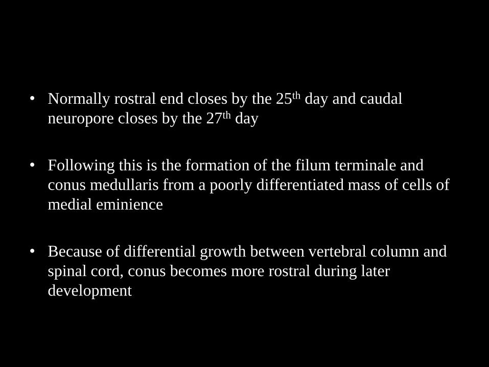
• Normally rostral end closes by the 25th day and caudal
neuropore closes by the 27th day
• Following this is the formation of the filum terminale and
conus medullaris from a poorly differentiated mass of cells of
medial eminience
• Because of differential growth between vertebral column and
spinal cord, conus becomes more rostral during later
development

Pathogenesis
• Meningomyelocoele is due to primary failure
of closure of neural tube or possible disruption
of already closed neural tube between 18-28
days of gestation

Etiology
• Usually multifactorial
• Specific etiology include:– Nutritional deficiency of Folic acid in mothers
– Hypervitaminosis A
– Chromosomal abnormalities- Trisomy 13,18
– Maternal obesity /Maternal insulin dependent diabetes mellitus
– Maternal hypothermia
– Intrauterine drug exposure to Valproate, Carbamazepine and ovulation inducing drugs
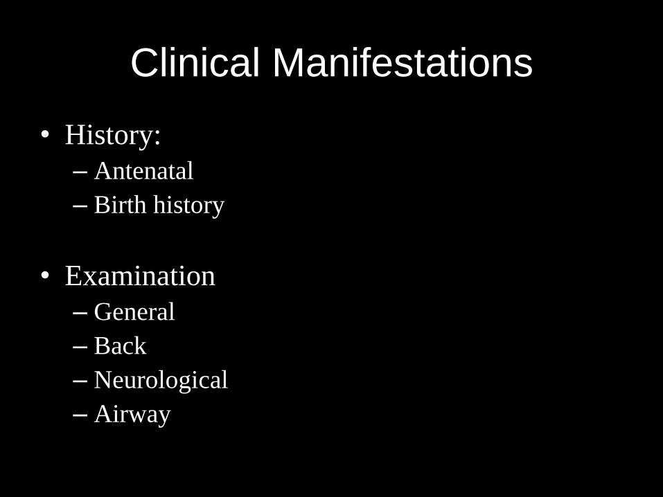
Clinical Manifestations
• History:
– Antenatal
– Birth history
• Examination
– General
– Back
– Neurological
– Airway

• Clinical findings:
– Paraplegia
– Hydrocephalus
– Cranial nerve dysfunction
– Seizures
– Neurogenic Bladder and bowel
– Renal failure
– Progressive bony, spine and joint deformities
– Pathological fractures

• Associated syndromes and anomalies:– Club feet
– Arnold-Chiari malformation
– Hydrocephalus
– Neurogenic bladder
– Musculoskeletal defects
– Urogenital abnormalities
– Hip dislocation
– Facial clefts
– Anorectal malformations
– Umbilical hernia
– Congenital heart disease- ASD/VSD
– VACTER
– Latex Allergy
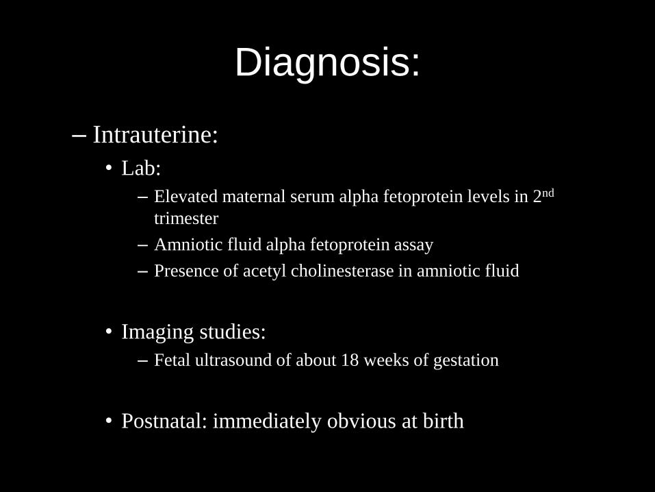
Diagnosis:
– Intrauterine:
• Lab:
– Elevated maternal serum alpha fetoprotein levels in 2nd
trimester
– Amniotic fluid alpha fetoprotein assay
– Presence of acetyl cholinesterase in amniotic fluid
• Imaging studies:
– Fetal ultrasound of about 18 weeks of gestation
• Postnatal: immediately obvious at birth

• Investigations :
– Hemogram
– S. Creatinine
– Urine routine and microscopy
– X-ray chest
– CT Brain and MRI brain and spine
– 2D Echo
– Ultrasound of urinary tract
– Urodynamic study

Anesthetic Management
• Problems anticipated in the perioperative
period:
– Age:
• Mainly neonate/infant
– Airway:
• Large head
• Associated facial clefts
• Paediatric airway and its implications

• Problems related to other syndromes
anomalies:
– Hydrocephalus
– Arnold-Chiari syndrome
– Congenital Heart disease
– Renal problems
– Musculoskeletal problems (Scoline)

• Problems related to surgery:
– Surgery in prone position:
• Extreme head flexion in Chiari malformation may cause
brain stem compression
• Improper positioning may lead to venous congestion of
face, neck and tongue, reducing lung compliance
• Increased abdominal pressure cause vena caval
compression that can cause increase bleeding through
engorged epidural veins

– Hypothermia
– Blood loss can be difficult to assess in view of simultaneous CSF loss
– Nerve studies may be needed intraoperatively to help in identification in few cases
– At dural closure, Valsalva manueuvre is needed to test the integrity of closure

• Problems in the post operative period:
– Due to nursing in the prone position
• Acute hydrocephalus:
– If not present preoperatively can occur due to
closure of the defect
– May occur due to shunt malfunction

• Premedication:– Need to premedicate will depend on the age of the child
– Since defect correction is done early, neonate will not need anxiolysis for premedication
– Antisialogogues may be given as the patient will be placed prone
– In case if the parents bring child in infancy premed to allay separation anxiety will be needed Choice based on presence of raised ICT – if present ketamine –best avoided

• Induction:– In syndromic babies and abnormal facies- difficult airway
is anticipated Induction of choice –inhalational anesthetic
– In presence of neurological deficit , SCh avoided
– Meningomyelocoele should be placed with adequate padding and rest of upper part of the body can rest on a adequate sized pillow
– Altenatively anesthetic and intubation can be in the lateral postition

• Postioning:
– Prone positioning with adequate sized bolster and head well supported , all bony points and eyes padded, abdomen should be free
– Anesthesia maintained with inhalational agents and opioids for pain relief
– Use of relaxants should be timed properly in case of nerve studies needed for nerve identification

• Monitoring:– Routine Monitoring – ECG, Pulse oximetry, capnometry,
NIBP
– Invasive BP in patients with large defect and difficult anatomy
– Temperature monitoring
– Hourly urine output
– Careful and accurate measurement of blood loss

Thank You


