Meninges, Ventricles and CSF · From (2) lateral ventricle it flows: through the (3)...
10
Meninges, Ventricles and CSF Neuroanatomy block-Anatomy-Lecture 19 Editing file
Transcript of Meninges, Ventricles and CSF · From (2) lateral ventricle it flows: through the (3)...
Meninges, Ventricles and CSFNeuroanatomy block-Anatomy-Lecture 19
Editing file

01 Describe the cerebral meninges & list the main dural folds.
02 Describe the spinal meninges & locate the level of the termination of each of them.
03 Describe the importance of the subarachnoid space.
04 List the Ventricular system of the CNS and locate the site of each of them.
05 Describe the formation, circulation, drainage, andfunctions of the CSF.
06 Know some clinical point about the CSF.
Color guide ● Only in boys slides in Green● Only in girls slides in Purple● important in Red● Notes in Grey
At the end of the lecture, students should be able to:
Objectives
3
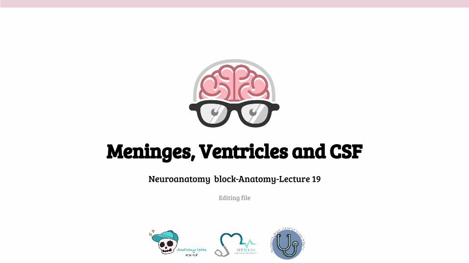
MeningesThe brain and spinal cord are invested by three concentric membranes:
Pia Mater The innermost layer.
Dura MaterThe outermost layer.
Arachnoid MaterThe middle layer.
Dura MaterThe cranial dura is a two layered tough, fibrous thick membrane that surrounds the brain.
Formed of two layers
periosteal :attached to the skull. meningeal :is folded forming the dural folds : Two large reflection of dura extend into the cranial cavity:
Sensory innervation of the dura mater: is mostly from : meningeal branches of the: trigeminal and vagus nerves & C1 to C3.
Falx cerebri:-It is a vertical sickle shaped sheet of dura, in the midline.-Extends from the cranial roof into the great longitudinal fissure between the two cerebral hemispheres. -It has an attached border adherent to the skull and a free border lies above the corpus callosum.
Tentorium cerebelli:-A horizontal shelf of dura, lies between the posterior part of the cerebral hemispheres and the cerebellum. -It has a free border that encircles the midbrain. -Its superior surface in the middle line it is continuous above with the falx cerebri, separated by straight sinus.

4
Arachnoid Mater & Pia MaterArachnoid Mater
- It is a soft, translucent membrane loosely envelops the brain.
- It is separated from the dura by a narrow subdural space.
Pia Mater - It is the innermost, thin, delicate & highly vascular
membrane that is closely adherent to the gyri and fitted into the sulci.
Between the pia and arachnoid mater lies the subarachnoid space which contains; fibrous trabeculae, main blood vessels and CSF.
Subarachnoid SpaceThe subarachnoid space is varied in depth forming; subarachnoid cisterns.
01Cisterna magna, or
cerebllomedullary cistern:
● lies between the inferior surface of thecerebellum and the back of the medulla.
● At this cistern CSF flows out of the fourth ventricle via the 2 lateral aperture and median aperture.
02 Interpeduncular cistern:● Is located at the base of the brain, where the arachnoid
spans between the two cerebral peduncles of midbrain.● It contains the optic chiasma & circulus arteriosus of
Wills .
5
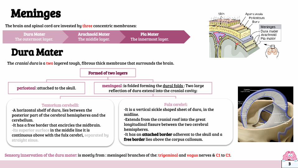
Spinal Meninges
● Spinal cord terminates at level L1-L2. ● The dura and arachnoid and, subarachnoid space, continue caudally to S2. ● Pia extends downwards forming the filum terminale which pierces the arachnoid and dural
sacs and passes through the sacral hiatus (11) to be attached to the back of the coccyx.● The spinal cord, is invested by three meningeal coverings:
Meningeal coverings
Dura Mater Pia mater
-The innermost covering, is a delicate fibrous membrane
closely envelops the cord and nerve roots.
-It is attached through the arachnoid to the dura by the
denticulate ligament.
-is a translucent membrane lies between the pia and dura, -Between arachnoid and pia lies the subarachnoid space
contains CSF.
-The outer covering; is a thick, tough fibrous membrane.
-It envelopes the cord loosely. -It is separated from
arachnoid mater by the subdural space, and from the
bony wall of the vertebral canal by the epidural space.
Arachnoid Mater
6
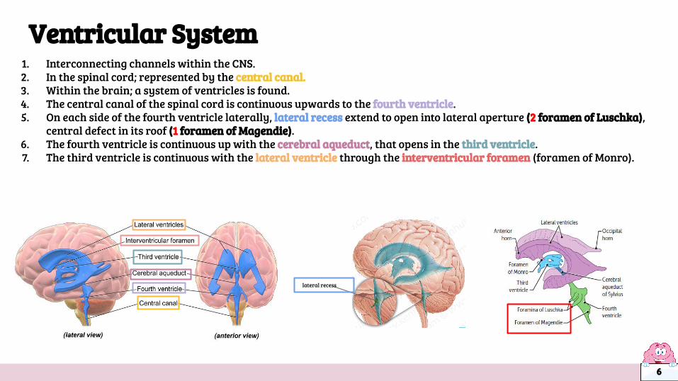
Ventricular System1. Interconnecting channels within the CNS. 2. In the spinal cord; represented by the central canal. 3. Within the brain; a system of ventricles is found. 4. The central canal of the spinal cord is continuous upwards to the fourth ventricle. 5. On each side of the fourth ventricle laterally, lateral recess extend to open into lateral aperture (2 foramen of Luschka),
central defect in its roof (1 foramen of Magendie).6. The fourth ventricle is continuous up with the cerebral aqueduct, that opens in the third ventricle. 7. The third ventricle is continuous with the lateral ventricle through the interventricular foramen (foramen of Monro).
lateral recess

● Present in the ventricular system, together with the cranial and spinal subarachnoid spaces.● It is colourless clear fluid containing little protein and few cells. It is about 150 ml.● It acts as a cushion for the brain from sudden movements of the head.● It’s circulates in the subarachnoid space.
Cerebrospinal Fluid
It is produced by the (1) choroid plexus, which is located in the lateral, third & fourth ventricles.
From (2) lateral ventricle it flows: through the (3) interventricular foramen or foramen of monro into the (4) 3rd ventricle and, by way of the (5) cerebral aqueduct (aqueduct of sylvius), into the (6) 4th ventricle.
It leaves the ventricular system through the three apertures of the 4th ventricle ( 7) median foramen of Magendie & (8) 2 lateral foramina of Luschka, to enters the subarachnoid space.
Reabsorbed finally into the venous system along (9) arachnoid villi, and arachnoid granulation (larger than villi) that project into the dural venous sinuses , mainly (10)superior sagittal sinus.
1
2
3
45
6
7
foramina of Luschka8
7
01
02
03
04
109 Arachnoid villi
Clinical point
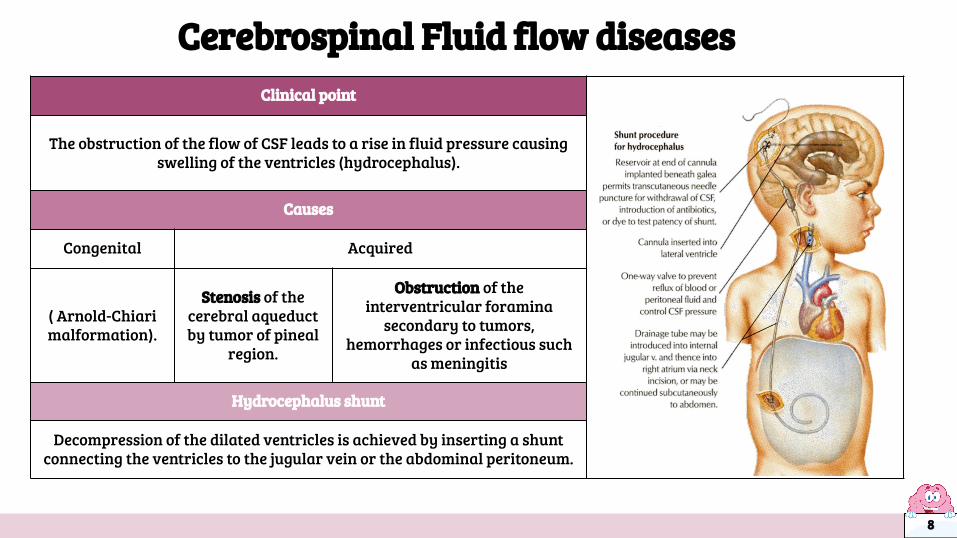
The obstruction of the flow of CSF leads to a rise in fluid pressure causing swelling of the ventricles (hydrocephalus).
Causes
Congenital Acquired
( Arnold-Chiarimalformation).
Stenosis of the cerebral aqueduct by tumor of pineal
region.
Obstruction of the interventricular foramina
secondary to tumors, hemorrhages or infectious such
as meningitis
Hydrocephalus shunt
Decompression of the dilated ventricles is achieved by inserting a shunt connecting the ventricles to the jugular vein or the abdominal peritoneum.
Cerebrospinal Fluid flow diseases
8
Practice Q1: The subarachnoid space terminates at:
A. Coccyx
B. L1-L2
C. S2
D. S1
Q2: The CSF is about:
A.50 ml
B. 150 ml
C. 250ml
D. 300 ml
Q3: cerebrospinal fluid is produced by:
A. 3rd ventricle
B. Choroid plexus
C. Lateral ventricle
D. 4th ventricle
Q4: which layer of dura mater that form falx cerebri and tentorium cerebelli ?
A. meningeal layer
B. pia mater layer
C. periosteal layer
D. subarachnoid layer
Q5: the CSF flows through foramen of monro (interventricular foramen) from (...) to (...)
A. Cerebral aqueduct , 3rd ventricle
B. 4th ventricle , central canal
C. 3rd ventricle, lateral ventricle
D. Lateral ventricle, 3rd ventricle
Q6: which one of these does not supply the dura mater ?
A. Facial
B. vagus
C. C1-C3
D. Trigeminal
Q7: the Cisterna magna is between ?
A. posterior surface of the cerebellum and the back of the medulla
B. cerebrum and the back of the medulla
C. at the base of the brain between the two cerebral peduncles of midbrain
D. inferior surface of the cerebellum and the back of the medulla
Q8 : hydrocephalus shunt is connecting the ventricles to
A. Carotid artery
B. jugular vein
C. Abdominal peritoneum
D. B & C
Answers: Q1(C) Q2(B) Q3(B) Q4(A) Q5(D) Q6(A) Q7(D) Q8(D) 9

Girls team :
● Ajeed Al Rashoud● Taif Alotaibi● Noura Al Turki● Amirah Al-Zahrani● Alhanouf Al-haluli● Sara Al-Abdulkarem● Renad Al Haqbani● Nouf Al Humaidhi● Jude Al Khalifah● Nouf Al Hussaini● Rahaf Al Shabri● Danah Al Halees● Rema Al Mutawa● Amirah Al Dakhilallah● Maha Al Nahdi ● Razan Al zohaifi ● Ghalia Alnufaei
Boys team:
● Mohammed Al-huqbani (legend)- LR
● Salman Alagla● Ziyad Al-jofan● Ali Aldawood● Khalid Nagshabandi● Omar Alammari● Sameh nuser● Abdullah Basamh (ily) -lecture reviewer
● Alwaleed Alsaleh● Mohaned Makkawi● Abdullah Alghamdi
Team leaders
● Ateen Almutairi● Abdulrahman Shadid
Contact us:
Editing file
Members board
most probably you don't need this
There are 3 easter eggs in this lecture, try to find them - Lecture reviewer بدر الشھري الظریف