Memory Metal Dennis Kok in Lumbar Spinal Fusion
173
Memory Metal in Lumbar Spinal Fusion Biological, mechanical, clinical and radiological studies Dennis Kok Dennis Kok Memory Metal in Lumbar Spinal Fusion
Transcript of Memory Metal Dennis Kok in Lumbar Spinal Fusion

Memory Metal in Lumbar Spinal FusionBiological, mechanical, clinical and radiological studies
Dennis Kok
Den
nis K
ok
Mem
ory
Metal in
Lum
bar Spin
al Fusio
n

Memory Metal in Lumbar Spinal FusionBiological, mechanical, clinical and radiological studies
Dennis Kok

Memory Metal in Lumbar Spinal FusionBiological, mechanical, clinical and radiological studiesThesis, Rijksuniversiteit Groningen, The Netherlands
Copyright © Dennis Kok, The NetherlandsAll rights reserved. No part of this thesis may be reproduced, stored in retrieval system or transmitted, in any form and by any means, without prior permission of the author.
Cover Design & Layout: High Woltage | Margreet Bruins - WoltmanPrinting: Print.com

Memory Metal inLumbar Spinal Fusion
Biological, mechanical, clinical and radiological studies
Proefschrift
ter verkrijging van de graad van doctor aan de
Rijksuniversiteit Groningen
op gezag van de
rector magnificus, prof. dr. C. Wijmenga
en volgens besluit van het College voor Promoties.
De openbare verdediging zal plaatsvinden op
woensdag 12 mei 2021 om 16.15 uur
door
Dennis Kok
geboren op 10 mei 1978
te Zwolle

Promotores Prof. dr. S.K. BulstraDr. M. Stevens
Copromotor Dr. F.H. Wapstra Beoordelingscommissie Prof. dr. F.C. ÖnerProf. dr. L.W. van RhijnProf. dr. P.C. Jutte


Contents
Chapter 1 General introduction, aims and outline
Chapter 2 Is remaining intervertebral disc tissue interfering with bone generation during fusion of two vertebrae?
Chapter 3 A New Lumbar Posterior Fixation System, the Memory Metal Spinal System: An In-vitro Mechanical Evaluation.
Chapter 4 The Memory Metal Spinal System in a Posterior Lumbar Interbody Fusion (PLIF) procedure: A Prospective, Non-Comparative Study to Evaluate the Safety and Performance.
Chapter 5 Biomechanical Evaluation of Two Minimal Access Interbody Cage Designs in a Cadaveric Model.
Chapter 6 The Memory Metal Minimal Access Cage: A New Concept in Lumbar Interbody Fusion. A Prospective, Non-Comparative Study to Evaluate the Safety and Performance.
Chapter 7 Changes in Bone Mineral Density in the Intertransverse Fusion Mass After Instrumented Single-Level Lumbar Fusion: A Prospective 1-Year Follow-up
Chapter 8 Conclusions, discussion and future perspective
Chapter 9 Summary
Chapter 10 Samenvatting
Appendices Dankwoord Curriculum Vitae
10
32
46
64
80
96
114
130
150
158
166

The Ecstasy of Gold” (Italian: L’estasi dell’oro) is a musical composition by
Ennio Morricone, part of his score for the 1966 Sergio Leone film The Good, the
Bad and the Ugly. It is played while Tuco (Eli Wallach) is frantically searching
a cemetery for the grave that holds $200,000 in gold coins. Sung by Edda Dell’Orso,
it stands as one of the most well known of Morricone’s themes.
American metal band Metallica has used “The Ecstasy of Gold” as the
introductory music for its concerts since 1983.

11
General introduction, aims and outline

12 Introduction, aims and outline
In order to gain better insight into different aspects of lumbar spinal fusion and its effect on chronic low back pain, this thesis will start with an overview of about 50 years of lumbar fusion. Different types of fusion devices as well as clinical and radiological fusion results will be reviewed. After looking at the background and history of lumbar fusion, the focus will be on the present and the future. The use of different implant devices to achieve spondylodesis and the evaluation of the results after spondylodesis will be specifically discussed in this introduction.
Background
Chronic low back pain is an insidious problem. Patients suffer from discomfort, anxiety and disability. The one-year prevalence ranges from 22 to 65%, and lifetime prevalence reportedly reaches 84% (1). Chronic low back pain belongs to the group of disorders that cause the highest burden of disease among 15-65-year-olds (the working population) in the Netherlands; it leads to a total of 112,700 Disability Adjusted Life Years (DALY) (2). In 2017, low back pain was even the leading cause of years lived with disability (YLD) globally, and next to headaches and depressive disorders has been the main cause of non-fatal health loss for the past three decades (3). The global prevalence of low back pain was almost 577 million in 2017, with a global incidence exceeding 245 million (3). Around 20% of those who suffer from low back pain seek medical attention (4). The economic burden to society is significant and can be categorized into direct costs, healthcare utilization costs and indirect costs. About 90% of the costs associated with low back pain are indirect costs, such as sickness absence and disability benefits (5). In the Netherlands, the costs for chronic low back pain represented 0.6% of the gross national product in 2007 (6). The most common cause of chronic low back pain is degeneration of the intervertebral disc (7). Treatment can be either conservative or operative. The majority of patients will find relief with conservative measures like behavioural-based exercise therapy (8), intensive rehabilitation programmes (9), or the use of Non Steroid Anti-Inflammatory Drugs (NSAIDs). For those with significant continuous specific symptoms like severe, disabling back pain with radiculopathy, surgical intervention may be appropriate. The main goals of surgical intervention are decompression of the neural structures and obtaining a solid arthrodesis. However, the role of spinal fusion in the treatment of chronic low back pain remains controversial (10,11). In a Cochrane review (12) no evidence was found of the effectiveness of spinal fusion for lumbar degenerative disc disease (DDD) or chronic low back pain, as compared to natural history, conservative treatment or placebo. A Swedish randomized controlled trial (RCT) from 2001 did show

13
Chap
ter 1 | Gen
eral intro
du
ction
, aims an
d o
utlin
e
a better outcome in patients treated with spinal fusion compared to patients who received conservative care, although this beneficial effect attenuated at longer follow-up (13).
Lumbar Spinal Fusion
HistorySuccessful interbody fusion has mechanical advantages over other types of fusion. Restoring disc height, segmental alignment and balance as well as load-bearing to anterior structures are some of the important features of interbody fusion (14). Hibbs and Albee (15) were the first to introduce stabilization of the spine in 1911. They reported the use of an interlaminar fusion technique for the treatment of an unstable spine secondary to Pott’s disease. This technique was ultimately extended to other pathological lesions and regions of the spine. In 1936, Mercer (16) theorized that the ideal operation for stabilization of the spine was interbody fusion. He lamented that the procedure was impossible to perform at that time, given the existing equipment and lack of sophistication of spinal surgery techniques. In 1943, Meyerding (17) reported posterior fusion of the laminae and spinous processes using an autograft in patients with back pain and spondylolisthesis. In the early years, lumbar interbody fusion was performed using allograft or autograft without instrumentation. These techniques were hampered by a very high failure rate (18,19). The high donor site morbidity of structural iliac crest bone graft was another problem with lumbar interbody fusion in the early years (20,21). To prevent collapse, displacement and/or extrusion of corticocancellous, autogenous or allogenous interbody bone grafts the use of cages emerged (20-34). These cages improved the long-term outcomes of spinal arthrodesis tremendously (35-37).
Bagby developed one of the first lumbar interbody fusion devices. This device, named the ‘Bagby Basket’, was essentially a stainless-steel basket. Other surgeons adopted this technique and modified it by adding threads to the cage for additional pullout and compressive strength, thus making the cage suitable for posterior approaches (38). A large number of interbody fusion devices are currently available on the market, and undoubtedly even more will become available in the future.
DevicesThe use of unilateral pedicle screw fixation was proposed to decrease the stiffness of the lumbar spinal fusion device and would be as effective as bilateral pedicle fixation. In 2000 Soo Suk et al. (39) showed that unilateral pedicle screw fixation was indeed as effective as bilateral pedicle screw fixation in lumbar spinal fusion, independently of the

14 number of fusion segments (one or two segments) or pedicle screw systems. Based on their results, unilateral fixation could be used even in bisegmental lumbar spinal fusion.
Interbody fusion cages can all be classified based on their design into one of two types: threaded or non-threaded. Threaded cages, also known as cylindrical or conical cages, are usually paired in the lumbar region, and are inserted parallel in an anteroposterior direction. These implants require preparation of the bony endplates with a reamer, followed by a threading device. The bony endplate’s integrity is thus partially destroyed, but the implant can achieve a good surface match with the underlying cancellous bone bed. The distractive height of cylindrical cages is limited, because the construct’s tolerated lateral width is restricted by vertebral anatomy. The non-threaded (box-shaped or rectangular) cages are placed singularly or in pairs. Most allow placement of bone graft inside and around the cage. To achieve fusion the bone graft contact area has to be as large as possible. Preparation of the cage bed requires removal of the endplate cartilage, so that bleeding bone is exposed to the graft material. There are various designs to achieve the largest contact surface and best anchorage possible.
MaterialsThe ideal spinal fusion device is one that is rigid enough to maintain stability, but with a similar elastic modulus of bone to prevent subsidence and stress-shielding. Additionally, osteoconductive properties vary by material and other factors such as radiolucencies, allowing for convenience during fusion assessments (40). The mismatch in the modulus of elasticity between the metal cage and the vertebral body may cause stress shielding, resulting in a delayed fusion and increased risk of cage fatigue failure (41). This is why different materials are available, each with its own benefits and drawbacks.
Carbon fiber cages better approximate the modulus of elasticity of vertebral bone, but there are some reports on carbon fiber release (wear) causing synovitis (42). Titanium implants (38,43) offer a radio-opaque alternative to carbon fibre materials, which also exhibit the necessary biomechanical strength as well as facilitate radiographic location of the cage. The problem with most of these cages, however, is the small contact area of the bone graft and therefore the high rate of pseudoarthrosis (44,45).
The drawbacks of non-resorbable cages have motivated exploration of the possible utility of polymer-based resorbable polylactide (PLA) cages. These cages have a modulus of elasticity close to that of vertebral bone and gradual resorption after interbody fusion is obtained (46). These bioresorbable cages may therefore avoid complications related to stress shielding. Another advantage is that they are
15
Chap
ter 1 | Gen
eral intro
du
ction
, aims an
d o
utlin
e
radiolucent, improving the radiographic evaluation of fusion (47). Mechanical failure, osteolysis and tissue reaction have been reported though (48). Much more research (level I/II) is required.
Interbody devices are also made from PEEK, which has an elastic modulus comparable to bone, allowing for relatively lower subsidence rates (49-51). Unlike titanium cages that are biocompatible, PEEK cages have a hydrophobic surface and may limit osseointegration (50,52,53). The need for greater endplate preparation and problems with overdistraction additionally compromise the effectiveness of PEEK cages (50,54). Still, a major advantage of PEEK implants is their radiolucent properties, which allow for better fusion assessment on imaging (50). For purposes of identification, these radiolucent cages often have metallic markers. Despite these differences, fusion rates between PEEK cages are comparable to titanium cages (55).
Although the recent introduction of Trabecular Metal Technology (Zimmer) into the field of spinal surgery has opened up new perspectives (56-58), additional (long-term) research on this technology is needed.
A different, relatively unexplored material in spinal surgery with interesting properties is shape memory metal. In 1962 Buehler and co-workers at the US Naval Ordnance Laboratory discovered the shape memory effect of the equiatomic alloy of nickel-titanium (Ni-Ti) (59). Ni-Ti alloys consisting of equal atomic amounts of Ni and Ti (49-51 mol%Ni) show unique mechanical properties such as shape memory, superelasticity and dumping. With the shape memory effect, the original shape can be recovered after deformation by heating; with superelasticity, any apparent plastic deformation can be returned to the original shape by releasing the load. The range of composition for the Ni-Ti alloy displaying these properties is very narrow, nearly a 1:1 atomic ratio. This Ni-Ti alloy has an optimal transformation temperature as a biomaterial, with an austenitic phase (A phase) at a higher temperature and a martensitic phase (M phase) at a lower temperature that is also heat-elastic. When the temperature of the alloy is decreased from the A phase, the martensitic transformation begins at Ms (1) and all phases transform to the M phase at Mf (2). On the other hand, when the temperature is increased from the M phase, austenitic transformation begins at As (3) and all phases transform to the A phase at Af (4). This is shown in Figure 1.

16 Fig. 1. Transformation of Ni-Ti alloy and mechanism of shape memory effect.
The mechanical properties of the martensite condition are very different from those of the austenite condition. The martensite is easily deformed to low-percentage strains at low stresses, whereas the austenite has much higher-yield stresses. In practice this means that the low-temperature martensite phase is soft and ductile and can be easily deformed, while the high-temperature austenite phase is quite strong and hard (similar to titanium) (60,61). Because of its special properties, the nearly equiatomic nickel-titanium alloy could be very suitable for minimally invasive applications. At present, the shape memory nickel-titanium alloy is used clinically in wires for orthodontic tooth alignment, osteosynthesis staples and vascular applications, for instance in a stent and a vena cava filter (62-67). Wever et al. looked at the cytotoxic, allergic and genotoxic activity of a nickel-titanium alloy (68); corrosion and fretting processes were not observed, and no adverse tissue reactions were evident (69). In the early 1970s, Schmerling and co-workers experimented in the field of spinal surgery with a shape memory metal rod to replace a standard Harrington rod in a human cadaver (70).

17
Chap
ter 1 | Gen
eral intro
du
ction
, aims an
d o
utlin
e
Preliminary investigations were also conducted with an anterior system, using a shape memory metal wire with Dwyer instrumentation (71). In China, Lu reported comparable results on surgical procedures in patients with an idiopathic scoliosis using shape memory metal rods instead of Luque rods (72). This literature evidences the merits of memory metal in the correction of scoliosis. To this date, there is no consensus on which material is superior for interbody fusion cages.
Biomechanics Interbody fusion cages have been stated to provide good segmental distraction, offer axial load support and reduce segmental mobility, but there are reports of failed fusions because of implant failure. The biomechanical function of an interbody implant is to support the anterior column, maintain tension on the annulus fibres, provide adequate foramina distraction, and confer immediate motion segment stability (41,73). From a materials and design perspective, the chosen interbody graft must be structurally competent to withstand the compressive loads experienced in the lower lumbar spinal segments during all forms of physical activity (74). When the disc is replaced by an interbody fusion implant, most of the axial load is transmitted through the implant. The implant-endplate contact area, through which the load also is transmitted, is related to the size and shape of the implant face that is in contact with the endplate. If the implant-endplate contact area is too small or incongruent, the implant can create excessive stress at the implant-bone interface, which may lead to implant subsidence. Conversely, if the implant-endplate contact area is very large, there remains too little surface for the housed graft material to be exposed to the host bone and incorporated into the fusion (75). Goh et al. (76) tested the significance of cage size (width and height) on segmental stability in response to various applied force vectors. They showed that medium-sized cages adequately restored extension and lateral bending stiffness, whereas torsional stiffness was restored only by using the larger-sized cages. Unlimited size selection, however, is restricted by the ability to place the cage safely within the intervertebral space without the risk of neurological injury. It has been argued (77,78) that preservation of bony endplate is desirable for the prevention of implant subsidence. On the other hand, partial removal of the endplate may facilitate the incorporation of the graft material, thereby increasing the likelihood of a solid interbody fusion (79). Some authors recommend (80-82) complete removal of the bony endplate to allow the implant and/or graft material to rest on cancellous bone. However, these studies arbitrarily chose a 50% cut-off of expected trabecular compressive strength (80) or were based on four specimens only, with no statistics provided (82). Hollowell et al. (81) found that complete removal of the bony endplate did not alter compressive strength. Steffen et al. (83) concluded that lumbar interbody fusion implant with approximately 30% endplate surface contact through a rim resting on the peripheral endplate offers compressive strength similar to that of an implant with full-surface area

18 in contact with the endplate. Further spinal stability, through enhanced load bearing across the anterior column, may be achieved by adding segmental pedicular fixation in a static or compressive mode (84). This has been shown to increase fusion rates by augmenting rigidity at the fusion site (85,86).
OutcomeWhen looking at the literature on lumbar spinal fusion, we focus on clinical outcome and radiological fusion. There is a large volume of data in the literature detailing clinical and radiological outcomes following specific interbody surgical interventions, yet there is little robust class 1 or 2 clinical and biomechanical data comparing the various available techniques. Surgeons who have been trained in one specific interbody technique will tend to favour that technique, irrespective of the pathology being treated or the number of operative levels performed. The literature uniformly supports the concept of interbody techniques over on-lay posterior spinal fusion for sagittal and coronal plane deformities (87), with deformity reduction correlating with positive clinical outcomes in a number of well-conducted studies (88). Stable fusion is the ultimate goal for any cage instrumentation. The main purpose of the placement of interbody cage devices is to create a proper mechanical environment for successful fusion. Bony ingrowths are enhanced by a very rigid postoperative structure (89). The larger the interface between bone graft and a correctly prepared host bed, the higher fusion rates will be (83).
Overall it can be concluded that outcome measures differ considerably between clinical cage studies, making comparison very hard. A few prospective clinical studies have been published, most of them investigating clinical and radiological performance of a cage used with different follow-up schedules. Yuan et al. (21), Kuslich et al. (38) and Alpert (90) published large prospective multicenter clinical trials. A total of 947 patients received the Bagby and Kuslich (BAK) device (Spine Tech Inc., Minneapolis, MN, USA), which consists of two titanium screw cages, through either a posterior (38%) or anterior approach (62%). Outcome was analyzed by using the Prolo scale developed by Prolo et al. (91) and a numerical scale that assesses functional outcome on seven parameters. Eighty-five percent of patients experienced a decrease in pain and improved functional outcome 24 months postoperatively. On both pain and functional outcome, improvement was better at 12 months than at 24 months, suggesting that improvement can still be expected after one year. Yuan et al. (21) also showed in their study that of the patients who were eligible for work, 68% had indeed returned to work one year after the operation. Twenty-four months postoperatively 78% of these patients were working again. Fusion was seen in 91% of their patients, and was defined as absence of significant radiolucencies and less than 5° vertebral motion in the sagittal plane.

19
Chap
ter 1 | Gen
eral intro
du
ction
, aims an
d o
utlin
e
Another study that reviewed the use of the Ray cage, published in 1997, showed 96% fusion after a minimum of 24 months in 211 patients (92). Fusion in that study was defined as absence of motion on flexion and extension radiographic evaluation, absence of radio-opacity and bony continuity within the cage.
Brantigan et al. (93) studied the results of 271 Brantigan cages filled with autogenous bone in combination with pediclescrew instrumentation. They compared these with a control group of 75 patients in which ethylene-oxide-sterilized allograft bone blocks and pedicle screws were used. In the group of patients who were treated with a Brantigan cage, 97% fusion was achieved versus 79% in the control group. Clinical success was analyzed using Prolo’s scale. A successful clinical result was achieved in 87% of those patients managed with a Brantigan cage and 80% who had been managed with an allograft.
Sasso et al. (94) conducted a prospective, randomized controlled clinical trial comparing a cylindrical threaded titanium cage to a femoral ring allograft control for anterior lumbar interbody fusion. At 12 months, 97% of the cylindrical threaded titanium cage device group and 40% of the control group demonstrated radiographic fusion. At 24 months, 97% of the cylindrical threaded titanium cage group and 52% of the control group showed radiographic fusion (P < 0.001). The Oswestry Low Back Pain Questionnaire and Health-Related Quality of Life (The Short Form (36) Health Survey) scores were not significantly different between groups.
A wide range of fusion rates (56-95%) after interbody fusion with varying techniques is reported (21,78,95-105). As stated in one review article (26), the rate of fusion ‘depends to a great extent on the investigator’s interpretation’. Because there is no single definition of what constitutes fusion, it is difficult if not impossible to compare results of different studies. It is likewise hard to determine radiographically whether fusion has occurred. Besides, findings of biomechanical tests on stability do not always correspond directly with radiographic evidence of fusion. The rates of fusion are approximately 20% higher when the sole criterion is loss of motion (determined by comparing lateral flexion and extension radiographs) rather than continuous trabeculae across the graft-vertebrae interfaces (79,89,96,98,102,106-114).
As mentioned, monitoring clinical fusion outcome is difficult (115). The most sensitive diagnostic methods for detecting a pseudoarthrosis are computed tomography (CT) and magnetic resonance imaging (MRI). However, steel and titanium implants cause artefacts, rendering fusion assessment unreliable. Even plain radiographs pose a significant limitation, in that the radio-opaque implant is often superimposed on the fusion mass. Fenestrated cages may allow for an improved view, but the entire fusion

20 mass is never visible for radiographic assessment. Functional radiographs in flexion/extension would allow residual segmental mobility to be quantified, thus serving as an indirect measure for monitoring fusion success, but the absence of detectable motion does not imply that solid fusion has occurred (98). A fibrous pseudoarthrosis may well limit gross segmental motion, but micro-motion may persist, impeding fusion. Gross segmental stability, even in the absence of pain, does not imply successful fusion. The only acceptable criterion for clinical fusion assessment is the presence of bridging trabecular bone in continuity across two adjacent vertebrae (116).
21
Chap
ter 1 | Gen
eral intro
du
ction
, aims an
d o
utlin
e
Aims and outline of this thesis
Over the past half-century, both the surgical techniques and the instrumentation required for lumbar spinal fusion have changed significantly. The common goals of these changes were to improve fusion rates and optimize clinical outcomes. The specific aim of the present thesis was to further study different potential improvements in lumbar spinal fusion, particularly the use of a novel material in spinal surgery with interesting properties.
What is the effect of remaining intervertebral disc tissue on the fusion process? (Chapter 2) Important features of interbody fusion devices are sufficient axial support, a large contact area of the graft facilitating bony ingrowth, and ease in minimal access implantation. With the development of minimally invasive surgery, the importance of complete removal of intervertebral disc (IVD) tissue in order to achieve optimal placement has become more a challenge.
How does the Memory Metal Spinal System behave biomechanical according to ASTM F1717-96, ‘Standard Test Methods for Static and Fatigue for Spinal Implant Constructs in a Corpectomy Model’. (Chapter 3)The development of the minimally invasive Memory Metal Spinal System (MMSS) and the Memory Metal Minimal Access Cage (MAC) are steps in the evolution of spinal surgery. The MMSS is a posterior system, consisting of a single spinal rod used in conjunction with pedicle screws and connection bridges. The single rod, manufactured from memory metal that offers more elasticity than stainless steel or titanium and therefore ease of use, should also reduce operating times, in turn leading to other desirable outcomes such as reduced blood loss (118). As mentioned, spinal systems that are currently available use components manufactured out of stainless steel or titanium. The square spinal rod component used in this system is manufactured from Nitinol (NiTi), a nickel-titanium alloy. There should be less degeneration of adjacent segments (adjacent level disease), and better fusion is expected because of reduced rigidity in the memory metal spinal system. With current systems there may be loss of achieved reposition due to the viscous properties of the spine. By using a memory metal in this new system the expectation was that there is better maintenance of the reposition thanks to the metal’s inherent shape-memory properties (continuous reposition force).
In vivo performance of the Memory Metal Spinal System: Is it safe? How does it perform clinically and radiologically? (Chapter 4)The Memory Metal Spinal System was implanted in humans for the first time and used
22 in conjunction with Brantigan IF® carbon fibre reinforced polymer fusion cage. The biomechanical behaviour of the device showed good results. The next step is to study the Memory Metal Spinal System for the treatment of spondylolisthesis, symptomatic spinal stenosis and degenerative disc disease in humans.
Biomechanical behaviour of the Memory Metal Minimal Access Cage (MAC) in a cadaveric model: Which of the new developed cage designs of Memory Metal performs best, with regard to stability and subsidence? (Chapter 5)The MAC builds on the concept of sufficient axial support in combination with a large contact area of the graft, facilitating bony ingrowths and ease in minimal access implantation owing to its high deformability. The MAC is also constructed from the memory metal Nitinol and will have the same modulus of elasticity as the vertebral body. It is a hollow horseshoe-shaped implant, which results in larger spaces for additional bone grafts. The combination of improved elasticity with a larger contact area should theoretically yield higher rates of solid fusion.
Clinical safety and performance of the Memory Metal Minimal Access Cage (MAC). (Chapter 6)As shown above, selection of the cage is an important aspect of the performance of a lumbar interbody fusion. However, clinical results with the newly developed Memory Metal Minimal Access Cage are not yet available. We therefore conducted a pilot study to evaluate the performance and safety of this new interbody fusion device in a relatively small group of patients.
Is the use of modern DEXA a useful tool in the prediction of lumbar spinal fusion? (Chapter 7) The process of bone graft remodelling and final bone formation after surgery to achieve a spondylodesis is difficult to follow. Modern Dual Energy X-ray Absorption (DEXA) technique has the ability to quantify bone mineral density (BMD) changes and could therefore be helpful towards gaining further insight into the biological process of bone graft remodelling as well as quantifying BMD changes in the fusion mass in time.
Discussion with conclusions and future perspectives. (Chapter 8)Finally, the findings presented in the preceding Chapters are discussed. The different steps to gain further insight in various aspects of lumbar spinal fusion are reviewed, and the conclusions of these studies and their shortcomings are described. Furthermore some future perspectives are given.
Summary of this thesis. (Chapter 9)
Summary of this thesis in Dutch. (Chapter 10)

23
Chap
ter 1 | Gen
eral intro
du
ction
, aims an
d o
utlin
e
References
1. Walker BF. The prevalence of low back pain: a systematic review of the literature from 1966 to 1998. J Spinal Disord. 2000;13(3):205-217.
2. Berg M van den, Schoemaker CG (red.). Effecten van preventie. Deelrapport van de Volksgezondheid Toekomst Verkenning 2010 Van gezond naar beter. RIVM Rijksinstituut voor Volksgezondheid en Milieu -rapport nr. 270061007. Bilthoven: RIVM, 2010
3. GBD 2017 Disease and Injury Incidence and Prevalence Collaborators. Global, regional, and national incidence, prevalence, and years lived with disability for 354 diseases and injuries for 195 countries and territories, 1990-2017: a systematic analysis for the Global Burden of Disease Study 2017 [published correction appears in Lancet. 2019 Jun 22;393(10190):e44]. Lancet. 2018;392(10159):1789-1858.
4. RIVM. Jaarprevalentie van nek- en rugklachten 2017.
5. Lambeek LC, van Tulder MW, Swinkels IC, Koppes LL, Anema JR, van Mechelen W. The trend in total cost of back pain in The Netherlands in the period 2002 to 2007. Spine (Phila Pa 1976). 2011;36(13):1050-1058.
6. Geurts JW, Willems PC, Kallewaard JW, van Kleef M, Dirksen C. The Impact of Chronic Discogenic Low Back Pain: Costs
and Patients’ Burden. Pain Res Manag. 2018;2018:4696180. Published 2018 Oct 1.
7. Hedman TP, Kostuik JP, Fernie GR, Hellier WG. Design of an intervertebral disc prosthesis. Spine (Phila Pa 1976). 1991;16(6 Suppl):S256-S260.
8. Brox JI, Sørensen R, Friis A, et al. Randomized clinical trial of lumbar instrumented fusion and cognitive intervention and exercises in patients with chronic low back pain and disc degeneration. Spine (Phila Pa 1976). 2003;28(17):1913-1921.
9. Fairbank J, Frost H, Wilson-MacDonald J, et al. Randomised controlled trial to compare surgical stabilisation of the lumbar spine with an intensive rehabilitation programme for patients with chronic low back pain: the MRC spine stabilisation trial [published correction appears in BMJ. 2005 Jun 25;330(7506):1485]. BMJ. 2005;330(7502):1233.
10. Krismer M. Fusion of the lumbar spine. A consideration of the indications. J Bone Joint Surg Br. 2002;84(6):783-794.11. Turner JA, Ersek M, Herron L, et al. Patient outcomes after lumbar spinal fusions. JAMA. 1992;268(7):907-911.
12. Gibson JN, Waddell G. Surgery for degenerative lumbar spondylosis: updated Cochrane Review. Spine (Phila Pa 1976). 2005;30(20):2312-2320.

24 13. Fritzell P, Hägg O, Wessberg P, Nordwall A; Swedish Lumbar Spine Study Group. 2001 Volvo Award Winner in Clinical Studies: Lumbar fusion versus nonsurgical treatment for chronic low back pain: a multicenter randomized controlled trial from the Swedish Lumbar Spine Study Group. Spine (Phila Pa 1976). 2001;26(23):2521-2534.
14. Panjabi MM. Biomechanical evaluation of spinal fixation devices: I. A conceptual framework. Spine (Phila Pa 1976). 1988;13(10):1129-1134.
15. Hibbs RA, Albee FH. An operation for progressive spinal deformities. NY State J Med. 1911;93: 1013.
16. Mercer W. Spondylolisthesis: With a Description of a New Method of Operative Treatment and Notes of Ten Cases. Edinb Med J. 1936;43(9):545-572.17. Meyerding HW. Spondylolisthesis: Surgical treatment and results. J Bone Joint Surg Am. 1943;25: 65-77.
18. Jaslow IA. Intercorporal bone graft in spinal fusion after disc removal. Surg Gynecol Obstet. 1946;82:215-218.
19. Stauffer RN, Coventry MB. Anterior interbody lumbar spine fusion. Analysis of Mayo Clinic series. J Bone Joint Surg Am. 1972;54(4):756-768.
20. Fernyhough JC, Schimandle JJ, Weigel MC, Edwards CC, Levine AM. Chronic donor site pain complicating bone graft
harvesting from the posterior iliac crest for spinal fusion. Spine (Phila Pa 1976). 1992;17(12):1474-1480.
21. Yuan HA, Kuslich SD, Dowdle JA Jr., et al. Prospective multicenter clinical trial of the BAK interbody fusion system. Read at the Annual Meeting of the North American Spine Society, New York, N.Y., Oct. 22, 1997.
22. Amuso SJ, Neff RS, Coulson DB, Laing PG. The surgical treatment of spondylolisthesis by posterior element resection. J Bone Joint Surg Am. 1970;52(3):529-536.
23. Boucher HH. A method of spinal fusion. J Bone Joint Surg Br. 1959;41-B(2):248-259.
24. Boxall D, Bradford DS, Winter RB, Moe JH. Management of severe spondylolisthesis in children and adolescents. J Bone Joint Surg Am. 1979;61(4):479-495.
25. Dickman CA (1997). Internal fixation and fusion of the lumbar spine using threaded interbody cages. BNI Quarterly 3: 4-25.
26. Dunsker SB (1988). Lumbar spine stabilization: Indications, in Black PM (ed.). Clin Neurosurg. Baltimore, Williams and Wilkins 36: 147-58.
27. Evans JH. Biomechanics of lumbar fusion. Clin Orthop Relat Res. 1985;(193):38-46.

25
Chap
ter 1 | Gen
eral intro
du
ction
, aims an
d o
utlin
e
28. Loguidice VA, Johnson RG, Guyer RD, et al. Anterior lumbar interbody fusion. Spine (Phila Pa 1976). 1988;13(3):366-369.
29. O’Brien JP. The role of fusion for chronic low back pain. Orthop Clin North Am. 1983;14(3):639-647.
30. Pizzutillo PD, Hummer CD 3rd. Nonoperative treatment for painful adolescent spondylolysis or spondylolisthesis. J Pediatr Orthop. 1989;9(5):538-540.
31. Pfeiffer M, Griss P, Haake M, Kienapfel H, Billion M. Standardized evaluation of long-term results after anterior lumbar interbody fusion. Eur Spine J. 1996;5(5):299-307.
32. Simmons JW. Posterior lumbar interbody fusion, in Frymoyer JW (ed.). The Adult Spine. New York, Raven Press. 1991, pp 1961-87.
33. Soini J. Lumbar disc space heights after external fixation and anterior interbody fusion: a prospective 2-year follow-up of clinical and radiographic results. J Spinal Disord. 1994;7(6):487-494.
34. Truumees E, Sidhu K, Fischgrund JS. Indications for fusion in lumbar disc disease. Semin Spine Surg. 1999; 11: 147-62.
35. Brantigan JW, Steffee AD. A carbon fiber implant to aid interbody lumbar fusion. Two-year clinical results in the
first 26 patients. Spine (Phila Pa 1976). 1993;18(14):2106-2107.
36. Carson WL (1990). Biomechanical studies, ISOLA spinal system manual, AcroMed Corporation, Cleveland.
37. Whitecloud TS, Wolfe MW. Indications for internal fixation and fusion in the degenerative lumbar spine. In: Bridwell KH, DeWald RL (eds). The Textbook of Spinal Surgery. 2nd ed. Philadelphia, 1997, pp 1581-1600.
38. Kuslich SD, Ulstrom CL, Griffith SL, Ahern JW, Dowdle JD. The Bagby and Kuslich method of lumbar interbody fusion. History, techniques, and 2-year follow-up results of a United States prospective, multicenter trial. Spine (Phila Pa 1976). 1998;23(11):1267-1279.
39. Suk KS, Lee HM, Kim NH, Ha JW. Unilateral versus bilateral pedicle screw fixation in lumbar spinal fusion. Spine (Phila Pa 1976). 2000;25(14):1843-1847.
40. Kienle A, Krieger A, Willems K, Wilke HJ. Resistance of coated polyetheretherketone lumbar interbody fusion cages against abrasion under simulated impaction into the disc space. J Appl Biomater Funct Mater. 2019;17(2):2280800018782854.
41. Sandhu HS, Turner S, Kabo JM, et al. Distractive properties of a threaded interbody fusion device. An in vivo model. Spine (Phila Pa 1976). 1996;21(10):1201-1210.
26 42. Parsons JR, Bhayani S, Alexander H, Weiss AB. Carbon fiber debris within the synovial joint. A time-dependent mechanical and histologic study. Clin Orthop Relat Res. 1985;(196):69-76.
43. Ray CD. Threaded titanium cages for lumbar interbody fusions. Spine (Phila Pa 1976). 1997;22(6):667-680.
44. McAfee PC, Cunningham BW, Lee GA, et al. Revision strategies for salvaging or improving failed cylindrical cages. Spine (Phila Pa 1976). 1999;24(20):2147-2153.
45. Wetzel FT, LaRocca H. The failed posterior lumbar interbody fusion. Spine (Phila Pa 1976). 1991;16(7):839-845.
46. van Dijk M, Smit TH, Burger EH, Wuisman PI. Bioabsorbable poly-L-lactic acid cages for lumbar interbody fusion: three-year follow-up radiographic, histologic, and histomorphometric analysis in goats. Spine (Phila Pa 1976). 2002;27(23):2706-2714.
47. McAfee PC, Boden SD, Brantigan JW, et al. Symposium: a critical discrepancy-a criteria of successful arthrodesis following interbody spinal fusions [published correction appears in Spine 2001 May 1;26(9):1103]. Spine (Phila Pa 1976). 2001;26(3):320-334.
48. Kandziora F, Pflugmacher R, Scholz M, Eindorf T, Schnake KJ, Haas NP. Bioabsorbable interbody cages in a sheep cervical spine fusion model. Spine (Phila Pa 1976). 2004;29(17):1845-1856.
49. van Dijk M, Smit TH, Sugihara S, Burger EH, Wuisman PI. The effect of cage stiffness on the rate of lumbar interbody fusion: an in vivo model using poly-Lactic Acid) and titanium cages. Spine (Phila Pa 1976). 2002;27(7):682-688.
50. Schimmel JJ, Poeschmann MS, Horsting PP, Schönfeld DH, van Limbeek J, Pavlov PW. PEEK Cages in Lumbar Fusion: Mid-term Clinical Outcome and Radiologic Fusion. Clin Spine Surg. 2016;29(5):E252-E258.
51. Vadapalli S, Sairyo K, Goel VK, et al. Biomechanical rationale for using polyetheretherketone (PEEK) spacers for lumbar interbody fusion-A finite element study [published correction appears in Spine. 2007 Mar 15;32(6):710]. Spine (Phila Pa 1976). 2006;31(26):E992-E998.
52. Torstrick FB, Safranski DL, Burkus JK, et al. Getting PEEK to Stick to Bone: The Development of Porous PEEK for Interbody Fusion Devices. Tech Orthop. 2017;32(3):158-166.
53. Tsou HK, Chi MH, Hung YW, Chung CJ, He JL. In Vivo Osseointegration Performance of Titanium Dioxide Coating Modified Polyetheretherketone Using Arc Ion Plating for Spinal Implant Application. Biomed Res Int. 2015;2015:328943.54. Spruit M, Falk RG, Beckmann L, Steffen T, Castelein RM. The in vitro stabilising effect of polyetheretherketone cages versus a titanium cage of similar design for anterior lumbar interbody fusion. Eur Spine J. 2005;14(8):752-758.

27
Chap
ter 1 | Gen
eral intro
du
ction
, aims an
d o
utlin
e
55. Seaman S, Kerezoudis P, Bydon M, Torner JC, Hitchon PW. Titanium vs. polyetheretherketone (PEEK) interbody fusion: Meta-analysis and review of the literature. J Clin Neurosci. 2017;44:23-29.
56. Levine BR, Sporer S, Poggie RA, Della Valle CJ, Jacobs JJ. Experimental and clinical performance of porous tantalum in orthopedic surgery. Biomaterials. 2006;27(27):4671-4681.
57. Löfgren H, Engquist M, Hoffmann P, Sigstedt B, Vavruch L. Clinical and radiological evaluation of Trabecular Metal and the Smith-Robinson technique in anterior cervical fusion for degenerative disease: a prospective, randomized, controlled study with 2-year follow-up. Eur Spine J. 2010;19(3):464-473.
58. Lequin MB, Verbaan D, Bouma GJ. Posterior lumbar interbody fusion with stand-alone Trabecular Metal cages for repeatedly recurrent lumbar disc herniation and back pain. J Neurosurg Spine. 2014;20(6):617-622.
59. Buehler WJ, Wang FE. A summary of recent research on nitinol alloys and their potential application. Ocean Eng 1968;1:105-120.
60. Duerig TW, Melton KN, Stoeckel D, Wayman CM (1990). Engeneering Aspects of Shape Memory Alloys Butterworth-Heinemann Ltd, London.
61. Sanders MM (1993). Amemory metal based scoliosis correction system. PhD Thesis, University of Twente, The Netherlands.
62. Andreasen G. A clinical trial of alignment of teeth using a 0.019 inch thermal nitinol wire with a transition temperature range between 31 degrees C. and 45 degrees C. Am J Orthod. 1980;78(5):528-537.
63. Baumgart F, Bensmann G, Haasters J, Nölker A, Schlegel KF. Zur Dwyerschen Skoliosenoperation mittels Drähten aus Memory-Legierungen. Eine experimentelle Studie [On Dwyer’s scoliosis operation using memory alloy wire (author’s transl)]. Arch Orthop Trauma Surg. 1978;91(1):67-75.
64. Cragg AH, De Jong SC, Barnhart WH, Landas SK, Smith TP. Nitinol intravascular stent: results of preclinical evaluation. Radiology. 1993;189(3):775-778.
65. Kambic H, Sutton C, Oku T, et al. Biological performance of TiNi shape memory alloy vascular ring prostheses: a two year study. Int J Artif Organs. 1988;11(6):487-492.
66. Prince MR, Salzman EW, Schoen FJ, Palestrant AM, Simon M. Local intravascular effects of the nitinol wire blood clot filter. Invest Radiol. 1988;23(4):294-300.
67. Simon M, Athanasoulis CA, Kim D, et al. Simon nitinol inferior vena cava filter: initial clinical experience. Work in progress. Radiology. 1989;172(1):99-103.

28 68. Wever DJ, Veldhuizen AG, Sanders MM, Schakenraad JM, van Horn JR. Cytotoxic, allergic and genotoxic activity of a nickel-titanium alloy. Biomaterials. 1997;18(16):1115-1120.
69. Wever DJ, Veldhuizen AG, de Vries J, Busscher HJ, Uges DR, van Horn JR. Electrochemical and surface characterization of a nickel-titanium alloy. Biomaterials. 1998;19(7-9):761-769.
70. Schmerling MA, Wilkov MA, Sanders AE, Woosley JE. Using the shape recovery of nitinol in the Harrington rod treatment of scoliosis. J Biomed Mater Res. 1976;10(6):879-892.
71. Baumgart F, Bensmann G, Haasters J, Nölker A, Schlegel KF. Zur Dwyerschen Skoliosenoperation mittels Drähten aus Memory-Legierungen. Eine experimentelle Studie [On Dwyer’s scoliosis operation using memory alloy wire (author’s transl)]. Arch Orthop Trauma Surg. 1978;91(1):67-75.
72. Lu S (1990). Medical applications of Ni-Ti alloys in China. In: Duerig TW, Melton KN, Stöckel CM, Wayman CM (eds) Engineering aspects of shape memory alloys. Butterworth-Heinemann, London, pp 445–451
73. Steffen T, Tsantrizos A, Fruth I, Aebi M. Cages: designs and concepts. Eur Spine J. 2000;9 Suppl 1(Suppl 1):S89-S94.
74. Weiner BK, Fraser RD. Spine update lumbar interbody cages [published correction appears in Spine 1998 Jun 15;23(12):1428]. Spine (Phila Pa 1976). 1998;23(5):634-640.
75. Wimmer C, Krismer M, Gluch H, Ogon M, Stöckl B. Autogenic versus allogenic bone grafts in anterior lumbar interbody fusion. Clin Orthop Relat Res. 1999;(360):122-126.
76. Goh JC, Wong HK, Thambyah A, Yu CS. Influence of PLIF cage size on lumbar spine stability. Spine (Phila Pa 1976). 2000;25(1):35-40.
77. Kozak JA, Heilman AE, O’Brien JP. Anterior lumbar fusion options. Technique and graft materials. Clin Orthop Relat Res. 1994;(300):45-51.
78. Lin PM, Cautilli RA, Joyce MF. Posterior lumbar interbody fusion. Clin Orthop Relat Res. 1983;(180):154-168.
79. Boden SD, Sumner DR. Biologic factors affecting spinal fusion and bone regeneration. Spine (Phila Pa 1976). 1995;20(24 Suppl):102S-112S.
80. Closkey RF, Parsons JR, Lee CK, Blacksin MF, Zimmerman MC. Mechanics of interbody spinal fusion. Analysis of critical bone graft area. Spine (Phila Pa 1976). 1993;18(8):1011-1015.
29
Chap
ter 1 | Gen
eral intro
du
ction
, aims an
d o
utlin
e
81. Hollowell JP, Vollmer DG, Wilson CR, Pintar FA, Yoganandan N. Biomechanical analysis of thoracolumbar interbody constructs. How important is the endplate?. Spine (Phila Pa 1976). 1996;21(9):1032-1036.
82. Pearcy MJ, Evans JH, O’Brien JP. The load bearing capacity of vertebral cancellous bone in interbody fusion of the lumbar spine. Eng Med. 1983;12(4):183-184.
83. Steffen T, Tsantrizos A, Aebi M. Effect of implant design and endplate preparation on the compressive strength of interbody fusion constructs. Spine (Phila Pa 1976). 2000;25(9):1077-1084.
84. Truchly G, Thompson WA. Posterolateral fusion of the lumbosacral spine. J Bone Joint Surg Am. 1962;44-A:505-512.
85. Kabins MB, Weinstein JN, Spratt KF, et al. Isolated L4-L5 fusions using the variable screw placement system: unilateral versus bilateral. J Spinal Disord. 1992;5(1):39-49.
86. Knox BD, Harvell JC Jr, Nelson PB, Hanley EN Jr. Decompression and luque rectangle fusion for degenerative spondylolisthesis. J Spinal Disord. 1989;2(4):223-228.
87. Kowalski RJ, Ferrara LA, Benzel EC. Biomechanics of bone fusion. Neurosurg Focus. 2001;10(4):E2. Published 2001 Apr 15.
88. Dorward IG, Lenke LG, Stoker GE, Cho W, Koester LA, Sides BA. Radiographical and Clinical Outcomes of Posterior Column Osteotomies in Spinal Deformity Correction. Spine (Phila Pa 1976). 2014;39(11):870-880.
89. Rapoff AJ, Ghanayem AJ, Zdeblick TA. Biomechanical comparison of posterior lumbar interbody fusion cages. Spine (Phila Pa 1976). 1997;22(20):2375-2379.
90. Alpert, S (1996). Summary of safety and effectiveness — BAK interbody fusion system — PMA P950002, PMA Document Mail Center (HFZ-401), Center for Disease and Radiological Health. Washington D.C., Food and Drug Administration.
91. Prolo DJ, Oklund SA, Butcher M. Toward uniformity in evaluating results of lumbar spine operations. A paradigm applied to posterior lumbar interbody fusions. Spine (Phila Pa 1976). 1986;11(6):601-606.
92. Ray CD. Threaded fusion cages for lumbar interbody fusions. An economic comparison with 360 degrees fusions. Spine (Phila Pa 1976). 1997;22(6):681-685.
93. Brantigan JW, Steffee AD, Lewis ML, Quinn LM, Persenaire JM. Lumbar interbody fusion using the Brantigan I/F cage for posterior lumbar interbody fusion and the variable pedicle screw placement system: two-year results from a Food and Drug Administration investigational device exemption clinical trial. Spine (Phila Pa 1976). 2000;25(11):1437-1446.

30 94. Sasso RC, Kitchel SH, Dawson EG. A prospective, randomized controlled clinical trial of anterior lumbar interbody fusion using a titanium cylindrical threaded fusion device. Spine (Phila Pa 1976). 2004;29(2):113-122.
95. Blumenthal SL, Gill K. Can lumbar spine radiographs accurately determine fusion in postoperative patients? Correlation of routine radiographs with a second surgical look at lumbar fusions. Spine (Phila Pa 1976). 1993;18(9):1186-1189.
96. Brantigan JW. Pseudarthrosis rate after allograft posterior lumbar interbody fusion with pedicle screw and plate fixation. Spine (Phila Pa 1976). 1994;19(11):1271-1280.
97. Cloward RB. Spondylolisthesis: treatment by laminectomy and posterior interbody fusion. Clin Orthop Relat Res. 1981;(154):74-82.
98. Collis JS. Total disc replacement: a modified posterior lumbar interbody fusion. Report of 750 cases. Clin Orthop Relat Res. 1985;(193):64-67.
99. Hutter CG. Spinal stenosis and posterior lumbar interbody fusion. Clin Orthop Relat Res. 1985;(193):103-114.
100. Lee CK, Vessa P, Lee JK. Chronic disabling low back pain syndrome caused by internal disc derangements. The results of disc excision and posterior lumbar interbody fusion. Spine (Phila Pa 1976). 1995;20(3):356-361.
101. Meril AJ. Direct current stimulation of allograft in anterior and posterior lumbar interbody fusions. Spine (Phila Pa 1976). 1994;19(21):2393-2398.
102 Rothman SL, Glenn WV Jr. CT evaluation of interbody fusion. Clin Orthop Relat Res. 1985;(193):47-56.
103. Simmons JW. Posterior lumbar interbody fusion with posterior elements as chip grafts. Clin Orthop Relat Res. 1985;(193):85-89.
104. Steffee AD, Brantigan JW. The variable screw placement spinal fixation system. Report of a prospective study of 250 patients enrolled in Food and Drug Administration clinical trials. Spine (Phila Pa 1976). 1993;18(9):1160-1172.
105. Fraser RD. Interbody, posterior, and combined lumbar fusions. Spine (Phila Pa 1976). 1995;20(24 Suppl):167S-177S.
106. Brodsky AE, Kovalsky ES, Khalil MA. Correlation of radiologic assessment of lumbar spine fusions with surgical exploration. Spine (Phila Pa 1976). 1991;16(6 Suppl):S261-S265.
107. Cloward RB. The treatment of ruptured lumbar intervertebral discs by vertebral body fusion. I. Indications, operative technique, after care. J Neurosurg. 1953;10(2):154-168.
108. Lenke LG, Bridwell KH, Bullis D, Betz RR, Baldus C, Schoenecker PL. Results of in situ fusion for isthmic spondylolisthesis. J Spinal Disord. 1992;5(4):433-442.

31
Chap
ter 1 | Gen
eral intro
du
ction
, aims an
d o
utlin
e
109. Ma GW. Posterior lumbar interbody fusion with specialized instruments. Clin Orthop Relat Res. 1985;(193):57-63.
110. McAfee PC, Regan JJ (1997). Laparoscopy of the spine. In: The Textbook of Spinal Surgery, edited by K. H. Bridwell and R. L. DeWald. Ed. 2, Philadelphia, Lippincott-Raven, pp 2333-2345.
111. Rish BL. A critique of posterior lumbar interbody fusion: 12 years’ experience with 250 patients. Surg Neurol. 1989;31(4):281-289.
112. Schechter NA, France MP, Lee CK. Painful internal disc derangements of the lumbosacral spine: discographic diagnosis and treatment by posterior lumbar interbody fusion. Orthopedics. 1991;14(4):447-451.
113. Van Horn JR, Bohnen LM. The development of discopathy in lumbar discs adjacent to a lumbar anterior interbody spondylodesis. A retrospective matched-pair study with a postoperative follow-up of 16 years. Acta Orthop Belg. 1992;58(3):280-286.
114. Wetzel FT, LaRocca SH, Lowery GL, Aprill CN. The treatment of lumbar spinal pain syndromes diagnosed by discography. Lumbar arthrodesis. Spine (Phila Pa 1976). 1994;19(7):792-800.
115. Heithoff KB, Mullin WJ, Holte D, et al. (1999). The failure of radiographic detection of pseudoarthrosis in patients with titanium lumbar interbody fusion cages. Read at the Annual Meeting of the North American Spine Society, Chicago, pp 20–24.
116. McAfee PC. Interbody fusion cages in reconstructive operations on the spine. J Bone Joint Surg Am. 1999;81(6):859-880.
117. Xin Z, Li W. Unilateral versus bilateral pedicle screw fixation in short-segment lumbar spinal fusion: a meta-analysis of randomised controlled trials. Int Orthop. 2016;40(2):355-364.
118. Lestini WF, Fulghum JS, Whitehurst LA. Lumbar spinal fusion: advantages of posterior lumbar interbody fusion. Surg Technol Int. 1994;3:577-590.
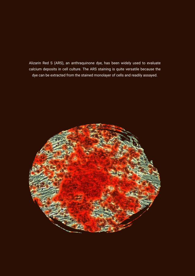
Alizarin Red S (ARS), an anthraquinone dye, has been widely used to evaluate
calcium deposits in cell culture. The ARS staining is quite versatile because the
dye can be extracted from the stained monolayer of cells and readily assayed.

33
D. Kok, MD1,2 C.M.M. Peeters, MD2 Z. Mardina3
S.K. Bulstra, MD, PhD2
R. Kuijer, PhD3
F.H. Wapstra, MD2, PhD2
1Department of Orthopaedics, Rijnstate Hospital, Arnhem, The Netherlands. 2Department of Orthopaedics, 3Department of Biomedical Engineering, University of
Groningen, University Medical Center Groningen, Groningen, The Netherlands.
PLoS One. 2019 Apr 25;14(4)
Is remaining intervertebral disc tissue interfering with bone formation during fusion of two vertebrae?

34 Abstract
Study designlaboratory research
Background Through the increasing number of minimally invasive procedures in spinal fusion surgery, the complete removal of intervertebral disc (IVD) tissue has become more a challenge. Remaining IVD may interfere with the biological process of bone formation.
Objective In order to establish whether complete removal of IVD tissue will improve or inhibit the fusion process, the effects of different concentrations of extracts of inflamed disc tissue on the mitochondrial activity of mesenchymal stem cells (MSCs), and the capacity to mineralize their extracellular matrix by osteoblasts and differentiated MSCs were tested in vitro.
Methods A MTT assay was conducted to measure the mitochondrial activity of MSCs, and an Alizarin Red S staining quantification assay to measure the deposition of calcium by osteoblasts and differentiated, bone marrow-derived MSCs.
Results A significantly higher mitochondrial activity was shown in MSCs co-cultured with extracts of IVD tissue (10%, 50%, and 100%) compared with the control group after 48 hours of incubation, indicating that the IVD tissue extracts stimulated the mitochondrial activity of MSCs. This effect appeared to be inversely proportional to the concentration of IVD tissue extract. No significant differences in mineralization by human osteoblasts or differentiated MSCs were found between the samples incubated with IVD tissue extracts (3% and 33%) and the control samples.
Conclusion Our findings indicate that remaining IVD tissue has more of a stimulating than inhibiting effect on the activity of MSCs. Even if inflammatory cytokines are produced, these do not result in a net inhibition of cellular activity or osteogenic differentiation of MSCs.
Keywordsspinal fusion surgery, intervertebral disc tissue, bone healing, mesenchymal stem cells, osteoblasts.

35Introduction
The number of spinal fusion surgeries has been increased considerably in the last few decades (1). For a solid fusion of two vertebrae it is essential to perform a discectomy and remove the tissue of the intervertebral disc (IVD) as well as both the vertebral endplate cartilages (2). However, during minimally invasive procedures using the transforaminal lumbar interbody fusion technique (TLIF), the complete removal of intervertebral tissue and the vertebral cartilages is a challenge (2,3).
Remaining disc tissue may interfere with the biological process of bone formation, and so increase the likelihood of a poor fusion of the vertebrae. In particular nucleus pulposus (NP) tissue is described to have inflammatory properties and secretes cytokines which may intervene in the metabolism of mesenchymal stem cell (MSC) or osteoblasts (4,5). Since osteoblasts are responsible for the synthesis of the bone matrix, and MSCs have the ability to differentiate towards osteoblasts, these cells play a crucial role in the bone formation process (6). It is well known that inflammation is necessary to regulate MSC osteogenesis (7). The environment of MSCs has been shown to fulfill an important role in behavior of the cells and differentiation processes (7). The exact interplay between the presence and concentrations of different cytokines and their influence on MSC behaviour, remains unknown. The presence of inflammatory cytokines secreted by remaining IVD tissue could for example decrease the cell viability of MSCs or interfere with the differentiation of MCSs to osteoblasts. Indirectly, this would have a negative impact on bone matrix formation. Besides possible interactions with MSCs, the cytokines of remaining IVD tissue could also inhibit directly osteoblast activity, and in this way result in less bone matrix formation. In order to establish whether complete removal of IVD tissue will improve the fusion process, we have tested the effects of different concentrations of inflamed disc tissue extracts on the viabiliy of MSCs (i), the matrix production of osteoblasts (ii), and differentiated MSCs (iii) in vitro.
Methods
Preparation of tissue extraxtsNucleus pulposis (NP) and annulus fibrosus (AF) tissues were obtained from six random patients who underwent posterior lumbar interbody fusion (PLIF) surgery at the University Medical Center Groningen, after informed consent of the patients and according to the legal procedures for the use of to be discarded body tissue for experimental research. The Medical Ethical Committee of the University Medical Center Groningen had approved the use of removed tissue samples for scientific purposes, provided the samples were anonymised. The mean age of the patients was 56.5 and ranged from 30 to 70 years old (table 1). The male to female ratio was 1:1. Tissues
Chap
ter 2 | Is remain
ing in
tervertebral disc tissu
e interferin
g with
bon
e form
ation
du
ring fu
sion
of tw
o vertebrae?

36 were transferred to the laboratory in pre-weighed sterile vials filled with transport medium consisting of Dulbecco’s Modified Eagle’s Medium (DMEM)-high glucose (Life Technology, Bleiswijk, The Netherlands) supplemented with 2% antibiotics, 0.2 mM ascorbic acid-2-phosphate and 10% fetal bovine serum (FBS). In the laboratory, the vials were weighed again to establish the weight of the collected tissue. The mean weight of tissue was 3.01 g. The tissues were washed once with phosphate buffered saline (PBS) and twice with α-MEM complete medium consisting of 90% α-MEM (Life Technology), 10% of FBS, 1% of antibiotics, and 0.2 mM of ascorbic acid-2-phosphate. Subsequently, this α-MEM complete culture medium was added to the disc tissue samples up a 5% suspension (5g / 100 ml). The tissues were extracted at 37°C in a humidified atmosphere of 5% CO2 and 95% air for 24 hours. After that, the extract suspensions were centrifuged at 1500 rpm for 15 minutes. The supernatants were collected and frozen at -20°C until further testing. The mean obtained extract volume was 15.6 ml.
Table 1: Patient characteristics
Patient/extract Gender Age (y) Weight of tissue (gram) Obtained Extract (ml)
1 M 57 3.3357 16.67852 M 30 3.6362 18.18103 M 54 1.7919 8.95954 F 70 4.9388 24.69405 F 66 3.5960 19.69006 F 62 1.0485 5.2425
Abbreviations: F, female; M, male
Human Mesenchymal Stem Cell cultureHuman MCSs were obtained from the MSC bank, containing MSCs isolated from bone marrow obtained from patients during total hip or knee replacement. The MSC bank was set up by Arina Buizer in our laboratory, who characterized all samples according to the guidelines of the International Society of Cellular Therapy (Supplementary data) (8). MCSs of the third passage (P-3) were cultured in T75 flasks in α-MEM complete culture medium until 50-60% confluence at 37 °C in a humidified atmosphere at 5% CO2. The cells were counted with a Bürker-Türk haemocytometer using a Leica inverted phase-contrast microscope (Leica DMIL LED, Leica Microsystems, Rijswijk, The Netherlands).

37Human Osteoblast culturePrimary human osteoblasts, previously isolated from bone chips derived from femoral heads of patients at total hip surgery were isolated according to the procedure described by Gartland et al. (9). Briefly, femoral heads removed during total hip surgery and obtained from the operating room were transferred to the laboratory in sterile DMEM-F12 culture medium (Gibco-Life Technology), supplemented with 2% anti-anti. In the lab the bone was crushed using bone mills and bone marrow was removed during incubation in collagenase type II solution for 24 hours. The bone chips were rinsed five times in DMEM/F12 supplemented with 2% anti-anti and then placed in cell culture flasks in culture medium. Medium was refreshed twice weekly. Cells that grew out of the bone chips were harvested when the cultures were 70% confluent. Cells were passaged and then frozen in liquid nitrogen, using standard cells culture procedures. Osteoblasts were characterized by assessment of alkaline phosphatase activity (Leukocyte alkaline phosphatase kit, Sigma, Steinheim, Germany) according to the manufacturer’s instructions. Osteoblasts were used at their third passage (P-3). The culture medium consisted for 88% of DMEM-F12 (1:1), 10% of FBS, 2% of anti-anti, and 0.2 mM of L-Ascorbic acid 2-phosphate. The osteoblasts were cultured in T75 flasks until 60% confluence.
Influence of intervertebral disc tissue extracts on viability of MCSs To evaluate the influence of extracts of IVD tissue on the mitochondrial activity of MSCs first a MTT assay was conducted, according to protocol BME-I-R-002 of the department of biomedical engineering, following the ISO 10993-5 standard.
MCSs were seeded in a 96-well plate (2000 cells/well), and allowed to adhere for 24 hours. Then the cells were incubated with extracts of IVD tissue from 5 patients at three different concentrations: 10%, 50% and 100% for 48 hours. The assay was performed in 8 replicate measurements. The control group consisted of MSCs that were not incubated with extract. The incubation was stopped by removing the culture medium and adding culture medium supplemented with 0.5 mg/ml 3-(4,5-dimethylthiazol -2-yl)-2,5-diphenyltetrazolium bromide (MTT) (Sigma-Aldrich, Zwijndrecht, The Netherlands). After an additional incubation of 3 hours, the culture medium was carefully removed and 2-propanol (Merck, EMD Millipore, Darmstadt, Germany) was added. The 96-well plate was shaken for 15 minutes and absorbance was read at 570 nm using the fluorostar Optima plate reader (BMG Labtech, De Meern, The Netherlands).
Influence of IVD tissue extracts on osteoblast activityIn order to establish whether the extracts of IVD tissue would affect the ability of osteoblasts to mineralize their environment, the deposition of calcium by these cells in the extracellular matrix (ECM) was measured in samples with and without disc tissue
Chap
ter 2 | Is remain
ing in
tervertebral disc tissu
e interferin
g with
bon
e form
ation
du
ring fu
sion
of tw
o vertebrae?

38 extracts using the Alizarin Red S staining quantification assay (Sciencell Inc, Carlsbad, CA, USA; ARed-Q, Catalog #8678, 100 Tests) as described by the manufacturer. Disc tissue extracts (3% and 33%) from two patients were added to cultures of primary osteoblasts at 60 % confluence for 48 hours, after which the Alizarin red S assay was performed. Controls were osteoblast cultures to which no extract was added. Next to measuring the amount of calcium deposits in the ECM, we also checked the confluence of the osteoblast cultures to ensure that the density of the osteoblast cultures was comparable with the control group.
Influence of IVD extracts on osteogenic differentiation of MSCs.In order to evaluate the influence of disc tissue extracts on the osteogenic differentiation of MCSs, the deposition of calcium in the mineralized ECM by the differentiated MSCs was measured in samples with and without disc tissue extracts. After the MCSs reached 50-60% confluence and were incubated for 24 hours in an osteogenic differentiation medium, disc tissue extracts from two patients were added at concentrations of 12% and 1.2% in a 6-well plate. After 48 hours of incubation, the Alizarin Red S assay was performed. Two samples in osteogenic medium were used as control (osteogenic), as well as two samples in a proliferation medium (non osteogenic).
Statistical AnalysisDifferences between samples in both MTT assay and Alizarin assay were tested for significance using Sigma plot 13 software. First, the data were tested for normal distribution with a Shapiro-Wilk test, followed by a equal variance test (Brown-Forsythe).For all our data one of these tests failed, so the data were analyzed with a Kruskall Wallis ANOVA on ranks; followed by a Dunn’s (not normally distributed) or Student-Newman-Keuls (unequal variance) post hoc test.
Results
Influence of IVD extracts on viability of MCSs The MTT assay assesses the activity of the mitochondrial β-nicotinamide adenine dinucleotide phosphate (NADPH)-depentent cellular oxidoreductase enzymes in converting MTT into water-insoluble formazan. This assay is generally used as a measure for cellular activity, proliferation of cells, or more specific, metabolic activity. In this study we will further stick to cellular activity.Figure 1 shows the cellular activity of MSC which were exposed to extracts of IVD tissue at three different concentrations after 48 hours of incubation. Significantly higher

39levels of cellular activity were assessed in the group of MSCs co-cultured with extracts of IVD tissue compared to those of the control group (P= <0.001). This effect appeared to be inversely proportional to the concentration of IVD tissue extract. Significantly higher cellular activity was detected in MSCs incubated at 10% of IVD tissue extracts compared with the control group, those incubated at 50% of IVD tissue extracts in extracts 1, 2, and 3, and those incubated at 100% of extracts 1, 2, 3, and 5 (table 2).
Figure 1: Effects of extracts of intervertebral disc tissue on the metabolic activity of human MSCs.
Effects of extracts of IVD tissue from 5 different patients, applied at 3 concentrations on the metabolic activity of human MSCs, assessed using a MTT assay. The dashed horizontal line at 0.2 represents the mean of the control samples. All samples were tested in 8 fold. The * denotes a statistically significant difference (P<0.05) compared to the control samples.
Chap
ter 2 | Is remain
ing in
tervertebral disc tissu
e interferin
g with
bon
e form
ation
du
ring fu
sion
of tw
o vertebrae?

40 Table 2: Effects of extracts of intervertebral disc tissue on the metabolic activity of humanMSCs. P-values after statistical analysis.
Extract 1 Extract 2 Extract 3 Extract 4 Extract 5Contr vs 10% <0.001 <0.001 <0.001 <0.001 <0.001Contr vs 50% 0.002 <0.001 0.004 <0.001 <0.001Contr vs 100% <0.001 ns ns <0.001 <0.00110% vs 50% <0.001 <0.001 <0.001 ns ns10% vs 100% <0.001 <0.001 <0.001 ns 0.02350% vs 100% 0.027 <0.001 0.003 ns <0.001
Abbreviations: ns, non significant.
Influence of IVD tissue extracts on osteoblast activityFigure 2 shows the results of the Alizarin Red S assay of osteoblasts exposed to two different concentrations of extracts of IVD tissue after 48 hours of incubation. No significant differences in the amount of deposited calcium by human osteoblasts were established between the samples incubated with IVD tissue extracts at 3%, at 33% and the control sample. All osteoblast cultures had a similar confluency.
Figure 2: Alizarin Red assay quantification of obsteoblast activity.
Effects of extracts of intervertebral disc tissue obtained from 4 different patients on mineralization by human osteoblasts using an Alizarin red S assay. No significant differences were obtained compared to the control (osteoblast without extract) set at 1.

41Influence of IVD tissue extracts on osteogenic differentiation of MSCsFigure 3 shows the results of the Alizarin Red S assay of MSCs. No significant differences were found in the amount of deposited calcium by the differentiated MSCs, the MSCs incubated with IVD tissue extracts (3 and 33%) and the control samples. The confluency of cells cultured in osteogenic medium of the three patients and the control group were comparable with each other. As expected, MSCs in non-osteogenic medium produced less (no) mineral.
Figure 3: Alizarin Red assay quantification of human MCSs osteogenesis.
Effects of extracts of IVD tissues from three different patients, applied at two, indicated concentration, on human MSCs in osteogenic medium, using an Alizarin red S assay. No significant differences were obtained compared to the control MSCs in osteogenic medium without extract. MSCs in non-osteogenic medium (non-osteo) produced less mineral.
Chap
ter 2 | Is remain
ing in
tervertebral disc tissu
e interferin
g with
bon
e form
ation
du
ring fu
sion
of tw
o vertebrae?
42 Discussion
The aims of this in vitro study were to evaluate the influence of IVD tissue extracts on (i) the cellular activity of human MCSs, (ii) their osteogenic differentiation, and (iii) the mineral production of osteblasts. The results of this study indicate that IVD tissue extracts might stimulate the cellular activity of MSCs, when present in low concentrations. The results also indicate that IVD tissue extracts have no influence on the osteogenic differentiation of MSCs, and on the mineralizing capacity of mature primary osteoblasts.
The increase in cellular activity of MSCs in the presence of low concentrations of IVD tissue extract is most probably cytokine mediated (10,11). The positive influence of cytokines TNF-α,interleukins, IFN-γ onmitochondrial- and NADPH oxidase-generated reactive oxygen species production has been decribed before (12). We did not observe inhibition of cellular activity of MSCs (compared to control condition) in the presence of any concentration of IVD tissue extract. From these observations, we conclude that IVD tissue extracts do not impede with MSC activity.
The in vitro study of Li et al. (2000) also evaluated the influence of IVD tissue extracts on the metabolism of osteoblast-like cells, and found that osteoblast proliferation as well as maturation was stimulated when IVD tissue was applied to osteoblasts in culture. Their results showed a stimulation of alkaline phosphatase production (maturation), cell proliferation measured by [3H]thymidine incorporation, and collagen type I production (13). Chan et al. (2015) described, on the contrary, that primary IVD tissue cells inhibit osteogenesis of MSCs. In their study the incubation of MSCs with IVD tissue cells was maintained for 21 days, and resulted in a reduction of calcium deposition as observed by reduced alizarin red staining. A reduction in alkaline phosphatase activity in cocultures of MSCs with NP cells and RT-PCR analyses confirmed these results (14). A possible explanation of the conflicting results with current study could be the differences in length of incubation time and concentration of IVD tissue material.
Li et al. (2002) conducted an in vivo study on pigs analysing the influence of IVD tissue on anterior spinal interbody fusion. They compared the bone fusion rate between the lumbar spine level with an implantation of Brantigan cage filled with a mixture of autograft and the NP tissue harvested from the removed disc level, and the spine level with a cage filled with autologous iliac crest bone graft in equal amounts. After 12 weeks CT evaluation showed that the level with NP tissue had a 20% fusion rate, while the level with pure autograft had a 70% fusion rate (P=0.07). In their conclusion they stated that NP tissue mixed with autologous bone graft can cause a delay or decrease

43in bone formation inside the cage (4). A possible explanation of the conflicting results with current study could be again the amount of IVD tissue material, but also the use of healthy IVD tissue. In our study the extracts of diseased IVD tissue were used, which might cause differences in inflammatory cytokines releases.
The results of our study indicated that IVD tissue extracts might stimulate the viability of MSCs when they are present in low concentrations, but higher concentrations of IVD tissue extracts seemed to result in lower metabolic activity of MSCs. We consider it likely that this effect is caused by the more optimal concentrations of the effect-producing cytokines in the solution with the lower concentration of the extracts. Gabeen et al. (2014) show the stimulating effect of IL-4 at low concentration and a strong inhibiting effect at high concentrations(15).
Inflammation is the process by which the body tries to heal damaged tissue. Damaged IVD tissue will thus contain inflammatory cells and cytokines. In our study we used IVD tissue from patients who underwent PLIF surgery, which we considered to be inflamed tissue, containing inflammatory cells and cytokines, such as TNF-α, IL-4, -6,-12 and interferon-γ(16). In spinal fusion surgery, remnants of inflamed NP en AF tissue will be a source of cytokines and chemokines, which could interfere with the bone forming process(17). Considering that the degree of inflammation varied in the samples of herniated disc tissue that were collected in the operating room, and that during the extraction period many cells underwent necrosis also releasing cytokines and chemokines, an excessive amount of cytokine and chemokine release could lead to unwanted effects in vivo and make these in vitro tests less representative. The number of cytokines, which affected the results of this study is namely considered to be small compared to the number of cytokines in the human body. Therefore, it remains hard to predict what the effect of remaining IVD material will be on bone fusion in vivo. Future studies should include in vivo studies, investigating the effect of different amounts of remaining IVD tissue on spinal fusion, using an animal model with an already inflamed IVD. The involving cytokines could then be observed by intensive histological analyses and ELISA measurement.
Limitations of our study were the fact that a complete fusion environment could not be reproduced in vitro, as decribed above, and that we were also not able to use the same dilutions of tissue extracts for our experiments.
In conclusion, remaining disc material is not specific inhibiting the viability of MSCs when they are present in low concentrations. Even more, it might have more of a stimulating effect. Even if inflammatory cytokines are produced, these do not result in a net inhibition of cellular activity and osteogenic differentiation of MSCs and in the osteoblast metabolism.
Chap
ter 2 | Is remain
ing in
tervertebral disc tissu
e interferin
g with
bon
e form
ation
du
ring fu
sion
of tw
o vertebrae?

44 References
1. Memtsoudis SG, Vougioukas VI, Ma Y, Gaber-Baylis LK, Girardi FP. Perioperative morbidity and mortality after anterior, posterior, and anterior/posterior spine fusion surgery. Spine. 2011 Oct 15;36(22):1867-77.
2. Rihn JA, Gandhi SD, Sheehan P, Vaccaro AR, Hilibrand AS, Albert TJ, et al. Disc space preparation in transforaminal lumbar interbody fusion: a comparison of minimally invasive and open approaches. Clinical orthopaedics and related research. 2014 Jun;472(6):1800-5.
3. Javernick MA, Kuklo TR, Polly DW, Jr. Transforaminal lumbar interbody fusion: unilateral versus bilateral disk removal-an in vivo study. Am J Orthop (Belle Mead NJ). 2003 Jul;32(7):344-8; discussion 8.
4. Li H, Zou X, Laursen M, Egund N, Lind M, Bunger C. The influence of intervertebral disc tissue on anterior spinal interbody fusion: an experimental study on pigs. European spine journal : official publication of the European Spine Society, the European Spinal Deformity Society, and the European Section of the Cervical Spine Research Society. 2002 Oct;11(5):476-81.
5. Kanerva A, Kommonen B, Gronblad M, Tolonen J, Habtemariam A, Virri J, et al. Inflammatory cells in experimental intervertebral disc injury. Spine. 1997 Dec 01;22(23):2711-5.
6. Birmingham E, Niebur GL, McHugh PE, Shaw G, Barry FP, McNamara LM. Osteogenic differentiation of mesenchymal stem cells is regulated by osteocyte and osteoblast cells in a simplified bone niche. European cells & materials. 2012 Jan 12;23:13-27.
7. Deshpande S, James AW, Blough J, Donneys A, Wang SC, Cederna PS, et al. Reconciling the effects of inflammatory cytokines on mesenchymal cell osteogenic differentiation. The Journal of surgical research. 2013 Nov;185(1):278-85.
8. Buizer AT, Veldhuizen AG, Bulstra SK, Kuijer R. Static versus vacuum cell seeding on high and low porosity ceramic scaffolds. Journal of biomaterials applications. 2014 Jul;29(1):3-13.
9. Gartland A, Rumney RM, Dillon JP, Gallagher JA. Isolation and culture of human osteoblasts. Methods in molecular biology. 2012;806:337-55.
10. Lim SW, Loh HS, Ting KN, Bradshaw TD, Allaudin ZN. Reduction of MTT to Purple Formazan by Vitamin E Isomers in the Absence of Cells. Tropical life sciences research. 2015 Apr;26(1):111-20.

4511. Dunigan DD, Waters SB, Owen TC. Aqueous soluble tetrazolium/formazan MTS as an indicator of NADH- and NADPH-dependent dehydrogenase activity. BioTechniques. 1995 Oct;19(4):640-9.
12. Yang D, Elner SG, Bian ZM, Till GO, Petty HR, Elner VM. Pro-inflammatory cytokines increase reactive oxygen species through mitochondria and NADPH oxidase in cultured RPE cells. Experimental eye research. 2007 Oct;85(4):462-72.
13. Li H, Laursen M, Lind M, Sun C, Bunger C. The influence of human intervertebral disc tissue on the metabolism of osteoblast-like cells. Acta orthopaedica Scandinavica. 2000 Oct;71(5):503-7.
14. Chan SC, Tekari A, Benneker LM, Heini PF, Gantenbein B. Osteogenic differentiation of bone marrow stromal cells is hindered by the presence of intervertebral disc cells. Arthritis research & therapy. 2015 Dec 25;18:29.
15. Gabeen AA, Abdel-Hamid FF, El-Houseini ME, Fathy SA. Potential immunotherapeutic role of interleukin-2 and interleukin-12 combination in patients with hepatocellular carcinoma. Journal of hepatocellular carcinoma. 2014;1:55-63.
16. Shamji MF, Setton LA, Jarvis W, So S, Chen J, Jing L, et al. Proinflammatory cytokine expression profile in degenerated and herniated human intervertebral disc tissues. Arthritis and rheumatism. 2010 Jul;62(7):1974-82. 17. Phillips KL, Chiverton N, Michael AL, Cole AA, Breakwell LM, Haddock G, et al. The cytokine and chemokine expression profile of nucleus pulposus cells: implications for degeneration and regeneration of the intervertebral disc. Arthritis research & therapy. 2013;15(6):R213.
18. Fusellier M, Colombier P, Lesoeur J, Youl S, Madec S, Gauthier O, et al. Longitudinal Comparison of Enzyme- and Laser-Treated Intervertebral Disc by MRI, X-Ray, and Histological Analyses Reveals Discrepancies in the Progression of Disc Degeneration: A Rabbit Study. BioMed research international. 2016;2016:5498271.
Chap
ter 2 | Is remain
ing in
tervertebral disc tissu
e interferin
g with
bon
e form
ation
du
ring fu
sion
of tw
o vertebrae?

Nickel Titanium (NiTiNOL) is a metal alloy that has the unique ability to change
shape. Consisting of near equal amounts of nickel and titanium, this mechanically
active alloy gets its name from the location of its initial discovery, the Naval
Ordinal Laboratory, in 1959.
NiTiNOL exhibits shape memory when it can be deformed easily and maintain
that deformed shape at room temperature. Upon heating to a certain temperature
(i.e. body temperature in biomedical applications), shape memory NiTiNOL will
recover back to its original “memorized” shape.

47
D. Kok, MD*
P. Firkins, PhD#
F.H. Wapstra, MD PhD*
A.G. Veldhuizen, MD PhD*
*Universitair Medisch Centrum Groningen #DePuy Spine International †
BMC Musculoskelet Disord. 2013 Sep 18;14:269.
A New Lumbar Posterior Fixation System, the Memory Metal Spinal SystemAn In-vitro Mechanical Evaluation
48 Abstract
BackgroundSpinal systems that are currently available for correction of spinal deformities or degeneration such as lumbar spondylolisthesis or degenerative disc disease use components manufactured from stainless steel or titanium and typically comprise two spinal rods with associated connection devices (for example: DePuy Spines Titanium Moss Miami Spinal System). The Memory Metal Spinal System of this study consists of a single square spinal rod made of a nickel titanium alloy (Nitinol) used in conjunction with connecting transverse bridges and pedicle screws made of Ti-alloy. Nitinol is best known for its shape memory effect, but is also characterized by its higher flexibility when compared to either stainless steel or titanium. A higher fusion rate with less degeneration of adjacent segments may result because of the elastic properties of the memory metal. In addition, the use of a single, unilateral rod may be of great value for a TLIF procedure. Our objective is to evaluate the mechanical properties of the new Memory Metal Spinal System compared to the Titanium Moss Miami Spinal System.
Methods An in-vitro mechanical evaluation of the lumbar Memory Metal Spinal System was conducted. The test protocol followed ASTM Standard F1717-96, “Standard Test Methods for Static and Fatigue for Spinal Implant Constructs in a Corpectomy Model.”1. Static axial testing in a load to failure mode in compression bending,2. Static testing in a load to failure mode in torsion,3. Cyclical testing to estimate the maximum run out load value at 5.0 x 10^6 cycles.
ResultsIn the biomechanical testing for static axial compression bending there was no statistical difference between the 2% yield strength and the stiffness of the two types of spinal constructs. In axial compression bending fatigue testing, the Memory Metal Spinal System construct showed a 50% increase in fatigue life compared to the Titanium Moss Miami Spinal System.In static torsional testing the Memory Metal Spinal System constructs showed an average 220% increase in torsional yield strength, and an average 30% increase in torsional stiffness.
Conclusions The in-vitro mechanical evaluation of the lumbar Memory Metal Spinal System showed good results when compared to a currently available spinal implant system. Throughout testing, the Memory Metal Spinal System showed no failures in static and dynamic fatigue.

49
Chap
ter 3 | A N
ew Lu
mbar P
osterio
r Fixatio
n Sy
stem, th
e Mem
ory
Metal Sp
inal Sy
stem. A
n In
-vitro M
echan
ical Evaluatio
n.
Key words Memory Metal Spinal System, NiTi, DePuy Spines Titanium Moss Miami, in-vitro mechanical evaluation, ASTM Standard F1717-96
Key points• In-vitro mechanically evaluation (ASTM Standard F1717-96) of the new lumbar Memory Metal Spinal System• The Memory Metal spinal rod showed no failures in static and dynamic fatigue
50 Introduction
Chronic low back pain can be the result of spondylolisthetic or degenerative lumbar segmental instability (1,2). Surgical treatment of this condition by fusion of the involved segments was introduced in the mid-1920s (1,3). Posterior lumbar interbody fusion (PLIF) has become a clinically established and increasingly popular procedure since its introduction by Cloward (4-7). A successful PLIF can restore disc height, decompress the dural sac and nerve roots, immobilize the unstable intervertebral disc, and restore load bearing to anterior structures (8). Rigid instrumentation was added to improve initial stability and to improve fusion rates of interbody fusion. On the other hand however, instrumented spinal fusion plays a major part in the development of adjacent segments degeneration because of increased stiffness of the fused motion segment (9-12).
We developed a system that can be used in the treatment of diseases in the lumbar region (short construct) as well as the 3-dimensial corrections of scoliotic deformities (13). We started with the development of the short lumbar construct, to evaluate its use for lumbar degenerative conditions. There may be specific advantages of this unique system for lumbar use, and it will generate proof of concept for lumbar as well as long constructs, as used for the treatment of deformities. The Memory Metal Spinal System is a posterior system, consisting of a single spinal rod used in conjunction with pedicle screws and connecting transverse bridges. The use of a unilateral single rod with pedicle screw fixation was proposed to decrease the stiffness of the implant and would be as effective as the conventional system with two rods and bilateral pedicle fixation (14,15). In addition, a unilateral may facilitate a TLIF procedure, because the rod will not obstruct a TLIF cage at the contra-lateral side.
Spinal systems that are currently available use components manufactured from stainless steel or titanium. Before implantation into humans the new Memory Metal Spinal System must be proven to be at least substantially equivalent in performance and safety to current deformity systems. The spinal rod component used in this system is manufactured from Nitinol (NiTi), a nickel-titanium alloy. The characteristics of this alloy were first described by Buehler and Wang (16). NiTi is a Memory Metal and is mainly characterized by its shape memory effect. At present, the characteristics of this NiTi alloy are used clinically in wires for orthodontic tooth alignment, osteosynthesis staples, vena cava filters and other vascular applications (17-22). Wever et al. looked at the biocompatibility and functionality of this new Memory Metal Spinal System (23). In the experimental animal study on six pigs, nickel levels measured post-operatively were similar to the results recorded preoperatively. Corrosion and fretting processes were not observed; no adverse tissue reactions were evident.
51
Chap
ter 3 | A N
ew Lu
mbar P
osterio
r Fixatio
n Sy
stem, th
e Mem
ory
Metal Sp
inal Sy
stem. A
n In
-vitro M
echan
ical Evaluatio
n.
Use of a single rod manufactured from memory metal, which offers increased elasticity compared to stainless steel or titanium and therefore ease of use, should lead to a reduction in the length of time for operation, which in turn should lead to other desirable outcomes as reduced blood loss. There should also be less degeneration of adjacent segments (adjacent level disease) and better fusion is expected because of less rigidity in the memory metal spinal system. With current systems there may be loss of achieved reposition due to the viscous properties of the spine. By using a memory metal in this new system the expectation was that there is a better maintenance of the reposition due to the metal’s inherent shape memory properties (continuous reposition force).
The purpose of this study is to fully biomechanically test the Memory Metal Spinal System according to ASTM F1717-96, “Standard Test Methods for Static and Fatigue for Spinal Implant Constructs in a Corpectomy Model” (24).
We used the DePuy Spine’s Titanium Moss Miami Spinal System as a comparison control. This system has proven clinical efficacy and safety, and is used to treat similar spinal disorders, as the Memory Metal Spinal System will be used for.
Materials and Methods
Memory Metal Spinal components
RODS:The Spinal Rods used are manufactured from medical grade Nickel Titanium Alloy according to ASTM F2063-00 standard. This standard references the acceptable biocompatibility of the material. The austenite start temperature (As) was set between 0 & 10C. This meant that at room and body temperature the rod was fully austenitic (Af) and the rod was super elastic. Being able to quote the ASTM standard allows international regulatory acceptance of the material to obtain the CE mark for the implants. The cross sectional shape of the rod is square which allows good torsional stability, and correction possibilities. A square cross-sectional rod profile may additionally allow deformity correction in the transverse plane when engaged in squared head pedicle screws or connection bridge (25).
The rod has a curved shape to fit anatomically in the lumbar spine, as shown in Figure 1.The rods have a 6.35mm square cross-section; current systems often have a round rod of this cross section. The square design of the rod adds torsional stability within the

52 connecting bridge. Rods are available straight or pre-bent (pre-lordosed). A surgeon using conventional systems will generally apply a bend to the rod to achieve lordosis in line with the anatomy of the spine in the lumbar region. Therefore, providing a surgeon with a pre-bent rod should make the surgical technique quicker and simpler. The length of the rods can be cut to the specific length required by the surgeon by using a specially designed cutter. During manufacturing, shape setting was done by heat treatment, and surface treatment carried out to give appropriate corrosion resistance.
Figure 1. Square, anatomically shaped NiTi alloy Spinal Rod.
PEDICLE SCREWS:Anchoring the spinal rod to the spine is achieved by pedicle screws. Pedicle screws have been used extensively in the lumbar region due to their strong fixation capabilities. The pedicle screws used in this study will be from DePuy Spine’s CE marked Spinal System called ’Monarch’ TM. They are manufactured from a medical grade titanium alloy (Titanium Per MS-401 Grade 01).
CONNECTOR BRIDGE:Attached between 2 pedicle screws on one vertebra is a transverse connecting bridge, available in various sizes. Offset on the bridge is a channel, which the square spinal rod is attached to. In vivo this allows the rod to be set lateral of the spinous process. The rod is fixed in the channel by a cap and setscrew combination, as shown in Figure 2.
The transverse connecting bridge is a device which is fixed between two polyaxial pedicle screws to produce a stable, rigid construct used to hold the spinal rod in place. This rigid construct aids the transfer of correctional forces from the rod to the vertebrae. The flexibility of the system comes from the rod and not the connector or screws. The rod is connected to the bridge and locked in place by a set screw and sliding cap. The rod can be approximated to the bridge using a mini approximator instrument. Connecting bridges are available in a range of standard sizes to accommodate the range of different anatomical sizes of the human vertebrae. All connecting bridges are manufactured from medical grade Titanium Alloy which is considered safe to use with Nitinol. The simplest lumbar construct would consist of four pedicle screws, two connector bridges, and one rod, and would be implanted over two adjacent vertebral levels.

53
Chap
ter 3 | A N
ew Lu
mbar P
osterio
r Fixatio
n Sy
stem, th
e Mem
ory
Metal Sp
inal Sy
stem. A
n In
-vitro M
echan
ical Evaluatio
n.
Figure 2. Assembled Memory Metal Spinal System Construct: Posterior View
The titanium MOSS Miami componentsThe titanium MOSS Miami System includes rods and polyaxial screws with inner screws and outer locking nuts. Bilateral constructs were assembled using the smallest size pedicle screws (5.5 mm). Only 5.5 mm rods are available in this system. These components were mounted onto two ultra high molecular weight polyethylene (UHMWPE) blocks to create a bilateral construct. The gap between the blocks ensured that no load sharing took place, i.e. the assembled bilateral MOSS Miami constructs alone resisted the applied load. Each polyaxial screw was tightened down so that the screw head was tight against the test block and then turned back 90 degrees. The construct blocks were aligned so that the ends were parallel to one another. Two 5.5 mm diameter rods were positioned into the slots of the polyaxial screws and the inner screws and outer nuts were hand tightened onto the polyaxial screws. The gauge length (the distance between the center of the upper screws to the center of the lower screws) and the moment arm from the loading point to the center of the longitudinal member were kept constant at 76.0 ±1.0 mm and 48.5 ±1.0 mm, respectively, for all constructs. A cross connector was placed halfway between the upper and lower UHMWPE blocks. The inner screws were tightened to 6 Nm, the outer nuts were tightened to 10 Nm and cross connector set screws were tightened to 6 Nm. The tightened sequence for the inner and screws/outer nuts was: 1. inner screw, 2. outer nut, 3. inner screw.

54 Testing ProtocolTesting was undertaken to determine the mechanical properties of the Memory Metal Spinal System compared to DePuy Spine’s Titanium Moss Miami Spinal System. If the lumbar Memory Metal Spinal System performs at least as well as the Moss Miami system in the biomechanical tests, it is assumed that the system will perform appropriately in the clinical setting from a biomechanical perspective. The objectives were to evaluate the Memory Metal Spinal System when compared to the DePuy Spine’s Titanium Moss Miami Spinal System in the following test methods: 1. Static axial testing in a load to failure mode in compression bending,2. Static testing in a load to failure mode in torsion,3. Cyclical testing to estimate the maximum run out load value at 5.0 x 10⁶ cycles.
The test protocol followed ASTM Standard F1717-96, “Standard Test Methods for Static and Fatigue for Spinal Implant Constructs in a Corpectomy Model.
Spinal implants are generally composed of several components which, when connected together, form a spinal implant assembly. Spinal implant assemblies are designed to provide some stability to the spine while arthrodesis takes place. These test methods outline standard materials and methods for the evaluation of different spinal implant assemblies so that comparison between different designs may be facilitated. These test methods are used to quantify the static and dynamic mechanical characteristics of different designs of spinal implant assemblies. The mechanical tests are conducted in vitro using simplified load schemes and do not attempt to mimic the complex loads of the spine. These test methods set out guidelines for load types and methods of applying loads. Methods for three static load types and one fatigue test are defined for the comparative evaluation of spinal implant assemblies. And these test methods establish guidelines for measuring displacements, determining the yield load, and evaluating the stiffness and strength of the spinal implant assembly.
A Corpectomy model simulates a worst-case scenario for a spinal construct, i.e. the model simulates the implants being implanted in two vertebral bodies with a vertebral body missing in-between therefore loading the implants fully. Normally implants share load with the actual spinal column.
All static torsion tests were conducted using an INSTRON 8874 Bi-Axial Table Top Servohydraulic Dynamic Testing System (INSTRON, Canton, MA) with a 25kN and 100 Nm load cell. All axial compression bending tests were conducted using an INSTRON 8872 Axial Table Top Servohydraulic Dynamic Testing System (INSTRON, Canton, MA) with a 10kN load cell. All tests were conducted using an environmental chamber,

55
Chap
ter 3 | A N
ew Lu
mbar P
osterio
r Fixatio
n Sy
stem, th
e Mem
ory
Metal Sp
inal Sy
stem. A
n In
-vitro M
echan
ical Evaluatio
n.
holding Phosphate Buffered Saline at 37°C±3°C, for the duration of the tests. Stainless steel fixtures were used to minimize corrosion products from being generated in the test environment.
Static Axial Compression Bending TestFive (5) Memory Metal Spinal System constructs and titanium 5.5 mm diameter Moss Miami constructs were tested on an INSTRON 8872 Axial Table Top Servohydraulic Dynamic Testing System in static axial compression bending to measure the compression bending yield load (N), compression bending stiffness (N/mm), and compression bending peak load (N). The failure mode of each construct was also recorded. The axial static compression bending tests were conducted in displacement control at a rate of 0.1mm/sec, collecting load and displacement data. The ramp waveform was conducted until the construct experienced a permanent deformation, the test blocks touched (25mm relative actuator displacement), or gross failure occurred. The test specimens were assembled per manufacturer instructions, and mounted in UHMWPE blocks. Statistical analysis included calculation of the mean and standard deviation of each measured value.
Static Torsion TestFive (5) Memory Metal Spinal System constructs and titanium 5.5 mm diameter Moss Miami constructs were statically tested on an INSTRON 8874 Bi-Axial Table Top Servohydraulic Dynamic Testing System in torsion, measuring the yield torque (N-m), torsional stiffness (N-m/degree) and the peak torque (N-m). The failure mode of each construct was recorded. The test specimens were assembled per manufacturer instructions, and mounted in UHMWPE blocks (per ASTM F1717). The torsion static tests were conducted with a static axial compression preload (20N), in angular displacement control at a rate of 1°/sec, collecting torque and angular displacement data. The ramp waveform was conducted until the construct experienced a permanent deformation or reached 80° of angular displacement. Statistical analysis included calculation of the mean and standard deviation of each measured value.
Dynamic Axial Compression Bending TestSix (6) Memory Metal Spinal System constructs and titanium 5.5 mm diameter Moss Miami constructs were tested in axial compression bending fatigue using a servo hydraulic testing machine. The testing configuration matched that of the static axial compression bending tests. A cyclic load with a constant frequency of 3 Hz was applied to each construct. The loads were maintained with a constant sinusoidal load amplitude control and a constant load ratio (R=min/max) equal to 10. Load values were chosen to develop a fatigue curve with two (2) specimens reaching 5,000,000 cycles without evidence of failure. Testing was terminated when the construct experienced

56 permanent deformation (actuator axial displacement greater than ±2mm) or reached 5,000,000 cycles. Dynamic stiffness (Force/Displacement) was calculated during the first 2000 cycles by capturing the peak and valley values from both the force and displacement sine waves. The failure mode of each construct and the corresponding cycle count were recorded. A fatigue curve with 95% confidence limits was also generated using TableCurve 2D (Jandel Scientific, Chicago, IL).
Results
Static Axial Compression Bending TestThe graphics of the compression bending stiffness (N/mm) for the static axial compression bending testing of the Memory Metal Spinal System constructs and the titanium 5.5 mm diameter Moss Miami constructs are shown in Figure 3. Table 1 contains the data, mean values and standard deviations for the compression bending yield load (N), compression bending stiffness (N/mm) and compression bending peak load (N) and failure mode. The peak load is defined as the highest values attained during the testing. The specimens did not experience a fracture or gross failure. The test was stopped when the test blocks touched. However, the Memory Metal test specimens did experience rotation of the superior block about the transverse bridge. Specimens AC1, AC4, and AC5 also experienced rotation of the rod about the inferior transverse bridge, as seen by movement of the inferior washers. A typical failure for the MOSS Miami test specimen was slippage in the polyaxial connections of the screws, followed by rod deformation after the polyaxial head could not slip further. Figure 3. Static axial compression Bending Testing

57
Chap
ter 3 | A N
ew Lu
mbar P
osterio
r Fixatio
n Sy
stem, th
e Mem
ory
Metal Sp
inal Sy
stem. A
n In
-vitro M
echan
ical Evaluatio
n.
Table 1. Static Axial Compression Bending Testing
MOSS MiamiSpecimen # Compressive stifness (N/mm) 2% yield load (N) Peak load (N)1 28.80 284.6 647.02 29.92 292.6 652.43 30.04 249.7 617.54 29.42 288.6 625.55 29.55 249.7 625.5Mean: 29.35 273.0 633.6SD: 0.50 21.50 15.20 Memory Metal Spinal SystemSpecimen # Compressive stifness (N/mm) 2% yield load (N) Peak load (N)1 27.62 227.16 335.722 24.82 258.27 365.933 26.12 264.87 396.694 21.19 365.97 434.755 24.32 306.71 409.48Mean: 24.81 284.60 388.51SD: 2.395 53.600 38.541
Static Torsion TestThe graphics of the compression bending stiffness (N/mm) for the static torsion testing of the Memory Metal Spinal System constructs and titanium 5.5 mm diameter Moss Miami constructs are shown in Figure 4. Table 2 contains the data, mean values and standard deviations for the yield torque (N-m), torsional stiffness (N-m/degree) and the peak torque (N-m) of each of the tested constructs. The peak torque is defined as the highest values attained during the testing. The specimens did not experience a fracture and the test was stopped when the test blocks rotated. However, the Memory Metal test specimens did experience rotation of the superior block about the transverse bridge, and the inferior transverse bridge experienced bending about the center of the test block. Typical failure for the MOSS Miami test specimens was cross-connector rod deformation at the j-hook, slippage of the open j-hooks on the rod and rotation of the polyaxial screw head.
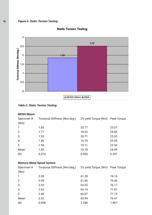
58 Figure 4. Static Torsion Testing
Table 2. Static Torsion Testing
MOSS MiamiSpecimen # Torsional Stiffness (Nm/deg.) 2% yield Torque (Nm) Peak Torque (Nm)1 1.85 20.77 25.572 1.77 18.62 24.833 1.95 20.71 25.924 1.89 19.75 25.555 1.94 19.11 25.56Mean: 1.85 19.78 25.49SD: 0.074 0.955 0.397 Memory Metal Spinal SystemSpecimen # Torsional Stiffness (Nm/deg.) 2% yield Torque (Nm) Peak Torque (Nm)1 2.38 61.28 74.162 2.59 61.66 76.463 2.53 65.53 76.174 2.63 65.14 77.815 2.48 66.07 77.74Mean: 2.52 63.94 76.47SD: 0.098 2.280 1.487

59
Chap
ter 3 | A N
ew Lu
mbar P
osterio
r Fixatio
n Sy
stem, th
e Mem
ory
Metal Sp
inal Sy
stem. A
n In
-vitro M
echan
ical Evaluatio
n.
Dynamic Axial Compression Bending TestSix (6) Memory Metal Deformity Implant System constructs and titanium 5.5 mm diameter Moss Miami constructs were tested in axial compression bending fatigue. Table 3 outlines the axial compression bending fatigue results for each specimen, including the applied load, the cycles to failure, and failure mode. The r2 value for the curve was calculated to be 0.832 (TableCurve 2D, Jandel Scientific), and is shown with 95% confidence limits in Figure 5.
Figure 5. Axial Compression Bending Fatigue Curves for the Memory Metal Spinal System (SMA) & the Moss Miami System.
Table 3. Dynamic Axial Compression Bending Testing
Memory Metal Spinal System Specimen # Load (N) Cycles to failure (n) Failure Mode1 198 5000000 No observed failure2 237 5000000 No observed failure3 237 5000000 No observed failure4 269 771901 Superior screws rotate inferiorly about superior transverse bridge5 296 500 Superior screws rotate inferiorly about superior transverse bridge6 355 250 Superior screws rotate inferiorly about superior transverse bridge

60 MOSS MiamiSpecimen # Load (N) Cycles to failure (n) Failure Mode1 160 5000000 No observed failure2 170 5000000 No observed failure3 172 80000 Screw head/shank interface4 175 134537 Screw head/shank interface5 180 90811 Screw head/shank interface6 220 44251 Screw head/shank interface
Discussion
In the biomechanical testing for static axial compression bending there was no statistical difference between the 2% yield strength and the stiffness of the two types of spinal constructs. Even though the Memory Metal Spinal System construct consists of only one rod compared to two rods for Moss Miami. For the Memory Metal Spinal System constructs, failure occurred at the bridge / screw connection. A typical failure for the Moss Miami constructs was slippage in the polyaxial connections of the screws. The square rod and the rigid bridge / screw connections help with its overall strength. In axial compression bending fatigue testing, the Memory Metal Spinal System construct showed a 50% increase in fatigue life compared to Moss Miami. Fatigue failure for the Memory Metal Spinal System constructs failed again at the bridge / screw connection, Moss Miami construct failure was due to fracture of the screw shank. It is suspected that the super elastic property of the Memory Metal Spinal System rod relieves stress from the rest of the rigid construct.
In static torsional testing the Memory Metal Spinal System constructs showed an average 220% increase in torsional yield strength, and an average 30% increase in torsional stiffness. The former was due to the super elastic properties of the Memory Metal Spinal System rod, the latter was due to the square cross section of the Memory Metal Spinal System rod. It is important to note that in all tests no failure of the Memory Metal Spinal System rods occurred.
After these encouraging results the further development with a lumbar construct is underway and has shown clinical efficacy. Twenty-seven patients have been implanted with the simple lumbar construct for the treatment of lower back disorders. To date all patients are doing well.
The next stages in development will include implantation of the full scoliosis construct to prove the system can stabilize the spine using the same material specification as the
61
Chap
ter 3 | A N
ew Lu
mbar P
osterio
r Fixatio
n Sy
stem, th
e Mem
ory
Metal Sp
inal Sy
stem. A
n In
-vitro M
echan
ical Evaluatio
n.
simple spinal construct, and finally a full scoliosis system based on the Memory Metal Spinal System generating 3D correctional forces using temperature controlled shape memory specification material. If the development is successful, this unique technology may allow the future development of a non-fusion system, which is recognized as the ultimate goal in treating patients with scoliosis.
Conclusion
Biomechanical testing showed good results when compared to currently available spinal implant systems. Throughout testing, the Memory Metal spinal rod showed no failures in static and dynamic fatigue.
Competing interests
The authors declare that they have no competing interests.
Acknowledgements
DePuy International contributed materials essential for the study.

62 References
1. Hibbs RA, Swift W. Developmental abnormalities at the lumbosacral juncture causing pain and disability. Surg Gynecol Obstet. 1929; 48: 604-12.
2. White AA, Panjabi M. Clinical Biomechanics of the Spine. Philadelphia: JB Lippincott 1978.
3. Schulitz KP. Lumbar spine fusion-indication under special consideration of spinal instrumentation. Wittenberg RH, Steffen R, eds. Instumented Spinal Fusion. Stuttgart, New York: Georg Thieme Verlag. 1994: 86-106.
4. Cloward RB. Lesions of the intervertebral disks and their treatment by interbody fusion methods. Clin Orthop. 1963; 27: 51-77.
5. Cloward RB. Rationing the treatment of ruptured lumbar intervertebral discs by vertebral body fusion. J Neurosurg. 1953; 10: 154-68.
6. Cloward RB. Spondylolisthesis: treatment by laminectomy and posterior lumbar interbody fusion. Clin Orthop. 1981; 154: 74-82.
7. Cloward RB. Posterior lumbar interbody fusion (P.L.I.F.) surgical techniques. Randolph, MA: Codman, undated.
8. Panjabi MM. Biomechanical evaluation of spinal fixation devices: I. A conceptual framework. Spine. 1988; 13: 1129-34.
9. Ha K-Y, Schendel MJ, Lewis JL, et al. Effects of immobilization and configuration on lumbar adjacent-segment biomechanics. J Spinal Disord. 1993; 6: 99-105.
10. Lee CK, Langrana NA. Lumbosacral spinal fusion: a biomechanical study. Spine. 1984; 9: 574-81.
11. Nagata H, Schendel MJ, Transfeldt EE, et al. The effects of immobilization of long segments of the spine on the adjacent and distal facet force and lumbosacral motion. Spine. 1993; 18: 2471-9.
12. Shono Y, Kaneda K, Abumi K, et al. Stability of posterior spinal instrumentation and its effects on adjacent motion segments in the lumbosacral spine. Spine. 1998; 23: 1550-8.
13. Veldhuizen AG, Sanders MM, Cool JC. A scoliosis correction device based on memory metal, Medical Engineering Physics. 1997; 19(2): 171-179.
14. Kabins MB, Weinstein JN, Spratt KF, et al. Isolated L4-L5 fusions using the variable screw placement system: unilateral versus bilateral. J Spinal Disord. 1992;5:39-49
15. Suk KS, Lee HM, Kim NH, et al. Unilateral versus bilateral pedicle screw fixation in lumbar spinal fusion. Spine. 2000; 25: 1843-7.

63
Chap
ter 3 | A N
ew Lu
mbar P
osterio
r Fixatio
n Sy
stem, th
e Mem
ory
Metal Sp
inal Sy
stem. A
n In
-vitro M
echan
ical Evaluatio
n.
16. Buehler WJ, Wang FE. A summary of recent research on Nitinol alloys and their potential application. Ocean. 1968; 1: 105-120.
17. Andreasen G. A clinical trial of alignment of teeth using 0.019 inch thermal Nitinol wire with transitional temperature range between 31 degrees C. and 45 degrees C. Am J Orthop. 1980; 78: 528-537.
18. Bensman G, Baumgart F, Haaster J. Osteosyntheseklammern aus nickel titan: Herstelung, versuche und klinischer einsatz. Tech Mitt Forsch Ber. 1982; 40:123-134
19. Cragg AH, De Jong SC, Barnhart WH, Landas SK, Smith TP. Nitinol intra-vascular stent: results of preclinical evaluation. Radiology. 1993; 189: 775-778.
20. Kambic H, Sutton C, Oku T, Sugita Y, Murabayashi S, Harasaki H, Kasick J, Shirey E, Nose Y. Biological performance of TiNi shape memory alloy vascular ring protheses: a two year study. Int J Artif Organs. 1988; 11: 487-492.
21. Prince MR, Salzman EW, Schoen FJ, Palestrant AM, Simon M. Local intravascular effect of the Nitinol blood clot filter. Invest Radiol. 1988; 23: 249-300.
22. Simon M, Athanasoulis CA, Kim D, Steinberg FL, Porter DH, Byse BH, Kleshinsky S, Geller S, Orron DE, Waltman AC. Simon Nitinol inferior vena cava filter: Initial clinical experience: Work in progress. Radiology. 1989; 172: 99-103.
23. Wever DJ, Veldhuizen AG, Sanders MM et al. Cytotoxic, allergic and genotoxic activity of a nickel-titanium alloy. Biomaterials. 1997; 18: 1-6.
24. ASTM International. Standard Test Methods for Spinal Implant Constructs in a Corpectomy Model. ASTM Standard F1717, 1996, West Conshohocken, PA, 1996, DOI: 10.1520/F1717-96, www.astm.org.
25. Newton PO, Farnsworth CL, Upasani VV, et al. Dual and single memory rod construct comparison in an animal study. Spine. 2011; 14: 904-13.

An X-ray, or X-radiation, is a penetrating form of high-energy
electromagnetic radiation.
In many languages, X-radiation is referred to as Röntgen radiation, after the
German scientist Wilhelm Conrad Röntgen, who discovered it on November 8,
1895. He named it X-radiation to signify an unknown type of radiation.
A CT scan or computed tomography scan is a medical imaging technique that
uses computer-processed combinations of multiple X-ray measurements taken
from different angles to produce tomographic (cross-sectional) images (virtual
“slices”) of a body, allowing the user to see inside the body without cutting.

65
D. Kok1
M. Grevitt2
FH. Wapstra1 A.G. Veldhuizen*1
1University Medical Center Groningen, The Netherlands2Nottingham University Hospitals NHS Trust, UK
Open Orthop J. 2012;6:220-5.
The Memory Metal Spinal System in a Posterior Lumbar InterbodyFusion (PLIF) Procedure A Prospective, Non-Comparative Study to
Evaluate the Safety and Performance

66 Abstract
The Memory Metal Spinal System, different from other devices on the market with regard to material and the one-rod configuration, was implanted in human subjects for the first time and used in a PLIF procedure. The Memory Metal Spinal System is safe for implantation into human subjects and clinically effective in the treatment of spondylolisthesis, symptomatic spinal stenosis or degenerative disc disease.
Study DesignA prospective, non-comparative study of 27 patients to evaluate the safety and performance of the Memory Metal Spinal System used in a PLIF procedure in the treatment of spondylolisthesis, symptomatic spinal stenosis or degenerative disc disease (DDD).
Objective To evaluate the clinical performance, radiological outcome and safety of the Memory Metal Spinal System, used in a PLIF procedure, in the treatment of spondylolisthesis, symptomatic spinal stenosis or degenerative disc disease in human subjects.
Summary of Background DataSpinal systems that are currently available for correction of spinal deformities or degeneration such as lumbar spondylosis or degenerative disc disease, use components manufactured from stainless steel or titanium and typically comprise two spinal rods with associated connection devices. The Memory Metal Spinal System consists of a single square spinal rod made from a nickel titanium alloy (Nitinol) used in conjunction with connection devices. Nitinol is characterized by its shape memory effect and is a more flexible material than either stainless steel or titanium. With current systems there is loss of achieved reposition due to the elastic properties of the spine. By using a memory metal in this new system the expectation was that this loss of reposition would be overcome due to the metal’s inherent shape memory properties. Furthermore, we expect a higher fusion rate because of the elastic properties of the memory metal.
Methods Twenty-seven subjects with primary diagnosis of spondylolisthesis, symptomatic spinal stenosis or degenerative disc disease (DDD) were treated with the Memory Metal Spinal System in conjunction with the Brantigan IF® Cage in two consecutive years. Clinical performance of the device was evaluated over 2 years using the Oswestry Disability Index (ODI), Short Form 36 questionnaire (SF-36) and pain visual analogue scale (VAS) scores. Safety was studied by collection of adverse events intra-operative

67
Chap
ter 4 | Th
e Mem
ory
Metal Sp
inal Sy
stem in
a Po
sterior Lu
mbar In
terbod
y Fu
sion
(PLIF) P
roced
ure
and during the follow-up. Interbody fusion status was assessed using radiographs and a CT scan.
ResultsThe mean pre-operative ODI score of 40.9 (±14.52) significantly improved to 17.7 (±16.76) at 24 months post-operative. Significant improvement in the physical component from the SF36 questionnaire was observed with increases from the baseline result of 42.4 to 72.7 at 24 months (p<.0001). The emotional component in the SF36 questionnaires mean scores highlighted a borderline significant increase from 56.5 to 81.7 at 24 months (p=0.0441). The average level of leg pain was reduced by more than 50% post-operation (VAS values reduced from 5.7 (±2.45) to 2.2 (±2.76) at 24 month post-operation with similar results observed for back pain.CT indicated interbody fusion rate was not significantly faster compared to other devices in literature. No device related adverse events were recorded in this study.
ConclusionsThe Memory Metal Spinal System, different from other devices on the market with regard to material and the one rod configuration, is safe and performed very well by improving clinically important outcomes in the treatment of spondylolisthesis, symptomatic spinal stenosis or degenerative disc disease. In addition the data compares favorably to that previously reported for other devices in the literature.
Key wordsMemory Metal Spinal System, PLIF, spondylolisthesis, degenerative disc disease, spinal fusion
Key points• The Memory Metal Spinal System was implanted in humans for the first time and used in a PLIF procedure.• The Memory Metal Spinal System is safe for implantation into humans with spondylolisthesis or degenerative disc disease.• The Memory Metal Spinal System performed very well by improving clinically important outcomes in the treatment of spondylolisthesis, symptomatic spinal stenosis or degenerative disc disease. The Oswestry Disability Index mean score pre-operative was 40.9 (±14.52). This significantly improved to 17.7 (±16.76) at 24 months post-operative (p<.0001).

68 Introduction
Chronic low back pain can be the result of spondylolisthetic or degenerative lumbar segmental instability (1,2). Surgical treatment of this condition by fusion of the involved segments was introduced in the mid-1920s (1,3). Treatment of this condition is one of the most resources demanding in the Western world (4-7). Indications of this operative technique, and outcome of this surgery are intensely debated (8-15). The idea of lumbar or lumbosacral arthrodesis is to eliminate motion and thus to relieve pain (16). King in 1944 (17) introduced internal spinal fixation in the lumbosacral region. The use of the pedicle for screw placement was introduced in 1969 (18) and efficient screw-rod connections have also been developed (19,20).
Our goal was to develop a less rigid fixation device to enhance intervertebral fusion (21).
The Memory Metal Spinal System is a posterior system, consisting of Titanium pedicle screws and bridges and a single square memory metal rod. Spinal systems that are currently available use components manufactured from stainless steel or titanium. The spinal rod component used in this system is manufactured from Nitinol (NiTi), a nickel-titanium alloy. The characteristics of this alloy were first described by Buehler and Wang (22). At present, the characteristics of this NiTi alloy are used clinically in wires for orthodontic tooth alignment, osteosynthesis staples, vena cava filters and other vascular applications (23-28). The biocompatibility and safety of memory metal are discussed in literature (29-30).
NiTi is a Memory Metal and is mainly characterized by its shape memory effect and its superelasticity. By using a memory metal in this new system the expectation was that there is a better maintenance of the reposition due to the metal’s inherent shape memory properties (continuous reposition force) and enhancement of the fusion due to a less rigid system. Although correction in deformity (reposition) was not specifically tested in the current study, shape memory is an important property of memory metal and its potential in spinal correction has yet to be fully explored.
In this study the Memory Metal Spinal System was implanted in humans for the first time and used in conjunction with Brantigan IF® carbon fibre reinforced polymer fusion cage. Brantigan IF® cages have been used in the clinical setting for ten years with excellent outcomes reported at both 2 years (31,32) and 10 years post-surgery (33). The main objectives of this study were to evaluate the fusion, the clinical performance, and safety of the Memory Metal Spinal System, used in a PLIF procedure, in the treatment of spondylolisthesis, symptomatic spinal stenosis or degenerative disc disease in humans.

69
Chap
ter 4 | Th
e Mem
ory
Metal Sp
inal Sy
stem in
a Po
sterior Lu
mbar In
terbod
y Fu
sion
(PLIF) P
roced
ure
Materials and Methods
This was a multi-centre, prospective, non-comparative, post marketing surveillance (PMS) study to evaluate the Memory Metal Spinal System, used in conjunction with the Brantigan IF® Cage, in subjects who underwent a PLIF procedure.
PatientsTwenty-seven consecutive patients (9 male and 18 female) with a diagnosis of a symptomatic single level degenerative lumbar disc consented were treated with the Memory Metal Spinal System, used in conjunction with the Brantigan IF® Cage, following Research Ethics Committee approval. Inclusion criteria required all patients aged 18 years and over, with disabling back and/or refractory radicular pain who have had at least six weeks of conservative management, with moderate to severe degenerative changes in one or two lumbar disc levels based on MRI performed not more than three months prior to study entry. In addition discography had been provocative for patients back pain. Exclusion criteria ruled out patients with more than two abnormal lumbar disc levels, evidence of infection in the disc or spine, spinal tumor(s), who are immunocompromized, pregnant, and/or have a condition which would compromise their participation and follow-up in this study. Conservative treatment mostly entailed a combination of appropriate analgesics, physical therapy, and epidural and/or facet injections.
Implant featuresThe Memory Metal Spinal System consists of pedicle screws, bridge connectors and one single square rod, made of a nickel-titanium alloy (Figure 1). All the spinal rods used in the study were manufactured from medical grade nickel titanium alloy (also referred to as Nitinol or Shape Memory Alloy) according to ASTM F2063-00 standard. The rods have a 6.35mm square cross-section, similar to current systems and are available straight or pre-bent (pre-lordosed). The length of the rods can be cut to the specific length required by the surgeon by using a specially designed cutter. Standard pedicle screws were used (Monarch TM pedicle screw system, DePuy International). The transverse connector bridge is a device, which is fixed between two pedicle screws to produce a stable, rigid construct. The flexibility of the system comes from the rod. The rod is connected to the bridge and locked in place by a setscrew and sliding cap. The rod can be approximated to the bridge using a mini approximator instrument. All connection bridges are manufactured from medical grade Titanium Alloy that is considered safe to use with Nitinol.

70 Figure 1. The single rod Memory Metal Spinal System
Two experienced spine surgeons performed all surgeries in two consecutive years. A standard PLIF procedure was performed using the Memory Metal Spinal System (DePuy International) where after the Brantigan IF® Cage (DePuy International) was filled with autologous bone and placed in the intervertebral disc space.
Clinical and radiological outcomePatients were evaluated pre-operatively at 1, 3, 6, 12 and 24 month after surgery. Evaluation at each interval included physical and neurological examination, concomitant medication, additional surgical procedures, subject completed questionnaires (Oswestry Disability Index, Short Form-36 Health) and Visual Analogue Scale for Pain. Any adverse events and complications were recorded.
Routine lateral and AP radiographs were obtained at each interval. Routine radiographs were used to evaluate the total intervertebral height and subsidence. The CT scan at two years follow-up was used to determine fusion. Interbody fusion was defined as complete bridging at any one or more points within the central area of the vertebral body as determined by CT. One independent radiologist who was not otherwise involved in the study determined intervertebral fusion assessments. Fusion was recorded as Yes/No/Can’t Assess. Complications were divided into intra-operative and post-operative adverse events.
StatisticsFor statistical analysis, comparisons between pre- and postoperative scores were made using paired t-tests.

71
Chap
ter 4 | Th
e Mem
ory
Metal Sp
inal Sy
stem in
a Po
sterior Lu
mbar In
terbod
y Fu
sion
(PLIF) P
roced
ure
Results
Clinical DataAll 27 patients completed the 24 months of follow-up without any major adverse event. The average age of the patients was 44.3 (range 23.1-73.9). All patients had symptomatic single level degenerative lumbar disc (one patient L3-4, ten patients L4-5 and sixteen patients L5-S1).
The Oswestry Disability Index (ODI) functional outcome data compared baseline and post-operative results. The ODI mean score pre-operative was 40.9 (±14.52). This significantly improved to 17.7 (±16.76) at 24 months post-operative (p<.0001). Significant improvement in the physical component from the SF36 questionnaire was observed with increases from the baseline result of 42.4 to 72.7 (±21.74) at 24 months (p<.0001); the emotional component in the SF36 questionnaires mean scores highlighted a borderline significant increase from 56.5 to 81.7 (±29.57) at 24 months (p=0.0441).
The average level of leg pain was reduced by more than 50% post-operation (VAS values reduced from 5.7 (±2.45) to 2.1 (±2.30) at 1 month post-operation). This reduction remained constant over the 24 months post-operation (2.2 (±2.76) at 24 month post-operation). A similar reduction in back pain was also revealed. Bivariate analysis indicated that gender, previous non-surgical treatment, smoking history, and obesity had no statistical effect on clinical or fusion success.
Radiological AssessmentX-rays were performed on standing patients with both AP and lateral views being taken. The pre-operative and post-operative radiographic assessments were carried out on the patient population. A CT scan and X-rays were performed at 24 months post-operative. The data are illustrated in Figure 2. An example of solid fusion on X-ray and CT is illustrated in Figures 3a and 3b.

72 Figure 2. Fusion Assessment at 24 Months – X-Ray/CT L3-L4, L4-L5, L5-S1
Figure 3a. “Solid fusion” status (X-ray) Figure 3b. “Solid fusion” status (CT)
SafetyDuring the 27 surgical implanting procedures, four intra-operative complications occurred. There was 1 (3.7%) malpositioning of a pedicle screw, 1 (3.7%) breakage of a cage and there were 2 (7.4%) dural tears. The implant breakage involved the inserted interbody fusion cage. The fracture was located around the threaded hole for the insertion tool and occurred on impaction into the disc space. The 2 dural tears were repaired during surgery, there was no neurologic injury and the hospital course was not affected. None of the adverse events were related to the device under investigation (i.e. biocompatibility issues).

73
Chap
ter 4 | Th
e Mem
ory
Metal Sp
inal Sy
stem in
a Po
sterior Lu
mbar In
terbod
y Fu
sion
(PLIF) P
roced
ure
Discussion
Twenty-seven subjects with primary diagnosis of spondylolisthesis, symptomatic spinal stenosis or degenerative disc disease (DDD) were treated with the Memory Metal Spinal System in conjunction with the Brantigan IF® Cage. The mean pre-operative ODI score of 40.9 (±14.52) significantly improved to 17.7 (±16.76) at 24 months post-operative. Significant improvement in the physical component from the SF36 questionnaire was observed with increases from the baseline result of 42.4 to 72.7 at 24 months (p<.0001). The emotional component in the SF36 questionnaires mean scores highlighted a borderline significant increase from 56.5 to 81.7 at 24 months (p=0.0441). The average level of leg pain was reduced by more than 50% post-operation (VAS values reduced from 5.7 (±2.45) to 2.2 (±2.76) at 24 month post-operation with similar results observed for back pain.
The clinical outcome in this study is comparable to the literature. A study of 60 patients with posterior lumbar interbody fusion combined with instrumented postero-lateral fusion reported by Freeman et al. (36) indicated stable circumferential fixation as shown by radiographs and tomograms confirming the presence of a bridging fusion mass. Of the 48 ODI questionnaires completed after 5 years, 79% had an ODI <30. In the present study 74% (17/23) of the patients indicated an ODI < 30. McKenna et al. reported a prospective, randomized controlled trial of femoral ring allograft (FRA) versus a titanium cage (TC) in circumferential lumbar spinal fusion with minimum 2 years clinical results (37). Comparison of change in ODI results indicated a significantly greater improvement in the FRA group (reduced from 57 to 42) when compared to the TC group (54 reduced to 48). The corresponding change in ODI results from baseline over 2 years in the current Memory Metal Spinal System study was greater than that of either the FRA or TC groups (23 versus 15 and 6). Both FRA and TC patients showed a significant improvement in VAS for back pain (change in VAS 1.9 and 1.1 respectively). However with leg pain VAS scores only FRA patients demonstrated a significant improvement (change in VAS of 1.3) whereas the TC group had more leg pain increasing the VAS scores postoperatively by 0.4 points. VAS results from the Memory Metal Spinal System study for both back and leg pain show a much greater improvement when compared to both groups in the FRA / TC study. Part of the explanation is that our patient group had significant more grade spondylolisthesis and therefore more leg pain. The review of Boos & Webb (38) suggests that PLIF in these cases do better than fusion alone. A study with two different patient groups of 30 subjects having spondylolisthesis, which were subjected to different surgeries: posterior lumbar fusion with pedicle screws (Group I) and posterior lumbar interbody fusion with pedicle screws (Group II) has also been reported (39). The ODI mean scores pre-operatively and 2 years post-operatively were 28.5 and 18.6 respectively for Group I and 31.3 and 13.3 respectively for Group II. The ODI scores in the current study show a comparable result. Glassman et al. reviewed the ODI and SF36 outcomes in a multicentre lumbar fusion study with follow
74 up after 2 years (40). The minimal clinically important difference (MCID) seeks to differentiate a magnitude of change, which is not only statistically valid but also of real clinical value. Figures for MCID for ODI results have been reported as low as a 4 points decrease (41) and also a 10 points decrease (42). The Food and Drug Administration (FDA) standards suggest a 15 point decrease in ODI and either maintenance of or any improvement in SF-36 Physical Composite Score (PCS) (43). Ware et al. (44) reported that an increase of 5.42 points in the SF-36 PCS is clinically important. A more recent study (45) has reported the following MCID values: 12.8 points for ODI, 4.9 points for SF-36 PCS, 1.2 points for back pain and 1.6 points for leg pain. The improvement in ODI values for the various fusion treatments in the multicentre review ranged from 9.9 to 22.2 points whereas the improvement in SF-36 data ranged from 13.8 to 6.3 points. The improvement in the corresponding ODI and SF-36 values in the current Memory Metal Spinal System study were 23 and 31. The improvement in back and leg pain were 2.5 and 3.5 respectively. The results obtained for the Memory Metal Spinal System have therefore satisfied the MCID reported in the literature. Radiological assessment indicated that interbody fusion rate was good after two years compared to other more traditional devices in literature. At 24 months post-operative there was a big difference in fusion status between X-Ray (91.0%) and CT (66.7%). These results are comparable to literature (34). Most studies only use plain X-ray for fusion assessment that gives high false positive results. Shah et al. (35) showed that CT provides a more sensitive assessment of interbody fusion than plain radiographs, with a more robust inter-observer agreement comparable to our study results. The memory metal system in this study proves to be safe in comparison to the reports of West et al. (46) and Pihajamski (47) for pedicle srew studies, Hall et al. (48) for degenerative disc disease treated with the Isola pedicle screw system and Brantigan et al. (32) for PLIF procedure.
Conclusion
The Memory Metal Spinal System, different from other devices on the market with regard to material and the one-rod configuration, is safe and performed well (in regard to fusion) in the treatment of spondylolisthesis, symptomatic spinal stenosis or degenerative disc disease. The clinical outcome is comparable to traditional devices in the literature. The results of this study were used to support the further development of the 3-dimensional treatment system of scoliotic deformities.
Acknowledgments
The study was sponsored by DePuy International.

75
Chap
ter 4 | Th
e Mem
ory
Metal Sp
inal Sy
stem in
a Po
sterior Lu
mbar In
terbod
y Fu
sion
(PLIF) P
roced
ure
References
1. Hibbs RA, Swift W. Developmental abnormalities at the lumbosacral juncture causing pain and disability. Surg Gynecol Obstet. 48 (1929) pp. 604-12.
2. White AA, Panjabi M. Clinical Biomechanics of the Spine. Philadelphia: JB Lippincott (1978)
3. Schulitz KP. Lumbar spine fusion-indication under special consideration of spinal instrumentation. Wittenberg RH, Steffen R, eds. Instumented Spinal Fusion. Stuttgart, New York: Georg Thieme Verlag. (1994) pp. 86-106
4. Aaron H, Schwartz WB. Rationing health care: the choice before us. Science. 247 (1990) pp. 418-22.
5. Chercin DC, Dayo RA, Loeser JD, et al. An International comparison of back surgery rates. Spine. 19 (1994) pp.1201-6.
6. Davis H. Increasing rates of cervical and lumbar spinal surgery in the United States, 1979-1990. Spine. 19 (1994) pp.1117-24.
7. Herkowitz HN, Sidhu K. Lumbar Spine fusion in the treatment of degenerative conditions: Current indications and recommendations. J Am Acad Surg. 3 (1996) pp. 123-35.
8. Zdeblick TA, Hanley EN Jr, Sonntag VK, et al. Indications for lumbar spinal fusion. Introduction. 1995 Focus Issue Meeting on Fusion. Spine. 20 (24 Suppl) (1995) pp 124S-125S.
9. Esses SI, Huler RJ. Indications for lumbar spine fusion in the adult. Clin Orthop. 279 (1992) pp 87-100.
10. Frymoyer JW, Low back pain. The role of spine fusion. Neurosurg Clin North Am. 2 (1991) pp. 933-54.
11. Greenough C. Results of treatment of lumbar spine disorders. Effects of assessment techniques and confounding factors. Acta Orthop Scand Suppl. 251 (1993) pp. 129-9.
12. Nachemson A. Instrumented Fusion of the Degenerative Lumbar Spine, State of the Art, Questions, and Controversies. Philadelphia: Lippincott-Raven Publishers. (1996) pp. 281-9.
13. Nachemson A. Instrumented Fusion of the lumbar spine for degenerative disorders: A critical look. In: Szpalski M, Gunzburg R, Spengler D, Nachemson A, (eds). Instrumented Fusion of the Degenerative Lumbar Spine, State of the Art, Questions, and Controversies. Philadephia: Lippincott-Raven Publishers. (1996) pp. 307-17.
14. Schulitz KP. Lumbar spine fusion-indication under special consideration of spinal instrumentation. In: Wittenberg RH, Steffen R, (eds). Instrumented Spinal Fusion. Stuttgart, New York: Georg Thieme Verlag. (1994) pp. 86-106.

76 15. Willner S, Lumbar spine fusion-conclusions. Acta Orthop Scand Suppl. 251 (1993) pp. 123-4.
16. Hanley EN Jr. The indications for lumbar spinal fusion with and without instrumentation. Spine. 20 (1995) pp. 143S-53S.
17. King D. Internal fixation for lumbosacral fusion. J Bone Joint Surg [Am]. 30 (1948) pp. 560-5.
18. Harrington PR, Tullos HS. Spondylolisthesis in children. Clin Orthop. 79 (1971) pp. 75-84.
19. Roy-Camille R, Roy-Camille M, Demeulenaere C. Osteosynthesis of dorsal, lumbar, and lumbosacral spine with metallic plates screwed into vertebral pedicles and articular apophyses. Press Medicale. 78 (1970) pp. 1447-8.
20. Steffen AD, Biscup RS, Sitkowski DJ. Segmental spine plates with pedicle screw fixation. A new internal fixation device for disorders of the lumbar and thoracolumbar spine. Clin Orthop. 203 (1986) pp. 45-53.
21. Veldhuizen AG, Sanders MM, Cool JC. A scoliosis correction device based on memory metal, Medical Engineering Physics. 19(2) (1997) pp. 171-179.
22. Buehler WJ, Wang FE. A summary of recent research on Nitinol alloys and their potential application. Ocean. 1 (1968) pp. 105-120.
23. Andreasen G. A clinical trial of alignment of teeth using 0.019 inch thermal Nitinol wire with transitional temperature range between 31 degrees C. and 45 degrees C. Am J Orthop. 78 (1980) pp. 528-537.
24. Bensman G, Baumgart F, Haaster J. Osteosyntheseklammern aus nickel titan: Herstelung, versuche und klinischer einsatz. Tech Mitt Forsch Ber. 40 (1982) pp. 123-134.
25. Cragg AH, De Jong SC, Barnhart WH, Landas SK, Smith TP. Nitinol intra-vascular stent: results of preclinical evaluation. Radiology. 189 (1993) pp. 775-778.
26. Kambic H, Sutton C, Oku T, Sugita Y, Murabayashi S, Harasaki H, Kasick J, Shirey E, Nose Y. Biological performance of TiNi shape memory alloy vascular ring protheses: a two year study. Int J Artif Organs. 11 (1988) pp. 487-492.
27. Prince MR, Salzman EW, Schoen FJ, Palestrant AM, Simon M. Local intravascular effect of the Nitinol blood clot filter. Invest Radiol. 23 (1988) pp. 249-300.
28. Simon M, Athanasoulis CA, Kim D, Steinberg FL, Porter DH, Byse BH, Kleshinsky S, Geller S, Orron DE, Waltman AC. Simon Nitinol inferior vena cava filter: Initial clinical experience: Work in progress. Radiology. 172 (1989) pp. 99-103.

77
Chap
ter 4 | Th
e Mem
ory
Metal Sp
inal Sy
stem in
a Po
sterior Lu
mbar In
terbod
y Fu
sion
(PLIF) P
roced
ure
29. Gil FJ, Planell JA. Shape memory alloys for medical applications. Proc. Instn Mech. Engrs. 212 (1998) pp. 473-488.
30. Wever DJ, Veldhuizen AG, Sanders MM et al, Cytotoxic, allergic and genotoxic activity of a nickel-titanium alloy, Biomaterials. 18 (1997) pp. 1-6.
31. Brantigan JW, Steffee AD. A carbon fiber implant to aid interbody lumbar fusion. Two-year clinical results in the first 26 patients, Spine. 18(14) (1993) pp. 2106-7.
32. Brantigan JW, Steffee AD, Lewis ML et al. Lumbar interbody fusion using the Brantigan I/F cage for posterior lumbar interbody fusion and the variable pedicle screw placement system: Two-year results from a Food and Drug Administration investigational device exemption clinical trial. Spine. 25(11) (2000) pp. 1437-46.
33. Brantigan JW, Neidre A, Toohey JS. The lumbar I/F cage for posterior lumbar interbody fusion with the variable screw placement system: 10-year results of a Food and Drug Administration investigational device exemption clinical trial. The Spine Journal. 4 (2004) pp. 681-668.
34. Leah YC, Steven DG, Mladen D, Reliability and agreement between fine-cut CT scans and plain radiography in the evaluation of posterolateral fusions. The Spine Journal. 7 (2007) pp. 39–43.
35. Shah RR, Mohammed S, Saifuddin A et al. Comparison of plain radiographs with CT scan to evaluate interbody fusion following the use of titanium interbody cages and transpedicular instrumentation. Eur Spine J 12 (2003) pp. 378-385
36. Freeman BJC, Licina P, Mehdian SH. Posterior lumbar interbody fusion combined with instrumented postero-lateral fusion: 5-year results in 60 patients, Eur Spine J. 9 (2000) pp. 42-46.
37. McKenna PJ, Freeman BJC, Mulholland et al. A prospective, randomized controlled trial of femoral ring allograft versus a titanium cage in circumferential lumbar spinal fusion with minimum 2-year results, Eur Spine J. 14 (2005) pp. 727-737.
38. Boos N, Webb JK. Pedicle screw fixation in spinal disorders: a European view, Eur Spine J. 6 (1997) pp. 2-18.
39. Dantas FLR, Prandini MN, Ferreira MAT. Comparision between posterior lumbar fusion with pedicle screws and posterior lumbar interbody fusion with pedicle screws in adult spondylolisthesis, Arq Neuropsiquitar. 65(3B) (2007) pp. 764-770.
40. Glassman S, Gornet MF, Branch C et al. MOS Short Form 36 and Oswestry Disability Index outcomes in lumber fusion: a multicentre experience, The Spine Journal. 6 (2006) pp. 21-26.

78 41. Meade TW, Dyer S, Browne W et al. Low back pain of mechanical origin: randomized comparison of chiropractic and hospital outpatient treatment. BMJ. 300 (1990) pp. 1431-7.
42. Hagg O, Fritzell P, Nordwall A. The clinical importance of changes in outcome scores after treatment for chronic low back pain, Eur Spine J. 12 (2003) pp. 20.
43. FDA Protocol G000137. A prospective, randomized clinical investigation of recombinant human bone morphogenic protein-2 and compression resistant matrix with the CD Horizon spinal system for posterolateral lumber fusion in patients with symptomatic degenerative disc disease. Medtronic Sofamor Danek, (2002) Memphis, Tennessee.
44. Ware JE, Kosinski M, Keller SK. SF-36 physical and mental health summary scales: a user’s manual. Boston MA: The Health Institute, (1994).
45. Copay AG, Glassman SD, Subach BR et al. The minimum clinically important difference in lumbar spine surgery patients: a choice of methods using the Oswestry Disability Index, medical outcomes study questionnaire Short Form 36, and pain scales. The Spine Journal. (2008); in press
46. West JL, Ogilvie JW, Bradford DS. Complications of the variable screw platepedicle screw fixation. Spine. 16 (1991) pp. 576–9.
47. Pihajamski H. Complications of transpedicular lumbosacral fixation fornontraumatic disorders. J Bone Joint Surg [Br]. 79 (1997) pp. 183–9.
48. Hall BB, Asher MA, Zang RH, Quinn LM. The safety and efficacy of theIsola Spinal Implant System for the surgical treatment of degenerative disc disease: A prospective study. Spine. 21 (1996) pp. 982–94.

79
Chap
ter 4 | Th
e Mem
ory
Metal Sp
inal Sy
stem in
a Po
sterior Lu
mbar In
terbod
y Fu
sion
(PLIF) P
roced
ure

Subsidence is the sudden sinking or gradual downward settling of the ground’s
surface with little or no horizontal motion. It may be caused by natural processes
or by human activities. Ground subsidence is of global concern to geologists,
geotechnical engineers, surveyors, engineers, urban planners, landowners, and
the public in general.
81
D. Kok, MD1,2
C.M.M. Peeters, MD2
F.H. Wapstra, MD PhD2
S.K. Bulstra, MD PhD2
A.G. Veldhuizen, MD PhD2
1Department of Orthopaedics, Rijnstate Hospital, Arnhem, The Netherlands. 2Department of Orthopaedics, University of Groningen, University Medical Center
Groningen, Groningen, The Netherlands.
J Exp Orthop. 2018 Dec 19;5(1):51.
Biomechanical Evaluation of Two Minimal Access Interbody Cage Designs in a Cadaveric Model

82 Abstract
BackgroundDifferent interbody grafts have been employed and evaluated for spinal fusion surgery. The Memory Metal Minimal Access Cage (MAC) is a hollow horseshoe shaped interbody fusion concept which provides a potentially major advantage with their small cage contact area and large graft space in comparison with other vertical cages.
MethodsThis Biomechanical Cadaveric Study evaluates the primary stability and the amount of acute subsidence occurring in two new MAC designs; the Niti-l and Niti-s. Both cages were made of nitinol in the form of a wedge-shaped horseshoe with spikes on the edges. Differences were the higher weight and larger tranverse section area of the Niti-l due to his specific design with two different layers of thickness. Biomechanical axial compression tests were performed on ten fresh-frozen T11-L5 vertebral bodies.
ResultsA direct relation between force at failure and BMD was found (p< 0.001). The displacements in the vertebral body at an axial force of 800N were 1.91mm and 1.88mm for the NiTi-l and NiTi-s cage, respectively. The mean failure load for the NiTi-l cages was 2043N, and 1866N for de NiTi-s cages. No significant difference was established between the two cages.
ConclusionThe biomechanical strength of both NiTi-l and NiTi-s cages is good and comparable to each other with a limited amount of short-term subsidence after the initial implantation of the cage spikes into the bone.
KeywordsInterbody fusion, cages, spinal fusion, Memory Metal Minimal Access Cage, nitinol

83
Chap
ter 5 | Bio
mech
anical evalu
ation
of tw
o m
inim
al access interbo
dy
cage design
s in a cad
averic mo
del
Background
Posterior lumbar interbody fusion (PLIF) is introduced independently in the 1940s by Jaslow and Cloward for the treatment of refractory discogenic back pain (1-4). The surgical goals of PLIF are to immobilize the unstable degenerated intervertebral disc area with direct neural decompression, to restore normal disc height, to provide segmental alignment and balance, and to restore load-bearing to anterior structures (5,6).Different interbody grafts have been employed and evaluated in the last decades. Generally good results have been reported for allogenic or autogenous corticocancellous interbody bone grafts (7). However, the use of these bone grafts alone leads to several limitations in biomechanical strength and donor-site morbidity (7-11). Interbody fusion cages, on the contrary, are thought to fulfil both mechanical and biological requirements for fusion as they are designed to withstand high axial loads without graft subsidence, and to allow insertion of bone graft or other osteoconductive materials (5,12,13). Although the popularity of metal cages has increased rapidly, the mismatch in the elastic modulus between the cage and the vertebral bone can lead to stress shielding, resulting in a delayed fusion and increased risk of cage failure (14). Carbon fiber cages are closest to the elastic modulus of the vertebral body, but synovitis related to the carbon fiber debris has been reported (15). Titanium implants, developed by Bagby and Kuslich, Ray, and Harms, also exhibit the necessary biomechanical strength and offer a radiopaque alternative to carbon fibre materials (16,17). Their open design provides large graft surface areas that allow for sufficient bone ingrowth.
The Memory Metal Minimal Access Cage (MAC) builds on the developments made in PLIF procedures, but uses the experience gained from titanium mesh technology. The MAC is a hollow horseshoe shaped interbody fusion concept which can be inserted through a more minimal approach with the use of a new delivery system (DePuy Spine International). During implantation into the disc space the device deploys from a straight configuration into a curved configuration due to the shape memory of nitinol (Figure 1). With this technique, the cage can be positioned well in the front, close to the cortex of the vertebral body. Once in-situ, the MAC has been thought to provide appropriate structural support to the concerned vertebral bodies. Another major advantage of this device is the relatively small cage contact area with the vertebrae. This results in larger spaces for additional bone grafts, what theoretically should lead to higher rates of solid fusion. Great biomechanical strength and high primary stability of the intervertebral device are important prerequisites for the translational application of new interbody cage designs in patients.
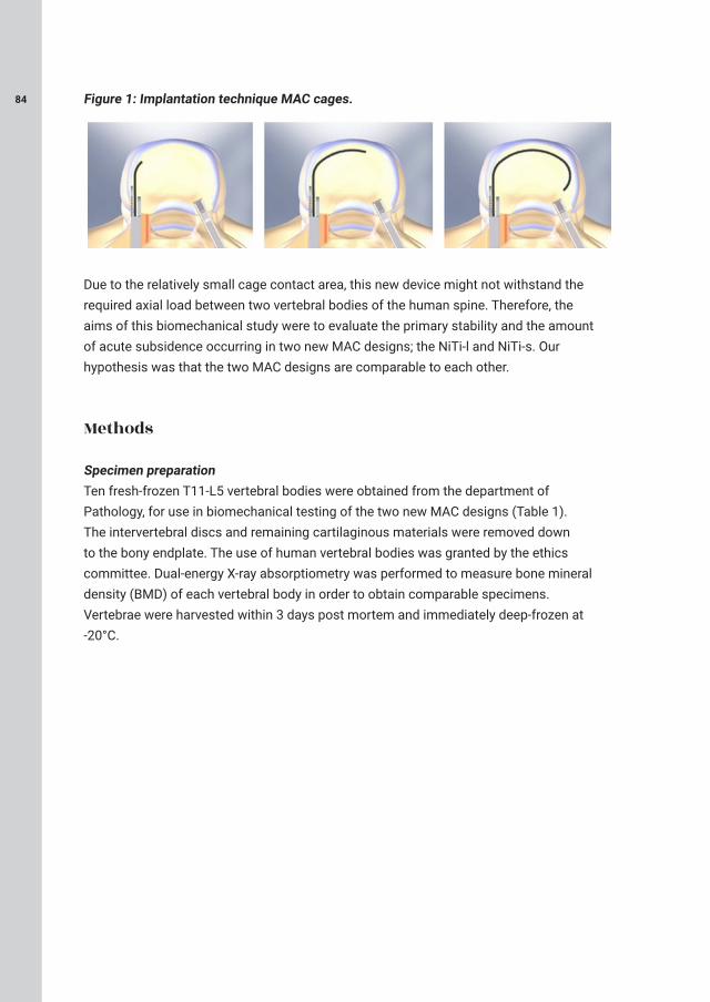
84 Figure 1: Implantation technique MAC cages.
Due to the relatively small cage contact area, this new device might not withstand the required axial load between two vertebral bodies of the human spine. Therefore, the aims of this biomechanical study were to evaluate the primary stability and the amount of acute subsidence occurring in two new MAC designs; the NiTi-l and NiTi-s. Our hypothesis was that the two MAC designs are comparable to each other.
Methods
Specimen preparationTen fresh-frozen T11-L5 vertebral bodies were obtained from the department of Pathology, for use in biomechanical testing of the two new MAC designs (Table 1). The intervertebral discs and remaining cartilaginous materials were removed down to the bony endplate. The use of human vertebral bodies was granted by the ethics committee. Dual-energy X-ray absorptiometry was performed to measure bone mineral density (BMD) of each vertebral body in order to obtain comparable specimens. Vertebrae were harvested within 3 days post mortem and immediately deep-frozen at -20°C.
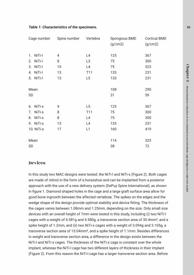
85
Chap
ter 5 | Bio
mech
anical evalu
ation
of tw
o m
inim
al access interbo
dy
cage design
s in a cad
averic mo
del
Table 1: Characteristics of the specimens.
Cage number Spine number Vertebra Spongious BMD Cortical BMD (g/cm2) (g/cm2)
1. NiTi-I 4 L4 125 3672. NiTi-I 8 L3 75 3003. NiTi-I 10 L4 75 3234. NiTi-I 13 T11 133 2315. NiTi-I 13 L5 133 231
Mean 108 290SD 31 59
6. NiTi-s 4 L5 125 3677. NiTi-s 8 T11 75 3008. NiTi-s 8 L4 75 3009. NiTi-s 13 L4 133 23110. NiTi-s 17 L1 160 419
Mean 114 323SD 38 72
Devices
In this study two MAC designs were tested: the NiTi-l and NiTi-s (Figure 2). Both cages are made of nitinol in the form of a horseshoe and can be implanted from a posterior approach with the use of a new delivery system (DePuy Spine International), as shown in figure 1. Diamond shaped holes in the cage and a large graft surface area allow for good bone ingrowth between the affected vertebrae. The spikes on the edges and the wedge shape of the design provide optimal stability and device fitting. The thickness of the cages varies between 1.08mm and 1.25mm, depending on the size. Only small size devices with an overall height of 7mm were tested in this study, including (i) two NiTi-l cages with a weight of 4.581g and 4.580g, a transverse section area of 30.4mm², and a spike height of 1.2mm, and (ii) two NiTi-s cages with a weight of 3.094g and 3.105g, a transverse section area of 15.04mm², and a spike height of 1.1mm. Besides differences in weight and transverse section area, a difference in the design exists between the NiTi-l and NiTi-s cages. The thickness of the NiTi-s cage is constant over the whole implant, whereas the NiTi-l cage has two different layers of thickness in their implant (Figure 2). From this reason the NiTi-l cage has a larger transverse section area. Before

86 biomechanical testing the devices were placed in a water bath of 60°C to make sure that the material was in its super elastic phase. Figure 2: The new prototype cages (left: NiTi-l, right: NiTi-s).
Axial compression testsThe biomechanical tests were performed with the use of a servo-hydraulic material testing system (MTS Bionix 858.2). The load was applied in force control mode with a constant loading rate of 50N/s.
Compressive force and displacement data were electronically recorded. The actuator of the MTS was equipped with a metal cylinder which provided perfect fitting with the MAC devices in order to prevent stability failures during the load application (Figure 3). Hot glue was used at three points for the attachment of the cage to the cylinder. The vertebral bodies were embedded in special holders using Polymethyl methacrylate(Technovit®) and placed on a XY-table. The cages were positioned approximately 3mm behind the front of the vertebral bodies according to instructions of DePuy International. In some cases the vertebral endplate did not make contact with the entire interface prior to the test due to irregularity of the endplate. Subsidence was assessed at a load of 800N. The compression test was continued until a subsidence of 7mm occurred. The failure load was defined as the maximum load reached before fracture of the vertebral endplate.
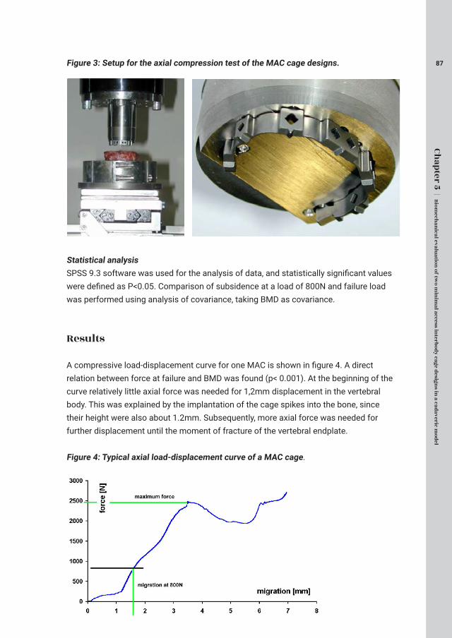
87
Chap
ter 5 | Bio
mech
anical evalu
ation
of tw
o m
inim
al access interbo
dy
cage design
s in a cad
averic mo
del
Figure 3: Setup for the axial compression test of the MAC cage designs.
Statistical analysisSPSS 9.3 software was used for the analysis of data, and statistically significant values were defined as P<0.05. Comparison of subsidence at a load of 800N and failure load was performed using analysis of covariance, taking BMD as covariance.
Results
A compressive load-displacement curve for one MAC is shown in figure 4. A direct relation between force at failure and BMD was found (p< 0.001). At the beginning of the curve relatively little axial force was needed for 1,2mm displacement in the vertebral body. This was explained by the implantation of the cage spikes into the bone, since their height were also about 1.2mm. Subsequently, more axial force was needed for further displacement until the moment of fracture of the vertebral endplate.
Figure 4: Typical axial load-displacement curve of a MAC cage.

88 The displacements in the vertebral body at an axial force of 800N were 1.91mm and 1.88mm for the NiTi-l and NiTi-s cage, respectively. As figure 5 indicates, no significant difference was established between the two cages. Figure 5: Mean displacement at a force of 800N and spike height of the cage.
The failure load and cage contact area of both cages are presented in figure 6. The mean failure load for the NiTi-l cage was 2043N, and 1866N for the NiTi-s cage. The failure load of NiTi-l group was slightly higher than the NiTi-s group, but no significant difference was established. Figure 6: Maximum force together with cage contact area (means and standard deviation).

89
Chap
ter 5 | Bio
mech
anical evalu
ation
of tw
o m
inim
al access interbo
dy
cage design
s in a cad
averic mo
del
Discussion
The aims of this biomechanical study were to evaluate the primary stability and the amount of acute subsidence occurring in two MAC designs. A servo-hydraulic material testing system was used to perform the axial compression test.
The Niti-l and Niti-s were tested in this study. Both cages are made of nitinol in the form of a wedge-shaped horseshoe with spikes on the edges. Differences are the higher weight and larger tranverse section area of the Niti-l due to his specific design with two different layers of thickness. The acute subsidence at a force of 800N was similar between the two cages. The failure load of the NiTi-l cage slightly exceeded that of the NiTi-s cage, although this difference was not statistically significant. Conform previous studies, a direct relation between force at failure and BMD was found in this study (18,19).
The primary subsidence in the vertebra was the result of the implantation of the cage spikes into the bone, and proved to be approximately 1.2mm. This result has been thought to be necessary for good adhesion of the device to the endplate and for optimal stability. Secondly, the primary subsidence might stimulate bone fusion because of the additional compression of the bone graft between the vertebrae. In order to prevent loss of height by subsidence of the two-sided spikes, an instrument can be used to enlarge the intervertebral space during the implantation of the MAC.
To prevent failure after intervertebral cage implantation in patients, knowledge about the minimum required load is warranted. The failure load of the MAC devices proved to be 2043N for the NiTi-l cage, and 1866N for the NiTi-s cage. The study of Wilke et al. (2001) measured intradiscal pressure in a non-degenerated L4-5 disc of a 45 year old male volunteer and discovered that intradiscal pressure depended on the kind of preceding activity, posture, external loads, and muscle activity (20). Kandziora et al. (2002) reported that the L4-L5 motion segment of a 80kg weighing patient could experience peak loads on the order of 2.24kN (21). Regarding the results of failure load from our study, this suggests that a risk of failure under physiological loads is present. However, considering the use of only small size devices in this study and the additional biomechanical strength obtained from the bone graft and posterior stabilization with pedicle screws, the MAC devices might therefore still meet the threshold for short-term as well as long-term clinical stability. Future studies should be performed to prove this statement.
In cylindrical cages generally higher failure loads have been observed after axial compression tests in cadaveric human spines (21). For example, the failure load

90 observed with a threaded, hollow, porous titanium BAK cage with a diameter of 15mm and a length of 24mm averaged 7.42kN (21). However, several complications have been reported in the use of BAK cages, including risk for long-term subsidence and corrosive effects, that contribute to an increase in revision surgery (22). For this reason, interbody devices with large graft spaces for optimal bone fusion are warranted. Although vertical cages like the MAC have lower failure loads, they certainly provide larger graft spaces and might therefore prolong the survival of the PLIF construction. The MOSS vertebral body replacement spacer (“Harms mesh cage”) is another example of a vertical cage.
The study of Knop et al. (2001) showed a mean maximum compressive force of 2.72kN for this device, which exceeds a little the failure load of the MAC (23). However, the contact area of the MOSS was 302mm2, whereas the NiTi-l and NiTi-s only have a contact area of 30.4 mm² and 15.04 mm², respectively. The MAC devices thereby, and through their wide and open design, offer a larger space for additional bone grafts. It has been well known that the surface area between graft bed and bone graft is one of the most important factors for optimal spinal fusion and secondary stability. Interbody graft area should be significantly greater than 30% of the total endplate area to provide sufficient compressive strength (24,25). Many cage designs do not provide such graft area, with the result that the contact area of local bone inside the cage might be insufficient for load transmission (26). Furthermore, the study of Lee et al. (2010) determined contact areas of fused local bone inside titanium cages using 3-dimensional thin-section computed tomography, and discovered that the ratio of fused area of local bone to total graft area inside cages was less than 50% (26). Although a fusion rate of 96.2% was reported, this indicates that only <50% of the exposed graft area inside cages contributes to the compressive strength of the bone fusion. For these reasons, MAC devices could possibly provide an relevant advantage with their small cage contact area and large graft space in comparison with other vertical cages. However, this statement is for the time being based on an assumption and future research should determine which vertical cage design is superior for optimal spinal fusion. Considering the little smaller cage contact area and thus larger graft space of the NiTi-s design, this cage type is favoured over the NiTi-l and should be used in further research study’s. The surgical technique should also be investigated in future studies, because theoretically the bone graft could migrate to the lumbar canal due to the open posterior design of the MAC. We believe, however, that this will not be a problem in practice, because of the standing posterior longitudinal ligament and because only a small opening is necessary to remove the discus and insert the MAC. Subsequently, through the lordosis of the lumbar spine the pressure of the bone grafts is mostly anteriorly.
The acute subsidence at a force of 800N and the failure load were comparable between the two MAC devices. Considering the small cage contact area, relatively high axial

91
Chap
ter 5 | Bio
mech
anical evalu
ation
of tw
o m
inim
al access interbo
dy
cage design
s in a cad
averic mo
del
force was needed for displacement in the vertebral body. This could be explained by the close position of implant to the cortex of the vertebra. A limitation in the current study was the inaccuracy of the measurement of subsidence. A spondylodesis is often performed in a degenerated spine without equal surfaces of endplates. Therefore, the amount of subsidence probably differs over the surface area while in this study only the mean subsidence in the vertebral body was measured.
A second limitation of the current study was that only an axial compression test was performed in cadaveric models. The flexion, extension, bending, and rotational stiffness or range of motion of the MAC cage designs were not evaluated. However, it has been thought that segmental stability will largely depend on the posterior fixation with pedicle screws and rods system. This was confirmed by the study of Wang et al. (2014). They showed a statistically significant improvement in stability when the cage or bone graft was supplemented with posterior instrumentation compared to biomechanical tests with cages alone (6). With this and the spikes on the edges and the wedge shape of the MAC design in consideration, we did not expect clinical relevant limitations in stability of the construction, and therefore only performed axial compression tests. Finally, a limitation could be the fact that these tests only reflect short-term subsidence. The long-term biomechanical strength of the design is unknown and difficult to test in vitro. Due to the small cage contact area of the MAC devices in comparison with other cages, it is highly likely that this design will fail earlier without additional bone graft supplementation. Therefore, good bone ingrowth is a prerequisite for a successful PLIF construction.
Conclusion
In conclusion, the biomechanical strength of both NiTi-l and NiTi-s cages is good and comparable to each other with a limited amount of short-term subsidence after the initial implantation of the cage spikes into the bone.
List of abbreviations
PLIF: Posterior lumbar interbody fusion MAC: Memory Metal Minimal Access CageBMD: bone mineral density
Ethics approval and consent to participateThe use of human vertebral bodies was granted by the ethics committee of University Medical Center Groningen.

92 Consent for publication
Not applicable.
Availability of data and material
The study data was collected by DePuy International. The data management and statistical analysis were performed by the Clinical Data Group of DePuy International Ltd. The datasets used and/or analysed during the current study are available from the corresponding author on reasonable request.
Competing interests
The authors declare that they have no competing interests.
Funding
No funding was received.
Authors’ contributions
DK, CP, FW have made substantial contributions to conception and design, and/or analysis and interpretation of data. All authors have participated in drafting the article or revising it critically for important intellectual content; and all authors give final approval of the version to be submitted and any revised version.
Acknowledgements
Not applicable.

93
Chap
ter 5 | Bio
mech
anical evalu
ation
of tw
o m
inim
al access interbo
dy
cage design
s in a cad
averic mo
del
References
1. Jaslow IA. Intercorporal bone graft in spinal fusion after disc removal. Surgery, gynecology & obstetrics. 1946, 82:215-218.
2. Cloward RB. Lesions of the intervertebral disks and their treatment by interbody fusion methods. The painful disk. Clinical orthopaedics and related research. 1963, 27:51-77.
3. Cloward RB. The treatment of ruptured lumbar intervertebral discs by vertebral body fusion. I. Indications, operative technique, after care. Journal of neurosurgery. 1953, 10(2):154-168.
4. Cloward RB. Spondylolisthesis: treatment by laminectomy and posterior interbody fusion. Clinical orthopaedics and related research. 1981(154):74-82.
5. Panjabi MM. Biomechanical evaluation of spinal fixation devices: I. A conceptual framework. Spine. 1988, 13(10):1129-1134.
6. Wang L, Huang H, Zhang Z, Zhang Z, Zhang L, Li J. Biomechanical Evaluation of a Novel Autogenous Bone Interbody Fusion Cage for Posterior Lumbar Interbody Fusion in a Cadaveric Model. Spine. 2014.
7. Loguidice VA, Johnson RG, Guyer RD, Stith WJ, Ohnmeiss DD, Hochschuler SH, Rashbaum RF. Anterior lumbar interbody fusion. Spine. 1988, 13(3):366-369.
8. Pfeiffer M, Griss P, Haake M, Kienapfel H, Billion M. Standardized evaluation of long-term results after anterior lumbar interbody fusion. European spine journal : official publication of the European Spine Society, the European Spinal Deformity Society, and the European Section of the Cervical Spine Research Society. 1996, 5(5):299-307.
9. Soini J. Lumbar disc space heights after external fixation and anterior interbody fusion: a prospective 2-year follow-up of clinical and radiographic results. Journal of spinal disorders. 1994, 7(6):487-494.
10. Fernyhough JC, Schimandle JJ, Weigel MC, Edwards CC, Levine AM. Chronic donor site pain complicating bone graft harvesting from the posterior iliac crest for spinal fusion. Spine. 1992, 17(12):1474-1480.
11. Younger EM, Chapman MW. Morbidity at bone graft donor sites. Journal of orthopaedic trauma. 1989, 3(3):192-195.
12. Boucher HH. A method of spinal fusion. The Journal of bone and joint surgery British volume. 1959, 41-B(2):248-259.
13. Evans JH. Biomechanics of lumbar fusion. Clinical orthopaedics and related research. 1985(193):38-46.
94 14. Cunningham BW, Kotani Y, McNulty PS, Cappuccino A, McAfee PC. The effect of spinal destabilization and instrumentation on lumbar intradiscal pressure: an in vitro biomechanical analysis. Spine. 1997, 22(22):2655-2663.
15. Parsons JR, Bhayani S, Alexander H, Weiss AB. Carbon fiber debris within the synovial joint. A time-dependent mechanical and histologic study. Clinical orthopaedics and related research. 1985(196):69-76.
16. Kuslich SD, Ulstrom CL, Griffith SL, Ahern JW, Dowdle JD. The Bagby and Kuslich method of lumbar interbody fusion. History, techniques, and 2-year follow-up results of a United States prospective, multicenter trial. Spine. 1998, 23(11):1267-1278; discussion 1279.
17. Ray CD. Threaded titanium cages for lumbar interbody fusions. Spine. 1997, 22(6):667-679; discussion 679-680.
18. Bisschop A, Mullender MG, Kingma I, Jiya TU, van der Veen AJ, Roos JC, van Dieen JH, van Royen BJ. The impact of bone mineral density and disc degeneration on shear strength and stiffness of the lumbar spine following laminectomy. European spine journal : official publication of the European Spine Society, the European Spinal Deformity Society, and the European Section of the Cervical Spine Research Society. 2012, 21(3):530-536.
19. Hussein AI, Jackman TM, Morgan SR, Barest GD, Morgan EF. The intravertebral distribution of bone density: correspondence to intervertebral disc health and implications for vertebral strength. Osteoporosis international : a journal established as result of cooperation between the European Foundation for Osteoporosis and the National Osteoporosis Foundation of the USA. 2013, 24(12):3021-3030.
20. Wilke H, Neef P, Hinz B, Seidel H, Claes L. Intradiscal pressure together with anthropometric data-a data set for the validation of models. Clinical biomechanics. 2001, 16 Suppl 1:S111-126.
21. Kandziora F, Pflugmacher R, Kleemann R, Duda G, Wise DL, Trantolo DJ, Lewandrowski KU. Biomechanical analysis of biodegradable interbody fusion cages augmented with poly(propylene glycol-co-fumaric acid). Spine. 2002, 27(15):1644-1651.
22. Beutler WJ, Peppelman WC, Jr.. Anterior lumbar fusion with paired BAK standard and paired BAK proximity cages: subsidence incidence, subsidence factors, and clinical outcome. The spine journal : official journal of the North American Spine Society. 2003, 3(4):289-293.

95
Chap
ter 5 | Bio
mech
anical evalu
ation
of tw
o m
inim
al access interbo
dy
cage design
s in a cad
averic mo
del
23. Knop C, Lange U, Bastian L, Oeser M, Blauth M. Biomechanical compression tests with a new implant for thoracolumbar vertebral body replacement. European spine journal : official publication of the European Spine Society, the European Spinal Deformity Society, and the European Section of the Cervical Spine Research Society. 2001, 10(1):30-37.
24. Sukovich W. Progress, Challenges And Opportunities In Disc Space Preparation For Lumbar Interbody Fusion. The Internet Journal of Spine Surgery. 2004, 1(2).
25. Closkey RF, Parsons JR, Lee CK, Blacksin MF, Zimmerman MC. Mechanics of interbody spinal fusion. Analysis of critical bone graft area. Spine. 1993, 18(8):1011-1015.
26. Lee JH, Jeon DW, Lee SJ, Chang BS, Lee CK. Fusion rates and subsidence of morselized local bone grafted in titanium cages in posterior lumbar interbody fusion using quantitative three-dimensional computed tomography scans. Spine. 2010, 35(15):1460-1465.

Minimally invasive procedures encompass surgical techniques that limit the size
of incisions needed and so lessen wound healing time, associated pain and risk
of infection. Minimally invasive procedures have been enabled by the advance of
various medical technologies.

97
D. Kok 1
R. D. Donk 2 F. H.Wapstra 1 A. G. Veldhuizen 1
1Department of Orthopedics, Universitair Medisch Centrum Groningen2Department of Orthopedics, Canisius Wilhelmina Ziekenhuis Nijmegen
Adv Orthop. 2012;2012:898606.
The Memory Metal Minimal Access Cage: A New Concept in Lumbar Interbody FusionA Prospective, Non-comparative Study to
Evaluate the Safety and Performance
98 Abstract
Study Design A single-centre, prospective, non-comparative study of 25 patients to evaluate the safety and performance of the Memory Metal Minimal Access Cage (MAC) in Lumbar Interbody Fusion.
Objective To evaluate the solid intervertebral fusion in two years and the safety of this new device, the Memory Metal Minimal Access Cage (MAC).Summary of Background Data: Interbody fusion cages in general are designed to withstand high axial loads which may otherwise lead to collaps of the intercorporal bone graft and in the meantime to allow ingrowth of new bone for bony fusion. In many cages the contact area with the endplate is rather large leaving a relatively small contact area for the bone graft with the adjacent host bone.
The Memory Metal Minimal Access Cage (MAC) is constructed from the memory metal Nitinol and builds on the concept of sufficient axial support in combination with a large contact area of the graft facilitating bony ingrowth and ease in minimal access implantation due to its high deformability.
Methods Twenty five subjects with a primary diagnosis of disabling back and radicular leg pain from a single level degenerative lumbar disc underwent an interbody fusion using the Memory Metal Minimal Access Cage (MAC) and pedicle screws.
Clinical performance was evaluated prospectively over 2 years using the Oswestry Disability Index (ODI), Short Form 36 questionnaire (SF-36) and pain visual analogue scale (VAS) scores. The interbody fusion status was assessed using conventional radiographs at regular time intervals and CT scan at 2 year follow-up. Safety of the device was studied by registration of intra- and post-operative adverse effects.
ResultsThe ODI, SF-36 and VAS for both back and leg pain all had improved significantly as compared to baseline scores (p<0.0018) Ongoing interbody fusion was observed on conventional radiographs and CT scan confirmed solid fusion in all 25 patients at two year follow-up. In two patients migration of the cage occurred, which was resolved uneventfully by placing a larger size at the subsequent revision.

99
Chap
ter 6 | Th
e Mem
ory
Metal M
inim
al Access Cage: A
New
Con
cept in
Lum
bar Interbo
dy
Fusio
n
Conclusions We conclude that the Memory Metal Minimal Access Cage (MAC) resulted in 100% solid fusions in 2 years and proved to be safe, although two patients required revision surgery in order to achieve solid fusion.
Key wordsMemory Metal Minimal Access Cage (MAC), PLIF, Fusion, CT.
Key points• The Memory Metal Minimal Access Cage (MAC) was implanted in humans for the first time and showed 100 percent fusion after two years, confirmed by CT.• The MAC is safe for implantation into humans with disabling back and/or refractory radicular pain with moderate to severe degenerative change in one or two lumbar disc levels based on MRI• The MAC performed very well by improving clinically important outcomes. The Oswestry Disability Index significantly improved from 38.32 ± 10.64 pre-operative to 8.4 ± 9.49 at 24 months post-operative.
100 Introduction
Chronic low back pain is an insidious problem. Individuals suffer from prolonged discomfort, anxiety and disability. Low back pain has been shown as the leading cause of man-hours lost to disease or injury. Degeneration of the intervertebral disc is the most common cause of low back pain (1).
Conservative treatment for low back pain may include rest, heat, physical therapy, medication, bracing and education. Most individuals will find relief given conservative treatments. However, for those with significant continuing specific symptoms, surgical intervention may be appropriate. One of the interventions is posterior lumbar interbody fusion (PLIF). The goal of spinal fusion is to obtain a solid arthrodesis. There is a wide range of fusion rates (56-95%) reported after PLIF with varying techniques (5,13,21-31).The PLIF procedure was introduced independently by Jaslow (2) and Cloward (3-6) in the 1940s to treat painful intervertebral disc damaged by degeneration or herniation. A PLIF has the advantages over other types of fusion allowing neural decompression while in the meantime restoration of the disc height, and segmental alignment is maintained (7).
In order to eventually achieve a solid interbody fusion a bone substitute has to be applied to the disc space. Without a mechanical support these grafts tend to collaps, displace or extrude (8-10). For this reason various metal and carbon fibre interbody cages have been developed (13-15). Interbody fusion cages aim to fulfil both mechanical and biological requirements for fusion, in that the cages are designed to withstand high axial loads (7,16,17) and in the meantime to allow ingrowth of vital host bone. Although cages have rapidly become popular, the mismatch in the modulus of elasticity between many available metal cages and the actual vertebral body may cause stress shielding, resulting in a delayed fusion and eventually pseudarthrosis (18,19). Carbon fiber cages better approximate this modulus of elasticity of the vertebral bone, however there are some reports on carbon fiber release causing synovitis (20). The titanium implants developed by Bagby and Kuslich, Ray, and Harms (14,15), offer a radio-opaque alternative to carbon fibre materials that also exhibit the necessary biomechanical strength as well as facilitating the cage to be located radiographically. Their open design means that the bone is exposed to a greater graft surface area that has been shown to facilitate good bony in growth. However, the problem with most cages is the small contact area of the bone graft and therefore a high rate of pseudoarthrosis.
The Memory Metal Minimal Access Cage (MAC) builds on the concept of sufficient axial support in combination with a large contact area of the graft facilitating bony ingrowth
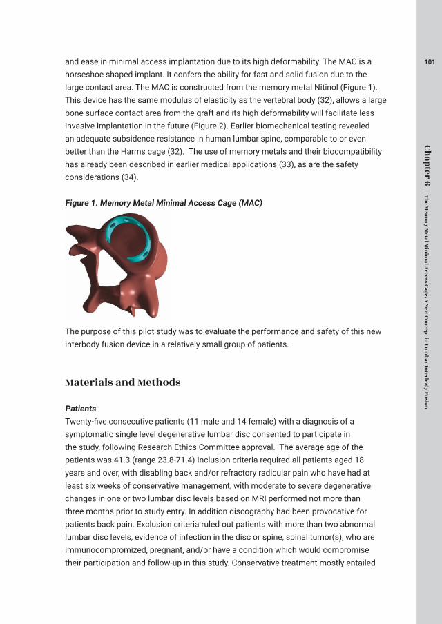
101
Chap
ter 6 | Th
e Mem
ory
Metal M
inim
al Access Cage: A
New
Con
cept in
Lum
bar Interbo
dy
Fusio
n
and ease in minimal access implantation due to its high deformability. The MAC is a horseshoe shaped implant. It confers the ability for fast and solid fusion due to the large contact area. The MAC is constructed from the memory metal Nitinol (Figure 1). This device has the same modulus of elasticity as the vertebral body (32), allows a large bone surface contact area from the graft and its high deformability will facilitate less invasive implantation in the future (Figure 2). Earlier biomechanical testing revealed an adequate subsidence resistance in human lumbar spine, comparable to or even better than the Harms cage (32). The use of memory metals and their biocompatibility has already been described in earlier medical applications (33), as are the safety considerations (34). Figure 1. Memory Metal Minimal Access Cage (MAC)
The purpose of this pilot study was to evaluate the performance and safety of this new interbody fusion device in a relatively small group of patients.
Materials and Methods
PatientsTwenty-five consecutive patients (11 male and 14 female) with a diagnosis of a symptomatic single level degenerative lumbar disc consented to participate in the study, following Research Ethics Committee approval. The average age of the patients was 41.3 (range 23.8-71.4) Inclusion criteria required all patients aged 18 years and over, with disabling back and/or refractory radicular pain who have had at least six weeks of conservative management, with moderate to severe degenerative changes in one or two lumbar disc levels based on MRI performed not more than three months prior to study entry. In addition discography had been provocative for patients back pain. Exclusion criteria ruled out patients with more than two abnormal lumbar disc levels, evidence of infection in the disc or spine, spinal tumor(s), who are immunocompromized, pregnant, and/or have a condition which would compromise their participation and follow-up in this study. Conservative treatment mostly entailed

102 a combination of appropriate analgesics, physical therapy, and epidural and/or facet injections.
Implant features and surgical procedure The Memory Metal Minimal Access Cage has a horseshoe shape, and comprises a material strip of 1.08mm thickness for the small sizes, and 1.25mm for the medium and large sizes. All cages have diamond shaped holes for bone through growth, spikes on the top and bottom edges for stability, and a wedged profile. The diamond shaped hole design aspect of the MAC is in line with surgical titanium mesh for similar product appearance, and seats on the bony outer cortical rim of the vertebral body.The cages are made of Nitinol, a shape memory alloy, which enables the surgeon to un-curve the strip completely, put it into an inserter, and insert it into the disc space while pushing it out of the inserter. The flat strip will henceforth curve into the original horseshoe shape (Figure 2). All surgeries were performed by two experienced spine surgeons between January 2004 and Oktober 2006. A standard PLIF procedure was performed using the Monarch TM pedicle screw system (DePuy International) where after the MAC was placed anteriorly in the intervertebral disc space (Figure 2) and locally available decompressive autologous bone was subsequently grafted into the disc space. Figure 2. Implantation technique
Clinical and radiological outcomePatients were evaluated pre-operatively at 1, 3, 6, 12 and 24 month after surgery. Evaluation at each interval included physical and neurological examination, concomitant medication, additional surgical procedures, subject completed questionnaires (Oswestry Disability Index, Short Form-36 Health) and Visual Analogue Scale for Pain. Any adverse events and complications were recorded in the case report forms.
Routine lateral and AP radiographs were obtained at each timer interval. Routine radiographs were used to evaluate the total intervertebral height and subsidence. The CT scan at two years follow-up was used to establish fusion. The total intervertebral

103
Chap
ter 6 | Th
e Mem
ory
Metal M
inim
al Access Cage: A
New
Con
cept in
Lum
bar Interbo
dy
Fusio
n
height (TIH) of two fused vertebral bodies was measured as distance between the mid-point of upper end plate of cranial vertebral body and the mid-point of lower end plate of caudal vertebral body on digital radiographs with built-in software (PACS viewer). The degree of subsidence (ΔTIH) was reflected by the difference between the immediate postoperative and follow-up TIH (Figure 3). With the same method, change of postoperative disc space height was reflected by the difference between TIH of the postoperative lateral plain radiograph and that of the preoperative lateral plain radiograph (Figure 3). Interbody fusion was defined as complete bridging at any one or more points within the central area of the vertebral body as determined by CT. Intervertebral fusion assessments were determined by one independent radiologist who was not otherwise involved in the study. Fusion was recorded as Yes/No/Can’t Assess. Figure 3. Measurement of the subsidence and total intervertebral height
Complications were divided into device-related and non–device-related complications. Non–device-related complications were listed as major and minor.
StatisticsFor statistical analysis, comparisons between pre- and postoperative scores were made using paired t-tests.

104 Results
Radiological AssessmentThe primary radiological objective was fusion rate. Fusion success was achieved in 25 (100%) of 25 patients. There was a solid bony fusion on CT at 2 years post-operative. The disc space height was restored to normal as part of the operative procedure. Disc height in the cage levels was increased from an average of 7.6 mm before surgery to an average of 12.4 mm after surgery losing 0.0 mm during healing in 2 years of follow-up.
Clinical DataAll 25 patients completed the 24 months of follow-up without any major adverse event. The clinical parameters are summarized in figures 4-6.The clinical outcome was the ODI score at 24 months post-treatment compared to baseline. The mean ODI score pre-operative was 38.32 ±10.64. This significantly improved to 8.4 ±9.49 at 24 months post-operative (p <.0001). Figure 4. Oswestry Disability Index at Baseline and 1, 3, 6, 12, 24 months Post-Operation
The Short-Form 36 health questionnaire (SF36) data assessed both physical and mental components. Physical (PCM) 36.15 ± 18.93 improved to 84.25 ± 22.29 (p <.0001) and mental (MCM) 60.54 ± 24.22 improved to 91.36 ± 12.76 (p <.0001).
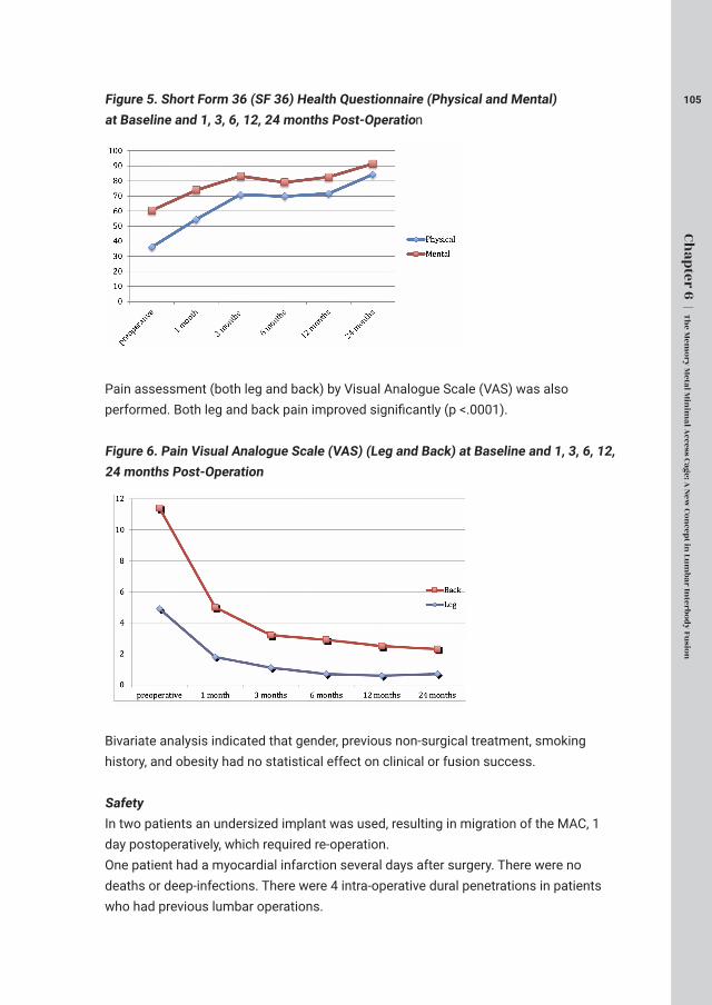
105
Chap
ter 6 | Th
e Mem
ory
Metal M
inim
al Access Cage: A
New
Con
cept in
Lum
bar Interbo
dy
Fusio
n
Figure 5. Short Form 36 (SF 36) Health Questionnaire (Physical and Mental) at Baseline and 1, 3, 6, 12, 24 months Post-Operation
Pain assessment (both leg and back) by Visual Analogue Scale (VAS) was also performed. Both leg and back pain improved significantly (p <.0001).
Figure 6. Pain Visual Analogue Scale (VAS) (Leg and Back) at Baseline and 1, 3, 6, 12, 24 months Post-Operation
Bivariate analysis indicated that gender, previous non-surgical treatment, smoking history, and obesity had no statistical effect on clinical or fusion success.
SafetyIn two patients an undersized implant was used, resulting in migration of the MAC, 1 day postoperatively, which required re-operation.One patient had a myocardial infarction several days after surgery. There were no deaths or deep-infections. There were 4 intra-operative dural penetrations in patients who had previous lumbar operations.

106 Discussion
In this study a prospective follow-up on clinical and radiographic parameters was performed in patients with a single level spondylodesis using a new interbody cage design.
Radiological Assessment Radiological assessment indicated that there was a 100 percent interbody fusion with the MAC device at 2 years on CT with no subsidence. Previous studies (5, 15, 35-41) report of PLIF fusion success with fusion in 85% of the cases. The difficulty in determining fusion success by standard roentgenographic methods was emphasized by Hibbs and Swift in 1929 (42), Cleveland et al. in 1948 (43), Prothero et al. in 1966 (44), Stauffer and Coventry in 1972 (45), Chow et al. in 1980 (46), Zinreich et al. in 1990 (47), and Brodsky et al. in 1991 (48). The recent use of pedicle screw fixation has added to the problem, because overlying shadows of the implants impaired radiographic visualization of posterolateral fusion mass (30, 49). Edward et al. in 2003 (50) emphasized that there is an overestimation of fusion on plain radiograph compared to CT.
In order to make a good estimation on interbody fusion we used CT in this study. Previous studies on interbody fusion reported significant loss of disc space height during healing of interbody grafts (5,10,22,51-54). In past reports, even pedicle screw stabilization has not prevented this loss of disc space height during the healing of interbody fusion (22,40,51). Loss of disc space height creates foraminal narrowing and the potential for nerve root compression. The fact that we recorded 100 percent fusion on CT and no subsidence is an advantage over other interbody fusion devices.
Clinical DataNumerous studies have provided subjective descriptions of criteria for excellent, good, fair, and poor results (38,49,55-60). We use the ODI as our primary clinical objective because the ODI is valid and vigorous measure and has been a worthwhile outcome measure (61, 62).
The Oswestry Disability Index mean score pre-operative was 38.32 ±10.64. This significantly improved to 8.4 ±9.49 at 24 months post-operative. Significant improvement in both physical and emotional components in the SF36 questionnaires mean scores were also observed, with increases from baseline results of 36.15 ±18.93 and 60.54 ±24.22 to 84.25 ±22.29 and 91.36 ±12.76 at 24 months respectively (p<.0001). The average level of leg pain was reduced by more than 50% post-operation (VAS values reduced from 4.88 ±2.96 to 1.78 ±1.97 at 1 month post-operation). This

107
Chap
ter 6 | Th
e Mem
ory
Metal M
inim
al Access Cage: A
New
Con
cept in
Lum
bar Interbo
dy
Fusio
n
reduction further improved over the 24 months post-operation (0.73 ±1.31 at 24 month post-operation). A similar reduction in back pain was also revealed. With both ODI and SF36 results, improvement in condition continued throughout the 24 months post-operation. Pain results indicated a rapid improvement post-operation, which was maintained during the 24 months post-operation.
A study of 60 patients with posterior lumbar interbody fusion combined with instrumented postero-lateral fusion reported by Freeman et al. (63) indicated stable circumferential fixation as shown by radiographs and tomograms confirming the presence of a bridging fusion mass. Of the 48 ODI questionnaires completed after 5 years, 79% had an ODI <30. In the present study 96% (24/25) of the patients indicated an ODI < 30. McKenna et al. reported a prospective, randomized controlled trial of femoral ring allograft (FRA) versus a titanium cage (TC) in circumferential lumbar spinal fusion with minimum 2 years clinical results (64). Comparison of change in ODI results indicated a significantly larger improvement in the FRA group (reduced from 57 to 42) when compared to the TC group (54 reduced to 48). The corresponding change in ODI results from baseline over 2 years in the current study was larger than that of either the FRA or TC groups (35 versus 15 and 6). SF36 results for the FRA patients showed a significant improvement in the Physical Function Component but not in the Mental Component (change in SF36 results of 17 and 2 respectively). In the TC patients the reverse was found (change in SF36 results of 5 and 9 for Physical Function and Mental Components respectively). The MAC in comparison gave a much greater improvement in both SF36 results (change in SF36 results of 63.1 and 27 for Physical and Mental Components respectively). Both FRA and TC patients showed a significant improvement in VAS for back pain (change in VAS 1.9 and 1.1 respectively). However with leg pain VAS scores only FRA patients demonstrated a significant improvement (change in VAS of 1.3) whereas the TC group had more leg pain increasing the VAS scores postoperatively by 0.4 points. In our study we found a significant reduction in both back and leg pain. With the MAC the back and VAS results were reduced by 6.4 and 5.8 points respectively. This indicates a significant improvement compared to the McKenna study. Cassinelli et al. published a prospective clinical study of revision fusion surgery in 19 patients with pseudoarthrosis who had received posterior lumber interbody fusion using stand-alone metallic cages (65). SF-36 and ODI data were collected prior to surgery and two years post operatively. Significant improvement was only noted in two of the eight SF-36 subcategories (Physical Functional and Role Mental). There was no significant difference in ODI scores. A study with two different patient groups of 30 subjects having spondylolisthesis, which were subjected to different surgeries: posterior lumbar fusion with pedicle screws (Group I) and posterior lumbar interbody fusion with pedicle screws (Group II) has also been reported (66). The ODI mean scores pre-operatively and 2 year post-operatively was 28.5 and 18.6

108 respectively for Group I and 31.3 and 13.3 respectively for Group II. The ODI scores in the current study show a greater improvement. Glassman et al. reviewed the ODI and SF36 outcomes in a multicentre lumbar fusion study with follow up after 2 years (67). The minimal clinically important difference (MCID) seeks to differentiate a magnitude of change, which is not only statistically valid but also of real clinical value. Figures for MCID for ODI results have been reported as low as a 4 point decrease (68) and also a 10 point decrease (69). The Food and Drug Administration (FDA) standards suggest a 15 point decrease in ODI and either maintenance of or any improvement in SF-36 Physical Composite Score (PCS) (70). Ware et al. (71) reported that an increase of 5.42 points in the SF-36 PCS is clinically important. A more recent study (72) has reported the following MCID values: 12.8 points for ODI, 4.9 points for SF-36 PCS, 1.2 points for back pain and 1.6 points for leg pain. The improvement in ODI values for the various fusion treatments in the multicentre review ranged from 9.9 to 22.2 points whereas the improvement in SF-36 data ranged from 13.8 to 6.3 points. The improvement in the corresponding ODI and SF-36 values in the current MAC study were 29.92 and 39.46. The improvement in back and leg pain were 4.88 and 4.15 respectively. In general, the ODI and VAS improved in all PLIF-procedures, according to the literature. The results obtained for the MAC have therefore satisfied the MCID reported in the literature.
SafetyThe device related adverse event recorded in this study was two undersized cages, resulting in migration. The migration problem lies within the operation technique. The dural penetrations all developed during decompression in patients who were previously operated on, not during cage insertion, and were repaired at surgery, not requiring reoperation, not causing neurologic injury, and not affecting the hospital course.
Conclusion
The Memory Metal Minimal Access Cage performed very well radiographically and clinically. There was a 100 percent interbody fusion at 2 years on CT, no subsidence and significant improvement of clinically important outcomes, although two patients required revision surgery in order to achieve solid fusion.
Acknowledgments
The study was sponsored by DePuy International.

109
Chap
ter 6 | Th
e Mem
ory
Metal M
inim
al Access Cage: A
New
Con
cept in
Lum
bar Interbo
dy
Fusio
n
References
1. Hedman TD. Design of an intervertebral disc prothesis. Spine. 1991; 16 (6C): 256-60.
2. Jaslow IA. Intercorporal bone graft in spinal fusion after disc removal. Surg Gyn Obst. 1946; 82: 215-8
3. Cloward RB. Lesions of the intervertebral disks and their treatment by interbody fusion methods. Clin Orthop 1963; 27: 51-77.
4. Cloward RB. Rationing the treatment of ruptured lumbar intervertebral discs by vertebral body fusion. J Neurosurg. 1953; 10: 154-68.
5. Cloward RB. Spondylolisthesis: treatment by laminectomy and posterior lumbar interbody fusion. Clin Orthop. 1981; 154: 74-82.
6. Cloward RB. Posterior lumbar interbody fusion (P.L.I.F.) surgical techniques. Randolph, MA: Codman, undated.
7. Panjabi MM. Biomechanical evaluation of spinal fixation devices: I. A conceptual framework. Spine. 1988; 13: 1129-34.
8. Loguidice VA, Johnson RG, Guyer RD, et al. Anterior lumbar interbody fusion. Spine. 1988; 13: 366-9.
9. Pfeiffer M, Griss P, Haake M, et al. Standardized evaluation of long-term results after anterior lumbar interbody fusion. Eur Spine J. 1996; 5: 299-307.
10. Soini J. Lumbar disc space heights after external fixation and anterior interbody fusion: A prospective 2 year follow-up of clinical and radiographic results. J Spinal Disord. 1994; 7: 487-94.
11. Fernyhough JC, Schimandle JJ, Weigel MC, et al. Chronic donor site pain complicating bone graft harvesting from the posterior iliac crest for spinal fusion. Spine. 1992; 17: 1474-80.
12. Younger EM, Chapman MW. Morbidity of bone graft donor sites. J Orthop Trauma. 1989; 3: 192-5.
13. Brantigan JW, Steffee AD, A carbon fiber implant to aid interbody lumbar fusion. Two-year clinical results in the first 26 patients, Spine. 1993; 18(14): 2106-7.
14. Kuslich SD, Ulstrom CL, Griffith SL, et al. The Bagby and Kuslich method of lumbar interbody fusion. History, techniques, and 2-year follow-up results of a United States prospective multicenter trial. Spine. 1998; 23: 1267-78.
15. Ray CD. Threaded titanium cages for lumbar interbody fusions. Spine. 1997; 22: 667-79.
16. Boucher HH. A method of spinal fusion. J Bone Joint Surg. 1959; 41B: 248-9.
17. Evans JH. Biomechanics of lumbar fusion. Clin Orthop. 1985; 193: 38-46.

110 18. Kanayama M, Cunningham BW, Haggarty CJ, et al. An in vitro biomechanical analysis on the stability and stress-shielding effect of interbody fusion implants. Presented at the annual meeting of the North American Spine Society, New York, NY, 1997.
19. Tullberg T. Failure of a carbon fiber implant. A case report. Spine. 1998; 23: 1804-6.
20. Parsons JR, Bhayani S, Alexander H, et al. Carbon fiber debris within the synovial joint. A time-dependent mechanical and histological study. Clin Orthop. 1985; 196: 69-76.
21. Berkley LR. A critique of posterior lumbar interbody fusion: 12 years’ experience with 250 patients. Surg Neurol. 1989; 31: 281-89.
22. Brantigan JW. Pseudoarthrosis rate after allograft posterior lumbar interbody fusion with pedicle screw and plate fixation. Spine. 1994; 19: 1271-80.
23. Collis JS. Total disc replacement: a modified posterior lumbar interbody fusion. Clin Orthop. 1985; 193: 64-7.
24. Hutter CG. Spinal stenosis and posterior lumbar interbody fusion. Clin Orthop. 1985; 193: 103-14.
25. Lee CK, Vessa P, Lee JK. Chronic disabling low back pain syndrome caused by internal disc derangements: the results of disc excision and posterior lumbar interbody fusion. Spine. 1995; 20: 356-61.
26. Lin PM, Cautilli RA, Joyce MF. Posterior lumbar interbody fusion. Clin Orthop. 1983; 180: 154-68.
27. Meril AJ. Direct current stimulation of allograft in anterior and posterior lumbar interbody fusions. Spine. 1994; 21: 2393-98.
28. Rothman SLG, Glenn MW. CT evaluation of interbody fusion. Clin Orthop. 1985; 193: 47-56.
29. Simmons JW. Posterior lumbar interbody fusion with posterior elements as chip grafts. Clin Orthop. 1985; 193: 85-9.
30. Steffee AD, Brantigan JW. The variable screw placement spinal fixation system. Report of a prospective study of 250 patients enrolled in food and drug administration clinical trials. Spine. 1993; 18: 1160-72.
31. Wiltberger BR. The dowel intervertebral-body fusion as used in lumbar-disc surgery. J Bone Joint Surg [Am]. 1957 ; 39: 284-331.
32. Kok D, Wapstra FH, Sanders MM et al, Minimal Access Interbody Cage Designs. A biomechanical comparison. Submitted.
33. Gil FJ, Planell JA. Shape memory alloys for medical applications. Proc. Instn Mech. Engrs. 1998,; 212(H): 473-488.
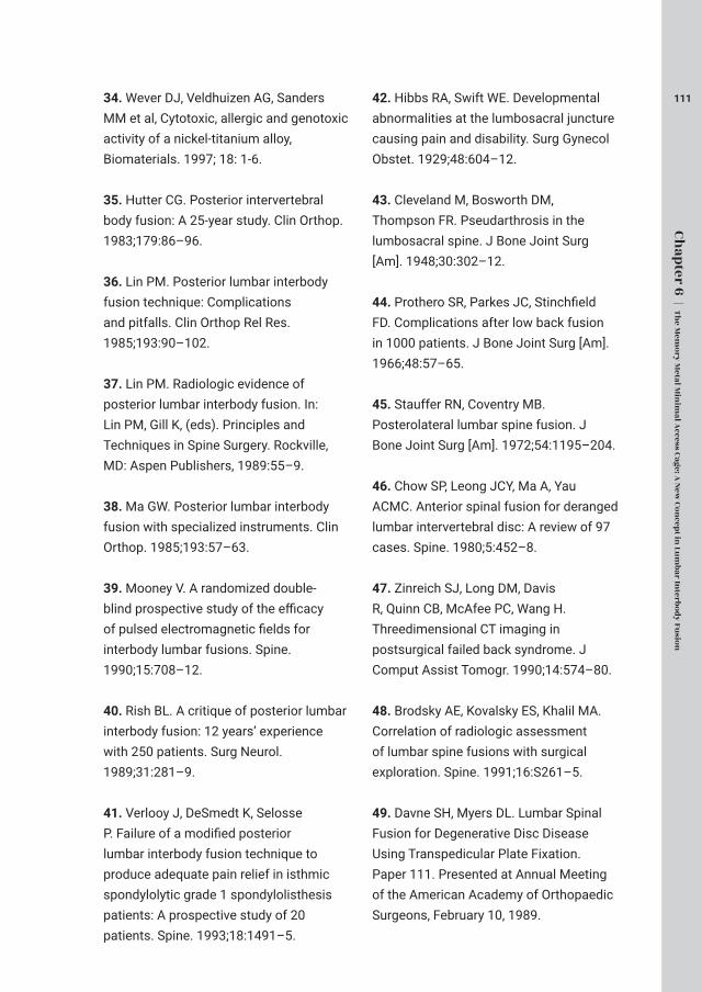
111
Chap
ter 6 | Th
e Mem
ory
Metal M
inim
al Access Cage: A
New
Con
cept in
Lum
bar Interbo
dy
Fusio
n
34. Wever DJ, Veldhuizen AG, Sanders MM et al, Cytotoxic, allergic and genotoxic activity of a nickel-titanium alloy, Biomaterials. 1997; 18: 1-6.
35. Hutter CG. Posterior intervertebral body fusion: A 25-year study. Clin Orthop. 1983;179:86–96.
36. Lin PM. Posterior lumbar interbody fusion technique: Complications and pitfalls. Clin Orthop Rel Res. 1985;193:90–102.
37. Lin PM. Radiologic evidence of posterior lumbar interbody fusion. In: Lin PM, Gill K, (eds). Principles and Techniques in Spine Surgery. Rockville, MD: Aspen Publishers, 1989:55–9.
38. Ma GW. Posterior lumbar interbody fusion with specialized instruments. Clin Orthop. 1985;193:57–63.
39. Mooney V. A randomized double-blind prospective study of the efficacy of pulsed electromagnetic fields for interbody lumbar fusions. Spine. 1990;15:708–12.
40. Rish BL. A critique of posterior lumbar interbody fusion: 12 years’ experience with 250 patients. Surg Neurol. 1989;31:281–9.
41. Verlooy J, DeSmedt K, Selosse P. Failure of a modified posterior lumbar interbody fusion technique to produce adequate pain relief in isthmic spondylolytic grade 1 spondylolisthesis patients: A prospective study of 20 patients. Spine. 1993;18:1491–5.
42. Hibbs RA, Swift WE. Developmental abnormalities at the lumbosacral juncture causing pain and disability. Surg Gynecol Obstet. 1929;48:604–12.
43. Cleveland M, Bosworth DM, Thompson FR. Pseudarthrosis in the lumbosacral spine. J Bone Joint Surg [Am]. 1948;30:302–12.
44. Prothero SR, Parkes JC, Stinchfield FD. Complications after low back fusion in 1000 patients. J Bone Joint Surg [Am]. 1966;48:57–65.
45. Stauffer RN, Coventry MB. Posterolateral lumbar spine fusion. J Bone Joint Surg [Am]. 1972;54:1195–204.
46. Chow SP, Leong JCY, Ma A, Yau ACMC. Anterior spinal fusion for deranged lumbar intervertebral disc: A review of 97 cases. Spine. 1980;5:452–8.
47. Zinreich SJ, Long DM, Davis R, Quinn CB, McAfee PC, Wang H. Threedimensional CT imaging in postsurgical failed back syndrome. J Comput Assist Tomogr. 1990;14:574–80.
48. Brodsky AE, Kovalsky ES, Khalil MA. Correlation of radiologic assessment of lumbar spine fusions with surgical exploration. Spine. 1991;16:S261–5.
49. Davne SH, Myers DL. Lumbar Spinal Fusion for Degenerative Disc Disease Using Transpedicular Plate Fixation. Paper 111. Presented at Annual Meeting of the American Academy of Orthopaedic Surgeons, February 10, 1989.
112 50. Edward RG, Santos, David GG, et al. Radiologic Assessment of Interbody Fusion Using Carbon Fiber Cages. Spine. 2003; 28: 997-1001.
51. Holte DC, O’Brien JP, Renton P. Anterior lumbar fusion using a hybrid interbody graft: A preliminary radiographic report. Eur Spine J. 1994;3:32–8.
52. Kumar A, Kozak JA, Doherty BJ, Dickson JH. Interspace distraction and graft subsidence after anterior lumbar fusion with femoral strut allograft. Spine. 1993;18:2393–400.
53. Tan SB, Kozak JA, Graham JM. A modified technique of anterior lumbar fusion with femoral cortical allograft. J Orthop Surg Techniques. 1990;5:83–93.
54. Dennis S, Watkins R, Landaker S, Dillin W. Comparison of disc space heights after anterior lumbar interbody fusion. Spine. 1989;14:876–8.
55. Henderson ED. Results of the surgical treatment of spondylolisthesis. J Bone Joint Surg [Am]. 1966;48:619–42.
56. Mardjetko SM, Connolly PJ, Shott S. Degenerative lumbar spondylolisthesis: A meta-analysis of literature 1970–1993. Spine. 1994;19:2256S–65S.
57. Naylor A. The late results of laminectomy for lumbar disc prolapse. J Bone Joint Surg [Br]. 1974;56:17–29.
58. Prolo DJ, Oklund SA, Butcher M. Toward uniformity in evaluating results of lumbar spine operations: A paradigm applied to PLIF. Spine. 1986;11:601–6.
59. Spengler DM, Ouellette EA, Battie M. Elective discectomy for herniation of a lumbar disc. J Bone Joint Surg [Am]. 1990;72:230–7.
60. White AH, Rogov RV, Zucherman J, Heiden D. Lumbar laminectomy for herniated disc: A prospective controlled comparison with internal fixation fusion. Spine. 1988;12:305–7.
61. Fairbank JC, Pynsent PB. The Oswestry Disability Index. Spine. 2000;25(22):2940-52; discussion 2952.
62. Davidson M, Keating JL. A comparison of five low back disability questionnaires: reliability and responsiveness. Phys Ther. 2002; 82(1):8-24.
63. Freeman BJC, Licina P, Mehdian SH. Posterior lumbar interbody fusion combined with instrumented postero-lateral fusion: 5-year results in 60 patients, Eur Spine J. 2000; 9: 42-46.
64. McKenna PJ, Freeman BJC, Mulholland et al. A prospective, randomized controlled trial of femoral ring allograft versus a titanium cage in circumferential lumbar spinal fusion with minimum 2-year results, Eur Spine J. 2005; 14: 727-737.
113
Chap
ter 6 | Th
e Mem
ory
Metal M
inim
al Access Cage: A
New
Con
cept in
Lum
bar Interbo
dy
Fusio
n
65. Cassinelli EH, Wallach C, Hanscom B, Vogt M et al. Prospective clinical outcomes of revision fusion surgery in patients with pseudoarthrosis after posterior lumber interbody fusion using stand-alone metallic cages, The Spine Journal. 2006; 6: 428-434.
66. Dantas FLR, Prandini MN, Ferreira MAT. Comparision between posterior lumbar fusion with pedicle screws and posterior lumbar interbody fusion with pedicle screws in adult spondylolisthesis, Arq Neuropsiquitar. 2007; 65(3B); 764-770.
67. Glassman S, Gornet MF, Branch C et al. MOS Short Form 36 and Oswestry Disability Index outcomes in lumber fusion: a multicentre experience, The Spine Journal. 2006; 6: 21-26.
68. Meade TW, Dyer S, Browne W et al. Low back pain of mechanical origin: randomized comparison of chiropractic and hospital outpatient treatment. BMJ. 1990; 300: 1431-7.
69. Hagg O, Fritzell P, Nordwall A. The clinical importance of changes in outcome scores after treatment for chronic low back pain. Eur Spine J. 2003; 12: 20.
70. FDA Protocol G000137. A prospective, randomized clinical investigation of recombinant human bone morphogenic protein-2 and compression resistant matrix with the CD Horizon spinal system for posterolateral lumber fusion in patients with symptomatic degenerative disc disease. Medtronic Sofamor Danek, Memphis, Tennessee, Feb. 2002.
71.Ware JE, Kosinski M, Keller SK. SF-36 physical and mental health summary scales: a user’s manual. Boston MA: The Health Institute, 1994.
72.Copay AG, Glassman SD, Subach BR et al. The minimum clinically important difference in lumbar spine surgery patients: a choice of methods using the Oswestry Disability Index, medical outcomes study questionnaire Short Form 36, and pain scales. The Spine Journal. 2008.

Bone Mineral Density (BMD), is the amount of bone mineral in bone tissue.
Bone density measurement is used in clinical medicine as an indirect indicator
of osteoporosis and fracture risk.
Dual-energy X-ray absorptiometry (DEXA) is a means of measuring bone mineral
density (BMD) using spectral imaging. Two X-ray beams, with different energy
levels, are aimed at the patient’s bones. When soft tissue absorption is subtracted
out, the bone mineral density (BMD) can be determined from the absorption of
each beam by bone.
115
Changes in Bone Mineral Density in the Intertransverse Fusion Mass After Instrumented Single-Level Lumbar Fusion 1-year follow-up
F. Hagenmaier, MSc * D. Kok , MD* A. Hol , MSc†
T. Rijnders , MD‡ F. C. Oner, MD, PhD§ J. L. C. van Susante , MD, PhD*
*Department of Orthopaedics, Rijnstate Hospital, Arnhem, The Netherlands.† Physiotherapy, Rijnstate Hospital, Arnhem, the Netherlands.‡ Nuclear Medicine, Rijnstate Hospital, Arnhem, the Netherlands.§ Department of Orthopaedics, University Medical Center Utrecht, the Netherlands.
Spine (Phila Pa 1976). 2013 Apr 15;38(8):696-702.
116 Abstract
Study Design Prospective cohort.
ObjectiveThe purpose of this study was to evaluate changes in bone mineral density (BMD) in the intertransverse fusion mass as representative for the process of bone remodeling after spinal fusion.
Summary of Background DataIntertransverse bone graft is frequently applied to facilitate bony fusion between 2 spinal levels. The biological process of bone graft remodeling leading to eventual fusion is, however, poorly understood.
MethodsIn 20 patients with a single-level instrumented posterolateral lumbar fusion for low-grade spondylolisthesis, radiographs, and clinical outcome scores (visual analogue scale for back and leg pain, Oswestry Disability Index , Short Form-36) were obtained. Locally harvested laminectomy bone was used as intertransverse bone graft. The BMD in selected “regions of interest” at both intertransverse fusion areas was assessed on days 4 and 3, after a period of 6 and 12 months after surgery using dual-energy x-ray absorptiometry scans. Eventual fusion status was assessed on computed tomography scan at 12 months.
ResultsAll clinical outcome scores significantly improved at the final follow-up ( P < 0.05). Baseline BMD in both paraspinal fusion areas was expressed as 100%, which significantly decreased from 81% to 75% and 77% to 70% at 3 and 6 months, for regions of interest 1 and 2, respectively (P < 0.001). From 6 to 12 months, there was an increase in BMD from 90% to 80%, for regions of interest 1 and 2 (P = 0.296). On computed tomography scan a complete fusion was noticed in 70% of the patients.
Conclusion Repeated dual-energy x-ray absorptiometry was able to elucidate the biological process of bone graft remodeling in the intertransverse fusion mass. An active bone remodeling process was quantified with profound resorption or demineralization of the graft during the first 6 months, followed by subsequent bone apposition and restoration of BMD at the final follow-up. No difference in trend in BMD change between patients with and without fusion could be established; however, no firm conclusions can be drawn from small patient numbers.

117
Chap
ter 7 | Chan
ges in bo
ne m
ineral d
ensity
in th
e intertran
sverse fusio
n m
ass after instru
men
ted sin
gle-level lum
bar fusio
n
Key wordsbone mineral density, intertransverse bone graft, bone remodeling, posterolateral lumbar fusion, DEXA.
Mini Abstract A prospective analysis of BMD-changes in the intertransverse fusion mass was performed using DEXA in 20 patients after lumbar fusion. A significant BMD decrease was quantified with restoration of BMD at final follow-up, as a representative for active bone remodeling. No difference in trend in BMD-change between fusion and non-fusion patients could be detected, however, the small number of patients did not allow firm conclusion on this matter.

118 Introduction
Lumbar spinal fusion is frequently performed as surgical treatment of symptomatic spondylolisthesis. Many different fusion techniques have been described, aiming for stabilization and achievement of solid arthrodesis between the involved vertebral segments. Frequently a bone grafting technique is used to facilitate fusion, most commonly using iliac crest bone graft (ICBG). Although reasonable union percentages and good clinical outcome have been achieved after lumbar spinal fusion (1,2), relatively high pseudarthrosis rates up to 65% have also been reported (3,4).
The biological process of bone graft remodeling most likely plays an important role in the establishment of true bony fusion. This process is however still poorly understood and there is no truly sensitive diagnostic tool to demonstrate a solid spinal fusion yet. Fusion status is frequently assessed on plain static or flexion-extension radiographs, which has been shown to be accurate in only 69% in the detection of non-union (5). Up to now fine-cut CT-scan is the best option to monitor spinal fusion, demonstrating accuracy up to 90% compared with surgical exploration (5). Deployment of CT-scan on a routine basis is however limited, due to concerns regarding radiation exposure and costs.
Recent improvements in modern Dual Energy X-ray Absorptiometry (DEXA)-scan techniques allow us to prospectively evaluate changes in bone mineral density (BMD) in specific regions of interest. In addition, DEXA is widely available, has a low radiation dose and is relatively inexpensive. These strengths enable DEXA to play a potential role in the evaluation of the process of bony healing after spinal fusion. Previous experimental studies have shown that changes in bone densities of healing fractures and tricortical iliac crest bone grafts correlated with biomechanical strength of remodeling bone (6). BMD changes in the posterolateral fusion mass may also prove to correspond with bone graft remodeling during the process of fusion, thereby providing a non-invasive technique to monitor and possibly predict successful bony fusion.
Prospective assessment of BMD changes in the intertransverse fusion mass after lumbar spinal fusion with DEXA-scan techniques has not been reported yet. So far, only two studies have addressed volumetric changes of the intertransverse bone graft in an attempt to determine the natural pattern of bone graft remodeling after lumbar spinal fusion (7,8). In these studies repeated CT-scan measurements were performed at 12 and 18 months. A decrease of bone graft volume was reported before the graft consolidated into a thick bone mass (6-8).
Purpose of the current study was to gain further insight in the biological process of bone graft remodeling and to quantify BMD changes in the fusion mass in time. The

119
Chap
ter 7 | Chan
ges in bo
ne m
ineral d
ensity
in th
e intertran
sverse fusio
n m
ass after instru
men
ted sin
gle-level lum
bar fusio
n
potential use of modern DEXA could be evaluated as an adjunctive diagnostic tool to assess BMD change and predict successful bony fusion after lumbar spinal fusion.
Materials and methods
Between January 2010 and January 2012, 20 patients with symptomatic spondylolisthesis treated with decompression and single-level instrumented posterolateral lumbar spinal fusion were consecutively recruited for this study. Patients were included if they suffered from back and leg pain caused by lumbar instability from an isthmic or degenerative spondylolisthesis at levels L3-S1 and if single-level instrumented posterolateral fusion was needed. Prior to surgery, all patients had been non-responsive to at least 6 months of conservative treatment, consisting of activity modification, physiotherapy and non-steroidal analgesia. Patients were excluded if they had a history of fusion attempts or an active spinal and/or systemic infection.
Treatment was performed according to a well-defined standardized surgical protocol (9) and consisted of decompression and single-level posterolateral lumbar fusion using pedicle screw and rod instrumentation (Stryker XIA® System and F.M.A.C™ Multi Axial Connector, StrykerSpine, Allendale, NJ). To facilitate fusion, local bone from the decompressive laminectomy was placed in the posterolateral gutters from one decorticated transversal process to the other (Fig. 2). The volumes of obtained bone were not measured, however consisted of one lamina in all cases and volumes were therefore approximately the same. All patients were prospectively followed, radiographs and clinical outcome scores were obtained preoperatively, at 6 weeks and 3, 6 and 12 months after surgery. DEXA-scans were obtained on day 4 and at 3, 6 and 12 months after surgery. Fusion status and pedicle screw positioning were assessed on CT-scan at final follow-up.
Approval was obtained from the regional ethics committee with issue number NL 28493.091.0 and informed consent was obtained in all cases.
Radiographic outcome measurements Standard standing anteroposterior (AP) and lateral radiographs were taken preoperatively, at 6 weeks, 3, 6 and 12 months after surgery. Fusion status and pedicle screw positioning were determined independently by a radiologist and a spinal surgeon (JvS) on the CT-scan at 12 months follow-up. Fusion status was classified according to Christensen et al. (9) as “successful”, “no fusion” or “doubtful” fusion. This classification focuses on the presence or absence of a uni- or bilateral intact bony bridge at the operated vertebral segment visible on a CT scan slice. Successful fusion

120 was defined as presence of such a uni- or bilateral bony bridge at the operated level on CT scan. A doubtful fusion indicated that the fusion mass could not be classified as an intact bony bridge crossing the fusion segment with certainty, including a fusion mass hidden behind the instrumentation. Definite absence of fusion at the intended sites was classified as non-union. Both observers also judged pedicle screw positioning with special focus on the possible presence of medial/superior/inferior pedicle wall perforation on CT scan since subsequent nerve root entrapment could have consequences on clinical outcome.
Bone DensitometryBone mineral density (BMD, g/cm2) in both paraspinal intertransverse fusion areas was measured by DEXA (Lunar Prodigy, GE Healthcare, United Kingdom) at 4 consecutive time intervals; day 4 and 3, 6 and 12 months after surgery. BMD values were expressed in g/cm2 and in percentages to enable expression of subsequent BMD values as a ratio of the baseline BMD (BMDratio).The first postoperative scan was used as the reference scan and this baseline BMD in both paraspinal fusion areas was expressed as 100%. To ensure validation of measurements between consecutive DEXA-scans, digital templates of specific “regions of interest” (ROI) were designed covering both posterolateral fusion masses for each patient using well-defined radiographic landmarks (Fig. 1). Figure 1. Typical example of BMD measurement in both intertransverse fusion masses (ROI 1&2) with Dual Energy X-ray Absorptiometry
The templates were saved into the available software package (13.60.033) and could subsequently be transported to the other postoperative (3, 6 and 12 months) DEXA scans. To assess reproducibility of BMD measurements, a subset of 5 patients of our study population underwent two sequential DEXA-scans on the same day, with

121
Chap
ter 7 | Chan
ges in bo
ne m
ineral d
ensity
in th
e intertran
sverse fusio
n m
ass after instru
men
ted sin
gle-level lum
bar fusio
n
repositioning between each scan. BMD was measured twice by an independent radiologist and the precision error between those measurements was expressed as the coefficient of variance percentage (CV%), calculated according to Aldinger et al (11).
Clinical Outcome measurementsClinical outcome measurements were completed preoperative, at 6 weeks and 3, 6 and 12 months after surgery. Back and leg pain were assessed on a 0-100 mm Visual Analogue Scale (VAS) (12). In case both legs were affected, the highest score at baseline was considered to represent the worst affected leg and this side was used for prospective evaluation at all postoperative intervals. Functional outcome was assessed by the Oswestry Disability Index (ODI, validated Dutch version modified from Fairbanks et al. (13)) and the SF-36. The ODI is a disease specific validated questionnaire, used to assess the effect of (low) back pain on functional outcome and quality of life in spinal pathology. This Dutch version of the ODI is divided into 10 different questions, recording the level of difficulty associated with the activity described. Outcome was recorded on a 0-100% scale (0% no disability, 100% total disability). The SF-36 is a multipurpose, generic health survey consisting of 36 questions, yielding an eight-scale profile covering different aspects of physical and mental health. Raw scale scores were transformed to a 0-100 point scale to facilitate statistical analysis (14).
Statistical analysis All statistical analysis were conducted with SPSS 18.0 (SPSS Inc., Chicago, IL, 2009). Data were controlled for normal distribution by means of the Shapiro-Wilk test. In normally distributed data, mean, standard deviation and parametric tests (paired t-test) were used to analyze differences between pre- and postoperative data. In addition, repeated analysis of variance (one-way ANOVA) was used to assess change in (normally distributed) BMD in both regions at each study interval. Median and range were used for description of non-normally distributed data. In (non-normally distributed) clinical outcome measures, Wilcoxon-signed-rank test was used to assess changes pre- and postoperative. Differences were considered statistically significant at p-value <0.05. According to the regional ethics committee a power analysis was not necessary due to the explorative character of the study. Furthermore, because of the explorative nature, the committee granted us permission to include only 20 patients in this study at this point, which also contraindicated a power analysis.

122 Results
Study population Patient demographics are summarized in Table 1. The final study population consisted of 20 patients (11 men, 9 women). Mean age at surgery was 51.5 (± 12.1) years. Origin of the instability was isthmic spondylolisthesis in 19 cases (95%) and degenerative spondylolisthesis in 1 case (5%). In 6 patients level L4-L5 was fused and in 14 patients level L5-S1.
Table 1. Demographic and Clinical data
Total (N=20)
Mean age at operationin years (± SD)+ 51.5 (± 12.1)
Gender Male 11 (55%)Female 9 (45%)
Body Mass Index (± SD)+ 27.4 (±4.6)
Radiographic outcomeOn repeated standard AP and lateral radiographs adequate single-level instrumentation was visible in all 20 patients. The bilateral intertransverse fusion mass could be clearly identified in all cases (Fig. 2). In time the intertransverse fusion mass appeared to consolidate and decrease in volume. This radiographic trend can only be described since it was not quantified.
Total (N=20)
Smoking Yes 5No 15
Fusion level L4-L5 6 (30%)L5-S1 14 (70%)
Origin of instability Isthmic spondylolisthesis 19 (95%) Degenerative spondylolisthesis 1 (5%)

123
Chap
ter 7 | Chan
ges in bo
ne m
ineral d
ensity
in th
e intertran
sverse fusio
n m
ass after instru
men
ted sin
gle-level lum
bar fusio
n
Figure 2. Typical example of a single-level posterolateral instrumented fusion. The intertransverse fusion mass is clearly visible on the AP-view.
According to the earlier described classification, on CT-scan a complete uni- or bilateral bony fusion was seen in 14 patients (70%) by both reviewers. Out of the remaining 6 patients in 2 cases, the two observers classified differently with respect to “doubtful” or “non-union”. Discrimination between a “doubtful” and “no fusion” appeared sometimes to be difficult with this classification system and for that reason we decided to take the “doubtful” and “no fusion” patients together and thus a “non-union” or “doubtful” fusion was seen in 6 patients (30%). CT-scan revealed adequate pedicle screw positioning in all patients. No (medial) pedicle wall violation had occurred, occasionally the pedicle screw had penetrated through the anterior cortex of the vertebral body.
Bone densitometry Mean BMD (g/cm²) values for each of two intertransverse fusion masses (ROI 1&2) obtained during the 12-month follow-up period are presented in Table 2, mean changes in BMDratio for ROI 1 and 2 are shown in Figure 3.
Table 2. Mean BMD (g/cm²) and (SD) for each ROI at different study intervals
Time (months) ROI 1 P* ROI 2 P*0 0.710 (0.210) 0.697 (0.126) 3 0.572 (0.190) 0.001 0.531 (0.188) 0.0006 0.527 (0.204) 0.000 0.478 (0.203) 0.00012 0.593 (0.255) 0.287 0.552 (0.279) 0.147*p-levels account for difference with baseline levels

124 For ROI 1, the mean BMDratio decreased significantly from 100% baseline level (DEXA scan 4 days after surgery) to 81.1% (16.2) and 76.5% (22.0) at 3 and 6 months follow-up, respectively (both p<0.001). At twelve months the mean BMD ratio had increased again to 89.7% (54.2), which was no longer significantly different from the baseline levels (p=0.406). A similar trend in BMD change was observed in the contralateral intertransverse fusion mass (ROI 2) with a significant decrease from 100% baseline to 75.4% (21.4) and 69.8% (26.3) after 3 and 6 months, followed by a similar increase up to 80.0% (41.6) again at 12 months (p<0.001, p<0.001 and p=0.045, respectively). Figure 3. Graph of BMD ratios, expressed as percentage of baseline values (100%, day 4 after surgery) for the separate ROI’s- ROI 1: left paraspinal region of interest- ROI 2: right paraspinal region of interest
Reproducibility of BMD measurements in our study was calculated from two sequential DEXA-scans in a subset of 5 consecutive patients (3 males and 2 females, 4 times L5S1 level and one single L4L5 level, mean age of 45 years (± 6.5)) as has been described earlier (15). The CV% was 4.7 for ROI 1 and 5.9 for ROI 2 (mean 5.3, SD 0.6).
Clinical outcomeAll clinical outcome scores improved significantly after surgery. Preoperative VAS for leg pain on the most affected side decreased from a median preoperative score of 62.5 (0.0-94.0) to 15.0 (0.0-82.0), 15.5 (0.0-83.0), 12.0 (0.0-77.0) and 10.0 (0.0-93.0) at 6 weeks, 3, 6 and 12 months after surgery (p=0.002, p=0.002, p=0.002 and p=0.003, respectively). Median preoperative VAS for back pain improved from a score of 56.5 (2.0-92.0) to 24.5 (0.0-79.0) at 12 months follow-up (p=0.006). The ODI significantly improved from a median preoperative score of 36.0% (14.0-96.0)

125
Chap
ter 7 | Chan
ges in bo
ne m
ineral d
ensity
in th
e intertran
sverse fusio
n m
ass after instru
men
ted sin
gle-level lum
bar fusio
n
to 26.0% (10.0-57.7), 22.1% (0.0-51.1), 15.0% (0.0-40.0) and 9.4% (0.0-62.0) at 6 weeks, 3, 6 and 12 months after surgery (p=0.020. p=0.002, p<0.001 and p<0.001, respectively).Both physical and mental component scores of the SF-36 improved significantly (p<0.05) from 42.5 (5.0-80.0) to 82.5 (25.0-100.0) points and from 62.0 (48.0-96.0) to 84.0 (48.0-100.0) points at 12 months follow-up (p<0.001 and p=0.003, respectively).
Discussion
In this prospective study we showed that approximately 30% of the original mineral content of the bone graft was lost in both paraspinal intertransverse fusion masses during the first 6 months after posterolateral lumbar spinal fusion surgery. Thereafter, subsequent bone apposition or remineralization led to restoration of BMD in ROI 1 and 2 up to 90% and 79% of baseline levels, respectively, at final follow-up. There was a large improvement in all clinical outcome parameters at final follow-up.
Although spinal fusion is frequently performed as surgical treatment of various spinal disorders, there are very few reports on the process of intertransverse bone graft remodeling. Results of the current study indicate that bone graft resorption occurs specifically in the first 6 months after lumbar spinal fusion, manifesting as a profound decrease in BMD. Consecutive revitalization of the graft, apposition of new bone and consolidation into a thick bone mass results in an increase in BMD again. Usually the fusion mass does not exceed initial graft volume during consolidation, although larger initial bone grafts seem to result in a larger fusion mass (3,6-8).
A similar pattern of bone graft remodeling was established by two reports from the literature that specifically assessed volumetric changes of intertransverse bone graft with repeated CT-scan measurements. The first prospective cohort study by Kim et al. (1999) (8) evaluated 15 patients with isthmic and degenerative spondylolisthesis after single-level lumbar intertransverse fusion. After a mean follow-up of 18 months 55% of the initial bone graft volume was lost. No significant differences were encountered between the instrumented versus non-instrumented group and the isthmic versus degenerative spondylolisthesis group. The second study by Ha et al. (2009) (7) evaluated 31 patients with lumbosacral disease treated with instrumented spinal fusion using ICBG. Twelve months after surgery a mean volume loss of 34.3% of the graft had occurred. Majority of this decrease happened between 6 and 12 months after surgery. Clinical outcome (VAS, ODI) also improved significantly in these patients compared to preoperative scores, despite the loss of bone graft volume. These two studies focused particularly on volumetric changes in the bone graft, whereas our study evaluated the actual change in bone mineral density in time. These are two different parameters, each with its own relevance. A well consolidated intertransverse bone graft may have
126 decreased in volume in time while BMD has increased during that same period. To our opinion more information about the actual biological process of bone graft remodeling can be obtained by combining data from prospective changes in BMD in conjunction with data on volumetric changes.
BMD change in the intertransverse bone graft may also have a predictive role in whether true solid fusion is obtained or non-union occurs. Once BMD change would indeed proof to be able to predict ongoing fusion in a patient then this technique could become a valuable diagnostic tool in clinical practice, especially since it is readily available and relatively inexpensive. In the current study, however, at this point no clear difference in BMD change for the “fused” and the “non-union/doubtful fusion” group could be established. Similar trends in the BMD change curve were obtained for both groups. It has to be noted however, that our explorative study is certainly underpowered to draw a firm conclusion as to whether the graphic trend in BMD change in the fusion mass and therefore DEXA as a diagnostic technique, is able to predict ongoing fusion or not. Future studies with a larger number of patients will have to resolve this item.
In the present study radiographic outcome was satisfactory. Pedicle screw positioning was adequate in all patients and 70% of our study population was classified as having a complete uni- or bilateral fusion on CT-scan. One can argue whether the addition of iliac crest bone graft may have lead to a higher fusion rate in our patients, however, earlier studies comparing iliac crest and local bone graft have already showed equally high fusion percentages (16,17). Furthermore, our clinical outcome scores showed a significant decrease in VAS leg pain and ODI irrespective of fusion status. It has to be noted that in our study for pragmatic reasons we presented only the VAS improvement in the most affected leg. As compared to the contralateral side the most affected leg also proved to reveal a relatively more significant improvement for obvious reasons and this may have positively influence our outcome measure for leg pain. A highly significant increase was encountered in both physical and mental component scores of the SF-36. This is in agreement with other studies suggesting that neither presence nor absence of solid bony fusion has influence on clinical improvement after lumbar fusion surgery (18-24). Limitations of this study should also be taken into consideration. First, our study population consisted of a relatively small number of patients. Nevertheless, we observed a consistent and significant trend in intertransverse bone graft BMD change as representative for the biological process of bone graft remodeling.
Secondly, only the worst affected leg was prospectively followed after surgery. Many patients originally had pain in both legs and often the less affected leg also improved after surgery, to a lesser degree though. Third, the precision error of BMD measurements (CV%) was approximately 5% for both ROI’s, which is relatively high

127
Chap
ter 7 | Chan
ges in bo
ne m
ineral d
ensity
in th
e intertran
sverse fusio
n m
ass after instru
men
ted sin
gle-level lum
bar fusio
n
compared to available data on DEXA measurements conducted to evaluate peri-prosthetic BMD changes after hip arthroplasty where percentages between 2 and 3 are common (11,15). Since prospective follow-up of BMD changes in fusion masses of the lumbar spine are not yet available in the literature we do not have a clear reference value on the precision error. Presence of pedicle screws and rod instrumentation as well as the nature of the grafted bone however offer a plausible explanation for the somewhat higher precision error we obtained for the spine in our study. Furthermore, DEXA measurements may appear to be less reliable at L5S1 then L4L5 since the L5S1 level is orientated more horizontal then L4L5 and DEXA is essentially an AP test. Since the precision error of BMD measurements was calculated from 5 patients out of which 4 patients had had an L5S1 fusion we feel that the calculation of our precision would probably only have been positively affected by the inclusion of more L4L5 patients. Taking these disturbing parameters into consideration, we believe a percent coefficient of variation of 5% is quite acceptable.
We conclude that with this explorative study we were able to gain further understanding of the biological process of intertransverse bone graft remodeling in vivo after instrumented spinal fusion. Bone graft resorption or demineralization occurs especially during the first 6 months after surgery, whereafter revitalization of the graft and restoration of bone mineral density occurs. Modern DEXA-scans can be applied to the spine to follow BMD changes in specific regions of interest such as the fusion graft. Due to the explorative character of this study we were unable to draw firm conclusions as to whether the graphic trend in BMD change in the fusion mass is able to predict ongoing fusion or not. Larger studies are necessary to conclude whether the shape of the curve of this trend in BMD change can actually predict ongoing fusion or formation of a non-union.
Acknowledgement
Financial support was received for this study from a Start-up Grant from AO Spine, Switzerland.

128 References
1. Dimar JR, Glassman SD, Burkus JK, Pryor PW, Hardacker JW, Carreon LY. Two-year fusion and clinical outcomes in 224 patients treated with a single-level instrumented posterolateral fusion with iliac crest bone graft. The spine journal. 2009;9(11):880-5.
2. Schwab FJ, Nazarian DG, Mahmud F, Michelsen CB. Effects of spinal instrumentation on fusion of the lumbosacral spine. Spine. 1995;20(18):2023-8.
3. Boden SD, Schimandle JH, Hutton WC. An experimental lumbar intertransverse process spinal fusion model. Radiographic, histologic, and biomechanical healing characteristics. Spine. 1995;20(4):412-20.
4. Singh K, Smucker JD, Gill S, Boden SD. Use of recombinant human bone morphogenetic protein-2 as an adjunct in posterolateral lumbar spine fusion: a prospective CT-scan analysis at one and two years. Journal of spinal disorders & techniques. 2006;19(6):416-23.
5. Djurasovic M, Glassman SD, Dimar JR, 2nd, Howard JM, Bratcher KR, Carreon LY. Does fusion status correlate with patient outcomes in lumbar spinal fusion? Spine. 2011;36(5):404-9.
6. Andersen T, Christensen FB, Langdahl BL, Ernst C, Fruensgaard S, Ostergaard J, et al. Fusion mass bone quality after uninstrumented spinal fusion in older patients. Eur Spine J. 2010;19(12):2200-8.
7. Ha KY, Lee JS, Kim KW. Bone graft volumetric changes and clinical outcomes after instrumented lumbar or lumbosacral fusion: a prospective cohort study with a five-year follow-up. Spine. 2009;34(16):1663-8.
8. Kim KW, Ha KY, Moon MS, Kim YS, Kwon SY, Woo YK. Volumetric change of the graft bone after intertransverse fusion. Spine. 1999;24(5):428-33.
9. Delawi D, Dhert WJ, Rillardon L, Gay E, Prestamburgo D, Garcia-Fernandez C, et al. A prospective, randomized, controlled, multicenter study of osteogenic protein-1 in instrumented posterolateral fusions: report on safety and feasibility. Spine. 2010;35(12):1185-91.
10. Christensen FB, Laursen M, Gelineck J, Eiskjaer SP, Thomsen K, Bunger CE. Interobserver and intraobserver agreement of radiograph interpretation with and without pedicle screw implants: the need for a detailed classification system in posterolateral spinal fusion. Spine. 2001;26(5):538-43.
11. Aldinger PR, Sabo D, Pritsch M, Thomsen M, Mau H, Ewerbeck V, et al. Pattern of periprosthetic bone remodeling around stable uncemented tapered hip stems: a prospective 84-month follow-up study and a median 156-month cross-sectional study with DXA. Calcified tissue international. 2003;73(2):115-21.
12. Huskisson EC. Measurement of pain. Lancet. 1974;2(7889):1127-31.
13. Fairbank JC, Couper J, Davies JB, O’Brien JP. The Oswestry low back pain disability questionnaire. Physiotherapy. 1980;66(8):271-3.
129
Chap
ter 7 | Chan
ges in bo
ne m
ineral d
ensity
in th
e intertran
sverse fusio
n m
ass after instru
men
ted sin
gle-level lum
bar fusio
n
14. Ware JE, Jr. SF-36 health survey update. Spine. 2000;25(24):3130-9.
15. Smolders JM, Hol A, Rijnders T, van Susante JL. Changes in bone mineral density in the proximal femur after hip resurfacing and uncemented total hip replacement: A prospective randomised controlled study. The Journal of bone and joint surgery [Br] 2010;92(11):1509-14.
16. Ohtori S, Suzuki M, Koshi T, Takaso M, Yamashita M, Yamauchi K, et al. Single-level instrumented posterolateral fusion of the lumbar spine with a local bone graft versus an iliac crest bone graft: a prospective, randomized study with a 2-year follow-up. Eur Spine J. 2011;20(4):635-9.
17. Sengupta DK, Truumees E, Patel CK, Kazmierczak C, Hughes B, Elders G, et al. Outcome of local bone versus autogenous iliac crest bone graft in the instrumented posterolateral fusion of the lumbar spine. Spine. 2006;31(9):985-91.
18. Diedrich O, Perlick L, Schmitt O, Kraft CN. Radiographic characteristics on conventional radiographs after posterior lumbar interbody fusion. A comparative study between radiotranslucent and radiopaque cages. Journal of spinal disorders. 2001;14(6):522-32.
19. Herkowitz HN, Kurz LT. Degenerative lumbar spondylolisthesis with spinal stenosis. A prospective study comparing decopression with decompression and intertransverse process arthrodesis. The Journal of bone and joint surgery [Am]. 1991;73(6):802-8.
20. Jacobs WC, Vreeling A, De Kleuver M. Fusion for low-grade adult isthmic spondylolisthesis: a systematic review of the literature. Eur Spine J. 2006;15(4):391-402.
21. Poussa M, Remes V, Lamberg T, Tervahartiala P, Schlenzka D, Yrjonen T, et al. Treatment of severe spondylolisthesis in adolescence with reduction or fusion in situ: long-term clinical, radiologic, and functional outcome. Spine. 2006;31(5):583-90.
22. Fischgrund JS, Mackay M, Herkowitz HN, Brower R, Montgomery DM, Kurz LT. 1997 Volvo Award winner in clinical studies. Degenerative lumbar spondylolisthesis with spinal stenosis: a prospective, randomized study comparing decompressive laminectomy and arthrodesis with and without spinal instrumentation. Spine. 1997;22(24):2807-12.
23. Park Y, Ha JW, Lee YT, Sung NY. The effect of a radiographic solid fusion on clinical outcomes after minimally invasive transforaminal lumbar interbody fusion. The spine journal : official journal of the North American Spine Society. 2011;11(3):205-12.
24. Spruit M, van Jonbergen JP, de Kleuver M. A concise follow-up of a previous report: posterior reduction and anterior lumbar interbody fusion in symptomatic low-grade adult isthmic spondylolisthesis. Eur Spine J. 2005;14(9):828-32.

In the consolidation stage, new bone trabeculae appear in the fibrocartilaginous
callus, which then converts into a bony callus. This occurs 3-4 weeks after injury
and continues until firm union is formed 2-3 months later.
Consolidation is a key stage in bone healing and ensures the bone will remodel
properly. After a bone is healed, it will be just as strong as the surrounding bone.

131
Conclusions, discussion and future perspective

132 Conclusions, discussion and future perspective
The literature contains a large volume of data on clinical and radiological outcomes following specific lumbar spinal surgical interventions. A variety of surgical approaches, devices and materials are used. All these interventions share common goals: higher fusion rates and optimization of clinical outcome. As fusion rates and success rates of spinal fusion remain somewhat unsatisfactory, scientific research could aid towards a better understanding of the process of fusion and therefore a higher succes rate of spinal fusion surgery. Possible improvement can be found in surgical techniques as well as in the use of different materials and designs of surgical implants.
This thesis was primarily designed to investigate these different aspects of lumbar spinal fusion, particularly the use of a novel material in spinal surgery with interesting properties. We studied the importance of technical details during surgery and their effect on fusion rates, described the use Dual Energy X-ray Absorptiometry (DEXA)-scans in the prediction of fusion rates and, mostly, examined the use of memory metal in lumbar spinal fusion, both biomechanically and clinically. In this way we contributed to the evidence-based scientific knowledge of treating patients with chronic low back pain with radiculopathy as a result of disc degeneration.
In this chapter we review the different steps in order to gain further insight into various aspects of lumbar spinal fusion, and will describe the conclusions of these studies and their shortcomings.
Step 1: A study on the influence of surgical technique on the biological process of fusionIt is important to look at technical details during spinal fusion surgery in order to enhance fusion. For a solid fusion of two vertebrae it is essential to perform a discectomy and remove the tissue of the intervertebral disc (IVD) as well as both vertebral endplate cartilages (1). During minimally invasive procedures the complete removal of intervertebral tissue and the vertebral cartilages is a challenge though (1, 2). Remaining disc tissue may interfere with the biological process of bone formation, increasing the likelihood of poor vertebral fusion. In particular, nucleus pulposus (NP) tissue is described as having inflammatory properties – it secretes cytokines, which may intervene in the metabolism of mesenchymal stem cells (MSC) or osteoblasts (3, 4).
Chapter 2 describes the effects of different concentrations of extracts of inflamed disc tissue on the mitochondrial activity of MSCs, and the capacity of osteoblasts and differentiated MSCs to mineralize their extracellular matrix in vitro. The results of this

133
Chap
ter 8 | Con
clusio
ns, d
iscussio
n an
d fu
ture p
erspectives
llow
-up
study indicate that IVD has no inhibiting effect on the viability of MSCs when they are present in low concentrations. Even when inflammatory cytokines are produced, these do not result in a net inhibition of cellular activity, osteogenic differentiation of MSCs or osteoblast metabolism.
In literature we see conflicting results. The in-vitro study of Li et al. (5) shows that osteoblast proliferation as well as maturation was stimulated when IVD tissue was applied to osteoblasts in culture. Their results show stimulation of alkaline phosphatase production (maturation), cell proliferation measured by [3H] thymidine incorporation, and collagen type I production.
By contrast, Chan et al. (6) describe that primary IVD tissue cells inhibit osteogenesis of MSCs. In their study the incubation of MSCs with IVD tissue cells was maintained for 21 days, resulting in a reduction of calcium deposition as observed by less alizarin red staining. A reduction in alkaline phosphatase activity in co-cultures of MSCs with NP cells and RT-PCR analyses confirm these results.
The results of our study indicate that IVD tissue extracts might stimulate the viability of MSCs when present in low concentrations, but higher concentrations of IVD tissue extracts seemed to result in lower metabolic activity of MSCs. We consider this effect to be likely caused by the more ideal concentrations of the effect-producing cytokines in the solution with the lower concentration of extracts. This theory is supported by a study of Gabeen et al. (7) that showed the stimulating effect of IL-4 at low concentrations and a strong inhibiting effect at high concentrations.
Inflammation is the process by which the body tries to heal damaged tissue. Damaged IVD tissue will thus contain inflammatory cells and cytokines. In our study we used IVD tissue from patients who underwent Posterior Lumbar Interbody Fusion (PLIF) surgery, which we considered to be inflamed tissue containing inflammatory cells and cytokines, such as TNF-α, IL-4, -6, -12 and interferon-γ (8). In spinal fusion surgery, remnants of inflamed NP and annulus fibrosus (AF) tissue will be a source of cytokines and chemokines that could interfere with the bone-forming process (9). Considering that the degree of inflammation varied in the samples of herniated disc tissue collected in the operating room, and that during the extraction period many cells underwent necrosis (also releasing cytokines and chemokines), an excessive amount of cytokine and chemokine release could lead to unwanted effects in vivo and make these in-vitro tests less representative. The number of cytokines affecting the results of our study is namely considered to be small compared to the number of cytokines in the human body. It therefore remains hard to predict what the effect of remaining IVD material would be on bone fusion in vivo.

134 In 2002, Li et al. conducted an in-vivo study on pigs analyzing the influence of IVD tissue on anterior spinal interbody fusion. They compared the bone fusion rate between the lumbar spine level with an implantation of Brantigan cage filled with a mixture of autograft and IVD tissue harvested from the removed disc level, and the spinal level with a cage filled with autologous iliac crest bone graft in equal amounts. After 12 weeks CT evaluation showed that the level with IVD tissue had a 20% fusion rate, while the level with pure autograft had a significantly higher fusion rate of 70%. In their conclusion they stated that IVD tissue mixed with autologous bone graft can cause a delay or decrease in bone formation inside the cage (3). A possible explanation for the conflicting results with our study could be the differences in length of incubation time and concentration of IVD tissue material. Another explanation could be the use of healthy IVD tissue. We used the extracts of degenerative IVD tissue, which might cause differences in releases of inflammatory cytokines. Future research should include in-vivo studies investigating the effect of different amounts of remaining IVD tissue on spinal fusion, using an animal model with an already inflamed IVD. The involved cytokines could then be observed by intensive histological analyses and enzyme-linked immunosorbent assay (ELISA) measurements.
Limitations of our study were the inability to reproduce a complete fusion environment in vitro, as described above, or the inability to use the same dilutions of tissue extracts for our experiments.
Step 2: Testing a new material in spinal fusionNew materials and new cage designs could aid towards higher success and fusion rates. Hence we embarked on a study, described in Chapter 3, to examine a relatively new material that can be used in the treatment of diseases in the lumbar region. The minimally invasive Memory Metal Spinal System (MMSS) was biomechanically tested according to ASTM F1717-96, ‘Standard Test Methods for Static and Fatigue for Spinal Implant Constructs in a Corpectomy Model’ (10). The DePuy Spine’s Titanium Moss Miami Spinal System was used as a comparison control. This system has proven clinical efficacy and safety, and is used to treat similar spinal disorders as the Memory Metal Spinal System. The in-vitro mechanical evaluation of the lumbar Memory Metal Spinal System showed good results when compared to the Titanium Moss Miami Spinal System. Throughout the testing, the Memory Metal Spinal System showed no failures in static or dynamic fatigue.
An important drawback of this study is the small sample size. For example, with a large range in data points (250-5.000.000) it is a risk to claim that the Memory Metal System had a 50% increase in fatigue life. We also have to take in account that an in-vitro study is not comparable to in-vivo results. This notwithstanding, the results of the previous
135
Chap
ter 8 | Con
clusio
ns, d
iscussio
n an
d fu
ture p
erspectives
llow
-up
studies encouraged us to take the next step and perform in vivo studies with the new Memory Metal based spinal system.
Step 3: A study on the use of the Memory Metal Spinal System (MMSS) in the Brantigan systemIn Chapter 4 the MMSS was used in conjunction with a Brantigan IF® carbon fiber reinforced polymer fusion cage. Brantigan IF® cages have been used in clinical settings for ten years with excellent outcomes reported at both 2 years (11,12) and 10 years postoperatively (13). From this study we concluded that the Memory Metal Spinal System is safe and performed well (in terms of fusion) in the treatment of spondylolisthesis, symptomatic spinal stenosis and degenerative disc disease.
The mean preoperative Oswestry Disability Index (ODI) score of 40.9 (±14.52) significantly improved to 17.7 (±16.76) at 24 months postoperatively. Significant improvement in the physical component from the SF36 questionnaire was observed with increases from the baseline result of 42.4 to 72.7 at 24 months. The emotional component in the SF36 questionnaires mean scores highlighted a borderline significant increase from 56.5 to 81.7 at 24 months. The average level of leg pain was reduced significantly by more than 50% postoperatively (VAS values went down from 5.7 (±2.45) to 2.2 (±2.76) at 24 months postoperatively, with similar results observed for back pain). In literature we see clinical and radiological results comparable to those of our study. McKenna et al. reported a prospective, randomized controlled trial of femoral ring allograft (FRA) versus a titanium cage (TC) in circumferential lumbar spinal fusion with minimum 2 years of clinical results (14). The corresponding change in ODI results from baseline over 2 years in the current Memory Metal Spinal System study was greater than that of either the FRA or TC groups. VAS results from the Memory Metal Spinal System study for both back and leg pain show a much greater improvement when compared to both groups in the FRA/TC study. Part of the explanation is that our patient group had significantly more grade spondylolisthesis and therefore more leg pain preoperatively. Freeman et al. (15) indicated stable circumferential fixation as shown by radiographs and tomograms confirming the presence of a bridging fusion mass. Of the 48 ODI questionnaires completed after five years, 79% had an ODI <30. In the present study, 74% (17/23) of patients indicated an ODI <30. At 24 months postoperatively there was a big difference in fusion status between X-Ray (91.0%) and CT (66.7%) in our study. Still, these results are comparable to literature (16). Most studies only use plain X-rays for fusion assessment, which gives high false-positive results. In 2003, Shah et al. (17) showed that CT provides a more sensitive assessment of interbody fusion than plain radiographs, with a more robust interobserver agreement comparable to our study results.

136 Adverse events that occurred during our study are comparable to those described in literature (12,18-20). None were related to the device under investigation (i.e. biocompatibility issues).
There are conflicting reports when we evaluate posterolateral fusion and the need for instrumentation (pedicle screws with or without cages). The review of Boos & Webb (21) suggests that PLIF in these cases does better than pedicle screws alone. A study (22) with two different patient groups subjected to different surgeries – posterior lumbar fusion with pedicle screws (Group I) and PLIF with pedicle screws (Group II) – showed equal results. The latest study from Sweden reflecting this topic concluded that with the exception of short-term back pain, the outcomes of fusion surgery for symptomatic isthmic spondylolisthesis are independent of fusion technique (23).
In the previous clinical study the MMSS was used in conjunction with a Brantigan IF® carbon fiber-reinforced polymer fusion cage for a posterior interbody fusion (PLIF). The surgical goals of PLIF are to immobilize the unstable degenerated intervertebral disc area with direct neural decompression, restore normal disc height, provide segmental alignment and balance, and restore load bearing to anterior structures (24,25). The Memory Metal Minimal Access Cage (MAC) was developed as a stand-alone cage or to be used in conjunction with the MMSS. The MAC is a hollow horseshoe-shaped interbody fusion concept that can be inserted through a more minimal approach by using a new delivery system (DePuy Spine International). During implantation into the disc space the device deploys from a straight configuration into a curved configuration due to the shape memory of Nitinol. In the initial situation the MAC is a shape memory metal cage well below the transition temperature (martensite phase). In this phase the cage can be deformed quite easily in order to enhance implantation. When the cage is subsequently heated to the higher body temperature form (austenite phase), it regains its original shape and rigidity. With this technique, the cage can be positioned well in the front, close to the anterior cortex of the vertebral body, and provide adequate structural support. Another major advantage of this device is the relatively small area of contact of the cage with the vertebrae. This results in larger spaces for additional bone grafts, which theoretically leads to higher rates of fusion. Great biomechanical strength and high primary stability of the intervertebral device are important prerequisites for the translational application of new interbody cage designs in patients. Due to its relatively small contact area, this new MAC device might not withstand the required axial load between two vertebral bodies of the human spine.

137
Chap
ter 8 | Con
clusio
ns, d
iscussio
n an
d fu
ture p
erspectives
llow
-up
Step 4: Testing the mechanical properties of two new Memory Metal Minimal Access Cage (MAC) designs in cadaveric modelsIn Chapter 5 the primary stability and the amount of acute subsidence occurring were tested in two new Memory Metal Minimal Access Cage (MAC) designs. These two designs, Niti-l and Niti-s, evidenced a direct, significant association between force at failure and BMD. The displacements in the vertebral body at an axial force of 800N and the mean failure rates of the two cages were not significant. This shows that the biomechanical strength of both NiTi-l and NiTi-s cages is good and comparable, with a limited amount of short-term subsidence after the initial implantation of the cage spikes into the bone. A limitation of the current study was the inaccuracy of the measurement of subsidence. A spondylodesis is often performed in a degenerated spine with irregular endplates. Hence the amount of subsidence probably differs over the surface area, while in this study only the mean subsidence in the vertebral body was measured. A second limitation was that only an axial compression test was performed in cadaveric models. The flexion, extension, bending, and rotational stiffness or ranges of motion of the MAC designs were not evaluated. However, monosegmental decompression of the lumbar spinal canal does not essentially destabilize the motion segment. A biomechanical study conducted in 2010 (26) showed that segmental stability increases following laminectomy and facetectomy, but only to a marginal extent. This might be linked to the fact that the interspinous and supraspinous ligaments were preserved, so that a dorsal tension band remained.
A biomechanical study conducted on calf spines showed that laminectomy with resection of interspinous ligaments leads to a significantly higher destabilization of the operated motion segment than bilateral laminotomy (27). This supports the notion that segmental stability will largely depend on posterior fixation with a pedicle screws and rods system, which was confirmed by the study of Wang et al. (2014). They showed a statistically significant improvement in stability when the cage or bone graft was supplemented with posterior instrumentation compared to biomechanical tests with cages alone (25). Considering the spikes on the edges and the wedge shape of the MAC cage design, we did not expect clinically relevant limitations in construction stability and therefore only performed axial compression tests.
Lastly, a limitation could be the fact that these tests only reflect short-term subsidence. The long-term biomechanical strength of the design is unknown and difficult to test in vitro.

138 Step 5: Use of the minimally invasive Memory Metal Cage in vivoEarlier biomechanical testing revealed an adequate subsidence resistance of the MAC in human lumbar spine, comparable to and even better than the Harms cage. The use of memory metals and their biocompatibility has also been described in earlier medical applications (28), as have the safety considerations (29). The next step was to evaluate the performance and safety of this new interbody fusion device in a pilot study. Chapter 6 shows the results of the use of Memory Metal in a single-centre, prospective, non-comparative study of 25 patients after 2 years follow-up. We concluded that the Memory Metal Minimal Access Cage performed very well radiographically and clinically. There was 100% interbody fusion at 2 years on CT, no subsidence, and significant improvement of clinically important outcomes, although two patients required revision surgery in order to achieve solid fusion.
The minimal clinically important difference (MCID) seeks to differentiate a magnitude of change which is not only statistically valid but also of real clinical value. MCID figures for ODI results have been reported as low as a 4-point decrease (30) as well as a 10-point decrease (31). The Food and Drug Administration (FDA) standards suggest a 15-point decrease in ODI and either maintenance of or any improvement in SF-36 Physical Composite Score (PCS) (32). Ware et al. (33) reported that an increase of 5.42 points in the SF-36 PCS is clinically important. A more recent study (34) has reported the following MCID values: 12.8 points for ODI, 4.9 points for SF-36 PCS, 1.2 points for back pain and 1.6 points for leg pain. The improvement in ODI values for the various fusion treatments in the multicentre review ranged from 9.9 to 22.2 points, the improvement in SF-36 data from 13.8 to 6.3 points. The improvement in the corresponding ODI and SF-36 values in the current MAC study was 29.92 and 39.46, respectively. The improvement in back and leg pain was 4.88 and 4.15, respectively. In general, the ODI and VAS improved in all PLIF procedures, according to literature. The results obtained for the MAC therefore satisfy the MCID reported in the literature.
In previous studies (35-43) a successful fusion was achieved in 85% of cases. It is important to emphasize that there is an overestimation of fusion on plain radiographs compared to CT (44). In this study we used CT to estimate solid interbody fusion. CT-scan is still the gold standard for noninvasive assessment of spinal fusion.
This study has some limitations. First, our study population consisted of a relatively small number of patients. Nevertheless, we observed a significant improvement of clinically important outcomes and a 100% interbody fusion at 2 years on CT. Another limitation was that the cage was not tested as stand-alone or in combination with the Memory Metal Spinal System, which would be most interesting because of the minimally invasive implantation properties of the Memory Metal Minimal Access Cage.

139
Chap
ter 8 | Con
clusio
ns, d
iscussio
n an
d fu
ture p
erspectives
llow
-up
We additionally discovered that migration of the cage occurred because of undersizing. This probably has something to do with the learning curve for this surgical technique.
Step 6: Radiological evaluations of lumbar fusionDespite the clinical success after lumbar spinal fusion (45,46), pseudarthrosis rates are high (47,48). The biological process of bone graft remodeling most likely plays an important role in the establishment of true bony fusion. This process is however still poorly understood and there is no truly sensitive diagnostic tool to demonstrate a solid spinal fusion yet. Historically, the standard for determining spinal fusion has been open exploration and direct examination of the fused segment (49,50). Although this is certainly useful in the experimental setting and an important observation in revision cases, its practicality and usefulness in the clinical setting is often limited and open to unfair bias. As a result, several radiologic methods have been described to assess spinal fusion. The matter is further complicated by anatomical differences in the cervical, thoracic and lumbar spine that prevent uniform assessment. As development continues of spinal implants, techniques and biological agents, a standardized, reliable and safe assessment of fusion is required to allow adequate comparisons between different techniques and implants (51). Fusion status is frequently assessed on plain static or flexion-extension radiographs, which has been shown to be 69% accurate in detections of non-union (52). So far fine-cut CT-scan is the best option to monitor spinal fusion, demonstrating up to 90% accuracy compared with surgical exploration (52,53). Deployment of CT-scan on a routine basis is limited though, due to concerns about radiation exposure and costs. It is important for clinicians constantly directing the use of ionizing radiation to adhere to the ALARA (as low as reasonably achievable) principle (54). MRI is promising because it does not carry the radiation penalty of CT. For the patient who is not progressing well, fine-cut helical CT with sagittal and coronal reconstruction seems to be the preferred option to assess the fusion mass (55).
MRI remains unproven in this area but holds some promise for the assessment of interbody fusion, particularly in cases using non-metallic cages. The quest for higher-resolution with MRI has led to the development of scanners of increasing magnetic field strength, with 3.0-Tesla magnets becoming more common. As the magnet strength (i.e. Tesla value) increases, so does susceptibility to metallic artifact. Paradoxically, lower-strength magnets (i.e. 1.0 to 1.5 Tesla) are superior for assessing fusion. In general, MRI is more time-consuming and more expensive than CT of the same region. MRI does have a distinct advantage in the postoperative evaluation of neural elements and other possible causes of ongoing symptoms following spinal fusion (56,57). Further work is needed to determine the most suitable MRI sequences for assessing the adequacy of spinal fusion. Recent improvements in modern dual energy X-ray absorptiometry (DEXA) scanning techniques allow us to prospectively evaluate changes in bone mineral

140 density (BMD) in specific regions of interest (ROI). In addition, DEXA is widely available, has a low radiation dose and is relatively inexpensive. These strengths enable DEXA to play a potential role in the evaluation of the process of bone healing after spinal fusion. In Chapter 7 the potential use of modern DEXA is evaluated as an adjunctive diagnostic tool to assess BMD change and predict successful bony fusion after lumbar spinal fusion. In this exploratory, prospective study we wanted to gain further understanding of the biological process of bone graft remodeling in vivo after posterolateral lumbar fusion without an interbody device. Bone graft resorption or demineralization occurs especially during the first 6 months post-surgery, and in our study 30% of the original mineral content of the bone graft was lost in both paraspinal intertransverse fusion masses (ROI’s) during these first 6 months after posterolateral lumbar spinal fusion surgery. Thereafter, subsequent bone apposition or remineralization led to restoration of BMD.
Studies conducted by Kim et al. (1999) (58) and Ha et al. (2009) (59) showed a similar pattern of bone graft remodeling that specifically assessed volumetric changes of intertransverse bone graft with repeated CT-scan measurements. These two studies focused particularly on volumetric changes in the bone graft, whereas our study evaluated the actual change in bone mineral density in time. These are two different parameters, each with its own relevance. A well-consolidated intertransverse bone graft may have decreased in volume in time while BMD increased during that same period. In our opinion, more information about the actual biological process of bone graft remodeling can be obtained by combining data from prospective changes in BMD in conjunction with data on volumetric changes. BMD change in the intertransverse bone graft may also have a predictive role in whether true solid fusion is obtained or non-union occurs. Once BMD change indeed proves its capability to predict ongoing fusion in a patient, this technique could become a valuable diagnostic tool in clinical practice, especially since it is available and relatively inexpensive. In the current study, however, at this point no clear difference in BMD change beween the ‘fused’ and the ‘non-union/doubtful fusion’ group could be established.
It should be noted that our exploratory study is certainly underpowered to draw a firm conclusion as to whether the graphic trend in BMD change in the fusion mass and therefore DEXA as a diagnostic technique is capable of predicting ongoing fusion. Future studies with a larger number of patients will have to resolve this query. In those studies it would also be interesting to assess BMD changes after posterolateral lumbar spinal fusion surgery with and without instrumentation and changes in interbody fusion.
141
Chap
ter 8 | Con
clusio
ns, d
iscussio
n an
d fu
ture p
erspectives
llow
-up
Step 7: Future perspectivesThere are numerous cages and approaches, all of which are reportedly safe and clinically successful. Possible areas where progress can be made in the future would be ease of implantation, reduction of complication rates, technical properties of the implant material and fusion rate. It is likewise important to examine the biomechanical effect of spinal constructs to facilitate an understanding of the predicted consequences of these devices on spinal motion.
Sagittal balance
The concept of sagittal plane balance considers the relationship of the lumbosacral junction and pelvis relative to the centre of gravity of the torso to estimate an ideal lumbar curvature. If a goal of operative stabilization of lumbar spinal disorders is to maintain distal lumbar lordosis, then one may be concerned about placement of interbody devices that may reduce the difference between the anterior and posterior intervertebral heights, resulting in more parallel end plates. The MAC has a potential advantage here. During implantation into the disc space the device deploys from a straight configuration into a curved configuration due to the shape memory property of Nitinol. With this technique, the cage can be positioned well at the anterior cortex of the vertebral body. Once in situ, the MAC is thought to provide proper structural support to the concerned vertebral bodies and thereby restore the sagittal balance. Often there is a compensatory rotation or translation of the pelvis for changes in lumbar lordosis. Additionally, alterations in hip, knee and ankle position can affect the pelvic position in the setting of spinal rigidity (60). There remains uncertainty regarding the ideal balance position and its clinical significance. The association between biomechanical properties and clinical outcomes is unknown. In Sweden (2017) a prospective study was conducted which compared the results of posterolateral fusion with and without instrumentation (23). The findings suggest that the short-term outcomes of fusion surgery for symptomatic isthmic spondylolisthesis are slightly better when instrumentation is used. However, this advantage is successively attenuated at longer follow-up and not seen after a mean of 6.9 years follow-up. By using a Memory Metal based System the expectation is that there would be better maintenance of the reposition and sagittal balance owing to the metal’s inherent shape memory properties (continuous repositioning force at a body temperature of 37°C) and therefore better long-term outcomes.
142 Minimal Access Spine Technique
The evolution of MAST (Minimal Access Spine Technique) in the last two decades has been nothing less than spectacular. Taking a cue from the dramatic success of laparoscopic and endoscopic techniques in other surgical disciplines, spinal surgeons have also focused on minimalist techniques. A number of commonly performed lumbar fusion procedures can now be conducted in a minimally invasive fashion. These procedures hold the promise of decreased iatrogenic soft-tissue injury and approach-related morbidity, while allowing the surgeon to perform the operation as effectively as in conventional open surgery (61).
Studies by Kasis et al. (62), Regan et al. (63) and Ntoukas et al. (64) all show promising results. For example, hospital stay shortened significantly. Minimally invasive techniques theoretically have the advantage of preserving posterior elements, avoiding far lateral dissection, having fewer neurologic complications, and avoiding iliac crest autograft. It seems likely that less soft-tissue necrosis is caused when using minimally invasive techniques, but no reduction in infection or wound breakdown have been proven yet. Minimally invasive surgical techniques remain technically demanding, and a significant complication rate has been reported during surgeons’ initial learning curve for the procedures. Improvements in surgeon training along with long-term prospective studies will be needed for advancements in this area of spinal surgery and to determine its advantages and disadvantages compared with conventional open surgeries (65,66).
Patient-reported outcome measures
It should be noted, however, that patients with demonstrated technical success on radiologic assessment may not necessarily demonstrate clinical success and vice versa. Radiologic imaging has the capability to demonstrate a solid arthrodesis or a pseudarthrosis. Demonstration of such non-union is most critical for patients with ongoing postoperative symptoms in a presumed non-united segment for which reoperation is being considered. It would therefore be very valuable to have a reliable, readily available and relatively inexpensive instrument to assess solid fusion (67). Clinical success often comprises other factors, such as pain reduction, improved function and patient satisfaction. In determining a successful outcome careful patient selection and realistic outcome measures may ultimately be more important than the radiologic outcome of surgery. Patient-reported outcome measures (PROMs) are tools that allow clinicians to assess the impact of spinal pathology on their patients and are increasingly being used to assess the effectiveness of interventions. In recent years the cost and efficacy of healthcare interventions have come under growing scrutiny
143
Chap
ter 8 | Con
clusio
ns, d
iscussio
n an
d fu
ture p
erspectives
llow
-up
in developed countries. PROMs are a potential measure of efficacy and they could be used to compare different outcomes. There already exist spinal registries with incorporated PROMs assessments (68-70). The registries include measures such as the EQ-5D (71), Oswestry Disability Index (ODI) (72) and visual analogue scale (VAS) assessments for pain, SF36 and Core Outcome Measures Index (COMI). PROMs are considered a valuable tool for use in clinical settings and research, with the potential to contribute to the reduction of ongoing controversies in lumbar spinal care. It is unclear, however, what evidence these recommendations regarding PROMs are based on. The science surrounding the development and validation of PROMs has developed enormously in recent years, partly due to the increasing need to demonstrate positive patient outcomes. Stokes et al. (73) conducted a review to identify the main outcome measures that are used to assess outcomes of spinal surgery and to determine if the measures currently used are accurate and reliable. Their conclusion was that a fundamental reappraisal of these instruments is required. Different measures should be used to address different objectives. This would allow two fundamental questions to be answered with more convenience and accuracy – does this treatment make patients better and is it cost-effective?
In conclusion, lumbar fusion surgery for degenerative conditions has been extensively studied. Although our understanding of indications and outcomes is steadily increasing, rigorous evaluation of indications and characterization of risks and outcomes is still required. Technologies and techniques have proliferated to the great benefit of patients and surgeons. Comparison between different studies remains very hard due to the use of different outcome measures and a lack of comparative studies. Unfortunately, extensive, large and well-designed clinical testing has yet to be reported for many new techniques.

144 References
1. Rihn JA, Gandhi SD, Sheehan P, et al. Disc space preparation in transforaminal lumbar interbody fusion: a comparison of minimally invasive and open approaches. Clin Orthop Relat Res. 2014;472(6):1800-1805.
2. Javernick MA, Kuklo TR, Polly DW Jr. Transforaminal lumbar interbody fusion: unilateral versus bilateral disk removal-an in vivo study. Am J Orthop (Belle Mead NJ). 2003;32(7):344-348.
3. Li H, Zou X, Laursen M, Egund N, Lind M, Bünger C. The influence of intervertebral disc tissue on anterior spinal interbody fusion: an experimental study on pigs. Eur Spine J. 2002;11(5):476-481.
4. Kanerva A, Kommonen B, Grönblad M, et al. Inflammatory cells in experimental intervertebral disc injury. Spine (Phila Pa 1976). 1997;22(23):2711-2715.
5. Li H, Laursen M, Lind M, Sun C, Bünger C. The influence of human intervertebral disc tissue on the metabolism of osteoblast-like cells. Acta Orthop Scand. 2000;71(5):503-507.
6. Chan SC, Tekari A, Benneker LM, Heini PF, Gantenbein B. Osteogenic differentiation of bone marrow stromal cells is hindered by the presence of intervertebral disc cells. Arthritis Res Ther. 2015;18:29. Published 2015 Dec 25.
7. Gabeen AA, Abdel-Hamid FF, El-Houseini ME, Fathy SA. Potential immunotherapeutic role of interleukin-2 and interleukin-12 combination in patients with hepatocellular carcinoma. J Hepatocell Carcinoma. 2014;1:55-63. Published 2014 Jun 9.
8. Shamji MF, Setton LA, Jarvis W, et al. Proinflammatory cytokine expression profile in degenerated and herniated human intervertebral disc tissues. Arthritis Rheum. 2010;62(7):1974-1982.
9. Phillips KL, Chiverton N, Michael AL, et al. The cytokine and chemokine expression profile of nucleus pulposus cells: implications for degeneration and regeneration of the intervertebral disc. Arthritis Res Ther. 2013;15(6):R213.
10. ASTM International: Standard Test Methods for Spinal Implant Constructs in a Corpectomy Model. ASTM Standard F1717, 1996, West Conshohocken, PA, 1996, DOI: 10.1520/F1717-96, www.astm.org.
11. Brantigan JW, Steffee AD. A carbon fiber implant to aid interbody lumbar fusion. Two-year clinical results in the first 26 patients. Spine (Phila Pa 1976). 1993;18(14):2106-2107.
12. Brantigan JW, Steffee AD, Lewis ML, Quinn LM, Persenaire JM. Lumbar interbody fusion using the Brantigan I/F cage for posterior lumbar interbody fusion and the variable pedicle screw placement system: two-year results from a Food
145
Chap
ter 8 | Con
clusio
ns, d
iscussio
n an
d fu
ture p
erspectives
llow
-up
and Drug Administration investigational device exemption clinical trial. Spine (Phila Pa 1976). 2000;25(11):1437-1446.
13. Brantigan JW, Neidre A, Toohey JS. The Lumbar I/F Cage for posterior lumbar interbody fusion with the variable screw placement system: 10-year results of a Food and Drug Administration clinical trial. Spine J. 2004;4(6):681-688.
14. McKenna PJ, Freeman BJ, Mulholland RC, Grevitt MP, Webb JK, Mehdian SH. A prospective, randomised controlled trial of femoral ring allograft versus a titanium cage in circumferential lumbar spinal fusion with minimum 2-year clinical results. Eur Spine J. 2005;14(8):727-737.
15. Freeman BJ, Licina P, Mehdian SH. Posterior lumbar interbody fusion combined with instrumented postero-lateral fusion: 5-year results in 60 patients. Eur Spine J. 2000;9(1):42-46.
16. Carreon LY, Glassman SD, Djurasovic M. Reliability and agreement between fine-cut CT scans and plain radiography in the evaluation of posterolateral fusions. Spine J. 2007;7(1):39-43.
17. Shah RR, Mohammed S, Saifuddin A, Taylor BA. Comparison of plain radiographs with CT scan to evaluate interbody fusion following the use of titanium interbody cages and transpedicular instrumentation. Eur Spine J. 2003;12(4):378-385.
18. West JL 3rd, Ogilvie JW, Bradford DS. Complications of the variable screw plate pedicle screw fixation. Spine (Phila Pa 1976). 1991;16(5):576-579.
19. Pihlajämaki H, Myllynen P, Böstman O. Complications of transpedicular lumbosacral fixation for non-traumatic disorders. J Bone Joint Surg Br. 1997;79(2):183-189.
20. Hall BB, Asher MA, Zang RH, Quinn LM. The safety and efficacy of the Isola Spinal Implant System for the surgical treatment of degenerative disc disease. A prospective study. Spine (Phila Pa 1976). 1996;21(8):982-994.
21. Boos N, Webb JK. Pedicle screw fixation in spinal disorders: a European view. Eur Spine J. 1997;6(1):2-18.
22. Dantas FL, Prandini MN, Ferreira MA. Comparison between posterior lumbar fusion with pedicle screws and posterior lumbar interbody fusion with pedicle screws in adult spondylolisthesis. Arq Neuropsiquiatr. 2007;65(3B):764-770.
23. Endler P, Ekman P, Möller H, Gerdhem P. Outcomes of Posterolateral Fusion with and without Instrumentation and of Interbody Fusion for Isthmic Spondylolisthesis: A Prospective Study. J Bone Joint Surg Am. 2017;99(9):743-752.24. Panjabi MM. Biomechanical evaluation of spinal fixation devices: I. A conceptual framework. Spine (Phila Pa 1976). 1988;13(10):1129-1134.

146 25. Wang L, Malone KT, Huang H, et al. Biomechanical Evaluation of a Novel Autogenous Bone Interbody Fusion Cage for Posterior Lumbar Interbody Fusion in a Cadaveric Model. Spine (Phila Pa 1976). 2014;39(11):E684-E692.
26. Delank KS, Gercek E, Kuhn S, et al. How does spinal canal decompression and dorsal stabilization affect segmental mobility? A biomechanical study. Arch Orthop Trauma Surg. 2010;130(2):285-292.
27. Rao RD, Wang M, Singhal P, McGrady LM, Rao S. Intradiscal pressure and kinematic behavior of lumbar spine after bilateral laminotomy and laminectomy. Spine J. 2002;2(5):320-326.28. Gil FJ, Planell JA. Shape memory alloys for medical applications. Proc Inst Mech Eng H. 1998;212(6):473-488.
29. Wever DJ, Veldhuizen AG, Sanders MM, Schakenraad JM, van Horn JR. Cytotoxic, allergic and genotoxic activity of a nickel-titanium alloy. Biomaterials. 1997;18(16):1115-1120.
30. Meade TW, Dyer S, Browne W, Townsend J, Frank AO. Low back pain of mechanical origin: randomised comparison of chiropractic and hospital outpatient treatment. BMJ. 1990;300(6737):1431-1437.
31. Hägg O, Fritzell P, Nordwall A; Swedish Lumbar Spine Study Group. The clinical importance of changes in outcome scores after treatment for chronic low back pain. Eur Spine J. 2003;12(1):12-20.
32. FDA Protocol G000137. A prospective, randomized clinical investigation of recombinant human bone morphogenic protein-2 and compression resistant matrix with the CD Horizon spinal system for posterolateral lumber fusion in patients with symptomatic degenerative disc disease. Medtronic Sofamor Danek, Memphis, Tennessee, Feb. 2002.
33. Ware JE, Kosinski M, Keller SK. SF-36 physical and mental health summary scales: a user’s manual. Boston MA: The Health Institute, 1994.
34. Copay AG, Glassman SD, Subach BR, Berven S, Schuler TC, Carreon LY. Minimum clinically important difference in lumbar spine surgery patients: a choice of methods using the Oswestry Disability Index, Medical Outcomes Study questionnaire Short Form 36, and pain scales. Spine J. 2008;8(6):968-974.
35. Cloward RB. Spondylolisthesis: treatment by laminectomy and posterior interbody fusion. Clin Orthop Relat Res. 1981;(154):74-82.
36. Ray CD. Threaded titanium cages for lumbar interbody fusions. Spine (Phila Pa 1976). 1997;22(6):667-680.
37. Hutter CG. Posterior intervertebral body fusion. A 25-year study. Clin Orthop Relat Res. 1983;(179):86-96.
38. Lin PM. Posterior lumbar interbody fusion technique: complications and pitfalls. Clin Orthop Relat Res. 1985;(193):90-102.

147
Chap
ter 8 | Con
clusio
ns, d
iscussio
n an
d fu
ture p
erspectives
llow
-up
39. Lin PM. Radiologic evidence of posterior lumbar interbody fusion. In: Lin PM, Gill K, (eds). Principles and Techniques in Spine Surgery. Rockville, MD: Aspen Publishers, 1989:55–9.
40. Ma GW. Posterior lumbar interbody fusion with specialized instruments. Clin Orthop Relat Res. 1985;(193):57-63.
41. Mooney V. A randomized double-blind prospective study of the efficacy of pulsed electromagnetic fields for interbody lumbar fusions. Spine (Phila Pa 1976). 1990;15(7):708-712.
42. Rish BL. A critique of posterior lumbar interbody fusion: 12 years’ experience with 250 patients. Surg Neurol. 1989;31(4):281-289.
43. Verlooy J, De Smedt K, Selosse P. Failure of a modified posterior lumbar interbody fusion technique to produce adequate pain relief in isthmic spondylolytic grade 1 spondylolisthesis patients. A prospective study of 20 patients. Spine (Phila Pa 1976). 1993;18(11):1491-1495.
44. Selby MD, Clark SR, Hall DJ, Freeman BJ. Radiologic assessment of spinal fusion. J Am Acad Orthop Surg. 2012;20(11):694-703.
45. Dimar JR 2nd, Glassman SD, Burkus JK, Pryor PW, Hardacker JW, Carreon LY. Two-year fusion and clinical outcomes in 224 patients treated with a single-level instrumented posterolateral fusion
with iliac crest bone graft. Spine J. 2009;9(11):880-885.
46. Schwab FJ, Nazarian DG, Mahmud F, Michelsen CB. Effects of spinal instrumentation on fusion of the lumbosacral spine. Spine (Phila Pa 1976). 1995;20(18):2023-2028.
47. Boden SD, Schimandle JH, Hutton WC. An experimental lumbar intertransverse process spinal fusion model. Radiographic, histologic, and biomechanical healing characteristics. Spine (Phila Pa 1976). 1995;20(4):412-420.
48. Singh K, Smucker JD, Gill S, Boden SD. Use of recombinant human bone morphogenetic protein-2 as an adjunct in posterolateral lumbar spine fusion: a prospective CT-scan analysis at one and two years [published correction appears in J Spinal Disord Tech. 2007 Apr;20(2):185. Gill, Sanjitpal [added]]. J Spinal Disord Tech. 2006;19(6):416-423.
49. Cook SD, Patron LP, Christakis PM, Bailey KJ, Banta C, Glazer PA. Comparison of methods for determining the presence and extent of anterior lumbar interbody fusion. Spine (Phila Pa 1976). 2004;29(10):1118-1123.
50. Buchowski JM, Liu G, Bunmaprasert T, Rose PS, Riew KD. Anterior cervical fusion assessment: surgical exploration versus radiographic evaluation. Spine (Phila Pa 1976). 2008;33(11):1185-1191.

148 51. Larsen JM, Rimoldi RL, Capen DA, Nelson RW, Nagelberg S, Thomas JC Jr. Assessment of pseudarthrosis in pedicle screw fusion: a prospective study comparing plain radiographs, flexion/extension radiographs, CT scanning, and bone scintigraphy with operative findings. J Spinal Disord. 1996;9(2):117-120.
52. Djurasovic M, Glassman SD, Dimar JR 2nd, Howard JM, Bratcher KR, Carreon LY. Does fusion status correlate with patient outcomes in lumbar spinal fusion? Spine (Phila Pa 1976). 2011;36(5):404-409.
53. Carreon LY, Djurasovic M, Glassman SD, Sailer P. Diagnostic accuracy and reliability of fine-cut CT scans with reconstructions to determine the status of an instrumented posterolateral fusion with surgical exploration as reference standard. Spine (Phila Pa 1976). 2007;32(8):892-895.
54. Biswas D, Bible JE, Bohan M, Simpson AK, Whang PG, Grauer JN. Radiation exposure from musculoskeletal computerized tomographic scans. J Bone Joint Surg Am. 2009;91(8):1882-1889.
55. Lebwohl NH, Williams AL, Gornet MF, Burkus JK. Radiographic evaluation of the postoperative interbody fusion patient: is CT the study of choice? AJNR Am J Neuroradiol. 2005;26(8):1885-1887.
56. Kröner AH, Eyb R, Lange A, Lomoschitz K, Mahdi T, Engel A. Magnetic resonance imaging evaluation of posterior lumbar interbody fusion. Spine (Phila Pa 1976). 2006;31(12):1365-1371.
57. Tohtz SW, Rogalla P, Taupitz M, Perka C, Winkler T, Putzier M. Inter- and intraobserver variability in the postoperative evaluation of transpedicular stabilization: computed tomography versus magnetic resonance imaging. Spine J. 2010;10(4):285-290.
58. Kim KW, Ha KY, Moon MS, Kim YS, Kwon SY, Woo YK. Volumetric change of the graft bone after intertransverse fusion. Spine (Phila Pa 1976). 1999;24(5):428-433.
59. Ha KY, Lee JS, Kim KW. Bone graft volumetric changes and clinical outcomes after instrumented lumbar or lumbosacral fusion: a prospective cohort study with a five-year follow-up. Spine (Phila Pa 1976). 2009;34(16):1663-1668.
60. Przybylski GJ. The biomechanics of lumbar interbody fusion and sagittal plane balance. Clin Neurosurg. 2000;47:461-469.
61. Foley KT, Holly LT, Schwender JD. Minimally invasive lumbar fusion. Spine (Phila Pa 1976). 2003;28(15 Suppl):S26-S35.
62. Kasis AG, Marshman LA, Krishna M, Bhatia CK. Significantly improved outcomes with a less invasive posterior lumbar interbody fusion incorporating total facetectomy. Spine (Phila Pa 1976). 2009;34(6):572-577.

149
Chap
ter 8 | Con
clusio
ns, d
iscussio
n an
d fu
ture p
erspectives
llow
-up
63. Regan JJ, Yuan H, McAfee PC. Laparoscopic fusion of the lumbar spine: minimally invasive spine surgery. A prospective multicenter study evaluating open and laparoscopic lumbar fusion. Spine (Phila Pa 1976). 1999;24(4):402-411.
64. Ntoukas V, Müller A. Minimally invasive approach versus traditional open approach for one level posterior lumbar interbody fusion. Minim Invasive Neurosurg. 2010;53(1):21-24.
65. Kim CW, Siemionow K, Anderson DG, Phillips FM. The current state of minimally invasive spine surgery. Instr Course Lect. 2011;60:353-370.
66. Garg B, Mehta N. Minimally invasive transforaminal lumbar interbody fusion (MI-TLIF): A review of indications, technique, results and complications. J Clin Orthop Trauma. 2019;10(Suppl 1):S156-S162.
67. Gruskay JA, Webb ML, Grauer JN. Methods of evaluating lumbar and cervical fusion. Spine J. 2014;14(3):531-539.
68. Schluessmann E, Diel P, Aghayev E, et al. SWISSspine: a nationwide registry for health technology assessment of lumbar disc prostheses. Eur Spine J. 2009;18(6):851-861.
69. Röder C, Chavanne A, Mannion AF, Grob D, Aebi M. SSE Spine Tango-content, workflow, set-up. www.eurospine.org - Spine Tango. Eur Spine J. 2005;14(10):920-924.
70. British Spine Registry. http://bsrcentre.org.uk/. Accessed 28 Nov 2014
71. EuroQol Group. EuroQol-a new facility for the measurement of health-related quality of life. Health Policy. 1990;16(3):199-208.
72. Fairbank JC, Pynsent PB. The Oswestry Disability Index. Spine (Phila Pa 1976). 2000;25(22):2940-2952.
73. Stokes OM, Cole AA, Breakwell LM, Lloyd AJ, Leonard CM, Grevitt M. Do we have the right PROMs for measuring outcomes in lumbar spinal surgery? Eur Spine J. 2017;26(3):816-824.

English is a West Germanic language first spoken in early medieval England
which eventually became the leading language of international discourse in
today’s world.
It is named after the Angles, one of the ancient Germanic peoples that migrated
to the area of Great Britain that later took their name, England. Both names
derive from Anglia, a peninsula on the Baltic Sea. w

151
English summary of this thesis

152 Summary
Over the past half-century, both the surgical techniques and the instrumentation required for lumbar spinal fusion have changed significantly. The common goals of these changes were to improve fusion rates and optimize clinical outcomes.
The specific aim of the present thesis was to study different aspects of lumbar spinal fusion. What influences the fusion between vertebrae? Can we improve clinical and radiological results with the introduction of new spinal devices made of Memory Metal? Is there an instrument that can help us in the prediction of lumbar spinal fusion?
The introduction in Chapter 1 posits the problem of chronic low back pain, one of the main causes of disability in the Western world with a vast economic burden on society. For those who experience significant, continuous specific symptoms despite conservative measures, surgical intervention may be appropriate. There is conflicting evidence regarding spinal fusion for lumbar degenerative disease. An overview of about 50 years of lumbar spinal fusion is given in order to gain better insight into different aspects of lumbar spinal fusion and its effect on chronic low back pain. Different types of spinal devices, materials and their clinical and radiological fusion (and other) results are reviewed. A better understanding of the technical details during interbody fusion surgery and better diagnostic tools are as important. The common goals of lumbar spinal fusion are to improve fusion rates and optimize clinical outcomes. The development of new materials and designs may improve the results of surgery.
Chapter 2 describes the effect of remaining disc tissue on the biological process of bone formation. In particular, nucleus pulposus (NP) tissue is described as having inflammatory properties; it secretes cytokines, which may intervene in the metabolism of mesenchymal stem cell (MSC) or osteoblasts. Through the increasing number of minimally invasive procedures in spinal fusion surgery, complete removal of intervertebral disc (IVD) tissue has become more a challenge. In order to establish whether complete removal of IVD tissue will improve the fusion process, we tested the effects of different concentrations of inflamed disc tissue extracts on the viabiliy of MSCs (i), the matrix production of osteoblasts (ii) and differentiated MSCs (iii) in vitro. An MTT assay was conducted to measure the metabolic activity of MSCs, and an Alizarin Red S staining quantification assay to measure the deposition of calcium produced by osteoblasts and differentiated MSCs. Significantly higher absorbance was assessed in the group of MSCs co-cultured with extracts of IVD tissue (10%, 50% and 100%) compared with the control group after 48 hours of incubation, indicating that the IVD tissue extracts stimulated the viability of the MSCs. This effect appeared to be inversely proportional to the concentration of IVD tissue extract. No significant

153
Chap
ter 9 | English
sum
mary
of th
is thesis
differences in the amount of calcium deposited by human osteoblasts or differentiated MSCs were found between the samples incubated with IVD tissue extracts (3% and 33%) and the control samples. Our findings indicate that remaining disc material has more of a stimulating than an inhibiting effect on the viability of MSCs when they are present in low concentrations, and probably does not produce inflammatory cytokines, which intervene in the osteogenic differentiation of MSCs and in osteoblast metabolism.
An in-vitro biomechanical evaluation of the lumbar Memory Metal Spinal System according to ASTM F1717-04, ‘Standard Test Methods for Static and Fatigue for Spinal Implant Constructs in a Corpectomy Model’ was conducted in Chapter 3. Spinal systems that are currently available for correction of spinal deformities or degeneration such as lumbar spondylolisthesis or degenerative disc disease use components manufactured from materials like stainless steel or titanium, and typically comprise two spinal rods with associated connection devices. The Memory Metal Spinal System of this study consists of a single square spinal rod made of a nickel titanium alloy (Nitinol) used in conjunction with connecting transverse bridges and pedicle screws made of Ti-alloy. Nitinol is best known for its shape memory effect, and characterized by its higher flexibility when compared to stainless steel or titanium. A higher fusion rate with less degeneration of adjacent segments may result because of the elastic properties of the memory metal. Our objective was to evaluate the mechanical properties of the new Memory Metal Spinal System compared to the Titanium Moss Miami Spinal System.
The in-vitro mechanical evaluation according to ASTM F1717-04 tested the following:1. Static axial testing in a load to failure mode in compression bending,2. Static testing in a load to failure mode in torsion,3. Cyclical testing to estimate the maximum runout load value at 5.0 x 10^6 cycles.
Biomechanical testing for static axial compression bending showed no statistical difference between the 2% yield strength and the stiffness of the two types of spinal constructs. In axial compression bending fatigue testing, the Memory Metal Spinal System construct showed a 50% increase in fatigue life compared to the Titanium Moss Miami Spinal System. In static torsional testing the Memory Metal Spinal System constructs showed an average 220% increase in torsional yield strength, and an average 30% increase in torsional stiffness. We concluded that the in-vitro mechanical evaluation of the lumbar Memory Metal Spinal System showed good results when compared to a currently available spinal implant system. Throughout testing, the Memory Metal Spinal System showed no failures in static and dynamic fatigue.

154 Chapter 4 describes a prospective, non-comparative study of 27 patients to evaluate the safety and performance of the Memory Metal Spinal System used in a PLIF procedure for the treatment of spondylolisthesis, symptomatic spinal stenosis or degenerative disc disease (DDD). The Memory Metal Spinal System is made of a nickel-titanium alloy (Nitinol). With current spinal systems there is loss of achieved reposition due to the elastic properties of the spine. By using a memory metal in this new system the expectation was that this loss of reposition would be overcome due to the metal’s inherent shape memory properties. We also expected a higher fusion rate, as mentioned before. Twenty-seven subjects with primary diagnosis of spondylolisthesis, symptomatic spinal stenosis or degenerative disc disease (DDD) were treated with the Memory Metal Spinal System in conjunction with the Brantigan IF® Cage in two consecutive years. Clinical performance of the device was evaluated over two years using the Oswestry Disability Index (ODI), Short Form 36 questionnaire (SF-36) and pain visual analogue scale (VAS) scores. Safety was studied by collecting adverse events intraoperatively and during follow-up. Interbody fusion status was assessed using radiographs and a CT scan. The mean preoperative ODI score of 40.9 (±14.52) significantly improved to 17.7 (±16.76) at 24 months postoperatively. Significant improvement in the physical component from the SF36 questionnaire was observed with increases from the baseline result of 42.4 to 72.7 at 24 months (p<.0001). The emotional component in the SF36 questionnaire’s mean scores highlighted a borderline significant increase from 56.5 to 81.7 at 24 months (p=0.0441). The average level of leg pain was reduced by more than 50% postoperatively (VAS values went down from 5.7 (±2.45) to 2.2 (±2.76) at 24 months postoperatively), with similar results observed for back pain. CT indicated that interbody fusion rate in literature was not significantly faster compared to other devices. No device-related adverse events were recorded in this study.
Chapter 5 describes a biomechanical cadaveric study that was conducted to evaluate the primary stability and the amount of acute subsidence occurring in two new Memory Metal Minimal Access Cage (MAC) designs, the Niti-l and Niti-s. Both cages were made of nitinol in the form of a wedge-shaped horseshoe with spikes on the edges. Differences were the higher weight and larger tranverse section area of the Niti-l due to its specific design with two different layers of thickness. Biomechanical axial compression tests were performed on ten fresh-frozen T11-L5 vertebral bodies. A direct relation between force at failure and bone mineral density (BMD) was found (p<0.001). The displacements in the vertebral body at an axial force of 800N were 1.91mm and 1.88mm for the NiTi-l and NiTi-s cages, respectively. Mean failure load was 2043N for the NiTi-l cage and 1866N for the NiTi-s cage. No significant difference was established between the two cages. We concluded that the biomechanical strength of both NiTi-l and NiTi-s cages is good and comparable, with a limited amount of short-

155
Chap
ter 9 | English
sum
mary
of th
is thesis
term subsidence after the initial implantation of the cage spikes into the bone.As by now we have proven the effectiveness and safety of the memory metal and new cage design, we felt confident to take things further and test results and safety in an in-vivo pilot study. Chapter 6 describes our single-centre, prospective, non-comparative study of 25 patients to evaluate the safety and performance of the Memory Metal Minimal Access Cage (MAC) in Lumbar Interbody Fusion. Twenty-five subjects with a primary diagnosis of disabling back and radicular leg pain from a single-level degenerative lumbar disc underwent an interbody fusion using the Memory Metal Minimal Access Cage (MAC) and pedicle screws. Clinical performance was evaluated prospectively over two years using the Oswestry Disability Index (ODI), Short Form 36 questionnaire (SF-36) and pain visual analogue scale (VAS) scores. Interbody fusion status was assessed using conventional radiographs at regular time intervals and CT-scan at 2 years follow-up. Safety of the device was studied by registering intraoperative and postoperative adverse effects. The ODI, SF-36 and VAS for both back and leg pain all had improved significantly as compared to baseline scores (p<0.0018). Ongoing interbody fusion was observed on conventional radiographs, and CT-scan confirmed solid fusion in all 25 patients at 2 years follow-up. In two patients migration of the cage occurred, which was resolved uneventfully by placing a larger-size cage at the subsequent revision. We concluded that the Memory Metal Minimal Access Cage (MAC) resulted in 100% solid fusions in 2 years and proved to be safe, although two patients required revision surgery in order to achieve solid fusion.
The problem with lumbar spinal fusion is determining whether fusion is complete. Many different techniques have been described, but the Holy Grail is yet to be discovered. The purpose of the study we present in Chapter 7 was to evaluate changes in bone mineral density (BMD) in the intertransverse fusion mass as representative of the process of bone remodeling after spinal fusion. Intertransverse bone graft is frequently applied to facilitate bony fusion between two spinal levels. The biological process of bone graft remodeling leading to eventual fusion is poorly understood though. Radiographs and clinical outcome scores (visual analogue scale for back and leg pain, Oswestry Disability Index, Short Form-36) were obtained for 20 patients with a single-level instrumented posterolateral lumbar fusion for low-grade spondylolisthesis. Locally harvested laminectomy bone was used as intertransverse bone graft. The BMD in selected ‘regions of interest’ at both intertransverse fusion areas was assessed on day 4 and after 3, 6 and 12 months postoperatively using dual-energy X-ray absorptiometry scans. Eventual fusion status was assessed on computed tomographic scan at 12 months. All clinical outcome scores significantly improved at the final follow-up (p<0.05). Baseline BMD in both paraspinal fusion areas was expressed as 100%, which significantly decreased from 81% to 75% and from 77% to 70% at 3 and 6 months for regions of interest 1 and 2, respectively (p<0.001). From 6 to 12 months there was

156 an increase in BMD from 90% to 80% for regions of interest 1 and 2 (p=0.296). On computed tomography scan a complete fusion was noticed in 70% of the patients. Repeated dual-energy X-ray absorptiometry elucidated the biological process of bone graft remodeling in the intertransverse fusion mass. An active bone remodeling process was quantified with profound resorption or demineralization of the graft during the first six months, followed by subsequent bone apposition and restoration of BMD at final follow-up. No difference in trend in BMD change between patients with and without fusion could be established, although no firm conclusions can be drawn from small patient numbers.

157
Chap
ter 9 | English
sum
mary
of th
is thesis

Het Nederlands is een West-Germaanse taal en de officiële taal van Nederland,
Suriname, en een van de drie officiële talen van België. Binnen het Koninkrijk
der Nederlanden is het Nederlands ook een officiële taal van Aruba, Curaçao en
Sint-Maarten. Het Nederlands is de derde meest gesproken Germaanse taal.
Het Afrikaans, een van de officiële talen van Zuid-Afrika, is een dochtertaal van
het Nederlands en beide talen zijn onderling verstaanbaar.

159
Dutch summary of this thesis

160 Samenvatting
In de afgelopen 50 jaar zijn zowel de chirurgische technieken als het instrumentarium voor lumbale fusie significant veranderd. Het doel van deze veranderingen is vanzelfsprekend het verbeteren van de slagingspercentages van lumbale fusie, en daarmee de klinische resultaten.
Het specifieke doel van dit proefschrift sluit daarop aan; het bestuderen van verschillende aspecten van lumbale fusie, met als einddoel het verbeteren van de resultaten. Welke factoren zijn van invloed op het ontstaan van een succesvolle fusie? Kunnen we de klinische en radiologische resultaten verbeteren met de introductie van nieuwe implantaten gemaakt van geheugenmetaal? Bestaat er een instrument dat ons kan helpen bij het beter voorspellen van een lumbale fusie?
In hoofdstuk 1, de introductie van dit proefschrift, wordt het probleem van chronische rugpijn beschreven. Chronische rugpijn is een medisch probleem met zeer grote economische gevolgen voor de westerse wereld. Patiënten met significante klachten, bij wie conservatieve therapie gefaald heeft, komen soms in aanmerking voor operatieve therapie. In het geval van degeneratieve ziekte kan een lumbale fusie overwogen worden. De wetenschappelijk gepubliceerde resultaten hiervan spreken elkaar op sommige punten tegen. In de afgelopen 50 jaar is veel over dit onderwerp gepubliceerd. Derhalve wordt in de introductie een overzicht gegeven van de ontwikkeling van lumbale spinale fusie. Aspecten zoals de verschillende implantaten, materialen waarvan deze zijn gemaakt en hun klinische en radiologische resultaten komen in dit hoofdstuk aan bod. Een beter inzicht in de technische details van lumbale spinale fusie, alsmede in de diagnostische hulpmiddelen zou moeten leiden tot betere radiologische en klinische uitkomsten. De ontwikkeling van nieuwe implantaten door onder andere gebruik te maken van nieuwe materialen zou mogelijk de resultaten van spinale fusie chirurgie kunnen verbeteren.
In hoofdstuk 2 wordt daarom gestart met basaal onderzoek. We hebben onderzocht of het achterlaten van discusweefsel tijdens het verrichten van een lumbale fusie, in theorie ook het biologische proces van botvorming beïnvloedt.
Met name het weefsel van de nucleus pulposus (NP) zou volgens de literatuur inflammatoire eigenschappen hebben en cytokines afscheiden, die kunnen ingrijpen op het metabolisme van mesenchymale stamcellen (MSC) of osteoblasten.
Door het toenemende aantal minimaal invasieve procedures bij spinale fusie chirurgie is de volledige verwijdering van weefsel van de tussenwervelschijf (IVD) een grotere

161
Chap
ter 10 | D
utch
sum
mary
of th
is thesis
uitdaging geworden. Om vast te stellen of volledige verwijdering van dit weefsel het fusieproces zal verbeteren, hebben we de effecten van verschillende concentraties van ontstoken IVD-weefselextracten op de levensvatbaarheid van MSC’s (I), de matrixproductie van osteoblasten (II) en gedifferentieerde MSC’s (III) in vitro getest. Er werd een MTT test uitgevoerd om de metabole activiteit van MSC’s te meten, en een Alizarin Red S-kleuring-kwantificeringstest om de depositie van calcium te meten dat door osteoblasten en gedifferentieerde MSC’s werd geproduceerd.
Significant hogere absorptie werd gevonden in de groep van MSC’s die waren gekweekt met extracten van IVD-weefsel (10%, 50% en 100%) vergeleken met de controlegroep na 48 uur incubatie, wat aangeeft dat de IVD-weefselextracten de levensvatbaarheid van de MSC’s beïnvloeden.
Dit effect bleek omgekeerd evenredig te zijn met de concentratie van IVD-weefselextract. Er werden geen significante verschillen gevonden in de hoeveelheid gedeponeerd calcium door menselijke osteoblasten of gedifferentieerde MSC’s tussen de monsters geïncubeerd met IVD-weefselextracten (3% en 33%) en de controlemonsters. Onze bevindingen geven aan dat het resterende IVD materiaal meer een stimulerend dan remmend effect heeft op de levensvatbaarheid van MSC’s wanneer ze in lage concentraties aanwezig zijn. Daarnaast zorgt achtergelaten IVD weefsel waarschijnlijk niet voor een hogere concentratie inflammatoire cytokines, die potentieel ingrijpen in de osteogene differentiatie van MSC’s en in het osteoblastmetabolisme.
In hoofdstuk 3 wordt de in vitro biomechanische evaluatie van het lumbale Memory Metal Spinal System, volgens het ASTM F1717-04, “Standard Test Methods for Static and Fatigue for Spinal Implant Constructs in a Corpectomy Model” beschreven.
Spinale systemen die momenteel beschikbaar zijn voor correctie van spinale misvormingen of degeneratie zoals lumbale spondylolisthesis of degeneratieve discusziekte gebruiken componenten die zijn vervaardigd van bijvoorbeeld roestvrij staal of titanium en omvatten typisch twee spinale staven met bijbehorende verbindingen.
Het Memory Metal Spinal System van deze studie bestaat uit een enkele vierkante spinale staaf gemaakt van een nikkel-titaniumlegering (Nitinol) die wordt gebruikt in combinatie met verbindingen en pedikelschroeven van een Titaniumlegering. Nitinol is vooral bekend om zijn ‘geheugeneigenschap’terug te kunnen keren naar zijn oorspronkelijke vorm, maar wordt ook gekenmerkt door zijn hogere flexibiliteit in vergelijking tot roestvrij staal of titanium.

162 Een hogere fusiegraad met daarbij minder degeneratie van aangrenzende segmenten kan het gevolg zijn van de elastische eigenschappen van het geheugenmetaal. Ons doel is om de mechanische eigenschappen van het nieuwe Memory Metal Spinal System te evalueren in vergelijking met het Titanium Moss Miami Spinal System.
De in-vitro mechanische evaluatie volgens ASTM F1717-04 testte het volgende: 1. Statische axiale testen in een belasting-tot-faalmodus bij compressiebuigen, 2. Statische testen in een belasting-tot-faalmodus in torsie, 3. Cyclisch testen om de maximale uitloopbelasting te schatten bij 5,0 x 10 ^ 6 cycli.
Bij de biomechanische testen voor statische axiale compressie was er geen statistisch verschil tussen de 2% torsiekracht en de stijfheid van de twee typen spinale constructies. Bij axiale compressie metaalmoeheidtesten vertoonde het Memory Metal Spinal System een toename van 50% in de levensduur in vergelijking met het Titanium Moss Miami Spinal System. Bij statische torsietesten vertoonden de constructies van het Memory Metal Spinal System een gemiddelde toename van 220% van de torsiesterkte en een gemiddelde toename van 30% van de torsiestijfheid. We concludeerden dat de in vitro mechanische evaluatie van het lumbale Memory Metal Spinal System goede resultaten liet zien in vergelijking met een momenteel beschikbaar spinaal implantaatsysteem. Tijdens het testen vertoonde het Memory Metal Spinal System geen tekorten tijdens zowel het testen van statische eigenschappen als de dynamische.
In hoofdstuk 4 wordt een prospectieve, niet-vergelijkende studie van 27 patiënten beschreven om de veiligheid en prestaties van het Memory Metal Spinal System te evalueren. Dit systeem wordt gebruikt middels een posterieure benadering van de wervelkolom bij de behandeling van spondylolisthesis (wervelafglijding), symptomatische spinale stenose (wervelkanaalvernauwing) of degeneratieve tussenwervelschijfziekte(DDD). Het Memory Metal System bestaat uit een nikkel-titaniumlegering (Nitinol) en werd reeds in het vorige hoofdstuk beschreven. Bij de huidige wervelkolomsystemen bestaat er een risico op repositie verlies vanwege de elastische eigenschappen van de wervelkolom. Door een geheugenmetaal in dit nieuwe systeem te gebruiken, was de verwachting dat dit verlies aan repositie zou worden overwonnen vanwege de inherente vormgeheugeneigenschappen van het metaal. Bovendien verwachtten we een hogere fusiegraad zoals ook hiervoor beschreven. Zevenentwintig proefpersonen met een primaire diagnose van spondylolisthesis, symptomatische spinale stenose of degeneratieve schijfziekte (DDD) werden in twee opeenvolgende jaren behandeld met het Memory Metal Spinal System in combinatie met de Brantigan IF® Cage.

163
Chap
ter 10 | D
utch
sum
mary
of th
is thesis
De klinische prestaties van het systeem werden gedurende 2 jaar geëvalueerd met behulp van de Oswestry Disability Index (ODI), Short Form 36-vragenlijst (SF-36) en de visuele analoge schaal (VAS) voor pijnscores. De veiligheid is onderzocht door het verzamelen van bijwerkingen tijdens de operatie en gedurende de follow-up. De fusie status tussen opeenvolgende wervels werd beoordeeld met behulp van röntgenfoto’s en een CT-scan. De gemiddelde pre-operatieve ODI-score van 40,9 (± 14,52) verbeterde significant tot 17,7 (± 16,76) 24 maanden na de operatie. Significante verbetering in de fysieke component van de SF36-vragenlijst werd waargenomen met stijgingen van het uitgangsresultaat van 42,4 tot 72,7 na 24 maanden (p <.0001); De emotionele component in de gemiddelde SF36-vragenlijsten duidde op een kleine, doch net significante, stijging van 56,5 naar 81,7 na 24 maanden (p = 0,0441). Het gemiddelde niveau van pijn in de benen nam met meer dan 50% na de operatie af (VAS-waarden verlaagd van 5,7 (± 2,45) tot 2,2 (± 2,76) na 24 maanden na de operatie), met vergelijkbare resultaten voor rugpijn. De snelheid van fusie bij het Memory Metal Spinal System, die beoordeeld werd met behulp van een CT scan, was niet significant veranderd ten opzichte van andere systemen. Er deden zich geen complicaties voor tijdens deze studie.
Hoofdstuk 5 beschrijft een biomechanische kadaverstudie die werd uitgevoerd om de primaire stabiliteit en de mate van acute verzakking te evalueren bij het gebruik van twee nieuwe Memory Metal Minimal Access Cage (MAC) ontwerpen; de Niti-l en Niti-s. Beide cages waren gemaakt van nitinol in de vorm van een wigvormig hoefijzer met schroeven aan de uiteinden. Ze verschillen van elkaar door het hogere gewicht en het grotere dwarsdoorsnede oppervlak van de Niti-l vanwege zijn specifieke ontwerp met twee verschillende laagdiktes. Biomechanische axiale compressie testen werden uitgevoerd op tien vers ingevroren T11-L5-wervellichamen. Er werd een directe relatie gevonden tussen kracht bij falen en botmineraaldichtheid (BMD) (p <0,001). Bij 800N axiale kracht ontstond een verzakking in het wervellichaam van respectievelijk 1,91mm en 1,88mm voor de NiTi-l -en NiTi-s cage. De gemiddelde belasting tot falen van het materiaal voor de NiTi-l cages was 2043N en 1866N voor de NiTi-s-cages. Er werd geen significant verschil vastgesteld tussen deze twee ontwerpen. We concludeerden hieruit dat de biomechanische sterkte van zowel NiTi-l als de NiTi-s cages goed is en dat ze vergelijkbaar zijn. Daarnaast bleek sprake van een maar beperkte verzakking van de cage in het bot kort na implantatie.
Omdat inmiddels de effectiviteit en veiligheid van het geheugenmetaal en het nieuwe ontwerp bewezen was, hadden we er vertrouwen in om verder te gaan en de resultaten en veiligheid ervan te testen in een in vivo pilotstudie.

164 Hoofdstuk 6 beschrijft deze single-center, prospectieve, niet-vergelijkende studie om de veiligheid en prestaties van de Memory Metal Minimal Access Cage (MAC) in Lumbar Interbody Fusion te evalueren. Vijfentwintig patiënten met een primaire diagnose van degeneratieve lumbale discusziekte op één enkel niveau ondergingen een interbody fusie met behulp van de Memory Metal Minimal Access Cage (MAC) in combinatie met een pedikelschroefsysteem.
Klinische prestaties werden prospectief geëvalueerd over een periode van 2 jaar met behulp van de Oswestry Disability Index (ODI), Short Form 36-vragenlijst (SF-36) en visuele analoge schaal (VAS) pijnscores. De interbody fusiestatus werd beoordeeld met conventionele röntgenfoto’s op regelmatige tijdsintervallen en een CT-scan na 2 jaar follow-up.
Zowel de intra- als postoperatieve complicaties werden geregistreerd. De ODI, SF-36 en VAS voor zowel rug- als beenpijn waren allemaal significant verbeterd in vergelijking met de basisscores (p <0,0018).
Vorderende consolidatie werd waargenomen op conventionele röntgenfoto’s en een CT-scan bevestigde solide fusie bij alle 25 patiënten na twee jaar follow-up. Bij twee patiënten trad migratie van het materiaal op, die probleemloos werd opgelost door bij de revisie ingreep een grotere cage maat te plaatsen. Daarom concludeerden we dat de Memory Metal Minimal Access Cage (MAC) in 2 jaar 100% solide fusies veroorzaakte en veilig bleek te zijn, hoewel twee patiënten een revisieoperatie nodig hadden om solide fusie te bereiken.
Het probleem met lumbale spinale fusie is dat een voltooide consolidatie moeilijk vast te stellen is. Er zijn verschillende technieken beschreven, maar de Heilige Graal moet nog worden ontdekt.
Het doel van de studie die we in hoofdstuk 7 presenteren was om veranderingen in botmineraaldichtheid (BMD) in de transversale fusiemassa te evalueren als afgeleidde voor het proces van botremodellering na spinale fusie.
Intertransversale bottransplantatie wordt vaak toegepast om botfusie tussen 2 spinale niveaus te vergemakkelijken. Het biologische proces van remodellering van bottransplantaten dat tot uiteindelijke fusie leidt, is echter niet geheel ontrafeld.
Bij 20 patiënten met een enkelvoudig geïnstrumenteerde postero-laterale lumbale fusie voor laaggradige spondylolisthesis, werden röntgenfoto’s en klinische uitkomstscores (visuele analoge schaal voor rug- en beenpijn, Oswestry Disability Index, Short Form-

165
Chap
ter 10 | D
utch
sum
mary
of th
is thesis
36) onderzocht. Lokaal geoogst laminectomiebot werd gebruikt als intertransversaal bottransplantaat.
De BMD in geselecteerde “interessegebieden” in beide transversale fusiegebieden werd beoordeeld met behulp van dual-energy röntgenabsorptiometrie-scans op de 4de dag en 3, 6 en 12 maanden na chirurgie. De uiteindelijke mate van consolidatie werd beoordeeld op een CT scan na 12 maanden.
Alle klinische uitkomstscores verbeterden significant bij de laatste follow-up (p <0,05). Baseline BMD in beide paraspinale fusiegebieden werd uitgedrukt als 100%. Deze nam significant af van 81% tot 75% en 77% tot 70% na 3 en 6 maanden, voor respectievelijk de interessegebieden 1 en 2 (P <0,001). Van 6 tot 12 maanden was er een toename in BMD van 90% tot 80% voor de interessegebieden 1 en 2 (P = 0,296). Op de CT scans werd een volledige fusie geconstateerd bij 70% van de patiënten.
Een serie dual-energy röntgenabsorptiometrie was in staat om het biologische proces van remodellering van bottransplantaten in de transversale fusiemassa aan te tonen. Een actief botremodelleringsproces werd gekenmerkt door diepgaande resorptie of demineralisatie van het transplantaat gedurende de eerste 6 maanden, gevolgd door daaropvolgende botappositie en herstel van BMD bij de laatste follow-up. Er kon geen verschil in trend in BMD-verandering tussen patiënten met en zonder fusie worden vastgesteld; uit kleine patiëntenaantallen kunnen echter geen definitieve conclusies worden getrokken.

Appendix, formally vermiform appendix, in anatomy, a vestigial hollow tube that
is closed at one end and is attached at the other end to the cecum, a pouchlike
beginning of the large intestine into which the small intestine empties its contents.
It is not clear whether the appendix serves any useful purpose in humans.
Of course, appendix also just means a section or table of subsidiary matter
at the end of a book or document.

167
Appendices

168 Dankwoord
Het dankwoord, als laatste geschreven, vaak als eerste (en enige) gelezen.Het zal rond 2006 zijn geweest toen Bertram The, in zijn dankwoord, mij succes wenste met mijn voorgenomen promotie. Het heeft even geduurd…. maar de finish van deze marathon komt nu dan toch eindelijk in zicht. Het traject is me niet altijd meegevallen en het moge duidelijk zijn dat dit proefschrift niet tot stand is gekomen door alleen mijn eigen inzet. Veel mensen hebben een bijdrage geleverd aan de totstandkoming van deze promotie. Met name de personen die een wetenschappelijke bijdrage hebben geleverd zou ik hier willen noemen.
Prof. Dr. A.G. Veldhuizen,Beste Albert, jij bent de initiator van dit alles. Ik kan mij nog goed herinneren dat je samen met Prof. Dr. Jim van Horn op een doordeweekse avond champagne zat te drinken op de stafgang van het UMCG. Ik had dienst die avond. Jullie riepen mij en deelden mij mee dat ik zou gaan promoveren op geheugenmetaal in de wervelkolom. Die dag was er een patiënt geopereerd aan de rug en er was voor het eerst gebruik gemaakt van een implantaat gemaakt van geheugenmetaal. Dank voor het vertrouwen en het feit dat ik dit proefschrift mocht schrijven
Prof. Dr. S.K. Bulstra (Promotor),Beste Sjoerd, heel veel dank ben ik je verschuldigd dat je toen het project dreigde vast te lopen mij de juiste richting hebt gewezen en hebt geholpen alsnog mijn proefschrift af te ronden.
Dr. M. Stevens (Promotor),Beste Martin, ook al ben je geen wervelkolom inhoudsdeskundige: Jij stond altijd voor mij klaar en hebt met jou ervaring in het begeleiden van promovendi een zeer belangrijke bijdrage geleverd aan dit proefschrift.
Dr. F.H. Wapstra (Co-promotor),Frits Hein, zonder jou was dit nooit gelukt! Onwijs veel dank ben ik je verschuldigd. Je enthousiasme, relativeringsvermogen en kennis van de wervelkolom hebben mij tot de afronding van dit promotietraject gebracht.
Leden van de manuscriptcommissie, Prof. dr. F.C. Öner, prof. dr. L.W. van Rhijn en prof. dr. P.C. Jutte, hartelijk dank voor de nauwkeurige beoordeling en goedkeuring van mij proefschrift.

169Carlo Peeters (mijn belangrijkste medeauteur),Carlo, topper! Je was een periode ANIOS orthopedie in het Rijnstate ziekenhuis. Wat ben ik blij dat ik je toen heb gevraagd mij te helpen met mijn onderzoek. Uiteindelijk is het mede dankzij deze hulp gelukt mijn promotie af te ronden. Ik wens je heel veel succes met de afronding van je eigen promotietraject.
Mijn paranimfen,Ramon, waar moet ik beginnen. We zullen een jaar of 6 zijn geweest toen onze vriendschap begon bij voetbalvereniging CSV’28. Onder de bezielende leiding van jouw vader zette we onze eerste stappen op het voetbalveld. Mede dankzij jou (ik stond nogal vaak op het voetbalveld in Deventer) heb ik mijn vwo-diploma gehaald en konden we gaan studeren in Groningen. Jij technische natuurkunde, ik geneeskunde. Wat hebben we veel moois meegemaakt en hopelijk ligt er nog veel meer in het verschiet. Heel veel succes met je eigen promotie.
Peer, mijn orthopedische maat met wie ik vanaf het begin een geweldige klik heb. We zijn in dezelfde sollicitatieronde aangenomen terwijl er eigenlijk maar plek was voor één. Wat hebben we veel lol met elkaar. Naast een vakgroepgenoot ben je een echte vriend. Super gaaf dat je mij met je geweldige schat aan wetenschappelijke ervaring wilt bijstaan tijdens mijn verdediging.
Vakgroepen Orthopedie Isala Klinieken en Universitair Medisch Centrum Groningen,Bedankt voor de ondersteuning die ik van jullie heb mogen ontvangen tijdens het uitvoeren van dit promotieonderzoek, maar het meest dankbaar ben ik jullie voor de fantastische opleiding tot orthopedisch chirurg die ik onder jullie begeleiding heb genoten.
Vakgroep orthopedie en chirurgie Rijnstate,Dagelijks rijd ik van Zwolle naar Arnhem met een ongelofelijk gevoel van trots, dat ik onderdeel mag uitmaken van deze geweldige groep medisch specialisten. Ik voel mij zeer gewaardeerd, dank daarvoor.
Ap
pen
dices

170 En dan is er natuurlijk een speciale plaats voor diegenen die wetenschappelijk geen bijdrage hebben gehad, maar zo belangrijk voor mij zijn dat ik ze absoluut wil benoemen.
Mijn Zwolse vrienden, Als ik het over jullie heb, dan gaat het over een heel belangrijk deel van mijn leven. Ik denk onder andere aan onze passie voor muziek, sport en eigenlijk al het moois dat het leven te bieden heeft! Fantastisch dat jullie er altijd voor mij zijn en we met elkaar zulke geweldige avonturen mogen beleven.
Mijn schoonouders,Lieve Edith en Wouter,Dank voor alles wat jullie voor ons gezin betekenen. Zonder jullie steun en liefde was het leven op deze manier niet mogelijk geweest.
Mijn ouders,Lieve papa en mama, Het is niet in woorden te vatten hoe jullie mij hebben gevormd. De onvoorwaardelijke liefde, interesse en steun. De hulp bij maken van belangrijke keuzes in het leven en ga zo maar door. Dankzij jullie ben ik geworden wie ik ben en kan ik dit fantastische leven leiden!
Lieve Annet, Huffie, Sjakie,Lastig om te bepalen op welke plek jij moest staan in dit dankwoord. Jij hebt namelijk wel degelijk ook een zeer belangrijke wetenschappelijk bijdrage geleverd. Om toch in de traditie van een proefschrift te blijven noem ik je aan het einde. Wat ben je toch een onwaarschijnlijke top vrouw. Alles wat je voor ons gezin doet, de ruimte die je mij geeft en dan ook nog zo’n geweldige plastisch chirurg en topsporter zijn. Lieverd, dat we maar samen oud mogen worden met onze kleine Kokkies!

171
Ap
pen
dices

172 Curriculum Vitae
The author of this thesis, Dennis Kok, was born on May the 10th of 1978 in Zwolle, the Netherlands. He grew up with one brother, attended primary school in Zwolle and loved playing soccer. During high school, he played soccer for Go Ahead Eagles Deventer, on a national level. In 1996 Dennis graduated from high school (Atheneum, Van der Capellen scholengemeenschap, Zwolle).
Sadly, a knee injury put an end to his soccer dream and he commenced to study Medicine at the Rijksuniversity Groningen. During his study, he took his first steps as a student researcher at the orthopedic department in the University medical center Groningen (supervision prof. dr. J.R. van Horn and dr. S.J. Ham). For his scientific thesis, Dennis undertook a scientific traineeship at Department of Orthopaedics at Baylor University Medical Center, Dallas, Tx, USA (supervision prof. dr. R.W. Jackson).
After obtaining his Medical Degree in 2002 he started his residency in general surgery (2003-2005) at the Martini hospital in Groningen (supervision dr. P.C. Baas). He did his orthopedic training (2005-2009) in the Isala Klinieken in Zwolle (supervision dr. N.J.A. Tulp and dr. C.C.P.M. Verheijen) and in the University medical center Groningen (supervision prof. dr. S.J. Bulstra).
In 2010 Dennis started working as a Chef de Clinique orthopedics at the Rijnstate hospital in Arnhem. After one year he became a full member of the orthopedics/general surgery faculty at that same hospital. He still works there with great joy as an orthopedic trauma surgeon.

173
SHA
RE-p
reviou
s dissertatio
ns
2021
Keizer MNJNeuromuscular control of knee laxity after an anterior cruciate ligament reconstruction(prof E Otten, dr JM Hijmans, dr RW Brouwer)
Habtewold TDData-driven subphenotypic dissection of the clinical heterogeneity of schizophrenia spectrum disorders(prof HM Boezen, prof R Bruggeman, dr BZ Alizadeh, dr EJ Liemburg)
Ferdiana AQuality of life, work, and social participation among individuals with spinal cord injury(prof U Bültmann, prof JJL van der Klink, prof MWM Post)
Tuvdenjorj AEconomic evaluation of tobacco control in Asia; dynamic population health impact assessment in Mongolia(prof E Buskens, prof TL Feenstra)
Nanninga CSHome & place making after stroke; exploring the gap between rehabilitation and living environment(prof K Postema, prof LB Meijering, dr AT Lettinga, dr MC Schönherr)06.01.2021 EXPAND
2020
Wassink-Vossen SPhysical activity and functional recovery in late-life depression(prof RC Oude Voshaar, dr P Naarding, dr R Collard)
Hovenkamp-Hermelink AThe long-term course of anxiety disorders; an epidemiological perspective(prof RA Schoevers, dr H Riese, dr B Jeronimus)
Moazzen SNutrients and diet quality in gastrointestinal cancers(prof GH de Bock, dr BZ Alizadeh)
Research Institute SHARE
This thesis is published within the Research Institute SHARE (Science in Healthy Ageing and healthcaRE) of the University Medical Center Groningen / University of Groningen.Further information regarding the institute and its research can be obtained from our internet site: http://www.share.umcg.nl/
More recent theses can be found in the list below.(supervisors are between brackets)

174 Poonsiri JExploring cycling and sports in people with a lower limb amputation: prosthetic aspects(prof JHB Geertzen, prof PU Dijkstra, prof R Dekker, dr JM Hijmans)
Rausch CPGeriatric syndromes prevalence: associated factors and outcomes(prof U Bültmann, prof SEJA de Rooij, prof L Laflamme, dr J Möller)
Shahabeddin Parizi ASelf-reported health status after solid-organ transplantation(prof PFM Krabbe, prof SJL Bakker, prof E Buskens, dr KM Vermeulen)
Vervoort DAdaptability of gait and balance across the adult lifespan(dr CJC Lamoth, prof T Hortobagyi, dr N Vuillerme, dr AR den Otter)
Munck L deBreast cancer: screening, stage and outcome; studies based on the Netherlands Cancer Registry(prof GH de Bock, prof S Siesling)
Wijnen ARehabilitation policies following total hip arthroplasty; across borders(prof SK Bulstra, prof D Lazovic, dr M Stevens)
Spinder NMaternal occupational exposure and congenital anomalies(prof HM Boezen, prof H Kromhout, dr HEK de Walle, dr JEH van Kammen-Bergman)
Driel-de Jong TJW vanFactors associated with the persistance of medically unexplained symptoms in later life(prof RC Oude Voshaar, prof JGM Rosmalen, Dr PH Hilderink, dr DJC Hanssen)
Timkova VSelf-reported health outcomes in patients with obstructive sleep apnoea; unraveling the role of bio-psycho-social factors(prof U Bültmann, prof R Tkacova, dr JP van Dijk, dr I Nagyova)
Alma HJDiscovering the dynamics of the minimal clinically important difference of health status instruments in patients with chronic obstructive pulmonary disease(prof T van der Molen, prof R Sanderman, dr C de Jong)
For earlier theses visit our website