Integrated Management of Neonatal and Childhood Illness DR.ARVIND GARG.

www.thelancet.com/child-adolescent Published online January 9, 2018 http://dx.doi.org/10.1016/S2352-4642(17)30180-3 1
Review
Lancet Child Adolesc Health 2018
Published Online January 9, 2018 http://dx.doi.org/10.1016/S2352-4642(17)30180-3
Intensive Care and Department of Paediatric Surgery (R Schiller MSc, H IJsselstijn PhD, Prof D Tibboel MD) and Department of Child and Adolescent Psychiatry/Psychology (R Schiller, T White PhD, Prof F Verhulst MD), Erasmus MC-Sophia Children’s Hospital, Rotterdam, Netherlands; Cardiac Intensive Care, Great Ormond Street Hospital NHS Foundation Trust, London, UK (A Hoskote MD); Department of Radiology and Nuclear Medicine, Erasmus MC, Rotterdam, Netherlands (T White); Department of Neonatology, Radboud University Medical Centre, Nijmegen, Netherlands (A van Heijst PhD); and Department of Clinical Medicine at the Faculty of Health and Medical Sciences, University of Copenhagen, Copenhagen, Denmark (Prof F Verhulst)
Correspondence to: Prof Dick Tibboel, Intensive Care and Department of Paediatric Surgery, Erasmus MC-Sophia Children’s Hospital, 3015 CN Rotterdam, Netherlands [email protected]
Memory deficits following neonatal critical illness: a common neurodevelopmental pathwayRaisa Schiller, Hanneke IJsselstijn, Aparna Hoskote, Tonya White, Frank Verhulst, Arno van Heijst, Dick Tibboel
Over the past decade, evidence has emerged that children growing up after neonatal critical illness, irrespective of underlying diagnosis, are at risk of memory impairment and academic problems. These difficulties are manifest even when intelligence is within the normal range. In this Review, we propose a common neurodevelopmental pathway following neonatal critical illness by showing that survivors of preterm birth, congenital heart disease, and severe respiratory failure share an increased risk of long-term memory deficits and associated hippocampal alterations. Rather than a consequence of underlying diagnosis, we suggest that this shared vulnerability is probably related to common conditions associated with neonatal critical illness, including hypoxia, neuroinflammation, stress, exposure to anaesthetics, or a complex interplay of these factors at different postconceptional ages. Future work should be aimed at improvement of early identification of patients at risk and evaluation of intervention modalities, such as exercise or cognitive training.
IntroductionThe number of children admitted to neonatal intensive care units (ICUs) has increased substantially worldwide over the past decade.1,2 Because of medical improvements, most of these children now survive to discharge,1,2 necessitating a broadening of focus from prevention of mortality to long-term outcome. Many children survive neonatal critical illness without overt brain abnormalities such as cerebral haemorrhage or periventricular leukomalacia.3–5 However, evidence has emerged that children growing up after neonatal critical illness, irrespective of gestational age or underlying diagnosis, are at risk of neuropsychological impairments and academic problems, which exist even when intelligence is within the normal range.6–9
Memory deficits are frequently reported following neonatal critical illness. The hippocampus—the crucial hub for memory formation10—is particularly vulnerable to conditions associated with critical illness such as hypoxia and neuroinflammation.11,12 Therefore, we speculate that a common neurodevelopmental pathway exists in critically ill neonates in whom early hippocampal alterations result in long-term memory deficits. This so-called growing into deficit phenomenon,13 in which subtle brain injuries acquired in early life only become evident when those brain regions are required for higher cognitive functioning, can potentially be delineated following three major causes of neonatal critical illness: preterm birth, congenital heart disease, and severe respiratory failure (necessitating neonatal extracorporeal membrane oxygenation [ECMO] treatment).
In this Review, we describe these abnormalities in long-term memory functioning and summarise findings on memory and its neurobiological substrates, specifically those pertaining to the hippocampus, in children following preterm birth, congenital heart disease, and neonatal ECMO treatment. We discuss why the hippocampus might be particularly vulnerable in these children and we propose a common neurodevelopmental pathway following neonatal critical
illness. Last, we address potential clinical implications and future directions of research.
Memory and the hippocampus after neonatal critical illnessAlthough intelligence is generally within normal range, incidence of academic difficulties is particularly high following preterm birth, congenital heart disease, and severe respiratory failure,6,7,9,14–16 which is suggestive of an alternative explanation related to specific neuro psycho-logical deficit rather than general intellectual functioning (table). Memory deficits can adversely affect daily activities and academic achievement and have been reported in 19–41% of children born preterm,7,19 28–64% of children with congenital heart disease,20 and 50–70% of children with severe respiratory failure, treated with or without neonatal ECMO,18 compared with 16% in the general population.17 These deficits become particularly evident as children get older, suggesting that they grow into their deficits.
Memory encoding, consolidation, and retrieval are dependent on the hippocampus and its connections,
Key messages
• A common neurodevelopmental pathway seems to exist across various types of neonatal critical illness, irrespective of underlying disease or gestational age, in which early hippocampal alterations result in long-term memory deficits. Broadening of our focus from prevention of mortality to long-term outcome in critically ill infants is imperative. Long-term follow-up is needed that incorporates standardised assessment of specific neuropsychological functions, such as memory, in combination with neuroimaging.
• The hippocampus shows pronounced and selective vulnerability to conditions associated with critical illness such as hypoxia, neuroinflammation, stress, and use of anaesthetics.
• Early identification of patients at risk by use of non-invasive structural MRI becomes feasible once normative infant hippocampal volumes are available.
• Future studies on memory rehabilitation using cognitive or exercise training are needed in survivors of neonatal critical illness.
2 www.thelancet.com/child-adolescent Published online January 9, 2018 http://dx.doi.org/10.1016/S2352-4642(17)30180-3
Review
See Online for appendix
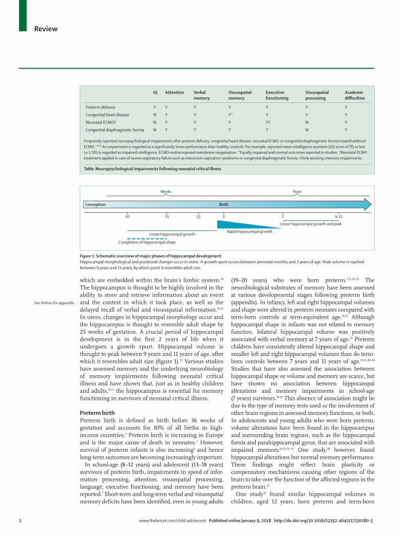
which are embedded within the brain’s limbic system.10 The hippocampus is thought to be highly involved in the ability to store and retrieve information about an event and the context in which it took place, as well as the delayed recall of verbal and visuospatial information.10,21 In utero, changes in hippocampal morphology occur and the hippocampus is thought to resemble adult shape by 25 weeks of gestation. A crucial period of hippocampal development is in the first 2 years of life when it undergoes a growth spurt. Hippocampal volume is thought to peak between 9 years and 11 years of age, after which it resembles adult size (figure 1).22 Various studies have assessed memory and the underlying neurobiology of memory impairments following neonatal critical illness and have shown that, just as in healthy children and adults,10,11 the hippocampus is essential for memory functioning in survivors of neonatal critical illness.
Preterm birthPreterm birth is defined as birth before 36 weeks of gestation and accounts for 10% of all births in high-income countries.2 Preterm birth is increasing in Europe and is the major cause of death in neonates.2 However, survival of preterm infants is also increasing2 and hence long-term outcomes are becoming increasingly important.
In school-age (8–12 years) and adolescent (13–18 years) survivors of preterm birth, impairments in speed of infor-mation processing, attention, visuospatial processing, language, executive functioning, and memory have been reported.7 Short-term and long-term verbal and visuo spatial memory deficits have been identified, even in young adults
(19–20 years) who were born preterm.7,21,23–25 The neurobiological substrates of memory have been assessed at various developmental stages following preterm birth (appendix). In infancy, left and right hippocampal volumes and shape were altered in preterm neonates compared with term-born controls at term-equivalent age.26,27 Although hippocampal shape in infants was not related to memory function, bilateral hippocampal volume was positively associated with verbal memory at 7 years of age.23 Preterm children have consistently altered hippocampal shape and smaller left and right hippo campal volumes than do term-born controls between 7 years and 11 years of age.19,23,28–30 Studies that have also assessed the association between hippocampal shape or volume and memory are scarce, but have shown no association between hippocampal alterations and memory impairments in school-age (7 years) survivors.19,29 This absence of association might be due to the type of memory tests used or the involvement of other brain regions in assessed memory functions, or both. In adolescents and young adults who were born preterm, volume alterations have been found in the hippocampus and surrounding brain regions, such as the hippocampal fornix and parahippocampal gyrus, that are associated with impaired memory.24,25,31–33 One study,28 however, found hippocampal alterations but normal memory perfor mance. These findings might reflect brain plasticity or compensatory mechanisms causing other regions of the brain to take over the function of the affected regions in the preterm brain.21
One study34 found similar hippocampal volumes in children, aged 12 years, born preterm and term-born
IQ Attention Verbal memory
Visuospatial memory
Executive functioning
Visuospatial processing
Academic difficulties
Preterm delivery Y Y Y Y Y Y Y
Congenital heart disease N Y Y Y* Y Y Y
Neonatal ECMO† N Y Y Y Y‡ N Y
Congenital diaphragmatic hernia N Y Y Y Y N Y
Frequently reported neuropsychological impairments after preterm delivery, congenital heart disease, neonatal ECMO, or congenital diaphragmatic hernia treated without ECMO.7,16–18 An impairment is regarded as a significantly lower performance than healthy controls. For example, reported mean intelligence quotient (IQ) score of 85 or less (≤–1 SD) is regarded as impaired intelligence. ECMO=extracorporeal membrane oxygenation. *Equally impaired and normal outcomes reported in studies. †Neonatal ECMO treatment applied in case of severe respiratory failure such as meconium aspiration syndrome or congenital diaphragmatic hernia. ‡Only working-memory impairments.
Table: Neuropsychological impairments following neonatal critical illness
Figure 1: Schematic overview of major phases of hippocampal developmentHippocampal morphological and positional changes occur in utero. A growth spurt occurs between perinatal months and 2 years of age. Peak volume is reached between 9 years and 11 years, by which point it resembles adult size.
10 0 2 9–1125 33
Completion of hippocampal shape
Linear hippocampal growthRapid hippocampal growth
Weeks
Conception Birth
Years
Linear hippocampal growth and peak

www.thelancet.com/child-adolescent Published online January 9, 2018 http://dx.doi.org/10.1016/S2352-4642(17)30180-3 3
Review
controls, despite poor memory outcome in preterm children. These memory impairments might be caused by alterations in other unassessed brain areas responsible for memory functioning. However, methodological issues, such as small sample size, assessment of only one type of memory, and use of two different MRI scanners within the same cohort, might also explain these contradictory results. The severity of prematurity could also affect findings as previous studies35–37 have shown positive associations between gestational age and hippocampal volume.
Congenital heart diseaseChildren with congenital heart disease who have been critically ill in the neonatal period and have had major cardiac surgery are at risk of severe neurodevelopmental problems later in life. The Boston Circulatory Arrest Trial8 assessed school-age children (8 years) and adolescents (16 years) with dextro-transposition of the great arteries (d-TGA) who had the arterial switch operation in the first year of life and found less than average academic achievement, visuospatial skills, working memory, and attention. These impairments were found even though intelligence was within normal range.8,38 A meta-analysis16 in children aged 5–8 years who had heart surgery for congenital heart disease found similar impairments in executive functioning, attention, and visuomotor integration. Survivors had lower verbal memory than did healthy controls, whereas their non-verbal memory was within normal range. In children with congenital heart disease tested 4 years after heart surgery treated with tight glucose control, worse working memory and immediate verbal memory were found compared with healthy controls.16 A study39 in children with d-TGA aged 8–16 years reported deficits in verbal and visual delayed memory. Similar deficits have also been found in adolescent and young adult survivors of congenital heart disease of differing complexity.20 A small number of studies have examined memory and its neurobiological correlates in survivors of congenital heart disease (appendix). Smaller bilateral hippocampal volumes were found in 40% of school-age children (8–16 years) who had d-TGA and cyanosis than in healthy controls. Reductions in hippocampal volume were associated with poor verbal and visuospatial memory.39 Furthermore, children aged 13 years who had cardio pulmonary bypass surgery in infancy had smaller bilateral hippocampal volumes and volume loss in other parts of the limbic system’s grey matter than did healthy controls.40
Underlying cardiac anomaly and treatment might influence neurodevelopmental outcome. For instance, adolescents who had the Fontan and Norwood procedures scored lower than the population norm on general memory, whereas patients who had a different operation had normal outcomes.15 Although assessed in a small sample size, patients with cyanotic congenital heart disease had more pronounced hippocampal volume loss
than did patients with acyanotic disease. Memory was not assessed.40
Neonatal ECMO in severe respiratory failureSince the first neonatal ECMO treatment in 1975, nearly 40 000 neonates have been treated with ECMO worldwide.41 The annual number of neonatal ECMO runs has decreased and treatment has shifted from respiratory to cardiac runs. However, the most frequent underlying diagnoses for neonatal ECMO remain meconium aspiration syndrome and congenital diaphragmatic hernia. Survival following meconium aspiration syndrome is over 90%. Congenital diaphragmatic hernia is a rare congenital anatomical malformation associated with high mortality and morbidity due to pulmonary hypoplasia and pulmonary hypertension. In the most severe cases necessitating treatment with ECMO, mortality is 49%.41 Over the past decade, standardised treatment protocols for patients with congenital heart disease have reduced mortality and the need for ECMO.42
In survivors of neonatal ECMO aged 8–12 years, extensive neuropsychological assessment has shown mainly deficits in attention, verbal, and visuospatial memory.17 Similar deficits were found in adolescent survivors, whereas other domains remained mostly unaffected.9 Memory deficits have also been found in children following severe respiratory failure as well as congenital diaphragmatic hernia18 who were not treated with ECMO.37
Studies have examined the neurobiological substrates of long-term neuropsychological outcome following severe respiratory failure with neonatal ECMO treatment (appendix). We found microstructure alterations in global white matter and regional alterations in the limbic system in neonatal ECMO survivors, aged 11 years, compared with healthy controls.43 Reductions in hippocampal volume were associated with increased delay in verbal memory in survivors of neonatal ECMO.17 White matter microstructure alterations in the parahippocampal region of the cingulum—a white matter tract connecting the medial temporal lobe with the parietal and occipital lobes—were associated with worse visuospatial and verbal memory.17 Similar structure–function relationships were found in patients with congenital heart disease not treated with ECMO.17 In line with these findings, smaller hippocampal volumes were identified in school-age (8–15 years) survivors of acute hypoxic respiratory failure, either treated with conventional ventilator management or ECMO, than in healthy controls.37 These reduced volumes were associated with impaired memory of daily events and verbal and visuospatial memory deficits.37
Hippocampal vulnerability and neonatal critical illnessMemory deficits are associated with hippocampal alterations following neonatal critical illness, irrespective of underlying diagnosis. These hippocampal alterations

4 www.thelancet.com/child-adolescent Published online January 9, 2018 http://dx.doi.org/10.1016/S2352-4642(17)30180-3
Review
are probably a result of the timing and type of insults critically ill neonates are exposed to. The brain, including the hippocampus and the rest of the limbic system, develops rapidly in the third trimester and throughout the neonatal period.44 During this period, critically ill infants are at risk of exposure to hypoxia, neuroinflammation, stress, and clinical procedures requiring general anaesthesia. The hippocampus has been found to have a selective vulnerability to these conditions associated with critical illness. Animal and in-vitro models of the hippocampus have shown that subregions of the hippocampus—CA1, CA3, the dentate gyrus, and subiculum—display differential vulnerability,11,45 which might explain why the hippocampus is affected by a wide range of conditions (figure 2).
HypoxiaCerebral hypoperfusion, hypoxaemia, or hypoperfusion with hypoxaemia resulting in hypoxia, are common complications in preterm infants or (near) term infants with congenital heart disease or severe respiratory failure (treated with neonatal ECMO). Studies using animal and in-vitro models have shown that the hippocampus displays more pronounced changes following hypoxia-ischaemia than other brain structures (reviewed by Schmidt-Kastner).47 Furthermore, differential vulnerability to hypoxia-ischaemia in the hippocampal subregions has been suggested; CA1 seems more vulnerable to acute hypoxia-ischaemia than CA3 and the dentate gyrus, in which relative sparing has been found.47 However, prolonged periods of cerebral ischaemia have been shown to damage CA3 and the dentate gyrus as well.47 Differential vulnerability within the hippocampus might result from regional differences in antioxidant enzymes, inflammatory response, or the distribution of glutamatergic N-methyl-D-aspartate (NMDA) receptors, or all of these factors.11 This variation in vulnerability might also lead to different types of memory impairments
since differential functional organisation within the hippocampal formation has been suggested as well.45 However, this mechanism needs further study in human beings.
In addition to the hippocampus, its surrounding white matter, and particularly the periventricular white matter, seems specifically susceptible to hypoxic-ischaemic insults. Animal models have identified that oxidative stress selectively affects premyelinating oligodendro-cytes in cerebral white matter. These cells account for approximately 90% of the total oligodendroglial population at 28 weeks of gestation and approximately 50% at term.48 Increased regional susceptibility of periventricular white matter is suggested to be due to the distribution of these premyelinating oligodendrocytes and relative underdevelopment of distal arterial fields in these areas.48,49 Therefore, neonates exposed to hypoxic-ischaemic injuries, even at term, might be at increased risk of myelin and axonal disruptions resulting in white matter abnormalities, which have been shown to correlate with impaired neurodevelopmental outcome.50
InflammationCritical illness is often accompanied by a marked pro-inflammatory response in the brain to underlying factors such as stress, infection, or hypoxia-ischaemia.51 The hippocampus is involved in the regulation of inflammation in the brain due to its high density of microglia, which in the (immature) central nervous system respond rapidly to infection or injury. In case of deleterious conditions, microglia in the hippocampus show a dynamic process of neuroprotective and pro-inflammatory responses. The pro-inflammatory response leads to the production of pro-inflammatory and neurotoxic factors, which have been shown to negatively affect hippocampal neurogenesis and cellular composition in the developing brain in rodent models.52 Clinical studies in neonates have shown an elevated risk of brain injury following inflammation,51 but specific effects on the hippocampus need further research. Behavioural effects of inflammatory damage to the hippocampus have been studied, though mostly through use of experimental models, showing an association between pro -inflam-matory cytokines in the hippocampus and memory impairments.52
Elevated glucocorticoid concentrationsEnvironmental stressors in the neonatal ICU have been found to elicit physiological stress responses in critically ill infants and affect brain structure and function.53 In response to stress, the brain’s hypothalamus–pituitary–adrenal (HPA) axis is activated, causing the release of cortisol into the blood by the adrenal gland. The hippocampus is a key regulator in this system by reducing HPA axis activity following stress exposure. Cortisol binds to mineralocorticoid receptors and glucocorticoid
Figure 2: Vulnerability of the hippocampus to conditions associated with neonatal critical illness. Differential vulnerability of CA1 and CA3, the dentate gyrus (DG) and subiculum (Sub) to hypoxia-ischaemia, neuroinflammation, stress, and anaesthetics is shown. Subset figure in the bottom-left corner shows the neural circuitry of the hippocampal formation. EC=entorhinal cortex. EC2=entorhinal cortex layer 2. EC3=entorhinal cortex layer 3. Adapted from Ramón y Cajal.46
Neuroinflammation
EC
Sub
DGCA3
CA1
Multimodal stress
Anaesthesia Hypoxia-ischaemia
EC2DG CA3
CA1Sub
EC3
EC deep

www.thelancet.com/child-adolescent Published online January 9, 2018 http://dx.doi.org/10.1016/S2352-4642(17)30180-3 5
Review
receptors, which are highly expressed in the hippocampus and excess cortisol causes hippocampal alterations such as hippocampal dendritic atrophy and inhibited neurogenesis.54 Animal models have shown pronounced effects of acute stress on CA1. However, in reaction to chronic or multimodal stress (eg, hours-long light and loud noise), or both, which might be more similar to experiences in the neonatal ICU, the number of synapses was reduced in CA3 leading to a decrease in object recognition memory.55 One study56 showed that stressors in the neonatal ICU were associated with altered brain microstructure and functional connectivity within the temporal lobes but not in the frontal lobe of the brain in preterm infants (born <30 weeks). Although memory function was not assessed, high stress exposure increased neurobehavioural problems at term-equivalent age.56 The mother might experience stress during pregnancy due to prenatally identified congenital anomalies or during the neonatal ICU period. Increased prenatal and postnatal maternal stress affects infants’ hippocampal growth in the first 6 months of life.54 Maternal stress exposure is thought to have programming effects on the activity of the fetal HPA axis. Increased maternal cortisol secretion might reach the fetus, increasing fetal HPA activity by reducing the number of mineralocorticoid and glucocorticoid receptors in the hippocampus.54 Although further research in human beings is required, maternal stress might contribute to increased risk of long-term memory impairments in critically ill neonates.54
Studies23,26 in preterm children have shown that postnatal treatment with dexamethasone—a cortico steroid used in preterm infants to accelerate lung development—negatively affected hippocampal morphology. However, dexamethasone might not affect children treated with neonatal ECMO and congenital heart disease to the same degree because corticosteroids are less frequently used in these patients.43
AnaestheticsPossible negative effects on the developing brain of prolonged, general anaesthesia has resulted in a warning from the US Food and Drug Administration about its use in children younger than 3 years.57 Although clinical studies in human beings are scarce and findings are mostly based on experimental studies, hippocampal development might be affected by commonly used anaesthetic agents. Commonly used anaesthetics bind either to γ-aminobutyric acid (GABA) or NMDA receptors. These receptor systems are crucial for neuronal connection and communication in the developing brain and unavailability of NMDA and GABA receptors causes neuro apoptosis.57 Various anaesthetics such as midazolam, propofol, and ketamine disrupt memory formation through their effects on the hippocampus.58 Memory formation and recall are dependent on persistent strengthening of synapses following high levels of stimulation (long-term potentiation), which mainly
happens in the hippocampus and relies heavily on NMDA. In a rat model, midazolam affected pyramidal neurons in CA1 and memory by suppressing long-term potentiation.59 However, the transferability of findings from animal models to the developing human brain is restricted60 and research in human beings and specific disease models is crucial. In very preterm neonates (24–32 weeks of gestation), high exposure to midazolam was negatively associated with hippocampal growth from birth to term-equivalent age, even when adjusted for clinical confounders including gestational age, days of mechanical ventilation, and number of surgeries;61 however, memory outcome was not assessed. Children aged 6–11 years who received general anaesthesia before 1 year of age had significantly worse memory recall than untreated controls while IQ remained unaffected.57 The association between these memory deficits and altered hippocampal morphology was not assessed.
A common neurodevelopmental pathway following neonatal critical illnessIn this Review we present evidence to suggest a shared vulnerability of the hippocampus that is associated with long-term memory impairments in critically ill neonates. On the basis of this evidence, we speculate that a final common neurodevelopmental pathway exists following neonatal critical illness (figure 3). The patient groups described in this Review have varying brain development trajectories due to variations in illness onset (eg, congenital anomaly developing in utero vs postnatal sepsis necessitating neonatal ECMO), sex, and gestational age. These factors are likely to interact with exposure to harmful conditions associated with neonatal critical illness and might influence how and when the hippocampus is affected. The exact pathophysiological mechanisms of hippocampal alterations in each of these patient groups remains unknown and needs further research.
Clinical implications and future directionsEarly risk predictionIdentification of patients at risk of academic problems relies solely on neuro psychological assessment. However, higher order cognitive functions, such as memory, cannot be reliably evaluated until about 8 years of age.7 Patients at risk of academic problems are often identified too late because neuropsychological deficits, which might have remained unidentified or unspecified, could have already led to academic problems. Neuro-psychological assessment should therefore be primarily used as a diagnostic tool, instead of a prediction tool, and patients at risk should be identified before academic difficulties emerge. Therefore, early predictors of long-term memory impairments, preferably measurable in infancy, are needed.
Alterations in hippocampal volume, if detected in infancy, could be a predictor of impaired long-term memory. MRI is a non-invasive neuroimaging technique

6 www.thelancet.com/child-adolescent Published online January 9, 2018 http://dx.doi.org/10.1016/S2352-4642(17)30180-3
Review
and thus a useful tool to assess the hippocampus in infants. Findings in preterm infants have shown that MRI can be reliably done without sedation, and infant hippocampal volumes (measured at term-equivalent age) correlate with memory outcomes at 2 years and 7 years of age.23,27 Future studies are needed to identify cutoff points to separate hippocampal volumes within and outside the normal range in infancy through use of a healthy control group. Furthermore, differences in timing and type of hippocampal injury are likely to exist across the population of critically ill infants because of differences in brain development associated with different causes of disease. For example, brain alterations have been found as early as in the third trimester in patients with congenital heart disease5 and the immature preterm brain might respond differently to the term brain to the adverse conditions associated with neonatal critical illness.49 Longitudinal studies are therefore imperative to accurately delineate the trajectories of longitudinal hippocampal growth across these patient groups. Since hippocampal growth is rapid during the first 2 years of life,22 longitudinal studies will help to determine the best time to assess hippocampal morphology as an early predictor of memory function.
The hippocampus has been primarily quantified through assessment of volumes using structural MRI. This method is robust and useful to accurately parcellate hippocampal volume. Future studies should focus on the details of hippocampal parcellation by obtaining high resolution images and multi-contrast imaging, or using alternative modalities such as diffusion imaging. These studies might contribute to a better understanding of the differential vulnerability of the hippocampal subregions and, if combined with neuropsychological assessment, their suggested differential involvement in various memory processes.11,45 However, MRI does not provide
information on the exact anatomical or molecular mechanisms underlying hippocampal alterations. Therefore, the specific contribution of the risk factors outlined in this Review to hippocampal alterations and long-term memory deficits remains speculative.
Hypoxia consistently affects hippocampal morphology. The severity of cerebral hypoperfusion sustained in the perinatal period might be another risk factor of long-term memory problems following neonatal critical illness. Adequate ways to monitor cerebral metabolism, haemodynamics, and autoregulation in the neonatal ICU are urgently needed since existing methods, such as transcranial Doppler ultrasonography or near-infrared spectroscopy, do not have sufficient resolution to target the brain region of interest in this context.
Treatment opportunities Although the hippocampus is a highly vulnerable brain structure, it has also been shown to exhibit more plasticity and capability of long-term neurogenesis than other brain structures.11 These properties make it a promising target for intervention or treatment to improve long-term memory following neonatal critical illness.
Another group of critically ill neonates at high risk of hypoxic-ischaemic injuries are survivors of perinatal asphyxia. By contrast with the patient groups described in this Review, large numbers of neonates with perinatal asphyxia also have overt chronic neurological abnormalities and severe long-term morbidities, such as cerebral palsy and intellectual disability.62 Whole body therapeutic hypothermia is standard care in full-term neonates with perinatal asphyxia and has been shown to improve neurological outcomes.63 Using animal and in-vitro models, mild to moderate therapeutic hypothermia has been shown to reduce hippocampal cell death following hypoxic or ischaemic insult.64 Although patients with perinatal asphyxia experience an acute phase of hypoxia,65 prolonged exposure to hypoxia might exist in the patient groups described in this Review. Furthermore, hypothermia probably does not protect the hippocampus against other types of harmful conditions associated with critical illness. Future randomised trials are needed to assess if and to what extent therapeutic hypothermia affects the hippocampus and memory in these patient groups before it can be recommended as a routine neuroprotective strategy.
As discussed, stress might negatively affect the hippocampus and memory.54 Reduction of stress exposure during their stay in the neonatal ICU could therefore be beneficial for critically ill infants and feasible in clinical practice. Decreased light and sound in the unit and encouragement of physical parent–infant contact during hospital stay can positively affect brain development.66 Future studies assessing whether such measures of stress reduction specifically influence hippocampal development and memory improvements in survivors of neonatal critical illness are needed.
Figure 3: A common neurodevelopmental pathway following neonatal critical illnessSurvivors of neonatal critical illness share an increased risk of hippocampal alterations due to vulnerability to common conditions associated with neonatal critical illness, which leads to long-term memory deficits. ECMO=extracorporeal membrane oxygenation.
Hypoxia-ischaemia
Inflammation
Glucocorticoidconcentration
Anaesthesia
Preterm birth | Congenital heart disease | Neonatal ECMOCongenital diaphragmatic hernia |
Rapid development of limbic system
Hippocampal alterations
Memory deficitsLearning problems
Hippocampal vulnerability

www.thelancet.com/child-adolescent Published online January 9, 2018 http://dx.doi.org/10.1016/S2352-4642(17)30180-3 7
Review
Studies evaluating brain training or computerised cognitive training programmes have increased over the past decade; however, the effectiveness of this approach remains controversial. Cognitive training programmes are based on the idea that repetitive mental exercise of one cognitive task will improve functioning that might generalise to other tasks with a similar underlying system. Cogmed working-memory training is a cognitive training programme for children and adults that has been widely evaluated.67 Studies have consistently shown improvements in the trained verbal and visuospatial working-memory skills, but have less consistently shown improvements in untrained functions such as delayed memory recall.68 Working memory, the target function of Cogmed, relies primarily on frontal-parietal networks.67 How or whether Cogmed influences the plastic nature of the hippocampus is unclear. Neuroimaging studies assessing the exact effects of Cogmed on hippocampal function and different types of memory are required in survivors of neonatal critical illness. Randomised trials are being done with school-age (8–12 years) survivors of neonatal ECMO (trial number NTR4571), and children and adolescents with congenital heart disease (trial numbers NCT03023644 and NCT02759263). Other types of cognitive training programmes used specifically for memory rehabilitation might be effective but require further research in survivors of neonatal critical illness.69
Identification of these survivors that have substantial memory deficits, and are thus in need of treatment, is an important part of the effective use of cognitive inter-ventions. Although early identification of these patients is ideal, identification of children with memory deficits remains reliant on neuropsychological assessment from 8 years of age. Neuropsychological assessment is essential in which, in addition to intelligence, specific neuro psychological outcomes are the primary focus following neonatal critical illness.
Exercise might enhance memory and learning by targeting the hippocampus. Good aerobic fitness has been associated with hippocampal volume as well as improvements in memory in children.70 Whether improvements persist in the long term remains largely unknown and needs further study in survivors of neonatal critical illness.
ConclusionsIn this Review, we propose a common neuro develop-mental pathway following neonatal critical illness; survivors of preterm birth, congenital heart disease, and severe respiratory failure share an increased risk of long-term memory deficits and related hippo campal alterations. Rather than a consequence of underlying diagnosis, we suggest shared vulnerability is related to common complications or conditions associated with neonatal critical illness including hypoxia, neuro-inflammation, stress, exposure to anaesthetics, or a complex interplay of these factors at different
postconceptional ages. Our findings underline the need to broaden focus from prevention of mortality to long-term outcome in critically ill infants. Follow-up of this patient population should incorporate standardised assessment of specific neuropsychological functions, such as memory, from 8 years of age. Early identification of patients at risk might be feasible by use of non-invasive structural MRI to assess infant hippocampal volumes. Future prospective studies on memory rehabilitation with the use of exercise or cognitive training are needed in survivors of neonatal critical illness. Increased awareness of the vulnerability of the hippocampus and memory deficits following neonatal critical illness is crucial to prepare survivors for potential future academic difficulties and full societal participation.ContributorsRS searched the literature and drafted the initial manuscript. RS, HI, AH, TW, FV, AvH, and DT contributed equally to the conception and design of the study and revised the manuscript critically for important intellectual content. All authors gave final approval for the manuscript to be published and agreed to be accountable for all aspects of the work.
Declaration of interestsFV publishes Dutch translations of the Achenbach System of Empirically Based Assessment for which he receives renumeration. AH is a co-investigator in a study on selection, definition, and evaluation of important early morbidities associated with paediatric cardiac surgery, funded by the National Institute of Health Research, Health Services Delivery and Organisation UK, to Great Ormond Street Hospital for Children’s NHS Foundation Trust. This study will not be completed until 2018. All other authors declare no competing interests.
AcknowledgmentsRS was funded by the Sophia Stichting Wetenschappelijk Onderzoek (SSWO): S14–21.
References1 Harrison W, Goodman D. Epidemiologic trends in neonatal
intensive care, 2007–2012. JAMA Pediatr 2015; 169: 855–62.
Search strategy and selection criteria
We searched PubMed for published articles in English between Jan 1, 2000, and June 29, 2017, using the search terms in the title or abstract: (“brain imaging” OR “brain” OR “neuroimaging” OR “magnetic resonance imaging” OR MR* OR hippocamp* OR “limbic”) AND (“memory” OR “learning”) AND ([“preterm” OR “preterm birth” OR “premature birth”] OR [“congenital heart disease” OR complex heart anomal* OR “complex heart disease”] OR [“neonatal respiratory failure” OR “acute respiratory failure” OR “neonatal extracorporeal membrane oxygenation” OR “neonatal ECMO”]). We reduced 348 references identified from this search to 258 by restricting findings to human studies. Studies that did not assess the hippocampus specifically or did not assess memory, or both, and studies including patients with severe neurological abnormalities or genetic syndromes known to affect neurodevelopmental outcome, were excluded. Searches were supplemented by hand searching of reference lists of identified articles. The final reference list was generated on the basis of relevance to the scope of this Review. In total, 27 studies were included.

8 www.thelancet.com/child-adolescent Published online January 9, 2018 http://dx.doi.org/10.1016/S2352-4642(17)30180-3
Review
2 Zeitlin J, Mohangoo AD, Delnord M, Cuttini M, EURO-PERISTAT Scientific Committee. The second European Perinatal Health Report: documenting changes over 6 years in the health of mothers and babies in Europe. J Epidemiol Community Health 2013; 67: 983–85.
3 van Heijst AF, de Mol AC, Ijsselstijn H. ECMO in neonates: neuroimaging findings and outcome. Semin Perinatol 2014; 38: 104–13.
4 Hamrick SE, Miller SP, Leonard C, et al. Trends in severe brain injury and neurodevelopmental outcome in premature newborn infants: the role of cystic periventricular leukomalacia. J Pediatr 2004; 145: 593–99.
5 Khalil A, Bennet S, Thilaganathan B, Paladini D, Griffiths P, Carvalho JS. Prevalence of prenatal brain abnormalities in fetuses with congenital heart disease: a systematic review. Ultrasound Obstet Gynecol 2016; 48: 296–307.
6 Schiller RM, Madderom MJ, Reuser JJ, et al. Neuropsychological follow-up after neonatal ECMO. Pediatrics 2016; 138: e20161313.
7 Anderson PJ. Neuropsychological outcomes of children born very preterm. Semin Fetal Neonatal Med 2014; 19: 90–96.
8 Bellinger DC, Wypij D, duPlessis AJ, et al. Neurodevelopmental status at eight years in children with dextro-transposition of the great arteries: the Boston Circulatory Arrest Trial. J Thorac Cardiovasc Surg 2003; 126: 1385–96.
9 Madderom MJ, Schiller RM, Gischler SJ, et al. Growing up after critical illness: verbal, visual-spatial, and working memory problems in neonatal extracorporeal membrane oxygenation survivors. Crit Care Med 2016; 44: 1182–90.
10 Squire LR. Memory and brain systems: 1969–2009. J Neurosci 2009; 29: 12711–16.
11 Bartsch T, Wulff P. The hippocampus in aging and disease: from plasticity to vulnerability. Neuroscience 2015; 309: 1–16.
12 Back SA, Riddle A, McClure MM. Maturation-dependent vulnerability of perinatal white matter in premature birth. Stroke 2007; 38 (suppl 2): 724–30.
13 Rourke BP, Bakker DJ, Fisk JL, Strang JD. Child neuropsychology. An introduction to theory, research, and clinical practice. New York: The Guilford Press, 1983.
14 Madderom MJ, Reuser JJ, Utens EM, et al. Neurodevelopmental, educational and behavioral outcome at 8 years after neonatal ECMO: a nationwide multicenter study. Intensive Care Med 2013; 39: 1584–93.
15 Bellinger DC, Watson CG, Rivkin MJ, et al. Neuropsychological status and structural brain imaging in adolescents with single ventricle who underwent the fontan procedure. J Am Heart Assoc 2015; 4: e002302.
16 Sterken C, Lemiere J, Vanhorebeek I, Van den Berghe G, Mesotten D. Neurocognition after paediatric heart surgery: a systematic review and meta-analysis. Open Heart 2015; 2: e000255.
17 Schiller RM, IJsselstijn H, Madderom MJ, et al. Neurobiological correlates of attention and memory deficits following critical illness in early life. Crit Care Med 2017: 45: 1742–50.
18 Leeuwen L, Schiller RM, Rietman AB, et al. Risk factors of impaired neuropsychologic outcome in school-aged survivors of neonatal critical illness. Crit Care Med 2017; published online Nov 30. DOI:10.1097/CCM.0000000000002869.
19 Omizzolo C, Thompson DK, Scratch SE, et al. Hippocampal volume and memory and learning outcomes at 7 years in children born very preterm. J Int Neuropsychol Soc 2013; 19: 1065–75.
20 Pike NA, Woo MA, Poulsen MK, et al. Predictors of memory deficits in adolescents and young adults with congenital heart disease compared to healthy controls. Front Pediatr 2016; 4: 117.
21 Nosarti C, Froudist-Walsh S. Alterations in development of hippocampal and cortical memory mechanisms following very preterm birth. Dev Med Child Neurol 2016; 58: 35–45.
22 Uematsu A, Matsui M, Tanaka C, et al. Developmental trajectories of amygdala and hippocampus from infancy to early adulthood in healthy individuals. PLoS One 2012; 7: e46970.
23 Thompson DK, Adamson C, Roberts G, et al. Hippocampal shape variations at term equivalent age in very preterm infants compared with term controls: perinatal predictors and functional significance at age 7. Neuroimage 2013; 70: 278–87.
24 Aanes S, Bjuland KJ, Skranes J, Løhaugen GC. Memory function and hippocampal volumes in preterm born very-low-birth-weight (VLBW) young adults. Neuroimage 2015; 105: 76–83.
25 Nosarti C, Nam KW, Walshe M, et al. Preterm birth and structural brain alterations in early adulthood. Neuroimage Clin 2014; 6: 180–91.
26 Thompson DK, Wood SJ, Doyle LW, et al. Neonate hippocampal volumes: prematurity, perinatal predictors, and 2-year outcome. Ann Neurol 2008; 63: 642–51.
27 Beauchamp MH, Thompson DK, Howard K, et al. Preterm infant hippocampal volumes correlate with later working memory deficits. Brain 2008; 131: 2986–94.
28 Brunnemann N, Kipp KH, Gortner L, et al. Alterations in the relationship between hippocampal volume and episodic memory performance in preterm children. Dev Neuropsychol 2013; 38: 226–35.
29 Thompson DK, Omizzolo C, Adamson C, et al. Longitudinal growth and morphology of the hippocampus through childhood: impact of prematurity and implications for memory and learning. Hum Brain Mapp 2014; 35: 4129–39.
30 Peterson BS, Vohr B, Staib LH, et al. Regional brain volume abnormalities and long-term cognitive outcome in preterm infants. JAMA 2000; 284: 1939–47.
31 Salvan P, Froudist Walsh S, Allin MP, et al. Road work on memory lane—functional and structural alterations to the learning and memory circuit in adults born very preterm. Neuroimage 2014; 102: 152–61.
32 Gimenez M, Junque C, Narberhaus A, et al. Hippocampal gray matter reduction associates with memory deficits in adolescents with history of prematurity. Neuroimage 2004; 23: 869–77.
33 Isaacs EB, Lucas A, Chong WK, et al. Hippocampal volume and everyday memory in children of very low birth weight. Pediatr Res 2000; 47: 713–20.
34 Fraello D, Maller-Kesselman J, Vohr B, et al. Consequence of preterm birth in early adolescence: the role of language on auditory short-term memory. J Child Neurol 2011; 26: 738–42.
35 Brumbaugh JE, Conrad AL, Lee JK, et al. Altered brain function, structure, and developmental trajectory in children born late preterm. Pediatr Res 2016; 80: 197–203.
36 Ball G, Boardman JP, Rueckert D, et al. The effect of preterm birth on thalamic and cortical development. Cereb Cortex 2012; 22: 1016–24.
37 Cooper JM, Gadian DG, Jentschke S, et al. Neonatal hypoxia, hippocampal atrophy, and memory impairment: evidence of a causal sequence. Cereb Cortex 2015; 25: 1469–76.
38 Bellinger DC, Wypij D, Rivkin MJ, et al. Adolescents with d-transposition of the great arteries corrected with the arterial switch procedure: neuropsychological assessment and structural brain imaging. Circulation 2011; 124: 1361–69.
39 Munoz-Lopez M, Hoskote A, Chadwick MJ, et al. Hippocampal damage and memory impairment in congenital cyanotic heart disease. Hippocampus 2017; 27: 417–24.
40 von Rhein M, Buchmann A, Hagmann C, et al. Brain volumes predict neurodevelopment in adolescents after surgery for congenital heart disease. Brain 2014; 137: 268–76.
41 Extracorporeal Life Support Organization. ECLS Registry Report International Summary January, 2017. https://www.elso.org/Portals/0/Files/Reports/2017/International%20Summary%20January%202017.pdf (accessed May 20, 2017).
42 Snoek KG, Reiss IK, Greenough A, et al. Standardized postnatal Management of infants with congenital diaphragmatic hernia in Europe: the CDH EURO Consortium Consensus—2015 Update. Neonatology 2016; 110: 66–74.
43 Schiller RM, van den Bosch GE, Muetzel RL, et al. Neonatal critical illness and development: white matter and hippocampus alterations in school-age neonatal extracorporeal membrane oxygenation survivors. Dev Med Child Neurol 2017; 59: 304–10.
44 Dubois J, Dehaene-Lambertz G, Kulikova S, Poupon C, Huppi PS, Hertz-Pannier L. The early development of brain white matter: a review of imaging studies in fetuses, newborns and infants. Neuroscience 2014; 276: 48–71.
45 Small SA, Schobel SA, Buxton RB, Witter MP, Barnes CA. A pathophysiological framework of hippocampal dysfunction in ageing and disease. Nat Rev Neurosci 2011; 12: 585–601.
46 Ramón y Cajal, S. Histologie du système nerveux de l’homme et des vertébrés, vol 1 and 2. Paris: Maloine, 1909 (in French).
47 Schmidt-Kastner R. Genomic approach to selective vulnerability of the hippocampus in brain ischemia-hypoxia. Neuroscience 2015; 309: 259–79.

www.thelancet.com/child-adolescent Published online January 9, 2018 http://dx.doi.org/10.1016/S2352-4642(17)30180-3 9
Review
48 Volpe JJ, Kinney HC, Jensen FE, Rosenberg PA. The developing oligodendrocyte: key cellular target in brain injury in the premature infant. Int J Dev Neurosci 2011; 29: 423–40.
49 Back SA. Cerebral white and gray matter injury in newborns: new insights into pathophysiology and management. Clin Perinatol 2014; 41: 1–24.
50 Ferriero DM, Miller SP. Imaging selective vulnerability in the developing nervous system. J Anat 2010; 217: 429–35.
51 Hagberg H, Mallard C, Ferriero DM, et al. The role of inflammation in perinatal brain injury. Nat Rev Neurol 2015; 11: 192–208.
52 Green HF, Nolan YM. Inflammation and the developing brain: consequences for hippocampal neurogenesis and behavior. Neurosci Biobehav Rev 2014; 40: 20–34.
53 Peng NH, Bachman J, Jenkins R, et al. Relationships between environmental stressors and stress biobehavioral responses of preterm infants in NICU. J Perinat Neonatal Nurs 2009; 23: 363–71.
54 Lupien SJ, McEwen BS, Gunnar MR, Heim C. Effects of stress throughout the lifespan on the brain, behaviour and cognition. Nat Rev Neurosci 2009; 10: 434–45.
55 McEwen BS, Nasca C, Gray JD. Stress effects on neuronal structure: hippocampus, amygdala, and prefrontal cortex. Neuropsychopharmacology 2016; 41: 3–23.
56 Smith GC, Gutovich J, Smyser C, et al. Neonatal intensive care unit stress is associated with brain development in preterm infants. Ann Neurol 2011; 70: 541–49.
57 Andropoulos DB. Effect of anesthesia on the developing brain: infant and fetus. Fetal Diagn Ther 2017; published online June 7. DOI:10.1159/000475928.
58 Haiying G, Mingjie H, Lingyu Z, Qingxiang W, Haisong W, Bingxi Z. Anesthetics inhibit extracellular signal-regulated Kinase1/2 phosphorylation via NMDA receptor, phospholipase C and protein kinase C in mouse hippocampal slices. Neurochem Int 2017; 103: 36–44.
59 Tokuda K, O’Dell KA, Izumi Y, Zorumski CF. Midazolam inhibits hippocampal long-term potentiation and learning through dual central and peripheral benzodiazepine receptor activation and neurosteroidogenesis. J Neurosci 2010; 30: 16788–95.
60 Disma N, Mondardini MC, Terrando N, Absalom AR, Bilotta F. A systematic review of methodology applied during preclinical anesthetic neurotoxicity studies: important issues and lessons relevant to the design of future clinical research. Paediatr Anaesth 2016; 26: 6–36.
61 Duerden EG, Guo T, Dodbiba L, et al. Midazolam dose correlates with abnormal hippocampal growth and neurodevelopmental outcome in preterm infants. Ann Neurol 2016; 79: 548–59.
62 Vannucci RC. Hypoxic-ischemic encephalopathy. Am J Perinatol 2000; 17: 113–20.
63 Azzopardi D, Brocklehurst P, Edwards D, et al. The TOBY Study. Whole body hypothermia for the treatment of perinatal asphyxial encephalopathy: a randomised controlled trial. BMC Pediatr 2008; 8: 17.
64 Gregersen M, Lee DH, Gabatto P, Bickler PE. Limitations of mild, moderate, and profound hypothermia in protecting developing hippocampal neurons after simulated ischemia. Ther Hypothermia Temp Manag 2013; 3: 178–88.
65 de Vries LS, Jongmans MJ. Long-term outcome after neonatal hypoxic-ischaemic encephalopathy. Arch Dis Child Fetal Neonatal Ed 2010; 95: F220–24.
66 Vandenberg KA. Individualized developmental care for high risk newborns in the NICU: a practice guideline. Early Hum Dev 2007; 83: 433–42.
67 Klingberg T. Training and plasticity of working memory. Trends Cogn Sci 2010; 14: 317–24.
68 Melby-Lervag M, Redick TS, Hulme C. Working memory training does not improve performance on measures of intelligence or other measures of “far transfer”: evidence from a meta-analytic review. Perspect Psychol Sci 2016; 11: 512–34.
69 Piras F, Borella E, Incoccia C, Carlesimo GA. Evidence-based practice recommendations for memory rehabilitation. Eur J Phys Rehabil Med 2011; 47: 149–75.
70 Chaddock L, Erickson KI, Prakash RS, et al. A neuroimaging investigation of the association between aerobic fitness, hippocampal volume, and memory performance in preadolescent children. Brain Res 2010; 1358: 172–83.