Department of Biomedical Physics & Technology University ...
MEDICAL PHYSICS AND BIOMEDICAL
ENGINEERING AT THE FACULTY OF MEDICINE,
MASARYK UNIVERSITY, BRNO
Daniel Vlk*, Vojtech Mornstein* and Carmel J. Caruana**
*Department of Biophysics, Medical Faculty, Masaryk University, Brno, Czech Republic
**Biomedical Physics, Institute of Health Care, University of Malta, Malta
Friday, Oct 15

Introduction We teach students of three different faculties, who represent several
healthcare professionals. About one fifth of the General and DentalMedicine students are enrolled in the English study programme. Thebiggest group involves students of General Medicine, followed by otherhealthcare professional groups from the Medical Faculty. A small groupof students comes from the Biophysics and Medical Physics programmesof the Faculty of Science. Since the academic year 2008/2009 we alsostarted education of students from the newly designed BSc programme“Biomedical Technology and Bioinformatics” of the Faculty of ElectricalEngineering and Communication (FEEC), University of Technology, Brno.
We have prepared individual syllabi for students for the differentprogrammes. We focus on the effective and safe use of biomedical devices(students of Medical Faculty), medical and technical aspects of biomedicalphysics (students of Medical Physics and Biophysics from the Facultyof Science) and general biophysics (students from FEEC).

Students at the department of Biophysics of the Faculty of Medicine,
Masaryk University, Brno
Faculty Specialisation Number of students
General Medicine 480
Dentistry 90
Nurse – midwife 30
General Nurse 40
Physiotherapy 40
Optometrics 40
Human Nutrition 20
Laboratory Assistant 50
Radiology Assistant 10
Biophysics 15
Medical physics 8
70
Faculty of MedicineMasaryk
University, Brno
Faculty of Sciences, Masaryk
University, Brno
FEEC, Technical University, BrnoBiomedical Technology and
Bioinformatics

Biomedical physics and other theoretical subjects
Theoretical subject
G M Dentistry BTB
Medical physics 105 (1) 90 (1) 75 (1)
Chemistry and
Biochemistry225 (3) 165 (3) 60 (1)
Biology 135 (2) 90 (2) 60 (1)
Anatomy 240 (3) 195 (3) 30 (1)
Physiology 232 (2) 120 (2) 75 (1)
Histology and
Embryology150 (2) 105 (2) 30 (1)
Study programme
Hours per semester (number of semesters)
In comparison with other biomedical subjects, biomedical physics
has less curriculum time in the case of General Medicine and
Dentistry programmes. However, it has a higher number of hours in
the case of the programme in Biomedical Technology.

Practical tasksThe practical exercises usually represent about 60% of the complete course of
biomedical physics. Moreover, practical exercises (tasks) include also an introduction to
healthcare informatics. The time available for practical tasks oriented on biomedical
physics proper is the same for GM, Dentistry and BTB – 45 hours. The intended
learning objectives of the practical sessions are: the development of the measurement
and instrument user skills, the awareness of the scientific, effective, efficient and safe
use of biomedical devices, basics of the biophysical quantities behind clinical
diagnostics and the fundamentals of medical imaging and physiological monitoring
techniques. One of the target learning outcomes of the practical tasks is for all
healthcare professions to share a common conceptual understanding and unified
terminology regarding medical devices. Our syllabus is based on the theoretical model
which considers Biomedical Physics as a discipline that creates a bridge between
physics, technology and medicine.
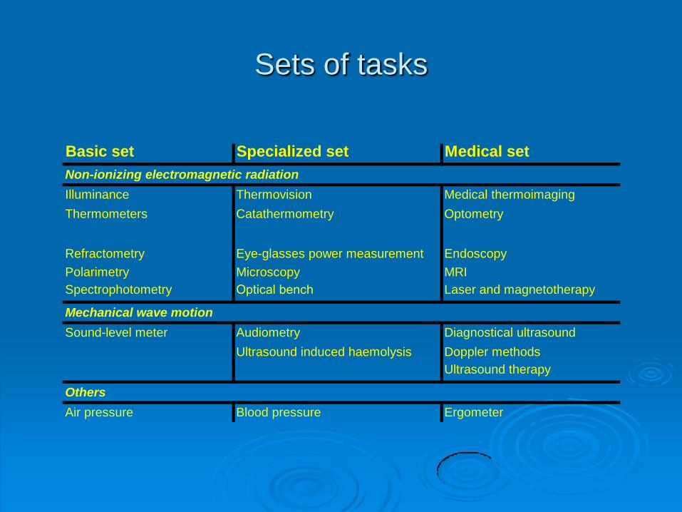
Sets of tasks
Basic set Specialized set Medical set
Illuminance Thermovision Medical thermoimaging
Thermometers Catathermometry Optometry
Polarimetry Microscopy MRI
Spectrophotometry Optical bench Laser and magnetotherapy
Sound-level meter Audiometry Diagnostical ultrasound
Ultrasound induced haemolysis Doppler methods
Ultrasound therapy
Air pressure Blood pressure Ergometer
Others
Mechanical wave motion
Non-ionizing electromagnetic radiation
Refractometry Eye-glasses power measurement Endoscopy
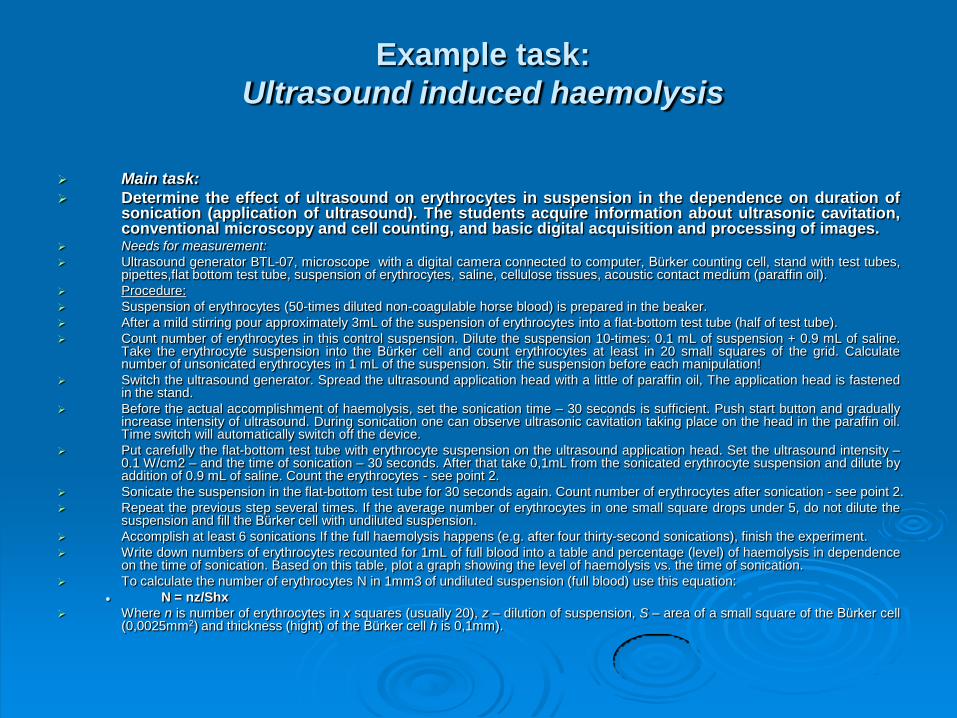
Example task:
Ultrasound induced haemolysis
Main task:
Determine the effect of ultrasound on erythrocytes in suspension in the dependence on duration ofsonication (application of ultrasound). The students acquire information about ultrasonic cavitation,conventional microscopy and cell counting, and basic digital acquisition and processing of images.
Needs for measurement:
Ultrasound generator BTL-07, microscope with a digital camera connected to computer, Bürker counting cell, stand with test tubes,pipettes,flat bottom test tube, suspension of erythrocytes, saline, cellulose tissues, acoustic contact medium (paraffin oil).
Procedure:
Suspension of erythrocytes (50-times diluted non-coagulable horse blood) is prepared in the beaker.
After a mild stirring pour approximately 3mL of the suspension of erythrocytes into a flat-bottom test tube (half of test tube).
Count number of erythrocytes in this control suspension. Dilute the suspension 10-times: 0.1 mL of suspension + 0.9 mL of saline.Take the erythrocyte suspension into the Bürker cell and count erythrocytes at least in 20 small squares of the grid. Calculatenumber of unsonicated erythrocytes in 1 mL of the suspension. Stir the suspension before each manipulation!
Switch the ultrasound generator. Spread the ultrasound application head with a little of paraffin oil, The application head is fastenedin the stand.
Before the actual accomplishment of haemolysis, set the sonication time – 30 seconds is sufficient. Push start button and graduallyincrease intensity of ultrasound. During sonication one can observe ultrasonic cavitation taking place on the head in the paraffin oil.Time switch will automatically switch off the device.
Put carefully the flat-bottom test tube with erythrocyte suspension on the ultrasound application head. Set the ultrasound intensity –0.1 W/cm2 – and the time of sonication – 30 seconds. After that take 0,1mL from the sonicated erythrocyte suspension and dilute byaddition of 0.9 mL of saline. Count the erythrocytes - see point 2.
Sonicate the suspension in the flat-bottom test tube for 30 seconds again. Count number of erythrocytes after sonication - see point 2.
Repeat the previous step several times. If the average number of erythrocytes in one small square drops under 5, do not dilute thesuspension and fill the Bürker cell with undiluted suspension.
Accomplish at least 6 sonications If the full haemolysis happens (e.g. after four thirty-second sonications), finish the experiment.
Write down numbers of erythrocytes recounted for 1mL of full blood into a table and percentage (level) of haemolysis in dependenceon the time of sonication. Based on this table, plot a graph showing the level of haemolysis vs. the time of sonication.
To calculate the number of erythrocytes N in 1mm3 of undiluted suspension (full blood) use this equation:
N = nz/Shx
Where n is number of erythrocytes in x squares (usually 20), z – dilution of suspension, S – area of a small square of the Bürker cell(0,0025mm2) and thickness (hight) of the Bürker cell h is 0,1mm).

Ultrasound generator Microscope connected with computer

Student´s responsibility
The practical exercises in Biophysics are compulsory. Theoreticalknowledge of the principles of the methods used is regularlychecked by oral examination. For each task the students have towrite a comprehensive report which is marked. At the end of thecourse students sit for a multiple-choice test which consists usuallyof 20 questions and must achieve 50% of the mark to be consideredsuccessful.
Students will only be allowed to sit for the final exam if successful inthe multiple choice test. The exam consists of a written testconsisting of 25 multiple choice questions and an oral part. Thestudent can continue with the oral part only when the number ofcorrectly answered questions in the multiple choice test is at least 15.The oral part consists of two questions. The examined student mustbe able to explain physical and biophysical problems and explaintheir clinical importance.
Acknowledgements
This contribution is supported by a development project
of the Czech Ministry of Education, Youth and Sports
entitled “C42 - Inter-university co-operation in biomedical
techniques and biomedical engineering using state-of-
the-art technologies”.