Medical Management of VAD Patients - UCSF CME. Liviu Klein.pdf · Medical Management of VAD...
32
Medical Management of VAD Patients Liviu Klein MD, MS Assistant Professor of Medicine Associate Director, Mechanical Circulatory Support and Heart Failure Device Program University of California San Francisco
Transcript of Medical Management of VAD Patients - UCSF CME. Liviu Klein.pdf · Medical Management of VAD...

Medical Management of VAD Patients
Liviu Klein MD, MSAssistant Professor of Medicine
Associate Director, Mechanical Circulatory Support and Heart Failure Device Program
University of California San Francisco

Relevant Financial Relationship Disclosure Statement
Medical Management of VAD Patients: Liviu Klein, MD, MS
I will discuss off label use and/or investigational use of products.
The following relevant financial relationships exist: None.

Prevalence of Heart Failure
Roger VL et al. Circulation. 2012;125:188-197.

Current Estimate of Stage D Patients
Miller LW et al. Circulation. 2011;123:1552-1558.
310 million US population
2% with HF => ~ 6 million
55% with DHF ~ 3-3.5 million
45% with SHF~ 2.5-3 million
35% NYHA class I35% NYHA class II
20% NYHA class IIIA5% NYHA class IIIB5% NYHA class IV
NYHA class IIIB/ IV and < 75 years
~ 150,000 ptsVADs asBTT/ DT

• Pulsatile flow with valve volume deplascement (Heart Mate XVE, Novacor)
• Continuous axial flow (Heart Mate II)
• Continuous centrifugal flow (HeartWare)
Longterm Assist Devices

Longterm Assist Devices
Baughman KL et al. N Engl J Med. 2007;357:846-849.

Impact on Organ Function
Russell SD et al. Circulation. 2009;120:2352-2357.
• 309 pts in HMII BTT and CAP
• Baseline:– ~ 50 yo– EF 16%, LVEDD
70 mm– 89% on inotrope– 44% on IABP– CI 2.1, PCW 25

QOL and Functional Capacity
Rogers JG et al. J Am Coll Cardiol. 2010;55:1826-1834.

KCCQ
Better QoL / Best Health
Status
Worst QoL/
Health Status
6 Min WalkClinical Summary │ Overall Summary
All paired differences p<0.001
EQ-5D VAS
Δ = +77%Δ = +106%
Δ = +91%
Δ = +106%
QOL and Functional Capacity
Aaronson KD et al. Circulation. 2012;125:3191-3200.

VAD Physiology
Frazier OH et al. Circulation. 2002;105:2855-2860.
• VAD pulls blood from LV -> pumps in ascend Ao
• RV functions to fill the VAD
• LV becomes giant atrium
VAD Physiology: Heart Mate II• Power: amount of energy required to rotate the
impeller (watts) – measured.• RPM: speed of the impeller – set by operator.
• Flow: pump output (L/min) – assumed, not measured.• Pulsatility index (PI): indirect measure of contribution
of native ventricular contraction to VAD filling –assumed, not measured.

Physiologic Goals for VAD Function• Maximize output (days)• Reduce LV filling pressures (days)• Decrease LV size (over time)• Reduce PA pressures (over time)
• Avoid suction events
• Keep aortic valve

Physiologic Goals for VAD Function• Maximize output (days)• Reduce LV filling pressures (days)• Decrease LV size (over time)• Reduce PA pressures (over time)
• Avoid suction events
• Keep aortic valve open/ closed/ partially open?

VAD Knowhow• Low end of speed
– AoV opens with every beat
– No HF symptoms
• High end of speed– Septal flattening– AoV closed
• Optimal speed– Mid point– Intermittent AoV opening
Slaughter MS et al. J Heart Lung Transplant. 2010;29:S1-39.

Adverse Events with VADs
Slaughter MS et al. N Engl J Med. 2009;361:2241-2251.
Anticoagulation
Infections
RV Failure
Arrhythmias
Hypertension

RV Failure: Identification/ Avoidance• Pre implant risk prediction of RV failure is difficult.• RV failure post VAD implant:
– Higher length of stay, higher ICU stay.– End organ dysfunction, higher mortality.
• Measures of RV function:– Echo: RV size, fractional area change, RVESD, TAPSE– Cath: CVP, PAP, RVSWI [(PAM-CVP) x SVI]
• Multivariate models of risk prediction of post op RV failure/ need for RVAD (Michigan, Penn).

RV Failure: Avoidance
Slaughter MS et al. J Heart Lung Transplant. 2010;29:S1-39.

RV Failure: Identification• Clinical diagnosis:
– Hypotension, low urine output, high JVP
• Hemodynamics:– Poor VAD flows (< 3 L/min)– Low CO, high CVP on Swan-Ganz cath.
• Echo:– RV enlargement, RV free wall not moving, TAPSE very low
• Caveat: hypovolemia/ bleeding & tamponade mimic !!

RV Failure: Management• Prevention key: go in dry, not vasodilated, with good RV!• Intra-op:
– iNO 5 ppm, lower tidal volumes, isoproterenol, milrinone/ dobutamine, vasopressin to keep MAP > 75, avoid RV flooding with PRBC/ FFPs/ Platelets.
• Post-op:– iNO 20 ppm x 24-48 hrs, milrinone/ dobutamine, AV pacing.– Volume removal, use CVVH if needed, keep CVP 10-15.– Drop VAD speed to “unload” RV into LV, allow septal bounce.– Open chest, RVAD if needed.

Anticoagulation with VADs• Historical Heart Mate II:
– High thrombosis rate, pump was redesigned.– Establishing high INR 2.5-3.5 (Expert Opinion).
• Current practice Heart Mate II:– No post-op heparin or start very low after 48 hrs (PTT 40-50”).– Start warfarin and ASA when chest tube drain minimally or out.– Goal INR 1.5 – 2.5 (HeartWare may be higher).– OK to stop for elective surgeries temporarily (HeartWare ?)
Slaughter MS et al. J Heart Lung Transplant. 2010;29:S1-39.
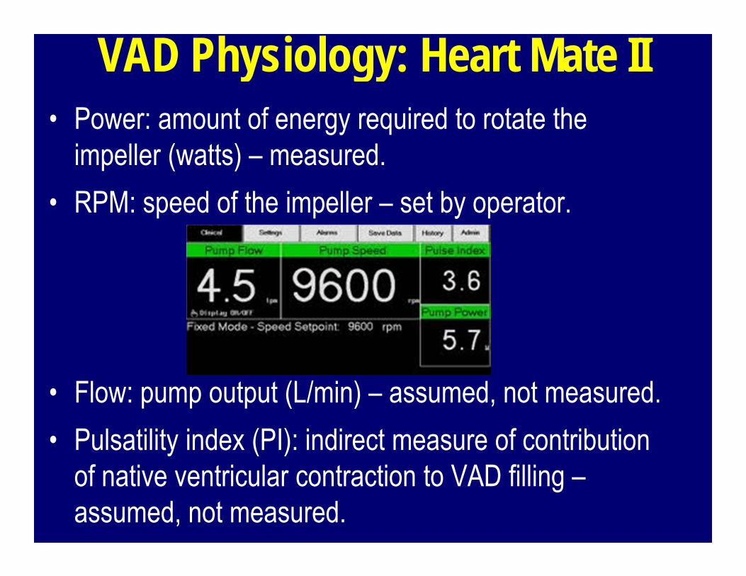
GI Bleeding with VADs: Common
• 101 pts at Mayo
• 46 XVE• 55 HM II• Higher
bleeding with CF VADs
Crow S et al. J Thorac Cardiovasc Surg. 2009;137:208-215.

GI Bleeding with VADs
Crow S et al. Ann Thorac Surg. 2010;90:1263-1269.
Uriel N et al. J Am Coll Cardiol. 2010;56:1207-1213.

GI Bleeding with VADs
• Mostly AVMs, rare gastric/ duodenal ulcers.• Diagnosis:
– EGD (low yield if no pre-op history of PUD); colonoscopy (low yield if pre-op normal)
– Capsule endoscopy– Tagged RBCs– Selective angiography
• Often no active bleeding, so nothing to treat.

GI Bleeding with VADs: Treatment• INR goal (~ 1.5), some may need to come off warfarin.• Eliminate ASA and keep low INR.• Lower rpm to increase pulsatility and flow to watershed
areas (mostly affected by AVMs).• If low MAPs, decrease anti-hypertensive meds.• PRBC transfusion if Hb < 8.• Octreotide for continuous bleeding (100-200 mg sc q 12
hrs, then 20 mg im depot).• Misoprostol (200 mcg TID).• Premarin 0.3 mg daily

Thrombosis with VADs: Rare• Pretty bad, catastrophic most of the time.• Stroke, peripheral emboli (leg, arm), pump stoppage.• Hemolysis (high LDH, low haptoglobin, high plasma free
Hb, high bilirubin).• Increased power, increased flow.• Loss of the VAD hum on exam, VAD “choking”.• IR or cath lab to do TPA intra LV.• Peripheral TPA, Integrillin, bivalirudin -> bad bleeds….• May require pump replacement.

Infections with VADs: Common• 15-20% of pts in trials and registry.• Percutaneous lead, pocket, systemic.• High morbidity and mortality.• Peri-op abx: vancomycin, fluconazole, +/- rifampicin (48
hrs usually), G negative coverage (institution specific).• Nasal mupirocin pre-op, hexidine scrub pre-op x 2.• Judicious arterial line, central line, PA catheter use.• Correct percutaneous lead placement and stabilization.

Infections with VADs: PreventionNutritionOperative technique (meticulous)Tubes out
Drive line stabilizationEducationAmbulationDrug therapy

Arrhythmias with VADs: VT• Causes:
– Underlying substrate: scar (ischemic pts).– Cannula position (towards septum or free wall instead of pointing
towards mitral valve).– Suction events.– Electrolyte abnormalities.– Inotropic agents.
• Assessment:– ECG (12 lead: see VT location), telemetry, ICD interrogation, echo.
• Treatment:– Amiodarone, mexiletine, ICD reprogramming for ATP, ablation ?

Hypertension with VADs• Common; remember we measure MAPs.• Goal MAPs 70-80 (afterload sensitive pumps).• High MAPs (> 95-100) -> high stroke rates.• Treatment:
– ACE-I/ ARB if Cr and K stable.– Beta blocker only if RV function good (carvedilol better BP
control).– Hydralazine, amlodipine.– Diuretics: carefully (VAD needs volume to fill).

Pulmonary HTN with VADs• Majority of chronic heart failure pts have PH.• VADs over time decrease LVEDP and PA pressures.• May need to start sildenafil, especially if marginal RV
function needing iNO post-op.• If pre-VAD PH present, recheck PA pressures by echo
after 6 months and if abnormal, consider right heart cath to confirm.
• Do not list unless PVR < 3 and PA pressures near normal.
• May need to continue sildenafil post transplant.

VADs: What Am I Taking Home?• Reality of current heart failure practice and will increase.• Not all VADs and pts are the same; management has
nuances (“art more than science”).• Optimizing pump speed allows symptomatic improvement,
normalization of PA pressures, LV remodeling. • Prevent RV failure and infection (nutrition !)• Tailor anticoagulation to pts not to guidelines.• Manage VT with meds and ICDs.• Treat A Fib?• Optimize BP control.