
Medical Image Fusion Methods- A Comparative Analysis · eg. MRI-T2 and CT, MRI-FLAIR and CT, MRI-T2...
9
International Journal of Scientific & Engineering Research, Volume 7, Issue 9, September-2016 1379 ISSN 2229-5518 IJSER © 2016 http://www.ijser.org Medical Image Fusion Methods- A Comparative Analysis Sumit Narayan Jarholiya, Dr. ShachiAwasthi Abstract—Image fusion is defined as the process by which several images of the same scene, or some of their features are combined together to form a single Image. In this paper we present a several fusion techniques to increase the information content of the fused image. It helps to diagnose the diseases like tumor, cancer, fracture in bones, ulcer and stones in the body etc effectively. The goal of image fusion (IF) is to integrate complementary multisensory, multitemporal and/or multiview information into one new image containing information the quality of which cannot be achieved otherwise. Various fusion applications have appeared in medical imaging like simultaneous evaluation of CT, MRI, and / or PET images. Index Terms—Medical Image Fusion, Wavelet Transform, Discrete Wavelet Transform (DWT ), Principal Component Analyses ( PCA), CT, MRI, and/or PET images , SPECT images, DTCWT, RDWT, Radon Transform , SIDWT and many more —————————— ————————— 1 INTRODUCTION OF IMAGE FUSION The term Image fusion means in general an approach to extraction of information acquired in number of domains. The main objective of this technique is to obtain an image that is more suitable for visual perception.[9] The goal of image fusion (IF) is to integrate complementary multisensor, multiview and multitemporal information into one new image containing information the quality of which cannot be achieved otherwise. [1] Medical image fusion is the idea to improve the image content by fusing images taken from different imaging tools like Computed Tomography (CT), Magnetic Resonance Imaging (MRI),Positron Emission Tomography (PET) and Single Photon Emission Computed Tomography (SPECT). [17] (A) Different Types of Medical Images:- In Medical field there are different types of medical scan are available in order to diagnosis a tumour of a patient in absolute manner. Some of the examples of medical scan are CT image, MRI image, PET image and SPECT image. 1) Computed Tomography (CT) Image: A CT Image provides the best information on denser / hard tissue with less distortion.[17] Fig. 1 CT Image 2) Magnetic Resonance Imaging (MRI) Image: MRI image provides better information on soft tissue with more distortion. Fig. 2 MRI Image 3) Positron Emission Tomography (PET) Image: A PET image shows chemical and other changes in the brain on comparing to that of CT and MRI images [18]. Fig. 3 PET Image 4)Single Photon Emission Computed Tomography (SPECT)Image: A Single Photon Emission Computed Tomography (SPECT) scan is a type of nuclear test that shows how blood flow changes in brain, tissues and organs. Fig. 4 SPECT Image At present, there are three types of fusion operators are used for wavelet image fusion: pixel, area and region.[4] Typically, the field of medical image analysis is divided into six categories: Post-acquisition, Segmentation, Registration, Computation, Visualization and Security. [5] Another approach of Medical imaging is Image guided interventions and therapy (IGIT) sometimes it is also called IJSER
Transcript of Medical Image Fusion Methods- A Comparative Analysis · eg. MRI-T2 and CT, MRI-FLAIR and CT, MRI-T2...

International Journal of Scientific & Engineering Research, Volume 7, Issue 9, September-2016 1379 ISSN 2229-5518
IJSER © 2016 http://www.ijser.org
Medical Image Fusion Methods- A Comparative Analysis
Sumit Narayan Jarholiya, Dr. ShachiAwasthi
Abstract—Image fusion is defined as the process by which several images of the same scene, or some of their features are combined together to form a single Image. In this paper we present a several fusion techniques to increase the information content of the fused image. It helps to diagnose the diseases like tumor, cancer, fracture in bones, ulcer and stones in the body etc effectively. The goal of image fusion (IF) is to integrate complementary multisensory, multitemporal and/or multiview information into one new image containing information the quality of which cannot be achieved otherwise. Various fusion applications have appeared in medical imaging like simultaneous evaluation of CT, MRI, and / or PET images. Index Terms—Medical Image Fusion, Wavelet Transform, Discrete Wavelet Transform (DWT ), Principal Component Analyses ( PCA), CT, MRI, and/or PET images , SPECT images, DTCWT, RDWT, Radon Transform , SIDWT and many more
——————————————————— 1 INTRODUCTION OF IMAGE FUSION
The term Image fusion means in general an approach to
extraction of information acquired in number of domains. The main objective of this technique is to obtain an image that is more suitable for visual perception.[9] The goal of image fusion (IF) is to integrate complementary multisensor, multiview and multitemporal information into one new image containing information the quality of which cannot be achieved otherwise. [1] Medical image fusion is the idea to improve the image content by fusing images taken from different imaging tools like Computed Tomography (CT), Magnetic Resonance Imaging (MRI),Positron Emission Tomography (PET) and Single Photon Emission Computed Tomography (SPECT). [17] (A) Different Types of Medical Images:- In Medical field there are different types of medical scan are available in order to diagnosis a tumour of a patient in absolute manner. Some of the examples of medical scan are CT image, MRI image, PET image and SPECT image. 1) Computed Tomography (CT) Image: A CT Image provides the best information on denser / hard tissue with less distortion.[17]
Fig. 1 CT Image 2) Magnetic Resonance Imaging (MRI) Image: MRI image provides better information on soft tissue with more distortion.
Fig. 2 MRI Image 3) Positron Emission Tomography (PET) Image: A PET image shows chemical and other changes in the brain on comparing to that of CT and MRI images [18].
Fig. 3 PET Image 4)Single Photon Emission Computed Tomography (SPECT)Image: A Single Photon Emission Computed Tomography (SPECT) scan is a type of nuclear test that shows how blood flow changes in brain, tissues and organs.
Fig. 4 SPECT Image At present, there are three types of fusion operators are used for wavelet image fusion: pixel, area and region.[4] Typically, the field of medical image analysis is divided into six categories: Post-acquisition, Segmentation, Registration, Computation, Visualization and Security. [5] Another approach of Medical imaging is Image guided interventions and therapy (IGIT) sometimes it is also called
IJSER

International Journal of Scientific & Engineering Research, Volume 7, Issue 9, September-2016 1380 ISSN 2229-5518
IJSER © 2016 http://www.ijser.org
image-guided therapy (IGT)or image-guided intervention(IGI) or image-guided surgery (IGS). [7][43]The minimally invasive nature of Image guided interventions and therapy (IGIT)is often preferred over open surgeries because less trauma to the patient’s body is caused, which generally is associated with easier and faster recovery.[7] 2 Image Fusion Methods There are many image fusion methods that can be used to produce high-resolution multispectral images from a high-resolution panchromatic image and low-resolution multispectral images.[8] The term CBIR (content-based image retrieval) designates the retrieval of images based on their visual similarity to a user-supplied query image or to a set of user-specified image features. CBIR can be widely used in many areas, such as photograph archives, medical diagnosis, the military, retail catalogs, and remote sensing systems. [10] There are so many image fusion methods are available. Most of these image fusion methods are as follows:- • Wavelet Transform Fusion • The Discrete Wavelet Transform ( DWT ) • Pixel Based Wavelet Transform Fusion • The Dual Tree Complex Wavelet Transform (DTCWT) • Edge Based Wavelet Transform Fusion • Operator Based Image Fusion Method • Fusion of Multimodal Brain Images using Redundant
Discrete Wavelet Transform ( RDWT) • Image Fusion Using The Convolution Of Meridian
Distributions • Image Fusion Method Based On Principal Component
Analysis (PCA) • Image Fusion Method Based On Brovey Transform • Image Fusion Method Based On Ehlers Transform • Simple Average Based Image Fusion • Image Fusion Based On Radon Transform • Shift Invariant Discrete Wavelet Transform For Image
Fusion • Fusion Of Images In Multi Resolution Domain • Complex Wavelet Image Fusion • Morphological Methods • Knowledge Based Methods • Neural Network Based Image Fusion Methods • Fuzzy Logic Based Image Fusion Methods • Discrete Cosine Transform(DCT) • Daubechies Discrete Wavelet Transform Based Image
Fusion • Symlet Based Image Fusion • Image Fusion Technique Using SVD For Radiotherapy,
Prostate Biopsy And Lesion Targeted Prostate Intervention
• Wavelet Transform Fusion In all wavelet based image fusion schemes the wavelet transforms W of the two registered input images I1(x,y )and
I2(x, y)are computed and these transforms are combined using some kind of fusion rule∅ (Fig 5). Then the inverse wavelet transform W−1is computed and the fused image I(x, y)is reconstructed . [3][15]
I (x, y) = W−1 {∅ (W (I1(x, y)), W (I2))) } { 1} Where x= Scaling parameter and y= Location parameter. Wavelet means “small wave” so wavelet analysis is about analyzing signal with short duration finite energy functions [17]. Wavelet Transform is reduces the storage cost but it is not able to maintain edge information efficiently[18]
Fig. 5Fusion of the wavelet transforms of two images.[3][15][19] • Discrete Wavelet Transform ( DWT ) The Discrete Wavelet Transform (DWT), which is based on sub-band coding is found to yield a fast computation of Wavelet Transform. It is easy to implement and reduces the computation time and resources required. [3] It provides some other advantages over other simple methods, like: energy compaction, larger SNR, reduced features, etc.[16] The discrete wavelet transform DWT is a spatial frequency decomposition that provides a flexible multi resolution analysis of an image in one dimension. The aim of the wavelet transform is to represent the signal as a superposition of wavelets, If a discrete signal is represented by f(t) its wavelet decomposition is then given by
f( t) = ∑ 𝐶𝐶𝐶𝐶,𝑛𝑛𝐶𝐶 ,𝑛𝑛 𝛹𝛹𝐶𝐶,𝑛𝑛(𝑡𝑡), {2} where m, n( t) is the dilated and or translated version of the mother wavelet given by the equation
𝛹𝛹𝐶𝐶,𝑛𝑛( t) = 2 –m /2𝛹𝛹[2 -m t –n ] {3}
where m and n are integers This ensures that the signal is decomposed into normalized wavelets at octave scales .It also provide More accurate clinical information for medical diagnosis & evaluation but it offers Poor directionality, Shift sensitivity, Absence of phase information.[18] • Pixel Based Wavelet Transform Fusion In the pixel based wavelet transform fusion all respective wavelet coefficients from the input images are combined using the fusion rule since wavelet coefficients having large absolute values contain the information about the salient features of the images such as edges and lines a good fusion rule is to take the maximum of the absolute values of the corresponding wavelet coefficients.[3] • The Dual Tree Complex Wavelet Transform
(DTCWT)
IJSER
International Journal of Scientific & Engineering Research, Volume 7, Issue 9, September-2016 1381 ISSN 2229-5518
IJSER © 2016 http://www.ijser.org
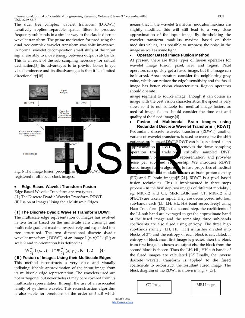
The dual tree complex wavelet transform (DTCWT) iteratively applies separable spatial filters to produce frequency sub bands in a similar way to the classic discrete wavelet transform. The prime motivation for producing the dual tree complex wavelet transform was shift invariance. In normal wavelet decomposition small shifts of the input signal are able to move energy between output sub bands. This is a result of the sub sampling necessary for critical decimation.[3] Its advantages is to provide better image visual eminence and its disadvantages is that it has limited directionality[18].
Fig. 6 The image fusion process using the DTCWT and two registered multi focus clock images. • Edge Based Wavelet Transform Fusion Edge Based Wavelet Transform are two types:- ( I ) The Discrete Dyadic Wavelet Transform DDWT. (II)Fusion of Images Using their Multiscale Edges. ( I ) The Discrete Dyadic Wavelet Transform DDWT The multiscale edge representation of images has evolved in two forms based on the multiscale zero crossings and multiscale gradient maxima respectively and expanded to a tree structured. The two dimensional discrete dyadic wavelet transform ( DDWT) of an image I (x, y)Є L2 (R2) at scale 2j and in orientation k is defined as
W 𝑘𝑘2𝑗𝑗 f (x, y) = I * 𝛹𝛹 𝑘𝑘
2𝑗𝑗 (x, y ) , K= 1, 2 {4} ( II ) Fusion of Images Using their Multiscale Edges This method reconstructs a very close and visually indistinguishable approximation of the input image from its multiscale edge representation. The wavelets used are not orthogonal but nevertheless I may bere covered from its multiscale representation through the use of an associated family of synthesis wavelet. This reconstruction algorithm is also stable for precisions of the order of 3 dB which
means that if the wavelet transform modulus maxima are slightly modified this will still lead to a very close approximation of the input image By thresholding the wavelet transform modulus maxima based on their modulus values, it is possible to suppress the noise in the image as well as some light. • Operator Based Image Fusion Method At present, there are three types of fusion operators for wavelet image fusion: pixel, area and region. Pixel operators can quickly get a fused image, but the image can be blurred. Area operators consider the neighboring gray value, which can reduce the edge's sensitivity and the fused image has better vision characteristics. Region operators should operate image segment to source image. Though it can obtain an image with the best vision characteristics, the speed is very slow, so it is not suitable for medical image fusion, as medical image fusion should consider the time cost and quality of the fused image.[4] • Fusion of Multimodal Brain Images using
Redundant Discrete Wavelet Transform ( RDWT) Redundant discrete wavelet transform (RDWT) another variant of wavelet transform, is used to overcome the shift variance problem of DWT.RDWT can be considered as an approximation to DWT that removes the down sampling operation from traditional critically sampled DWT, produces an over-complete representation, and provides noise per sub-band relationship. We introduce RDWT based image fusion algorithm to fuse properties of medical images of different modalities such as brain proton density (PD) and T1 brain images[5][21]. RDWT is a pixel based fusion techniques. This is implemented in three steps process:- In the first step two images of different modality ( eg. MRI-T2 and CT, MRI-FLAIR and CT, MRI-T2 and SPECT) are taken as input. They are decomposed into four sub-bands each (LL, LH, HL, HH band respectively) using Haar Transform [23].In the second step, the coefficients of the LL sub band are averaged to get the approximate band of the fused image and the remaining three sub-bands coefficients are also fused using entropy. The three high sub-bands namely (LH, HL, HH) is further divided into blocks of 3*3 and the entropy of each block is calculated. If entropy of block from first image is greater, then the block from first image is chosen as output else the block from the second block is chosen. Thus the LH, HL, HH sub-bands of the fused images are calculated [23].Finally, the inverse discrete wavelet transform is applied to the fused coefficients to reconstruct the resultant fused image .The block diagram of the RDWT is shown in Fig. 7 [23].
CT Image MRI Image
IJSER

International Journal of Scientific & Engineering Research, Volume 7, Issue 9, September-2016 1382 ISSN 2229-5518
IJSER © 2016 http://www.ijser.org
Fig. 7Block diagram of Redundancy Discrete Wavelet Transform RDWT
• Image Fusion Using The Convolution Of
Meridian Distributions
The Meridian distribution is a member of the GCD family (when p=1) for modeling heavy tailed non-Gaussian behavior. The pdf of the Meridian model has the form [6]:
P (x; μ, γ ) = γ2�(𝑥𝑥−μ)+ γ�^2
{5}
Where μ (−∞ < μ < ∞) is the location parameter, specifying the location of the peak of the distribution, and γ (γ >0) is the scale parameter of the distribution that determines the spread of the distribution centered on μ. It can be shown that if two independent random variables X1 and X2 follow Meridian distributions with parameters (μ1, γ1) and (μ2, γ2), respectively, then the random variable Y = X1 + X2 follows the convolution of the distributions of X1 and X2, which is also a Meridian distribution.
P(y ; μ ,γ ) = P(x ; μ1,γ1 ) * P( x ; μ2 ,γ2 ) = P( x; μ1 + μ2 , γ1 + γ2 ) {6}
More generally, the weighted sum of two independent Meridian random variables, Z w1X1,w2X2, also follows the Meridian distribution with parameters μ and γ defined
μ= w1 μ1 + w2μ2 , γ= w1 γ1 + w2 γ2{7} Here we assume that there are two input images, which are decomposed in the wavelet domain. Z represents the weighted combination of the two source images, whereas X1 and X2 are their corresponding wavelet coefficients. This is represented in figures given below. • Image Fusion Method Based On Principal
Component Analysis (PCA) The PCA is a statistical technique which is used to transform the multivariate dataset of correlated variables into a dataset of uncorrelated linear combinations of the
original variables [20]. There's involvement of mathematical formula for change of factors which can be called key components. It computes a tight and optimum explanation of the info set. The very first key aspect corresponds to the maximum amount of difference probable in the info and every subsequent aspect corresponds to the rest of the variance. First key aspect is taken across the direction of optimum variance. The 2nd key aspect is limited to lay in the subspace at a 90 level angle of the first. Within this Subspace, this aspect items the direction of optimum variance. The next key aspect is taken in the optimum difference direction in the subspace at a 90 level angle to the former two. [18]
Fig. 8 Image Fusion Method Based On Principal Component Analysis (PCA) [12][20]
Fig. 9 Image fusion scheme employing PCA and fused image [12] • Image Fusion Method Based On Brovey
Transform The Brovey transform is based on the mathematical combination of the multispectral images and high resolution Pan image. Each multispectral image is normalized based on the other spectral bands and multiplied by the Pan image to add the spatial information to the output image. The following equation shows the mathematical algorithm for the Brovey method[11] [8]
Fi= 𝑀𝑀𝑀𝑀∑ 𝑀𝑀𝑗𝑗 +𝐶𝐶𝑁𝑁𝑗𝑗=1
𝑥𝑥𝑥𝑥{8}
Where Fi is a fused band , Mi is the multispectral band to be fused, and P is the Panchromatic band . In some cases there it be an area with zero DN values for all bands; therefore a
RDWT RDWT
Low sub-band
High Sub-band
Low sub-band
High Sub-band
Average EN
IRDWT
Fused Image
IJSER
International Journal of Scientific & Engineering Research, Volume 7, Issue 9, September-2016 1383 ISSN 2229-5518
IJSER © 2016 http://www.ijser.org
constant C has to be added in the denominator to produce meaningful output digital numbers. • Image Fusion Method Based On Ehlers
Transform The basic idea behind this method is to modify the input Pan image to look more like the intensity component. In the first step in order to manipulating HIS components, three low resolution multispectral RGB bands are selected and transformed into the IHS domain. Then, the intensity component and the panchromatic image are transformed into the spectral domain via a two-dimensional Fast Fourier Transform (FFT). Low pass (LP) and high pass (HP) filter were directly performed in the frequency domain on the intensity component and the high resolution panchromatic image respectively. The idea is to replace the high frequency part of the intensity component with that from the Pan image [11]. To return both components back into the spatial domain an inverse FFT transform was used. Then the high pass filtered panchromatic band and low pass filter intensity are added and matched to the original intensity histogram. Finally, an inverse IHS transform converts the fused image back into the RGB colour domain to obtain the high resolution multispectral image. • Simple Average Based Image Fusion This is a very basic technique of image fusion. It is shown in fig. 10.Image fusion could be achieved by simple averaging corresponding pixels in each input image as follows[12][17][22]
If (x, y) = 𝐼𝐼1 (𝑥𝑥 ,𝑦𝑦)+ 𝐼𝐼2 (𝑥𝑥 ,𝑦𝑦 )2
{9}
Fig. 10:Sample images ,PET (left ) and MRI (right).
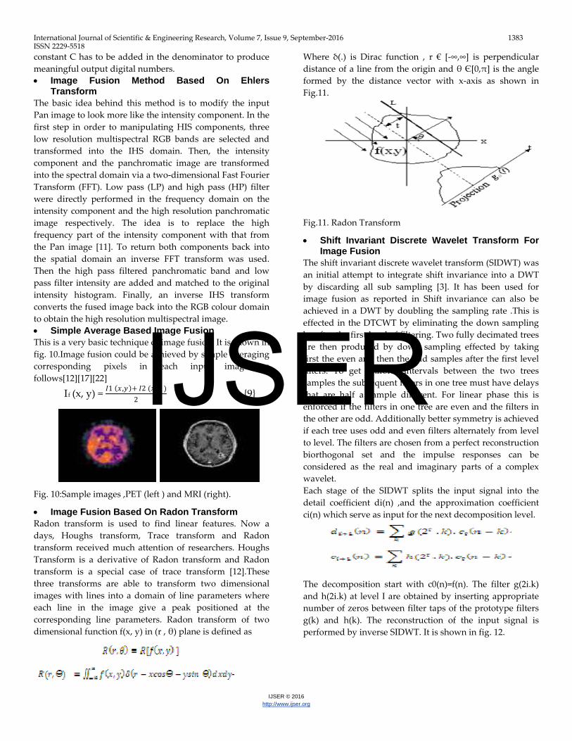
• Image Fusion Based On Radon Transform Radon transform is used to find linear features. Now a days, Houghs transform, Trace transform and Radon transform received much attention of researchers. Houghs Transform is a derivative of Radon transform and Radon transform is a special case of trace transform [12].These three transforms are able to transform two dimensional images with lines into a domain of line parameters where each line in the image give a peak positioned at the corresponding line parameters. Radon transform of two dimensional function f(x, y) in (r , θ) plane is defined as
Where δ(.) is Dirac function , r € [-∞,∞] is perpendicular distance of a line from the origin and θ Є[0,π] is the angle formed by the distance vector with x-axis as shown in Fig.11.
Fig.11. Radon Transform
• Shift Invariant Discrete Wavelet Transform For Image Fusion
The shift invariant discrete wavelet transform (SIDWT) was an initial attempt to integrate shift invariance into a DWT by discarding all sub sampling [3]. It has been used for image fusion as reported in Shift invariance can also be achieved in a DWT by doubling the sampling rate .This is effected in the DTCWT by eliminating the down sampling by after the first level of filtering. Two fully decimated trees are then produced by down sampling effected by taking first the even and then the odd samples after the first level filters. To get uniform intervals between the two trees samples the subsequent filters in one tree must have delays that are half a sample different. For linear phase this is enforced if the filters in one tree are even and the filters in the other are odd. Additionally better symmetry is achieved if each tree uses odd and even filters alternately from level to level. The filters are chosen from a perfect reconstruction biorthogonal set and the impulse responses can be considered as the real and imaginary parts of a complex wavelet. Each stage of the SIDWT splits the input signal into the detail coefficient di(n) ,and the approximation coefficient ci(n) which serve as input for the next decomposition level.
The decomposition start with c0(n)=f(n). The filter g(2i.k) and h(2i.k) at level I are obtained by inserting appropriate number of zeros between filter taps of the prototype filters g(k) and h(k). The reconstruction of the input signal is performed by inverse SIDWT. It is shown in fig. 12.
IJSER
International Journal of Scientific & Engineering Research, Volume 7, Issue 9, September-2016 1384 ISSN 2229-5518
IJSER © 2016 http://www.ijser.org
Fig.12 Shift invariant discrete wavelet transforms • Fusion Of Images In Multi Resolution Domain Several algorithms developed up to now can be classified in three primary categories: space field, methods of optimization and transformed field or multi-resolution. We were interested to evaluate the results of the algorithms of fusion in the multi resolution field moved two facts: the human visual system (HVS) is mainly sensitive to the change of local contrast. In addition, the MRI and the PET are currently the best techniques of anatomical and functional imageries, respectively of the human brain[13]. The imagery by magnetic resonance (MRI) constitute some of advanced most significant of the medicine of the 20th centuries. The basic idea is to apply a transform multi-scale to each image source, then builds a composite representation multi-scale of those by certain techniques of fusion [13]. • Complex Wavelet Image Fusion The areas of the images more in focus give rise to larger magnitude coefficients within that region. A simple choose maximum scheme is used to produce the combined coefficient map. The resulting fused image is then produced by transforming the combined coefficients map using the inverse complex wavelet transform. The wavelet coefficient images show the orientated nature of the complex wavelet sub-bands[15]. • Morphological Methods The morphology operators has been explored by image processing community for long, and the concept is used by the medical imaging community to detect spatially relevant information from the medical images. The morphological filtering methods for medical image fusion have been applied, for example, in brain diagnosis [2]. An example of modalities used in morphology based fusion can be seen in the fusion of CT and MR images [2].The operators such as averaging, morphology towers, K-L transforms and morphology pyramids are used for achieving the data fusion. These methods are highly sensitive to the inter-image variability resulting from outliers, noise, size and shape of the features. • Knowledge Based Methods In medical imaging, there are several instances where the medical practitioner’s knowledge can be used in designing segmentation, labeling and registration of the images. There are a range of applications where the domain-dependent knowledge is useful for image fusion such as for
segmentation, micro-calcification diagnosis, tissue classification, brain diagnosis, classifier fusion, breast cancer tumor detection and delineation & recognition of anatomical brain object. The knowledge based systems can used in combination with other methods such as pixel intensity. These methods place a significant amount of trust in the medical expert in labeling and identifying the domain knowledge relevant to the fusion task. The advantage is the ability to benchmark the images with the known human vision standards, while the drawback is the limitations imposed by human judgment in images that are prone to large pixel intensity variability. The use of preprocessing techniques in images can improve the imaging quality and increase the accuracy of ground truths [2]. • Neural Network Based Image Fusion Methods Artificial neural networks (ANN) are inspired from the idea of biological neural network having the ability to learn from inputs for preprocessing features and for making global decisions. The artificial neural network models require an input training set to identify the set of parameters of the network referred to as weights. The ability to train the neural network to adopt to these changes enable several applications for medical image fusion such as solving the problems of feature generation , classification , data fusion , image fusion , micro-calcification diagnosis , breast cancer detection, medical diagnosis , cancer diagnosis , natural computing methods and classifier fusion [2]. • Fuzzy Logic Based Image Fusion Method The conjunctive, disjunctive and compromise properties of the fuzzy logic have been widely explored in image processing and have proved to be useful in image fusion. The fuzzy logic is applied both as a feature transform operator or a decision operator for image fusion. There are several applications of fuzzy logic based image fusion such as brain diagnosis , cancer treatment , image segmentation and integration , maximization mutual information , deep brain stimulation , brain tumor segmentation , image retrieval , spatial weighted entropy , feature fusion , multimodal image fusion , ovarian cancer diagnosis , sensor fusion , natural computing methods and gene expression [2] • Discrete Cosine Transform (DCT) The fusion strategies which are used in DCT domain are extremely effective when the feedback photographs are numbered and merged photographs are restored in JPEG standard .For using the JPEG development , an image (in color or gray scales) is divided into blocks of 8x8 pixels firstly. The Discrete Cosine Transform (DCT) is a while later used on each block leading to the technology of 64 coefficients that are quantized to decrease their magnitude. The coefficients are then changed into a one-dimensional
IJSER
International Journal of Scientific & Engineering Research, Volume 7, Issue 9, September-2016 1385 ISSN 2229-5518
IJSER © 2016 http://www.ijser.org
variety in a crisscross fashion prior to entropy encoding. The compression is obtained in two measures; the initial through quantization and the next through the entropy development process. For reducing the problems undergone in the fusion of real time programs and enhancing the quality of merged picture, DCT fusion strategy is applied. Difference of 8×8 blocks computed from DCT coefficients is used as a comparison qualification for the experience evaluate. Fig 13 shows the Discrete Cosine transform [18].
Fig. 13 Shows the Discrete Cosine transform • Daubechies Discrete Wavelet Transform Based
Image Fusion The Daubechies wavelet (db2) can be decomposed to n level based on the size of the image. Here first level of decomposition is considered for image fusion. Daubechies wavelets are orthogonal wavelets and asymmetric in nature. The two dimensional DWT leads to a decomposition of approximation coefficients at level j in four components: the approximation at level j+1, and the details in three orientations (horizontal, vertical, and diagonal) at j+1. Image Fusion is carried out by averaging the contents[21]. Image reconstruction process is performed using inverse of DWT (IDWT). IDWT uses the wavelet db2to compute the single-level reconstruction of fused Image IF, based on approximation matrix (A) and detailed matrices H, V and D (horizontal, vertical and diagonal respectively). Daubechies db2 is giving entropy results better than Haar, but unable give better than SIDWT and RDWT. To get better entropy one has process to the higher level of decomposition[21]. • Symlet Based Image Fusion Symlet based image fusion similar to „db2‟. The steps are exactly same as the “db2”. Even the results are almost same[21]. Therefore this method is not mostly used for Image Fusion. • Image Fusion Technique Using SVD SVD is a method for identifying and ordering the dimensions along which data points exhibit the most variations. Using SVD it’s possible to find the best approximation of the original data points using fewer dimensions. SVD takes a high dimensional, highly variable set of data points and reducing it to a lower dimensional space that exposes the substructure of the original data
more clearly and orders it from most variation to the least. SVD is based on a theorem from linear algebra which says that a rectangular matrix A can be broken down into the product of three matrices - an orthogonal matrix U, a diagonal matrix S, and the transpose of an orthogonal matrix V. The theorem is usually presented like this [22]:
A = USVT {10} where UTU = I, VTV = I; the columns of U are orthonormal eigenvectors of AAT, the columns of V are orthonormal eigenvectors of ATA, and S is a diagonal matrix containing the square roots of eigen values from U or V in descending order. This method is robust , simple and efficiency is good in the presence of noise but noise can be present [22]. The input images referred in the below block diagram may be a multimodality medical images such as CT, MRI, PET etc. Fig. 14 Block Diagram of Image Fusion Using SVD • Image Fusion For Radiotherapy, Prostate Biopsy
And Lesion Targeted Prostate Intervention The image-fusion of prostate imaging were reported in the field of radiotherapy including external beam radiation therapy and brachy therapy, using fusions of CT, MR, ultrasound, and/or fluoroscopy[14].In addition, the potential value of image fusion of Doppler TRUS with MRI in the staging of prostatic cancer was discussed. However, recent attention to image fusion technology for prostate cancer is more toward its value in improving the quality of prostate biopsy by precisely targeting the image-suspicious area, in mapping the 3D localization of biopsy-proven prostate cancer, as well as its value in navigating image-guided focal therapy. 3 Conclusion Medical image fusion plays an important role in clinical applications. This is a major source for the doctors to diagnose the diseases. Whatsoever the medical imaging has its own kinds of imaging techniques like X-ray, computed tomography (CT), magnetic resonance imaging (MRI). However the characteristics and results of each of these medical imaging techniques are unique. For instance, X-ray and CT can provide images as dense like structure with which the physiological changes could not be detected whereas in MRI images even the soft pathological tissues
I/P Image1 Tensor Decomposition
Sigmoid Function
I/P Image2 Tensor Decomposition
Sigmoid Function
Fused O/P Image Performance Analysis IJSER
International Journal of Scientific & Engineering Research, Volume 7, Issue 9, September-2016 1386 ISSN 2229-5518
IJSER © 2016 http://www.ijser.org
can be visualized better therefore the combination of CT and MRI medical image fusion analysis can plays better role. For Medical image fusion applications computation time and accuracy are two main parameter. After studies of all those above image fusion methods we can conclude that Discrete Wavelet Transform( DWT ) will give better result since it is easy to implement, reduces the computation time and resources required. It also provides energy compaction, larger SNR and More accurate clinical information for medical diagnosis &evaluation. But at the same time it also offers Poor directionality, Shift sensitivity, Absence of phase information and presence of spectra degradation. To remove the drawback of Discrete Wavelet Transform we can apply Dual Tree Complex Wavelet Transform (DTCWT) followed by DWT because it provides better image visual eminence , shift invariance and better directionality. References [1] Jan Flusser, Filip Sroubek, and Barbara Zitov´a,“ Image Fusion: Principles, Methods, and Applications”, Institute of Information Theory and Automation ,Academy of Sciences of the Czech Republic (2007) [2] A.P. James, B. V. Dasarathy, “Medical Image Fusion: A survey of the state of the art”, Nazarbayev University Information Fusion, 2014 [3] StavriNikolov, Paul Hill, David Bull, NishanCanagarajah, “Wavelets For Imaged Fusion”,Image Communications Group Centre for Communications Research,University of Bristol [4] Zhi-haiXu,Ling-xiang Liu, Lei Tong, Lin-hong Zhou, Chao-min Chen, “Wavelet medical image fusion algorithm based on local area feature”, Research Article/Biological and Biomedical Reports (ISSN: 2162-4186), 2012, 2(1), 25-31 [5] Richa Singh, MayankVatsa, AfzelNoore,“ Multimodal Medical Image Fusion using Redundant DiscreteWavelet Transform”, West Virginia University, Morgantown, WV, USA [6]Swathi.P.S, Sheethal.M.S, Vince Paul , “Survey on Multimodal Medical Image Fusion Techniques”, IJCSET , January 2016 , Vol 6, Issue 1, ISSN:2231-0711, pp 33-39 [7] Dani¨elRuijters, “Multi-modal image fusion during minimally invasive treatment”, TechnischeUniversiteit Eindhoven, 2010. [8]Zhijun Wang, DjemelZiou, Costas Armenakis, Deren Li, and Qingquan Li “A Comparative Analysis of Image Fusion Methods”,IEEE Transactions On Geoscience And Remote Sensing , Vol. 43, No. 6, June 2005 [9] M.D. Nandeesh and Dr.M. Meenakshi, “Image Fusion Algorithms for Medical Images-A Comparison “,Bonfring International Journal of Advances in Image Processing, Vol. 5, No. 3, July 2015, pp 23-26.
[10] S. Yang, I. Atmosukarto, J. Franklin, J. F. Brinkley, D. Suciu, and L. G. Shapiro, “A Model of Multimodal Fusion for Medical Applications”, University of Washington [11] R. Riyahi ,C. Kleinn , H. Fuchs,”Comparison Of Different Image Fusion Techniques ForIndividual Tree Crown Identification UsingQuickbird Images”,Department of Forest Inventory and Remote Sensing,Georg-August-University, Göttingen, Germany, pp 1-4 [12]ManjushaDeshmukh&UdhavBhosale ,“Image Fusion and Image Quality Assessment of Fused Images”, International Journal of Image Processing (IJIP),ISSN 1985-2304, Volume (4): Issue (5) , 2010, pp 484-508 [13]WalidAribi,AliKhalfallah, NoomèneElkadriand Mohammed Salim Bouhle , “Objective Evaluation of Image Fusion Techniques inNuclear Medicine”, IEEE Sciences of Electronics Technologies of Information and Telecommunication (SETIT) , Sousse 21-24, March 2012- Tunisia, pp 1-6 [14]Osamu Ukimura , “Image-fusion for Biopsy, Intervention, and Surgical Navigation in Urology” pp 415-428 [15] Paul Hill, NishanCanagarajah and Dave Bull , “Image Fusion using Complex Wavelets”,Dept. of Electrical and Electronic Engineering ,The University of Bristol, UK [16]LigiaChiorean, Mircea-Florin Vaida , “Medical Image Fusion Based on Discrete Wavelet Transform Using Java Technology” ,Proceedings of the ITI 2009 31st Int. Conf. on Information Technology Interfaces,June 22-25, 2009, Cavtat, Croatia [17]Hari Om Shanker Mishra and SmritiBhatnagar , “MRI and CT Image Fusion Based on Wavelet Transform”, International Journal of Information and Computation Technology. ISSN 0974-2239 Volume 4, Number 1 (2014), pp. 47-52 [18] Swathi.P.S, Sheethal.M.S and Vince Paul , “ Survey on Multimodal Medical Image Fusion Techniques”,ISSN 2231-0711 ,IJCSET , January 2016 , Vol 6, Issue 1, pp 33-39 [19]NayeraNahvi, Deep Mittal , “Medical Image Fusion Using Discrete Wavelet Transform”, Int. Journal of Engineering Research and Applications ,ISSN : 2248-9622, Vol. 4, Issue 9( Version 5), September 2014, pp.165-170 [20] Mr. Roshan ,P. Helonde, Prof. M.R. Joshi , “ Image Fusion Based on Medical Images Using DWT and PCA Methods”,International Journal of Computer Techniques , ISSN: 2394-2231, Volume 2 Issue 1, 2015 , pp 75-79 [21]Umesh B. Mantale, Prof. Vishwajit B. Gaikwad, “Image Fusion of Brain Images using Redundant Discrete Wavelet Transform”,International Journal of Computer Applications (0975 – 8887) Volume 74– No.4, July 2013 , pp 7-11 [22]P.AmbikaPriyadharsini, M.R.Mahalakshmi,“Multimodal Medical Image Fusion Based On SVD”, IOSR Journal of Computer Engineering
IJSER

International Journal of Scientific & Engineering Research, Volume 7, Issue 9, September-2016 1387 ISSN 2229-5518
IJSER © 2016 http://www.ijser.org
(IOSR-JCE) e-ISSN: 2278-0661, p- ISSN: 2278-8727Volume 16, Issue 1, Ver. III (Jan. 2014), PP 27-31 [23] Shrey Gupta, S Rajkumar, V Vijayarajan, K Marimuthu,“Quantitative Analysis of various ImageFusion techniques based on various metrics using different Multimodality MedicalImages”, International Journal of Engineering and Technology (IJET), ISSN : 0975-4024 Vol 5 No 1 Feb-Mar 2013 , pp 133-141 Author Profile
Sumit Narayan Jarholiya pursuing his Doctorate from Jayoti Vidyapeeth Women’s University, Jaipur , India. He did his Master of Engineering (Digital Communication) from Shri Ram Institute of Technology, Jabalpur, He had completed B. Tech. (ET &CS ) from Mahatma Gandhi Chitrakoot Gramodaya University in 1998.
ShachiAwasthi Tiwari is awarded her Doctorate from Rani Durgavati University, Jabalpur. She had Completed her Masters of Engineering ( Digital Communication ) from Shri Ram Institute of Technology, Jabalpur India.
IJSER