Genetic approaches to discover novel oncogenes in human cancer
Upload
eric-thomasCategory
view
241download
5
Medical BiochemistryMedical Biochemistry
Cancer and Oncogenes
Lecture 76
Cancer and Oncogenes
Lecture 76
Oncogenes
• Oncogenes may alter cellular metabolism and stimulate growth by several mechanisms
• sis - B chain of PDGF
• erb-B - truncated EGF receptor
• ras - small GTPase
• src - protein-tyrosine kinase
• c-myc - DNA-binding protein
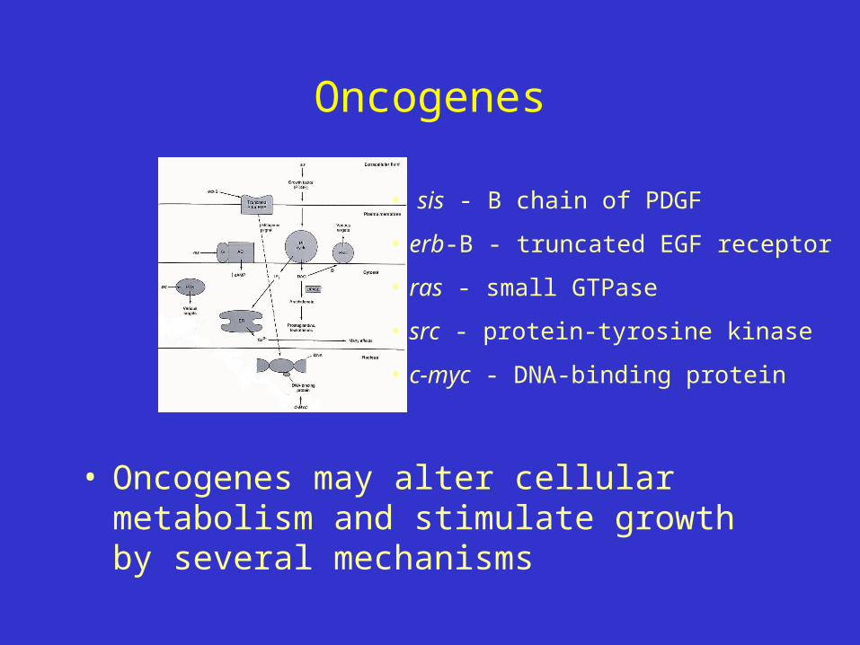
Oncogenes
• Qualitative and quantitative mechanisms for activation of proto-oncogenes to an oncogene
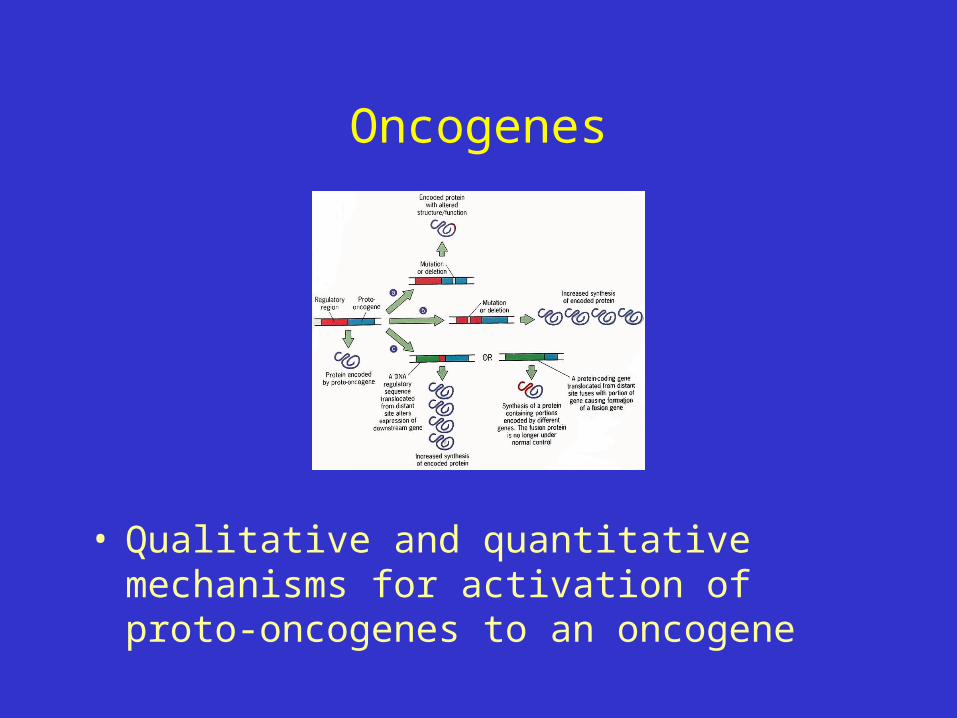
Oncogenes vs Tumor Suppressor Genes
Oncogenes• Mutation in one of two
alleles sufficient for activity; act dominant to wild-type
• “Gain of function” of protein that signals cell division
• Mutation arises in somatic tissue, not inherited
• Some tissue preference
Oncogenes vs Tumor Suppressor Genes
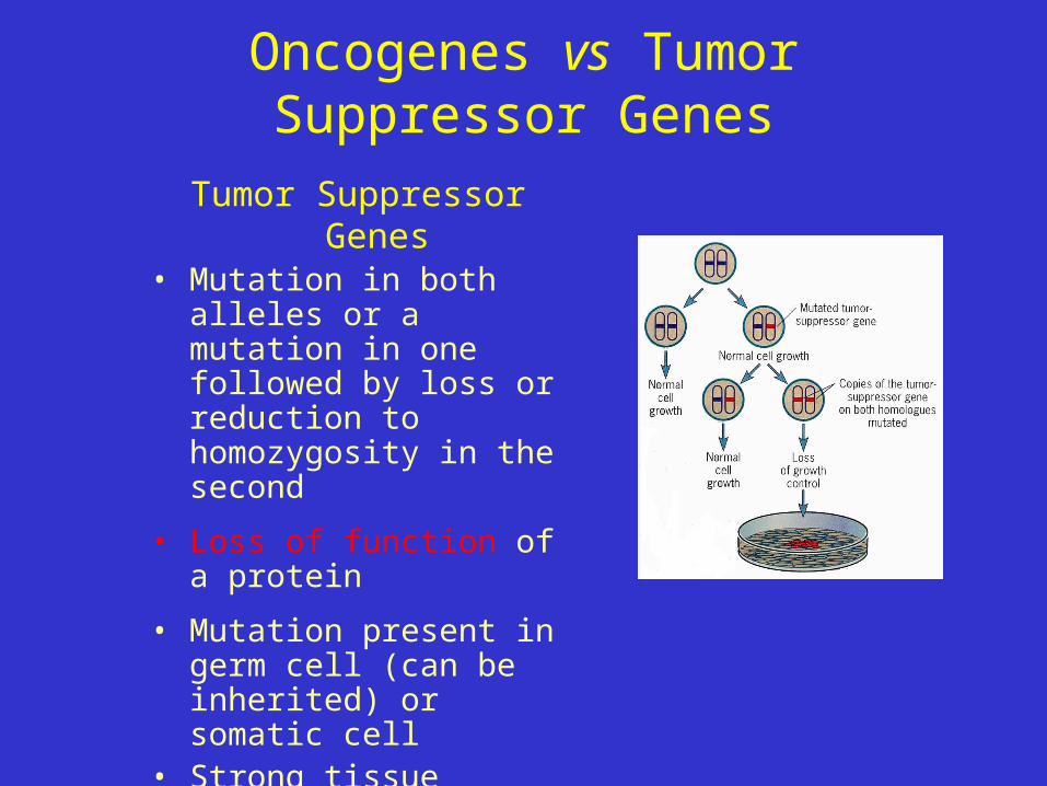
Tumor Suppressor Genes• Mutation in both alleles or a
mutation in one followed by loss or reduction to homozygosity in the second
• Loss of function of a protein
• Mutation present in germ cell (can be inherited) or somatic cell
• Strong tissue preference (e.g., effect of RB1 gene in the retina)
RB1, a Tumor Suppressor Gene
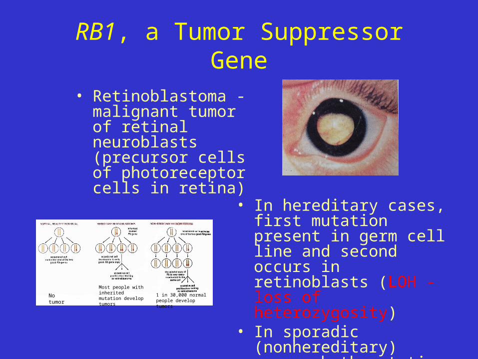
• Retinoblastoma - malignant tumor of retinal neuroblasts (precursor cells of photoreceptor cells in retina)
• In hereditary cases, first mutation present in germ cell line and second occurs in retinoblasts (LOH - loss of heterozygosity)
• In sporadic (nonhereditary) cases, both mutations occur in retinoblasts
No tumor
Most people with inherited mutation develop tumors
1 in 30,000 normal people develop tumors

RB1, a Tumor Suppressor Gene
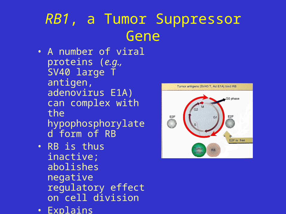
• pRB is a nuclear phosphoprotein
• During G0 or G1, RB is hypophosphorylated, binds E2F, blocks G1/S transition
• Phosphorylation increases in late G1 and early S phases, E2F released, transcribes genes required for S phase, cell enters S phase
RB1, a Tumor Suppressor Gene
• A number of viral proteins (e.g., SV40 large T antigen, adenovirus E1A) can complex with the hypophosphorylated form of RB
• RB is thus inactive; abolishes negative regulatory effect on cell division
• Explains increased multiplication by cells transformed by these viruses

p53 Tumor Suppressor Gene
• p53 is a DNA-binding protein that suppresses growth or triggers apoptosis
• at least 3 major functions(1) transcriptional activator, regulating certain genes
involved in the cell cycle (e.g., p21)
p53p21
p27
p16

p53 Tumor Suppressor Gene
(2) acts as a G1 checkpoint control for DNA damage– e.g., following UV irradiation, activity of p53 ,
inhibits cell cycle to allow time for repair
(3) participates in the initiation of apoptosis (programmed cell death)
p53 Tumor Suppressor Gene
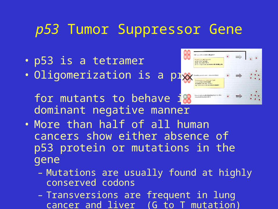
• p53 is a tetramer• Oligomerization is a prerequisite
for mutants to behave in a dominant negative manner
• More than half of all human cancers show either absence of p53 protein or mutations in the gene– Mutations are usually found at highly conserved codons– Transversions are frequent in lung cancer and liver (G
to T mutation)• benzo[a]pyrene - cigarette smoke• aflatoxin B1 (potent hepatocarcinogen)

Normal colon cells
Loss of APCtumor-suppressor gene
Activation of K-rasoncogene
Loss of DCC tumor-suppressor gene
Loss of p53 tumor-suppressor gene
Other changes
Dysplastic aberrantcrypt foci (ACF)
Early adenoma
Intermediate adenoma(class II)
Late adenoma(class III)
Carcinoma
Metastasis
Colorectal cancer: A model for cancer development
• Based on studies of familial adenomatous polyposis (autosomially dominant inherited disorder)– A number of steps involved (i.e., cancer is
a multistep process)– Mutations in APC initiate process– Mutations in K-ras involved– Mutaions of other tumor-suppressor genes,
including p53 and DCC, also implicated– Mutations affecting mismatch repair genes
accelerate overall process– Overall, 6 or 7 genes initially involved– Precise order of changes not as important
as accumulation of changes– Additional mutations necessary for
metastasis
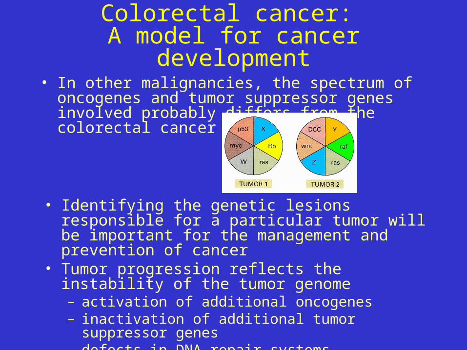
Colorectal cancer: A model for cancer development
• In other malignancies, the spectrum of oncogenes and tumor suppressor genes involved probably differs from the colorectal cancer model
• Identifying the genetic lesions responsible for a particular tumor will be important for the management and prevention of cancer
• Tumor progression reflects the instability of the tumor genome– activation of additional oncogenes– inactivation of additional tumor suppressor genes– defects in DNA repair systems
DNA Repair Systems
• Nucleotide excision repair– removes larger areas of DNA damaged by chemicals or
irradiation
– Xeroderma pigmentosum - autosomal recessive disease, affected individuals get melanomas and squamous cell carcinomas easily if skin exposed to UV light (unable to repair UV damage or remove bulky adducts from DNA bases
DNA Repair Systems
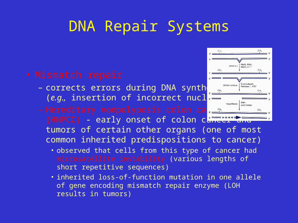
• Mismatch repair– corrects errors during DNA synthesis
(e.g., insertion of incorrect nucleotide)
– Hereditary nonpolyposis colon cancer (HNPCC) - early onset of colon cancer and tumors of certain other organs (one of most common inherited predispositions to cancer)
• observed that cells from this type of cancer had microsatellite instability (various lengths of short repetitive sequences)
• inherited loss-of-function mutation in one allele of gene encoding mismatch repair enzyme (LOH results in tumors)

DNA Repair Systems
• Transforming growth factor (TGF)– inhibits the growth of most cells– TGF receptor is heteromer of type I
(RI) and II (RII) receptor molecules– TGF binding activates Smad proteins
that translocate to the nucleus and activate gene encoding inhibitor of protease and cell-cycle inhibitor
– Due to loss of mismatch repair system, RII gene (contains A10 sequence) frequently mutated causing a frame shift, abolishes production of normal receptor
– Loss of TGF signaling promotes cell proliferation and development of malignancy

Cancer Chemotherapy• Seven major classes of compounds that have been
widely used in cancer treatment– Many of these agents used because they inhibit DNA
synthesis (malignant tumors typified by unrestrained cell division)
– Also likely to damage normal tissues undergoing continuous division (e.g., gut, bone marrow)
– growth fraction (% tumor cells constantly in cycle) is important for cancer chemotherapy
• tumors with high growth fraction usually more responsive than cells in G0

Resistance to cancer chemotherapy
• Development of resistance to agents used in cancer chemotherapy is a major problem– Tumor cells develop mechanisms that render
agents ineffective (acquired resistance)• Believed to reflect cells high spontaneous mutation
rates; thus, resistance not only to the drug but also to other structurally unrelated anticancer agents
• e.g., multidrug resistance - resistance to methotrexate develops, tumor cells may also be resistant to antitumor antibiotics (doxorubicin) and to plant compounds (vincristine)
• frequent cause of failure of cancer chemotherapy
Resistance to cancer chemotherapy• Molecular basis of multidrug resistance
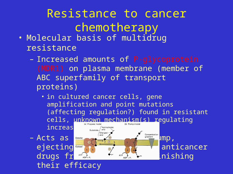
– Increased amounts of P-glycoprotein (MDR1) on plasma membrane (member of ABC superfamily of transport proteins)
• in cultured cancer cells, gene amplification and point mutations (affecting regulation?) found in resistant cells, unknown mechanism(s) regulating increase in vivo
– Acts as energy-dependent pump, ejecting a wide variety of anticancer drugs from cells, thus diminishing their efficacy

Resistance to cancer chemotherapy
• A particular drug may never be effective against a target tumor cell (intrinsic resistance)– e.g., high capacity for metabolic inactivation of
administered drug (usually mediated by cytochrome P450 species)
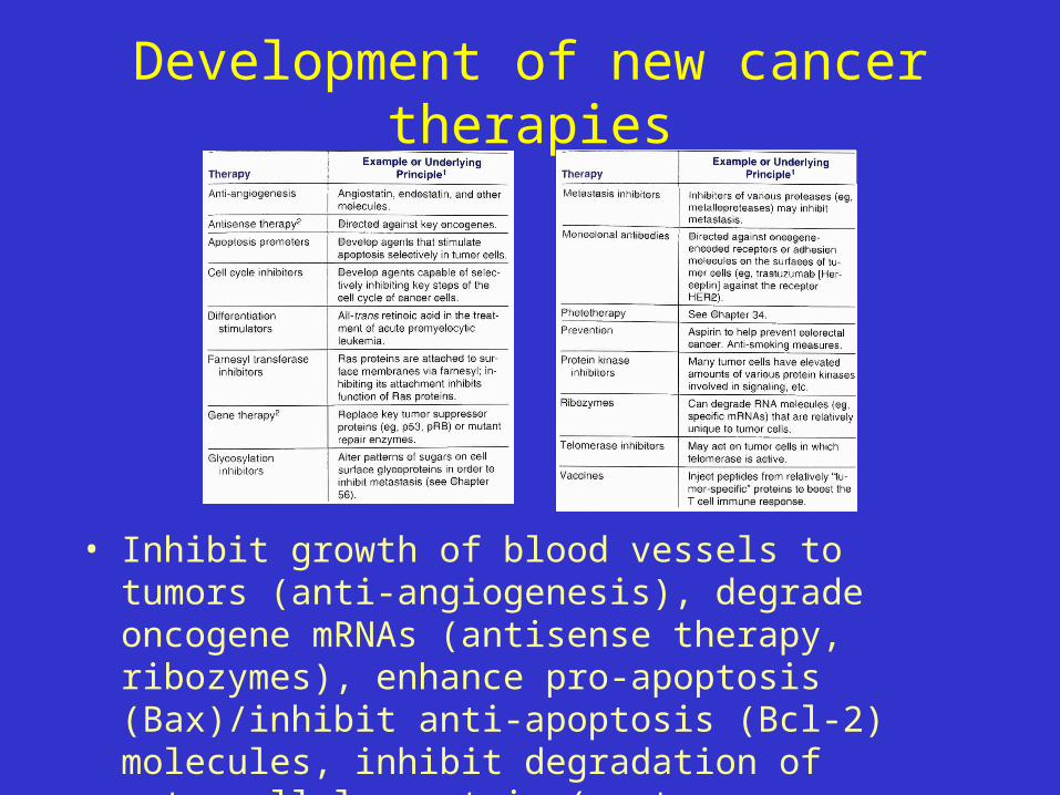
Development of new cancer therapies
• Inhibit growth of blood vessels to tumors (anti-angiogenesis), degrade oncogene mRNAs (antisense therapy, ribozymes), enhance pro-apoptosis (Bax)/inhibit anti-apoptosis (Bcl-2) molecules, inhibit degradation of extracellular matrix (protease inhibitors)