Medial Canthus Syndrome in Dogs – Chronic Tearing, Pigment ... · In this type of entropion, the...
14
______________________________ From Chronic Diseases, Proceedings of a Symposium sponsored by Schering-Plough Animal Health. Copyright ©2008 Schering-Plough Animal Health. All Rights Reserved. The opinions expressed in this article are those of the author and do not necessarily reflect the official label recommendations and point of view of the company or companies that manufacture and/or market any of the pharmaceutical agents referred to. Medial Canthus Syndrome in Dogs – Chronic Tearing, Pigment, Medial Entropion, and Trichiasis Sheryl G. Krohne, BA, DVM, MS, Diplomate ACVO School of Veterinary Medicine Purdue University West Lafayette, Indiana Abstract Chronic corneal and conjunctival irritation in dogs is often caused by anatomic eyelid abnormalities, particularly in brachycephalic and shallow-orbit breeds. This syndrome has been called “medial canthus syndrome” by some authors and may cause pigmentary keratitis, epiphora, and occasionally ulcers. Most of these abnormalities may be improved or controlled with surgery (blepharoplasty), resulting in the decrease of clinical symptoms and improved comfort of the patient. Key Concepts x Chronic tearing, corneal and conjunctival pigment, and conjunctivitis in dogs are often the result of breed-related conformational abnormalities. x The abnormalities may be grouped into two larger categories: 1) those often seen in brachycephalic and shallow-orbit breeds (the majority of the cases); and 2) those found in mesocephalic breeds with laxity of the eyelid margin. x The shallow-orbit breed abnormalities may include an abnormally large palpebral fissure (congenital euryblepharon); trichiasis from the nasal fold, medial caruncle and eyelids; medial inferior entropion; lagophthalmos with exposure keratoconjunctivitis, decreased corneal sensation, pigmentary keratitis or conjunctivitis; and chronic tearing. x Breeds with deeper-set eyes may have medial canthus eyelid furrows, and an ectropion and entropion combination or diamond-shaped eyelid openings. x Both groups may have the additional problems of distichia, ectopic cilia, imperforate puncta (uncommon), and secondary Staphylococcus sp. allergic conjunctivitis. x Most of these abnormalities may be improved or controlled with surgery (blepharoplasty), resulting in the decrease of clinical symptom, and improved comfort of the patient. x Complete resolution of the clinical signs is uncommon; therefore, client education is important so that expectations for improvement are realistic.
Transcript of Medial Canthus Syndrome in Dogs – Chronic Tearing, Pigment ... · In this type of entropion, the...
______________________________
From Chronic Diseases, Proceedings of a Symposium sponsored by Schering-Plough Animal Health. Copyright ©2008 Schering-Plough Animal Health. All Rights Reserved. The opinions expressed in this article are those of the author and do not necessarily reflect the official label recommendations and point of view of the company or companies that manufacture and/or market any of the pharmaceutical agents referred to.
Medial Canthus Syndrome in Dogs – Chronic Tearing, Pigment, Medial Entropion, and Trichiasis Sheryl G. Krohne, BA, DVM, MS, Diplomate ACVO School of Veterinary Medicine Purdue University West Lafayette, Indiana Abstract Chronic corneal and conjunctival irritation in dogs is often caused by anatomic eyelid abnormalities, particularly in brachycephalic and shallow-orbit breeds. This syndrome has been called “medial canthus syndrome” by some authors and may cause pigmentary keratitis, epiphora, and occasionally ulcers. Most of these abnormalities may be improved or controlled with surgery (blepharoplasty), resulting in the decrease of clinical symptoms and improved comfort of the patient. Key Concepts x Chronic tearing, corneal and conjunctival pigment, and conjunctivitis in dogs are often the result of
breed-related conformational abnormalities. x The abnormalities may be grouped into two larger categories: 1) those often seen in brachycephalic
and shallow-orbit breeds (the majority of the cases); and 2) those found in mesocephalic breeds with laxity of the eyelid margin.
x The shallow-orbit breed abnormalities may include an abnormally large palpebral fissure (congenital euryblepharon); trichiasis from the nasal fold, medial caruncle and eyelids; medial inferior entropion; lagophthalmos with exposure keratoconjunctivitis, decreased corneal sensation, pigmentary keratitis or conjunctivitis; and chronic tearing.
x Breeds with deeper-set eyes may have medial canthus eyelid furrows, and an ectropion and entropion combination or diamond-shaped eyelid openings.
x Both groups may have the additional problems of distichia, ectopic cilia, imperforate puncta (uncommon), and secondary Staphylococcus sp. allergic conjunctivitis.
x Most of these abnormalities may be improved or controlled with surgery (blepharoplasty), resulting in the decrease of clinical symptom, and improved comfort of the patient.
x Complete resolution of the clinical signs is uncommon; therefore, client education is important so that expectations for improvement are realistic.
Chronic Diseases x Symposium Proceedings
2
Chronic tearing, corneal and conjunctival pigment, and conjunctivitis in dogs are often the result of breed-related conformational abnormalities. This syndrome has been given many names, usually reflecting the abnormalities which are present in each individual patient. The abnormalities may be grouped into two larger categories:
x Those seen in brachycephalic and shallow-orbit breeds (the majority of the cases) x Those found in mesocephalic breeds with laxity of the eyelid margin
The shallow-orbit breed abnormalities may include an abnormally large palpebral fissure (congenital euryblepharon); trichiasis from the nasal fold in brachycephalic dogs, medial caruncle and eyelids; medial inferior entropion; lagophthalmos with exposure keratoconjunctivitis, decreased corneal sensation, pigmentary keratitis or conjunctivitis; and chronic tearing. Breeds with deeper-set eyes may have medial canthus eyelid furrows, and an ectropion and entropion combination or diamond-shaped eyelid openings. Both groups may have the additional problems of distichia, ectopic cilia, imperforate puncta (uncommon), and secondary Staphylococcus sp. allergic conjunctivitis. Most of these abnormalities may be improved or controlled with surgery (blepharoplasty), resulting in the decrease of clinical symptoms, and improved comfort of the patient. Complete resolution of the clinical signs is uncommon; therefore, client education is important so that expectations for improvement are realistic. A complete eye examination should be performed on all animals to rule out other causes of these symptoms. This exam should include flushing the nasolacrimal ducts to rule out blockage or abnormal ducts and puncta, corneal staining to check for ulceration, Schirmer tear tests to rule out dry eye, and intraocular pressure measurement to rule out uveitis and ocular hypertension. The syndrome is diagnosed when one or a combination of the abnormalities listed below is present. Some dogs will present with blepharospasm—intermittent or constant—depending on whether they have good corneal sensation, and whether the irritant is enough to induce squinting. Even when there is no squinting, the eyelid conformation and the trichiasis may be causing the clinical changes of epiphora and excess pigmentation and inflammation. Distichia usually will cause squinting when it is a clinical problem. Medial Canthus Syndrome in Shallow-Orbit Breeds
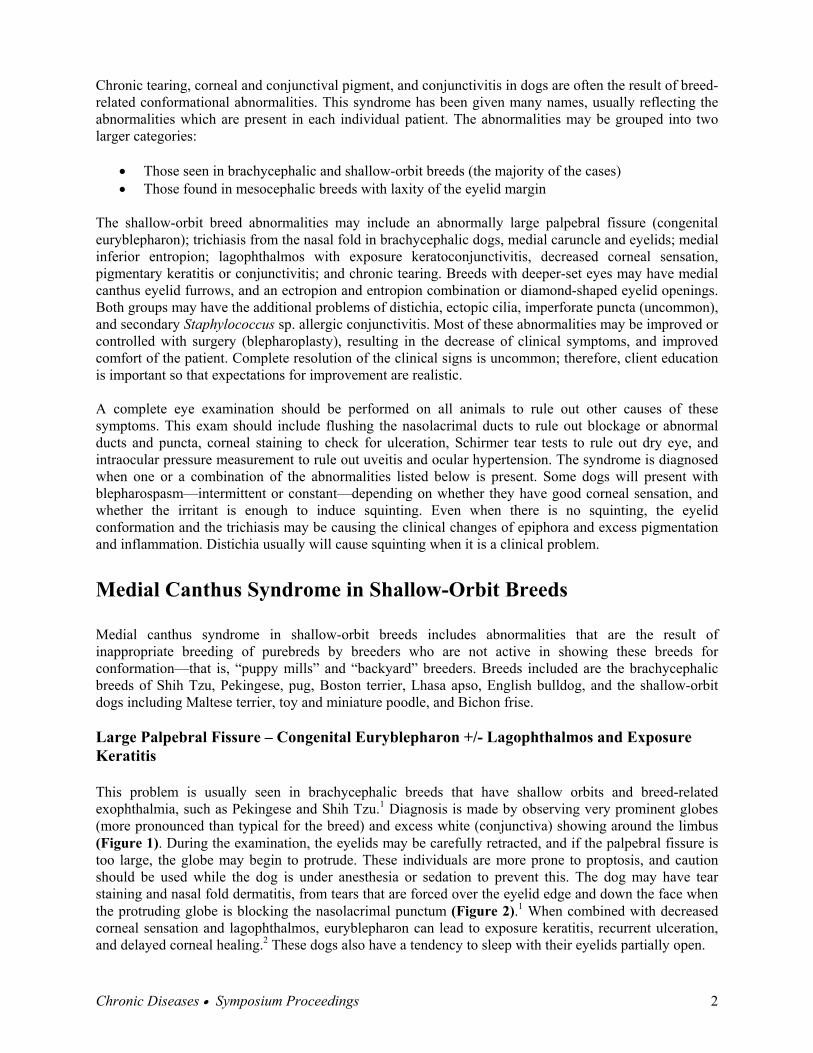
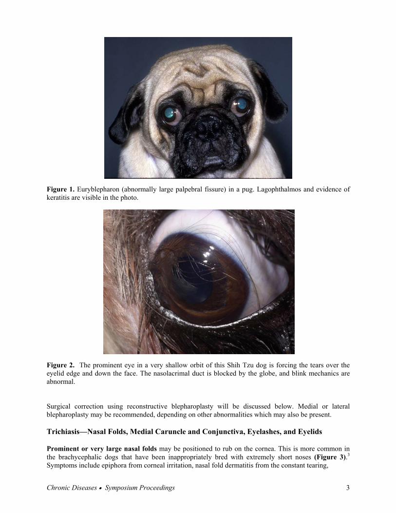
Medial canthus syndrome in shallow-orbit breeds includes abnormalities that are the result of inappropriate breeding of purebreds by breeders who are not active in showing these breeds for conformation—that is, “puppy mills” and “backyard” breeders. Breeds included are the brachycephalic breeds of Shih Tzu, Pekingese, pug, Boston terrier, Lhasa apso, English bulldog, and the shallow-orbit dogs including Maltese terrier, toy and miniature poodle, and Bichon frise. Large Palpebral Fissure – Congenital Euryblepharon +/- Lagophthalmos and Exposure Keratitis This problem is usually seen in brachycephalic breeds that have shallow orbits and breed-related exophthalmia, such as Pekingese and Shih Tzu.1 Diagnosis is made by observing very prominent globes (more pronounced than typical for the breed) and excess white (conjunctiva) showing around the limbus (Figure 1). During the examination, the eyelids may be carefully retracted, and if the palpebral fissure is too large, the globe may begin to protrude. These individuals are more prone to proptosis, and caution should be used while the dog is under anesthesia or sedation to prevent this. The dog may have tear staining and nasal fold dermatitis, from tears that are forced over the eyelid edge and down the face when the protruding globe is blocking the nasolacrimal punctum (Figure 2).1 When combined with decreased corneal sensation and lagophthalmos, euryblepharon can lead to exposure keratitis, recurrent ulceration, and delayed corneal healing.2 These dogs also have a tendency to sleep with their eyelids partially open.
Chronic Diseases x Symposium Proceedings
3
Figure 1. Euryblepharon (abnormally large palpebral fissure) in a pug. Lagophthalmos and evidence of keratitis are visible in the photo.
Figure 2. The prominent eye in a very shallow orbit of this Shih Tzu dog is forcing the tears over the eyelid edge and down the face. The nasolacrimal duct is blocked by the globe, and blink mechanics are abnormal. Surgical correction using reconstructive blepharoplasty will be discussed below. Medial or lateral blepharoplasty may be recommended, depending on other abnormalities which may also be present.
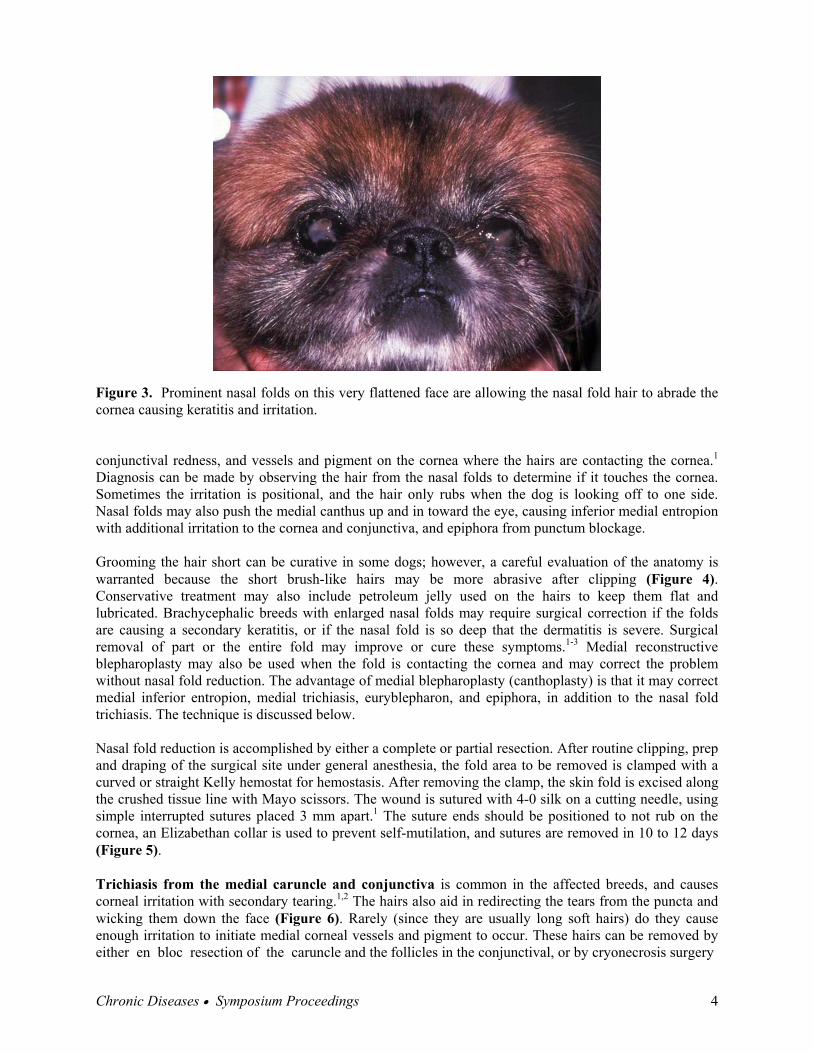
Trichiasis—Nasal Folds, Medial Caruncle and Conjunctiva, Eyelashes, and Eyelids Prominent or very large nasal folds may be positioned to rub on the cornea. This is more common in the brachycephalic dogs that have been inappropriately bred with extremely short noses (Figure 3).3 Symptoms include epiphora from corneal irritation, nasal fold dermatitis from the constant tearing,
Chronic Diseases x Symposium Proceedings
4
Figure 3. Prominent nasal folds on this very flattened face are allowing the nasal fold hair to abrade the cornea causing keratitis and irritation. conjunctival redness, and vessels and pigment on the cornea where the hairs are contacting the cornea.1 Diagnosis can be made by observing the hair from the nasal folds to determine if it touches the cornea. Sometimes the irritation is positional, and the hair only rubs when the dog is looking off to one side. Nasal folds may also push the medial canthus up and in toward the eye, causing inferior medial entropion with additional irritation to the cornea and conjunctiva, and epiphora from punctum blockage.
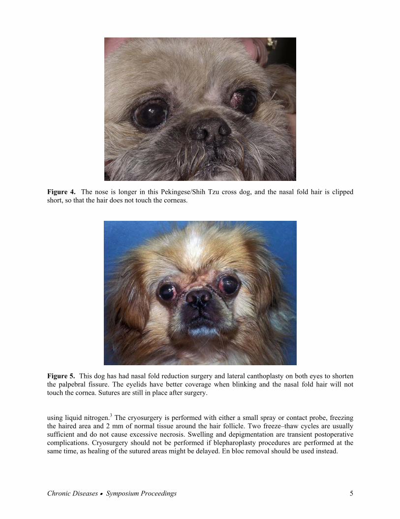
Grooming the hair short can be curative in some dogs; however, a careful evaluation of the anatomy is warranted because the short brush-like hairs may be more abrasive after clipping (Figure 4). Conservative treatment may also include petroleum jelly used on the hairs to keep them flat and lubricated. Brachycephalic breeds with enlarged nasal folds may require surgical correction if the folds are causing a secondary keratitis, or if the nasal fold is so deep that the dermatitis is severe. Surgical removal of part or the entire fold may improve or cure these symptoms.1-3 Medial reconstructive blepharoplasty may also be used when the fold is contacting the cornea and may correct the problem without nasal fold reduction. The advantage of medial blepharoplasty (canthoplasty) is that it may correct medial inferior entropion, medial trichiasis, euryblepharon, and epiphora, in addition to the nasal fold trichiasis. The technique is discussed below. Nasal fold reduction is accomplished by either a complete or partial resection. After routine clipping, prep and draping of the surgical site under general anesthesia, the fold area to be removed is clamped with a curved or straight Kelly hemostat for hemostasis. After removing the clamp, the skin fold is excised along the crushed tissue line with Mayo scissors. The wound is sutured with 4-0 silk on a cutting needle, using simple interrupted sutures placed 3 mm apart.1 The suture ends should be positioned to not rub on the cornea, an Elizabethan collar is used to prevent self-mutilation, and sutures are removed in 10 to 12 days (Figure 5). Trichiasis from the medial caruncle and conjunctiva is common in the affected breeds, and causes corneal irritation with secondary tearing.1,2 The hairs also aid in redirecting the tears from the puncta and wicking them down the face (Figure 6). Rarely (since they are usually long soft hairs) do they cause enough irritation to initiate medial corneal vessels and pigment to occur. These hairs can be removed by either en bloc resection of the caruncle and the follicles in the conjunctival, or by cryonecrosis surgery
Chronic Diseases x Symposium Proceedings
5
Figure 4. The nose is longer in this Pekingese/Shih Tzu cross dog, and the nasal fold hair is clipped short, so that the hair does not touch the corneas.
Figure 5. This dog has had nasal fold reduction surgery and lateral canthoplasty on both eyes to shorten the palpebral fissure. The eyelids have better coverage when blinking and the nasal fold hair will not touch the cornea. Sutures are still in place after surgery. using liquid nitrogen.3 The cryosurgery is performed with either a small spray or contact probe, freezing the haired area and 2 mm of normal tissue around the hair follicle. Two freeze–thaw cycles are usually sufficient and do not cause excessive necrosis. Swelling and depigmentation are transient postoperative complications. Cryosurgery should not be performed if blepharoplasty procedures are performed at the same time, as healing of the sutured areas might be delayed. En bloc removal should be used instead.
Chronic Diseases x Symposium Proceedings
6
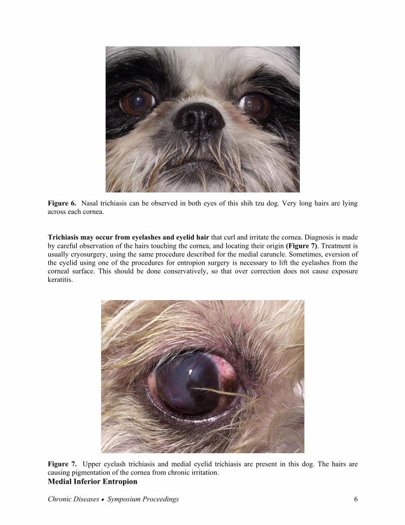
Figure 6. Nasal trichiasis can be observed in both eyes of this shih tzu dog. Very long hairs are lying across each cornea. Trichiasis may occur from eyelashes and eyelid hair that curl and irritate the cornea. Diagnosis is made by careful observation of the hairs touching the cornea, and locating their origin (Figure 7). Treatment is usually cryosurgery, using the same procedure described for the medial caruncle. Sometimes, eversion of the eyelid using one of the procedures for entropion surgery is necessary to lift the eyelashes from the corneal surface. This should be done conservatively, so that over correction does not cause exposure keratitis.
Figure 7. Upper eyelash trichiasis and medial eyelid trichiasis are present in this dog. The hairs are causing pigmentation of the cornea from chronic irritation. Medial Inferior Entropion
Chronic Diseases x Symposium Proceedings
7
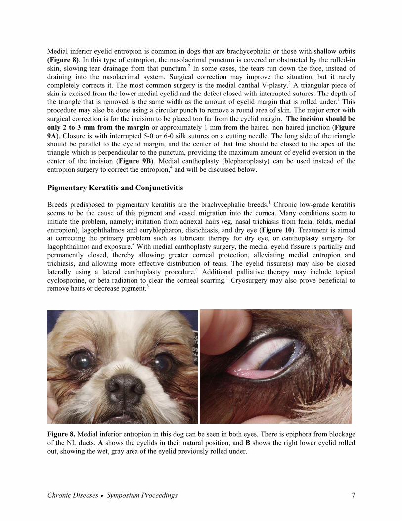
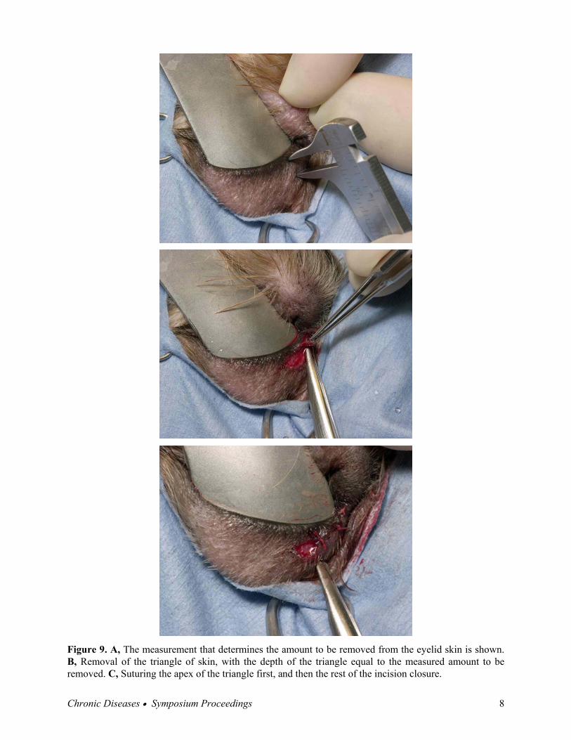
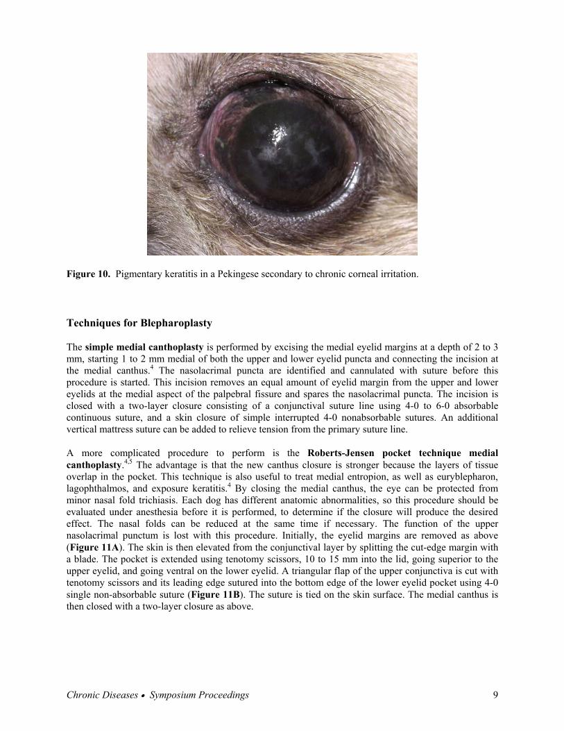
Medial inferior eyelid entropion is common in dogs that are brachycephalic or those with shallow orbits (Figure 8). In this type of entropion, the nasolacrimal punctum is covered or obstructed by the rolled-in skin, slowing tear drainage from that punctum.2 In some cases, the tears run down the face, instead of draining into the nasolacrimal system. Surgical correction may improve the situation, but it rarely completely corrects it. The most common surgery is the medial canthal V-plasty.2 A triangular piece of skin is excised from the lower medial eyelid and the defect closed with interrupted sutures. The depth of the triangle that is removed is the same width as the amount of eyelid margin that is rolled under.1 This procedure may also be done using a circular punch to remove a round area of skin. The major error with surgical correction is for the incision to be placed too far from the eyelid margin. The incision should be only 2 to 3 mm from the margin or approximately 1 mm from the haired–non-haired junction (Figure 9A). Closure is with interrupted 5-0 or 6-0 silk sutures on a cutting needle. The long side of the triangle should be parallel to the eyelid margin, and the center of that line should be closed to the apex of the triangle which is perpendicular to the punctum, providing the maximum amount of eyelid eversion in the center of the incision (Figure 9B). Medial canthoplasty (blepharoplasty) can be used instead of the entropion surgery to correct the entropion,4 and will be discussed below. Pigmentary Keratitis and Conjunctivitis Breeds predisposed to pigmentary keratitis are the brachycephalic breeds.1 Chronic low-grade keratitis seems to be the cause of this pigment and vessel migration into the cornea. Many conditions seem to initiate the problem, namely; irritation from adnexal hairs (eg, nasal trichiasis from facial folds, medial entropion), lagophthalmos and euryblepharon, distichiasis, and dry eye (Figure 10). Treatment is aimed at correcting the primary problem such as lubricant therapy for dry eye, or canthoplasty surgery for lagophthalmos and exposure.4 With medial canthoplasty surgery, the medial eyelid fissure is partially and permanently closed, thereby allowing greater corneal protection, alleviating medial entropion and trichiasis, and allowing more effective distribution of tears. The eyelid fissure(s) may also be closed laterally using a lateral canthoplasty procedure.4 Additional palliative therapy may include topical cyclosporine, or beta-radiation to clear the corneal scarring.1 Cryosurgery may also prove beneficial to remove hairs or decrease pigment.3
Figure 8. Medial inferior entropion in this dog can be seen in both eyes. There is epiphora from blockage of the NL ducts. A shows the eyelids in their natural position, and B shows the right lower eyelid rolled out, showing the wet, gray area of the eyelid previously rolled under.
Chronic Diseases x Symposium Proceedings
8
Figure 9. A, The measurement that determines the amount to be removed from the eyelid skin is shown. B, Removal of the triangle of skin, with the depth of the triangle equal to the measured amount to be removed. C, Suturing the apex of the triangle first, and then the rest of the incision closure.
Chronic Diseases x Symposium Proceedings
9
Figure 10. Pigmentary keratitis in a Pekingese secondary to chronic corneal irritation.
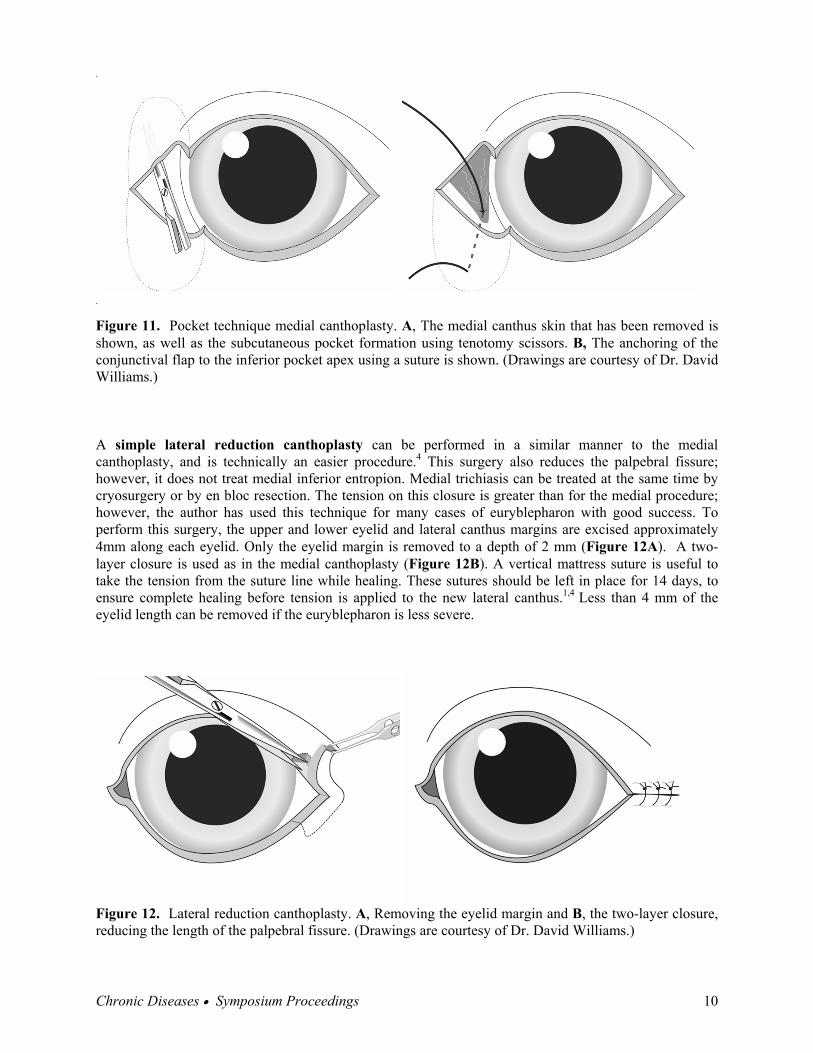
Techniques for Blepharoplasty The simple medial canthoplasty is performed by excising the medial eyelid margins at a depth of 2 to 3 mm, starting 1 to 2 mm medial of both the upper and lower eyelid puncta and connecting the incision at the medial canthus.4 The nasolacrimal puncta are identified and cannulated with suture before this procedure is started. This incision removes an equal amount of eyelid margin from the upper and lower eyelids at the medial aspect of the palpebral fissure and spares the nasolacrimal puncta. The incision is closed with a two-layer closure consisting of a conjunctival suture line using 4-0 to 6-0 absorbable continuous suture, and a skin closure of simple interrupted 4-0 nonabsorbable sutures. An additional vertical mattress suture can be added to relieve tension from the primary suture line. A more complicated procedure to perform is the Roberts-Jensen pocket technique medial canthoplasty.4,5 The advantage is that the new canthus closure is stronger because the layers of tissue overlap in the pocket. This technique is also useful to treat medial entropion, as well as euryblepharon, lagophthalmos, and exposure keratitis.4 By closing the medial canthus, the eye can be protected from minor nasal fold trichiasis. Each dog has different anatomic abnormalities, so this procedure should be evaluated under anesthesia before it is performed, to determine if the closure will produce the desired effect. The nasal folds can be reduced at the same time if necessary. The function of the upper nasolacrimal punctum is lost with this procedure. Initially, the eyelid margins are removed as above (Figure 11A). The skin is then elevated from the conjunctival layer by splitting the cut-edge margin with a blade. The pocket is extended using tenotomy scissors, 10 to 15 mm into the lid, going superior to the upper eyelid, and going ventral on the lower eyelid. A triangular flap of the upper conjunctiva is cut with tenotomy scissors and its leading edge sutured into the bottom edge of the lower eyelid pocket using 4-0 single non-absorbable suture (Figure 11B). The suture is tied on the skin surface. The medial canthus is then closed with a two-layer closure as above.
Chronic Diseases x Symposium Proceedings
10
Figure 11. Pocket technique medial canthoplasty. A, The medial canthus skin that has been removed is shown, as well as the subcutaneous pocket formation using tenotomy scissors. B, The anchoring of the conjunctival flap to the inferior pocket apex using a suture is shown. (Drawings are courtesy of Dr. David Williams.) A simple lateral reduction canthoplasty can be performed in a similar manner to the medial canthoplasty, and is technically an easier procedure.4 This surgery also reduces the palpebral fissure; however, it does not treat medial inferior entropion. Medial trichiasis can be treated at the same time by cryosurgery or by en bloc resection. The tension on this closure is greater than for the medial procedure; however, the author has used this technique for many cases of euryblepharon with good success. To perform this surgery, the upper and lower eyelid and lateral canthus margins are excised approximately 4mm along each eyelid. Only the eyelid margin is removed to a depth of 2 mm (Figure 12A). A two-layer closure is used as in the medial canthoplasty (Figure 12B). A vertical mattress suture is useful to take the tension from the suture line while healing. These sutures should be left in place for 14 days, to ensure complete healing before tension is applied to the new lateral canthus.1,4 Less than 4 mm of the eyelid length can be removed if the euryblepharon is less severe.
Figure 12. Lateral reduction canthoplasty. A, Removing the eyelid margin and B, the two-layer closure, reducing the length of the palpebral fissure. (Drawings are courtesy of Dr. David Williams.)
Chronic Diseases x Symposium Proceedings
11
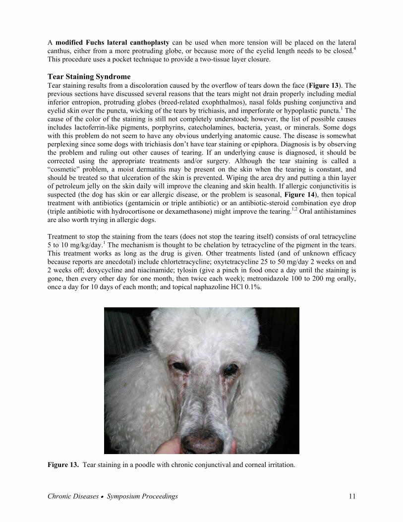
A modified Fuchs lateral canthoplasty can be used when more tension will be placed on the lateral canthus, either from a more protruding globe, or because more of the eyelid length needs to be closed.4 This procedure uses a pocket technique to provide a two-tissue layer closure. Tear Staining Syndrome Tear staining results from a discoloration caused by the overflow of tears down the face (Figure 13). The previous sections have discussed several reasons that the tears might not drain properly including medial inferior entropion, protruding globes (breed-related exophthalmos), nasal folds pushing conjunctiva and eyelid skin over the puncta, wicking of the tears by trichiasis, and imperforate or hypoplastic puncta.1 The cause of the color of the staining is still not completely understood; however, the list of possible causes includes lactoferrin-like pigments, porphyrins, catecholamines, bacteria, yeast, or minerals. Some dogs with this problem do not seem to have any obvious underlying anatomic cause. The disease is somewhat perplexing since some dogs with trichiasis don’t have tear staining or epiphora. Diagnosis is by observing the problem and ruling out other causes of tearing. If an underlying cause is diagnosed, it should be corrected using the appropriate treatments and/or surgery. Although the tear staining is called a “cosmetic” problem, a moist dermatitis may be present on the skin when the tearing is constant, and should be treated so that ulceration of the skin is prevented. Wiping the area dry and putting a thin layer of petroleum jelly on the skin daily will improve the cleaning and skin health. If allergic conjunctivitis is suspected (the dog has skin or ear allergic disease, or the problem is seasonal, Figure 14), then topical treatment with antibiotics (gentamicin or triple antibiotic) or an antibiotic-steroid combination eye drop (triple antibiotic with hydrocortisone or dexamethasone) might improve the tearing.1,2 Oral antihistamines are also worth trying in allergic dogs. Treatment to stop the staining from the tears (does not stop the tearing itself) consists of oral tetracycline 5 to 10 mg/kg/day.1 The mechanism is thought to be chelation by tetracycline of the pigment in the tears. This treatment works as long as the drug is given. Other treatments listed (and of unknown efficacy because reports are anecdotal) include chlortetracycline; oxytetracycline 25 to 50 mg/day 2 weeks on and 2 weeks off; doxycycline and niacinamide; tylosin (give a pinch in food once a day until the staining is gone, then every other day for one month, then twice each week); metronidazole 100 to 200 mg orally, once a day for 10 days of each month; and topical naphazoline HCl 0.1%.
Figure 13. Tear staining in a poodle with chronic conjunctival and corneal irritation.
Chronic Diseases x Symposium Proceedings
12
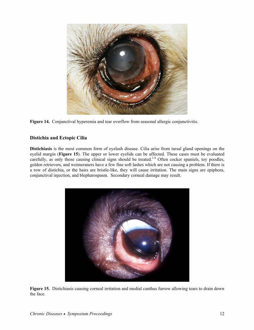
Figure 14. Conjunctival hyperemia and tear overflow from seasonal allergic conjunctivitis. Distichia and Ectopic Cilia
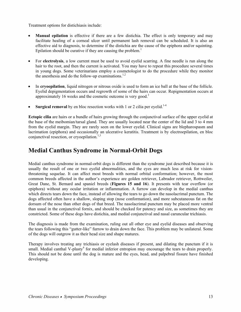
Distichiasis is the most common form of eyelash disease. Cilia arise from tarsal gland openings on the eyelid margin (Figure 15). The upper or lower eyelids can be affected. These cases must be evaluated carefully, as only those causing clinical signs should be treated.1-4 Often cocker spaniels, toy poodles, golden retrievers, and weimeraners have a few fine soft lashes which are not causing a problem. If there is a row of distichia, or the hairs are bristle-like, they will cause irritation. The main signs are epiphora, conjunctival injection, and blepharospasm. Secondary corneal damage may result.
Figure 15. Distichiasis causing corneal irritation and medial canthus furrow allowing tears to drain down the face.
Chronic Diseases x Symposium Proceedings
13
Treatment options for distichiasis include: x Manual epilation is effective if there are a few distichia. The effect is only temporary and may
facilitate healing of a corneal ulcer until permanent lash removal can be scheduled. It is also an effective aid to diagnosis, to determine if the distichia are the cause of the epiphora and/or squinting. Epilation should be curative if they are causing the problem.1
x For electrolysis, a low current must be used to avoid eyelid scarring. A fine needle is run along the
hair to the root, and then the current is activated. You may have to repeat this procedure several times in young dogs. Some veterinarians employ a cosmetologist to do the procedure while they monitor the anesthesia and do the follow-up examinations.1,2
x In cryoepilation, liquid nitrogen or nitrous oxide is used to form an ice ball at the base of the follicle.
Eyelid depigmentation occurs and regrowth of some of the hairs can occur. Repigmentation occurs at approximately 16 weeks and the cosmetic outcome is very good.3
x Surgical removal by en bloc resection works with 1 or 2 cilia per eyelid.1-4
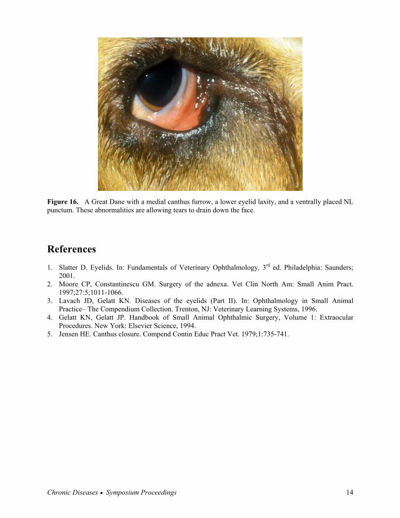
Ectopic cilia are hairs or a bundle of hairs growing through the conjunctival surface of the upper eyelid at the base of the meibomian/tarsal gland. They are usually located near the center of the lid and 3 to 4 mm from the eyelid margin. They are rarely seen on the lower eyelid. Clinical signs are blepharospasm and lacrimation (epiphora) and occasionally an ulcerative keratitis. Treatment is by electroepilation, en bloc conjunctival resection, or cryoepilation.1,3 Medial Canthus Syndrome in Normal-Orbit Dogs Medial canthus syndrome in normal-orbit dogs is different than the syndrome just described because it is usually the result of one or two eyelid abnormalities, and the eyes are much less at risk for vision-threatening sequelae. It can affect most breeds with normal orbital conformation; however, the most common breeds affected in the author’s experience are golden retriever, Labrador retriever, Rottweiler, Great Dane, St. Bernard and spaniel breeds (Figures 15 and 16). It presents with tear overflow (or epiphora) without any ocular irritation or inflammation. A furrow can develop in the medial canthus which directs tears down the face, instead of allowing the tears to go down the nasolacrimal punctum. The dogs affected often have a shallow, sloping stop (nose conformation), and more subcutaneous fat on the dorsum of the nose than other dogs of that breed. The nasolacrimal punctum may be placed more ventral than usual in the conjunctival fornix, and should be checked for patency and size, as sometimes they are constricted. Some of these dogs have distichia, and medial conjunctival and nasal caruncular trichiasis. The diagnosis is made from the examination, ruling out all other eye and eyelid diseases and observing the tears following this “gutter-like” furrow to drain down the face. This problem may be unilateral. Some of the dogs will outgrow it as their head size and shape matures. Therapy involves treating any trichiasis or eyelash diseases if present, and dilating the punctum if it is small. Medial canthal V-plasty4 for medial inferior entropion may encourage the tears to drain properly. This should not be done until the dog is mature and the eyes, head, and palpebral fissure have finished developing.
Chronic Diseases x Symposium Proceedings
14
Figure 16. A Great Dane with a medial canthus furrow, a lower eyelid laxity, and a ventrally placed NL punctum. These abnormalities are allowing tears to drain down the face.
References 1. Slatter D. Eyelids. In: Fundamentals of Veterinary Ophthalmology, 3rd ed. Philadelphia: Saunders;
2001. 2. Moore CP, Constantinescu GM. Surgery of the adnexa. Vet Clin North Am: Small Anim Pract.
1997;27:5;1011-1066. 3. Lavach JD, Gelatt KN. Diseases of the eyelids (Part II). In: Ophthalmology in Small Animal
Practice– The Compendium Collection. Trenton, NJ: Veterinary Learning Systems, 1996. 4. Gelatt KN, Gelatt JP. Handbook of Small Animal Ophthalmic Surgery, Volume 1: Extraocular
Procedures. New York: Elsevier Science, 1994. 5. Jensen HE. Canthus closure. Compend Contin Educ Pract Vet. 1979;1:735-741.