
MED12 controls the response to multiple cancer drugs through regulation of TGF-β receptor...
14
MED12 Controls the Response to Multiple Cancer Drugs through Regulation of TGF- b Receptor Signaling Sidong Huang, 1 Michael Ho ¨ lzel, 1,9 Theo Knijnenburg, 1 Andreas Schlicker, 1 Paul Roepman, 3,4 Ultan McDermott, 5 Mathew Garnett, 5 Wipawadee Grernrum, 1 Chong Sun, 1 Anirudh Prahallad, 1 Floris H. Groenendijk, 1 Lorenza Mittempergher, 1 Wouter Nijkamp, 1 Jacques Neefjes, 2 Ramon Salazar, 6 Peter ten Dijke, 7 Hidetaka Uramoto, 8 Fumihiro Tanaka, 8 Roderick L. Beijersbergen, 1 Lodewyk F.A. Wessels, 1 and Rene ´ Bernards 1,3,4, * 1 Division of Molecular Carcinogenesis, Cancer Genomics Center and Cancer Systems Biology Center 2 Division of Cell Biology The Netherlands Cancer Institute, Plesmanlaan 121, 1066 CX Amsterdam, The Netherlands 3 Agendia Inc., 22 Morgan Drive, Irvine, CA 92618, USA 4 Agendia NV, Science Park 406, 1098 XH Amsterdam, The Netherlands 5 Wellcome Trust Sanger Institute, Wellcome Trust Genome Campus, Hinxton, Cambridge CB10 1SA, UK 6 Institut Catala ` d’Oncologia (IDIBELL), 08908 l’Hospitalet de Llobregat, Spain 7 Department of Molecular Cell Biology and Centre for Biomedical Genetics, Leiden University Medical Center, Postbus 9600, 2300 RC Leiden, The Netherlands 8 Second Department of Surgery, School of Medicine, University of Occupational and Environmental Health, 1-1 Iseigaoka, Yahatanishi-ku, Kitakyushu 807-8555, Japan 9 Present address: Institute of Clinical Chemistry and Clinical Pharmacology, University Hospital Bonn, 53127 Bonn, Germany *Correspondence: [email protected] http://dx.doi.org/10.1016/j.cell.2012.10.035 SUMMARY Inhibitors of the ALK and EGF receptor tyrosine kinases provoke dramatic but short-lived responses in lung cancers harboring EML4-ALK translocations or activating mutations of EGFR, respectively. We used a large-scale RNAi screen to identify MED12, a component of the transcriptional MEDIATOR com- plex that is mutated in cancers, as a determinant of response to ALK and EGFR inhibitors. MED12 is in part cytoplasmic where it negatively regulates TGF- bR2 through physical interaction. MED12 suppres- sion therefore results in activation of TGF-bR signaling, which is both necessary and sufficient for drug resistance. TGF-b signaling causes MEK/ERK activation, and consequently MED12 suppression also confers resistance to MEK and BRAF inhibitors in other cancers. MED12 loss induces an EMT-like phenotype, which is associated with chemotherapy resistance in colon cancer patients and to gefitinib in lung cancer. Inhibition of TGF-bR signaling re- stores drug responsiveness in MED12 KD cells, sug- gesting a strategy to treat drug-resistant tumors that have lost MED12. INTRODUCTION Cancer therapy is often hampered by the rapid emergence of drug resistance. This is true not only for the conventional chemotherapies but also for the new generation of drugs target- ing those components that are mutated or deregulated in tumor cells. For example, treatment of metastatic non-small-cell lung cancers (NSCLCs) harboring activating mutations in the gene encoding the epidermal growth factor receptor (EGFR) leads to significant increases in progression-free survival. However, such responses are often short-lived, resulting in much less impressive patient benefit in terms of overall survival (Maemondo et al., 2010). This lack of long-term benefit is due to the emer- gence of drug-resistant variants. Development of resistance to targeted therapies is a general phenomenon and is also seen in BCR-ABL-translocated chronic myelogenous leukemias (CML) treated with imatinib (Gorre et al., 2001), BRAF mutant melanomas treated with the BRAF inhibitor vemurafenib (Chapman et al., 2011), and EML4-ALK-translocated NSCLCs treated with the ALK inhibitor crizotinib (Kwak et al., 2010). About half of the resistance seen in EGFR mutant NSCLCs treated with EGFR inhibitors can be explained by secondary mutations in the EGFR gene itself (Sequist et al., 2011). The T790M ‘‘gatekeeper’’ mutation in EGFR is critical for binding of competitive inhibitors to the ATP-binding pocket (Yun et al., 2008), allowing continued proliferation in the presence of the drug. Similar gatekeeper mutations have been found in BCR- ABL-positive CMLs treated with imatinib (Shah et al., 2002) and in EML4-ALK mutant NSCLCs treated with crizotinib (Choi et al., 2010). Resistance to targeted therapies that does not involve secondary mutations in the drug target itself is often caused by mutations in the signaling pathway downstream of the target. Thus, primary resistance to EGFR-targeted therapy in colon cancer is associated with mutations in KRAS (Karapetis et al., Cell 151, 937–950, November 21, 2012 ª2012 Elsevier Inc. 937
-
Upload
anirudh-prahallad -
Category
Documents
-
view
346 -
download
0
Transcript of MED12 controls the response to multiple cancer drugs through regulation of TGF-β receptor...
MED12 Controls the Response to MultipleCancer Drugs through Regulationof TGF-b Receptor SignalingSidong Huang,1 Michael Holzel,1,9 Theo Knijnenburg,1 Andreas Schlicker,1 Paul Roepman,3,4 Ultan McDermott,5
Mathew Garnett,5 Wipawadee Grernrum,1 Chong Sun,1 Anirudh Prahallad,1 Floris H. Groenendijk,1
Lorenza Mittempergher,1 Wouter Nijkamp,1 Jacques Neefjes,2 Ramon Salazar,6 Peter ten Dijke,7 Hidetaka Uramoto,8
Fumihiro Tanaka,8 Roderick L. Beijersbergen,1 Lodewyk F.A. Wessels,1 and Rene Bernards1,3,4,*1Division of Molecular Carcinogenesis, Cancer Genomics Center and Cancer Systems Biology Center2Division of Cell Biology
The Netherlands Cancer Institute, Plesmanlaan 121, 1066 CX Amsterdam, The Netherlands3Agendia Inc., 22 Morgan Drive, Irvine, CA 92618, USA4Agendia NV, Science Park 406, 1098 XH Amsterdam, The Netherlands5Wellcome Trust Sanger Institute, Wellcome Trust Genome Campus, Hinxton, Cambridge CB10 1SA, UK6Institut Catala d’Oncologia (IDIBELL), 08908 l’Hospitalet de Llobregat, Spain7Department of Molecular Cell Biology and Centre for Biomedical Genetics, Leiden University Medical Center, Postbus 9600,2300 RC Leiden, The Netherlands8Second Department of Surgery, School of Medicine, University of Occupational and Environmental Health, 1-1 Iseigaoka,
Yahatanishi-ku, Kitakyushu 807-8555, Japan9Present address: Institute of Clinical Chemistry and Clinical Pharmacology, University Hospital Bonn, 53127 Bonn, Germany*Correspondence: [email protected]
http://dx.doi.org/10.1016/j.cell.2012.10.035
SUMMARY
Inhibitors of the ALK and EGF receptor tyrosinekinases provoke dramatic but short-lived responsesin lung cancers harboring EML4-ALK translocationsor activating mutations of EGFR, respectively. Weused a large-scale RNAi screen to identify MED12,a component of the transcriptional MEDIATOR com-plex that is mutated in cancers, as a determinant ofresponse to ALK and EGFR inhibitors. MED12 is inpart cytoplasmic where it negatively regulates TGF-bR2 through physical interaction. MED12 suppres-sion therefore results in activation of TGF-bRsignaling, which is both necessary and sufficient fordrug resistance. TGF-b signaling causes MEK/ERKactivation, and consequently MED12 suppressionalso confers resistance to MEK and BRAF inhibitorsin other cancers. MED12 loss induces an EMT-likephenotype, which is associated with chemotherapyresistance in colon cancer patients and to gefitinibin lung cancer. Inhibition of TGF-bR signaling re-stores drug responsiveness in MED12KD cells, sug-gesting a strategy to treat drug-resistant tumorsthat have lost MED12.
INTRODUCTION
Cancer therapy is often hampered by the rapid emergence of
drug resistance. This is true not only for the conventional
chemotherapies but also for the new generation of drugs target-
ing those components that are mutated or deregulated in tumor
cells. For example, treatment of metastatic non-small-cell lung
cancers (NSCLCs) harboring activating mutations in the gene
encoding the epidermal growth factor receptor (EGFR) leads to
significant increases in progression-free survival. However,
such responses are often short-lived, resulting in much less
impressive patient benefit in terms of overall survival (Maemondo
et al., 2010). This lack of long-term benefit is due to the emer-
gence of drug-resistant variants. Development of resistance to
targeted therapies is a general phenomenon and is also seen
in BCR-ABL-translocated chronic myelogenous leukemias
(CML) treated with imatinib (Gorre et al., 2001), BRAF mutant
melanomas treated with the BRAF inhibitor vemurafenib
(Chapman et al., 2011), and EML4-ALK-translocated NSCLCs
treated with the ALK inhibitor crizotinib (Kwak et al., 2010).
About half of the resistance seen in EGFR mutant NSCLCs
treated with EGFR inhibitors can be explained by secondary
mutations in the EGFR gene itself (Sequist et al., 2011). The
T790M ‘‘gatekeeper’’ mutation in EGFR is critical for binding of
competitive inhibitors to the ATP-binding pocket (Yun et al.,
2008), allowing continued proliferation in the presence of the
drug. Similar gatekeeper mutations have been found in BCR-
ABL-positive CMLs treated with imatinib (Shah et al., 2002)
and in EML4-ALK mutant NSCLCs treated with crizotinib (Choi
et al., 2010).
Resistance to targeted therapies that does not involve
secondary mutations in the drug target itself is often caused by
mutations in the signaling pathway downstream of the target.
Thus, primary resistance to EGFR-targeted therapy in colon
cancer is associated with mutations in KRAS (Karapetis et al.,
Cell 151, 937–950, November 21, 2012 ª2012 Elsevier Inc. 937
A B
C
D
E
F
G
H
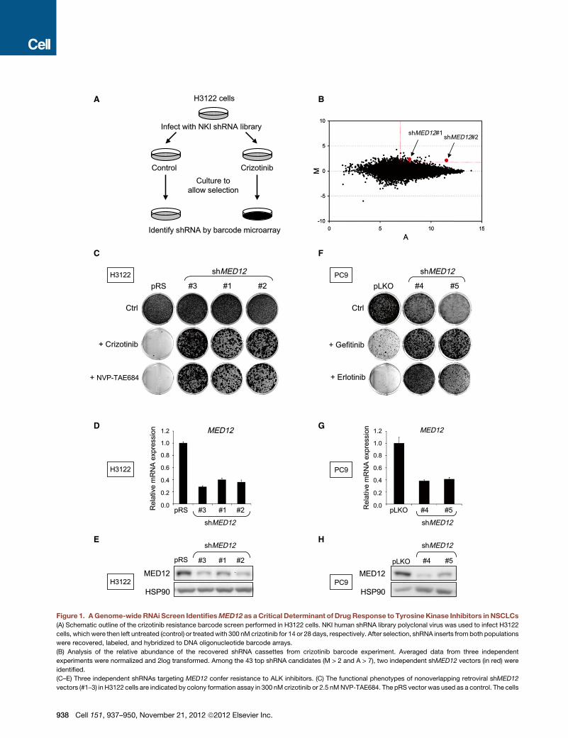
Figure 1. AGenome-wide RNAi Screen IdentifiesMED12 as aCritical Determinant of DrugResponse to Tyrosine Kinase Inhibitors in NSCLCs
(A) Schematic outline of the crizotinib resistance barcode screen performed in H3122 cells. NKI human shRNA library polyclonal virus was used to infect H3122
cells, whichwere then left untreated (control) or treatedwith 300 nM crizotinib for 14 or 28 days, respectively. After selection, shRNA inserts from both populations
were recovered, labeled, and hybridized to DNA oligonucleotide barcode arrays.
(B) Analysis of the relative abundance of the recovered shRNA cassettes from crizotinib barcode experiment. Averaged data from three independent
experiments were normalized and 2log transformed. Among the 43 top shRNA candidates (M > 2 and A > 7), two independent shMED12 vectors (in red) were
identified.
(C–E) Three independent shRNAs targeting MED12 confer resistance to ALK inhibitors. (C) The functional phenotypes of nonoverlapping retroviral shMED12
vectors (#1–3) in H3122 cells are indicated by colony formation assay in 300 nM crizotinib or 2.5 nMNVP-TAE684. The pRS vector was used as a control. The cells
938 Cell 151, 937–950, November 21, 2012 ª2012 Elsevier Inc.
2008). Similarly, acquired resistance to BRAF inhibition in
melanoma can result from an activating mutation in the MEK1
kinase that was not detectable in the primary tumor (Wagle
et al., 2011). Alternatively, resistance can result from activation
of a parallel pathway or in genes that feed into the downstream
signaling of the drug target. Thus, amplification of the MET
oncogene is found in EGFR drug-resistant NSCLC (Sequist
et al., 2011), and overexpression of COT, leading to activation
of MEK, can be a causal agent in BRAF resistance in melanoma
(Johannessen et al., 2010). At present, some 30% of the
resistance to EGFR-targeted therapies in NSCLCs cannot be
explained by any of the mechanisms described above (Sequist
et al., 2011).
Functional genetic screens provide a powerful tool to identify
novel components of signaling pathways and can help to identify
mechanisms of drug resistance in preclinical models of cancer
(Berns et al., 2007; Holzel et al., 2010). We describe here the
use of a large-scale loss-of-function genetic screen to identify
genes whose suppression can confer resistance to crizotinib in
a NSCLC cell line harboring an EML4-ALK translocation. We
identify a key component of the transcriptional MEDIATOR
complex, MED12, as a determinant of crizotinib response in
NSCLC. Remarkably, we find that suppression of MED12
also confers resistance to a range of cancer drugs, including
chemotherapy, in colon cancer, melanoma, and liver cancer.
We identify an unexpected activity of MED12 in regulating
transforming growth factor b (TGF-b) receptor signaling, as the
major mechanism of drug-resistance induction.
RESULTS
MED12 Suppression Confers Resistance to MultipleTyrosine Kinase Inhibitors in NSCLCsThe NSCLC cell line H3122 harbors an EML4-ALK translocation
and is exquisitely sensitive to the ALK inhibitors PF-02341066
(crizotinib) and NVP-TAE684 (McDermott et al., 2008). To identify
genetic determinants of resistance to ALK inhibitors in EML4-
ALK-translocated NSCLC, we performed a large-scale RNA
interference (RNAi) genetic screen with a collection of 24,000
short hairpin RNA (shRNA) vectors targeting 8,000 human genes
(Berns et al., 2004). As outlined in Figure 1A, we used a barcoding
technology to identify genes whose suppression causes resis-
tance to crizotinib in H3122 cells (Brummelkamp et al., 2006;
Holzel et al., 2010). The results are shown in Figure 1B. Each
dot in the M/A-plot represents one individual shRNA vector. M
and A values reflect relative enrichment and hybridization signal
intensity. Low-intensity spots are prone to technical artifacts and
are thus unreliable. Therefore we restricted our candidate selec-
tion by applying M/A cut-off values as indicated in Figure 1B. To
rule out ‘‘off-target’’ effects, we prioritized genes that are present
were fixed, stained, and photographed after 14 (untreated) or 28 days (treated). (D
MED12 mRNA levels by qRT-PCR. Error bars denote standard deviation (SD). (E)
(F–H) Suppression of MED12 also confers to EGFR inhibitors. (F) Colony forma
shMED12 vectors (#4 and #5) and that were cultured in 50 nM gefitinib or erlotinib.
(treated). (G) The level ofMED12KD by each of the shRNAs wasmeasured by exam
of knockdown of MED12 protein was measured by western blotting.
See also Figures S1 and S2.
with multiple shRNAs. Only one gene fulfilled these criteria:
MED12 encoding a component of the large MEDIATOR tran-
scriptional adaptor complex.
To validate MED12 as a gene whose suppression confers
resistance to crizotinib, we introduced the two MED12 shRNAs
(#1 and #2) from the library and one newly generated shRNA
(#3) into H3122 cells by retroviral infection. Empty vector (pRS)
or shRNA-targeting GFP (shGFP) served as controls. All three
distinct MED12 shRNAs conferred resistance to both crizotinib
and NVP-TAE684 (Figure 1C) and also suppressed MED12
mRNA and protein expression (Figures 1D and 1E). Expression
of additional independent lentiviral shMED12 vectors (#4 and
#5) in H3122 cells also conferred resistance to ALK inhibitors
(Figures S1A–S1C available online and data not shown). Further-
more, reconstitution of the RNAi-resistant murine Med12 cDNA
in MED12 knockdown (MED12KD) H3122 cells restored the
sensitivity of these cells to ALK inhibition (Figure S1). Suppres-
sion of MED12 also conferred resistance to the EGFR inhibitors
gefitinib or erlotinib in the EGFR mutant NSCLC cell lines PC9
and H3255 (Figures 1F–1H and data not shown). These results
establish a potential role for MED12 in resistance to ALK and
EGFR inhibitors.
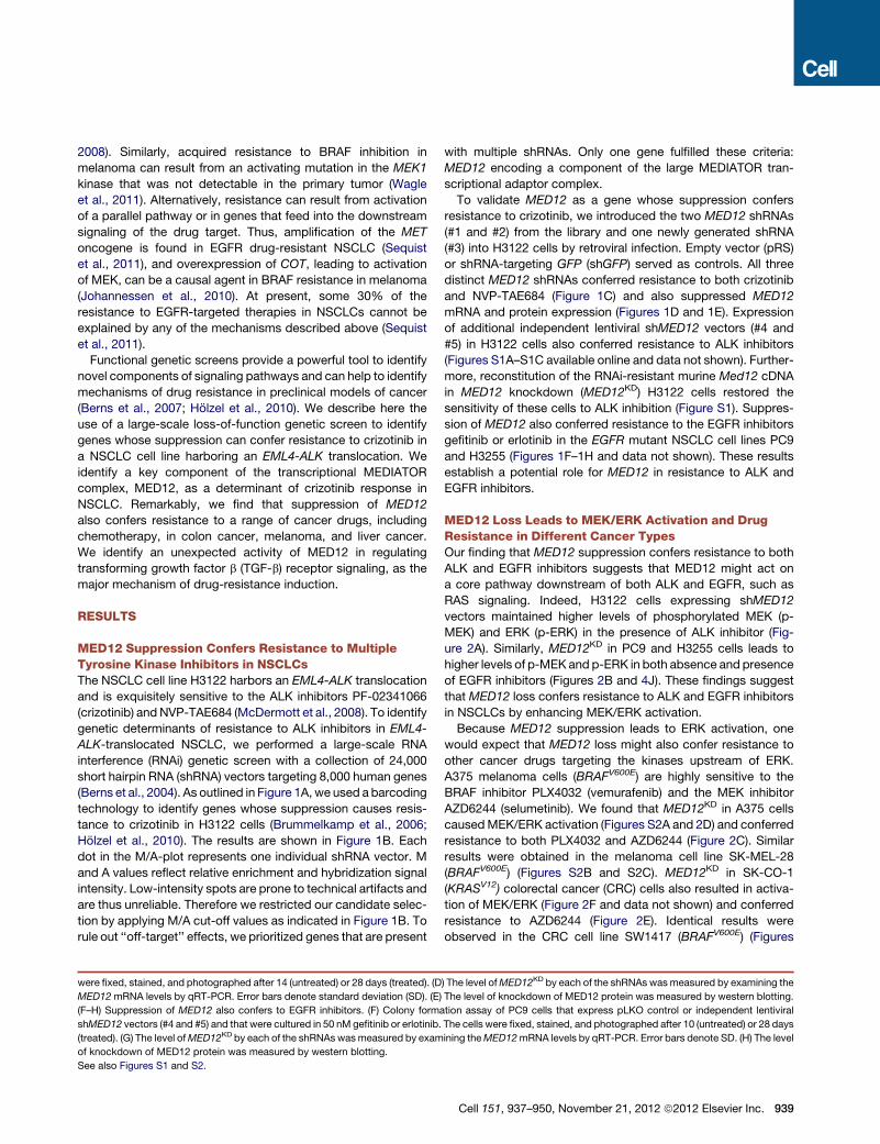
MED12 Loss Leads to MEK/ERK Activation and DrugResistance in Different Cancer TypesOur finding that MED12 suppression confers resistance to both
ALK and EGFR inhibitors suggests that MED12 might act on
a core pathway downstream of both ALK and EGFR, such as
RAS signaling. Indeed, H3122 cells expressing shMED12
vectors maintained higher levels of phosphorylated MEK (p-
MEK) and ERK (p-ERK) in the presence of ALK inhibitor (Fig-
ure 2A). Similarly, MED12KD in PC9 and H3255 cells leads to
higher levels of p-MEK and p-ERK in both absence and presence
of EGFR inhibitors (Figures 2B and 4J). These findings suggest
that MED12 loss confers resistance to ALK and EGFR inhibitors
in NSCLCs by enhancing MEK/ERK activation.
Because MED12 suppression leads to ERK activation, one
would expect that MED12 loss might also confer resistance to
other cancer drugs targeting the kinases upstream of ERK.
A375 melanoma cells (BRAFV600E) are highly sensitive to the
BRAF inhibitor PLX4032 (vemurafenib) and the MEK inhibitor
AZD6244 (selumetinib). We found that MED12KD in A375 cells
causedMEK/ERK activation (Figures S2A and 2D) and conferred
resistance to both PLX4032 and AZD6244 (Figure 2C). Similar
results were obtained in the melanoma cell line SK-MEL-28
(BRAFV600E) (Figures S2B and S2C). MED12KD in SK-CO-1
(KRASV12) colorectal cancer (CRC) cells also resulted in activa-
tion of MEK/ERK (Figure 2F and data not shown) and conferred
resistance to AZD6244 (Figure 2E). Identical results were
observed in the CRC cell line SW1417 (BRAFV600E) (Figures
) The level ofMED12KD by each of the shRNAs wasmeasured by examining the
The level of knockdown of MED12 protein was measured by western blotting.
tion assay of PC9 cells that express pLKO control or independent lentiviral
The cells were fixed, stained, and photographed after 10 (untreated) or 28 days
ining theMED12mRNA levels by qRT-PCR. Error bars denote SD. (H) The level
Cell 151, 937–950, November 21, 2012 ª2012 Elsevier Inc. 939
Figure 2. MED12 Suppression Leads to MEK/ERK Activation and Confers Multidrug Resistance in Different Cancer Types
(A and B)MED12KD results in an elevated level of phosphorylated MEK (p-MEK) and phosphorylated ERK (p-ERK). (A)MED12KD H3122 cells have higher p-MEK
and p-ERK levels. H3122 cells expressing pLKO or shMED12 vectors were grown in the absence or presence of 20 nM NVP-TAE684 for 6 hr, and the cell lysates
were harvested for western blotting analysis. (B) Elevated p-MEK and p-ERK levels in MED12KD PC9 cells were documented by western blotting. PC9 cells
expressing pLKO or shMED12 vectors were grown in the absence or presence of 25 nM gefitinib for 6 hr.
(C and D) MED12KD confers resistance to BRAF and MEK inhibitors in melanoma cells. (C) BRAFV600E A375 cells expressing pLKO or shMED12 vectors were
cultured in the absence or presence of 2.5 mM PLX4032 or 0.5 mM AZD6244. The cells were fixed, stained, and photographed after 10 (untreated) or 28 days
940 Cell 151, 937–950, November 21, 2012 ª2012 Elsevier Inc.
S4D and S4E). Similarly, Huh-7 hepatocellular carcinoma cells
became resistant to the multikinase inhibitor sorafenib after
MED12KD (Figures 2G and 2H). In addition, MED12KD also
conferred resistance to chemotherapy drugs such as cisplatin
and 5-Fluorouracil (5-FU) (Figures S2F and S2G). We conclude
that the effects of MED12 suppression are mostly context inde-
pendent as its consequences are readily apparent in several
cancer types.
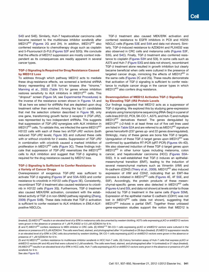
TGF-bSignaling IsRequired forDrugResistanceCausedby MED12 LossTo address through which pathway MED12 acts to mediate
these drug-resistance effects, we screened a lentiviral shRNA
library representing all 518 human kinases (the ‘‘kinome,’’
Manning et al., 2002) (Table S1) for genes whose inhibition
restores sensitivity to ALK inhibitors in MED12KD cells. This
‘‘dropout’’ screen (Figure 3A; see Experimental Procedures) is
the inverse of the resistance screen shown in Figures 1A and
1B as here we select for shRNAs that are depleted upon drug
treatment rather than enriched. Among the top 51 candidates
that met the selection criterion described in Figure 3B, only
one gene, transforming growth factor b receptor II (TGF-bR2),
was represented by two independent shRNAs. This suggests
that suppression of TGF-bR2 synergizes with ALK inhibition in
MED12KD cells. To validate this, we infected the sameMED12KD
H3122 cells with each of these two shTGF-bR2 vectors (both
reduced TGF-bR2 levels; Figure 3D) and cultured these cells
with or without crizotinib for 2 weeks. Suppression of TGF-bR2
in combination with crizotinib caused a marked inhibition of
proliferation in MED12KD cells (Figure 3C). These findings indi-
cate that suppression of TGF-bR2 resensitizes the MED12KD
cells to ALK inhibition and suggest that TGF-b signaling is
required for the drug resistance caused by MED12 loss.
TGF-b Signaling Is Sufficient to Confer Resistance toa Variety of Cancer DrugsOverexpression of exogenous TGF-bR2 was sufficient to
activate TGF-b signaling (Figures 3F and S3A–S3D) and confer
resistance to crizotinib in H3122 cells (Figure 3E). Consistently,
recombinant TGF-b treatment also caused resistance to crizoti-
nib in H3122 cells (Figure 3G). Furthermore, TGF-b treatment
also caused MEK/ERK activation, consistent with the estab-
lished activity of TGF-b in non-SMAD pathway signaling (Zhang,
2009) (Figure S4B). These data indicate that TGF-b activation
is sufficient to confer resistant to ALK inhibitors in EML4-ALK-
positive NSCLCs.
(treated). (D)MED12KD results in an elevated level of p-ERK inmelanoma cells doc
were grown in the absence or presence of 1 mM PLX4032 or 0.5 mM AZD6244 fo
(E and F) MED12KD confers resistance to MEK inhibitor in CRC cells. (E) KRASV
absence or presence of 0.5 mMAZD6244. The cells were fixed, stained, and photo
in an elevated level of p-ERK in CRC cells documented by western blotting. SK-C
presence of 1 mM AZD6244 for 6 hr.
(G and H) MED12KD confers resistance to multikinase inhibitor sorafenib in HCC
shMED12 vectors (#4 and #5) and that were cultured in 2 mM sorafenib. The cells
(H)MED12KD results in an elevated level of p-ERK in HCC cells. Huh-7 cells expres
sorafenib for 6 hr.
See also Figure S2.
TGF-b treatment also caused MEK/ERK activation and
conferred resistance to EGFR inhibitors in PC9 and H3255
NSCLC cells (Figures S3E and S4A and data not shown). Simi-
larly, TGF-b-induced resistance to AZD6244 and PLX4032 was
also observed in CRC cells and melanoma cells (Figures S3F,
S3G, and S4C). Finally, TGF-b treatment also conferred resis-
tance to cisplatin (Figures S3H and S3I). In some cells such as
A375 and Huh-7 (Figure S3G and data not shown), recombinant
TGF-b treatment alone resulted in growth inhibition but clearly
became beneficial when cells were cultured in the presence of
targeted cancer drugs, mimicking the effects of MED12KD in
the same cells (Figures 2C and 2G). These results demonstrate
that activation of TGF-b signaling is sufficient to confer resis-
tance to multiple cancer drugs in the cancer types in which
MED12KD also confers drug resistance.
Downregulation of MED12 Activates TGF-b Signalingby Elevating TGF-bR2 Protein LevelsOur findings suggested that MED12 acts as a suppressor of
TGF-b signaling. We explored this by studying gene-expression
analysis using transcriptome sequencing (RNA-Seq) in a panel of
cells lines (H3122, PC9, SK-CO-1, A375, andHuh-7) andmultiple
MED12KD derivatives thereof. The genes deregulated by
MED12KD (>2-fold) in at least three out of five cell lines used
are listed in Table S2A and are referred to asMED12KD signature
genes henceforth (237 genes up- and 22 genes downregulated).
Strikingly, many of these genes are bona fide TGF-b targets.
Upregulation of these TGF-b target genes upon MED12KD was
confirmed by quantitative RT-PCR (qRT-PCR) (Figures 4A–4D).
We also observed induction of these TGF-b target genes upon
MED12KD in other tumor types including melanoma, colon
cancer, and hepatocellular carcinoma (HCC) (Figures S5A–
S5D). It is well-established that TGF-b induces an epithelial-
mesenchymal transition (EMT), leading to the induction of
several mesenchymal markers such as Vimentin (VIM) and
N-cadherin (CDH2) (Thiery et al., 2009). MED12KD also induced
expression of VIM and CDH2, indicating that an EMT-like
process is initiated in MED12KD cells (Figures 4E, 4F S5E, and
S5F). Accordingly, the protein products of these mesen-
chymal-specific genes were also detected in MED12KD cells
(Figures 4J and S5L and data not shown) at levels similar to those
induced by TGF-b treatment in the same cells (Figure S5M).
Expression of the epithelial marker E-cadherin (CDH1) was not
lost in MED12KD cells (data not shown), suggesting that
MED12KD induces a partial EMT. Together these unbiased
gene-expression studies support the notion that MED12 is
umented by western blotting. A375 cells expressing pLKO or shMED12 vectors
r 6 hr.12 SK-CO-1 cells expressing pLKO or shMED12 vectors were cultured in the
graphed after 14 (untreated) or 28 days (treated). (F)MED12 suppression results
O-1 cells expressing pLKO or shMED12 vectors were grown in the absence or
Huh-7 cells. (G) Colony formation assay of Huh-7 cells that express pLKO or
were fixed, stained, and photographed after 14 (untreated) or 21 days (treated).
sing pLKO or shMED12 vectors were grown in the absence or presence of 4 mM
Cell 151, 937–950, November 21, 2012 ª2012 Elsevier Inc. 941
sh
shTGFF- R2#2
TGFF- R2#1
A MED12KD H3122 cells
Infect with TRC kinome shRNA library
Control CrizotinibCulture to
allow selection
Identify shRNA by deepseq
B
C D
Ctrl cells + MED12KD cells +
pLKO
shTGF-
R2#1
shTGF-
R2#1
shTGF-
R2#2
shTGF-
R2#2
Crizotinib
Ctrl
pLKO
TGF- R2
0.0
0.2
0.4
0.6
0.8
1.0
1.2
Rel
ativ
em
RN
Aex
pres
sion
pLKOshTGF-
R2#1shTGF-
R2#2
shTGF-R2#1
shTGF-R2#2
pLKO
Ctrl cells + MED12KD cells +
100502512.56.250
TGF- [ M]
Crizotinib
Ctrl
TGF- R2GFP
Ctrl
Crizotinib
E F
p-SMAD2
SMAD2
TGF- R2
TGF-R2
GFP
p-ERK
ERK
G
Figure 3. TGF-b Signaling Is Required for the Drug Resistance Driven by MED12 Suppression
(A) Schematic outline of the ‘‘dropout’’ RNAi screen for kinases whose inhibition restores sensitivity to crizotinib in MED12KD cells. Human TRC
kinome shRNA library polyclonal virus was produced to infect H3122 cells stably expressing shMED12#3, which were then left untreated (control) or
treated with 300 nM crizotinib for 10 days. After selection, shRNA inserts from both populations were recovered by PCR and identified by next-generation
sequencing.
(B) Representation of the relative abundance of the shRNA barcode sequences from the shRNA screen experiment depicted in (A). The y axis is enrichment
(relative abundance of crizotinib treated/untreated), and x axis is the intensity (average sequence reads in untreated sample) of each shRNA. Among the 51 top
shRNA candidates (more than 2.5-fold depleted by crizotinib treatment andmore than 200 reads in untreated as indicated by the red dash lines), two independent
shTGF-bR2 vectors (in red) were identified.
(C) Suppression of TGF-bR2 restores the crizotinib sensitivity in MED12KD cells. Using lentiviral infection, pLKO or two independent shTGF-bR2 vectors were
introduced into H3122 control orMED12KD cells. After this, cells were cultured in the absence or presence of 300 nM crizotinib. The cells were fixed, stained, and
photographed after 14 (untreated) or 21 days (treated). The level of knockdown of TGF-bR2 by each of the shRNAs was measured by examining the TGF-bR2
mRNA levels by qRT-PCR. Error bars denote SD.
(E and F) Activation of TGF-b signaling by TGF-bR2 overexpression was sufficient to confer resistance to crizotinib in H3122 cells. (E) H3122 cells expressing
pQXCIP-GFP control or pQXCIP-TGF-bR2-HA were cultured in the absence or presence of 300 nM crizotinib. The cells were fixed, stained, and photographed
after 14 (untreated) or 21 days (treated). (F) Western blotting analysis showing that TGF-bR2 overexpression resulted in elevated levels of p-SAMD2 and p-ERK.
(G) Activation of TGF-b signaling by recombinant TGF-b treatment also leads to resistance to crizotinib in H3122 cells in a TGF-b dosage-dependent manner.
See also Table S1 and Figures S3 and S4.
942 Cell 151, 937–950, November 21, 2012 ª2012 Elsevier Inc.
a suppressor of TGF-b signaling in a wide range of cancer types
and that its loss activates TGF-b signaling.
To further study the mechanism by which MED12 suppresses
TGF-b signaling, we investigated the effect of MED12KD on key
components of the TGF-b pathway. We found thatMED12KD re-
sulted in a strong induction of TGF-bR2 protein levels (Figures
4G and 4H). As a result of the TGF-bR2 upregulation, SMAD2,
the key mediator of TGF-b signaling, was activated as indicated
by a strong increase in SMAD2 phosphorylation. Consistently,
affinity-labeling assayswith 125I-TGF-b1 showed strong increase
of the 125I-labeled cell-surface TGF-bR2 upon MED12KD in
H3122 cells (Figure S5H). As controls, 125I-BMP9 affinity-labeling
experiments showed no significant change in labeled BMP
receptors upon MED12KD. Similar results were obtained in
A375 melanoma and in SK-CO-1 CRC cells indicating that this
interplay between MED12 and TGF-b signaling is conserved
across different tumor types (Figures S5I and S5J). Thus the up-
regulation of TGF-bR2 inMED12KD cells causes the activation of
TGF-b signaling, which in turns leads to MEK/ERK activation
(Figure 4J). Supporting this notion that downregulation of TGF-
bR2 by RNAi suppressed the MEK/ERK activation in MED12KD
cells (Figures S4D–S4F and data not shown).
Because MED12 is part of the MEDIATOR transcriptional
complex that functions in the nucleus, we assumed that
MED12 would act on TGF-bR2 transcription. However, there
was only a modest increase in TGF-bR2 mRNA upon MED12KD
(Figure 4I). Moreover, we observed a progressive increase in
TGF-bR2 protein levels in time after MED12KD. These results
suggest that MED12 predominantly suppresses TGF-bR2 in
a posttranscriptional manner. To investigate this, we determined
the subcellular localization of MED12. We carried out nuclear
and cytoplasmic fractionation of PC9 cells expressing control
vector or shMED12 followed by western blotting (Figure 4K).
Lamin A/C and SP1 were used as controls for nuclear fractions,
a-TUBULIN and HSP90 for cytoplasmic fractions. Abundant
nuclear MED12 was detected, consistent with its function in
the MEDIATOR transcriptional complex. Unexpectedly, a signifi-
cant quantity of MED12 was also present in the cytoplasmic
fraction. Cytoplasmic MED12 was also seen in H3122 cells
(Figure S5K). No cytoplasmic CDK8, another subunit of the
MEDIATOR kinase module with which MED12 is known to asso-
ciate closely, was detected. This suggests that cytoplasmic
MED12 might have a second function distinct from its role in
the MEDIATOR complex. Consistent with this, downregulation
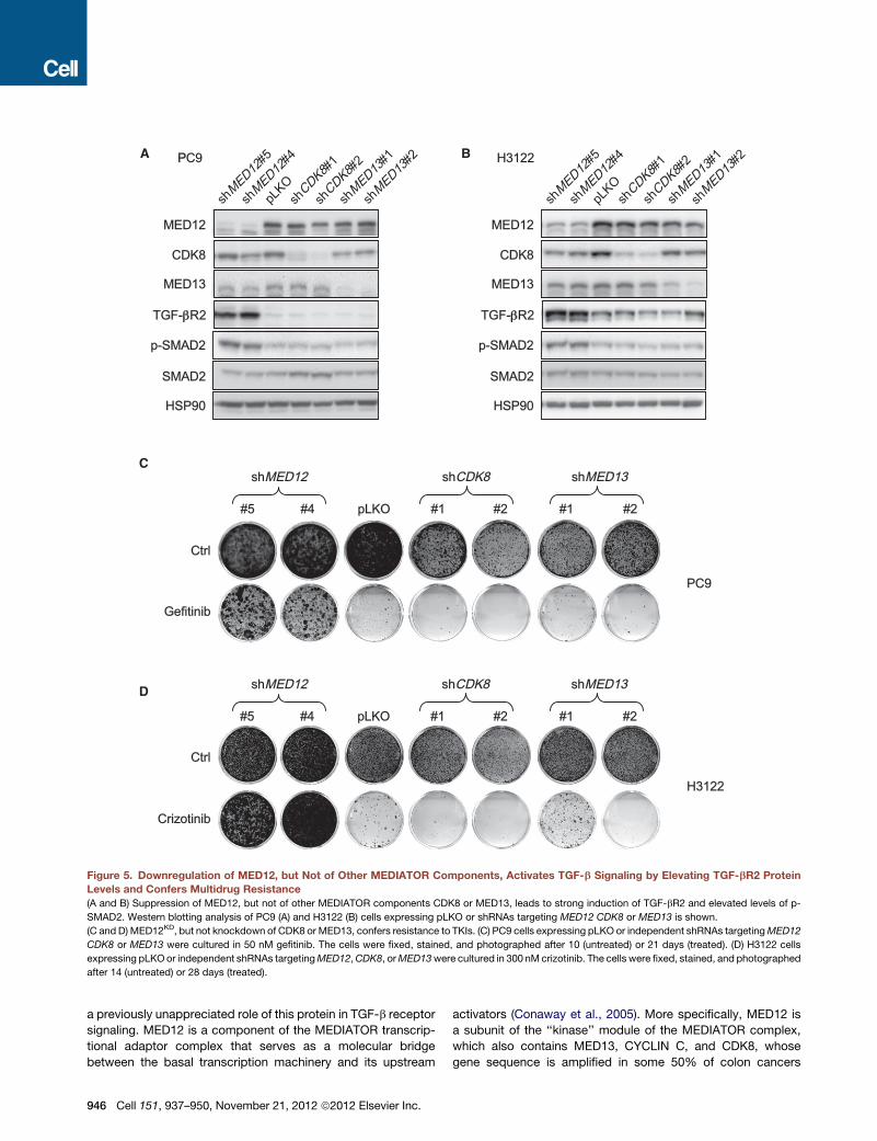
of other MEDIATOR subunits, such as CDK8 and MED13 in
PC9 and H3122 cells, did not lead to upregulation of TGF-bR2
or activation of SMAD2 (Figures 5A and 5B) and failed to confer
resistance to EGFR and ALK inhibitors (Figures 5C and 5D).
The unexpected cytoplasmic localization of MED12 promp-
ted us to examine a potential physical interaction between
MED12 and TGF-bR2. We first performed coimmunoprecipita-
tion (coIP) experiments with Phoenix cells cotransfected with
TGF-bR2 and MED12. As indicated in Figure 4L, TGF-bR2 coim-
munoprecipitated withMED12, and conversely MED12 coimmu-
noprecipitated with TGF-bR2, indicating that MED12 interacts
physically with TGF-bR2. Consistent with this, coIP experiments
with the cytoplasmic fraction of untransfected PC9 cells indicate
that endogenous TGF-bR2 interacts with endogenous MED12
(Figure 4M). As a second independent approach,weusedaprox-
imity ligation assay (PLA) to validate the TGF-bR2-MED12 inter-
action in situ. PLA technology allows sensitive detection of
protein-protein interaction and requires two primary antibodies
from different species against the proteins that are presumed
to interact. Because our best antibodies against TGF-bR2 and
MED12 were produced in rabbits, we generated MED12KD
PC9 cells reconstituted with Flag-Med12 to be able to use PLA
technology with mouse anti-Flag to detect Med12. These recon-
stituted cells expressed levels of MED12 and TGF-bR2 proteins
similar to those in parental cells (Figure S6A). The results shown
in Figure S6B indicate that there is a significant in situ interaction
of TGF-bR2 and MED12 in the cytoplasm of PC9 cells, which is
consistent with the data from the coIP experiments above.
The observation that MED12KD caused a strong increase of
cell-surface TGF-bR2 (Figure S5H) suggests that MED12
could inhibit TGF-bR signaling by preventing the maturation of
TGF-bR2. To test this, we performed coIP experiments with
antibodies against HA tag and MED12 on Phoenix cells cotrans-
fected with HA-TGF-bR2 and MED12 and incubated the immu-
noprecipitates with Endo H or PNGase F enzymes. Endo H
removes oligosaccharides of glycoproteins in the endoplasmic
reticulum (ER), but not the highly processed complex oligosac-
charides processed in the Golgi. In contrast, PNGase F deglyco-
sylates glycoproteins in both the ER and Golgi. As indicated in
Figure S6C, in the TGF-bR2 immunoprecipitate, we observed
three distinct forms of TGF-bR2: the 60 kDa form that was insen-
sitive to both Endo H and PNGase F corresponding to unglyco-
sylated TGF-bR2; the 70 kDa form that was sensitive to Endo H
corresponding to the partially glycosylated TGF-bR2 in the ER;
the smear from 80 to 100 kDa that was Endo H resistant but
PNGase F sensitive corresponding to the fully glycosylated
TGF-bR2. We found that only the nonprocessed and partially
processed forms of TGF-bR2 coimmunoprecipitated with
MED12. These data are consistent with a model in which
MED12 interferes with the proper glycosylation of TGF-bR2
and hence blocks cell-surface expression of the receptor (Kim
et al., 2012).
A MED12KD Gene Signature Has Features of EMTand Is Both Prognostic and PredictiveMED12 suppression leads to activation of TGF-b signaling and
expression of mesenchymal markers, suggestive of a partial
EMT-like process. Recently, EMT has been identified as
a program in human CRC that correlates with poor prognosis
(Loboda et al., 2011). We therefore asked whether MED12KD
indeed induces an EMT-like process and whether the processes
induced by MED12KD are likewise associated with poor prog-
nosis in CRC. We first compared the 237 genes that are upregu-
lated in the MED12KD signature (Table S2A) to the 229 genes
upregulated in a more general EMT signature (see Extended
Experimental Procedures; Table S2B). We found a significant
overlap of 31 genes between both signatures (p = 8.9 3 10�23;
Figure S7A and Table S2C). This further supports the notion
that MED12 loss initiates a partial EMT. Next we asked whether
genes that are deregulated after MED12KD predict survival in
CRC. Hierarchical clustering of a set of 231 CRC tumor samples
using the MED12KD signature genes led to the identification of
Cell 151, 937–950, November 21, 2012 ª2012 Elsevier Inc. 943

-TUBULIN
pLKO shMED12
Cyt Nuc Cyt Nuc
MED12
Lamin A/C
SP1
HSP90
Gefitinib: - + - + - + - +
CDK8
L
MED12 + TGF- R2 :
INPUT
MED12
TGF-R2
IgGco
ntrol
- + + + +
IP
MED12
HSP90
K
G H
HSP90
pLKOsh
MED12#4
shMED12
#5
p-SMAD2
SMAD2
MED12
PC9
shMED12
#5
p-SMAD2
TGF-- R2
TGF-- R2
TGF-- R2
TGF-- R2
TGF-- R2
HSP90
pLKO
shMED12
#4
SMAD2
MED12
H3122
E
pLKO
H3122
#4 #5
shMED12
pLKO
PC9
#4 #5
shMED12
VIM
0.0
0.6
0.8
1.0
1.2
Rel
ativ
em
RN
Aex
pres
sion
0.4
0.2
D
CTGF
0.0
0.6
0.8
1.0
1.2
Rel
ativ
em
RN
Aex
pres
sion
0.4
0.2
pLKO
H3122
#4 #5
shMED12
pLKO
PC9
#4 #5
shMED12
F
CDH2
0.0
0.6
0.8
1.0
1.2
Rel
ativ
em
RN
Aex
pres
sion
0.4
0.2
pLKO
H3122
#4 #5
shMED12
pLKO
PC9
#4 #5
shMED12
B
TAGLN
0.0
0.6
0.8
1.0
1.2
Rel
ativ
em
RN
Aex
pres
sion
0.4
0.2
pLKO
H3122
#4 #5
shMED12
pLKO
PC9
#4 #5
shMED12
A
ANGPTL4
0.0
0.6
0.8
1.0
1.2
Rel
ativ
em
RN
Aex
pres
sion
0.4
0.2
pLKO
H3122
#4 #5
shMED12
pLKO
PC9
#4 #5
shMED12
C
CYR61
0.0
0.6
0.8
1.0
1.2
Rel
ativ
em
RN
Aex
pres
sion
0.4
0.2
pLKO
H3122
#4 #5
shMED12
pLKO
PC9
#4 #5
shMED12
TGF- R2
0.0
1.5
2.0
2.5
1.0
0.5
shMED12 : +- +- +-Day 3 Day 5 Day 8
Rel
ativ
em
RN
Aex
pres
sion
PC9
I
J
IP
MED12IN
PUTMED12
IgG
PC9 Cytoplasmic fraction
M
MED12
shMED12 : +- +- +-Day 3 Day 5 Day 8
N-Cadherin
p-MEK
MEK
p-ERK
ERK
PC9
Phoenix
PC9
INPUT (Lo
ngExp
.)
Figure 4. MED12 Suppresses TGF-b Signaling by Negatively Regulating TGF-bR2
(A–F) MED12KD leads to induction of a panel of TGF-b target genes and EMT marker genes. mRNA expression analysis by qRT-PCR of TGF-b target genes
ANGPTL4 (A), TAGLN (B), CYR61 (C), and CTGF (D) and EMT marker genes VIM (E) and CDH2 (F) in H3122 and PC9 cells expressing pLKO controls or shRNAs
targeting MED12 is shown. Cells were cultured in normal condition without TGF-b stimulation. Error bars denote SD.
(G and H)MED12KD results in strong induction of TGF-bR2 protein and SMAD2 phosphorylation. Western blot analysis of H3122 (G) and PC9 (H) cells expressing
pLKO or shMED12 vectors. HSP90 was used as a loading control.
(I) MED12KD results in a modest induction of TGF-bR2 mRNA in a time course experiment. RNA samples from PC9 cells expressing pLKO or shMED12 were
collected at days 3, 5, and 8 post lentiviral infection, and TGF-bR2 mRNA was analyzed by qRT-PCR. Error bars denote SD.
944 Cell 151, 937–950, November 21, 2012 ª2012 Elsevier Inc.

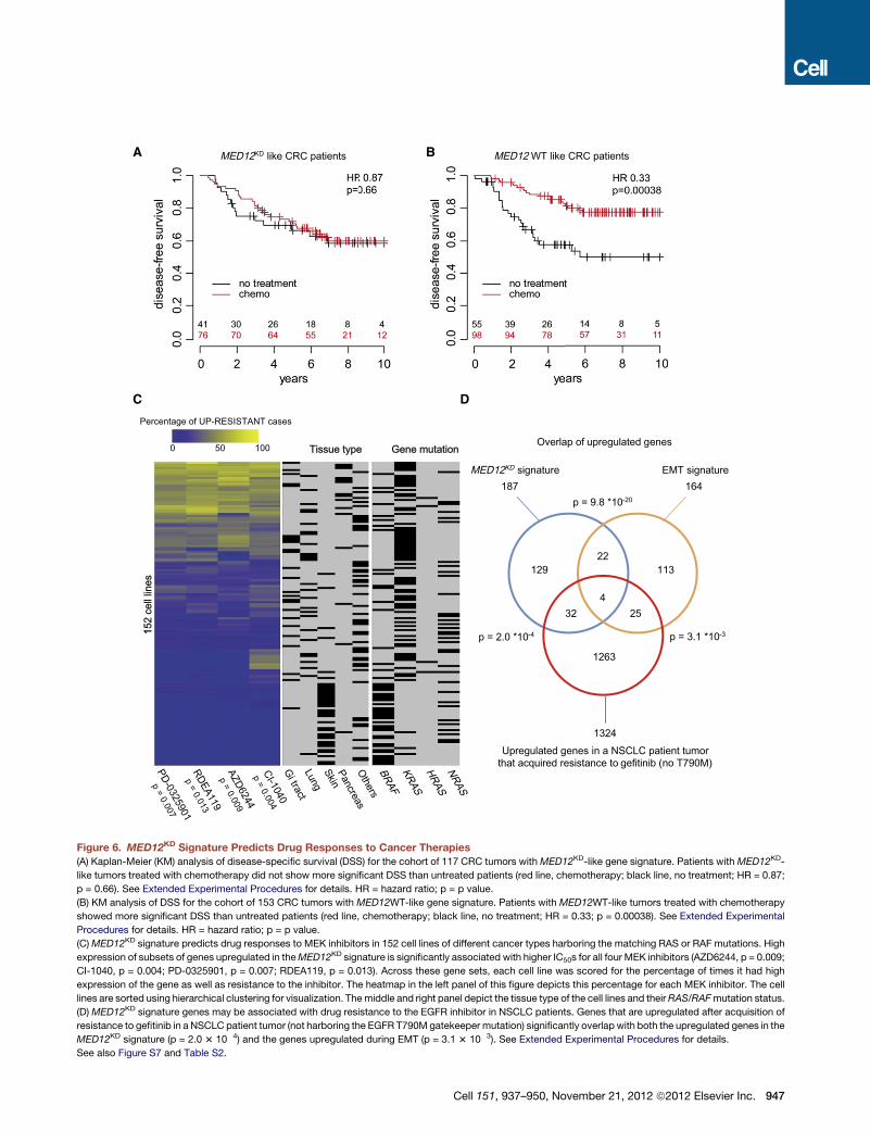
two groups of patients that have significantly different disease-
specific survival (DSS) (Figure S7B). The group with higher over-
all expression of signature genes that are upregulated upon
MED12KD had worse outcomes compared to the group with
lower expression of the same genes. These results indicate
that the processes induced by MED12KD are associated with
a poor survival in CRC patients.
Next, we examined a second cohort of 270 stage III CRC
patients, only some of whom were treated with 5-FU-based
chemotherapy and whose responses to chemotherapy are
known, to determine whether the MED12KD signature could
also predict responses to chemotherapy in patients (Salazar
et al., 2011; P.R., unpublished data). We used the MED12KD
signature genes that were also present in the microarray previ-
ously used for the expression analysis of these tumors to classify
patients as MED12KD like or MED12 wild-type (MED12WT) like
(see Extended Experimental Procedures; Tables S2D and
S2M). We found that chemotherapy did not lead to noticeable
change in DSS of patients withMED12KD-like tumors (Figure 6A)
whereas it did cause a significant increase in DSS of patients
with MED12WT-like tumors (Figure 6B). These results indicate
that the MED12KD signature predicts response to 5-FU-based
chemotherapy in CRC patients, consistent with our finding that
MED12KD confers resistance to 5-FU (Figure S2F).
To further substantiate our finding that MED12 suppression
confers resistance to cancer drugs targeting the MEK-ERK
pathway, we asked whether the MED12KD signature could
predict response to MEK inhibitors in a large and heterogeneous
panel of cancer cell lines of different tissue types. As MEK inhib-
itors are currently being evaluated to treat tumors that have
activating mutations in RAS or BRAF, we focused on 152 tumor
cell lines harboring either RAS or BRAF mutations for whom the
IC50 values of four different MEK inhibitors and gene-expression
patterns have been determined (Garnett et al., 2012) (see
Extended Experimental Procedures and Table S2F). Of the 237
genes that were upregulated by MED12KD as identified by
RNA-Seq, we could read the expression levels for 170 genes in
these 152 cell lines (Table S2E). We found that high expression
of these 170 genes is significantly associated with higher IC50s
for all four MEK inhibitors in these cell lines (AZD6244, p =
0.009; CI-1040, p = 0.004; PD-0325901, p = 0.007; RDEA119,
p = 0.013; Figure 6C and Table S2E). The analysis of one of these
genes, ZBED2, is shown as an example in Figure S7C. Thus the
group of genes that is upregulated upon MED12KD predicts
response to MEK inhibitors in a very heterogeneous panel of
cancer cell lines, consistent with our finding that MED12 acts
independently of cellular context to influence cancer drug
response (Figure 6C).
(J) There is a progressive increase in TGF-bR2 protein levels in time after MED
cadherin. Western blotting analysis of the total lysates from the PC9 cells describe
(K) MED12 localizes to both nucleus and cytoplasm. Western blotting analysis
control vector or shMED12 with or without 16 hr of 25 nM gefitinib treatment. La
a-TUBULIN and HSP90 were used as controls for cytoplasmic fractions.
(L) MED12 is capable of physically interacting with TGF-bR2. Western blotting ana
with TGF-bR2 and MED12 in a ratio of 5:1 is shown.
(M) Cytoplasmic MED12 interacts with TGF-bR2 in PC9 cells. Western blotting a
parental PC9 cells is shown.
See also Figures S5 and S6.
Finally, we asked whether expression of MED12KD signature
genes is associated with drug resistance to targeted agents in
the clinic. We obtained pairs of tumor samples derived from
three patients (cases 3, 6, and 10) that have NSCLC tumors
with EGFR-activating mutations both before and after develop-
ment of resistance to gefitinib (Uramoto et al., 2011). Two of
the resistant tumors did have the EGFR T790M gatekeeper
mutation (cases 3 and 6). RNA was isolated from these
formalin-fixed tumor slides followed by transcriptome se-
quencing by RNA-Seq. For each pair, we selected genes that
showed a greater than 2-fold upregulation after acquisition of
gefitinib resistance and then asked whether these genes overlap
with the MED12KD signature. For the tumor pair without the
EGFR T790M mutation (case 10), we did observe a significant
overlap of genes upregulated after acquisition of gefitinib resis-
tance with the MED12KD signature genes (Figure 6D and Tables
S2G–S2L), but not for the two tumor pairs with EGFR T790M
mutation (cases 3 and 6) (Figure S7B and Table S3). This result
indicates that in the patient of case 10, a gene-expression
program was activated upon gefitinib resistance that resembles
the program induced by MED12KD.
TGF-bR Inhibitor and TKIs Synergize to SuppressProliferation of MED12KD NSCLC CellsAs inhibition of TGF-bR2 by RNAi resensitizedMED12KD cells to
tyrosine kinase inhibitors (TKIs), we reasoned that TGF-bR inhib-
itors shouldsynergizewithTKIs to inhibit proliferation inMED12KD
cells. To test this, we cultured both parental andMED12KDH3122
cells in the absence and the presence of crizotinib, the TGF-bR
inhibitor LY2157299, or the combination of both drugs. Crizotinib
alone potently inhibited the growth of the control, but not of
theMED12KD cells. LY2157299 alone had little effect on all cells.
However, strong synergy was seen when crizotinib was
combined with LY2157299 (Figure 7A). The same synergistic
response was also obtained when LY2157299 was combined
with gefitinib in MED12KD PC9 cells (Figure 7B). Moreover, the
combination of LY2157299with crizotinib or gefitinib suppressed
the ERK activation driven by MED12KD in both H3122 and PC9
cells (Figures 7C and 7D). These biochemical data are in line
with our previous RNAi results where TGF-bR2KD suppressed
ERK activation in MED12KD cells (Figures S4D–S4F). Thus, the
combination of TGF-bR inhibitors and TKIs might be a strategy
for treating tumors with elevated TGF-b signaling.
DISCUSSION
We identify hereMED12 as a candidate biomarker of response to
a range of cancer drugs in a variety of cancer types through
12KD, and the increase is associated with increased p-MEK, p-ERK, and N-
d in (I). All cells were treated with 25 nMgefinitib for 6 hr before lysate collection.
of the nuclear and cytoplasmic fractions prepared from PC9 cells expressing
min A/C and SP1 were used as marker controls for nuclear fractions, whereas
lysis of coimmunoprecipitation experiments using Phoenix cells cotransfected
nalysis of coimmunoprecipitation experiments with a cytoplasmic fraction of
Cell 151, 937–950, November 21, 2012 ª2012 Elsevier Inc. 945
PPC9 H3122
MED12
TGF- R2
CDK8
MED13
shMED12
#4
pLKO
shMED12
#5
shCDK8#
1
shCDK8#
2
shMED13
#1
shMED13
#2
p-SMAD2
SMAD2
HSP90
MED12
TGF- R2
CDK8
MED13
shMED12
#4
pLKO
shMED12
#5
shCDK8#
1
shCDK8#
2
shMED13
#1
shMED13
#2
p-SMAD2
SMAD2
HSP90
pLKO#5 #4 #1 #2 #1 #2
Ctrl
Gefitinib
shMED12 shCDK8 shMED13
PC9
pLKO#5 #4 #1 #2 #1 #2
Ctrl
Crizotinib
shMED12 shCDK8 shMED13
H3122
A B
C
D
Figure 5. Downregulation of MED12, but Not of Other MEDIATOR Components, Activates TGF-b Signaling by Elevating TGF-bR2 Protein
Levels and Confers Multidrug Resistance
(A and B) Suppression of MED12, but not of other MEDIATOR components CDK8 or MED13, leads to strong induction of TGF-bR2 and elevated levels of p-
SMAD2. Western blotting analysis of PC9 (A) and H3122 (B) cells expressing pLKO or shRNAs targeting MED12 CDK8 or MED13 is shown.
(C and D) MED12KD, but not knockdown of CDK8 or MED13, confers resistance to TKIs. (C) PC9 cells expressing pLKO or independent shRNAs targetingMED12
CDK8 or MED13 were cultured in 50 nM gefitinib. The cells were fixed, stained, and photographed after 10 (untreated) or 21 days (treated). (D) H3122 cells
expressing pLKOor independent shRNAs targetingMED12,CDK8, orMED13were cultured in 300 nM crizotinib. The cells were fixed, stained, and photographed
after 14 (untreated) or 28 days (treated).
a previously unappreciated role of this protein in TGF-b receptor
signaling. MED12 is a component of the MEDIATOR transcrip-
tional adaptor complex that serves as a molecular bridge
between the basal transcription machinery and its upstream
946 Cell 151, 937–950, November 21, 2012 ª2012 Elsevier Inc.
activators (Conaway et al., 2005). More specifically, MED12 is
a subunit of the ‘‘kinase’’ module of the MEDIATOR complex,
which also contains MED13, CYCLIN C, and CDK8, whose
gene sequence is amplified in some 50% of colon cancers
A BMED12KD like CRC patients
C
Percentage of UP-RESISTANT cases
PD-0325901
p = 0.007
RDEA119
p = 0.013
AZD6244
p = 0.009
CI-1040
p = 0.004
1152
cell
lines
0 50 100
Gi tract
PancreasO
thers
LungSkin
BRAFKRASHRASNRAS
Tissue type Gene mutation
D
Upregulated genes in a NSCLC patient tumorthat acquired resistance to gefitinib (no T790M)
12922
113
4
1263
2532
EMT signatureMED12KD signature187 164
1324
p = 9.8 *10-20
p = 2.0 *10-4 p = 3.1 *10-3
Overlap of upregulated genes
MED12 WT like CRC patients
Figure 6. MED12KD Signature Predicts Drug Responses to Cancer Therapies
(A) Kaplan-Meier (KM) analysis of disease-specific survival (DSS) for the cohort of 117 CRC tumors withMED12KD-like gene signature. Patients with MED12KD-
like tumors treated with chemotherapy did not show more significant DSS than untreated patients (red line, chemotherapy; black line, no treatment; HR = 0.87;
p = 0.66). See Extended Experimental Procedures for details. HR = hazard ratio; p = p value.
(B) KM analysis of DSS for the cohort of 153 CRC tumors with MED12WT-like gene signature. Patients with MED12WT-like tumors treated with chemotherapy
showed more significant DSS than untreated patients (red line, chemotherapy; black line, no treatment; HR = 0.33; p = 0.00038). See Extended Experimental
Procedures for details. HR = hazard ratio; p = p value.
(C)MED12KD signature predicts drug responses to MEK inhibitors in 152 cell lines of different cancer types harboring the matching RAS or RAF mutations. High
expression of subsets of genes upregulated in theMED12KD signature is significantly associated with higher IC50s for all four MEK inhibitors (AZD6244, p = 0.009;
CI-1040, p = 0.004; PD-0325901, p = 0.007; RDEA119, p = 0.013). Across these gene sets, each cell line was scored for the percentage of times it had high
expression of the gene as well as resistance to the inhibitor. The heatmap in the left panel of this figure depicts this percentage for each MEK inhibitor. The cell
lines are sorted using hierarchical clustering for visualization. Themiddle and right panel depict the tissue type of the cell lines and theirRAS/RAFmutation status.
(D) MED12KD signature genes may be associated with drug resistance to the EGFR inhibitor in NSCLC patients. Genes that are upregulated after acquisition of
resistance to gefitinib in a NSCLC patient tumor (not harboring the EGFR T790M gatekeeper mutation) significantly overlap with both the upregulated genes in the
MED12KD signature (p = 2.0 3 10�4) and the genes upregulated during EMT (p = 3.1 3 10�3). See Extended Experimental Procedures for details.
See also Figure S7 and Table S2.
Cell 151, 937–950, November 21, 2012 ª2012 Elsevier Inc. 947

AA B
Ctrl
Crizotinib
LY2157299
pRS
shMED12
#3
shMED12
#2
Crizotinib+LY2157299
Ctrl
Gefitinib
LY2157299
pLKO
shMED12
#4
shMED12
#5
Gefitinib+LY2157299
PC9
shMED12 : - + - + - + - +
LY2157299 : - - + + - - + +Gefitinib : - - - - + + + +
H3122
shMED12 : - + - + - + - +
LY2157299 : - - + + - - + +
p-ERK
MED12
p-SMAD2
SMAD2
ERK
p-ERK
MED12
p-SMAD2
SMAD2
ERK
NVP-TAE684 : - - - - + + + +
C D
PC9H3122
Figure 7. TGF-bR Inhibitor and TKIs Syner-
gize to Suppress Proliferation of MED12KD
NSCLC Cells
(A) Combination of TGF-bR and ALK inhibitors
synergistically inhibits growth ofMED12KD NSCLC
cells harboring EML4-ALK translocation. H3122
cells expressing pRS or shMED12 vectors were
cultured in the absence and the presence of 1 mM
LY2157299, 300 nM crizotinib, or the combination
of 1 mM LY2157299 and 300 nM crizotinib. The
cells were fixed, stained, and photographed after
14 (untreated and LY2157299 alone) or 28 days
(crizotinib alone and LY2157299 plus crizotinib).
(B) Combination of TGF-bR and EGFR inhibitors
synergistically inhibits growth ofMED12KD NSCLC
cells harboring EGFR-activating mutation. PC9
cells expressing pLKO or shMED12 vectors were
cultured in the absence and the presence of 1 mM
LY2157299, 100 nM gefitinib, or the combination
of 1 mMLY2157299 and 100 nM gefitinib. The cells
were fixed, stained, and photographed after 10
(untreated and LY2157299 alone) or 28 days
(gefitinib alone and LY2157299 plus gefitinib).
(C and D) Combination of LY2157299 with crizo-
tinib or gefitinib suppressed the ERK activation
driven by MED12KD in both H3122 and PC9 cells.
(C) H3122 cells were grown in the absence
or presence of 20 mM NVP-TAE684, 5 mM
LY2157299, or the combination of 20 mM NVP-
TAE684 and 5 mM LY2157299 for 6 hr, and the cell
lysates were harvested for western blotting anal-
ysis. (D) PC9 cells were grown in the absence or
presence of 25 nM gefitinib, 5 mM LY2157299, or
the combination of 25 nM gefitinib and 5 mM
LY2157299 for 6 hr, and the cell lysates were
harvested for western blotting analysis.
See also Figure S4.
(Firestein et al., 2008). However, neither CDK8KD nor MED13KD
caused upregulation of TGF-bR2 or conferred drug resistance,
highlighting the unique role ofMED12 in both TGF-bR2 activation
and drug resistance. The involvement of MED12 in the response
to TKIs was unexpected as most of the known genes that
influence responses to TKIs involve components of signaling
pathways that act downstream of or in parallel to these recep-
tors. We reconcile this apparent discrepancy by demonstrating
that part of MED12 resides in the cytosol, where it interacts
with the immature forms of TGF-bR2 and inhibits its glycosyla-
tion, thereby preventing cell-surface expression (Kim et al.,
2012). Consequently, MED12KD strongly enhances cell-surface
expression of TGF-bR2 and activates TGF-b signaling. Activa-
tion of TGF-b signaling has also been linked to increased RAS-
MEK-ERK signaling (reviewed by Zhang, 2009). Indeed we
observed activation of ERK signaling by MED12 suppression,
which persists in the presence of drugs like crizotinib, gefitinib,
vemurafenib, seluteminib, and sorafenib (Figures 2 and S4),
thus providing a rationale for why suppression ofMED12 confers
resistance to these drugs.
Our data indicate that MED12 suppression also induces an
EMT-like phenotype and that this EMT-like phenotype induced
by MED12KD is associated with chemotherapy resistance in
both cell lines and patients. Our data are consistent with the
948 Cell 151, 937–950, November 21, 2012 ª2012 Elsevier Inc.
findings of others who also witnessed resistance to EGFR inhib-
itors in cell lines undergoing EMT (Fuchs et al., 2008; Yao et al.,
2010). In the clinic, EMT transdifferentiation was also seen in
NSCLC patients who developed resistance to EGFR TKIs
(Sequist et al., 2011; Uramoto et al., 2011). Consistent with
this, we observed in a NSCLC patient who developed resistance
to gefitinib without gatekeeper T790M mutation that a program
of gene expression that resembled the one induced byMED12KD
was activated (Figure 6D). It is at this point not clear whether
patients that acquire EMTduring drug resistancedo so as a result
of MED12 loss. This appears possible as MED12 is mutated in
some 70% of uterine leiomyomas and in 5% of prostate cancers
(Barbieri et al., 2012; Makinen et al., 2011). We note that these
mutations are highly clustered, raising the possibility that these
mutations are not null alleles. Consistent with this, we observe
thatMED12 suppression often confers a slow-growth phenotype
to cancer cells and that near-complete suppression ofMED12 is
not tolerated bymost cells. Thus suppression ofMED12may not
confer a selective advantage in the absence of drug but may only
become a benefit to the cancer cells when undergoing drug
selection pressure. Consistent with this, we observed that
PC9, NSCLC, A375, melanoma, and Huh-7 HCC cells are
growth-inhibited by MED12KD, but this turns into a proliferative
advantage when exposed to EGFR, BRAF, or MEK inhibitors or
the multikinase inhibitor sorafenib. Therefore, MED12 suppres-
sion may not be a marker of intrinsic drug resistance as its
constitutive suppression could well be disadvantageous to the
cancer cell, but it may be acquired during drug selection to resist
the therapy. That cancer cells can transiently assume a reversible
drug-tolerant state was recently shown (Sharma et al., 2010).
Finally, our data demonstrate that inhibition of TGF-b signaling
in MED12KD cells with small-molecule drugs can reverse resis-
tance to targeted cancer drugs (Figure 7). This raises the possi-
bility that EMT arising during drug-resistance development as
seen in NSCLC (Sequist et al., 2011; Uramoto et al., 2011) may
be countered by combination with a TGF-b antagonist, a notion
that can readily be tested in the clinic.
EXPERIMENTAL PROCEDURES
shRNA Screens
The NKI shRNA library and the barcode screen are as described (Berns et al.,
2004; Brummelkamp et al., 2006). Additional details can be found at http://
screeninc.nki.nl/. See the Extended Experimental Procedures for details on
the kinome ‘‘dropout’’ shRNA screen.
Cell Culture, Viral Transduction, and Long-Term Cell Proliferation
Assays
Experiments were performed as described (Huang et al., 2009). See the
Extended Experimental Procedures for details.
Gene-Expression and Statistical Analysis
Transcriptome sequencing analysis of cell lines was performed with RNA-Seq
to generate the MED12KD gene signature, which was employed to hierarchi-
cally cluster a data set consisting of gene expression data for 231 CRC tumor
samples for their outcome and to predict responses to chemotherapy in
a second cohort of 270 CRC patients. Differences in DSS were determined
using the Kaplan-Meier (KM) statistics.
See the Extended Experimental Procedures for details.
COSMIC Cell-Line Panel Analysis
Drug-response data (IC50 values) and gene-expression levels were obtained
from Catalogue Of Somatic Mutations In Cancer (COSMIC) (Forbes et al.,
2010). See the Extended Experimental Procedures for details for the analysis.
NSCLC Patient Samples
Tumor samples derived from three patients (cases 3, 6, and 10) that have
NSCLC tumors with EGFR-activating mutations both before and after acquisi-
tion of resistance to gefitinib are as described (Uramoto et al., 2011). The insti-
tutional review board’s approved informed consent for the use of the tumor
tissue specimens was obtained either from all the patients or from the patient’s
legal guardians.
SUPPLEMENTAL INFORMATION
Supplemental Information includes Extended Experimental Procedures, seven
figures, and three tables and can be found with this article online at http://dx.
doi.org/10.1016/j.cell.2012.10.035.
ACKNOWLEDGMENTS
We thank the members of the NKI Genomics Core Facility, Maarten van
Dinther, Jelle Wesseling, Ingrid Hofland, Ian Majewski, Kylie Greig, Johan
Kuiken, and Erik Voets for technical support and discussion. We are grateful
to Cinzia Pochet for support. This work was supported by grants from the
Dutch Cancer Society, a European Research Council grant, The Cancer
Systems Biology Center grant by NWO, The Netherlands Genomics Initiative
(NGI), and a Spanish BAE FIS travel grant by Instituto Carlos III (to R.S.).
P.R. and R.B. are employees and shareholders of Agendia Inc.
Received: November 23, 2011
Revised: August 10, 2012
Accepted: September 24, 2012
Published: November 21, 2012
REFERENCES
Barbieri, C.E., Baca, S.C., Lawrence, M.S., Demichelis, F., Blattner, M., Theur-
illat, J.P., White, T.A., Stojanov, P., Van Allen, E., Stransky, N., et al. (2012).
Exome sequencing identifies recurrent SPOP, FOXA1 and MED12 mutations
in prostate cancer. Nat. Genet. 44, 685–689.
Berns, K., Hijmans, E.M., Mullenders, J., Brummelkamp, T.R., Velds, A., Hei-
merikx, M., Kerkhoven, R.M., Madiredjo, M., Nijkamp, W., Weigelt, B., et al.
(2004). A large-scale RNAi screen in human cells identifies new components
of the p53 pathway. Nature 428, 431–437.
Berns, K., Horlings, H.M., Hennessy, B.T., Madiredjo, M., Hijmans, E.M.,
Beelen, K., Linn, S.C., Gonzalez-Angulo, A.M., Stemke-Hale, K., Hauptmann,
M., et al. (2007). A functional genetic approach identifies the PI3K pathway as
a major determinant of trastuzumab resistance in breast cancer. Cancer Cell
12, 395–402.
Brummelkamp, T.R., Fabius, A.W., Mullenders, J., Madiredjo, M., Velds, A.,
Kerkhoven, R.M., Bernards, R., and Beijersbergen, R.L. (2006). An shRNA bar-
code screen provides insight into cancer cell vulnerability to MDM2 inhibitors.
Nat. Chem. Biol. 2, 202–206.
Chapman, P.B., Hauschild, A., Robert, C., Haanen, J.B., Ascierto, P., Larkin,
J., Dummer, R., Garbe, C., Testori, A., Maio, M., et al.; BRIM-,3 Study Group.
(2011). Improved survival with vemurafenib in melanoma with BRAF V600E
mutation. N. Engl. J. Med. 364, 2507–2516.
Choi, Y.L., Soda, M., Yamashita, Y., Ueno, T., Takashima, J., Nakajima, T.,
Yatabe, Y., Takeuchi, K., Hamada, T., Haruta, H., et al.; ALK Lung Cancer
Study Group. (2010). EML4-ALK mutations in lung cancer that confer resis-
tance to ALK inhibitors. N. Engl. J. Med. 363, 1734–1739.
Conaway, R.C., Sato, S., Tomomori-Sato, C., Yao, T., and Conaway, J.W.
(2005). The mammalian Mediator complex and its role in transcriptional
regulation. Trends Biochem. Sci. 30, 250–255.
Firestein, R., Bass, A.J., Kim, S.Y., Dunn, I.F., Silver, S.J., Guney, I., Freed, E.,
Ligon, A.H., Vena, N., Ogino, S., et al. (2008). CDK8 is a colorectal cancer
oncogene that regulates beta-catenin activity. Nature 455, 547–551.
Forbes, S.A., Tang, G., Bindal, N., Bamford, S., Dawson, E., Cole, C., Kok,
C.Y., Jia, M., Ewing, R., Menzies, A., et al. (2010). COSMIC (the Catalogue
of Somatic Mutations in Cancer): a resource to investigate acquired mutations
in human cancer. Nucleic Acids Res. 38(Database issue), D652–D657.
Fuchs, B.C., Fujii, T., Dorfman, J.D., Goodwin, J.M., Zhu, A.X., Lanuti, M., and
Tanabe, K.K. (2008). Epithelial-to-mesenchymal transition and integrin-linked
kinase mediate sensitivity to epidermal growth factor receptor inhibition in
human hepatoma cells. Cancer Res. 68, 2391–2399.
Garnett, M.J., Edelman, E.J., Heidorn, S.J., Greenman, C.D., Dastur, A., Lau,
K.W., Greninger, P., Thompson, I.R., Luo, X., Soares, J., et al. (2012). System-
atic identification of genomic markers of drug sensitivity in cancer cells. Nature
483, 570–575.
Gorre, M.E., Mohammed, M., Ellwood, K., Hsu, N., Paquette, R., Rao, P.N.,
and Sawyers, C.L. (2001). Clinical resistance to STI-571 cancer therapy
caused by BCR-ABL gene mutation or amplification. Science 293, 876–880.
Holzel, M., Huang, S., Koster, J., Ora, I., Lakeman, A., Caron, H., Nijkamp, W.,
Xie, J., Callens, T., Asgharzadeh, S., et al. (2010). NF1 is a tumor suppressor in
neuroblastoma that determines retinoic acid response and disease outcome.
Cell 142, 218–229.
Huang, S., Laoukili, J., Epping, M.T., Koster, J., Holzel, M., Westerman, B.A.,
Nijkamp, W., Hata, A., Asgharzadeh, S., Seeger, R.C., et al. (2009). ZNF423 is
critically required for retinoic acid-induced differentiation and is a marker of
neuroblastoma outcome. Cancer Cell 15, 328–340.
Johannessen, C.M., Boehm, J.S., Kim, S.Y., Thomas, S.R., Wardwell, L.,
Johnson, L.A., Emery, C.M., Stransky, N., Cogdill, A.P., Barretina, J., et al.
Cell 151, 937–950, November 21, 2012 ª2012 Elsevier Inc. 949
(2010). COT drives resistance to RAF inhibition through MAP kinase pathway
reactivation. Nature 468, 968–972.
Karapetis, C.S., Khambata-Ford, S., Jonker, D.J., O’Callaghan, C.J., Tu, D.,
Tebbutt, N.C., Simes, R.J., Chalchal, H., Shapiro, J.D., Robitaille, S., et al.
(2008). K-ras mutations and benefit from cetuximab in advanced colorectal
cancer. N. Engl. J. Med. 359, 1757–1765.
Kim, Y.W., Park, J., Lee, H.J., Lee, S.Y., and Kim, S.J. (2012). TGF-b sensitivity
is determined by N-linked glycosylation of the type II TGF-b receptor.
Biochem. J. 445, 403–411.
Kwak, E.L., Bang, Y.J., Camidge, D.R., Shaw, A.T., Solomon, B., Maki, R.G.,
Ou, S.H., Dezube, B.J., Janne, P.A., Costa, D.B., et al. (2010). Anaplastic
lymphoma kinase inhibition in non-small-cell lung cancer. N. Engl. J. Med.
363, 1693–1703.
Loboda, A., Nebozhyn, M.V., James, W., Watters, J.W., Carolyne, A., Buser,
C.A., Shaw, P.M., Huang, P.S., Van’t Veer, L.J., Tollenaar, R.A.E.M., et al.
(2011). EMT is the dominant program in human colon cancer. BMC Medical
Genomics 2011, 4–9.
Maemondo, M., Inoue, A., Kobayashi, K., Sugawara, S., Oizumi, S., Isobe, H.,
Gemma, A., Harada, M., Yoshizawa, H., Kinoshita, I., et al.; North-East Japan
Study Group. (2010). Gefitinib or chemotherapy for non-small-cell lung cancer
with mutated EGFR. N. Engl. J. Med. 362, 2380–2388.
Makinen, N., Mehine, M., Tolvanen, J., Kaasinen, E., Li, Y., Lehtonen, H.J.,
Gentile, M., Yan, J., Enge, M., Taipale, M., et al. (2011). MED12, the mediator
complex subunit, 12 gene is mutated at high frequency in uterine leiomyomas.
Science 334, 252–255.
Manning, G.,Whyte, D.B., Martinez, R., Hunter, T., and Sudarsanam, S. (2002).
The protein kinase complement of the human genome. Science 298, 1912–
1934.
McDermott, U., Iafrate, A.J., Gray, N.S., Shioda, T., Classon, M., Maheswaran,
S., Zhou, W., Choi, H.G., Smith, S.L., Dowell, L., et al. (2008). Genomic alter-
ations of anaplastic lymphoma kinase may sensitize tumors to anaplastic
lymphoma kinase inhibitors. Cancer Res. 68, 3389–3395.
Salazar, R., Roepman, P., Capella, G., Moreno, V., Simon, I., Dreezen, C.,
Lopez-Doriga, A., Santos, C., Marijnen, C., Westerga, J., et al. (2011). Gene
950 Cell 151, 937–950, November 21, 2012 ª2012 Elsevier Inc.
expression signature to improve prognosis prediction of stage II and III
colorectal cancer. J. Clin. Oncol. 29, 17–24.
Sequist, L.V., Waltman, B.A., Dias-Santagata, D., Digumarthy, S., Turke, A.B.,
Fidias, P., Bergethon, K., Shaw, A.T., Gettinger, S., Cosper, A.K., et al. (2011).
Genotypic and histological evolution of lung cancers acquiring resistance to
EGFR inhibitors. Sci. Transl. Med. 3, 75ra26.
Shah, N.P., Nicoll, J.M., Nagar, B., Gorre, M.E., Paquette, R.L., Kuriyan, J., and
Sawyers, C.L. (2002). Multiple BCR-ABL kinase domainmutations confer poly-
clonal resistance to the tyrosine kinase inhibitor imatinib (STI571) in chronic
phase and blast crisis chronic myeloid leukemia. Cancer Cell 2, 117–125.
Sharma, S.V., Lee, D.Y., Li, B., Quinlan, M.P., Takahashi, F., Maheswaran, S.,
McDermott, U., Azizian, N., Zou, L., Fischbach, M.A., et al. (2010). A chro-
matin-mediated reversible drug-tolerant state in cancer cell subpopulations.
Cell 141, 69–80.
Thiery, J.P., Acloque, H., Huang, R.Y., and Nieto, M.A. (2009). Epithelial-
mesenchymal transitions in development and disease. Cell 139, 871–890.
Uramoto, H., Shimokawa, H., Hanagiri, T., Kuwano, M., and Ono, M. (2011).
Expression of selected gene for acquired drug resistance to EGFR-TKI in
lung adenocarcinoma. Lung Cancer 73, 361–365.
Wagle, N., Emery, C., Berger, M.F., Davis, M.J., Sawyer, A., Pochanard, P.,
Kehoe, S.M., Johannessen, C.M., Macconaill, L.E., Hahn, W.C., et al. (2011).
Dissecting therapeutic resistance to RAF inhibition in melanoma by tumor
genomic profiling. J. Clin. Oncol. 29, 3085–3096.
Yao, Z., Fenoglio, S., Gao, D.C., Camiolo, M., Stiles, B., Lindsted, T., Schle-
derer, M., Johns, C., Altorki, N., Mittal, V., et al. (2010). TGF-beta IL-6 axis
mediates selective and adaptive mechanisms of resistance to molecular tar-
geted therapy in lung cancer. Proc. Natl. Acad. Sci. USA 107, 15535–15540.
Yun, C.H., Mengwasser, K.E., Toms, A.V., Woo, M.S., Greulich, H., Wong,
K.K., Meyerson, M., and Eck, M.J. (2008). The T790Mmutation in EGFR kinase
causes drug resistance by increasing the affinity for ATP. Proc. Natl. Acad. Sci.
USA 105, 2070–2075.
Zhang, Y.E. (2009). Non-Smad pathways in TGF-beta signaling. Cell Res. 19,
128–139.


![Research Paper TGF-β and NF-κB signaling pathway crosstalk ... · expression via a Smad3-mediated signaling pathway [22]. Increased levels of TGF-β have been reported in several](https://static.fdocuments.in/doc/165x107/5dd0c05cd6be591ccb6284f7/research-paper-tgf-and-nf-b-signaling-pathway-crosstalk-expression-via.jpg)


![Impaired neurodevelopmental pathways in autism spectrum … · 2019. 6. 15. · hedgehog (SHH) [14, 15], fibroblast growth factor (FGF) [16], transforming growth factor β (TGF-β)[17,](https://static.fdocuments.in/doc/165x107/60ec180132b3841da052075f/impaired-neurodevelopmental-pathways-in-autism-spectrum-2019-6-15-hedgehog.jpg)


