
Mechanistic insights into a TIMP3- sensitive pathway ... · PDF filefeature of the disease,...
26
*For correspondence: anne. [email protected] Competing interests: The authors declare that no competing interests exist. Funding: See page 23 Received: 05 May 2016 Accepted: 30 July 2016 Published: 01 August 2016 Reviewing editor: David E Clapham, Howard Hughes Medical Institute, Boston Children’s Hospital, United States Copyright Capone et al. This article is distributed under the terms of the Creative Commons Attribution License, which permits unrestricted use and redistribution provided that the original author and source are credited. Mechanistic insights into a TIMP3- sensitive pathway constitutively engaged in the regulation of cerebral hemodynamics Carmen Capone 1,2 , Fabrice Dabertrand 3,4 , Celine Baron-Menguy 1,2 , Athena Chalaris 5,6 , Lamia Ghezali 1,2 , Vale ´ rie Domenga-Denier 1,2 , Stefanie Schmidt 5,6 , Cle ´ ment Huneau 1,2 , Stefan Rose-John 5,6 , Mark T Nelson 3,4,7 , Anne Joutel 1,2 * 1 Genetics and Pathogenesis of Cerebrovascular Diseases, INSERM, U1161, Universite ´ Paris Diderot, Sorbonne Paris Cite ´ , UMRS 1161, Paris, France; 2 DHU NeuroVasc, Sorbonne Paris Cite ´ , Paris, France; 3 Department of Pharmacology, University of Vermont, Burlington, United States; 4 College of Medicine, University of Vermont, United States; 5 Institute of Biochemistry, Christian Albrechts University, Kiel, Germany; 6 Medical Faculty, Christian Albrechts University, Kiel, Germany; 7 Institute of Cardiovascular Sciences, University of Manchester, Manchester, United Kingdom Abstract Cerebral small vessel disease (SVD) is a leading cause of stroke and dementia. CADASIL, an inherited SVD, alters cerebral artery function, compromising blood flow to the working brain. TIMP3 (tissue inhibitor of metalloproteinase 3) accumulation in the vascular extracellular matrix in CADASIL is a key contributor to cerebrovascular dysfunction. However, the linkage between elevated TIMP3 and compromised cerebral blood flow (CBF) remains unknown. Here, we show that TIMP3 acts through inhibition of the metalloprotease ADAM17 and HB-EGF to regulate cerebral arterial tone and blood flow responses. In a clinically relevant CADASIL mouse model, we show that exogenous ADAM17 or HB-EGF restores cerebral arterial tone and blood flow responses, and identify upregulated voltage-dependent potassium channel (K V ) number in cerebral arterial myocytes as a heretofore-unrecognized downstream effector of TIMP3-induced deficits. These results support the concept that the balance of TIMP3 and ADAM17 activity modulates CBF through regulation of myocyte K V channel number. DOI: 10.7554/eLife.17536.001 Introduction Small vessel disease (SVD) of the brain is a leading cause of stroke and age-related cognitive decline and disability for which there are currently no treatments (Pantoni, 2010). Our limited understanding of the pathogenesis of cerebral SVD is a major obstacle to the development of treatments. Mono- genic forms of these diseases, such as CADASIL (Cerebral Autosomal Dominant Arteriopathy with Subcortical Infarcts and Leukoencephalopathy), provide a window into the mechanism underlying much more common, but largely indistinguishable, sporadic forms of SVD (Joutel and Faraci, 2014). CADASIL, the most common hereditary cerebral SVD, is caused by dominant mutations in the NOTCH3 receptor that stereotypically lead to the extracellular deposition of NOTCH3 ectodomain (Notch3 ECD ) and aggregates of other proteins on vessels (Joutel et al., 2000; Chabriat et al., 2009; Monet-Lepre ˆtre et al., 2013). A deficit in cerebral blood flow (CBF) hemodynamics is an early Capone et al. eLife 2016;5:e17536. DOI: 10.7554/eLife.17536 1 of 26 RESEARCH ARTICLE
Transcript of Mechanistic insights into a TIMP3- sensitive pathway ... · PDF filefeature of the disease,...
*For correspondence: anne.
Competing interests: The
authors declare that no
competing interests exist.
Funding: See page 23
Received: 05 May 2016
Accepted: 30 July 2016
Published: 01 August 2016
Reviewing editor: David E
Clapham, Howard Hughes
Medical Institute, Boston
Children’s Hospital, United
States
Copyright Capone et al. This
article is distributed under the
terms of the Creative Commons
Attribution License, which
permits unrestricted use and
redistribution provided that the
original author and source are
credited.
Mechanistic insights into a TIMP3-sensitive pathway constitutively engagedin the regulation of cerebralhemodynamicsCarmen Capone1,2, Fabrice Dabertrand3,4, Celine Baron-Menguy1,2,Athena Chalaris5,6, Lamia Ghezali1,2, Valerie Domenga-Denier1,2,Stefanie Schmidt5,6, Clement Huneau1,2, Stefan Rose-John5,6, Mark T Nelson3,4,7,Anne Joutel1,2*
1Genetics and Pathogenesis of Cerebrovascular Diseases, INSERM, U1161,Universite Paris Diderot, Sorbonne Paris Cite, UMRS 1161, Paris, France; 2DHUNeuroVasc, Sorbonne Paris Cite, Paris, France; 3Department of Pharmacology,University of Vermont, Burlington, United States; 4College of Medicine, Universityof Vermont, United States; 5Institute of Biochemistry, Christian Albrechts University,Kiel, Germany; 6Medical Faculty, Christian Albrechts University, Kiel, Germany;7Institute of Cardiovascular Sciences, University of Manchester, Manchester, UnitedKingdom
Abstract Cerebral small vessel disease (SVD) is a leading cause of stroke and dementia.
CADASIL, an inherited SVD, alters cerebral artery function, compromising blood flow to the
working brain. TIMP3 (tissue inhibitor of metalloproteinase 3) accumulation in the vascular
extracellular matrix in CADASIL is a key contributor to cerebrovascular dysfunction. However, the
linkage between elevated TIMP3 and compromised cerebral blood flow (CBF) remains unknown.
Here, we show that TIMP3 acts through inhibition of the metalloprotease ADAM17 and HB-EGF to
regulate cerebral arterial tone and blood flow responses. In a clinically relevant CADASIL mouse
model, we show that exogenous ADAM17 or HB-EGF restores cerebral arterial tone and blood
flow responses, and identify upregulated voltage-dependent potassium channel (KV) number in
cerebral arterial myocytes as a heretofore-unrecognized downstream effector of TIMP3-induced
deficits. These results support the concept that the balance of TIMP3 and ADAM17 activity
modulates CBF through regulation of myocyte KV channel number.
DOI: 10.7554/eLife.17536.001
IntroductionSmall vessel disease (SVD) of the brain is a leading cause of stroke and age-related cognitive decline
and disability for which there are currently no treatments (Pantoni, 2010). Our limited understanding
of the pathogenesis of cerebral SVD is a major obstacle to the development of treatments. Mono-
genic forms of these diseases, such as CADASIL (Cerebral Autosomal Dominant Arteriopathy with
Subcortical Infarcts and Leukoencephalopathy), provide a window into the mechanism underlying
much more common, but largely indistinguishable, sporadic forms of SVD (Joutel and Faraci, 2014).
CADASIL, the most common hereditary cerebral SVD, is caused by dominant mutations in the
NOTCH3 receptor that stereotypically lead to the extracellular deposition of NOTCH3 ectodomain
(Notch3ECD) and aggregates of other proteins on vessels (Joutel et al., 2000; Chabriat et al., 2009;
Monet-Lepretre et al., 2013). A deficit in cerebral blood flow (CBF) hemodynamics is an early
Capone et al. eLife 2016;5:e17536. DOI: 10.7554/eLife.17536 1 of 26
RESEARCH ARTICLE

feature of the disease, suggesting that cerebrovascular dysfunction may have a key role in disease
pathogenesis (Chabriat et al., 2000; Pfefferkorn et al., 2001; Liem et al., 2009).
Small vessels of the brain have unique functional properties that ensure that the brain, which has
a limited capacity to store energy, maintains an adequate supply of blood-borne nutrients in the
face of variations in blood pressure and changing neuronal energy demands. Cerebral arteries exist
in a partially constricted state called ’myogenic tone’, which reflects an intrinsic contractile response
of arterial myocytes to increases in intravascular pressure. Thus, these arteries are positioned to
dilate, and thereby increase local CBF, in response to elevated neuronal activity. This phenomenon,
known as functional hyperemia, serves to satisfy enhanced glucose and oxygen demands of active
neurons (Iadecola and Nedergaard, 2007). Impaired functional hyperemia and CBF autoregulation,
attenuated CBF responses to topical application of vasodilators, and diminished myogenic
responses of cerebral arteries and arterioles are early and prominent features of the well-established
TgNotch3R169C CADASIL mouse model (Joutel et al., 2010; Dabertrand et al., 2015;
Capone et al., 2016). The mechanisms underlying this cerebrovascular dysfunction are poorly
understood.
Recently, we found that TIMP3 (tissue inhibitor of metalloproteinases-3) forms complexes with
Notch3ECD and abnormally accumulates in the extracellular matrix of brain vessels of patients and
mice with CADASIL (Monet-Lepretre et al., 2013). Remarkably, genetic overexpression of TIMP3
recapitulates both CBF and myogenic-response deficits of the CADASIL model; conversely, genetic
reduction of TIMP3 in CADASIL model mice restores normal function (Capone et al., 2016). TIMP
family members are key regulators of the metalloproteinases that degrade the extracellular matrix.
Within the TIMP family, TIMP3 has the broadest spectrum of substrates, which extends to members
of the ADAM (a disintegrin and metalloproteinases) family. These metalloproteinases proteolytically
release the extracellular domains of membrane-bound cytokines, cell adhesion molecules and
growth factors, such as tumor necrosis factor-a and several ligands of the epidermal growth factor
receptor (EGFR) family, including HB-EGF (heparin-binding EGF-like growth factor) (Brew and
Nagase, 2010; Khokha et al., 2013; Arpino et al., 2015). As such, in addition to being a powerful
regulator of extracellular matrix remodeling in various organs (Arpino et al., 2015), TIMP3 is a key
player in inflammatory pathologies and autoimmune diseases through regulation of cell surface
eLife digest There are currently no effective treatments or cures for small blood vessel diseases
of the brain, which lead to strokes and subsequent decreases in mental abilities. Normally, smooth
muscle cells that surround the vessels relax or contract to regulate blood flow and ensure the right
amount of oxygen and nutrients reaches the different regions of the brain. In a syndrome called
CADASIL, which is the most common form of inherited small vessel disease, a genetic mutation
causes the smooth muscle cells to weaken over time.
The accumulation of several proteins – including one called TIMP3 – around the smooth muscle
cells plays a key role in the smooth muscle cell weakening seen in CADASIL. Capone et al. have now
studied mice that display the symptoms of CADASIL to investigate how TIMP3 decreases blood flow
through blood vessels in the brain. This revealed that TIMP3 inactivates another protein called
ADAM17. The latter protein is normally responsible for starting a signaling pathway that helps
smooth muscle cells to regulate blood flow according to the needs of the brain cells. Artificially
adding more ADAM17 to the brains of the CADASIL mice reduced their symptoms of small vessel
disease.
Using smooth muscle cells freshly isolated from the brains of CADASIL mice, Capone et al. also
demonstrated that abnormal TIMP3-ADAM17 signaling increases the number of voltage-dependent
potassium channels in the membrane of the muscle cells. Having too many of these channels impairs
the flow of blood through vessels in the brain.
Further experiments are needed to investigate whether correcting TIMP3-ADAM17 signaling
could prevent strokes in people with inherited CADASIL. It also remains to be seen whether similar
signaling mechanisms are at play in other small vessel diseases.
DOI: 10.7554/eLife.17536.002
Capone et al. eLife 2016;5:e17536. DOI: 10.7554/eLife.17536 2 of 26
Research article Human Biology and Medicine Neuroscience
proteins (Brew and Nagase, 2010; Khokha et al., 2013). However, how metalloproteinase inhibi-
tion might dynamically regulate arterial tone and CBF hemodynamics is unclear.
In another recent study, we established that upregulation of voltage-gated potassium (KV) chan-
nels at the plasma membrane of arterial myocytes is responsible for the diminished myogenic
responses of cerebral arteries and penetrating arterioles in the TgNotch3R169C CADASIL model.
Notably, an influence of the endothelium in myogenic tone deficit was ruled out (Dabertrand et al.,
2015). KV channels play an important and dynamic role in opposing pressure-induced depolarization
and vasoconstriction (Longden et al., 2015). Furthermore, we (Dabertrand et al., 2015) and our
collaborators (Koide et al., 2007) have found that down-regulation of plasma membrane KV chan-
nels through activation of the EGFR pathway restores normal responses to pressure. Collectively,
our results suggest a fundamental linkage between the activity of TIMP3 in the extracellular matrix
of cerebral arterial myocytes and cerebral arterial tone.
Here, we find that the ADAM17/HB-EGF/EGFR (ErbB1/ErbB4) signaling axis is a key TIMP3-sensi-
tive signaling module that regulates CBF responses and the myogenic tone of cerebral arteries. We
further provide evidence that disruption of this TIMP3-sensitive pathway mediates cerebrovascular
dysfunction in the TgNotch3R169C CADASIL model and identify upregulated KV channel current den-
sity in cerebral arterial myocytes as a heretofore-unrecognized effector of this pathway. These
insights into the relationship between TIMP3 activity and cerebral arterial tone may ultimately lead
to therapeutic options in cerebral SVD.
Results
Exogenous TIMP3, but neither TIMP1 nor TIMP2, strongly attenuatesCBF responsesTo explore the role of TIMP3 in the regulation of CBF, we monitored CBF responses in wild-type
mice equipped with an open cranial window over the somatosensory cortex, before and after the
application of recombinant TIMP3 as well as TIMP1 or TIMP2 (Figure 1A; Figure 1—source data 1).
We initially ensured that a recombinant protein applied over the cranial window could enter the
brain. In the absence of robust anti-TIMP3 antibody suitable for immunohistochemistry and of an in
situ assay to detect TIMP3 activity, we assessed brain penetration of Fluorescein isothiocyanate-
labeled albumin (FITC-BSA, 66 kDa). After 30 min of continuous superfusion, fluorescence imaging
of fixed vibratome slices showed that FITC-BSA entered the cortex along penetrating arteries
beneath the cranial window (Figure 1—figure supplement 1), consistent with transport via the
glymphatic system (Iliff et al., 2012). We found that TIMP3 (40 nM) did not affect resting CBF
(Figure 1B), but did strongly reduce the increase in CBF evoked by whisker stimulation (Figure 1C,
D; Figure 1—source data 2,3). Superfusion of 8 nM TIMP3 similarly attenuated functional hyper-
emia (Figure 1—figure supplement 2; Figure 1—source data 2,3). To rule out a possible effect of
TIMP3 on neural activity, we recorded evoked neural activity during TIMP3 application (Figure 1E).
We found that the amplitude of the somatosensory fields potentials produced by electrical stimula-
tion of the whisker pad was unaltered by TIMP3 superfusion (Figure 1F).
The known TIMPs share 38–49% amino acid identity and inhibit most matrix metalloproteinases
(MMPs). However, differences in substrate selectivity and inhibitory properties between different
TIMPs have been described (Khokha et al., 2013; Stetler-Stevenson, 2008) (Figure 1—source
data 1). This prompted us to assess the effects of other TIMPs on functional hyperemia. In sharp
contrast to TIMP3, neither exogenous TIMP1 (50 nM) nor TIMP2 (50 nM) altered functional hyper-
emia (Figure 1C,D; Figure 1—source data 2,3).
We further assessed CBF responses to topical application of the endothelium-dependent and
smooth muscle-dependent vasodilators, acetylcholine and adenosine, respectively, upon superfusion
of TIMP3 (8 and 40 nM), TIMP1 (50 nM) or TIMP2 (50 nM). Again, the increases in CBF induced by
acetylcholine or adenosine were profoundly attenuated by TIMP3 but were unaffected by TIMP2 or
TIMP1, with the exception of a modest attenuation of the adenosine-induced increase in CBF by
TIMP1 (Figure 1G,H; Figure 1—figure supplement 2; Figure 1—source data 2,3). Thus, these find-
ings establish that elevation of TIMP3 is sufficient to induce CBF deficits in vivo and suggest that a
TIMP3-specific target accounts for these deficits.
Capone et al. eLife 2016;5:e17536. DOI: 10.7554/eLife.17536 3 of 26
Research article Human Biology and Medicine Neuroscience
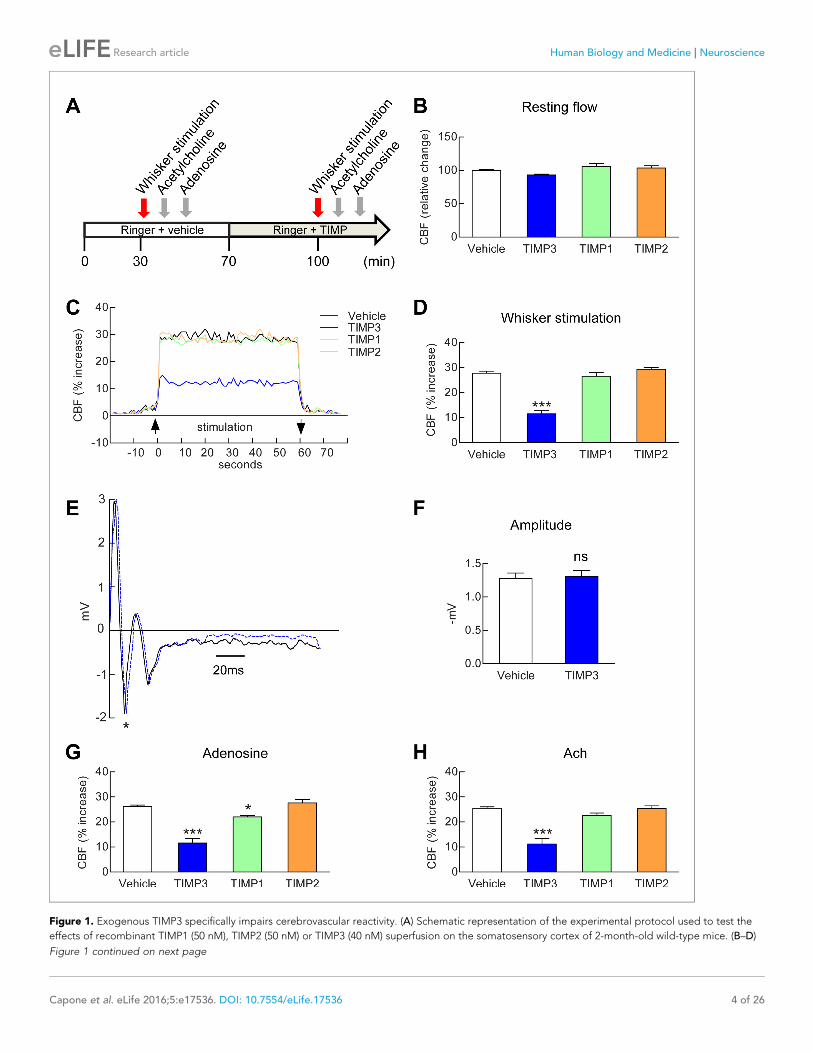
Figure 1. Exogenous TIMP3 specifically impairs cerebrovascular reactivity. (A) Schematic representation of the experimental protocol used to test the
effects of recombinant TIMP1 (50 nM), TIMP2 (50 nM) or TIMP3 (40 nM) superfusion on the somatosensory cortex of 2-month-old wild-type mice. (B–D)
Figure 1 continued on next page
Capone et al. eLife 2016;5:e17536. DOI: 10.7554/eLife.17536 4 of 26
Research article Human Biology and Medicine Neuroscience
ADAM17 is required for TIMP3-induced attenuation of CBF responsesOur efforts to identify the target of TIMP3 focused on ADAM17, which is uniquely inhibited by
TIMP3 (Xu et al., 2012) and is expressed in brain arteries, as demonstrated by our immunoblot anal-
yses (Figure 2A,B). If TIMP3 does indeed act through inhibition of ADAM17, its effects on CBF
responses should be mimicked by pharmacological inhibition of ADAM17. Here, we used the
hydroxamate-based GW413333X inhibitor, which specifically blocks both ADAM10 and ADAM17;
the ADAM10 inhibitor GI254023X was used as a control (Hundhausen et al., 2003) (Figure 2—
source data 1). We found that GW413333X (5 mM), but not GI254023X (5 and 20 mM), strongly
attenuated the increase in CBF produced by whisker stimulation or topical application of acetylcho-
line or adenosine (Figure 2C–E; Figure 2—figure supplement 1A; Figure 2—source data 2,3). To
further support a specific role for ADAM17 in these defects, we assessed CBF responses following
reduction of ADAM17 levels using a genetic approach. Complete ablation of ADAM17 is lethal
(Peschon et al., 1998). Therefore, we used hypomorphic mice with dramatically reduced expression
of ADAM17 (Adam17ex/ex) using the exon-induced translational stop strategy. These mice are viable,
but develop eye, skin and heart defects as a consequence of impaired EGFR signaling
(Chalaris et al., 2010). We found that genetic depletion of ADAM17 strongly attenuated CBF
responses in a dose-dependent manner (Figure 2F,G; Figure 2—figure supplement 1B; Figure 2—
source data 2,3). To confirm that the reduction in evoked CBF responses in these mice is caused by
reduced ADAM17 expression, we examined whether an enzymatically active extracellular domain of
ADAM17 (sADAM17) applied exogenously could prevent these CBF deficits. Topical neocortical
application of sADAM17 (16 nM) (Figure 2—source data 1) did not affect cerebrovascular responses
in wild-type mice, but did fully restore CBF responses in Adam17ex/+ mice with half-reduced
ADAM17 levels (Figure 2H–J; Figure 2—figures supplements 1C,2A; Figure 2—source data
2,3). Together, these results indicate that decreasing ADAM17 activity compromises CBF regulation.
To further confirm the direct connection between increased TIMP3 expression and reduced
ADAM17 activity and CBF deficits, we tested whether exogenous sADAM17 is capable of preventing
the CBF deficits produced by genetic overexpression of TIMP3. Superfusion with the enzymatically
active extracellular domain of ADAM17 (16 nM) increased resting CBF in TgBAC-TIMP3 mice
towards the same absolute values as wild-type mice and improved all evoked cerebrovascular
responses (Figure 2K–M; Figure 2—figures supplements 1D,2B; Figure 2—source data 2,3) sug-
gesting that TIMP3 induces CBF deficits by decreasing ADAM17 activity.
Figure 1 continued
Resting CBF (B) and CBF responses to whisker stimulation (C, D) were evaluated upon superfusion of vehicle or TIMP proteins. (C) Representative trace
of CBF responses to whisker stimulation upon superfusion of vehicle or TIMP proteins (C). (E) Representative trace of the field potentials evoked by
whisker stimulation upon vehicle or TIMP3 superfusion, showing typical sharp positive (P1)-negative (N1) waves followed by a slower positive-negative
waveform occurring within 80 ms post stimulus (Di and Barth, 1991). (F) The amplitude of the negative wave (N1, asterisk in E) of the field potential
was not affected by TIMP3 superfusion (p=0.79). (G, H) CBF responses to topical application of adenosine (G) or acetylcholine (H) upon superfusion of
vehicle or TIMP proteins. Significance was determined by one-way ANOVA followed by Tukey’s post-hoc test (B, D, G, H) or unpaired Student’s t-test
(F). (*p<0.05, ***p<0.001 compared to vehicle; n = 5 mice/groups). Error bars indicate SEM.
DOI: 10.7554/eLife.17536.003
The following source data and figure supplements are available for figure 1:
Source data 1. Reagents used for Figure 1.
DOI: 10.7554/eLife.17536.004
Source data 2. Main physiological variables of mice studied in Figure 1.
DOI: 10.7554/eLife.17536.005
Source data 3. Numerical data that were used to generate the bar charts in Figure 1.
DOI: 10.7554/eLife.17536.006
Figure supplement 1. Assessment of brain penetration of fluorescein isothiocyanate labelled serum albumin (FITC-BSA) superfused over the cranial
window.
DOI: 10.7554/eLife.17536.007
Figure supplement 2. Exogenous TIMP3 (8 nM) impairs cerebrovascular reactivity.
DOI: 10.7554/eLife.17536.008
Capone et al. eLife 2016;5:e17536. DOI: 10.7554/eLife.17536 5 of 26
Research article Human Biology and Medicine Neuroscience

Figure 2. Cerebrovascular responses are impaired by pharmacological or genetic inhibition of ADAM17, and rescued by exogenous sADAM17. (A)
Immunoblot of cerebral arteries dissected from Adam17+/+ and Adam17ex/ex mice (n = 3 biological samples/genotype) incubated with anti-ADAM17 or
Figure 2 continued on next page
Capone et al. eLife 2016;5:e17536. DOI: 10.7554/eLife.17536 6 of 26
Research article Human Biology and Medicine Neuroscience

HB-EGF and EGFR operate downstream of ADAM17 to regulate CBFresponsesTo elucidate the molecular factors that operate downstream of ADAM17 in the context of cerebro-
vascular regulation, we examined the role of the EGFR signaling pathway. This pathway consists of
four related receptor tyrosine kinases of the ErbB family—ErbB1/EGFR (Her1), ErbB2/Neu (Her2),
ErbB3 (Her3) and ErbB4 (Her4)—which are regulated by 11 different ligands, all of which are pro-
duced as membrane-bound precursor proteins and cleaved by cell surface proteases to yield the
active soluble species; ADAM17 is the critical sheddase of at least six of these ligands (Sahin et al.,
2004; Roskoski, 2014) (Figure 3A,B). A critical role for ADAM17 in EGFR signaling is supported by
the observation that mice deficient for ADAM17 (Peschon et al., 1998; Chalaris et al., 2010) resem-
ble mice lacking EGFR, exhibiting perinatal lethality, generalized epithelial defects, and defective
cardiac valves (Miettinen et al., 1995; Sibilia and Wagner, 1995; Threadgill et al., 1995). To inves-
tigate the role of the EGFR pathway, we recorded CBF responses evoked by whisker stimulation or
vasodilators before and after topical application of blockers of this pathway (Figure 3—source data
1).
Based on the decrease in evoked CBF responses to elevation of TIMP3 or reduction of ADAM17,
we predicted that inhibition of ErbB pathway would have a similar effect. Indeed, we found that neo-
cortical application of the selective ErbB1/EGFR and ErbB4 inhibitor, tyrphostin AG1478 (10 and 20
mM), strongly attenuated the evoked CBF responses, but did not impair resting CBF. In contrast,
CBF responses were unaffected by the selective ErbB2 inhibitor, tyrphostin AG825, at both 50 and
200 mM (Figure 3C–E; Figure 3—source data 2, 3). Of note, ErbB3 lacks kinase activity (Ros-
koski 2014). We then tested the effect of soluble recombinant decoy ErbB receptors, known as chi-
meric ErbB receptor traps, which comprise the truncated extracellular domain of the ErbB receptor
fused with the constant region (Fc) of human immunoglobulin (Stratman et al., 2010). Evoked CBF
responses were attenuated by superfusion of either ErbB1/EGFR (67 nM) or ErbB4 (71 nM) receptor
traps, which block the function of ErbB1 and ErbB4 ligands, respectively, but not by superfusion of
the ErbB3 (71 nM) receptor trap or by control IgG1 Fc (286 nM) and IgG2 Fc (286 nM) fragments
(Figure 3F–H; Figure 3—figure supplement 1A; Figure 3—source data 2, 3). Notably, the effects
of ErbB1 and ErbB4 receptor traps on evoked CBF responses were not additive (Figure 3G–H; Fig-
ure 3—source data 2, 3), even though neither ErbB1 nor ErbB4 receptor traps achieved maximum
inhibition. ErbB2 has no known ligand (Roskoski, 2014); thus, these data are consistent with a role
Figure 2 continued
anti-smooth muscle a-actin (a-SMA) antibody. (B) Quantification of relative protein level of ADAM17 in (A). (C–E) Resting CBF (C) and CBF responses to
whisker stimulation (D) or topical application of adenosine (E) were evaluated upon superfusion of the dual ADAM10/ADAM17 inhibitor GW413333X
(GW; 5 mM) or the ADAM10 inhibitor GI254023X (GI; 5 and 20 mM). ***p<0.05 compared with vehicle. (F, G) CBF responses to whisker stimulation (F) or
topical application of adenosine (G) were strongly reduced in Adam17ex/+ mice and further reduced in Adam17ex/ex mice compared with wild-type
littermate controls. (H–J) Exogenous sADAM17 (16 nM) significantly ameliorated CBF responses to whisker stimulation (I) or topical application of
adenosine (J) in Adam17ex/+ mice, whereas ADAM17 had no effect on wild-type littermates. (K–M) Resting CBF and CBF responses were evaluated in
TgBAC-TIMP3 mice and non-transgenic littermates (WT) before and after superfusion of ADAM17. CBF responses to whisker stimulation (L) or topical
application of adenosine (M) were strongly reduced in TgBAC-TIMP3 mice compared with those in WT mice, as previously reported (Capone et al.,
2016), and were normalized by sADAM17 superfusion. Significance was determined by one-way ANOVA followed by Tukey’s post-hoc test (B–G) and
two-way repeated measure ANOVA followed by Bonferroni post-hoc test (H–M) (n = 5 mice/group). Error bars indicate SEM.
DOI: 10.7554/eLife.17536.009
The following source data and figure supplements are available for figure 2:
Source data 1. Reagents used for Figure 2.
DOI: 10.7554/eLife.17536.010
Source data 2. Main physiological variables of mice studied in Figure 2.
DOI: 10.7554/eLife.17536.011
Source data 3. Numerical data that were used to generate the bar charts in Figure 2.
DOI: 10.7554/eLife.17536.012
Figure supplement 1. CBF responses to acetylcholine are attenuated by pharmacological or genetic inhibition of ADAM17 but rescued upon
superfusion of exogenous sADAM17.
DOI: 10.7554/eLife.17536.013
Figure supplement 2. Absolute measurements of resting CBF in Adam17ex/+ and TgBAC-TIMP3 mice in the presence and absence of sADAM17.
DOI: 10.7554/eLife.17536.014
Capone et al. eLife 2016;5:e17536. DOI: 10.7554/eLife.17536 7 of 26
Research article Human Biology and Medicine Neuroscience
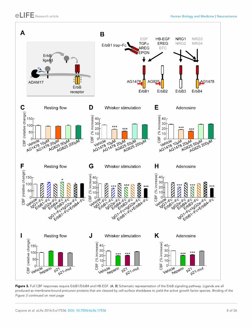
Figure 3. Full CBF responses require ErbB1/ErbB4 and HB-EGF. (A, B) Schematic representation of the ErbB signaling pathway. Ligands are all
produced as membrane-bound precursor proteins that are cleaved by cell-surface sheddases to yield the active growth factor species. Binding of the
Figure 3 continued on next page
Capone et al. eLife 2016;5:e17536. DOI: 10.7554/eLife.17536 8 of 26
Research article Human Biology and Medicine Neuroscience

for ErbB1/EGFR or ErbB4 activation in CBF responses, and suggest the involvement of bispecific
ligands with dual-specificity toward ErbB1 and ErbB4.
Next, we sought to pinpoint which ErbB ligand that requires ADAM17 cleavage for activation is
involved in CBF regulation. Heparin-binding EGF-like growth factor (HB-EGF) is one of three ligands
that can bind to both ErbB1 and ErbB4 and is expressed in the vasculature (Zhang et al., 2014).
ADAM17 is the major sheddase of HB-EGF (Sahin et al., 2004), and ADAM17-mediated shedding
of proHB-EGF largely regulates soluble, mature HB-EGF binding to and activating ErbB receptors
(Yamazaki et al., 2003). Moreover, mice lacking HB-EGF have reduced postnatal viability with
defective cardiac valvulogenesis, similar to mice lacking ADAM17 (Jackson et al., 2003), prompting
us to study the role of HB-EGF in cerebrovascular regulation. To do this, we examined the impact of
HB-EGF inhibition on CBF responses. Unlike all other EGF ligands apart from amphiregulin, HB-EGF
has a heparin-binding domain, and interactions through this domain with cell surface-associated hep-
aran sulfate proteoglycans (HSPGs) are necessary for binding and activation of ErbB receptors
(Higashiyama et al., 1993). We found that superfusion of heparin (40 ui/mL), which competitively
inhibits binding of HB-EGF to cell surface HSPGs (Higashiyama et al., 1993), impaired evoked CBF
responses without affecting resting CBF (Figure 3I–K; Figure 3—figure supplement 1B; Figure 3—
source data 2, 3). To further support a role for HB-EGF in evoked CBF responses, we examined the
effects of the synthetic peptide p21, which corresponds to the heparin-binding domain of murine
HB-EGF and similarly inhibits binding of HB-EGF to cell surface HSPGs (Higashiyama et al., 1993).
We found that superfusion of p21 (12 mM) similarly impaired evoked CBF responses without affect-
ing resting CBF (Figure 3I–K; Figure 3—figure supplement 1B; Figure 3—source data 2, 3); in
contrast, a mutated inactive p21 peptide (p21-mut; 12 mM) had no effect on evoked or resting CBF.
To assess the connection between HB-EGF and ADAM17 in the context of cerebrovascular regu-
lation, we tested the ability of a soluble form of HB-EGF (sHB-EGF) to counteract cerebrovascular
dysfunction induced by ADAM17 inhibition or depletion (Figure 4—source data 1). TIMP3 or the
ADAM10/17 inhibitor, GW413333X, was topically applied over the neocortex and CBF responses
were measured before and after superfusion with sHB-EGF. We found that sHB-EGF (20 nM) pre-
vented TIMP3 and GW-induced cerebrovascular deficits (Figure 4D–I; Figure 4—figure supplement
1A,B; Figure 4—source data 2,3). Also, sHB-EGF significantly improved evoked CBF responses in
Adam17ex/ex mice (Figure 4—figure supplement 2). Notably, sHB-EGF could not prevent CBF defi-
cits induced by pharmacological blockage of ErbB1/EGFR and ErbB4 (Figure 4A–C; Figure 4—
source data 1–3). These findings, collectively, suggest that ADAM17/HB-EGF/(ErbB1/ErbB4) is a
key TIMP3-sensitive signaling pathway for cerebrovascular regulation.
Figure 3 continued
soluble form of the ligand induces ErbB receptor homodimerization or heterodimerization, converting the receptor to an active dimeric conformation
(A). Ligands are grouped in four rows according to their receptor specificity (top; arrows); the six ligands for which ectodomain shedding is primarily
mediated by ADAM17 appear in black characters, and the remaining five are in grey characters (B). (C–K) Resting CBF (C, F, I) and CBF responses to
whisker stimulation (D, G, J) or topical application of adenosine (E, H, K) were evaluated before and after superfusion of various inhibitors of the ErbB
signaling pathway, including the ErbB1/ErbB4 inhibitor AG1478 (10 and 20 mM); the ErbB2 inhibitor AG825 (50 and 200 mM) (C–E), the soluble ErbB
receptor traps (ErbB1-Fc, 66.7 nM; ErbB3-Fc, 71.4 nM; ErbB4-Fc, 71.4 nM) and the respective control IgG1-Fc and IgG2-Fc fragments (286 nM) (F–H),
heparin and the synthetic peptide p21 (12 mM) and the control inactive peptide p21-mut (12 mM) (I–K). None of these compounds affected resting CBF,
except ErbB4-Fc, which produced a slight increase. (C–K) Significance was determined by one-way ANOVA followed by Tukey’s post-hoc test (*p<0.05,
**p<0.01, ***p<0.001 compared to vehicle; n = 5/group). Error bars indicate SEM.
DOI: 10.7554/eLife.17536.015
The following source data and figure supplement are available for figure 3:
Source data 1. Reagents used for Figure 3.
DOI: 10.7554/eLife.17536.016
Source data 2. Main physiological variables of mice studied in Figure 3.
DOI: 10.7554/eLife.17536.017
Source data 3. Numerical data that were used to generate the bar charts in Figure 3.
DOI: 10.7554/eLife.17536.018
Figure supplement 1. Blockade of ErbB1/ErbB4 or HB-EGF impairs CBF responses to acetylcholine.
DOI: 10.7554/eLife.17536.019
Capone et al. eLife 2016;5:e17536. DOI: 10.7554/eLife.17536 9 of 26
Research article Human Biology and Medicine Neuroscience
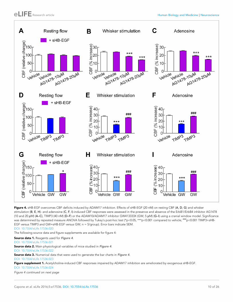
Figure 4. sHB-EGF overcomes CBF deficits induced by ADAM17 inhibition. Effects of sHB-EGF (20 nM) on resting CBF (A, D, G) and whisker
stimulation (B, E, H)- and adenosine (C, F, I)-induced CBF responses were assessed in the presence and absence of the ErbB1/ErbB4 inhibitor AG1478
(10 and 20 mM) (A–C), TIMP3 (40 nM) (D–F) or the ADAM10/ADAM17 inhibitor GW413333X (GW; 5 mM) (G–I) using a cranial window model. Significance
was determined by repeated measure ANOVA followed by Tukey’s post-hoc test (*p<0.05, ***p<0.001 compared to vehicle; ###p<0.001 TIMP3+sHB-
EGF versus TIMP3 and GW+sHB-EGF versus GW; n = 5/group). Error bars indicate SEM.
DOI: 10.7554/eLife.17536.020
The following source data and figure supplements are available for figure 4:
Source data 1. Reagents used for Figure 4.
DOI: 10.7554/eLife.17536.021
Source data 2. Main physiological variables of mice studied in Figure 4.
DOI: 10.7554/eLife.17536.022
Source data 3. Numerical data that were used to generate the bar charts in Figure 4.
DOI: 10.7554/eLife.17536.023
Figure supplement 1. Acetylcholine-induced CBF responses impaired by ADAM17 inhibition are ameliorated by exogenous sHB-EGF.
DOI: 10.7554/eLife.17536.024
Figure 4 continued on next page
Capone et al. eLife 2016;5:e17536. DOI: 10.7554/eLife.17536 10 of 26
Research article Human Biology and Medicine Neuroscience
The ADAM17/HB-EGF/(ErbB1/ErbB4) signaling module regulatespressure-induced myogenic tone in brain arteriesOur data support the concept that the ADAM17/HB-EGF/(ErbB1/ErB4) axis regulates CBF
responses, and any genetic or pharmacological maneuver that inhibits this pathway impairs CBF
responses to diverse stimuli, including topical application of vasodilators and neural activity (Fig-
ures 1–3). Notably, responses cannot be enhanced by stimulation of this pathway, implying that this
pathway is maximally activated in a physiological in vivo setting. Given that myogenic tone sets the
resting arterial diameter from which other stimuli can induce vasoconstriction or vasodilation, we
hypothesized that a reduction in the myogenic tone of cerebral arteries could represent a common
mechanism underlying these CBF deficits. To test this hypothesis, we assessed the effects of inhibi-
tors of this pathway on pressure-induced constriction of brain arteries (Figure 5—source data 1).
We found that pre-incubation of arterial segments with TIMP3 (8 nM) strongly attenuated myo-
genic tone at pressures of 40 mmHg and above compared to arteries incubated with vehicle,
whereas recombinant TIMP2 (10 nM) had no effect (Figure 5A–C; Figure 5—source data 2). Nota-
bly, attenuation of myogenic responses by TIMP3 was even more pronounced in intracerebral pene-
trating arterioles (Figure 5—figure supplement 1). Likewise, myogenic constriction to pressure was
strongly attenuated by the dual ADAM10/ADAM17 inhibitor GW413333X (1 mM), but not by the
ADAM10 inhibitor GI254023X (1 mM). Also, myogenic tone was reduced in heterozygous ADAM17
hypomorphic mice (Adam17ex/+) compared to wild-type littermates (Adam17+/+) but restored by
pre-incubating arterial segments of Adam17ex/+ mice with sADAM17 (3.2 nM) (Figure 5D,I; Fig-
ure 5—figure supplement 2; Figure 5—source data 2). Moreover, pre-incubation of arteries with
the ErbB1/ErbB4 inhibitor AG1478 (2 mM) or the HB-EGF inhibitor p21 peptide (2.4 mM), but not
with the p21-mut peptide (2.4 mM), strongly attenuated myogenic responses (Figure 5E,F; Fig-
ure 5—source data 2). Thus, these data indicate that myogenic tone is increased by tonic activity of
the ADAM17/HB-EGF/(ErbB1/ErbB4) pathway.
To provide further support for the concept that ADAM17 and HB-EGF function as part of a signal-
ing module to enhance the myogenic tone of cerebral arteries, we tested the ability of exogenous
sHB-EGF to counteract the effects of ADAM17 inhibition (Figure 5—source data 1). Pressurized
arteries were pre-incubated with recombinant TIMP3 (4 nM) or the ADAM10/ADAM17 inhibitor
GW413333 in the presence of sHB-EGF (3 nM) or vehicle. We found that co-incubation of arterial
segments with sHB-EGF significantly ameliorated the TIMP3-induced reduction in arterial tone
(Figure 5G; Figure 5—source data 2). Likewise, co-incubation of arterial segments with sHB-EGF
overcame the reduction in arterial tone caused by GW413333-mediated inhibition of ADAM17
(Figure 5H; Figure 5—source data 2). Moreover, sHB-EGF significantly increased myogenic tone in
arteries from heterozygous ADAM17-hypomorphic mice (Adam17ex/+), restoring a near-normal myo-
genic phenotype (Figure 5I; Figure 5—source data 2). Collectively, these data support the concept
that the TIMP3-sensitive pathway, ADAM17/HB-EGF/(ErbB1/ErbB4), increases myogenic constriction
in brain arteries.
Exogenous sADAM17 and exogenous sHB-EGF rescue CBF andmyogenic-response deficits in the TgNotch3R169C CADASIL modelOur findings above predict that excess TIMP3 impairs arterial tone and CBF responses in CADASIL
by suppressing the ADAM17/HB-EGF/(ErbB1/ErbB4) pathway. To test this, we examined whether
recombinant sADAM17 and sHB-EGF could restore normal pressure-induced myogenic constriction
of brain arteries and normal cerebrovascular responses in TgNotch3R169C CADASIL mice. We found
that preincubation of arterial segments with the enzymatically active extracellular domain of
ADAM17 (3.2 nM) increased myogenic tone in arteries from TgNotch3R169C CADASIL mice, whereas
sADAM17 had no detectable effect on arterial segments from wild-type mice at this concentration
(Figure 6A). We further found that sADAM17 (16 nM), locally applied on the necortex of
TgNotch3R169C CADASIL mice, significantly improved resting CBF and rescued the impaired
Figure 4 continued
Figure supplement 2. CBF deficits induced by ADAM17 deficiency are improved by sHB-EGF.
DOI: 10.7554/eLife.17536.025
Capone et al. eLife 2016;5:e17536. DOI: 10.7554/eLife.17536 11 of 26
Research article Human Biology and Medicine Neuroscience

Figure 5. The ADAM17/HB-EGF/(ErbB1/ErbB4) signaling module is involved in regulating the myogenic tone of cerebral arteries. (A–C) Effects of TIMP
proteins on the myogenic responses of posterior cerebral arteries to increasing intraluminal pressure. (A, B) Representative internal diameter recordings
in the presence of TIMP2 (10 nM) (A) or TIMP3 (8 nM) (B). (C) Summary data of results in (A) and (B). (D–F) Myogenic tone of posterior cerebral arteries,
tested in the presence and absence of the dual ADAM10/ADAM17 inhibitor GW413333X (GW; 1 mM), the ADAM10 inhibitor GI254023X (GI; 1 mM) (D),
the ErbB1/ErbB4 inhibitor AG1478 (2 mM) (E), the p21 peptide (2.4 mM), and the mutated inactive peptide p21-mut (2.4 mM) (F). (C–F) **p<0.01,
***p<0.001 versus vehicle. (G, H) Effects of TIMP3 (8 nM) (g) or GW (1 mM) (H) on the myogenic tone of posterior cerebral arteries were tested in the
presence of soluble HB-EGF (3 nM) or vehicle. ##p<0.01, ###p<0.001, TIMP3+HB-EGF versus TIMP3 and GW+HB-EGF versus GW. (I) Myogenic tone of
Figure 5 continued on next page
Capone et al. eLife 2016;5:e17536. DOI: 10.7554/eLife.17536 12 of 26
Research article Human Biology and Medicine Neuroscience
reactivity of brain vessels to whisker stimulation and vasodilators (Figure 6B–D; Figure 6—figure
supplement 1; Figure 6—source data 1,2). We previously reported that sHB-EGF restores myo-
genic responses in parenchymal arteries from TgNotch3R169C CADASIL mice (Dabertrand et al.,
2015). Here, we extend these observations, showing that exogenous sHB-EGF (20 nM) restored
evoked CBF responses in TgNotch3R169C mice (Figure 6E–G; Figure 6—source data 1,2). Collec-
tively, these findings support the concept that the diminished myogenic tone and CBF deficits in
CADASIL are caused by TIMP3-mediated suppression of the ADAM17/HB-EGF/(ErbB1/ErbB4)
pathway.
Excess TIMP3 drives upregulation of KV currents in cerebral arterialmyocytes from TgNotch3R169C miceOur prior work established that upregulation of KV channels in the plasma membrane of cerebral
arterial myocytes is responsible for the diminished myogenic response of cerebral arteries in the
TgNotch3R169C CADASIL model. Importantly, application of sHB-EGF was found to normalize KV cur-
rent density and restore myogenic responses in cerebral arteries from TgNotch3R169Cmice
(Dabertrand et al., 2015). In light of this and the above, we investigated the involvement of TIMP3
and ADAM17 in this upregulation of KV current density.
We first asked whether reducing TIMP3 expression in the TgNotch3R169C mice would decrease
the number of functional KV channels. To this end, we measured KV currents in freshly isolated myo-
cytes from cerebral arteries of TgNotch3R169C mice with normal expression of TIMP3
(TgNotch3R169C;Timp3+/+), which have reduced myogenic tone, and in freshly isolated myocytes
from cerebral arteries of TgNotch3R169C mice with reduced expression of TIMP3 (TgNotch3R169C;
Timp3+/-), in which myogenic responses are restored (Dabertrand et al., 2015). Currents were
recorded in response to 10-mV voltage steps from �70 mV to +60 mV. We found that KV current
density was significantly lower in myocytes from TgNotch3R169C;Timp3+/- mice than in myocytes
from TgNotch3R169C;Timp3+/+ at all voltage steps above +10 mV (Figure 7A–C; Figure 7—source
data 2). Conversely, incubation of wild-type arterial myocytes with recombinant TIMP3 (8 nM)
resulted in a significant increase in KV current density compared with myocytes incubated with vehi-
cle (Figure 7—figure supplement 1A,B; Figure 7—source data 2). Remarkably, half-maximal acti-
vation voltage (V0.5) and slope (k), determined by fitting normalized peak tail currents to the
Boltzmann equation, were statistically indistinguishable among arterial myocytes from the different
groups analyzed. Likewise, activation (tact) and deactivation (tdeact) time constants determined from
exponential fits of individual voltage-evoked current traces and current decay, respectively, were
comparable among the different groups. These current kinetics, attributable to KV1.5 channels, are
consistent with our previous report (Dabertrand et al., 2015). These results indicate that the TIMP3
pathway regulates the number of channels, and not channel properties (Figure 7—figure supple-
ment 2; Figure 7—source data 1,2). Using the Goldman–Hodgkin–Katz constant field equation and
a single-channel conductance of 15 pS (Aiello et al., 1998), we estimated the average number of
functional KV channels per myocyte. This analysis showed that exogenously applied TIMP3 increased
the number of KV channels in arteries from wild-type mice by ~25% (from 3120 to 3920 per myocyte).
Figure 5 continued
posterior cerebral arteries was tested in heterozygous Adam17ex/+ (ex/wt) and Adam17+/+(wt/wt) mice in the presence and absence of soluble HB-EGF
(3 nM). **p<0.01, ***p<0.001 Adam17ex/+ versus Adam17+/+ ; ##p<0.01, ###p<0.001, Adam17ex/+/HB-EGF versus Adam17ex/+). Significance was
determined by two-way repeated measures ANOVA followed by Bonferroni post-hoc test (n = 6–8 arteries/group). Error bars indicate SEM.
DOI: 10.7554/eLife.17536.026
The following source data and figure supplements are available for figure 5:
Source data 1. Reagents used for Figure 5.
DOI: 10.7554/eLife.17536.027
Source data 2. Numerical data that were used to generate the graphs in Figure 5.
DOI: 10.7554/eLife.17536.028
Figure supplement 1. TIMP3 strongly impairs myogenic tone of parenchymal arterioles.
DOI: 10.7554/eLife.17536.029
Figure supplement 2. sADAM17 ameliorates arterial tone in Adam17ex/+ mice.
DOI: 10.7554/eLife.17536.030
Capone et al. eLife 2016;5:e17536. DOI: 10.7554/eLife.17536 13 of 26
Research article Human Biology and Medicine Neuroscience
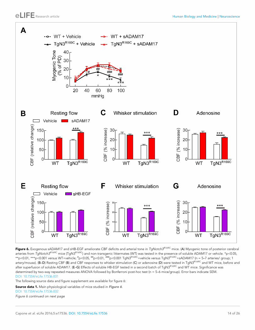
Figure 6. Exogenous sADAM17 and sHB-EGF ameliorate CBF deficits and arterial tone in TgNotch3R169C mice. (A) Myogenic tone of posterior cerebral
arteries from TgNotch3R169C mice (TgN3R169C) and non-transgenic littermates (WT) was tested in the presence of soluble ADAM17 or vehicle. *p<0.05,
**p<0.01, ***p<0.001 versus WT+vehicle; #p<0.05, ##p<0.01, ###p<0.001 TgN3R169C+vehicle versus TgN3R169C+sADAM17 (n = 5–7 arteries/ group; 1
artery/mouse). (B–D) Resting CBF (B) and CBF responses to whisker stimulation (C) or adenosine (D) were tested in TgN3R169C and WT mice, before and
after superfusion of soluble ADAM17. (E–G) Effects of soluble HB-EGF tested in a second batch of TgN3R169C and WT mice. Significance was
determined by two-way repeated measures ANOVA followed by Bonferroni post-hoc test (n = 5–6 mice/group). Error bars indicate SEM.
DOI: 10.7554/eLife.17536.031
The following source data and figure supplement are available for figure 6:
Source data 1. Main physiological variables of mice studied in Figure 6.
DOI: 10.7554/eLife.17536.032
Figure 6 continued on next page
Capone et al. eLife 2016;5:e17536. DOI: 10.7554/eLife.17536 14 of 26
Research article Human Biology and Medicine Neuroscience
A similar increase in KV channel number was observed in the TIMP3-overexpressing TgNotch3R169C;
Timp3+/+ genetic model, where channel density (4840/myocyte) was ~38% greater than that in
TgNotch3R169C;Timp3+/- mice (3510/myocyte).
Our model predicts that exogenous sADAM17 should counteract the increase in TIMP3 in cere-
bral arteries of TgNotch3R169Cmice by decreasing KV channel density. Consistent with the ability of
exogenous sADAM17 to restore normal myogenic responses in TgNotch3R169C mice, we found that
application of enzymatically active, sADAM17 (3.2 nM) significantly reduced KV current density in
TgNotch3R169C cerebral myocytes, decreasing the density of Kv channels by ~22% (from 4840 to
3760 channels per myocyte) (Figure 7D,E; Figure 7—source data 2). Thus, these new findings,
taken together with our previous studies, indicate that excess TIMP3 in the TgNotch3R169CCADASIL-
model drives increased KV channel density and diminished myogenic responses by reducing
ADAM17 activity and subsequently reducing the release of sHB-EGF.
DiscussionAlthough SVD of the brain is a heterogeneous group of disorders with different ultimate causes act-
ing through specific pathways, the recently emerging view is that perturbations of proteins constitut-
ing or associated with the extracellular matrix of cerebral vessels could be a convergent pathway
driving the functional and structural alterations of small brain vessels (Joutel et al., 2016). Previ-
ously, we demonstrated that elevated TIMP3, a protein tightly bound to the extracellular matrix of
brain arteries, contributes to cerebrovascular dysfunction in CADASIL, a genetic paradigm of small
vessel disease of the brain (Monet-Lepretre et al., 2013; Capone et al., 2016). In the present study,
we establish the novel concept that a TIMP3-sensitive pathway is constitutively engaged in the regu-
lation of cerebral hemodynamics, and we unravel the mechanism by which excess TIMP3 in brain ves-
sels compromises cerebrovascular regulation in a clinically relevant model of CADASIL.
By combining genetic and pharmacological approaches with in vivo analyses of CBF regulation
and ex vivo measurements of myogenic responses of brain arteries in physiological settings, we
found that ADAM17/HB-EGF/(ErbB1/ErbB4) is a key TIMP3-sensitive signaling module essential for
maintaining robust CBF responses to evoked neural activity or topically applied vasodilators as well
as for myogenic responses of brain arteries. Next, using the TgNotch3R169C model, we provided
pharmacological evidence that, in the setting of CADASIL, attenuated ADAM17 and HB-EGF-depen-
dent activation of ErbB1/ErbB4 underlies deficits in evoked CBF responses and cerebral arterial
tone. Further, by using patch clamp electrophysiology in combination with genetic and pharmaco-
logical approaches, we identified upregulated KV channel current density in cerebral arterial myo-
cytes as the heretofore-unrecognized downstream effector of this TIMP3-sensitive pathway by which
excess TIMP3 reduces arterial tone in the TgNotch3R169C CADASIL model. Collectively, these data
suggest that elevated TIMP3 blunts the activity of the ADAM17/HB-EGF/(ErbB1/ErbB4) pathway in
cerebral arterial myocytes, thereby attenuating myogenic responses in brain arteries and compromis-
ing CBF regulation in CADASIL (Figure 8).
Our data provide the first evidence for a mechanistic link between a change in a component of
the extracellular matrix of cerebral arteries—TIMP3—and a pathogenic alteration in the density of an
ion channel—KV—in cerebral arterial myocytes. KV channels are powerful negative regulators of arte-
rial tone, which act by exerting a tonic hyperpolarizing influence on the membrane potential of arte-
rial smooth muscle cells that serves to limit pressure-induced depolarization and vasoconstriction
(Longden et al., 2015). Our results introduce the novel concept that the concentration of TIMP3 in
brain vessels regulates arterial tone and blood flow by playing a critical role in adjusting KV channel
density. We surmise that such an extracellular matrix-dependent paradigm may be at play in more
Figure 6 continued
Source data 2. Numerical data that were used to generate the graphs and bar charts in Figure 6.
DOI: 10.7554/eLife.17536.033
Figure supplement 1. Resting CBF and acetylcholine-induced CBF responses impaired by the R169C Notch3 mutation are ameliorated by exogenous
sADAM17.
DOI: 10.7554/eLife.17536.034
Capone et al. eLife 2016;5:e17536. DOI: 10.7554/eLife.17536 15 of 26
Research article Human Biology and Medicine Neuroscience

Figure 7. TIMP3 haploinsufficiency and exogenous sADAM17 decrease KV channel current density in cerebral smooth muscle cells from TgNotch3R169C
mice. (A,B) Typical family of KV currents recorded in isolated cerebral smooth muscle cells from double-mutant TgNotch3R169C;Timp3+/- mice, with
Timp3 haploinsufficiency in the context of Notch3R169C overexpression (B), and TgNotch3R169C;Timp3+/+ mice, with wild-type Timp3 in the context of
Notch3R169C overexpression (A) elicited by voltage pulses from �70 mV to +60 mV in the presence of 1 mM paxilline (included to block BK channel
Figure 7 continued on next page
Capone et al. eLife 2016;5:e17536. DOI: 10.7554/eLife.17536 16 of 26
Research article Human Biology and Medicine Neuroscience

common forms of cerebral small vessel disease where remodeling of the vascular extracellular matrix
is a key feature (Joutel et al., 2016).
Our results indicate that, under physiological conditions, tonic activity of the ADAM17/HB-EGF/
(ErbB1/ErbB4) pathway prevents excess accumulation of KV channels at the plasma membrane and
thereby maintains myogenic tone and robust CBF responses to neural activity and vasodilators.
Interestingly, we found that only factors that inhibit this pathway had a functional effect; activating
this pathway in wild-type mice by providing sADAM17 or sHB-EGF did not enhance evoked CBF
responses or myogenic tone. This suggests that the set point of this pathway in a physiological in
vivo setting is already at maximum. In support of this interpretation is a recent study showing that
genetic overexpression of ADAM17 protein does not result in enhanced shedding activity in vivo
(Yoda et al., 2013). On the other hand, decreasing KV current density in cerebral artery myocytes,
which is at least 50% lower than that in peripheral artery myocytes (Dabertrand et al., 2015), could
be an in vivo rate-limiting step following physiological activation of this pathway. Notably however,
studies in experimental models of aneurysmal subarachnoid hemorrhage indicate that this pathway
can be further activated in a pathological context. Indeed, Wellman and colleagues have shown that
the blood component, oxyhemoglobin, causes suppression of KV currents in cerebral arterial myo-
cytes through HB-EGF–mediated activation of ErbB1/ErbB4, resulting in membrane depolarization
and enhanced tone of brain arteries (Nystoriak et al., 2011).
In the present study, we found that any genetic or pharmacological maneuver that blocked the
ADAM17/HB-EGF/(ErbB1/ErbB4) pathway attenuated both the myogenic tone of brain arteries and
the increase in CBF responses evoked by diverse stimuli; conversely, exogenous sADAM17 and sHB-
EGF could overcome the reduction in both myogenic tone and evoked CBF responses elicited by
the R169C Notch3 mutation or elevated TIMP3. Moreover, our previous (Dabertrand et al., 2015)
and current results collectively indicate that upregulation of KV channels is sufficient to explain the
decrease in myogenic tone, without an involvement of the endothelium or large conductance, volt-
age and Ca2+ activated K+ (BK) channels. Nonetheless, we cannot exclude an effect on other chan-
nels engaged by pressure. Given the key role of KV channels in the regulation of arterial tone, these
findings are consistent with the interpretation that the smooth muscle ADAM17/HB-EGF/(ErbB1/
ErbB4)/KV pathway regulates evoked CBF responses by elevating the physiological tone of brain
arteries, and that the reduction in myogenic tone caused by inhibition of this pathway by excess
TIMP3 in the extracellular matrix surrounding smooth muscle cells likely accounts for the attenuation
of evoked CBF responses in CADASIL (Figure 8). A previous study in acute brain slices provides
additional support for this interpretation, showing that the initial degree of arteriolar tone deter-
mines the diameter changes elicited by functional hyperemia (Blanco et al., 2008). On the other
hand, a transient loss of myogenic tone is expected to increase resting CBF, and vice versa. Unex-
pectedly, we found that acute pharmacological blockade of the ADAM17/HB-EGF/(ErbB1/ErbB4)
pathway did not affect resting CBF (or inconsistently increased it), despite its ability to profoundly
reduce myogenic responses of brain arteries ex vivo. Also, neocortical application of exogenous
Figure 7 continued
currents). (C) Summary of current density results, showing that current density is decreased in myocytes of TgNotch3R169C;Timp3+/- mice compared with
those of TgNotch3R169C;Timp3+/+ mice. (D) Typical family of KV currents recorded in isolated cerebral smooth muscle cells from TgNotch3R169C mice
incubated with soluble ADAM17 (3.2 nM). (E) Summary of current density results, showing that the current density of TgNotch3R169C mice is decreased
in the presence of sADAM17. Significance was analyzed by two-way repeated measures ANOVA followed by Bonferroni post-hoc test (n = 7–8 cells/
group; 1 cell/mouse). Error bars indicate SEM.
DOI: 10.7554/eLife.17536.035
The following source data and figure supplements are available for figure 7:
Source data 1. Comparison of cerebral KV current properties.
DOI: 10.7554/eLife.17536.036
Source data 2. Numerical data that were used to generate the graphs in Figure 7.
DOI: 10.7554/eLife.17536.037
Figure supplement 1. Exogenous TIMP3 increases voltage-gated potassium (KV) channel current density in cerebral smooth muscle cells.
DOI: 10.7554/eLife.17536.038
Figure supplement 2. Analyses of cerebral KV current properties.
DOI: 10.7554/eLife.17536.039
Capone et al. eLife 2016;5:e17536. DOI: 10.7554/eLife.17536 17 of 26
Research article Human Biology and Medicine Neuroscience

sADAM17 unexpectedly increased resting CBF in TgNotch3R169Cmice, despite its ability to increase
and normalize myogenic tone in these mice. However, although myogenic tone and myogenic
responses are known to contribute to the regulation of resting CBF, their relative importance are
hard to quantify and poorly understood; their contribution may also change depending on condi-
tions or disease states and other mechanisms —metabolic, neural, endothelial—also influence or
contribute to resting CBF (Cipolla, 2009). It is also possible that overall resting CBF does not change
despite seeing a change in myogenic tone in one portion of the vasculature because of compensa-
tory adjustments in vessels downstream. Simultaneous in vivo recordings of blood flow and vessel
diameters may be of interest to address this possibility. Moreover, it should be stressed that in our
experiments cell populations targeted by pharmacological compounds likely differ depending on
whether the compound is topically applied in vivo over the somatosensory cortex or incubated ex
vivo with isolated brain arteries. In particular, proteins or peptides topically applied in vivo are
thought to target only the abluminal surface of the vessel (smooth muscle cells) (Park et al., 2013)
and may target other brain cells (e.g., astrocytes), whereas ex vivo incubation targets only vascular
cells, including both abluminal and luminal (endothelial cell) surfaces. In this regard, involvement of
the ADAM17/HB-EGF/(ErbB1/ErbB4) pathway in cells other than arterial myocytes cannot be ruled
Figure 8. Proposed model of TIMP3 regulation of cerebral arterial tone and CBF responses. (A) Under physiological conditions (upper panel), TIMP3 is
present in a low abundance in the extracellular matrix of brain arteries. ADAM17 at the cell surface of cerebral arterial myocytes is therefore active and
able to cleave and release sHB-EGF, resulting in ErbB1/ErbB4 activation and KV1 channel endocytosis. The internalization of KV1 channels relieves the
tonic hyperpolarizing influence of these channels on the membrane potential of arterial myocytes, thereby allowing full development of pressure-
induced vasoconstriction (myogenic tone) of brain arteries and enabling full CBF responses to whisker stimulation and vasodilators. (B) In CADASIL
(lower panel), Notch3ECD accumulates at the surface of smooth muscle cells, leading to an increase in the amount of TIMP3, which binds to and inhibits
ADAM17, blunting sHB-EGF release and ErbB1/ErbB4 activity, and thereby decreasing KV1 endocytosis. The resulting increase in KV1 current density
hyperpolarizes arterial myocytes, acting as a brake to limit the development of myogenic tone and evoked CBF responses.
DOI: 10.7554/eLife.17536.040
Capone et al. eLife 2016;5:e17536. DOI: 10.7554/eLife.17536 18 of 26
Research article Human Biology and Medicine Neuroscience

out. Finally, our results may point toward the involvement of other downstream effectors in addition
to KV channels in the regulation of CBF by this pathway.
The fact that increasing TIMP3 or decreasing ADAM17 caused a concentration-dependent
impairment of cerebrovascular function taken together with the observation that exogenous
sADAM17 is capable of overcoming elevated TIMP3-induced cerebrovascular dysfunction indicates
that ADAM17 activity depends on the relative activity of ADAM17 and TIMP3 in brain arteries. Sev-
eral lines of evidence from cell systems indicate that the bulk of ADAM17 is intracellular, whereas
the majority of ADAM17 shedding activity occurs at the cell surface, where ADAM17 can associate
with its natural inhibitor TIMP3 (Xu et al., 2012; Chapnick et al., 2015). Thus, the ratio of TIMP3
and ADAM17 at the cell surface is likely a key determinant of ADAM17 activity. Biochemical confir-
mation of this in brain arteries will require further investigation, although the lack of ADAM17 and
TIMP3 antibodies suitable for immunohistochemistry, the tiny amount of material provided by seg-
ments of cerebral arteries for biochemical studies, and the lack of in situ or specific assay to assess
ADAM17 activity in tissues remain major technical obstacles.
Although many of the molecular details of the mechanism responsible for EGFR-mediated sup-
pression of KV channels in cerebral arterial myocytes remain unsettled, previous studies have shown
that activation of EGFR tyrosine kinase activity can suppress KV channel activity through enhanced
endocytosis (Koide et al., 2007; Ishiguro et al., 2006). Functional homo- or heteromeric KV chan-
nels are formed from four a-subunits, plus additional b-subunits. KV1.5, and to a lesser extent KV1.2,
are the predominant a-subunits in rodent brain arteries (Thorneloe et al., 2001; Straub et al.,
2009). Whereas direct tyrosine phosphorylation of the channel has been identified as the mechanism
regulating KV 1.2 endocytosis in HEK or neuronal cells (Nesti et al., 2004), a role for this mechanism
in KV1.5 endocytosis has not yet been demonstrated (Ishiguro et al., 2006). Whether other subunits
within the KV 1.5 channel complex or a closely associated protein is the target of phosphorylation
remains to be tested. On the other hand, KV channel suppression could be mediated by enhanced
lysosomal or proteasomal degradation, as recently shown for KV 1.5 in mesenteric arteries
(Kidd et al., 2015). Future experiments are needed to elucidate mechanisms responsible for the reg-
ulation and trafficking of KV1 channels in cerebral arterial myocytes.
In summary, our study has uncovered a novel and central role for the ADAM17/HB-EGF/ErbB/KV
signaling pathway in the physiological and pathological control of CBF and arterial tone. Our results
highlight a heretofore-unrecognized mechanistic link between pathological alterations of the vascu-
lar extracellular matrix and KV channel density that underlies cerebrovascular dysfunction in CADA-
SIL. We believe that this novel extracellular matrix-dependent mechanism establishes an important
paradigm for cerebrovascular regulation. Importantly, illumination of its dysfunction in cerebral small
vessel disease offers multiple points of potential therapeutic intervention that may prove to be more
easily druggable than pathological changes in vascular extracellular matrix.
Material and methods
ReagentsAcetylcholine, adenosine, the selective ErbB1/ErbB4 inhibitor tyrphostin AG1478, and the selective
ErbB2 inhibitor tyrphostin AG825 (Levitzki and Gazit, 1995) were purchased from Sigma Aldrich
(St. Louis, MO). Heparin was purchased from Merck Millipore (Molsheim, France). The ADAM inhibi-
tors GI254023X (ADAM 10) and GW413333X (ADAM10/ADAM17) (Hundhausen et al., 2003) were
synthesized by Iris Biotech (Marktredwitz, Germany). Murine recombinant TIMP1, murine soluble
ErbB1, ErbB3 and ErbB4 receptor traps (ErbB1-IgG1 Fc, ErbB3-IgG2 Fc and ErbB4-IgG2 Fc), and
control IgG1 Fc and IgG2 Fc fragments, as well as human bioactive ADAM17 were purchased from
R&D Systems (Lille, France). Murine recombinant TIMP2 and TIMP3 were purchased from Uscn Life
Science (Houston, TX, and murine sHB-EGF was purchased from BioVision (Milpitias, CA). The p21
peptide (H-KKK KKG KGL GKK RDP CLR KYK-OH), which competitively inhibits HB-EGF binding to
heparan sulfate proteoglycan (Higashiyama et al., 1993), and a mutated, inactive peptide in which
all lysine residues that are important for inhibitory activity are replaced with alanines (p21-mut;
H-AAA AAG AGL GAA RDP CLR AYA-OH) were purchased from Eurogenetec (Seraing, Belgium)
and resuspended at a concentration of 25 mM in DMSO, following the manufacturer’s directions.
Paxilline was purchased from A.G. Scientific (San Diego, CA). Apamin and charybdotoxin were
Capone et al. eLife 2016;5:e17536. DOI: 10.7554/eLife.17536 19 of 26
Research article Human Biology and Medicine Neuroscience

purchased from Enzo Life Sciences (Farmingdale, NY). Papain and collagenase type 4 were pur-
chased from Worthington Biochemical Corporation (Lakewood, NJ). All other chemicals were
obtained from Sigma Aldrich.
MiceExperiments were conducted in FVB/N mice (Charles River Laboratories, France); transgenic mice
overexpressing the R169C mutation of Notch3 (TgNotch3R169C, line 88), bred on an FVB/N back-
ground (Joutel et al., 2010); transgenic mice overexpressing human TIMP3 (TgBAC-TIMP3), bred
on a hybrid background (88% FVB/N/12% C57Bl/6) (Capone et al., 2016); homozygous Adam17ex/
ex mice, which express profoundly reduced ADAM17 protein levels in all tissues; and heterozygous
Adam17ex/+ mice and wild-type littermates (Chalaris et al., 2010), maintained on a C57BL/6-SV129
hybrid background. Genotyping analyses were performed by polymerase chain reaction (PCR) using
the following primer pairs: TgNotch3, 5’-TCA ACG CCT TCT CGT TCT TC-3’ (forward) and 5’-AAT
ACC GTC GTG CTT TCG AG-3’ (reverse); TgBAC-TIMP3, 5’-CCA GGA GAC AGC AAG TAG CC-3’
(forward) and 5’-GCT GCT GTT TAG GGA TCT GC-3’ (reverse); Adam17 mutant and wild-type allele,
5’- TAT GTG ATA GGT GTA ATG -3’ (forward) and 5’ CTT ATT ATT CTC GTG GTC ACC -
3’(reverse).
Mice were bred and housed in pathogen-free animal facilities and fed a standard diet ad libitum
with free access to water. All experiments described in this study were conducted in full accordance
with the guidelines of our local Institutional Animal Care and Use Committee (Lariboisiere-Villemin),
with every effort being made to minimize the number of animals used. All mice were male, aged 2
months, except for TgNotch3R169C, TgBAC-TIMP3 and non-transgenic littermate mice, which were 6
months old. We report this study in compliance with the ARRIVE guidelines.
Western blottingProtein extracts were prepared from cerebral pial arteries and immunoblotted using rabbit poly-
clonal anti-ADAM17 (18.2) (1:2000) (Chalaris et al., 2010) and anti-smooth muscle a-actin (Clone
1A4, Dako; Les Ulis, France) antibodies, as previously described (Monet-Lepretre et al., 2013). Den-
sitometric quantification of band intensity was performed using ImageJ (version 10.2, NIH).
In vivo analysis of cerebrovascular reactivitySurgical procedureMice were anesthetized with isoflurane (maintenance, 2%), tracheally intubated, and artificially venti-
lated with an oxygen-nitrogen mixture using a ventilator (Sar-830/P; CWE Inc.). The femoral artery
was cannulated for recording mean arterial pressure and collecting blood samples. A small craniot-
omy (2 � 2 mm) was performed to expose the whisker-barrel area of the somatosensory cortex, the
dura was removed, and the site was superfused with Ringer’s solution (37˚C, pH 7.3–7.4). After sur-
gery, isoflurane was gradually discontinued and anesthesia was maintained with urethane (750 mg
kg�1) and chloralose (50 mg kg�1). Rectal temperature was maintained at 37˚C, and arterial blood
gases were measured. The level of anesthesia was monitored by testing corneal reflexes and motor
responses to tail pinch. To minimize confounding effects of anesthesia on vascular reactivity, we
kept the time interval between the administration of urethane-chloralose and the testing of CBF
responses consistent among the different groups of mice studied. Arterial blood pressure, blood
gases, and rectal temperature were monitored and controlled.
CBF monitoringRelative CBF was continuously monitored at the site of the cranial window using a laser-Doppler
probe (Moor Instruments; Axminster, UK) positioned stereotaxically 0.5 to 1 mm from the cortical
surface. CBF values were expressed as percent increase relative to the resting level [(CBFstimulus–
CBFresting)/CBFresting]. Zero values for CBF were obtained after the heart was stopped by an over-
dose of isoflurane at the end of the experiment (Capone et al., 2012).
CBF recordings were started after arterial pressure and blood gases had reached a steady state,
as previously described (Capone et al., 2012). All pharmacological agents and drugs studied were
dissolved in a modified Ringer’s solution (Girouard et al., 2006). The increase in CBF produced by
somatosensory activation was assessed by stimulating the whiskers contralateral to the cranial
Capone et al. eLife 2016;5:e17536. DOI: 10.7554/eLife.17536 20 of 26
Research article Human Biology and Medicine Neuroscience
window by side-to-side deflection for 60 s. The endothelium-dependent vasodilator acetylcholine
(10 mmol/L; Sigma-Aldrich) was topically superfused for 5 min, and the resulting changes in CBF
were monitored. CBF responses to the smooth muscle-dependent relaxant adenosine (400 mM;
Sigma-Aldrich) were also examined.
PharmacologyThe effects of drugs on cerebrovascular reactivity were examined by testing CBF responses to whis-
ker stimulation and adenosine before superfusion, during superfusion of the cranial window with
Ringers’ solution containing the appropriate vehicle (first step), and after superfusion with Ringers’
solution containing the drug for 30 to 90 min (second step) (Figure 1A). In some studies, a third
step was added to test the joint effect of two compounds. Drug concentrations were based on prior
reports (Schwarz et al., 2013), initial experiments, and a report showing that 100- to 1000-fold
higher amounts of drugs are required to achieve effective concentrations in the brain in vivo
(Westerink and De Vries, 2001). Chemical inhibitors were used at concentrations ranging from 0.1
to 20 mM, recombinant proteins were used at concentrations ranging from 8 to 70 nM, and synthetic
peptides were used at 12 mM.
Assessment of brain penetration of recombinant protein topically appliedover the cranial windowSurgical procedures were performed as described above. Ringers’ solution containing fluorescein
isothiocyanate-labeled serum albumin (FITC-BSA) was topically superfused over the somatosensory
cortex for 30 min. The mouse was then transcardially perfused with 20 ml of phosphate-buffered
saline (PBS) and 30 ml of 4% paraformaldehyde, and, after sacrificing the animal, the brain was
removed and post-fixed in 4% paraformaldehyde overnight. The brain was sectioned in 50-mm-thick
coronal slices through the perfusion site using a vibratome, washed in PBS, immunostained with
Alexa 594-conjugated anti-smooth muscle a-actin (1:500, clone 1A4; Abcam, Paris, France) and
mounted on a glass slide in a drop of Dako fluorescence mounting medium (Dako; Les Ulis, France).
Stained sections were imaged with a Nikon Eclipse 80i microscope (Nikon; Champigny sur Marne,
France); images were captured with an Andor Neo sCMOS camera and NIS Elements BR v 4.0 soft-
ware (Nikon) using identical settings across compared groups.
Local field potential recordingsMice were anesthetized and surgically prepared as described above. Field potentials were recorded
using a stainless steel bipolar electrode placed in the somatosensory cortex contralateral to the acti-
vated whiskers (3 mm lateral and 1.5 mm caudal to bregma; depth, 0.5 mm). The somatosensory
cortex was activated by two needle electrodes (21 gauge) subdermally inserted in the whisker pad.
Each stimulation trial lasted for 1 min (0.65 mA; 0.5 Hz; pulse duration, 1 ms) and the interval
between two trials was 10 min. Eight consecutive stimulation trials were performed on each mouse.
The first four cycles were carried out in presence of vehicle, and the subsequent four trials were per-
formed in the presence of recombinant TIMP3 (40 nM); analyses were performed on the average of
four trials. Data were obtained and recorded using the MP36R System (Biopac System, CA) and ana-
lyzed off-line using AcqKnowledge Software (Biopac System, CA).
Pharmacology on pressurized brain arteries and parenchymal arteriolesAfter overdosing with CO2, mice were decapitated and their brains were harvested. Arterial seg-
ments of the posterior cerebral artery and precapillary segments of parenchymal arterioles that arise
from the middle cerebral artery M1 region and perfuse the neocortex were dissected, cannulated on
two glass micropipettes in an organ chamber containing physiological salt solution (PSS) maintained
at 37˚C (pH 7.4), and pressurized using an arteriograph system (Living Systems Instrumentation, Inc.,
St. Albans, VT) as previously described (Joutel et al., 2010). Once prepared, arteries were allowed
to stabilize for at least 60 min at a pressure of 60 mmHg until the development of basal tone. Pres-
sure was then switched to 20 mmHg and compounds were added to the chamber for 20 to 60 min
before increasing the intraluminal pressure to 40, 60, 80 and 100 mm Hg using a pressure-servo con-
trol pump. Vessel internal diameter was continuously recorded using a CCD camera and edge-detec-
tion software (Biopac MP150; Biopac Systems Inc., CA or AcqKnowledge Software; IonOptix,
Capone et al. eLife 2016;5:e17536. DOI: 10.7554/eLife.17536 21 of 26
Research article Human Biology and Medicine Neuroscience

Milton, MA). Diameters measured in PSS were considered active diameters. At the conclusion of
each experiment, maximal dilation was obtained in nominally Ca2+-free PSS containing EGTA (2–5
mM; Sigma). Artery diameters are given in micrometers. Myogenic tone was expressed as the per-
centage of passive diameter ([passive diameter – active diameter]/passive diameter � 100).
Compound concentrations were based on initial experiments of cerebrovascular reactivity and
used at approximately one fifth of the concentration used in vivo.
Arterial myocyte isolation and electrophysiologyAnterior, middle, and posterior cerebral arteries and arterioles were cleaned of connective tissue
and placed in cell-isolation solution. Single smooth muscle cells were isolated from cerebral arteries
by enzymatic digestion in papain (0.5 mg/mL) and dithioerythritol (1 mg/mL) for 12 min, followed by
a second digestion in collagenase type 4 (1 mg/mL) without Ca2+ for 10 min. Digested tissue was
washed out and gently triturated with a fire-polished glass pipette. The single-cell suspension of
myocytes was refrigerated until use (typically 4–6 hr). Outward K+ currents were recorded from sin-
gle cells in the presence of 1 mM paxilline (to block BK currents) at room temperature using the per-
forated-cell configuration of the patch-clamp technique. Recording electrodes with resistances of 2–
4 MW were pulled from borosilicate glass and backfilled with a pipette solution of appropriate com-
position. Currents were recorded from cells on an Axopatch 200B amplifier, filtered at 2 kHz using a
low-pass Bessel filter, and digitized at 10 kHz (Digidata 1322A; Molecular Devices). pCLAMP-9 soft-
ware (Molecular Devices) was used for data recording and analysis. The composition of cell isolation
solution was 60 mM NaCl, 85 mM Na-glutamate, 3 mM KCl, 2 mM MgCl2, 10 mM HEPES, 10 mM
glucose, 7 mM mannitol, pH 7.4. For patch-clamp experiments, the bath solution composition was
137 mM NaCl, 3 mM KCl, 0.1 mM CaCl2, 4 mM glucose, 10 mM HEPES (pH 7.3), and contained pax-
illine (1 mM); the pipette solution was 10 mM NaCl, 30 mM KCl, 110 mM K-aspartate, 1 mM MgCl2,
10 mM HEPES (pH 7.2), and contained 250 mg/mL amphotericin B. Families of outward KV currents
were elicited by series of 10-mV depolarizing steps from �70 mV to +60 mV, from a holding poten-
tial of �80 mV (Figure 7a). Current density was calculated by dividing membrane current amplitude
at the end of the pulse by cell capacitance.
The relationship between myocyte membrane voltage and the amplitudes of tail currents (I) was
fit to the Boltzmann equation,
I ¼Imax
1þ e V0:5þVð Þ=k
where Imax is the measured peak tail current, which allows determination of the half-maximal activa-
tion potential (V0.5) and slope (k).
Estimation of the number of channelsIn symmetrical, high extracellular K+ ([K+]o) and intracellular K+ ([K+]i) solutions, the Goldman-Hodg-
kin-Katz flux equation (Hodgkin and Katz, 1949) for K+ predicts a linear relationship between chan-
nel current amplitude, I, and membrane potential:
i¼ PK �EF
2
RT�
Kþ½ �
o� K
þ½ �i:exp EF= RTð Þð Þ
1� exp EF= RTð Þð Þ:
With [K+]o = [K+]i the equation becomes
i¼ PK �EF2K
þ½ �= RTð Þ;
where PK is the permeability to K+ of a single channel (in cm/s); E is the membrane potential (V); F, R
and T have their usual meanings; and the K+ concentration is given in mol/mL. Since K+ single-chan-
nel conductance is defined as g¼ i=E, permeability can be defined as
PK ¼ g= Kþ½ �� RT=F2
� �
:
Using a single-channel conductance of 15 pS recorded in inside-out patches with symmetrical [K+] =
140 mM (Aiello et al., 1998), we calculated PK = 2.83 � 10–14 cm/s at 23˚C. The single-channel cur-
rent amplitude, i, was then estimated using PK and the Goldman-Hodgkin-Katz flux equation at a
Capone et al. eLife 2016;5:e17536. DOI: 10.7554/eLife.17536 22 of 26
Research article Human Biology and Medicine Neuroscience

given voltage (�40 mV) with [K+]o = 3 mM and [K+]i = 140 mM. Finally, the number of channels (N)
was determined using the macroscopic current amplitude (I) equation, I = iNPo, with Po = 0.014 at
�40 mV (Aiello et al., 1998).
Statistical analysisData are expressed as means ± SEM. Sample size needed for CBF and myogenic tone analysis as
well as for electrophysiology experiments was determined based on our prior works
(Dabertrand et al., 2015), (Capone et al., 2016); n values indicate the number of biological repli-
cates. CBF responses were analyzed by one-way analysis of variance (ANOVA) or repeated-measure
ANOVA followed by Bonferroni or Tukey post-hoc tests. Evoked potential fields were analyzed using
unpaired Student’s t-test. Myogenic tone and current densities were analyzed by two-way repeated-
measure ANOVA followed by Bonferroni post-hoc tests. All statistics were performed using Graph
Pad Prism. Differences with p-values < 0.05 were considered statistically significant. The significance
level was set at p<0.05.
AcknowledgementsWe thank Serge Charpak for assistance with local field potential recordings, and David Hill-Eubanks
for discussions and editorial input. We thank TAAM-Orleans (Caroline Bertrand & Alexandre Diet)
and University Paris Denis Diderot-site Villemin (Suzanne Orville & Frederic Baudin) for animal
housing.
Additional information
Funding
Funder Grant reference number Author
United Leukodystrophy Foun-dation
CADASIL Research Grant Fabrice Dabertrand
Deutsche Forschungsge-meinschaft
DFG, SFB877 project A1 Athena ChalarisStefan Rose-John
Deutsche Forschungsge-meinschaft
The Cluster of ExcellenceInflammation at Interfaces
Athena ChalarisStefan Rose-John
Fondation Leducq Transatlantic Network ofExcellence on thePathogenesis of SmallVessel Disease of the Brain
Mark T NelsonAnne Joutel
European Union Horizon 2020 research andinnovation programmeSVDs@target, grantagreement No 666881
Mark T NelsonAnne Joutel
National Institutes of Health R37DK053832 Mark T Nelson
Totman Medical ResearchTrust
Mark T Nelson
National Institutes of Health PO1HL095488 Mark T Nelson
National Institutes of Health RO1HL44455 Mark T Nelson
National Institutes of Health R01HL121706 Mark T Nelson
National Institutes of Health R01HL131181 Mark T Nelson
Agence Nationale de la Re-cherche
ANR Genopath 2009-RAE09011HSA
Anne Joutel
Agence Nationale de la Re-cherche
ANR Blanc 2010-RPV11011HHA
Anne Joutel
The funders had no role in study design, data collection and interpretation, or the decision tosubmit the work for publication.
Capone et al. eLife 2016;5:e17536. DOI: 10.7554/eLife.17536 23 of 26
Research article Human Biology and Medicine Neuroscience

Author contributions
CC, Participated in the design of the study, Performed CBF experiments, Conducted local field
potential recordings, Analyzed the data and Contributed to drafting of the paper; FD, Carried out
myogenic tone analyses, Performed arterial myocyte electrophysiology, Analyzed the data and Con-
tributed to drafting of the paper; CB-M, Carried out myogenic tone analyses, Analyzed the data and
Contributed to drafting of the paper; AC, SS, Provided critical reagents and mice and participated
to drafting of the paper; LG, VD-D, CH, Contributed to acquisition and analysis of data, and to draft-
ing of the paper; SR-J, Provided critical reagents and mice and participated to drafting and revising
the paper; MTN, Participated in the design of the study and data analysis, Wrote the manuscript;
AJ, Conceived the study, Supervised the project, Analyzed the data and Wrote the manuscript.
Author ORCIDs
Anne Joutel, http://orcid.org/0000-0003-3963-3860
Ethics
Animal experimentation: All experiments were conducted in full accordance with the French guide-
lines for the use of animals in research and were approved by the "Lariboisiere-Villemin" Institutional
Animal Care and Use Committee (C2EA 09), with every effort made to minimize the number of ani-
mals used.
ReferencesAiello EA, Malcolm AT, Walsh MP, Cole WC. 1998. Beta-adrenoceptor activation and PKA regulate delayedrectifier K+ channels of vascular smooth muscle cells. The American Journal of Physiology 275:H448–459.
Arpino V, Brock M, Gill SE. 2015. The role of TIMPs in regulation of extracellular matrix proteolysis. MatrixBiology 44-46:247–254. doi: 10.1016/j.matbio.2015.03.005
Blanco VM, Stern JE, Filosa JA. 2008. Tone-dependent vascular responses to astrocyte-derived signals. AJP 294:H2855–2863. doi: 10.1152/ajpheart.91451.2007
Brew K, Nagase H. 2010. The tissue inhibitors of metalloproteinases (TIMPs): an ancient family with structuraland functional diversity. Biochimica Et Biophysica Acta 1803:55–71. doi: 10.1016/j.bbamcr.2010.01.003
Capone C, Faraco G, Peterson JR, Coleman C, Anrather J, Milner TA, Pickel VM, Davisson RL, Iadecola C. 2012.Central cardiovascular circuits contribute to the neurovascular dysfunction in angiotensin II hypertension.Journal of Neuroscience 32:4878–4886. doi: 10.1523/JNEUROSCI.6262-11.2012
Capone C, Cognat E, Ghezali L, Baron-Menguy C, Aubin D, Mesnard L, Stohr H, Domenga-Denier V, Nelson MT,Joutel A. 2016. Reducing Timp3 or vitronectin ameliorates disease manifestations in CADASIL mice. Annals ofNeurology 79:387–403. doi: 10.1002/ana.24573
Chabriat H, Pappata S, Ostergaard L, Clark CA, Pachot-Clouard M, Vahedi K, Jobert A, Le Bihan D, Bousser MG.2000. Cerebral hemodynamics in CADASIL before and after acetazolamide challenge assessed with MRI bolustracking. Stroke 31:1904–1912. doi: 10.1161/01.STR.31.8.1904
Chabriat H, Joutel A, Dichgans M, Tournier-Lasserve E, Bousser MG. 2009. Cadasil. The Lancet Neurology 8:643–653. doi: 10.1016/S1474-4422(09)70127-9
Chalaris A, Adam N, Sina C, Rosenstiel P, Lehmann-Koch J, Schirmacher P, Hartmann D, Cichy J, Gavrilova O,Schreiber S, Jostock T, Matthews V, Hasler R, Becker C, Neurath MF, Reiss K, Saftig P, Scheller J, Rose-John S.2010. Critical role of the disintegrin metalloprotease ADAM17 for intestinal inflammation and regeneration inmice. Journal of Experimental Medicine 207:1617–1624. doi: 10.1084/jem.20092366
Chapnick DA, Bunker E, Liu X. 2015. A biosensor for the activity of the "sheddase" TACE (ADAM17) revealsnovel and cell type-specific mechanisms of TACE activation. Science Signaling 8:rs1. doi: 10.1126/scisignal.2005680
Cipolla MJ. 2009. The Cerebral Circulation, Integrated Systems Physiology: From Molecule to Function. SanRafael: Morgan & Claypool Life Sciences.
Dabertrand F, Krøigaard C, Bonev AD, Cognat E, Dalsgaard T, Domenga-Denier V, Hill-Eubanks DC, BraydenJE, Joutel A, Nelson MT. 2015. Potassium channelopathy-like defect underlies early-stage cerebrovasculardysfunction in a genetic model of small vessel disease. PNAS 112:E796–E805. doi: 10.1073/pnas.1420765112
Di S, Barth DS. 1991. Topographic analysis of field potentials in rat vibrissa/barrel cortex. Brain Research 546:106–112. doi: 10.1016/0006-8993(91)91164-V
Girouard H, Park L, Anrather J, Zhou P, Iadecola C. 2006. Angiotensin II attenuates endothelium-dependentresponses in the cerebral microcirculation through nox-2-derived radicals. Arteriosclerosis, Thrombosis, andVascular Biology 26:826–832. doi: 10.1161/01.ATV.0000205849.22807.6e
Higashiyama S, Abraham JA, Klagsbrun M. 1993. Heparin-binding EGF-like growth factor stimulation of smoothmuscle cell migration: dependence on interactions with cell surface heparan sulfate. Journal of Cell Biology122:933–940. doi: 10.1083/jcb.122.4.933
Hodgkin AL, Katz B. 1949. The effect of sodium ions on the electrical activity of giant axon of the squid. Journalof Physiology 108:37–77. doi: 10.1113/jphysiol.1949.sp004310
Capone et al. eLife 2016;5:e17536. DOI: 10.7554/eLife.17536 24 of 26
Research article Human Biology and Medicine Neuroscience
Hundhausen C, Misztela D, Berkhout TA, Broadway N, Saftig P, Reiss K, Hartmann D, Fahrenholz F, Postina R,Matthews V, Kallen KJ, Rose-John S, Ludwig A. 2003. The disintegrin-like metalloproteinase ADAM10 isinvolved in constitutive cleavage of CX3CL1 (fractalkine) and regulates CX3CL1-mediated cell-cell adhesion.Blood 102:1186–1195. doi: 10.1182/blood-2002-12-3775
Iadecola C, Nedergaard M. 2007. Glial regulation of the cerebral microvasculature. Nature Neuroscience 10:1369–1376. doi: 10.1038/nn2003
Iliff JJ, Wang M, Liao Y, Plogg BA, Peng W, Gundersen GA, Benveniste H, Vates GE, Deane R, Goldman SA,Nagelhus EA, Nedergaard M. 2012. A paravascular pathway facilitates CSF flow through the brain parenchymaand the clearance of interstitial solutes, including amyloid b. Science Translational Medicine 4:147ra111. doi:10.1126/scitranslmed.3003748
Ishiguro M, Morielli AD, Zvarova K, Tranmer BI, Penar PL, Wellman GC. 2006. Oxyhemoglobin-inducedsuppression of voltage-dependent K+ channels in cerebral arteries by enhanced tyrosine kinase activity.Circulation Research 99:1252–1260. doi: 10.1161/01.RES.0000250821.32324.e1
Jackson LF, Qiu TH, Sunnarborg SW, Chang A, Zhang C, Patterson C, Lee DC. 2003. Defective valvulogenesis inHB-EGF and TACE-null mice is associated with aberrant BMP signaling. The EMBO Journal 22:2704–2716. doi:10.1093/emboj/cdg264
Joutel A, Andreux F, Gaulis S, Domenga V, Cecillon M, Battail N, Piga N, Chapon F, Godfrain C, Tournier-Lasserve E. 2000. The ectodomain of the Notch3 receptor accumulates within the cerebrovasculature ofCADASIL patients. Journal of Clinical Investigation 105:597–605. doi: 10.1172/JCI8047
Joutel A, Monet-Lepretre M, Gosele C, Baron-Menguy C, Hammes A, Schmidt S, Lemaire-Carrette B, DomengaV, Schedl A, Lacombe P, Hubner N. 2010. Cerebrovascular dysfunction and microcirculation rarefactionprecede white matter lesions in a mouse genetic model of cerebral ischemic small vessel disease. Journal ofClinical Investigation 120:433–445. doi: 10.1172/JCI39733
Joutel A, Faraci FM. 2014. Cerebral small vessel disease: insights and opportunities from mouse models ofcollagen IV-related small vessel disease and cerebral autosomal dominant arteriopathy with subcortical infarctsand leukoencephalopathy. Stroke 45:1215–1221. doi: 10.1161/STROKEAHA.113.002878
Joutel A, Haddad I, Ratelade J, Nelson MT. 2016. Perturbations of the cerebrovascular matrisome: A convergentmechanism in small vessel disease of the brain? Journal of Cerebral Blood Flow and Metabolism 36:143–157.doi: 10.1038/jcbfm.2015.62
Khokha R, Murthy A, Weiss A. 2013. Metalloproteinases and their natural inhibitors in inflammation andimmunity. Nature Reviews Immunology 13:649–665. doi: 10.1038/nri3499
Kidd MW, Leo MD, Bannister JP, Jaggar JH. 2015. Intravascular pressure enhances the abundance of functionalKv1.5 channels at the surface of arterial smooth muscle cells. Science Signaling 8:ra83. doi: 10.1126/scisignal.aac5128
Koide M, Penar PL, Tranmer BI, Wellman GC. 2007. Heparin-binding EGF-like growth factor mediatesoxyhemoglobin-induced suppression of voltage-dependent potassium channels in rabbit cerebral arterymyocytes. AJP 293:H1750–1759. doi: 10.1152/ajpheart.00443.2007
Levitzki A, Gazit A. 1995. Tyrosine kinase inhibition: an approach to drug development. Science 267:1782–1788.doi: 10.1126/science.7892601
Liem MK, Lesnik Oberstein SA, Haan J, Boom R, Ferrari MD, Buchem MA, Grond J. 2009. Cerebrovascularreactivity is a main determinant of white matter hyperintensity progression in CADASIL. American Journal ofNeuroradiology 30:1244–1247. doi: 10.3174/ajnr.A1533
Longden TA, Hill-Eubanks DC, Nelson MT. 2016. Ion channel networks in the control of cerebral blood flow.Journal of Cerebral Blood Flow & Metabolism 36. doi: 10.1177/0271678X15616138
Miettinen PJ, Berger JE, Meneses J, Phung Y, Pedersen RA, Werb Z, Derynck R. 1995. Epithelial immaturity andmultiorgan failure in mice lacking epidermal growth factor receptor. Nature 376:337–341. doi: 10.1038/376337a0
Monet-Lepretre M, Haddad I, Baron-Menguy C, Fouillot-Panchal M, Riani M, Domenga-Denier V, Dussaule C,Cognat E, Vinh J, Joutel A. 2013. Abnormal recruitment of extracellular matrix proteins by excess Notch3 ECD:a new pathomechanism in CADASIL. Brain 136:1830–1845. doi: 10.1093/brain/awt092
Nesti E, Everill B, Morielli AD. 2004. Endocytosis as a mechanism for tyrosine kinase-dependent suppression of avoltage-gated potassium channel. Molecular Biology of the Cell 15:4073–4088. doi: 10.1091/mbc.E03-11-0788
Nystoriak MA, O’Connor KP, Sonkusare SK, Brayden JE, Nelson MT, Wellman GC. 2011. Fundamental increasein pressure-dependent constriction of brain parenchymal arterioles from subarachnoid hemorrhage model ratsdue to membrane depolarization. AJP 300:H803–H812. doi: 10.1152/ajpheart.00760.2010
Pantoni L. 2010. Cerebral small vessel disease: from pathogenesis and clinical characteristics to therapeuticchallenges. Lancet Neurology 9:689–701. doi: 10.1016/S1474-4422(10)70104-6
Park L, Zhou P, Koizumi K, El Jamal S, Previti ML, Van Nostrand WE, Carlson G, Iadecola C. 2013. Brain andcirculating levels of Ab1-40 differentially contribute to vasomotor dysfunction in the mouse brain. Stroke 44:198–204. doi: 10.1161/STROKEAHA.112.670976
Peschon JJ, Slack JL, Reddy P, Stocking KL, Sunnarborg SW, Lee DC, Russell WE, Castner BJ, Johnson RS,Fitzner JN, Boyce RW, Nelson N, Kozlosky CJ, Wolfson MF, Rauch CT, Cerretti DP, Paxton RJ, March CJ, BlackRA. 1998. An essential role for ectodomain shedding in mammalian development. Science 282:1281–1284. doi:10.1126/science.282.5392.1281
Pfefferkorn T, von Stuckrad-Barre S, Herzog J, Gasser T, Hamann GF, Dichgans M. 2001. Reducedcerebrovascular CO(2) reactivity in CADASIL: A transcranial Doppler sonography study. Stroke 32:17–21. doi:10.1161/01.STR.32.1.17
Capone et al. eLife 2016;5:e17536. DOI: 10.7554/eLife.17536 25 of 26
Research article Human Biology and Medicine Neuroscience
Roskoski R. 2014. The ErbB/HER family of protein-tyrosine kinases and cancer. Pharmacological Research 79:34–74. doi: 10.1016/j.phrs.2013.11.002
Sahin U, Weskamp G, Kelly K, Zhou HM, Higashiyama S, Peschon J, Hartmann D, Saftig P, Blobel CP. 2004.Distinct roles for ADAM10 and ADAM17 in ectodomain shedding of six EGFR ligands. Journal of Cell Biology164:769–779. doi: 10.1083/jcb.200307137
Schwarz J, Broder C, Helmstetter A, Schmidt S, Yan I, Muller M, Schmidt-Arras D, Becker-Pauly C, Koch-Nolte F,Mittrucker HW, Rabe B, Rose-John S, Chalaris A. 2013. Short-term TNFa shedding is independent ofcytoplasmic phosphorylation or furin cleavage of ADAM17. Biochimica Et Biophysica Acta 1833:3355–3367.doi: 10.1016/j.bbamcr.2013.10.005
Sibilia M, Wagner EF. 1995. Strain-dependent epithelial defects in mice lacking the EGF receptor. Science 269:234–238. doi: 10.1126/science.7618085
Stetler-Stevenson WG. 2008. Tissue inhibitors of metalloproteinases in cell signaling: metalloproteinase-independent biological activities. Science Signaling 1:re6. doi: 10.1126/scisignal.127re6
Stratman AN, Schwindt AE, Malotte KM, Davis GE. 2010. Endothelial-derived PDGF-BB and HB-EGFcoordinately regulate pericyte recruitment during vasculogenic tube assembly and stabilization. Blood 116:4720–4730. doi: 10.1182/blood-2010-05-286872
Straub SV, Girouard H, Doetsch PE, Hannah RM, Wilkerson MK, Nelson MT. 2009. Regulation of intracerebralarteriolar tone by Kv channels: effects of glucose and PKC. AJP 297:C788–C796. doi: 10.1152/ajpcell.00148.2009
Thorneloe KS, Chen TT, Kerr PM, Grier EF, Horowitz B, Cole WC, Walsh MP. 2001. Molecular composition of 4-aminopyridine-sensitive voltage-gated K(+) channels of vascular smooth muscle. Circulation Research 89:1030–1037. doi: 10.1161/hh2301.100817
Threadgill DW, Dlugosz AA, Hansen LA, Tennenbaum T, Lichti U, Yee D, LaMantia C, Mourton T, Herrup K,Harris RC. 1995. Targeted disruption of mouse EGF receptor: effect of genetic background on mutantphenotype. Science 269:230–234. doi: 10.1126/science.7618084
Westerink BH, De Vries JB. 2001. A method to evaluate the diffusion rate of drugs from a microdialysis probethrough brain tissue. Journal of Neuroscience Methods 109:53–58. doi: 10.1016/S0165-0270(01)00401-0
Xu P, Liu J, Sakaki-Yumoto M, Derynck R. 2012. TACE activation by MAPK-mediated regulation of cell surfacedimerization and TIMP3 association. Science Signaling 5:ra34. doi: 10.1126/scisignal.2002689
Yamazaki S, Iwamoto R, Saeki K, Asakura M, Takashima S, Yamazaki A, Kimura R, Mizushima H, Moribe H,Higashiyama S, Endoh M, Kaneda Y, Takagi S, Itami S, Takeda N, Yamada G, Mekada E. 2003. Mice withdefects in HB-EGF ectodomain shedding show severe developmental abnormalities. Journal of Cell Biology163:469–475. doi: 10.1083/jcb.200307035
Yoda M, Kimura T, Tohmonda T, Morioka H, Matsumoto M, Okada Y, Toyama Y, Horiuchi K. 2013. Systemicoverexpression of TNFa-converting enzyme does not lead to enhanced shedding activity in vivo. PLoS One 8:e54412. doi: 10.1371/journal.pone.0054412
Zhang Y, Chen K, Sloan SA, Bennett ML, Scholze AR, O’Keeffe S, Phatnani HP, Guarnieri P, Caneda C, RuderischN, Deng S, Liddelow SA, Zhang C, Daneman R, Maniatis T, Barres BA, Wu JQ. 2014. An RNA-sequencingtranscriptome and splicing database of glia, neurons, and vascular cells of the cerebral cortex. Journal ofNeuroscience 34:11929–11947. doi: 10.1523/JNEUROSCI.1860-14.2014
Capone et al. eLife 2016;5:e17536. DOI: 10.7554/eLife.17536 26 of 26
Research article Human Biology and Medicine Neuroscience