Mechanisms of actions of coenzymesenzymatically into their corresponding coenzymes (Arsic et al.,...
31
Chemia Naissensis, Vol 1, Issue 1, 153-183 153 Mechanisms of actions of coenzymes Biljana Arsić University of Niš, Faculty of Sciences and Mathematics, Department of Chemistry, Višegradska 33, 18000 Niš, Republic of Serbia, e-mail: [email protected]
Transcript of Mechanisms of actions of coenzymesenzymatically into their corresponding coenzymes (Arsic et al.,...

Chemia Naissensis, Vol 1, Issue 1, 153-183
153
Mechanisms of actions of coenzymes
Biljana Arsić
University of Niš, Faculty of Sciences and Mathematics, Department of Chemistry, Višegradska 33,
18000 Niš, Republic of Serbia, e-mail: [email protected]
Chemia Naissensis, Vol 1, Issue 1, 153-183
154
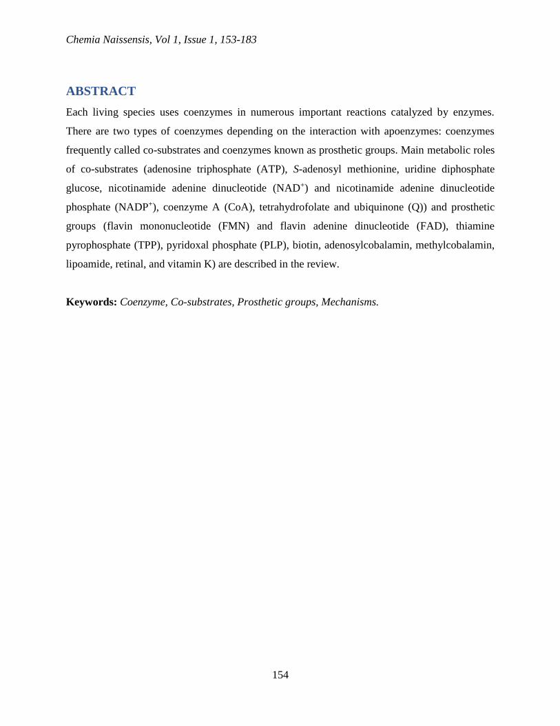
ABSTRACT
Each living species uses coenzymes in numerous important reactions catalyzed by enzymes.
There are two types of coenzymes depending on the interaction with apoenzymes: coenzymes
frequently called co-substrates and coenzymes known as prosthetic groups. Main metabolic roles
of co-substrates (adenosine triphosphate (ATP), S-adenosyl methionine, uridine diphosphate
glucose, nicotinamide adenine dinucleotide (NAD+) and nicotinamide adenine dinucleotide
phosphate (NADP+), coenzyme A (CoA), tetrahydrofolate and ubiquinone (Q)) and prosthetic
groups (flavin mononucleotide (FMN) and flavin adenine dinucleotide (FAD), thiamine
pyrophosphate (TPP), pyridoxal phosphate (PLP), biotin, adenosylcobalamin, methylcobalamin,
lipoamide, retinal, and vitamin K) are described in the review.
Keywords: Coenzyme, Co-substrates, Prosthetic groups, Mechanisms.
Chemia Naissensis, Vol 1, Issue 1, 153-183
155
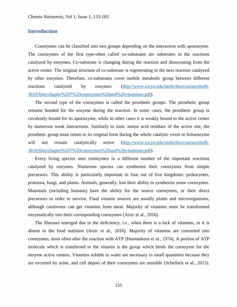
Introduction
Coenzymes can be classified into two groups depending on the interaction with apoenzyme.
The coenzymes of the first type-often called co-substrates are substrates in the reactions
catalyzed by enzymes. Co-substrate is changing during the reaction and dissociating from the
active center. The original structure of co-substrate is regenerating in the next reaction catalyzed
by other enzymes. Therefore, co-substrates cover mobile metabolic group between different
reactions catalyzed by enzymes (http://www.uwyo.edu/molecbio/courses/molb-
3610/files/chapter%207%20coenzymes%20and%20vitamines.pdf).
The second type of the coenzymes is called the prosthetic groups. The prosthetic group
remains bonded for the enzyme during the reaction. In some cases, the prosthetic group is
covalently bound for its apoenzyme, while in other cases it is weakly bound to the active center
by numerous weak interactions. Similarly to ionic amino acid residues of the active site, the
prosthetic group must return to its original form during the whole catalytic event or holoenzyme
will not remain catalytically active (http://www.uwyo.edu/molecbio/courses/molb-
3610/files/chapter%207%20coenzymes%20and%20vitamines.pdf).
Every living species uses coenzymes in a different number of the important reactions
catalyzed by enzymes. Numerous species can synthesize their coenzymes from simple
precursors. This ability is particularly important in four out of five kingdoms: prokaryotes,
protozoa, fungi, and plants. Animals, generally, lost their ability to synthesize some coenzymes.
Mammals (including humans) have the ability for the source coenzymes, or their direct
precursors in order to survive. Final vitamin sources are usually plants and microorganisms,
although carnivores can get vitamins from meat. Majority of vitamins must be transformed
enzymatically into their corresponding coenzymes (Arsic et al., 2016).
The illnesses emerged due to the deficiency, i.e., when there is a lack of vitamins, or it is
absent in the food nutrition (Arsic et al., 2016). Majority of vitamins are converted into
coenzymes, most often after the reaction with ATP (Huennekens et al., 1974). A portion of ATP
molecule which is transferred to the vitamin is the group which binds the coenzyme for the
enzyme active centers. Vitamins soluble in water are necessary in small quantities because they
are excreted by urine, and cell depots of their coenzymes are unstable (Schellack et al., 2015).

Chemia Naissensis, Vol 1, Issue 1, 153-183
156
On the other side, lipid vitamins, like vitamins A, D, E, I, K are stored in animals, and increased
intake can cause toxic states known as hypervitaminoses (Engelking, 2015).
The most important enzymes are listed in Table 1 together with their roles in metabolism and
their vitamin sources.
Table 1. The most important coenzymes (Horton et al., 2006)
Coenzyme Vitamin Main metabolic role Mechanistic role
Adenosine
triphosphate (ATP) -
Transfer of
phosphoryl or
nucleotidyl groups
Co-substrate
S-Adenosyl
methionine -
Transfer of metal
groups Co-substrate
Uridine diphosphate
glucose -
Transfer of glycosyl
groups Co-substrate
Nicotinamide adenine
dinucleotide (NAD+)
and nicotinamide
adenine dinucleotide
phosphate (NADP+)
Niacin
Oxidation-reduction
reactions involving 2-
electron transfers
Co-substrate
Flavin
mononucleotide
(FMN) and flavin
adenine dinucleotide
(FAD)
Riboflavin (B2)
Oxidation-reduction
reactions including
one and two electron
transfers
Prosthetic group
Coenzyme A (CoA) Pantothenate (B3) Transfer of acyl
groups Co-substrate
Thiamine
pyrophosphate (TPP) Thiamine (B1)
Transfer of fragments
from two carbons
containing carbonyl
group
Prosthetic group
Pyridoxal phosphate
(PLP) Pyridoxine (B6)
Transfer of groups
from and to amino Prosthetic group

Chemia Naissensis, Vol 1, Issue 1, 153-183
157
acids
Biotin Biotin
ATP dependent
carboxylation of
substrate or transfer
of carboxylic groups
between substrates
Prosthetic group
Tetrahydrofolate Folate
Transfer of
substituents with one
carbon, particularly
formyl and
hydroxymethyl
groups; giving methyl
group for thiamine in
DNA
Co-substrate
Adenosylcobalamin Cobalamine (B12) Intramolecular
rearrangement Prosthetic group
Methylcobalamin Cobalamine (B12) Transfer of methyl
groups Prosthetic group
Lipoamide -
Oxidation of
hydroxyalkyl group
from TPP and the
next transfer as an
acyl group
Prosthetic group
Retinal Vitamin A Eyesight Prosthetic group
Vitamin K Vitamin K
Carboxylation of
some glutamic
residues
Prosthetic group
Ubiquinone (Q) - Electron carrier
soluble in fats Co-substrate

Chemia Naissensis, Vol 1, Issue 1, 153-183
158
ATP and other nucleotide co-substrates
There are many nucleoside triphosphates which behave as coenzymes. Among them,
adenosine triphosphate (ATP) is the most abundant. Other frequent examples are GTP, S-
adenosyl methionine and nucleotide sugars such as uridine diphosphate glucose (UDP-glucose).
ATP (Figure 1a) can donate phosphoryl, pyrophosphoryl, adenylyl (AMP), or adenosyl groups in
reactions of group transfers.
a)
O
OHOH
HH
HH
CH2OP
O
O
OP
O
O
OP
O
O
O N
N
N
N
NH2
b)
O
OHOH
HH
HH
CH2S
CH3
CH2
CH2
CH
COO
H3N
N
N
N
N
NH2
Figure 1. a) Nitrogen base adenine is connected to the ribose which carries three phosphoryl
groups. Transfer of the phosphoryl group gives ADP, and the transfer of nucleotidyl group
(AMP) gives pyrophosphate; b) S-adenosyl methionine
The most usual reaction involving ATP is the transfer of the phosphoryl group. In the
reactions catalyzed by enzymes, for example, -phosphoryl group ATP is transferred to the

Chemia Naissensis, Vol 1, Issue 1, 153-183
159
nucleophile, leaving ADP. The second most usual reaction is a transfer of the nucleotidyl group
(transfer of AMP part), leaving pyrophosphate (PPi).
S-adenosyl methionine (Figure 1b) is synthesized in the reaction of methionine with ATP.
Different from a thiomethyl group of methionine, positively charged sulfonium S-adenosyl
methionine is highly reactive, so it reacts readily with nucleophilic acceptors, and practically it is
a donor of all methyl groups used in biosynthetic reactions (e.g., conversion of hormone
norepinephrine into epinephrine). Methylation reactions which require S-adenosyl methionine
involve methylation of phospholipids, proteins, DNA, and RNA. In plants, S-adenosyl
methionine is involved in the regulation of fruit ripening as a precursor of plant hormone
ethylene (Chiang et al., 1996).
Nucleotide-sugar coenzymes are involved in the metabolism of carbohydrates. The most
common nucleotide sugar, uridine diphosphate glucose (UDP-glucose) is formed in the reaction
of glucose 1-phosphate with uridine triphosphates (UTP) (Figure 2). UDP-glucose can donate its
glycosyl group to the corresponding acceptor releasing UDP. UDP-glucose is regenerated when
UDP accepted phosphoryl group from ATP and obtained UTP reacts with another molecule of
glucose-1-phosphate.

Chemia Naissensis, Vol 1, Issue 1, 153-183
160
Figure 2. The formation of UDP glucose catalyzed with UDP-glucose phosphorylase
(Horton et al., 2006)
In the mechanism of the formation of UDP glucose, the oxygen of the phosphoric group of α-D-
glucose 1-phosphate attacks α-phosphorus of UTP. The released PPi is hydrolyzed fast to 2 Pi by
the action of pyrophosphatase. This hydrolysis enables the occurring of the reaction catalyzed by
pyrophosphorylase.
Chemia Naissensis, Vol 1, Issue 1, 153-183
161
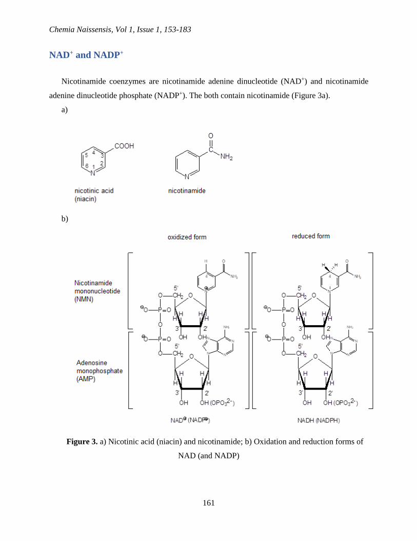
NAD+ and NADP+
Nicotinamide coenzymes are nicotinamide adenine dinucleotide (NAD+) and nicotinamide
adenine dinucleotide phosphate (NADP+). The both contain nicotinamide (Figure 3a).
a)
b)
Figure 3. a) Nicotinic acid (niacin) and nicotinamide; b) Oxidation and reduction forms of
NAD (and NADP)

Chemia Naissensis, Vol 1, Issue 1, 153-183
162
The deficiency of nicotinic acid (niacin) causes the pellagra disease. Nicotinic acid or
nicotinamide is essential as a precursor of NAD+ and NADP+ (pyridine nucleotide coenzymes)
(Figure 3b) (Sorci et al., 2010). Nicotinamide coenzymes play a role in numerous oxidation-
reduction reactions in the form of electron transfers from and to the metabolite. Pyridine ring
NAD+ is reduced by the addition of hydride ion onto C-4 when NAD+ is transformed into NADH
(and when NADP+ is transformed into NADPH) (Sorci et al., 2010).
NAD+ and NADP+ almost always behave as dehydrogenase substrates (Bellamacina, 1996).
Dehydrogenase catalyzes the oxidation of the substrate by transferring two electrons and proton
in the form of hydride ion (H-) onto C-4 of nicotinamide group NAD+ and NADP+. In this way,
the reduced forms are formed (NADH and NADPH), where new C-H bond is created on C-4
(Bellamacina, 1996).
NADH and NADPH (stable in solutions containing oxygen) possess reductive power
(Kukielka and Cederbaum, 1990). The stability of reduced pyridine nucleotides allows them to
carry their reduction potential from one enzyme to another; the characteristics not owned by
flavin coenzymes. The majority of reactions in which NADH and NADPH are formed are
catabolic reactions. The oxidation of NADH in mitochondria is coupled with ATP synthesis. The
most significant part of NADPH is used as a reduction agent in biosynthetic reactions (Kukielka
and Cederbaum, 1990).
NADH and NADPH show a maximum in ultraviolet region at 340 nm caused by
dihydropyridine ring, while NAD+ and NADP+ do not absorb the light at this wavelength. The
appearance and disappearance of the absorbance at 340 nm are useful for the measurement of the
rate of the oxidation (McComb et al., 1976).
In the mechanism of the oxidation of lactate to pyruvate catalyzed by lactate dehydrogenase
(Figure 4), the coenzyme accepts the hydride ion on C-4 in nicotinamide group. This
phenomenon leads to the bond rearrangement in the ring when electrons are moving to the
positively charged nitrogen atom. The enzyme represents acid-base catalyst and the suitable
place for binding coenzyme, and the substrate as well. Two hydrogens are moving from the
lactate to produce pyruvate. One of these hydrogens are moving to NAD+ as a hydride ion
bearing two electrons, and the another is transferring to His-195 as a proton. The second
hydrogen is then releasing as H+ to regenerate the base catalyst (His-195) (Kane, 2014; Speers
and Reguera, 2012).

Chemia Naissensis, Vol 1, Issue 1, 153-183
163
Figure 4. The mechanism of lactate dehydrogenase (Speers and Reguera, 2012)
FAD and FMN
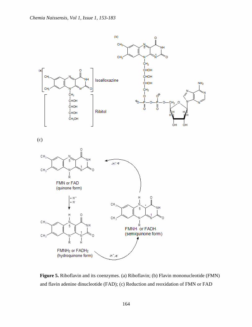
Coenzymes flavin adenine dinucleotide (FAD) and flavin mononucleotide (FMN) are derived
from riboflavin or vitamin B2. Riboflavin is synthesized by bacteria, protozoa, fungi, plants and
some animals. Mammals are getting riboflavin from food. Riboflavin consists from ribitol
connected to N-10 atom of heterocyclic ring system called isoalloxazine (Figure 5a). Similarly to
NAD+ and NADP+, FAD contains AMP and pyrophosphate bond (Figure 5b).
Chemia Naissensis, Vol 1, Issue 1, 153-183
164
(c)
Figure 5. Riboflavin and its coenzymes. (a) Riboflavin; (b) Flavin mononucleotide (FMN)
and flavin adenine dinucleotide (FAD); (c) Reduction and reoxidation of FMN or FAD

Chemia Naissensis, Vol 1, Issue 1, 153-183
165
FAD or FMN are necessary as prosthetic groups for many oxidoreductases. Reduced flavin
coenzymes can be easily oxidized in the presence of oxygen. FAD and FMN are reduced to
FADH2 and FMNH2 by taking proton and two electrons in the form of hydride ion (Figure 5c).
Oxidized enzymes are light yellow as a result of the system of conjugated double bonds of the
isoalloxazine cyclic system. Different from NADH and NADPH, which exclusively participate
in two-electron systems, FMNH2 and FADH2 are donated electrons (one (FADH· or FMNH· are
formed) or two). The intermediates are relatively stable free radicals called semiquinones.
Oxidation of FADH2 and FAMNH2 is often coupled with the reduction of metalloproteins
containing Fe3+ (in [Fe-S] cluster). Since iron-sulfur cluster can accept only one electron,
reduced flavin must be oxidized into two one-electron steps via semiquinone intermediate
(Ghisla and Massey, 1989).
Coenzyme A
Numerous metabolic processes depend on coenzyme A (CoA, or HS-CoA), including the
oxidation of fuel molecule and biosynthesis of some carbohydrates and fats. The coenzyme is
involved in the reactions of acyl group transfers (Leonardi et al., 2005). It has three main
components: 2-mercaptoethylamine unit with free -SH group, pantothenate vitamin (vitamin B3),
and ADP part (Figure 6a). Acetyl CoA is energetically rich compound because of the high
energy of thioester bond.
Phosphopantetheine, phosphate ester which contains 2-mercaptoethylamine and pantothenate
parts of coenzyme A, is a prosthetic group of a small protein (77 amino acid residues), known as
an acyl carrier protein (ACP). The prosthetic group is esterified to ACP via oxygen in the side
chain of a serine residue (Figure 6b). The intermediates acetylate SH of the prosthetic group
ACP in the fatty acids’ biosynthesis (Leonardi et al., 2005).

Chemia Naissensis, Vol 1, Issue 1, 153-183
166
Figure 6. (a) Coenzyme A; (b) Acyl carrier protein (ACP)
Thiamine pyrophosphate
Thiamine (or vitamin B1) contains pyrimidine ring and positively charged thiazoline ring
(Figure 7a). In mammals, thiamine is the essential vitamin, wide-spread in the rice peel and other
wheat. Its deficiency causes beriberi. Coenzyme is the thiamine pyrophosphate (TPP) (Figure
7b). TPP is synthesized from thiamine by the enzymatic transfer of pyrophosphoryl group from
ATP (Shepard and Broderick, 2010).
Numerous decarboxylases (carboxylases) require TPP as a coenzyme (e.g., pyruvate
decarboxylase of the yeast) (Figure 7c).
TPP is also the coenzyme involved in oxidative decarboxylation of α-keto acids, except for
pyruvates. The first steps in these reactions are occurring according to the mechanism shown in

Chemia Naissensis, Vol 1, Issue 1, 153-183
167
Figure 7c. Besides, TPP is a prosthetic group for the enzymes known as transketolases, which
catalyze the transfer between the sugar molecules of two carbon groups containing keto group.

Chemia Naissensis, Vol 1, Issue 1, 153-183
168
(c)
Figure 7. (a) Thiamine (vitamin B1); (b) Thiamine pyrophosphate (TPP); (c) The mechanism
of pyruvate decarboxylase from the yeast
Thiazoline ring of the coenzyme contains the reactive center. C-2 of TPP has unusual
activity; it is acidic despite high pKa in aqueous solution. Experiments show that pKa value for
the ionization of hydroxyethylamine pyrophosphate (HETPP) (i.e., forming of dipolar carbanion)

Chemia Naissensis, Vol 1, Issue 1, 153-183
169
changes from 15 in water to 6 on the active center of pyruvate decarboxylase. This increased
acidity is ascribed to low polarity of active center, which is also responsible for the increased
reactivity of TPP itself. The positive charge of thiazoline ring of TPP attracts electrons,
weakening the bond between C-2 and hydrogen. The proton is mostly removing by base part of
the enzyme. Ionization gives resonantly stable dipolar carbanion known as ylide. Negatively
charged C-2 attacks electron-deficit carbonyl carbon of pyruvate substrate, and the first product
(CO2) is releasing. Two carbons of pyruvate are now attached to thiazoline ring as a part of
resonance-stabilized carbanion. In the next step, the protonation of carbanion gives
hydroxyethylamine pyrophosphate (HETPP). HETPP is separating, releasing acetaldehyde (the
second product). TPP is forming again when ylide is protonating from the side of the enzyme
(Figure 7c) (Shepard and Broderick, 2010).
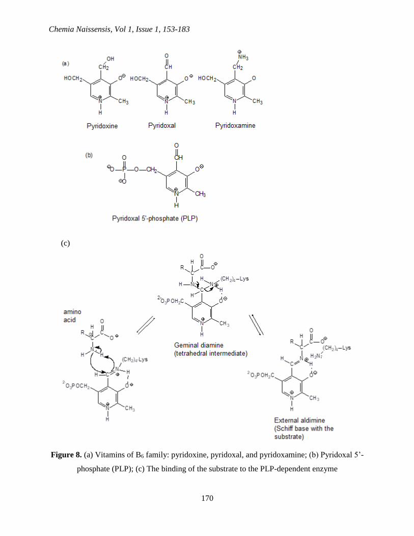
Pyridoxal phosphate
The family of B6 vitamins soluble in water consists of three closely connected molecules
differing only in the state of the oxidation or amination of the carbon bound to the position 4 of
the pyridine ring (Figure 8a). Induced deficiency of vitamin B6 in mice causes dermatitis and
various disorders connected to the metabolism of proteins; deficiencies of vitamin B6 in humans
are rare. Once B6 enters the cell, enzymatic transfer of -phosphoryl group from ATP forms
coenzyme pyridoxal 5’-phosphate (PLP) (Figure 8b) (Hayashi et al., 1990).
Chemia Naissensis, Vol 1, Issue 1, 153-183
170
(c)
Figure 8. (a) Vitamins of B6 family: pyridoxine, pyridoxal, and pyridoxamine; (b) Pyridoxal 5’-
phosphate (PLP); (c) The binding of the substrate to the PLP-dependent enzyme

Chemia Naissensis, Vol 1, Issue 1, 153-183
171
Pyridoxal phosphate is a prosthetic group for many enzymes which catalyze different
reactions, including isomerizations, decarboxylations, and eliminations in side chain or
substitutions. In enzymes dependent on PLP, the carbonyl group of the prosthetic group is
binding as Schiff base (imine) for -amino group of a lysine residue in the active center (Figure
8c) (Hayashi et al., 1990; Horton et al., 2006).
Transamination is most frequently dependent reaction on PLP, and the mechanism of this
type of the reaction is presented in Figure 9.
Figure 9. The mechanism of transaminase (Horton et al., 2006)

Chemia Naissensis, Vol 1, Issue 1, 153-183
172
In the first step of the mechanism, amino acid replaces lysine from internal aldimine which
binds PLP for the enzyme forming the external aldimine. In the second step, α-hydrogen of the
amino acid is taken with the base catalyst by the same lysine residue. Electronic rearrangement
leads to the quinoid intermediate. In the third step, the protonation of the intermediate with the
lysine residue gives ketoimine. In the fourth step, hydrolysis of the ketoimine gives α-keto acid,
which dissociates, and PMP remains bound to the enzyme. If another α-keto acid enters, each
step goes in reverse. The amino acid is transferred to α-keto acid, giving new amino acid and
regenerates the original PLP form of the enzyme (Figure 9) (Hayashi et al., 1990; Horton et al.,
2006).
Biotin
Biotin is a prosthetic group for enzymes which catalyze the reactions of the transfer of
carboxyl group and the reaction of carboxylation dependent on ATP. It is covalently bound to the
active center of its enzyme host by amide bond for -amino group of a lysine residue (Figure
10a).
(a)
Chemia Naissensis, Vol 1, Issue 1, 153-183
173
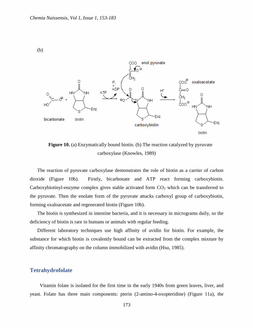
(b)
Figure 10. (a) Enzymatically bound biotin. (b) The reaction catalyzed by pyruvate
carboxylase (Knowles, 1989)
The reaction of pyruvate carboxylase demonstrates the role of biotin as a carrier of carbon
dioxide (Figure 10b). Firstly, bicarbonate and ATP react forming carboxybiotin.
Carboxybiotinyl-enzyme complex gives stable activated form CO2 which can be transferred to
the pyruvate. Then the enolate form of the pyruvate attacks carboxyl group of carboxybiotin,
forming oxaloacetate and regenerated biotin (Figure 10b).
The biotin is synthesized in intestine bacteria, and it is necessary in micrograms daily, so the
deficiency of biotin is rare in humans or animals with regular feeding.
Different laboratory techniques use high affinity of avidin for biotin. For example, the
substance for which biotin is covalently bound can be extracted from the complex mixture by
affinity chromatography on the column immobilized with avidin (Hsu, 1985).
Tetrahydrofolate
Vitamin folate is isolated for the first time in the early 1940s from green leaves, liver, and
yeast. Folate has three main components: pterin (2-amino-4-oxopteridine) (Figure 11a), the

Chemia Naissensis, Vol 1, Issue 1, 153-183
174
residue of p-aminobenzoic acid, and residue of the glutamate. Humans need folate in nutrition
because they are not in the position to synthesize pterin-p-aminobenzoic acid intermediate
(PABA) (Wallig and Keenan, 2013).
Coenzyme formed from the folate (Figure 11b) is known as tetrahydrofolate (Figure 11c)
(Wallig and Keenan, 2013).

Chemia Naissensis, Vol 1, Issue 1, 153-183
175
(d)
(e)
HN
N N
N
O
H2N
H
H
H
H
H
CH
OH
CH
OH
CH3
Figure 11. (a) Pterin; (b) Folate; (c) Tetrahydrofolate; (d) Monocarbonic derivatives of
tetrahydrofolates; (e) 5,6,7,8-tetrahydrobiopterin
The anionic polyglutamic residue, usually five to six residues long, takes part in the binding
of coenzymes for enzymes (Wallig and Keenan, 2013).

Chemia Naissensis, Vol 1, Issue 1, 153-183
176
Tetrahydrofolate is formed by the addition of hydrogens into positions 5, 6, 7 and 8 of pterin
cyclic system.
The reduction of dihydrofolate obtained during the formation of the methyl group of
thymidylates (dTMP) is the primary metabolic function of dihydrofolate reductase. This reaction
which uses derivative of tetrahydrofolate is the essential step in the synthesis of DNA. Because
the cell division cannot be achieved when DNA synthesis is stopped, dihydrofolate reductase is
intensively studied as an aim in chemotherapy for cancer treatment (Horton et al., 2006).
5,6,7,8-tetrahydrofolate is necessary to enzymes which catalyze biochemical transfers of
several monocarbon units. The groups bound to tetrahydrofolate are methylene, methyl, and
formyl. Figure 11d shows the structure of several monocarbon derivatives of tetrahydrofolate
and enzymatic interconversion happens between them (Horton et al., 2006).
Monocarbonic metabolic groups are covalently bound for the secondary amine N-5 or N-10
of tetrahydrofolates, or both in cyclic form. 10-Formyltetrahydrofolate is a donor of formyl
groups, and 5,10-methylenetetrahydrofolate is the donor of hydroxymethyl groups (Figure 11d).
The second pterin coenzyme, 5,6,7,8-tetrahydrobiopterin has a side chain with three carbons
on C-6 pterin part instead of long side chain which is situated in tetrahydrofolate (Figure 11e)
(Wallig and Keenan, 2013). This coenzyme is not derived from vitamin; it is synthesized by
animals alone and by other organisms. Tetrahydrobiopterin is a cofactor for several
hydroxylases, and it is a reducing agent in the conversion of phenylalanine to tyrosine. Also, it is
necessary to the enzyme which catalyzes the synthesis of nitrogen oxide from arginine.
Cobalamin
Cobalamin (vitamin B12) is the biggest B vitamin, and it is last isolated. The structure of
cobalamin includes corrin ring system which is similar to the porphyrinic cyclic system of heme.
Cobalamin contains cobalt instead of iron which is situated in heme. In coenzyme form of
cobalamin, R group is either methyl group (in methyl-cobalamin) or 5’-deoxyadenosyl group (in
adenosylcobalamin) (Banerjee and Ragsdale, 2003).
Cobalamin is necessary as a micro substance to all animals and some bacteria and algae, but
it is not necessary for plants, and they are not synthesizing them. Thus, humans will generally get
vitamin B12 from food of animal origin. Vegetarians are getting appropriate quantity from
microorganisms. The cobalamin deficiency can cause anemia, potentially fatal diseases in which
Chemia Naissensis, Vol 1, Issue 1, 153-183
177
there is decreasing in the production of blood cells by bone marrow. This type of anemia can
lead to neurological disorders. Majority of victims of this anemia do not excrete necessary
glycoprotein (called internal factor) (Arsic et al., 2016).
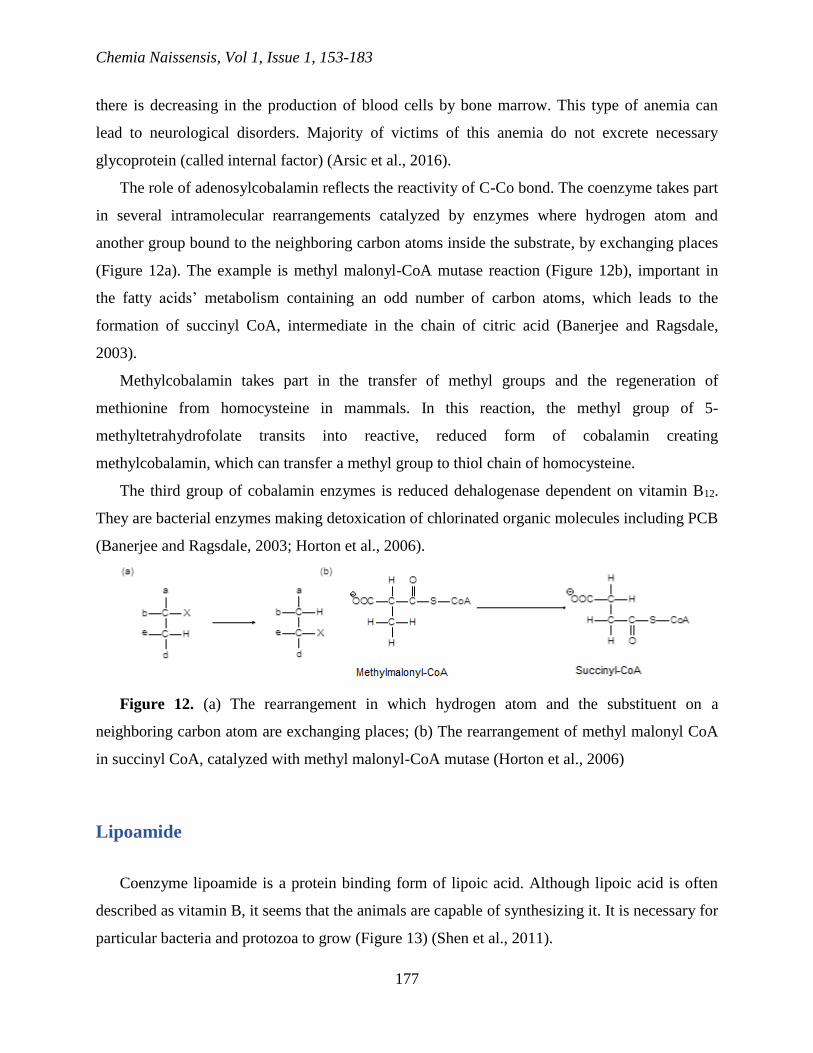
The role of adenosylcobalamin reflects the reactivity of C-Co bond. The coenzyme takes part
in several intramolecular rearrangements catalyzed by enzymes where hydrogen atom and
another group bound to the neighboring carbon atoms inside the substrate, by exchanging places
(Figure 12a). The example is methyl malonyl-CoA mutase reaction (Figure 12b), important in
the fatty acids’ metabolism containing an odd number of carbon atoms, which leads to the
formation of succinyl CoA, intermediate in the chain of citric acid (Banerjee and Ragsdale,
2003).
Methylcobalamin takes part in the transfer of methyl groups and the regeneration of
methionine from homocysteine in mammals. In this reaction, the methyl group of 5-
methyltetrahydrofolate transits into reactive, reduced form of cobalamin creating
methylcobalamin, which can transfer a methyl group to thiol chain of homocysteine.
The third group of cobalamin enzymes is reduced dehalogenase dependent on vitamin B12.
They are bacterial enzymes making detoxication of chlorinated organic molecules including PCB
(Banerjee and Ragsdale, 2003; Horton et al., 2006).
Figure 12. (a) The rearrangement in which hydrogen atom and the substituent on a
neighboring carbon atom are exchanging places; (b) The rearrangement of methyl malonyl CoA
in succinyl CoA, catalyzed with methyl malonyl-CoA mutase (Horton et al., 2006)
Lipoamide
Coenzyme lipoamide is a protein binding form of lipoic acid. Although lipoic acid is often
described as vitamin B, it seems that the animals are capable of synthesizing it. It is necessary for
particular bacteria and protozoa to grow (Figure 13) (Shen et al., 2011).

Chemia Naissensis, Vol 1, Issue 1, 153-183
178
Figure 13. Lipoamide
It is believed for lipoamide to function as a pendulum which carries acyl groups between
active centers in multi-enzymatic complexes. For example, in complex of pyruvate
dehydrogenase, disulfide ring of lipoamide prosthetic group reacts with HETPP, binding its
acetyl group on sulfur atom attached to C-8 of lipoamide and creating thioester. Then, the acyl
group is moved to the sulfur atom of coenzyme A giving reduced (dihydrolipoamide) form of
prosthetic group.
The last step catalyzed by pyruvate dehydrogenase complex is the oxidation of
dihydrolipoamide. In this reaction, NADH is formed by the action of the flavoprotein component
of the complex. The actions of multiple coenzymes of pyruvate dehydrogenase complex show
how coenzymes supplying reactive groups and thus increasing catalytic diversity of proteins are
used for storing energy and carbon building blocks (Horton et al., 2006; Shen et al., 2011).
Ubiquinone
Ubiquinone (coenzyme Q) (Figure 14a) is a coenzyme soluble in fats synthesized by all
species. In the membrane, ubiquinone transports electrons between enzymatic complexes
situated in the membrane. Some bacteria use menaquinone instead of ubiquinone. Ubiquinone
analog called plastoquinone (Figure 14b) has a similar function in the photosynthetic transport of
electrons in chloroplasts (DiNicolantonio et al., 2015).

Chemia Naissensis, Vol 1, Issue 1, 153-183
179
(c)
Figure 14. (a) Ubiquinone; (b) Plastoquinone; (c) Three oxidation states of ubiquinone
Ubiquinone is a stronger oxidation reagent than both NAD+ and flavin coenzymes. Similarly
to FMN and FAD, ubiquinone can accept or donate electrons (one or two) because it has three
oxidation states: oxidized Q, partially reduced semiquinone free radical and completely reduced
QH2 (ubiquinol) (Figure 14c) (Sohal, 2004).

Chemia Naissensis, Vol 1, Issue 1, 153-183
180
Coenzyme Q plays the leading role in the electron transport connected to the membrane. It is
responsible for the moving of protons from one side of the membrane to another by the process
known as cycle Q. The created protein gradient leads to ATP synthesis (Lenaz et al., 2007).
Protein coenzymes
Some proteins behave as coenzymes. They do not catalyze reactions, but they are necessary
from specific enzymes. These enzymes are called either protein for groups transfer or protein
coenzymes. They contain functional group either as a part of their protein skeleton or as a
prosthetic group (Horton et al., 2006).
Metal ions, iron-sulfur clusters, and heme groups are reactive centers usually found in these
protein coenzymes. Several protein coenzymes have two reactive centers. Thioredoxins are
observed as reduction agents (cycle of citric acid, photosynthesis, and synthesis of
deoxyribonucleotides). Disulfide reactive center of thioredoxin is on the surface of the protein,
so it is available to the active centers of corresponded enzymes (Horton et al., 2006; Johnson et
al., 2014).
Cytochromes
Cytochromes are protein coenzymes containing heme where Fe (III) atoms undergo
reversible one-electron reduction. They are classified as a, b and c based on their visible
absorption spectra. Heme of cytochrome b type is the same as that of hemoglobin and
myoglobin. Heme of cytochrome a has a hydrophobic chain of 17 carbons on C-2 porphyrin ring
and formyl group on C-8, while heme of type b has a vinyl group attached on C-2 and methyl
group on C-8. In cytochromes of type c, the heme is covalently bound to apoprotein with two
thioester bonds formed by the addition of thiol groups of two cysteine residues for vinyl groups
of the heme (Heldt and Piechulla, 2011).
The tendency to transfer an electron to another substance, measured as a reductive potential,
also varies among cytochromes. The range of reduction potentials among prosthetic groups is an

Chemia Naissensis, Vol 1, Issue 1, 153-183
181
essential property of membrane-connected electron transferred cycles and biosynthesis (Heldt
and Piechulla, 2011).
References
Arsic, B., Dimitrijevic, D., & Kostic, D. (2016). Chapter 1: Mineral and vitamin fortification. In:
A. M. Grumazescu (Ed.), Nutraceuticals: nanotechnology in the agri-food industry (pp. 1-40).
Amsterdam: Elsevier.
Banerjee, R., & Ragsdale, S. W. (2003). The many faces of vitamin B12: catalysis by cobalamin-
dependent enzymes. Annual Review of Biochemistry, 72, 209-247.
Bellamacina, C. R. (1996). The nicotinamide dinucleotide binding motif: a comparison of
nucleotide binding proteins. The FASEB Journal, 10, 1257-1269.
Chapter 7. "Coenzymes and Vitamins", http://www.uwyo.edu/molecbio/courses/molb-
3610/files/chapter%207%20coenzymes%20and%20vitamines.pdf, accessed 12/12/2018
Chiang, P. K., Gordon, R. K., Tal, J., Zeng, G. C., Doctor, B. P., Pardhasaradhi, K., & McCann,
P. P. (1996). S-Adenosylmethionine and methylation. The FASEB Journal, 10, 471-480.
DiNicolantonio, J. J., Bhutani, J., McCarty, M. F., & O’Keefe, J. H. (2015). Coenzyme Q10 for
the treatment of heart failure: a review of the literature. Open Heart, 2, e000326.
Engelking, L. R. (2015). Chapter 44 - Vitamin A. In: Textbook of Veterinary Physiological
Chemistry (Third Edition) (pp. 282-287). Academic Press.
Ghisla, S., & Massey, V., (1989). Mechanisms of flavoprotein-catalyzed reactions. European
Jiurnal of Biochemistry, 181, 1-17.
Hayashi, H., Wada, H., Yoshimura, T., Esaki, N., & Soda, K. (1990). Recent topics in pyridoxal
5’-phosphate enzyme studies. Annual Review of Biochemistry, 59, 87-110.
Heldt, H.-W., & Piechulla, B., (2011). 3 - Photosynthesis is an electron transport process, In:
Plant Biochemistry (Fourth Edition) (pp. 65-112). Academic Press.
Horton, H. R., Moran, L. A., Scrimgeour, K. G., Perry, M. D., & Rawn, J. D. (2006). Principles
of Biochemistry. (4th ed.). Pearson Prentice Hall, Pearson Education, Inc., New Jersey.
Hsu, S. M. (1985). Immunoperoxidase techniques using the avidin-biotin system. In: Ngo T.
T., Lenhoff H. M. (eds) Enzyme-Mediated Immunoassay. Boston, MA: Springer.
Chemia Naissensis, Vol 1, Issue 1, 153-183
182
Huennekens, F. M., Digirolamo, P. M., Fujii, K., Henderson, G. B., Jacobsen, D. W., Neef, V.
G., & Rader, J. I. (1974). Folic acid and vitamin B12: Transport and conversion to coenzyme
forms. Advances in Enzyme Regulation, 12(C).
Johnson, M. N. R., Londergan, C. H., & Charkoudian, L. K., (2014). Probing the
phosphopantetheine arm conformations of acyl carrier proteins using vibrational spectroscopy.
Journal of the American Chemical Society, 136, 11240-11243.
Kane, D. A., (2014). Lactate oxidation at the mitochondria: a lactate-malate-aspartate shuttle at
work. Frontiers in Neuroscience, 6, article: 366.
Knowles, J. R. (1989). The mechanism of biotin-dependent enzymes. Annual Review of
Biochemistry, 58, 195-221.
Kukielka, E., & Cederbaum, A. I., (1990). NADPH- and NADH-Dependent oxygen radical
generation by rat liver nuclei in the presence
of redox cycling agents and iron. Archives of Biochemistry and Biophysics,
283 (2), 326-333.
Lenaz, G., Fato, R., Formiggini, G., & Genova, M. L. (2007). The role of coenzyme Q in
mitochondrial electron transport. Mitochondrion, 7S, S8-S33.
Leonardi, R., Zhang, Y.-M., Rock, C. O., & Jackowski, S., (2005). Coenzyme A: back in action.
Progress in Lipid Research, 44, 125-153.
McComb, R. B., Bond, R. W., Burnett, R. W., Keech, R. C., & Bowers Jr. G. N., (1976).
Determination of the molar absorptivity of NADH. Clinical Chemistry, 22 (2), 141-150.
Schellack, G., Harirari, P., & Schellack N. (2015). B-complex vitamin deficiency and
supplementation. South African Pharmaceutical Journal, 82(4), 28-33.
Shen, W., Hao, J., Feng, Z., Tian, C., Chen, W., Packer, L., Shi, X., Zang, W., & Liu, J. (2011).
Lipoamide or lipoic acid stimulates mitochondrial biogenesis in 3T3-L1 adipocytes via the
endothelial NO synthase-cGMP-protein kinase G signalling pathway. British Journal of
Pharmacology, 162, 1213–1224.
Shepard, E. M., & Broderick, J. B., (2010). S-Adenosylmethionine and iron–sulfur clusters in
biological radical reactions: The radical SAM superfamily. Reference Module in Chemistry,
Molecular Sciences and Chemical Engineering, Comprehensive Natural Products II, Chemistry
and Biology, Volume 8, pp. 625-661.

Chemia Naissensis, Vol 1, Issue 1, 153-183
183
Sohal, R. S. (2004). Coenzyme Q and vitamin E interactions. Methods in Enzymology, 378, 146-
151.
Sorci, L., Kurnasov, O., Rodionov, D. A., & Osterman, A. L. (2010). 7.08 - Genomics and
Enzymology of NAD Biosynthesis. In: Reference Module in Chemistry, Molecular Sciences
and Chemical Engineering, Comprehensive Natural Products II, Chemistry and Biology, Volume
7 (pp. 213-257). Elsevier.
Speers, A. M., & Reguera, G. (2012). Electron donors supporting growth and electroactivity of
Geobacter sulfurreducens anode biofilms. Applied and Environmental Microbiology, 437-444.
Wallig, M. A., & Keenan, K. P., (2013). Chapter 36 - Nutritional Toxicologic Pathology,
Haschek and Rousseaux's Handbook of Toxicologic Pathology (Third Edition), Volume II, pp.
1077-1121.