Mechanism and Control of V(D)J Recombination versus Class Switch
70
Mechanism and Control of V(D)J Recombination versus Class Switch Recombination: Similarities and Differences Darryll D. Dudley, 1 Jayanta Chaudhuri, Craig H. Bassing, and Frederick W. Alt Howard Hughes Medical Institute, The Children’s Hospital Boston, CBR Institute for Biomedical Research, and Harvard Medical School, Boston, Massachusetts 02115 Abstract .................................................................................................. 43 1. Overview: V(D)J and Class Switch Recombination ............................................... 44 2. Antigen Receptor Gene Rearrangement ............................................................ 47 3. Regulation of V(D)J Recombination ................................................................ 61 4. Class Switch Recombination Employs Distinct Mechanisms for V(D)J Recombination ............................................................................. 69 5. CSR-Related Diseases ................................................................................. 93 6. Concluding Remarks ................................................................................... 95 References ............................................................................................... 97 Abstract V(D)J recombination is the process by which the variable region exons encoding the antigen recognition sites of receptors expressed on B and T lymphocytes are generated during early development via somatic assembly of component gene segments. In response to antigen, somatic hypermutation (SHM) and class switch recombination (CSR) induce further modifications of immunoglobulin genes in B cells. CSR changes the IgH constant region for an alternate set that confers distinct antibody effector functions. SHM introduces mutations, at a high rate, into variable region exons, ultimately allowing affinity maturation. All of these genomic alteration processes require tight regulatory control mechan- isms, both to ensure development of a normal immune system and to prevent potentially oncogenic processes, such as translocations, caused by errors in the recombination/mutation processes. In this regard, transcription of substrate sequences plays a significant role in target specificity, and transcription is mechanistically coupled to CSR and SHM. However, there are many mechanis- tic differences in these reactions. V(D)J recombination proceeds via precise DNA cleavage initiated by the RAG proteins at short conserved signal sequences, whereas CSR and SHM are initiated over large target regions via activation- induced cytidine deaminase (AID)–mediated DNA deamination of transcribed 1 Present address: Immunobiology and Cancer Research Program, Oklahoma Medical Research Foundation, Oklahoma City, Oklahoma 73104. 43 advances in immunology, vol. 86 ß 2005 Elsevier Inc. 0065-2776/05 $35.00 All rights reserved.
Transcript of Mechanism and Control of V(D)J Recombination versus Class Switch

Mechanism and Control of V(D)J Recombination versus ClassSwitch Recombination: Similarities and Differences
Darryll D. Dudley,1 Jayanta Chaudhuri,Craig H. Bassing, and Frederick W. Alt
Howard Hughes Medical Institute, The Children’s Hospital Boston,CBR Institute for Biomedical Research, and Harvard Medical School,
Boston, Massachusetts 02115
Abstract . . . . . . . . . . . . . . . . . . . . . . . . . . . . . . . . . . . . . . . . . . . . . . . . . . . . . . . . . . . . . . . . . . . . . . . . . . . . . . . . . . . . . . . . . . . . . . . . . . 431. Overview: V(D)J and Class Switch Recombination .. . . . . . . . . . . . . . . . . . . . . . . . . . . . . . . . . . . . . . . . . . . . . . 442. Antigen Receptor Gene Rearrangement. . . . . . . . . . . . . . . . . . . . . . . . . . . . . . . . . . . . . . . . . . . . . . . . . . . . . . . . . . . . 473. Regulation of V(D)J Recombination .. . . . . . . . . . . . . . . . . . . . . . . . . . . . . . . . . . . . . . . . . . . . . . . . . . . . . . . . . . . . . . . 614. Class Switch Recombination Employs Distinct Mechanisms
for V(D)J Recombination .. . . . . . . . . . . . . . . . . . . . . . . . . . . . . . . . . . . . . . . . . . . . . . . . . . . . . . . . . . . . . . . . . . . . . . . . . . . . 695. CSR-Related Diseases . . . . . . . . . . . . . . . . . . . . . . . . . . . . . . . . . . . . . . . . . . . . . . . . . . . . . . . . . . . . . . . . . . . . . . . . . . . . . . . . . 936. Concluding Remarks . . . . . . . . . . . . . . . . . . . . . . . . . . . . . . . . . . . . . . . . . . . . . . . . . . . . . . . . . . . . . . . . . . . . . . . . . . . . . . . . . . . 95
References . . . . . . . . . . . . . . . . . . . . . . . . . . . . . . . . . . . . . . . . . . . . . . . . . . . . . . . . . . . . . . . . . . . . . . . . . . . . . . . . . . . . . . . . . . . . . . . 97
Abstract
V(D)J recombination is the process by which the variable region exons encodingthe antigen recognition sites of receptors expressed on B and T lymphocytes aregenerated during early development via somatic assembly of component genesegments. In response to antigen, somatic hypermutation (SHM) and classswitch recombination (CSR) induce further modifications of immunoglobulingenes in B cells. CSR changes the IgH constant region for an alternate set thatconfers distinct antibody effector functions. SHM introduces mutations, at ahigh rate, into variable region exons, ultimately allowing affinity maturation. Allof these genomic alteration processes require tight regulatory control mechan-isms, both to ensure development of a normal immune system and to preventpotentially oncogenic processes, such as translocations, caused by errors in therecombination/mutation processes. In this regard, transcription of substratesequences plays a significant role in target specificity, and transcription ismechanistically coupled to CSR and SHM. However, there are many mechanis-tic differences in these reactions. V(D)J recombination proceeds via precise DNAcleavage initiated by the RAG proteins at short conserved signal sequences,whereas CSR and SHM are initiated over large target regions via activation-induced cytidine deaminase (AID)–mediated DNA deamination of transcribed
1Present address: Immunobiology and Cancer Research Program, Oklahoma Medical ResearchFoundation, Oklahoma City, Oklahoma 73104.
43advances in immunology, vol. 86 � 2005 Elsevier Inc.
0065-2776/05 $35.00 All rights reserved.

44 darryll d. dudley ET AL.
target DNA. Yet, new evidence suggests that AID cofactors may help provide anadditional layer of specificity for both SHM and CSR. Whereas repair ofRAG-induced double-strand breaks (DSBs) involves the general nonhomolo-gous end-joining DNA repair pathway, and CSR also depends on at least some ofthese factors, CSR requires induction of certain general DSB response factors,whereas V(D)J recombination does not. In this review, we compare and contrastV(D)J recombination and CSR, with particular emphasis on the role of theinitiating enzymes and DNA repair proteins in these processes.
1. Overview: V(D)J and Class Switch Recombination
The lymphoid arm of the vertebrate immune system has evolved to respondand protect against a diverse set of antigens constantly encountered bythe host. Lymphocytes generate a nearly limitless diversity of antigen recep-tors via processes that direct somatic rearrangements and mutations into thegermline DNA sequences of antigen receptor genes. Variable region exonsof antigen receptors expressed on B and T lymphocytes are generatedvia somatic assembly of component variable (V), diversity (D), and joining (J)gene segments in a process called V(D)J recombination. As the usage of partic-ular gene segments for a given locus is to a certain extent stochastic,this combinatorial joining process generates a highly diverse set of antigenreceptors from a limited number of germline gene segments. B cells are capableof undergoing two additional forms of genetic alteration that enhance the abilityof an antigen-specific B cell to recognize and respond to its cognate antigen.Somatic hypermutation (SHM) introduces a high rate of mutations into thegermline DNA sequences of assembled immunoglobulin heavy (IgH) and light(IgL) chain variable region exons and allows the selection of B cells withreceptors that have increased affinity for a given antigen. IgH class switchrecombination (CSR) adjoins a rearranged variable region exon initially asso-ciated with the Igm constant region (Cm) exons to one of several downstreamsets of CH exons (referred to as CH genes) through the deletion of interveninggermline DNA sequences. This allows expression of an antibody with the sameantigen-binding specificity but with altered CH effector function.
Initiation of the V(D)J recombination reaction requires the products ofrecombination activating genes 1 and 2 (RAGs) (Oettinger et al., 1990;Schatz et al., 1989), which are expressed only in developing lymphocytes(Chun et al., 1991; Mombaerts et al., 1992). RAGs were identified by theirability to confer recombinational activity to a fibroblast cell line harboring adrug-selectable recombination substrate (Oettinger et al., 1990; Schatz et al.,1989). Deficiency in either RAG-1 or RAG-2 leads to a complete block inlymphocyte development at progenitor stages, the first stages at which V(D)J

v(d)j versus class switch recombination 45
recombination normally takes place (Mombaerts et al., 1992; Shinkai et al.,1992). RAGs introduce a DNA double-strand break (DSB) precisely betweena variable region gene-coding segment and an associated recombination signal(RS) sequence (reviewed in Fugmann et al., 2000a; Jung and Alt, 2004). EachRS is made up of conserved heptamer and nonamer sequences and an inter-vening spacer sequence that is either 12 or 23 bp in length. RAGs will mediaterecombination only between antigen receptor gene segments that have RSspacer sequences of 12 and 23 bp, referred to as the 12/23 rule. RAG-inducedDNA breaks are repaired by ubiquitously expressed nonhomologous end-joining (NHEJ) proteins, forming precise signal end joints (SJs) and imprecisecoding end joints (CJs) (reviewed in Bassing et al., 2002b; Jung and Alt, 2004).
Lymphoid-specific expression of RAGs limits V(D)J recombination to B andT lymphocytes (reviewed in Nagaoka et al., 2000). However, to ensure thatT cell receptor (TCR) genes are rearranged to completion only in T cells andthat immunoglobulin genes are rearranged to completion only in B cells, theregulation of V(D)J recombination also involves the lineage-specific accessibil-ity of gene segments (Yancopoulos and Alt, 1985). Such regulated accessibilityof antigen receptor gene segments directs developmental stage-specific rear-rangement. In developing B cells IgH genes are assembled before IgL genes,whereas in developing ab T cells TCRb genes are assembled before TCRagenes (reviewed in Willerford et al., 1996). Regulated accessibility also likelycontributes to the ordered rearrangement of IgH and TCRb genes, whereinD-to-J rearrangements proceed to completion before the onset of V-to-DJrearrangements (Alt et al., 1984; Born et al., 1985; Sleckman et al., 2000).Recombinational accessibility correlates with transcriptional activity of a givenantigen receptor locus, as eliminating transcriptional enhancers often ablatesrearrangement of associated gene segments (reviewed in Bassing et al., 2002b;Sleckman et al., 1996).
CSR and SHM, unlike V(D)J recombination, are dependent on activation-induced cytidine deaminase (AID), a protein expressed only in activatedgerminal center B cells (Muramatsu et al., 2000). Conversely, CSR and SHMdo not require the presence of RAGs, as B cells derived by site-specifictargeting of rearranged IgH and IgL transgenes into the corresponding endog-enous loci of RAG-deficient mice undergo normal levels of CSR (Lansfordet al., 1998) and SHM (Zheng et al., 1998). AID was identified via subtractivecloning of a cell line capable of switching from IgM to IgA on appropriatecellular stimulation (Muramatsu et al., 1999). The absence of AID results inthe loss of CSR and SHM in humans and mice and eliminates gene conversionin chickens, a process related to SHM that allows gene diversification in someanimals (Arakawa et al., 2002; Muramatsu et al., 2000; Revy et al., 2000).In addition, expression of AID in nonlymphoid cell lines induces CSR and

46 darryll d. dudley ET AL.
SHM of transfected substrates, implying that AID is the only lymphoid-specific factor necessary to effect these processes (Okazaki et al., 2002;Yoshikawa et al., 2002). Evidence demonstrates that AID deaminates cyti-dines of single-stranded DNA (ssDNA), thereby introducing DNA lesionsthat effect CSR and SHM (Bransteitter et al., 2003; Chaudhuri et al., 2003;Petersen-Mahrt et al., 2002; Pham et al., 2003; Sohail et al., 2003; Yu et al.,2004). Multiple DNA repair pathways including base excision repair(BER), mismatch repair (MMR), and NHEJ appear to be required for theprocessing and resolution of the AID-initiated DNA lesions during SHM andCSR (Chaudhuri and Alt, 2004; Petersen-Mahrt et al., 2002). The NHEJfactors Ku and DNA-PKcs appear to be required for normal levels of CSR(Casellas et al., 1998; Manis et al., 1998a, 2002a) and may be involved in theresolution of DNA lesions, including DNA DSB intermediates induced byAID (Bross et al., 2000; Chen et al., 2001; Papavasiliou and Schatz, 2000;Wuerffel et al., 1997).
In contrast to the site-specific RSs that target V(D)J recombination, CSR istargeted to large regions (1–12 kb) of repetitive DNA sequences, known asswitch (S) regions, located upstream of all CH genes except Cd (which isregulated at the level of alternate RNA splicing) (Davis et al., 1980; Honjoand Kataoka, 1978; Kataoka et al., 1980). Likewise, SHM mutates noncon-served sequences of rearranged VHDJH and VLJL exons (reviewed in Harriset al., 1999; Jacobs and Bross, 2001). CSR requires the transcription of S regiontarget sequences, as disruption of specific S region transcriptional units elim-inates CSR to the corresponding isotype (reviewed in Manis et al., 2002b),whereas transcription has not been shown to be directly (i.e., mechanistically)involved in V(D)J recombination. In this regard, the transcriptional orientationof an S region is important, as inverted S regions are impaired in their ability tomediate CSR in vivo (Shinkura et al., 2003), in accord with a direct role oftranscription in the process of CSR, as opposed to V(D)J recombination, whichclearly involves a different mechanism. Thus, although enhanced Vk germlinetranscription in vivo enhances Vk rearrangement (Casellas et al., 2002),germline promoter location, rather than transcription through gene seg-ments, may target gene segment accessibility via chromatin remodeling in apolymerase-independent manner (Sikes et al., 1998).
The identification of RAG-1 and -2 was instrumental in elucidating theV(D)J recombination mechanism, which is now understood in some detail(Fugmann et al., 2000a). Likewise, the identification of AID has led to rapidadvances in our understanding of SHM and CSR mechanisms (reviewed inHonjo et al., 2002; Kenter, 2003; Manis et al., 2002b). This review comparesand contrasts the targeting, initiation, and resolution of V(D)J recombinationand CSR.

v(d)j versus class switch recombination 47
2. Antigen Receptor Gene Rearrangement
2.1. Genomic Organization of Murine Antigen Receptor Loci
The antigen receptor expressed on the surface of a B cell normally consists offour polypeptides that are made up of two identical IgH chains and twoidentical IgL chains, with IgL chains being derived from the rearrangementof either Igk or Igl genes (reviewed in Gorman and Alt, 1998). T cells expresssurface receptors made up of either ab or gd heterodimers (reviewed inKisielow and von Boehmer, 1995). The assembly of the variable region exonsof Igk and Igl in developing B cells, as well as the assembly of the variableregion exons of TCRa and TCRg in developing T cells, involves the rearrange-ment of V and J gene segments (reviewed in Bassing et al., 2002b). In contrast,IgH, TCRb, and TCRd variable region exons are assembled from componentV, D, and J gene segments, thus increasing the level of diversification ofrearranged products (reviewed in Bassing et al., 2002b). The variable regionexons of all antigen receptors are then linked to constant region exons via RNAsplicing and subsequently expressed at the cell surface (Fig. 1).
The murine IgH locus consists of some several hundred different V genesegments distributed throughout an approximate 1-Mb region beginning about
Figure 1 Schematic diagram of the murine IgH locus before and after V(D)J recombination. TheVH, DH, and JH gene segments are depicted as rectangles. The 12-bp RS sequences are shown asopen triangles, and the 23-bp RS sequences as solid triangles. The m constant region exons areshown as shaded rectangles, and the switch m region as an oval. The position of the iEm enhancer isindicated by a shaded diamond. The positions of the VH and I exon promoters are shown as solidcircles. Distances between the various elements are not drawn to scale.

Figure 2 Schematic diagram of the murine B cell receptor loci. The V, D, and J gene segments aredepicted as rectangles. The 12-bp RS sequences are shown as open triangles, and the 23-bp RSsequences as solid triangles. Only functional constant region exons are shown, represented bysquares. The positions of various enhancer elements are indicated by circles. The estimatednumber of antigen receptor gene segments for the VH and Vk loci is indicated above each locus.Distances between the various elements are not drawn to scale. Adapted from Hesslein and Schatz(2001).
48 darryll d. dudley ET AL.
100 kb upstream of C m on chromosome 12 (reviewed in Honjo and Matsuda,1995) (Fig. 2). Four J gene segments are positioned in a cluster about 7.5 kbupstream of C m, and 13 known D gene segments are dispersed between theVH and JH gene segments (reviewed in Hesslein and Schatz, 2001). The VH
gene segments are flanked at their 30 ends with RSs containing 23-bpsequences (23-bp RS), as are the JH gene segments at their 50 ends (reviewedin Hesslein and Schatz, 2001). The DH gene segments, on the other hand, areflanked on both sides by RSs with 12-bp spacer sequences (12-bp RS)(reviewed in Hesslein and Schatz, 2001). Thus the 12/23 rule prohibits directVH-to-JH joining and ensures the usage of DH gene segments during normalV(D)J rearrangement, augmenting junctional diversification.
The Igk locus spans approximately 3 Mb of chromosome 6 and containsabout 140 Vk gene segments that can rearrange to 1 of 4 functional Jk genesegments positioned just upstream of a single Ck gene (reviewed in Gormanand Alt, 1998; Schable et al., 1999) (Fig. 2). There is also one nonfunctional Jkgene segment (reviewed in Hesslein and Schatz, 2001). Unlike the IgH andIgl loci, Vk gene segments are found in both transcriptional orientations andthus allow for rearrangement by both deletion and inversion of interveningsequences (reviewed in Gorman and Alt, 1998). Vk gene segments are flankedby 12-bp RSs, and Jk segments by 23-bp RSs (reviewed in Gorman and Alt,1998).
The Igl locus in most mouse strains spans about 200 kb on chromosome 16and has only three functional Vl gene segments, each with a flanking 23-bp RS

v(d)j versus class switch recombination 49
(reviewed in Gorman and Alt, 1998; Selsing and Daitch, 1995; Fig. 2). Thereare three functional and one nonfunctional Cl genes, each of which is asso-ciated with an upstream Jl gene segment flanked by a 12-bp RS (reviewed inGorman and Alt, 1998; Selsing and Daitch, 1995). Two of the Vl genesegments are located upstream of all four Jl–Cl units whereas Vl1 is posi-tioned upstream of only the two 30-most Jl–Cl units and is therefore restrictedin potential rearrangements (reviewed in Gorman and Alt, 1998; Selsing andDaitch, 1995).
The TCRb locus contains two Cb genes, each associated with a single Db
and six functional Jb gene segments positioned upstream (reviewed in Glusmanet al., 2001) (Fig. 3). The entire locus spans nearly 700 kb of mouse chromo-some 6 (reviewed in Glusman et al., 2001; Fig. 3). The Jb gene segments areassociated with 12-bp RSs, whereas the Db segments are flanked on the 50 sideby 12-bp RSs and on the 30 side by 23-bp RSs (reviewed in Hesslein and Schatz,2001). There are about 34 Vb gene segments flanked by 23-bp RSs locatedupstream of the DJb clusters, 14 of which appear to be nonfunctional pseudo-genes (reviewed in Hesslein and Schatz, 2001). There is also one Vb segment,Vb14, found 30 of Cb2 that rearranges by inversion (reviewed in Hesslein andSchatz, 2001). The gene segments and associated RSs of the TCRb locus areorganized in such a way that according to the 12/23 rule, direct Vb-to-Jbrearrangement should be allowed, yet such rearrangements do not normallyoccur (Bassing et al., 2000a; Davis and Bjorkman, 1988; Ferrier et al., 1990).Additional constraints, referred to as beyond 12/23 restriction, ensure that Db
Figure 3 Schematic diagram of the murine T-cell receptor loci. The V, D, and J gene segments aredepicted as rectangles. The 12-bp RS sequences are shown as open triangles, and the 23-bp RSsequences as solid triangles. Only functional constant region exons are shown, represented by solidsquares. The positions of various enhancer elements are indicated by circles. The estimatednumber of antigen receptor gene segments for each locus is indicated above each locus. Distancesbetween the various elements are not drawn to scale. Adapted from Hesslein and Schatz (2001).

50 darryll d. dudley ET AL.
gene segments are utilized during Vb(D)J b rearrangement of the TCR b locusand limit direct Vb-to-J b joining (Bassing et al., 2000a; Jung et al., 2003;Sleckman et al., 2000).
Both TCRa and TCRd gene segments are spread throughout a regionspanning more than 1.3 Mb of mouse chromosome 14 (reviewed in Glusmanet al., 2001; Fig. 3). The single Cd, two Dd, and two Jd gene segments arepositioned between the 30-most Va and 50-most Ja segments, and thus aredeleted after Va-to-Ja rearrangement (reviewed in Hesslein and Schatz, 2001).There are more than 85 Va and 12 Vd gene segments, each adjoined by a 30
23-bp RS, some of which can function as either Va or Vd gene segments,located upstream of the Dd segments (reviewed in Hesslein and Schatz, 2001).There is also one Vd positioned 30 of Cd that has a promoter in the oppositetranscriptional orientation and undergoes inversional recombination (reviewedin Hesslein and Schatz, 2001). Like the Db gene segments, the Dd genesegments have 50 12-bp RSs and 30 23-bp RSs (reviewed in Hesslein andSchatz, 2001).
Furthermore, Jd gene segments have 50 12-bp RSs that according to the12/23 rule might allow for direct Vd-to-Jd joining, although, as with the TCRb
locus, this does not normally occur. At least 60 Ja gene segments are foundupstream of a single Ca gene, each associated with a 50 12-bp RS (reviewed inHesslein and Schatz, 2001).
The TCRg locus is distributed across a region spanning approximately 200 kbof mouse chromosome 13 (reviewed in Glusman et al., 2001; Fig. 3). There areseven Vg gene segments and one Vg pseudogene segment interspersed amongthree functional Jg–Cg units and one nonfunctional Jg–Cg unit (reviewedin Hesslein and Schatz, 2001). All gene segments are positioned in thesame transcriptional orientation, with Vg segments flanked by 23-bp RSs andJg gene segments flanked by 12-bp RSs (reviewed in Hesslein and Schatz,2001).
2.2. Initiation of V(D)J Recombination
2.2.1. Recombinant-Activating Genes 1 and 2
RAGs were identified by transfecting cDNAs into a fibroblast cell line carryingthe stable integration of a V(D)J recombination substrate that can confer drugresistance on successful completion of an RS-directed rearrangement (Schatzand Baltimore, 1988). RAG-1 and RAG-2 are each encoded within a singlecoding exon, and the RAG genes are located within 20 kb of one another in theopposite transcriptional orientation (Oettinger et al., 1990). The close proximi-ty of the two RAG genes, the lack of introns in their coding sequences, and

v(d)j versus class switch recombination 51
their inverted orientation led to the hypothesis that the RAGs were once partof a transposable element that integrated into the vertebrate genome (Agrawalet al., 1998; Lewis and Wu, 1997; Spanopoulou et al., 1996; Thompson, 1995;van Gent et al., 1996a). In support of this theory, RAGs have been shown tocarry out transposition of target sequences in vitro (Agrawal et al., 1998; Hiomet al., 1998) and have been implicated in mediating translocations that occur invivo (Messier et al., 2003; Zhu et al., 2002).
Null mutations in RAGs cause a severe combined immune deficiency(SCID) in humans (Schwarz et al., 1996) and mice (Mombaerts et al., 1992;Shinkai et al., 1992) caused by a complete block in B-and T-cell development.The block in lymphocyte development occurs at the B and T progenitor stages(Mombaerts et al., 1992; Shinkai et al., 1992), the stages at which B cellsnormally rearrange IgH genes and T cells rearrange TCRb, g, and d genes(reviewed in Fehling et al., 1999; Willerford et al., 1996). Furthermore, muta-tions that lead to partial RAG activity in humans cause Omenn syndrome (Villaet al., 1998, 1999; Wada et al., 2000), an SCID disorder characterized byhepatosplenomegaly, lymphadenopathy, eosinophilia, elevated IgE, lack ofcirculating B cells, and a variable number of oligoclonal T cells (reviewed inNotarangelo et al., 1999; Villa et al., 2001).
Aberrant RAG activity has been implicated in translocations betweenimmunoglobulin or TCR and oncogenes such as c-Myc, Bcl-2, and Bcl-6among human T and B lineage lymphomas (reviewed by Mills et al., 2003;Roth, 2003). Some such translocations may involve interchromosomal V(D)Jrecombination involving cryptic RSs in the oncogene loci; whereas others mayinvolve aberrant joining of RAG-initiated DSBs at antigen receptor loci togeneral DSBs on other chromosomes. Clear evidence for the latter process hascome from studies of mouse pro-B lymphomas that arise in an NHEJ- andp53-deficient background (Difilippantonio et al., 2002; Guidos et al., 1996;Zhu et al., 2002). In addition, work has suggested that RAGs initiate transloca-tions by introducing ssDNA nicks, which can be converted to DSBs, at crypticRS or other non-B form DNA structures at various chromosomal locations,such as around the major breakpoint cluster region of human Bcl-2, and,thereby, initiate translocations (Lee et al., 2004; Raghavan and Lieber, 2004;Raghavan et al., 2004).
2.2.2. RAGs Recognize Site-Specific Target Sequences
RAGs recognize and bind to site-specific RSs positioned adjacent to all antigenreceptor gene-coding segments (reviewed in Tonegawa, 1983). Each RS con-sists of a conserved 7-bp sequence (heptamer; consensus, 50-CACAGTG), aconserved 9-bp sequence (nonamer; consensus, 50-ACAAAAACC), and anintervening, relatively nonconserved 12 � 1 or 23 � 1 bp spacer sequence

52 darryll d. dudley ET AL.
(Early et al., 1980; Hesse et al., 1989; Max et al., 1979; Sakano et al., 1979).Although overall highly conserved, there is variation between heptamer andnonamer sequences of individual RSs, with those most closely resemblingthe consensus sequences being the most efficiently rearranged (reviewed inLewis, 1994a). Moreover, not all of the positions within the conserved hepta-mer and nonamer sequences appear to be important for RAG-mediatedcleavage. Whereas changes in the first three nucleotide positions of theheptamer or in the sixth or seventh positions of the nonamer greatly reduceRAG-mediated cleavage of plasmid substrates, changes at other positions arebetter tolerated (Hesse et al., 1989). The spacer sequences also play anessential role in V(D)J recombination, as RAG-mediated cleavage will occuronly when an RS with a 12-bp spacer sequence is paired in complex with an RSwith a 23-bp spacer sequence, a constraint referred to as the 12/23 rule(Eastman et al., 1996; Sakano et al., 1981; van Gent et al., 1996b). The 12/23rule appears to be enforced at the level of binding and assembly of RAGs topaired RSs (Hiom and Gellert, 1998; Mundy et al., 2002) as well as thesubsequent cleavage step (West and Lieber, 1998; Yu and Lieber, 2000).Although much less conservation exists in RS spacer sequences comparedwith heptamer and nonamer sequences, these sequences have also beenshown to influence RAG-mediated cleavage and RS usage (Jung et al., 2003;Nadel et al., 1998). As described above, like gene segments (e.g., allV segments) for any given antigen receptor locus are each associated withRSs with the same length spacer sequences, and thus the 12/23 rule preventsnonproductive V-to-V or J-to-J joining.
The configuration of the heavy chain locus ensures that D gene segmentsflanked with 12-bp RSs will be utilized in all successful VHDJH rearrange-ments, as VH and JH gene segments all have 23-bp RSs (Fig. 2). In contrast, theconfiguration of the TCRb locus, with 23-bp RSs flanking the Vbs and 12-bpRSs flanking the Jbs, should allow direct Vb-to-Jb rearrangement according tothe 12/23 rule, yet this rarely occurs in vivo (Bassing et al., 2000; Sleckmanet al., 2000; Wu et al., 2003) (Fig. 3). Even when Db1 was deleted on bothalleles in mice, Vb-to-Jb1 rearrangements rarely took place and subsequent abT-cell development was severely impaired (Bassing et al., 2000). Severalstudies have shown that this so-called beyond 12/23 restriction is enforced atthe level of specific RSs (Bassing et al., 2000; Jung et al., 2003; Tillman et al.,2003). Indeed, when a Vb 23-bp RS was replaced by the 30 Db1 23-bp RS, the‘‘beyond 12/23 restriction’’ was broken, and direct Vb-to-Jb rearrangement wasdetected (Wu et al., 2003). The strength and efficiency with which the 30 Db123-bp RS mediates rearrangement imply that this RS might contribute toordered rearrangement in which Db1-to-Jb rearrangement takes place beforethe onset of Vb-to-DJb rearrangement.

v(d)j versus class switch recombination 53
In a coupled cleavage reaction involving both 12- and 23-bp RSs, RAGsintroduce DNA DSBs between the heptamers and flanking coding sequences,followed by subsequent ligation of the two blunt RS ends and two modifiedcoding ends. Recombination that takes place between RSs found in theopposite chromosomal orientation will therefore result in the deletion ofintervening DNA sequences in the form of covalently sealed DNA circles(Fujimoto and Yamagishi, 1987; Okazaki et al., 1987; Sakano et al., 1979).Subsequent rounds of cell division result in the permanent loss of thesesequences from the genome (Kabat, 1972; Sakano et al., 1979; Tonegawa et al.,1977). On the other hand, recombination between RSs that are in the samechromosomal orientation leads to an inversion of intervening DNA sequencesand retention of these sequences in the genome (Alt and Baltimore, 1982; Lewiset al., 1982; Malissen et al., 1986; Weichhold et al., 1990; Zachau, 1993). As thepresence of an accessible RS is all that is necessary to render a piece of DNAsusceptible to RAG-mediated cleavage, plasmid substrates have been engi-neered that retain either the RS or coding ends, allowing detailed analysis ofeach type of DNA junction (Hesse et al., 1987; Lewis et al., 1985).
2.2.3. Assembly of Precleavage Complex
In vitro, RAGs are found to cooperatively associate with 12- and 23-bp RSsand their flanking coding gene segments to form a synaptic complex (Bailinet al., 1999; Hiom and Gellert, 1997; Leu and Schatz, 1995). Contacts betweenRAG-1 and nonamer sequences are essential for RS binding, whereas inter-actions with the heptamer or coding sequences appear to help provide speci-ficity to the RAG-binding complex and to promote efficient DNA cleavage(Difilippantonio et al., 1996; Roman and Baltimore, 1996). RAG-1 binding tononamer sequences involves the region between residues 376 and 477 ofRAG-1, with a GGRPR motif (amino acids 389–393 of murine RAG-1) thatis also found in members of the bacterial DNA invertase family forming themain site of interaction (Difilippantonio et al., 1996; Spanopoulou et al., 1996).Independently, RAG-1 binds only weakly to heptamer sequences; however, thepresence of RAG-2 has been shown to help stabilize this interaction (Aidiniset al., 2000; Akamatsu and Oettinger, 1998; Fugmann and Schatz, 2001;Spanopoulou et al., 1996; Swanson and Desiderio, 1999). The region ofRAG-1 that makes contact with the heptamer has been mapped to residues528–760 and also appears to contain the main site of RAG-2 interaction(Arbuckle et al., 2001; Peak et al., 2003). Although the RAG-2 protein doesnot bind DNA independently, RAG-2 does make contact with the RS hepta-mer sequence when in a complex with RAG-1 (Difilippantonio et al., 1996;Spanopoulou et al., 1996; Swanson and Desiderio, 1999).

54 darryll d. dudley ET AL.
Synaptic complex assembly begins in vitro with the binding of RAGs to asingle 12-bp RS referred to as a single complex (SC), followed by integration ofthe companion 23-bp RS target DNA into a paired complex (PC) (Jones andGellert, 2002; Mundy et al., 2002; Swanson, 2002b). The DNA-bending pro-teins HMG1 and HMG2 facilitate the integration of the 23-bp RS and assem-bly of the SC (Rodgers et al., 1999; Swanson, 2002a) and appear to promoteRAG-mediated cleavage (Swanson, 2002a; van Gent et al., 1997). The coordi-nated assembly of the PC and subsequent coupled cleavage requires thepresence of Mg2þ divalent cation, whereas in vitro the presence of Mn2þ
allows cleavage to take place on single RS-containing substrates (van Gentet al., 1996b). By replacing Mn2þ or Mg2þ divalent cations with Ca2þ in vitro,DNA cleavage by RAGs is blocked and the SC consisting of RAGs bound to asingle RS can be isolated as an intermediate of the reaction (Hiom and Gellert,1997). This made it possible to detect two distinct complexes that form on asingle RS, single complex 1 (SC1) and single complex 2 (SC2) (Mundy et al.,2002; Swanson, 2002b). The number of RAG-1 subunits in the SC1, SC2, andPC appears to be the same, although whether there are two (Swanson, 2002b)or more (Mundy et al., 2002) molecules of RAG-1 per complex is still not clear.Studies have consistently found that the slower migrating SC1 contains twosubunits of RAG-2, whereas only a single subunit of RAG-2 exists in SC2(Mundy et al., 2002; Swanson, 2002b). However, crystallization of the complexmay ultimately be required to unequivocally ascertain the stoichiometry.
2.2.4. Biochemistry of the Cleavage Reaction
After assembly of the PC, RAGs introduce a single-strand nick in the DNAbetween the border of the RS heptamer and the gene-coding segment in acoupled cleavage reaction that in vivo requires the presence of both a 12-bpRS and a 23-bp RS (McBlane et al., 1995; van Gent et al., 1996b). This createsa 30-OH on one DNA strand of the gene-coding segment and a 50-phosphategroup on the corresponding RS-containing DNA strand (Fig. 4). The 30-OHthen acts as a nucleophile in attacking the opposite DNA strand in a trans-esterification reaction, forming a covalently sealed hairpin coding end and ablunt, 50-phosphorylated RS end (McBlane et al., 1995; Roth et al., 1992)(Fig. 4). After cleavage, the DNA ends are held together in a postcleavagecomplex that includes the RAGs and all four DNA ends (Agrawal and Schatz,1997; Hiom and Gellert, 1998; Jones and Gellert, 2001; Qiu et al., 2001; Tsaiet al., 2002; Yarnell Schultz et al., 2001).
Mutational studies have identified active catalytic residues in RAG-1 thatwhen mutated result in defects in DNA nicking and hairpin formation,although these residues do not appear to be required for assembly of the PC(Fugmann et al., 2000b; Kim et al., 1999; Landree et al., 1999). The three

Figure 4 Biochemistry of V(D)J recombination. Standard V(D)J recombination results in theformation of precise signal joints and modified coding joints. Products of aberrant V(D)J recombi-nation include hybrid joints, open and shut joints, and transposition events. The rectanglesrepresent V, D, or J gene segments and the solid and open triangles represent 12- and 23-bpRSs, respectively. RAG cleavage and subsequent processing and joining via the NHEJ pathwayleads to the standard V(D)J recombination products shown on the left. Hybrid joints can formwhen the 30-OH of an RAG-liberated RS end attacks the hairpin-coding end of the partner genesegment in the coupled reaction, as shown in the center. RAG-mediated transposition of aliberated 30-OH into an intact piece of double-stranded DNA is depicted on the right.
v(d)j versus class switch recombination 55
identified acidic residues (D600, D708, and E782) are all contained within theactive core RAG-1 protein and likely constitute a DDE motif similar to thatfound in many integrase/transposase family proteins (reviewed in Haren et al.,1999). The DDE triad is thought to function in coordinating two divalentmetal ions (Mg2þ) that facilitate the trans-esterification reaction, one acting asa general base and the other as a general acid (reviewed in Haren et al., 1999).The presence of the DDE motif is consistent with the theory that RAGsstarted out as components of a transposable element that integrated into thevertebrate genome (Agrawal et al., 1998; Spanopoulou et al., 1996; Thompson,1995; van Gent et al., 1996a).
Full-length RAG proteins are relatively insoluble, and therefore elucidationof the biochemistry behind RAG-mediated V(D)J recombination has largelymade use of highly truncated ‘‘core’’ RAG proteins (Kirch et al., 1996;

56 darryll d. dudley ET AL.
McBlane et al., 1995; Sadofsky et al., 1993, 1994; Sawchuk et al., 1997).Although truncated core RAG proteins are capable of mediating completeV(D)J recombination in vitro, the core RAGs carry out the reaction at reducedefficiency both in vitro and in vivo (Akamatsu et al., 2003; Dudley et al., 2003;Kirch et al., 1998; Liang et al., 2002). In the absence of the C-terminal portionof RAG-2, V-to-DJ rearrangements appear more severely affected than D-to-Jrearrangements, suggesting that the noncore region of RAG-2 may play aspecific role during ordered rearrangement of IgH and TCR genes (Kirchet al., 1998; Roman et al., 1997).
In addition to normal CJs and SJs (see below), RAGs can mediate open andshut joints, hybrid joints (HJs), and transpositions both in vitro and in vivo(Fig. 4) (Agrawal et al., 1998; Lewis et al., 1988; Messier et al., 2003;Morzycka-Wroblewska et al., 1988; Sekiguchi et al., 2001). An HJ is definedas the joining of the liberated RS end from one coding segment to the partnerhairpin-coding end participating in the recombination reaction (Fig. 4) (Lewiset al., 1988). A transposition event is similar to a hybrid joint, with insertion ofthe liberated RS end into a double-stranded DNA target sequence instead ofjoining with an RAG-generated coding end (Fig. 4) (Agrawal et al., 1998; Hiomet al., 1998). The truncated core RAGs mediate an increased rate of HJs inNHEJ-deficient cells compared with full-length RAGs, suggesting that thenoncore regions normally function to suppress such aberrant joining events(Sekiguchi et al., 2001). Furthermore, there is an increase in the frequency oftransposition events in core RAG-2–expressing cells compared with controls(Elkin et al., 2003; Tsai and Schatz, 2003), which form by a similar mechanismas that of hybrid joints. Taken together, these studies imply that the noncoreregions of RAGs may have evolved to ensure that RAG-liberated DNA endsare properly joined, thus preventing transposition and other deleterious orineffective recombination reactions (Agrawal and Schatz, 1997; Elkin et al.,2003; Hiom et al., 1998; Messier et al., 2003; Sekiguchi et al., 2001; Steen et al.,1999; Tsai and Schatz, 2003).
2.2.5. Postcleavage Complex
After cleavage, the RAGs remain associated with the four DNA ends in apostcleavage complex, possibly playing a role in the protection of DNA endsfrom degradation, the juxtaposition of ends before rejoining, or recruitmentand activation of end-joining factors for both CJ and SJ formation (Fig. 5)(Agrawal and Schatz, 1997; Hiom and Gellert, 1998; Jones and Gellert, 2001;Qiu et al., 2001; Tsai et al., 2002; Yarnell Schultz et al., 2001). Stability of thepostcleavage complex may also function to inhibit DSB-induced cell cyclearrest and apoptosis, as well as to prevent potentially deleterious transpositionevents (Jones and Gellert, 2001; Perkins et al., 2002). However, studies have

Figure 5 Joining of RAG-mediated DNA double-strand breaks. (A) RAG-1 and RAG-2 cleavageoccurs between RS and coding segments. (B) Ku70 and Ku80 bind to the broken DNA ends.(C) DNA-PKcs and Artemis facilitate the opening and processing (opening) of covalently sealedhairpin coding ends. (D) TdT adds random nucleotides to opened coding ends. XRCC4 and Lig4seal the blunt signal ends and processed coding ends to produce precise signal joints and modifiedcoding joints. In addition, DNA-PKcs functions independently of Artemis to form normal signaljoints.
v(d)j versus class switch recombination 57
shown that signal ends must be deproteinized before rejoining by NHEJfactors in vitro (Leu et al., 1997; Ramsden et al., 1997). In this regard, it wasdemonstrated that the N terminus of RAG-1 has E3 ubiquitin ligase activity(Yurchenko et al., 2003), suggesting a function for RAG-1 in steps beyondrecognition and DNA cleavage. For instance, once the appropriate end-joiningproteins have been recruited or have performed their function, RAG-1–mediated ubiquitination could tag RAG-2 or NHEJ proteins within the com-plex for proteasomal degradation, thus promoting disassembly of the complexand ligation of the DNA ends.
2.2.6. Coding and Signal Joint Formation
RAG-mediated cleavage generates hairpin-coding ends that must be openedand processed before rejoining, whereas the RS ends do not require anyadditional processing and are religated by NHEJ proteins to form precise

58 darryll d. dudley ET AL.
SJs (reviewed in Fugmann et al., 2000a; Fig. 5). Although studies have demon-strated that the RAGs themselves can mediate hairpin opening in vitro(Besmer et al., 1998; Ma et al., 2002; Shockett and Schatz, 1999), evidencehas shown that the NHEJ protein Artemis, in association with DNA-PKcs,likely performs this role in vivo (Ma et al., 2002; Rooney et al., 2002, 2003)(Fig. 5). Hairpin-coding ends are opened via the introduction of a DNA nick,usually within four or five nucleotides 30 of the apex of the hairpin (reviewed inFugmann et al., 2000a; Lieber, 1991; Nadel et al., 1995). Once the hairpins areopened, the 30 overhangs can be filled in via DNA polymerases, thus generat-ing short stretches of palindromic sequences at the junctions of CJs, referredto as P nucleotides (Lafaille et al., 1989; Lewis, 1994b; reviewed in Lewis,1994a; Lieber, 1991). Alternatively, nucleases can chew back the 30 overhangs,resulting in a loss of germline nucleotides at the junction of CJs (reviewed inFugmann et al., 2000a; Lieber, 1991; Nadel et al., 1995). To further diversifyjunctions, the lymphoid-specific protein terminal deoxynucleotidyltransferase(TdT) adds random, nontemplated nucleotides to 30 coding ends and intro-duces so-called N-nucleotide additions, further increasing the diversity ofantigen receptor variable regions (Alt and Baltimore, 1982). Moreover, a splicevariant of TdT appears to function to remove nucleotides from coding junc-tions (Thai et al., 2002). Although TdT is not required for either V(D)Jrecombination or lymphocyte development, it does affect overall repertoirediversification (Gilfillan et al., 1993; Komori et al., 1993). Finally, DNA poly-merase m (polm)–deficient mice have a significant reduction in the length ofVk-to-Jk junctions, suggesting polm plays a role in maintaining CDR3 length ofIgk chains (Bertocci et al., 2003). It is still unclear whether polm regulates theprocessing of coding ends by protecting them from exonucleolytic attack or byfilling in 30 overhangs (Bertocci et al., 2003). Notably, polm shares homologywith TdT.
2.3. Joining of RAG-Mediated DNA Double-Strand Breaks
DNA DSBs can be induced by a variety of agents including ionizing radiation(IR), oxidative stress incurred during normal cellular metabolism, and RAGsduring V(D)J recombination. Mammalian cells have evolved two differentpathways to repair such potentially catastrophic lesions. Homologous recom-bination (HR) is a high-fidelity process that repairs breaks, using a homologouschromosome as a DNA template (reviewed in Hoeijmakers, 2001). NHEJrepairs broken DNA ends in the absence of long stretches of homology,allowing both the loss and addition of nucleotides at the repair junction(reviewed in Khanna and Jackson, 2001). HR takes place predominantly inS and G2 phases of the cell cycle, when homologous templates are both

v(d)j versus class switch recombination 59
available and in close proximity (reviewed in Hoeijmakers, 2001). Conversely,NHEJ appears to be the preferred repair pathway during the G1 phase of thecell cycle, corresponding to the stage at which RAGs are both expressed andactive for recombination (reviewed in Jackson, 2002; Lin and Desiderio, 1995).However, it is clear that NHEJ can function outside of the G1 phase andcomplement the repair activities of HR (Couedel et al., 2004; Mills et al.,2004). Studies involving the transfection of recombination substrates into IR-sensitive cell lines implicated several members of ubiquitously expressedNHEJ proteins as having direct roles in V(D)J recombination (reviewed inBassing et al., 2002b; Taccioli et al., 1993). Members of the NHEJ repairpathway known to be involved in V(D)J recombination include Ku70, Ku80,DNA-PKcs, XRCC4, ligase 4 (Lig4), and Artemis (reviewed in Mills et al.,2003; Rooney et al., 2004). Cells isolated from patients that are unable tocomplete RAG-initiated V(D)J recombination of transiently transfected plas-mid substrates and exhibit IR sensitivity have implicated a seventh potentialmember of the NHEJ group, as genetic analyses have ruled out defects inKu70, Ku80, DNA-PKcs, Artemis, XRCC4, or Lig4 (Dai et al., 2003).
2.3.1. Ku70 and Ku80
Ku70 and Ku80 form a heterodimer (Ku) that directly associates with DNADSBs as well as telomeric regions of chromosomes (reviewed in Critchlow andJackson, 1998). Ku-deficient cell lines are IR sensitive and defective in both CJand SJ formation of transiently transfected recombination substrates, demon-strating that these proteins are essential for normal V(D)J recombination (Guet al., 1997; Nussenzweig et al., 1996; Taccioli et al., 1993, 1994; Zhu et al.,1996). Potential functions for Ku during NHEJ include (1) the protection ofDNA ends generated during V(D)J recombination from unwanted processingor degradation (Boulton and Jackson, 1996; de Vries et al., 1989; Getts andStamato, 1994), (2) the juxtaposition of DNA ends produced by RAG cleavagebefore religation (Boulton and Jackson, 1996; Cary et al., 1997), and (3) therecruitment or activation of DNA repair or DNA damage-sensing proteins(Gottlieb and Jackson, 1993; Lieber et al., 1997; Ramsden and Gellert, 1998;West et al., 1998).
2.3.2. DNA-PKcs and Artemis
DNA-PKcs is a serine/threonine kinase and member of the phosphatidylino-sitol-3-kinase (PI-3 kinase) family that includes the DNA damage responseproteins ataxia telangiectasia mutated (ATM) and ataxia telangiectasia related(ATR) (reviewed in Smith and Jackson, 1999). DNA-PKcs–deficient cell linesdisplay varying degrees of IR sensitivity (Gao et al., 1998; Lees-Miller et al.,

60 darryll d. dudley ET AL.
1995; Taccioli et al., 1998). Furthermore, cell lines derived from SCID mice,which harbor a mutation in DNA-PKcs (Blunt et al., 1995), are severelyimpaired in their ability to form CJs, although SJ formation is relativelyunaffected in these cells (Blackwell et al., 1989; Lieber et al., 1988; Malynnet al., 1988). DNA-PKcs–deficient embryonic stem (ES) cells fail to make CJsbut make fully normal SJs (Gao et al., 1998); however, mouse embryonicfibroblasts from DNA-PKcs–deficient mice, which are also fully deficient forCJ formation, are also somewhat impaired in SJ formation, with many RS joinsin such cells harboring abnormal deletions (Bogue et al., 1998; Errami et al.,1998; Fukumura et al., 1998, 2000; Gao et al., 1998; Priestley et al., 1998).Therefore, although not fully required, DNA-PKcs has some unknown role inSJ formation, and this role is independent of Artemis (see below) and may besubstituted by other factors (e.g., in ES cells). Finally, hairpin-coding endswere shown to accumulate in lymphocytes derived from DNA-PKcs–deficientmice (Roth et al., 1992), thus implicating a function for DNA-PKcs in theprocessing of these V(D)J intermediates.
More recently, DNA-PKcs has been shown to phosphorylate Artemis, an-other member of the NHEJ repair pathway required for the formation of CJsbut not SJs (Ma et al., 2002; Nicolas et al., 1998; Rooney et al., 2002). Thephosphorylated form of Artemis has an endonuclease activity that in vitro iscapable of opening DNA hairpins produced by RAGs (Ma et al., 2002).Sequences of CJ junctions generated from both DNA-PKcs– and Artemis-deficient cells show an increased rate of P-nucleotide additions (Rooneyet al., 2003) consistent with aberrant opening of the hairpin ends (Kienkeret al., 1991; Lewis, 1994b; Rooney et al., 2002; Schuler et al., 1991). In contrastto DNA-PKcs deficiency, SJ formation is normal (both in quantity and quality)in all types of Artemis-deficient cells examined (Noordzij et al., 2003; Rooneyet al., 2003), again supporting a non-Artemis–mediated role for DNA-PKcs inV(D)J recombination and NHEJ.
2.3.3. XRCC4 and DNA Ligase 4
The role of XRCC4 in V(D)J was discovered by expression cloning via com-plementation of an IR-sensitive, V(D)J recombination-defective hamster cellline with a human cDNA library, which was shown to completely complementall IR sensitivity and V(D)J recombination defects of this line (Li et al., 1995).XRCC4 was then shown to associate with DNA Lig4 in vitro (Critchlow et al.,1997; Grawunder et al., 1997). XRCC4- and Lig4-deficient cells were bothshown to exhibit IR sensitivity and an inability to generate either SJs or CJs(Frank et al., 1998; Gao et al., 1998). Thus DNA Lig4 in association withXRCC4 rejoins the four broken DNA ends generated by RAG-mediatedcleavage and likely has a similar role in NHEJ in general.

v(d)j versus class switch recombination 61
3. Regulation of V(D)J Recombination
3.1. RAG-1 and RAG-2 Expression
3.1.1. Lymphoid-Specific Expression of RAGs
The expression of RAG-1 and RAG-2 is predominantly limited to developinglymphocytes, although low levels of RNA transcripts have been detected in themurine central nervous system and in peripheral lymphoid tissues (reviewed inNagaoka et al., 2000). In fact, transcription of the RAG-1 locus is detected inthe earliest lymphocyte progenitors isolated thus far from the bone marrow ofmice (Igarashi et al., 2002). However, the only known defect in RAG-deficientmice is a complete block in lymphocyte development, and therefore the RAGsdo not appear to play a role in the development or function of the centralnervous system (Mombaerts et al., 1992; Shinkai et al., 1992).
The RAG-2 promoter is lymphoid specific and differentially regulated in B andT cells, whereas the basal RAG-1 promoter in both mice and humans does notimpart lymphoid specificity (Lauring and Schlissel, 1999; Monroe et al., 1999a).A more distal element 50 of RAG-2 directs the coordinate and lymphoid-specificexpression of fluorescently tagged RAG-1 and RAG-2 from a bacterial artificialchromosome (BAC) transgene integrated into mice (Yu et al., 1999a). Moreover,RAG transcription appears to be regulated by different cis elements in B andT lymphocytes (Hsu et al., 2003), with both a silencer and antisilencer importantfor proper tissue- and stage-specific expression (Yannoutsos et al., 2004).
Several studies have detected RAG expression in mature B and T cells,leading to the hypothesis that RAGs could function to maintain self-tolerancevia secondary rearrangements of autoreactive receptors (Han et al., 1996;Hikida et al., 1996; Papavasiliou et al., 1997). However, targeted replacementof the RAG-2 gene with sequences encoding a RAG-2:GFP (green fluorescentprotein) fusion protein demonstrated that the low level of RAGs detected inthe periphery likely came from immature lymphocytes that had not completelyshut off RAG expression and yet migrated to the peripheral lymphoid tissues(Kuwata et al., 1999; Monroe et al., 1999b; Yu et al., 1999b). Moreover, RAG-2:GFP expression was not detected when GFP-negative lymphocytes isolatedfrom the periphery of RAG-2:GFP mice were adoptively transferred intoRAG-1–deficient host animals after immunization (Yu et al., 1999b), thusindicating that RAG-2 was not reexpressed in mature lymphocytes.
3.1.2. Allelic Exclusion and Feedback Regulation
Coincident with the onset of RAG expression, IgH rearrangements begin inB220þCD43þckitþCD19þ progenitor B cells (pro-B), and TCRb, TCRg, andTCRd rearrangements take place in CD4�CD8� double-negative (DN) T cells

62 darryll d. dudley ET AL.
(reviewed in Willerford et al., 1996). Because of the inherently imprecisenature of CJs, only one in three rearrangements will be in-frame and capableof expressing a functional protein. Theoretically, lymphocytes could make adifferent receptor chain for each allele and express multiple receptors, each ofa different specificity. However, almost all B cells express the functionalproducts of only one IgH allele and one IgL allele, and in mature ab T cellsonly one TCRb allele is functionally rearranged and expressed, a processreferred to as allelic exclusion (reviewed in Gorman and Alt, 1998; Kisielowand von Boehmer, 1995; Melchers et al., 1999). Thus for the IgH, Igk, Igl, andTCRb loci, only those cells in which the first V(D)J rearrangement is nonpro-ductive go on to rearrange their second allele, preventing the assembly ofmultiple antigen receptors in a single cell (Alt et al., 1984; Yancopoulos and Alt,1985). Both stochastic and regulated models have been proposed to explainallelic exclusion, but the absolute mechanism, which may involve differentmechanistic aspects for different loci, remains enigmatic; although it seemsclear that there must be some form of feedback regulation to prevent openingof the second allele (reviewed by Mostoslavsky et al., 2004). In this context,epigenetic factors, such as asynchronous replication, monoallelic demethyla-tion, and variegated, monoallelic transcriptional activation, which render onlya single allele capable of V-to-(D)J rearrangement initially (Liang et al., 2004;Mostoslavsky et al., 1998), may contribute to initiation of allelic exclusionbefore the feedback signals that maintain allelic exclusion in the face ofcontinued expression of RAG.
The product of a functionally rearranged IgH gene, mIgH, and expression ofa TCRb chain initiate signals that enforce feedback regulation at nonrear-ranged IgH and TCRb alleles, respectively, thus preventing further rearran-gements (reviewed in Gorman and Alt, 1998; Kisielow and von Boehmer,1995; Melchers et al., 1999). In this regard, mIgH associates with the surrogatelight chain proteins, Vpre-B and l5, to form the pre-B cell receptor (pre-BCR)(Melchers et al., 1993). A productive TCRb chain then associates with the pre-Ta protein to form the pre-T cell receptor (pre-TCR) (Saint-Ruf et al., 1994).Expression of the pre-BCR and pre-TCR provides the necessary signals tomediate feedback regulation, cellular expansion, and differentiation to theB220þCD43lo/ckit�CD19þ pre-B and CD4þCD8þ double-positive (DP)T-cell stages, respectively (reviewed in Kisielow and von Boehmer, 1995;Rolink et al., 2001; von Boehmer et al., 1999). Thus, introduction of rearrangedIgH (Spanopoulou et al., 1994; Young et al., 1994) or TCRb (Shinkai et al.,1993) transgene into an RAG-deficient background promotes development tothe pre-B or DP T-cell stages, respectively.
RAG expression is terminated during the phase of cellular proliferation thatoccurs during the transition from pro-B to pre-B in developing B cells, and

v(d)j versus class switch recombination 63
from DN to DP in developing T cells (Grawunder et al., 1995). To facilitatethis, RAG-2 is specifically tagged for degradation by cell cycle–dependentphosphorylation and ubiquitination (Lin and Desiderio, 1993; Mizuta et al.,2002). After the cellular proliferation signaled by expression of the pre-BCR orpre-TCR, RAGs are once again expressed, thus allowing rearrangement of IgLand TCRa genes in pre-B and DP T cells, respectively (reviewed in Nagaokaet al., 2000). During this second wave of RAG expression, further rearrange-ment of the IgH and TCRb loci does not occur, and therefore these loci havesomehow been rendered inaccessible to the RAGs (reviewed in Krangel,2003).
Individual B cells produce immunoglobulin containing either Igk or Igllight chains, but not both, referred to as IgL isotype exclusion (reviewed inMostoslavsky et al., 2004). In pre-B cells, the Igk locus is the first to rearrange,with subsequent rearrangement of Igl genes usually occurring only in cellsthat have failed to generate a productive Igk gene rearrangement (reviewed inGorman and Alt, 1998). The mechanism of sequential Igk versus Igl rear-rangement and whether it is regulated or stochastic also remain unsolvedproblems (reviewed in Mostoslavsky et al., 2004). Once a productive IgLchain is formed, it pairs with the previously rearranged IgH chain to create amature BCR in the form of IgM that is expressed at the surface of thedeveloping B cell. Surface expression of IgM provides further feedbackregulation and allelic exclusion, as well as signaling for the differentiation tothe immature B cell stage and beyond (reviewed in Rolink et al., 2001;Willerford et al., 1996). IgL rearrangements sometimes result in Igk and Iglprotein products that fail to associate with mIgH, and thus are functionallynonproductive (Alt et al., 1980).
Similarly, productive TCRa rearrangement in DP T cells allows for expres-sion of a functional TCRa/b complex that induces progression through posi-tive and negative selection steps that lead to the development of mature CD4þ
or CD8þ single-positive (SP) T cells (reviewed in Kisielow and von Boehmer,1995; Willerford et al., 1996). Unlike other antigen receptor gene loci, theTCRa, g, and d loci do not undergo feedback regulation or allelic exclusionat the level of gene rearrangement (Casanova et al., 1991; Davodeau et al., 1993;Malissen et al., 1988, 1992; Padovan et al., 1993; Sleckman et al., 1998).Preferential pairing of one TCRa chain to the expressed TCRb chain essen-tially prevents the expression of more than one antigen-specific receptor at thesurface of ab T cells carrying two productive TCRa rearrangements (reviewedin Fehling and von Boehmer, 1997; Malissen et al., 1992).
If a newly generated B cell expresses a productive but self-reactive receptor,signaling via the Ig receptor appears to prolong, or possibly reactivate, RAGexpression and allow further rearrangement of IgL chains (reviewed in

64 darryll d. dudley ET AL.
Nemazee and Weigert, 2000; Nussenzweig, 1998). This may lead to thereplacement of a self-reactive receptor with a non-self-reactive one in aprocess termed receptor editing (Gay et al., 1993; Radic et al., 1993; Tiegset al., 1993). There also is some evidence that suggests potential editing ofTCRb genes in peripheral T cells (reviewed by Mostoslavsky and Alt, 2004). Incontrast, both alleles of the TCRa locus appear to rearrange concomitantly inT cells and continue to rearrange throughout the DP T cell stage until positiveselection on self-MHC has successfully occurred (Borgulya et al., 1992;Malissen et al., 1988; Petrie et al., 1995).
3.1.3. Effect of Deregulated RAG Expression
Tight regulatory control over RAG expression is important for normal lympho-cyte development. Overall numbers of B and T cells are dramatically reducedin transgenic mice expressing RAGs continually during all stages of lymphocytedevelopment (Barreto et al., 2001; Wayne et al., 1994a). The reduction in T-cellnumbers reflects a selective reduction in ab T cells, with gd T cell developmentappearing relatively normal (Barreto et al., 2001). As ab T cells undergo a burstof proliferation after productive TCRb rearrangement and expression ofthe pre-TCR, the impairment would be consistent with p53-induced cellcycle arrest or apoptosis caused by an abundance of RAG-generated DNADSBs. Surprisingly, allelic exclusion is maintained even in mice continuallyexpressing RAGs throughout lymphocyte development, indicating that theregulation of antigen receptor gene accessibility is an extremely efficientprocess (Barreto et al., 2001; Wayne et al., 1994b). Finally, transgenic micethat ubiquitously express RAGs die prematurely and are significantly smallerthan control littermates, although it is unclear why (Barreto et al., 2001).
3.2. Regulated Accessibility of Antigen Receptor Gene Segments
3.2.1. Regulated Accessibility and V(D)J Recombination
IgH and TCRb rearrangements take place in an ordered fashion, with DH-to-JH (Alt et al., 1984) and Db-to-Jb (Born et al., 1985) rearrangements proceed-ing to completion on both IgH and TCRb alleles, respectively, before the onsetof subsequent V-to-DJ rearrangements. Lymphoid-restricted RAG expressionlimits V(D)J recombination to developing B and T lymphocytes but cannotaccount for the ordered or stage-specific rearrangement of immunoglobulinand TCR loci (Yancopoulos and Alt, 1985). Regulated gene accessibilityimparts ordered rearrangement of antigen receptor genes such that IgHgenes assemble before IgL genes in developing B cells, and TCRb genesassemble before TCRa genes in developing ab T cells (reviewed by Krangel,

v(d)j versus class switch recombination 65
2003; Mostoslavsky et al., 2003; Yancopoulos and Alt, 1986). Moreover, duringIgH and TCR b gene assembly, D-to-J rearrangements precede V-to-DJ rear-rangements and take place on both alleles before the onset of V-to-DJ rear-rangements (Alt et al., 1984; Born et al., 1985). In this regard, when nucleiisolated from RAG-deficient lymphocytes are incubated with RAGs in vitro,RS cleavage reflects the lineage and stage specificity of the cell from which thenuclei were isolated (Stanhope-Baker et al., 1996). Moreover, the efficiency ofRAG-mediated cleavage of RS-containing extrachromosomal substrates wasfound to be substantially reduced when the substrate was incorporated into anucleosome compared with the same substrate in the form of naked DNA(Golding et al., 1999; Kwon et al., 1998). Thus higher order chromatin struc-ture plays an integral role in the regulated accessibility of antigen receptorgene rearrangement (reviewed in Bassing et al., 2002b; Hesslein and Schatz,2001; Krangel, 2003).
3.2.2. Transcription and V(D)J Recombination
Recombinational activity of integrated immunoglobulin or TCR transgenes isoften largely dependent on the presence and function of associated transcrip-tional enhancer elements (reviewed in Ferrier et al., 1990; Krangel, 2003;Raulet et al., 1985; Sleckman et al., 1996). As the site of integration andnumber of integrated copies can influence the expression of transgenes,gene-targeted ablation of regulatory cis elements at endogenous loci wasused to assess more directly their function in recombination.
There are transcriptional enhancer and promoter elements associated withall antigen receptor loci (reviewed in Hempel et al., 1998; Hesslein and Schatz,2001). Early observations that V(D)J recombination generally correlated withthe appearance of germline transcripts at various antigen receptor loci impli-cated regulatory cis elements as playing a role in recombinational accessibility(Alessandrini and Desiderio, 1991; Fondell and Marcu, 1992; Goldman et al.,1993; Schlissel and Baltimore, 1989; Yancopoulos and Alt, 1985). In the IgHlocus, gene-targeted deletion of the intronic IgH enhancer (iEm) substantiallyreduced VH-to-DJH but not DH-to-JH rearrangement in developingB lymphocytes (Chen et al., 1993; Serwe and Sablitzky, 1993). In the Igklocus, elimination of both the intronic Igk (iEk) and 30 (30Ek) enhancerscompletely abolished Vk-to-Jk rearrangement (Inlay et al., 2002) and sepa-rate deletion of one or the other led to a substantial reduction in Vk-to-Jkrearrangement (Gorman et al., 1996; Xu et al., 1996).
In T cells, deletion of the TCRb enhancer (Eb) substantially reduced thelevel of TCR DJb and VbDJb transcripts, although some Vb-associated germ-line transcripts were still present (Bories et al., 1996; Bouvier et al., 1996;Mathieu et al., 2000). A corresponding decrease in levels of DJb and VbDJb

66 darryll d. dudley ET AL.
rearrangements in Eb�/� lymphocytes was also found, and overall ab T-celldevelopment was severely impaired (Bories et al., 1996; Bouvier et al., 1996).Gene-targeted ablation of the TCRa enhancer (Ea) resulted in a severereduction in germline Ja transcripts and TCRa rearrangement in developingT cells (Sleckman et al., 1997). On the other hand, deletion of Ea did notsubstantially alter the level of TCRd rearrangements in these same cells, eventhough TCRd gene segments are distributed among TCRa gene segments inthis locus (Sleckman et al., 1997). In contrast, mice carrying a deletion of theTCRd enhancer (Ed) had substantial impairment in VdDJd rearrangementsbut were normal for TCRa/b development (Monroe et al., 1999c). WhereasEd was required for TCRd transcripts in DN thymocytes, TCRd transcriptswere unaffected in the few gd T cells that developed in the absence of Ed
(Monroe et al., 1999c). Thus Ed differentially regulates early but not late gd
T-cell processes.Antigen receptor gene segment–associated germline promoters have also
been shown to affect V(D)J recombination. Deletion of the germline Db1promoter substantially reduced germline Db1 transcripts and Db-to-Jb1 rear-rangement levels but did not affect transcription or rearrangement involvingDb2/Jb2 gene segments (Whitehurst et al., 1999). Similarly, deletion of theT early a (TEA) germline promoter upstream of the 50-most Ja gene segmentseliminated germline transcripts associated with upstream Ja gene seg-ments and reduced Va-to-Ja rearrangements corresponding to these sameJa segments (Villey et al., 1996). However, germline Ja transcripts initiatingdownstream of TEA were detectable in thymocytes lacking TEA, and overalllevels of TCRa rearrangements were normal (Villey et al., 1996).
Regarding lineage specificity, targeted replacement of the TCRb enhancerwith iEm promoted transcription and rearrangement of the TCRb locus atlevels substantially higher than in developing T cells with Eb deleted (Borieset al., 1996; Bouvier et al., 1996), demonstrating that iEm can function out-side of the IgH locus to promote accessibility of heterologous sequences.VbDJb rearrangements did not occur at appreciable levels in B lineage cellscarrying iEm in place of Eb, although germline JbCb transcripts were readilydetectable in these same cells (Bories et al., 1996). However, when a largerregion encompassing Cb2 and Eb was replaced with iEm, significant levels ofDb-to-Jb rearrangements were detected in splenic B cells (Eyquem et al.,2002), suggesting that elements within the larger region have the ability tosuppress TCRb accessibility in B lineage cells. When Eb was replaced withthe TCRa enhancer (Ea), there was a significant reduction in Db germ-line transcripts in CD25þCD44�CD4�CD8� (DN3) thymocytes, the stageat which rearrangements of TCRb gene segments normally take place.However, levels of germline Db transcripts were normal in CD4þCD8þ

v(d)j versus class switch recombination 67
thymocytes carrying the Eb-to-Ea replacement, the stage during which TCRais normally active. Thus Ea affected TCRb transcription in a mannercorresponding to its normal functional stage. However, levels of Vb germlinetranscripts in DN3 thymocytes appeared normal, and overall levels of DJb andVbDJb rearrangements were only modestly reduced, demonstrating that Ea
could function to promote VbDJb recombination differently than it wouldnormally at the TCRa locus. Finally, TCRa rearrangements were dramaticallyreduced when Ea was replaced with either the TCRd enhancer (Ed) or iEm
(Bassing et al., 2003b), demonstrating that Ea must carry elements importantfor the regulated rearrangement of TCRa genes that cannot be replaced byother enhancer sequences. Thus, promoter/enhancer interactions clearlyinfluence transcription as well as RAG-mediated recombination at specificsites within antigen receptor loci (reviewed in Bassing et al., 2000; Krangel,2003).
3.2.3. Chromatin Modifications
Chromatin is made up of complexes of protein and DNA that allow thepackaging of approximately 1- to 10-cm lengths of unwound chromosomalDNA into a nucleus with a diameter of only 3–10mm (Alberts et al., 1983).The structure of chromatin begins with 146 bp of DNA wrapped around acomplex of histone proteins that form a nucleosome. Each nucleosome con-sists of eight histone molecules, two copies each of histone family proteins H3,H4, H2A, and H2B (Alberts et al., 1983). The histone protein H1 then linksnucleosomes into the higher ordered structure of 30-nm fibers, which arecondensed even further during interphase of the cell cycle (reviewed inBelmont et al., 1999). The regulation and control of higher order chromatinis an integral component of transcriptional activation and repression of eukary-otic genes (reviewed in Udvardy, 1999), as well as DNA replication (reviewedin Gerbi and Bielinsky, 2002). The epigenetic regulation of chromatin accessi-bility is associated with histone acetylation, phosphorylation, methylation, andubiquitination (reviewed in Berger, 2002), as well as DNA methylation(reviewed in Richards and Elgin, 2002).
Histone modifications have been associated with actively recombining extra-chromsomal substrates and endogenous antigen receptor loci (reviewed inKrangel, 2003; Oettinger, 2004). Treatment with histone deacetylase inhibitorshas been shown to induce RS accessibility and V(D)J recombination within theIgk, TCRg, and TCRb loci of cells otherwise inaccessible because of higherorder chromatin (Agata et al., 2001; Mathieu et al., 2000; McBlane and Boyes,2000). In addition, deletions of cis-regulatory elements necessary for endoge-nous V(D)J recombination have been linked to reduced levels of histoneacetylation of antigen receptor locus–associated sequences (Agata et al.,

68 darryll d. dudley ET AL.
2001; Mathieu et al., 2000). Furthermore, chromatin near the D b1J b1 regionof E b-deficient thymocytes, which have reduced levels of TCRb rearrange-ments (see above), contained alterations in histone acetylation, methylation,and phosphorylation (Spicuglia et al., 2002). These data are consistent with thehypothesis that such combinatorial interactions and modifications lead to anepigenetic regulatory system or a so-called ‘‘histone code’’ that in some mannermay promote recombinational accessibility at antigen receptor loci (reviewedin Bassing et al., 2002b; Jenuwein and Allis, 2001; Oettinger, 2004; Strahl andAllis, 2000). Methylation of specific lysine residues on histone H3 has alsobeen shown to correlate with recombinational activity at the IgH and TCR bloci (Morshead et al., 2003; Ng et al., 2003; Spicuglia et al., 2002; Su et al.,2003). However, targeted recruitment of a histone methyltransferase tochromosomal recombination substrates blocks transcription and recombina-tion of nearby segments (Osipovich et al., 2004), illuminating the complexity ofsuch regulation.
DNA hypomethylation at CpG dinucleotides has been shown to correlatewith transcription in general (Razin and Riggs, 1980), as well as with specificantigen receptor gene segments (Kelley et al., 1988; Mather and Perry, 1981,1983). Regarding recombinational accessibility, methylation at a single CpGsite within the 30 RS of Db1 did not allow cleavage by RAGs (Whitehurst et al.,2000). In addition, Vk-to-Jk rearrangements appear limited to hypomethylatedalleles, and thus DNA methylation may also play a role in allelic exclusion(Mostoslavsky et al., 1998). However, as not all actively recombining antigenreceptor loci display hypomethylated status (Villey et al., 1997), DNA methyl-ation does not always result in elevated recombinational accessibility (Cherryet al., 2000). Therefore, the overall state of recombinationally acces-sible antigen receptor gene segments likely involves a variety of interactingmodifications involving chromatin and DNA (reviewed in Krangel, 2003).
3.2.4. H2AX
Chromatin modifications along antigen receptor loci are also important formonitoring the chromosomal V(D)J recombination reaction to ensure thenormal NHEJ-mediated repair of RAG-generated DSBs. There are threesubfamilies of histone H2A, of which H2AX comprises 10–15% of total H2Aprotein in most mammalian cells (Mannironi et al., 1989). In response to IR-induced DNA DSBs, H2AX is phosphorylated on Ser-139, thus producingg-H2AX. g-H2AX is found in discrete foci at the site of DSBs, and these focioccur at a frequency comparable to the number of induced DSBs (Rogakouet al., 1999). Several DNA repair factors including Rad50, Rad51, andNijmegen breakage syndrome protein (NBS1) have been shown to colocalizewith g-H2AX after the induction of DSBs (Chen et al., 2000; Paull et al., 2000).

v(d)j versus class switch recombination 69
Both DNA-PKcs and ATM are able to phosphorylate H2AX in vitro and/orin vivo (Burma et al., 2001; Rogakou et al., 1998; Stiff et al., 2004).
g-H2AX and NBS1 have been shown to undergo RAG-dependent colocali-zation with the TCR a locus in normal thymocytes (Chen et al., 2000), andH2AX-deficient mice have a slight reduction in overall numbers of B andT lymphocytes (Bassing et al., 2003a; Celeste et al., 2002). Nevertheless, theformation of V(D)J-associated CJs and SJs was found to be normal in H2AX-deficient mice (Bassing et al., 2003a; Celeste et al., 2002) and from recombi-nation substrates transiently transfected into H2AX-deficient ES cells (Bassinget al., 2002a). Clearly, H2AX is not essential for the repair of RAG-inducedDNA DSBs. However, approximately 4% of nontransformed ab T cells fromH2AX-deficient mice contain potential TCR a/ d locus translocations (Celesteet al., 2002) and H2AX-deficient mice may exhibit an increased predispositionto thymic lymphomas with potential TCR a/d locus translocations (Bassinget al., 2003a). Thus, H2AX likely serves a critical role in the suppressionof aberrant V(D)J recombination, possibly through the proposed ‘‘anchoring’’function of H2AX in forming a nucleation site for a number of DNA–protein–protein–DNA interactions that might serve to stabilize synaptic complexes ofchromosomal RAG-cleaved antigen receptor loci (Bassing and Alt, 2004).
4. Class Switch Recombination Employs Distinct Mechanismsfor V(D)J Recombination
4.1. Overview of Class Switch Recombination andSomatic Hypermutation
The consequence of successful V(D)J recombination of IgH and IgL chains indeveloping B cells is the surface expression of IgM and/or IgD. Activation byantigen in the context of certain cytokine stimuli can induce the process ofCSR, whereby the V(D)JH exon initially associated with Cm exons is adjoinedto one of several groups of downstream CH exons (e.g., C g, Ce, and C a,referred to as CH genes) (Fig. 6). Recombination takes place between repeti-tive sequences, termed S regions, which lie just upstream of the various CH
genes (reviewed in Chaudhuri and Alt, 2004). The exchange in CH genes altersthe isotype of expressed antibody from IgM to either IgG, IgE, or IgA, alongwith associated changes in effector function, while maintaining antigen-binding specificity (reviewed in Manis et al., 2002b). DNA sequences locatedbetween the recombining S regions can be detected in the form of circularizedDNA that has been excised from the genome of the effected B cell (Iwasatoet al., 1990). The liberation of circular DNA in CSR is consistent withthe participation of DSB intermediates, analogous to excised SJs in V(D)J

Figure 6 Schematic diagram of the murine IgH locus before and after class switch recombinationbetween Sm and Se. The VH gene segments are depicted as shaded rectangles, the DH segments assolid rectangles, and the JH segments as open rectangles. The Sm regions exons are shown asstriped ovals and constant region exons as solid squares. The position of the iEm and 30 RRenhancers are indicated by diamonds. The positions of the VH and I exon promoters are shownas solid circles. Distances between the various elements are not drawn to scale.
70 darryll d. dudley ET AL.
recombination that require synapsis and repair over appreciable chromo-somal distances. CSR is a mature B-cell–specific process and, unlike V(D)Jrecombination, does not occur in T lineage cells.
In addition to CSR, activation of mature B cells in the context of a germinalcenter reaction can introduce mutations at a high rate (10 �3 to 10�4 per basepair per generation) into assembled IgH and IgL variable region exons via aprocess called SHM. Selection of B cells in which mutated V regions createan antigen receptor of higher affinity than the original results in ‘‘affinitymaturation’’ and the generation of a more effective immune response.
CSR and SHM rely on the activity of the aicda gene, which encodes activa-tion-induced deaminase (AID), which in turn deaminates cytidine residues onDNA and, thus, forms dU/dG mismatched DNA base pairs (Bransteitter et al.,2003; Chaudhuri et al., 2003; Dickerson et al., 2003; Petersen-Mahrt et al., 2002;Pham et al., 2003; Sohail et al., 2003; Yu et al., 2004). CSR and SHM likelyproceed via subsequent excision of the mismatched dU by the base excisionrepair protein uracil DNA glycosylase (UNG). UNG creates an abasic site, anddifferential repair of this lesion apparently leads to either SHM or CSR (Di Noiaand Neuberger, 2002; Petersen-Mahrt et al., 2002; Rada et al., 2002b). Themismatch repair (MMR) proteins Msh2/Msh6 can also bind and process the

v(d)j versus class switch recombination 71
dU:dG mismatch and contribute to the CSR and SHM. In this regard, UNG andMMR deficiencies can impair CSR and alter the spectrum of mutations sus-tained by V genes during SHM. Intriguingly, however, some UNG mutants thathave lost the uracil glycosylase activity retain the ability to mediate CSR in mice,leading to the suggestion that UNG may participate in an as yet unidentifiedmanner in CSR that extends beyond its known enzymatic activity (Begum et al.,2004a).
Transcription of target S region or variable region target sequences isessential for both CSR (Bottaro et al., 1994; Zhang et al., 1993) and SHM(Bachl et al., 2001; Betz et al., 1994; Fukita et al., 1998; Peters and Storb,1996). Each CH gene is organized into a transcriptional unit differentiallyregulated by cytokine-specific transcription factors, thus providing the neces-sary specificity for directing isotype-specific switching (Fig. 6) (reviewed inManis et al., 2002b; Stavnezer, 2000). AID deaminates cytidines of ssDNAin vitro (Bransteitter et al., 2003; Chaudhuri et al., 2003; Dickerson et al.,2003; Pham et al., 2003) but requires transcription of double-stranded dsDNAto generate an appropriate ssDNA substrate (Chaudhuri et al., 2003; Ramiroet al., 2003). Thus, transcription provides an appropriate target substrate forAID to act on (Chaudhuri et al., 2003; Ramiro et al., 2003). The remainder ofthis review focuses on CSR, although contrasts will be made between CSR andSHM where appropriate.
4.2. Organization of Heavy Chain Constant Region Genes
In mice there are eight CH genes located on chromosome 12 and positioneddownstream of the antigen receptor gene segments in a region spanningapproximately 200 kb (Fig. 6). Cm and Cd are the most JH-proximal group ofCH genes, and Cm is the first to associate with a functionally rearrangedV(D)JH exon, and therefore IgM is the first isotype expressed on the surfaceof a B cell. Later in development, expression of IgD occurs via alternative RNAsplicing of the V(D)JH exon to the Cd exons. Thus, IgM and IgD are oftensimultaneously expressed on the surface of the same B cell. Expression of allother immunoglobulin isotypes requires CSR between Sm and downstreamS region sequences (e.g., Sg, Se, or Sa), with subsequent loss of the interven-ing Cm and Cd genes (reviewed in Chaudhuri and Alt, 2004). All CH genes,except Cd, are integrated into transcriptional units consisting of an intervening(I) exon (Lutzker and Alt, 1988), S region, CH exons, and downstream poly-adenylation signal sequences corresponding to the membrane and secretedversions of immunoglobulin. Finally, a region downstream of Ca containingenhancer elements, referred to as the 30 regulatory region (30 RR), is important

72 darryll d. dudley ET AL.
for the production of germline transcripts (GTs) and CSR to the various CH
genes except C g1 (reviewed in Manis et al., 2002b).
4.3. Regulation of Class Switch Recombination
4.3.1. B-C ell Activation and Clas s Switc h Recomb ination
V(D)J recombination and early B-cell development take place in the bonemarrow. On successful expression of surface immunoglobulin in the form ofIgM, immature B cells migrate from the bone marrow to peripheral lymphoidtissues located in the spleen, lymph nodes, and gut-associated lymphoid tissue.Here, at discrete anatomic sites referred to as germinal centers, and often inassociation with T cells, B cells encounter antigen and undergo antigen-drivenclonal expansion that can lead to CSR and/or SHM; although CSR(Macpherson et al., 2001) and SHM (William et al., 2002) can also take placeoutside of germinal centers as well.
CSR is induced in vivo by both T-dependent (TD) and T-independent (TI)antigens. B-cell activation by TD antigens requires interaction of CD40 ligandexpressed on activated T cells and CD40 on the surface of B cells. T-indepen-dent antigens can activate B cells in the absence of direct T- and B-cellinteractions. Type 1 TI antigens, such as lipopolysaccharide (LPS), can act aspolyclonal B-cell activators at high concentrations and are able to activateB cells in the complete absence of T cells. Type 2 TI antigens, on the otherhand, which usually consist of highly repetitious molecules, do not requiredirect B- and T-cell interactions to induce B-cell activation or CSR, althoughB-cell activation and CSR occur inefficiently in the absence of T-cell–derivedcytokines. TD antigen stimulation can be mimicked in vitro by culturing B cellsin the presence of anti-CD40 along with specific cytokines, and TI activationcan be mimicked by treatment with LPS plus or minus the addition of specificcytokines (reviewed in Manis et al., 2002b; Stavnezer, 2000). In concert withantigen-dependent activation, cytokine-induced signaling provides specificityto CSR (reviewed in Manis et al., 2002b; Stavnezer, 2000). For instance, LPSinduces isotype switching to IgG2b and IgG3, whereas LPS plus interleukin 4(IL-4) induces isotype switching to IgG1 and IgE (reviewed in Manis et al.,2002b; Stavnezer, 2000).
4.3.2. Ger mline CH Tr anscrip ts
Isotype switching to a particular CH gene is preceded by transcription of thecorresponding germline sequences (reviewed by Chaudhuri and Alt, 2004;Manis et al., 2002b; Stavnezer, 2000). Transcripts initiate upstream of I exonsfound 50 of each CH gene and terminate at polyadenylation sites located

v(d)j versus class switch recombination 73
downstream of CH genes (Lutzker and Alt, 1988). These transcripts undergoRNA splicing between the I and CH exons to form processed GTs in which theintervening S region sequences are deleted (reviewed in Chaudhuri and Alt,2004). Transcription is regulated by I region promoter sequences that areactivated by CD40-, LPS-, and cytokine-mediated signals (Fig. 6) (reviewedin Stavnezer, 2000).
Deletion of I region promoters and subsequent loss of recombinationinvolving the associated CH gene demonstrated the dependence on S regiontranscription for CSR (Jung et al., 1993; Lorenz et al., 1995; Zhang et al.,1993). Furthermore, targeted replacement of I promoters with heterologous,constitutively active promoters rescues S region transcription and directsisotype switching to the corresponding CH genes (Bottaro et al., 1994;Lorenz et al., 1995; Seidl et al., 1998). GTs do not encode functional proteins,but several studies have implicated RNA splicing and processing as potentiallyplaying a role in the CSR process, as targeted mutations in specific splice sitesstrongly inhibited CSR to the corresponding CH gene (Hein et al., 1998;Lorenz et al., 1995). However, it now seems clear that the major role of GTsis to provide the appropriate ssDNA substrate for AID (see Sections 4.4 and4.5, below) (reviewed in Chaudhuri and Alt, 2004).
In the IgH locus, VH-to-DJH rearrangements are impaired in the absence ofthe intronic enhancer (iE m) that lies in the intronic region between the JH
gene segments and Cm just uptream of Sm; thus it appears that iE m serves anessential role in promoting VH-to-DJH rearrangement in developing B cells(Sakai et al., 1999a; Serwe and Sablitzky, 1993). In this regard, CSR isalso reduced in the absence of iEm, which serves a promoter function forI m-C m–containing GTs (Bottaro et al., 1998; Gu et al., 1993; Sakai et al., 1999a;Su and Kadesch, 1990). Conceivably, CSR that occurs in the absence of iEm
may be mediated by transcription of Im sequences initiated by heterologouspromoters such as those associated with VH or DH gene segments (Gu et al.,1993; Kuzin et al., 2000). Notably, removal of iEm along with a large region ofpotential upstream promoters resulted in reduced Sm recombination, but leftsubstantial recombination within downstream S regions, which manifest asinternal S region deletions (Gu et al., 1993). Thus, iEm does not appear tofunction as an essential enhancer element for promoting CSR to downstreamCH genes, which may be more dependent on the activity of the 30RR (seebelow).
The production of GTs is influenced by enhancer-like elements located inthe region found approximately 15 kb downstream of Ca. The 30RR is an �40-kb region composed of four elements corresponding to hypersensitivity sites(50-HS3a-HS1,2-HS3b-HS4-30), which in various combinations have beenshown to possess locus control region (LCR)–like activity (reviewed in

74 darryll d. dudley ET AL.
Madisen and Groudine, 1994; Manis et al., 2002b). Targeted replacement ofHS1,2 with a pgk-driven neomycin resistance gene cassette (pgk neor) disruptsthe production of GTs and CSR to all CH genes except IgG1 and IgA, some ofwhich are located as far as 120 kb upstream (Cogne et al., 1994). This suggeststhat either I promoter–driven transcription of CH genes is dependent on thissite, or that pgk neor blocked the activity of an unknown downstream element(Cogne et al., 1994; Manis et al., 1998b). In support of the latter possibility,production of GTs and CSR was rescued when the neor cassette was deletedfrom the locus, leaving a loxP site in place of HS1,2 (Manis et al., 1998b).Similar studies demonstrated that HS3a is also dispensable for CSR (Maniset al., 1998b). Furthermore, targeted insertion of the pgk neor cassette atseveral locations throughout the IgH locus inhibited the production of GTsand CSR of CH genes located upstream of the pgk neor insertion, but notdownstream (Seidl et al., 1999). Thus the pgk neor cassette appears to prefer-entially compete with CH promoters for an enhancer activity found in theregion downstream of HS1,2. Clean deletion of HS3b and HS4 together hasthe identical effect as the targeted replacement of HS1,2 or HS3a with the pgkneor cassette, namely a significant reduction in the production of all immuno-globulin isotypes except IgM and IgG1, with a modest reduction in IgA(Pinaud et al., 2001). Finally, insertion of the pgk neor cassette downstreamof HS4 did not reduce CSR or the production of GTs, which, together with theother data, strongly implies that the enhancer activity is contained within theHS3b or HS4 sequences, and possibly both (Manis et al., 2003).
4.3.3. Class Switch Recombination and Somatic HypermutationAre Region-Specific Events
CSR is targeted to S regions, found upstream of each CH gene, that consist of1–12 kb of repetitive sequences (Fig. 6) (Kataoka et al., 1980). Each uniqueS region is made up of tandem repeat units varying in length between 5 bp(Sm) and 80 bp (Sa) (reviewed in Honjo et al., 2002) with some varying degreesof overall homology (reviewed in Stavnezer, 1996). In contrast to the site-specific cleavage mediated by RAGs at RSs in V(D)J recombination, CSRbetween two S regions can occur throughout, and even outside, the corerepeat sequences (Dunnick et al., 1993; Lee et al., 1998; Luby et al., 2001).Sequencing the break points of CSR junctions failed to identify a consensustarget sequence either in relation to the overall S regions or in the context ofthe short pentameric repeats, confirming that CSR is a region-specific ratherthan site-specific event (Dunnick et al., 1993; Lee et al., 1998).
The lack of consensus sequences at CSR junctions suggests that rather thandepending on sequence-specific recognition, the CSR ‘‘recombinase’’ mightinstead be targeted via the formation of S region–specific higher order

v(d)j versus class switch recombination 75
structures. The S regions of most vertebrates are rich in G and C nucleotides,with a bias for purine nucleotides on the nontemplate strand of DNA(Shinkura et al., 2003). Such properties have been shown to promote theformation of RNA–DNA hybrids (Mizuta et al., 2003; Reaban and Griffin,1990; Reaban et al., 1994; Tian and Alt, 2000) that could lead to the generationof structures such as R loops (Tian and Alt, 2000; Shinkura et al., 2003; Yu,2003) and G quartets (Dempsey et al., 1999; Sen and Gilbert, 1988). Incontrast, the S region sequences in Xenopus contain approximately 60%A and T nucleotides, consistent with the overall nucleotide composition ofthe Xenopus genome (Mubmann et al., 1997). However, Xenopus S regions arehighly repetitive and contain palindromic sequences similar to those thattarget SHM, which conceivably could effect CSR in the absence of potentialhigher order structures. (Mubmann et al., 1997; Zarrin et al., 2004).
Deletion of the core tandem repeat sequences of Sm significantly reducedCSR in mice (Luby et al., 2001). The remaining level of CSR detected in thesemice might be due to the retention of a considerable amount of G-rich andshort palindromic sequences left upstream of Cm (Luby et al., 2001), althoughcomplete deletion of all Sm tandem repeats further reduces, but does noteliminate, CSR (Khamlichi et al., 2004). Thus, transcriptional activation fromthe iEm enhancer may by itself be enough to induce low levels of recombina-tion (Khamlichi et al., 2004). On the other hand, complete deletion of Sg1sequences in mice essentially blocks CSR to the deleted allele (Shinkura et al.,2003). Finally, the lack of an associated S region apparently prevents the usageof the cCg gene in humans, as it is located in the correct transcriptionalorientation and lacks mutations in its coding sequence that result in frameshiftor stop codons or would otherwise prevent functional expression (Bensmanaet al., 1988). Therefore, the presence of an S region appears critical for normalCSR. Potential functions of mammalian S regions are discussed in more detailbelow.
SHM is also region specific, as mutations begin in the region just down-stream of an IgH or IgL V promoter, are found throughout the variable regionexons, and are detected as far as 2 kb downstream of V promoters within theintronic region between J and C exons (reviewed in Harris et al., 1999).However, most of the introduced mutations occur within the assembled vari-able regions that form the antigen-binding portion of an antibody molecule orin nearby flanking sequences (reviewed in Harris et al., 1999; Papavasiliou andSchatz, 2002b). Mutations are frequently associated with RGYW sequencemotifs (where R is A or G, Y is C or T, and W is A or T). The most commonchanges are point mutations, with transitions being slightly favoredover transversions, although small deletions and duplications are also detected.Targeting to RGYW sequences likely reflects specific recruitment and

76 darryll d. dudley ET AL.
specificity of the recombinase machinery (Chaudhuri et al., 2004; seebelow), whereas mutations concentrated in sequences making up antigen-binding regions are partly the result of the selection for high-affinity antigenreceptors in response to antigen during affinity maturation (Griffiths et al.,1984).
4.3.4. Indu ced Muta tions in and Around S Regi on Sequenc es
Sequencing of CSR junctions reveals frequent DNA alterations in the form ofsingle-nucleotide mutations and small deletions (Dunnick et al., 1993; Leeet al., 1998). These mutations are AID dependent (Petersen et al., 2001) andcan be found both 5 0 and 30 of the Sm region in wild-type B cells activated for,but that have not undergone, CSR (Dudley et al., 2002; Nagaoka et al., 2002;Petersen et al., 2001). CSR is therefore frequently associated with mutationsthat resemble those induced by SHM, likely reflecting the common AIDdeamination event in the initiation of both CSR and SHM. Thus, the resolu-tion of a common DNA lesion generated by AID by different downstreamrepair pathways could result in either DNA recombination or mutation(reviewed in Chaudhuri and Alt, 2004).
4.4. Activation-Induced Cytidine Deaminase
4.4.1. Discovery and Isolation of Activation-Induced Cytidine Deaminase
The discovery of AID and the subsequent demonstration of its essential role inCSR and SHM have led to rapid advances toward the elucidation of themechanisms that effect CSR and SHM (reviewed in Kenter, 2003; Reynaudet al., 2003). AID was isolated via a subtractive cloning screen from a murineB-cell line (CH12) that on activation switches from IgM to IgA (Muramatsuet al., 1999). Expression of AID is limited to developing germinal center B cells(Muramatsu et al., 1999) and can be induced in vitro by culturing splenicB-cells in the presence of activating stimuli known to induce CSR (Muramatsuet al., 1999). AID deficiency completely abrogates CSR and SHM in bothhumans (Revy et al., 2000) and mice (Muramatsu et al., 2000), and expressionof AID in nonlymphoid cell lines induces at least limited CSR (Okazaki et al.,2002) and SHM (Martin et al., 2002b; Yoshikawa et al., 2002). Furthermore,overexpression of AID in bacteria can lead to mutations in several transcribedgenes (Petersen-Mahrt et al., 2002b; Ramiro et al., 2003). Thus, analogous toRAGs with respect to V(D)J recombination, AID is both necessary and suffi-cient to effect CSR and SHM in the context of proteins expressed in non-lymphoid cells. It is to be noted, however, that the rate of CSR and the

v(d)j versus class switch recombination 77
spectrum of mutations observed in nonlymphoid cells on artificial substratesdo not faithfully recapitulate that observed at endogenous loci in B cells,suggesting that B-cell–specific factors and/or AID modifications (see below)contribute to these processes.
4.4.2. Activation-Induced Cytidine Deaminase Expression
High-level expression of AID via stable integration of an AID transgene caninduce mutations within actively transcribed nonimmunoglobulin genes, in-cluding the AID transgene itself, both in lymphoid and nonlymphoid cell lines(Martin and Scharff, 2002b; Yoshikawa et al., 2002). Constitutive and ubiqui-tous expression of AID in mice via a transgene leads to T-cell lymphomas andadenosarcomas (Okazaki et al., 2003) and deregulated AID expression hasbeen detected in several types of human non-Hodgkin lymphomas (Greeveet al., 2003; Hardianti et al., 2004a,b). Moreover, mature human and mouseB lineage tumors often have translocations that fuse S regions with oncogeneloci (reviewed by Mills et al., 2003). Work has shown, in mouse models, thatsuch translocations are dependent on AID (Ramiro et al., 2004). Thus, tightregulatory control of AID expression is necessary to prevent generalizedgenomic mutations and genomic instability.
AID is expressed in activated B lymphocytes in the context of a germinalcenter reaction, precisely in those cells that undergo SHM and CSR in vivo(Muramatsu et al., 1999). Expression of AID is modulated by inhibitors ofdifferentiation (Id) proteins, as ectopic expression of Id2 or Id3 reduces AIDexpression in activated splenic B cells and inhibits CSR (Gonda et al., 2003;Sayegh et al., 2003). Id proteins are best known as antagonists of the E familyof transcription factors (E proteins), a class of basic helix–loop–helix proteinsthat bind DNA at conserved E box sites as homo- and heterodimers (reviewedin Quong et al., 2002; Sun, 2004). Id proteins form heterodimers withE proteins that are unable to bind DNA, thus negatively regulating transcrip-tional activation by E proteins (Benezra et al., 1990a,b; Christy et al., 1991;Riechmann et al., 1994; Sun et al., 1991). Id proteins have also been shown tointeract with members of Pax and Ets families of transcription factors, likewiseinhibiting their DNA-binding functions (Roberts et al., 2001; Yates et al.,1999). E12, E47, and Pax5 are vital for B-cell development (Bain et al.,1994; Urbanek et al., 1994; Zhuang et al., 1994), and their expression is highlyinduced in mature B cells by CSR-inducing stimuli (Gonda et al., 2003; Quonget al., 1999). E47 and Pax5 have both been shown to bind regulatory elementsupstream of AID in vivo (Gonda et al., 2003; Sayegh et al., 2003), and the Pax5element was shown to be essential for AID gene expression (Gonda et al.,2003).

78 darryll d. dudley ET AL.
4.4.3. Ac tivation- Induced Cytidine Deamin ase Deaminates dCResidue s of Sin gle-Strande d D NA Substrate s: Interac tionwith Replication Prote in A
AID is a ssDNA cytidine deaminase (Bransteitter et al., 2003; Chaudhuri et al.,2003; Dickerson et al., 2003; Pham et al., 2003; Sohail et al., 2003) and suchssDNA substrates are probably revealed during transcription through theS regions (reviewed in Chaudhuri and Alt, 2004). Transcription of S regions,in their physiological orientation, generates ssDNA in the context of R loopsin vitro (Shinkura et al., 2003; Tian and Alt, 2000; Yu et al., 2003) and in vivo(Yu et al., 2003) and such transcribed DNA can serve as targets of AIDdeamination in vitro (Chaudhuri and Alt, 2004; see below).
In addition to R loops, other mechanisms may operate to target AID activityto S regions (see below). In this regard, variable region exons do not haverepetitive sequences or unusual GC content that would lead to R loop forma-tion and do not form R loops when transcribed in vitro (reviewed inChaudhuri and Alt, 2004; Papavasiliou and Schatz, 2002b), yet they aretargeted by AID during SHM. These observations suggested a specific cofactorto target AID during SHM and such a cofactor was identified (Chaudhuri et al.,2004) as replication protein A (RPA), a heterotrimeric ssDNA-binding proteininvolved in replication, recombination, and repair (reviewed in Wold, 1997).RPA stabilizes ssDNA (Wold, 1997) and can bind short stretches of ssDNAbubbles and recruit nucleotide excision and base excision repair proteins(reviewed by Binz et al., 2004; Matsunaga et al., 1996). AID forms a specificcomplex with RPA that facilitates AID-induced DNA deamination of tran-scribed RGYW-containing substrates (Chaudhuri et al., 2004). The efficiencyof substrate binding and deamination by RPA�AID complexes was dependenton the number of RGYW motifs, and deamination was observed at or aroundthese sequences (Chaudhuri et al., 2004). Thus RPA likely functions to targetAID to transcribed SHM hot spots found in V region exons of IgH genes.
Significantly, the AID�RPA complex is B-cell specific, and this specificityappears regulated, at least in part, by the phosphorylation status of AID inB cells (Chaudhuri et al., 2004). Other findings support the notion that theAID�RPA complex may also be involved in CSR, particularly in the context ofXenopus S regions that lack R loop-forming ability but contain regions ofRGYW motifs (Zarrin et al., 2004; see below). Also, it is possible that RPAthat remains bound to the deaminated mammalian S region substrate, asproposed for SHM, also can actively recruit proteins that are downstream ofdeamination, such as UNG and MMR proteins, to the site of initial DNAlesions in CSR.

v(d)j versus class switch recombination 79
4.4.4. Apobec1 and Other Deaminase Family Members
AID shares significant sequence homology with Apobec-1 (34% amino acididentity), a known cytidine deaminase (Muramatsu et al., 1999), and has beenshown to catalyze the deamination of free CTP nucleotides in vitro(Muramatsu et al., 1999; Papavasiliou and Schatz, 2002a). Apobec-1 functionsas an RNA-editing enzyme, inducing a C-to-U conversion at position 6666 ofthe ApoB mRNA transcript, hence changing Gln-2153 into an in-frame stopcodon (reviewed in Chan et al., 1997). The edited transcript encodes ApoB-48,a protein that, although colinear with the N-terminal 2152 residues of full-length ApoB-100, has significantly altered biological function (reviewed inChan, 1992). The shared homology with Apobec-1 led to the proposal thatAID may edit an mRNA transcript of unknown function, thus generating anovel class switch recombinase and/or V region mutator (Muramatsu et al.,2000). This model, which contrasts with most data arguing for a DNA deami-nation activity for AID in CSR and SHM, was supported, albeit quite indirect-ly, by the finding that de novo protein synthesis is required for AID to induceCSR (Begum et al., 2004b; Doi et al., 2003).
The cytidine deaminase ApoBec3G acts as an inhibitor of the humanimmunodeficiency virus type (HIV-1) retrovirus, not by mutating the genomicviral RNA or RNA transcripts, but by introducing dG-to-dA mutations intothe newly synthesized viral DNA (reviewed in Neuberger et al., 2003).Furthermore, although RNA is the physiological substrate for Apobec-1,AID (Bransteitter et al., 2003; Chaudhuri et al., 2003; Dickerson et al.,2003; Ramiro et al., 2003) and other Apobec-1 family members (Harriset al., 2002; Petersen-Mahrt and Neuberger, 2003) can deaminate dC resi-dues of nontranscribed ssDNA or transcribed dsDNA substrates in vitro.Furthermore, overexpression of Apobec-1 in bacteria, as well as otherApobec-1 family members including AID, can lead to dC-to-dG mutations inbacterial DNA (Harris et al., 2002; Petersen-Mahrt et al., 2002). These muta-tions were dependent on the catalytic function of the transfected deaminasevectors, as mutations in Znþ coordination motifs required for deaminaseactivity abolished this mutagenic effect (Harris et al., 2002). Thus Apobec-1family members including AID, but not Apobec-1 itself, function via a DNAdeamination process that is dependent on ssDNA, rather than the previouslyproposed RNA-editing model, further weakening the argument that AID actsvia RNA editing. Notably, Apobec-1 is unable to induce CSR or SHM whenoverexpressed in B cells, as does AID (Eto et al., 2003; Fugmann et al., 2004),although Apobec-1 can function in vitro to deaminate DNA; it is conceivablethat lack of such complementing activity may, in part, reflect inability to recruitcofactors such as RPA. Overall, current evidence suggests the possibility that

80 darryll d. dudley ET AL.
AID may well have evolved from a family of DNA-editing proteins, withApobec-1 being the outlying protein that may have evolved a new RNA-editingfunction.
4.5. Mechanism of Class Switch Recombination andSomatic Hypermutation
4.5.1. S Region Transcription and Activation-Induced CytidineDeaminase Substrate Formation
Mammalian S regions display a strong G-rich nontemplate strand bias(reviewed in Manis et al., 2002b). In this regard, inversion of the Sg1 regionin vivo significantly reduces recombination of the corresponding allele(Shinkura et al., 2003). This strongly suggests that CSR, at least to Cg1, isinfluenced by the transcriptional orientation of S region sequences in vivo.Targeted replacement of Sg1 sequences with randomly generated purine- orpyrimidine-rich sequences supported these findings (Shinkura et al., 2003).Replacement of Sg1 with a 1-kb sequence that when transcribed produces ahighly purine-rich transcript was able to target recombination to Cg1, albeit ata reduced level compared with the endogenous Sg1 sequences (Shinkura et al.,2003). The reduction in CSR efficiency is likely due in significant part to thedifference in overall length of available target sequences (1 kb for the synthe-sized regions compared with 12 kb for the endogenous Sg1 region) (A. Zarrin,and F. Alt, unpublished data). When this 1-kb sequence was inverted, so thatpyrimidine-rich instead of purine-rich transcripts are generated, recombina-tion was reduced to levels comparable to that of an allele completely lackingSg1 (Shinkura et al., 2003).
In vitro, transcription of substrates with pyrimidine-rich sequences on thetemplate strand produces purine-rich transcripts that form stable RNA–DNAheteroduplexes (R loops and collapsed R loops) with the DNA template strandand result in stretches of ssDNA that have been shown to exist in vitro (Fig. 7)(Mizuta et al., 2003; Reaban and Griffin, 1990; Reaban et al., 1994; Tian andAlt, 2000). Furthermore, R loop formation is orientation dependent, as sub-stantial levels of R loops do not form when these same sequences are tran-scribed in the opposite transcriptional orientation (Shinkura et al., 2003; Tianand Alt, 2000). Thus R loop structures form under circumstances that also leadto CSR in vivo and promote stretches of ssDNA that could provide thenecessary substrate for AID deamination (Fig. 7). Notably, such stable R loopstructures were demonstrated to occur within endogenous S regions whenthey were transcribed in vivo, indicating that they may well serve the physio-logical function of providing an AID substrate that was generated from in vitrostudies (Yu et al., 2003).
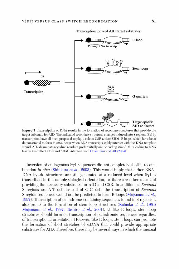
Figure 7 Transcription of DNA results in the formation of secondary structures that provide thetarget substrate for AID. The indicated secondary structural changes induced into S regions (Sx) bytranscription have all been proposed to play a role in CSR and/or SHM. R loops, which have beendemonstrated to form in vivo, occur when RNA transcripts stably interact with the DNA templatestrand. AID deaminates cytidine residues preferentially on the coding strand, thus leading to DNAlesions that effect CSR and SHM. Adapted from Chaudhuri and Alt (2004).
v(d)j versus class switch recombination 81
Inversion of endogenous Sg1 sequences did not completely abolish recom-bination in vivo (Shinkura et al., 2003). This would imply that either RNA–DNA hybrid structures are still generated at a reduced level when Sg1 istranscribed in the nonphysiological orientation, or there are other means ofproviding the necessary substrates for AID and CSR. In addition, as XenopusS regions are A-T rich instead of G-C rich, the transcription of XenopusS region sequences would not be predicted to form R loops (Mubmann et al.,1997). Transcription of palindrome-containing sequences found in S regions isalso prone to the formation of stem–loop structures (Kataoka et al., 1981;Mubmann et al., 1997; Tashiro et al., 2001). Unlike R loops, stem–loopstructures should form on transcription of palindromic sequences regardlessof transcriptional orientation. However, like R loops, stem loops can promotethe formation of short stretches of ssDNA that could provide appropriatesubstrates for AID. Therefore, there may be several ways in which the unusual

82 darryll d. dudley ET AL.
sequence content of S regions could lead to the generation of structurescontaining ssDNA that would be a substrate for AID activity. Finally, allS regions (from Xenopus to mammals) have a high concentration of theconsensus SHM motif (most notably AGCT), and evidence suggests that thissequence could target AID in CSR in the context of an AID�RPA complex as itdoes in SHM (Zarrin et al., 2004). It has been proposed that S regions mayhave evolved from sequences containing large numbers of SHM motifs (e.g.,AGCT) in lower organisms to the form found in mammals in which high levelsof such motifs remain (to target AID in the context of RPA), but in mammalianS regions, ssDNA generation may have been further augmented via evolutionof ability to form R loops (Zarrin et al., 2004).
4.5.2. AID-Induced Cytidine Deamination
Cytidine deaminases catalyze the conversion of dC to dU residues via thehydrolytic removal of the amino group at the fourth position of the pyrimidinering of cytidine (Betts et al., 1994). The deamination mechanism is likely toresemble that of adenosine deaminases as both involve a zinc atom in theactive site (Betts et al., 1994; Harris et al., 2002). Deamination of dC residuesby AID thereby induces dU/dG mismatches in DNA, the type of DNA lesionsnormally corrected by the base excision repair and MMR pathways (reviewedin Lindahl, 2000).
4.5.3. Base Excision Repair and Uracil DNA Glycosylase
The base excision repair pathway has evolved to provide protection againststructural alterations that can occur in DNA as a result of various endogenousalkylating agents and metabolic reactive oxygen species (reviewed in Lindahl,2000). Such alterations lead to the formation of aberrant nucleotide residuesthat are frequently recognized by DNA glycosylases (reviewed in Krokan et al.,1997). These glycosylases recognize the aberrant nucleotide and remove itfrom the DNA backbone, leaving behind an abasic site (reviewed in Krokanet al., 1997). In the case of cytidine deamination, this function is performed byUNG. The abasic site is then cleaved by an apyrimidic (AP) endonuclease,followed by nucleotide replacement via the action of a polymerase, which isnormally polymerase b (polb) (reviewed in Lindahl, 2000). The final repairstep is ligation, likely involving DNA ligase III (reviewed in Lindahl, 2000).
4.5.4. DNA Deamination Model
The DNA deamination mechanism for CSR and SHM was initially proposedon the basis of the observation that AID expression in bacteria caused muta-tions that somewhat resemble those induced by SHM (Petersen-Mahrt et al.,

Figure 8 The DNA deamination model for CSR and SHM. AID deaminates cytidine residues inS regions, and in the variable region exons of IgH and IgL genes. Steps that lead to SHM areshown on the left and those that effect CSR on the right. UNG, uracil deglycosylase; AID,activation-induced cytidine deaminase. The minor pathway for CSR refers to CSR in the absenceof UNG, likely involving mismatch repair (MMR). Adapted from Rada et al. (2002) and Di Noiaand Neuberger (2002).
v(d)j versus class switch recombination 83
2002). In this model, replication or repair of dU-containing DNA strands leadsto both CSR between S regions and SHM of immunoglobulin VH and VL exons(Fig. 8; Petersen-Mahrt et al., 2002). Mutations could be generated if the dUresidue resulting from AID-induced cytidine deamination is not removed bybase excision repair before DNA replication, wherein the dU is read as a dT,thus resulting in dC-to-dT and dG-to-dA transitions (Fig. 8; Petersen-Mahrtet al., 2002). Alternatively, if the abasic site produced by the function of UNGundergoes replication via a translesional polymerase (reviewed by Chaudhuriand Alt, 2004; Reynaud et al., 2003), subsequent repair of the abasic site wouldlead to both transitions and transversions (Fig. 8; Petersen-Mahrt et al., 2002).Moreover, if an error-prone polymerase were recruited to the abasic siteduring replication, then mutations could be introduced both in and aroundthe initial deaminase-induced lesion, explaining mutations that arise at non-dC

84 darryll d. dudley ET AL.
or non-dG residues in SHM (reviewed in Reynaud et al., 2003). The dU:dGmismatch could also be recognized and processed by components of the MMRmachinery, ultimately leading to the generation of DNA breaks.
Regarding CSR, if multiple cytidine residues are deaminated in close prox-imity and on opposite strands, then base excision repair could lead directlyto staggered DNA breaks (Petersen-Mahrt et al., 2002). Processing of theends of staggered DNA breaks could then result in the formation of DNADSBs that are either blunt or have short overhangs (reviewed in Reynaud et al.,2003). The subsequent repair of DNA DSBs induced in two different S regionsby the NHEJ pathway could thus result in CSR. In addition, there are modelswhereby MMR could also lead to DSBs after AID deamination (reviewed inMartin and Scharff, 2002a).
In support of the DNA deamination model with respect to SHM, thepattern of hypermutations observed in the hypermutating chicken cellline DT40 changes from transversion-dominated mutations to transitionswhen UNG function is inhibited (Di Noia and Neuberger, 2002).Furthermore, there is a dramatic shift in the SHM pattern to transitionsin immunoglobulin V genes of murine UNG-deficient B cells (Rada et al.,2002b). As transversions are primarily dependent on the removal of dU resi-dues by UNG, these data provide further support for the DNA deaminationmodel. With respect to class switching, CSR is substantially reduced in UNG-deficient mice and humans, in accordance with the predictions of the model(Rada et al., 2002b). In addition, as mentioned above, AID has been shown tobe capable of direct deamination of DNA (Bransteitter et al., 2003; Chaudhuriet al., 2003; Dickerson et al., 2003; Petersen-Mahrt et al., 2002; Pham et al.,2003; Ramiro et al., 2003; Sohail et al., 2003; Yu et al., 2004). Finally, theobservation that AID associates with transcribed S regions (Chaudhuri et al.,2004; Nambu et al., 2003) provides strong support for the DNA deaminationmodel.
As mentioned above, work has questioned the precise role of UNG in CSRand SHM, as certain UNG mutants that are catalytically inactive in U removalactivity in vitro are still proficient in mediating CSR and SHM in vivo,suggesting that the role of UNG in CSR and SHM is beyond its DNAglycosylase activity (Begum et al., 2004a). These results were surprisinggiven that human patients with similar mutations have profound defects inCSR (Imai et al., 2003a), leading to the speculation that there may be second-ary mutations in these patients that contribute to the phenotype (Begumet al., 2004a) or that there is some undetected UNG activity in vivo in themouse mutants; overall, these apparently conflicting findings await furtherexperimentation for full resolution.

v(d)j versus class switch recombination 85
4.5.5. Class Switch Recombination versus Somatic HypermutationSpecific Factors
Hyper-IgM syndrome (HIGM) is caused by defects in CSR leading to reducedlevels of IgG, IgA, and IgE (reviewed in Durandy, 2001). Until recently, the knowncauses of HIGM have been thought to affect both CSR and SHM (reviewedin Durandy, 2001). Some patients with HIGM have now been identified with anovel form of HIGM that results in impaired CSR but normal levels of SHM (Imaiet al., 2003b). B cells isolated from the peripheral blood of HIGM4 patientswere activated for CSR in vitro and shown to express substantial levels of AIDand GTs, yet failed to secrete detectable levels of IgE or IgG (Imai et al., 2003b).Furthermore, using a ligation-mediated polymerase chain reaction (LM-PCR)assay, DNA DSBs corresponding to Sm region sequences were readily detec-table in DNA isolated from activated HIGM4 and control B cells (Imai et al.,2003a). Thus HIGM4 may arise from defects in processes downstream of DNAdeamination that are distinct between CSR from SHM.
CSR and SHM share the requirements for AID-induced DNA deamination;however, AID mutants have now been identified that can differentially effectCSR or SHM. Mutations in the C terminus of AID retain SHM activity butare unable to promote CSR in AID�/� B cells (Barreto et al., 2003; Ta et al.,2003). AID C-terminal mutants retained DNA deamination function (Barretoet al., 2003; Ta et al., 2003), and loss of CSR was not due to failure in nucleartransport (Barreto et al., 2003). Furthermore, several mutations in theN terminus of AID had nearly normal CSR activity but were unable to mediateSHM of a retroviral GFP expression construct (Shinkura et al., 2004).Although some of these C-terminal mutants had defects in nuclear import,their ability to effect CSR suggests inefficient nuclear transport is not the causeof defective SHM (Shinkura et al., 2004). AID mutations that uncouple therelated but distinct processes of CSR and SHM suggest specific cofactorsmight exist that interact with these different domains of AID, with RPAbeing one such potential factor. In this regard, the C terminus of AID hasbeen shown to contain a nuclear export sequence that facilitates nuclear exportin a CRM1-dependent pathway (McBride et al., 2004). Although AID must bepresent in the nucleus to effect CSR and SHM, AID is predominantly found inthe cytoplasm (Rada et al., 2002a). Thus AID cofactors such as RPA may play arole in the retention of AID in the nucleus as well as target specificity.
4.5.6. AID-Induced Double-Strand Breaks
Past studies have documented DNA DSBs in S regions of cells stimulated forCSR (Chen et al., 2001; Wuerffel et al., 1997) and in variable regions of B cellsstimulated for SHM (Bross et al., 2000; Papavasiliou and Schatz, 2000). These

86 darryll d. dudley ET AL.
findings led to the proposal that DSBs may be intermediates in these process-es. On identification of AID, early efforts focused on determining whetherAID functioned in the induction or resolution of DNA DSBs (reviewed inChua et al., 2002). The IgH locus has been shown to colocalize with NBS-1and gH2AX foci, which are normally associated with DSBs, following CSRactivation of wild-type but not AID-deficient B cells (Begum et al., 2004b;Petersen et al., 2001). Colocalization was interpreted to reflect the induction ofDNA DSBs in S regions; and, therefore, these studies concluded that AIDdirectly participates in the formation of DNA DSBs during CSR (Petersen et al.,2001). However, H2AX was also found to be associated with V genes (Woo et al.,2003), and, therefore, it is not clear whether the H2AX foci indeed representbreaks at S regions or those at V genes, particularly given that H2AX foci canextend for up to 1 Mb from a DSB (Rogakou et al., 1999). In support ofDSB intermediates in CSR, LM-PCR assays have detected both AID- andUNG-dependent S region breaks (Catalan et al., 2003; Imai et al., 2003a).
In several studies, DNA breaks in V gene segments were found at similarfrequencies in both wild-type and AID-deficient B cells stimulated to undergoSHM (Bross et al., 2002; Papavasiliou and Schatz, 2002a). These results led tothe suggestion that AID was not involved in the induction of DNA DSBsduring SHM, and thus it was speculated that AID might instead be somehowinvolved in the repair of these DSBs (Papavasiliou and Schatz, 2002a).However, it remains unclear whether the DSBs observed in these studieswere actually related to SHM; so the significance of the findings remainsunclear (reviewed in Chua et al., 2002). Overall, it seems likely that CSRworks via a DSB intermediate, whereas SHM does not; this interpretationhas been reinforced by the requirement for factors involved in the DSBresponse (H2AX, DNA-PKcs, Ku, 53BP1, etc.) in CSR but not SHM (seebelow).
4.5.7. Inter nal S Region Deletions Are Anal ogou s to ClassSwitch Recomb ination
A high frequency of internal S region (intra-S) deletions is detected in the S mregion of normal B cells and B cell lines activated for CSR (Alt et al., 1982;Bottaro et al., 1998; Hummel et al., 1987; Winter et al., 1987). Intra-Smdeletions are largely AID dependent, can occur in the absence of an acceptorS region, and are accompanied by mutations in 30 flanking sequences analo-gous to those seen in CSR junctions (Dudley et al., 2002). Thus an intra-Sregion deletion probably reflects the normal CSR mechanism, but in whichrecombination has taken place within homologous sequences of a singleS region rather than between two heterologous S regions. This could result

v(d)j versus class switch recombination 87
via the failure to induce DNA lesions in downstream S regions or in theabsence of S region synapsis.
4.5.8. S Region Mutations
AID mutants with N-terminal alterations that result in selective loss of SHMactivity, but retain the ability to mediate CSR, also generate mutations in Sm(Shinkura et al., 2004). Mutations in AID that selectively lose SHM activitycould be the result of failure to target the AID mutant to V region sequences,for instance because of inability to interact with RPA (Chaudhuri et al., 2004).In this regard, induction of Sm mutations by SHM-defective AID mutantswould simply reflect the differential targeting of AID and not SHM-versusCSR-specific functions of AID. However, loss of AID-targeting activity doesnot preclude an SHM-specific function of AID not directly related to aseparate CSR-specific activity.
Separate studies involving human or murine C-terminal mutants of AIDthat promote SHM and gene conversion but not CSR have given conflictingresults regarding the induction of Sm mutations (Barreto et al., 2003; Shinkuraet al., 2004). CSR-defective mutants of murine AID induced normal levels ofSm mutations, whereas most human CSR-defective AID mutants were unableto promote Sm lesions. Sm mutations induced by CSR-defective murine AIDmutants (Barreto et al., 2003) could be due to the retention of low-level CSRactivity as suggested by an analogous human C-terminal AID mutant, and thusbe independent of SHM activity (Shinkura et al., 2004). This would imply thatAID contains distinct functions for promoting SHM and CSR; or that theCSR-defective mutant forms of AID fail to target S regions. Alternatively,the CSR-defective AID mutants may properly target S regions and effectDNA lesions, as evidenced by the ability to induce mutations in Sm sequencesbut fail to complete actual CSR. In this scenario, CSR-defective mutants mightbe unable to interact with cofactors essential for DNA repair or to facilitateS region synapsis (reviewed by Chaudhuri and Alt, 2004).
4.6. Class Switch Recombination and S Region Synapsis
Recombination between two different S regions takes place over large chro-mosomal distances (up to �175 kb), and these regions must be juxtaposedbefore being joined. Adjoining of S regions could be mediated via associationwith transcriptional promoters, enhancers, chromatin factors, DNA repairproteins, or AID-associated factors, or by interactions involving the S regionsequences themselves.

88 darryll d. dudley ET AL.
4.6.1. Promoter/Enhancer Interactions
CSR is reduced in the absence of iEm, even though the level of steady-statetranscription through Sm appeared unaffected (Bottaro et al., 1998). Promoter/enhancer interactions between iEm and downstream I promoters could effectjuxtaposition of Sm with the S regions of downstream CH genes. However, asSm transcription detected in the absence of iEm could be driven by VH or DH
promoters, normal levels of CSR may be dependent on the specific site oftranscriptional initiation rather than overall levels of transcription (Bottaroet al., 1998). In this regard, replacement of iEm with a pgk promoter returnsthe rate of CSR to approximately normal levels (Bottaro et al., 1998). Similarly,although deletions of HS3b and HS4 in the 30 RR located downstream of theIgH locus result in the reduction of GTs, a role for these sites in the synapsis ofS region sequences cannot be excluded (Pinaud et al., 2001).
4.6.2. H2AX
Effective long-range synapsis of S regions likely relies on chromatin modificationsand associated factors, as indicated by studies of H2AX deficiency (Reina-San-Martin et al., 2003). As noted above, AID-dependent H2AX foci are found at theIgH locus in conjunction with IgH CSR (Petersen et al., 2001). SHM is unaffectedin H2AX-deficient mice, whereas CSR is substantially impaired (Reina-San-Martin et al., 2003). Intra-S region deletions were detected in H2AX-deficientB cells activated for CSR, demonstrating that accessibility of S regions to theCSR machinery and the basic joining mechanism required for CSR is notimpaired by the absence of H2AX (Reina-San-Martin et al., 2003). The re-cruitment and assembly of repair factors at sites of DNA DSBs by g-H2AX hasbeen proposed to facilitate the juxtaposition of broken DNA ends andsubsequent repair by NHEJ proteins (Bassing and Alt, 2004). Thus gH2AXmight similarly promote long-range S region synapsis for the efficient recom-bination between heterologous S regions. In this regard, H2AX-deficient mice,in the absence of the cell cycle checkpoint protein p53, have been shown toundergo translocations involving S region sequences, perhaps indicating thatproper synapsis of S regions during CSR is important for genome stability aswell as CSR (Bassing et al., 2003a). The finding that another protein, 53BP1,proposed to work in the H2AX anchoring mechanism, is also required for CSR(but not SHM) further supports this general model (Manis et al., 2004).
4.6.3. DNA-PKcs
Pro-B cells that lack DNA-PKcs are defective for switching to the IgE isotype(Rolink et al., 1996). However, significant levels of CSR to all immunoglobulinisotypes were detected in a study involving SCID mice reconstituted with

v(d)j versus class switch recombination 89
rearranged IgH and IgL transgenes, which carry a catalytic mutation in DNA-PKcs that abrogates the kinase activity of DNA-PKcs (Bosma et al., 2002; Cooket al., 2003). In contrast, DNA-PKcs–deficient mice had a significant reductionin CSR to all isotypes except IgG1 (Manis et al., 2002). Expression of DNA-PKcs, albeit catalytically inactive, can be detected in cells from SCID mice,leading to the intriguing possibility that serine/threonine kinase activity ofDNA-PKcs is dispensable for CSR, whereas the presence of a noncatalyticDNA-PKcs can provide a necessary function for CSR (Bosma et al., 2002). Inthe context of this model, why there should still be CSR to IgG1 in DNA-PKcs–deficient mice remains a mystery. The fact that DNA-PKcs–deficientB cells switch to IgG1 and not other isotypes implies that recombina-tion between Sm and Sg1 may be mechanistically different than that ofCSR between Sm and other S regions. Alternatively, a general reduction inCSR efficiency in the absence of DNA-PKcs could result in the preferentialdetection of IgG1 simply because it occurs the most efficiently because of itslarge size. Whatever the case, it is notable that DNA-PKcs is able to promotesynapsis of broken DNA ends in vitro (DeFazio et al., 2002), consistent withsuch a function in CSR. In this regard, transformation/transcription domain-associated protein (TRRAP), a distantly related member of the PI-3 kinasefamily found in humans with homologs in both yeast and Caenorhabditiselegans, apparently lacks kinase activity and appears to instead function as ascaffolding protein during chromatin remodeling (McMahon et al., 1998).
4.6.4. Mismatch Repair
Mlh1- and Pms2-deficient mice have a modest reduction in CSR activity, andsequences isolated from S junctions of Mlh1- and Pms2-deficient B cells havean increased rate of microhomologies compared with wild-type B cells(Ehrenstein et al., 2001; Schrader et al., 2002). Yeast homologs of PMS2 andMLH1 can bind two different DNA molecules simultaneously (Hall et al.,2001), leading to the proposal that PMS2 and MLH1 might facilitate S regionsynapsis during CSR (Schrader et al., 2003).
4.6.5. Other Factors
LR-1 is a B-cell–specific heterodimeric protein composed of nucleolin andheterogeneous nuclear ribonucleoprotein D (hnRNP D), in which each sub-unit is capable of low-affinity binding to S region–specific duplex sequences,and with high affinity to sequences in the form of G quartets or G4 DNA(Dempsey et al., 1999; Williams and Maizels, 1991). Consequently, it has beenproposed that LR-1 might bind and capture DNA from two different S regionsand facilitate their synapsis, thus contributing to CSR (Dempsey et al., 1999).

90 darryll d. dudley ET AL.
4.7. Class Switch Recombination and Double-StrandBreak Repair
The detection of closed circles of DNA composed of intervening sequencesbetween two different S regions implied that intermediates of CSR occur inthe form of DNA DSBs (reviewed in Iwasato et al., 1990; Kenter, 2003).Sequences from CSR junctions demonstrate little or no sequence homologybetween donor and acceptor S regions, and CSR junctions frequently containshort deletions or insertions of nucleotides, all of which are consistent with theNHEJ pathway of DNA DSB repair (Dunnick et al., 1993). Lending furthersupport for DSBs as intermediates in CSR, deficiencies in assayed NHEJproteins reduce CSR in mice (Casellas et al., 1998; Manis et al., 1998a,2002a). Finally, deficiency for 53BP1, a DNA damage-sensing protein thatbecomes activated in response to DSBs and is found associated with H2AX,also leads to significantly reduced levels of CSR (Manis et al., 2004; Ward et al.,2004).
4.7.1. Ku
Ku-deficient mice do not develop B or T cells; therefore rearranged IgH andIgL genes must be introduced into these animals to derive mature B cells(Casellas et al., 1998; Manis et al., 1998a). The only detectable IgH isotype inthe serum of these mice is IgM, and splenic B cells isolated from these animalsand stimulated in vitro to undergo specific CSR fail to secrete anything otherthan IgM (Casellas et al., 1998; Manis et al., 1998a). The presence of GTs fromdownstream CH genes and DSBs detected in S g3 sequences suggested thatthe defect in CSR was not due to an inability to initiate the process (Casellaset al., 1998; Manis et al., 1998a). However, as Ku-deficient B cells are alsodefective in proliferation, the lack of CSR could be explained by decreasedsurvival of activated B cells (Manis et al., 1998a). Potentially counteringthis argument, cells that have undergone several rounds of cell division stilldo not undergo CSR (Reina-San-Martin et al., 2003), although it is not clearwhether these cells might represent those that have failed to be completelyactivated.
4.7.2. D NA-PKc s and Artemis
DNA-PKcs–deficient mice have significantly reduced levels of serum isotypes(Manis et al., 2002a), whereas SCID mice that carry DNA-PKcs kinase inactivemutations undergo CSR at nearly normal levels (Bosma et al., 2002; Cook et al.,2003). In this regard, CSR occurs normally in the absence of Artemis (Rooneyet al., submitted), which is activated on phosphorylation by DNA-PKcs (Maet al., 2002), whereas Artemis is essential for opening the hairpin-coding ends

v(d)j versus class switch recombination 91
generated during V(D)J recombination (Rooney et al., 2002). Therefore,DNA-PKcs may provide functions for the repair of DNA damage inducedduring CSR, such as stabilization of the repair complex, synapsis of targetsequence (see above), or recruitment of other essential proteins to the site ofDNA breaks, outside its role in Artemis activation that is required for V(D)J CJformation.
4.7.3. Ataxia Telangiectasia Mutated
Human patients with ataxia telangiectasia mutated (ATM) have normal levelsof SHM in their V region sequences, although an overall reduction in serumimmunoglobulin isotypes and an increase in homology at S region junctionssuggest that ATM does influence CSR (Pan et al., 2002; Pan-Hammarstromet al., 2003; Waldmann et al., 1983; reviewed in Regueiro et al., 2000). ATM isactivated by DNA damage, thereby phosphorylating and activating cell cyclecontrol proteins p53 and Chk2, and thus inducing cell cycle arrest in cellscontaining DSBs (reviewed in Khanna and Jackson, 2001; Shiloh, 2001).However, ATM likely functions beyond sensing DNA damage and cell cycleregulation, as indicated by its ability to phosphorylate the DNA repair proteinNBS1 (Gatei et al., 2000; Lim et al., 2000; Wu et al., 2000; Zhao et al., 2000). Inaddition to the increase in homology at CSR junctions, there are fewer muta-tions and insertions in the sequences around CSR junctions of ATM-deficientB cells than are found in control B cells (Pan et al., 2002). Thus it appears thatATM may function during the repair phase of CSR, although secondary effectscaused by defects in B- and T-cell development and survival could alsocontribute to the observed immunodeficiencies in patients with ATM. In thisregard, ATM-deficient mice initially were not found to have clear-cut defectsin the production of serum IgH isotypes (Barlow et al., 1996; Xu et al., 1996).However, more detailed analyses have now clearly shown a defect in CSR butnormal internal Sm deletions similar to what is seen in H2AX deficiency, whichsupports a role for the DNA DSB response in this process and potentiallysynapsis (Reina-San-Martin et al., 2004; see below).
4.7.4. 53BP1
The role of NHEJ proteins and the likely generation of DNA DSBs duringCSR imply the need to sense and respond to such DNA lesions. 53BP1 wasfound to interact with the DNA damage response and cell cycle checkpointprotein p53 (Xia et al., 2001). 53BP1 was rapidly phoshphorylated in responseto IR (Anderson et al., 2001) and was found in foci that are thought torepresent sites of DNA damage (Anderson et al., 2001; Schultz et al., 2000).Furthermore, 53BP1 colocalized with g-H2AX in nuclear foci that appear after

92 darryll d. dudley ET AL.
DSB induction (Rappold et al., 2001; Rogakou et al., 1999). 53BP1-deficientB cells were dramatically impaired for CSR; although the production of germ-line transcripts and induction of AID expression were normal (Manis et al.,2004; Ward et al., 2004). In contrast, both V(D)J recombination and SHMoccurred normally in 53BP1-deficient mice (Manis et al., 2004; Ward et al.,2004). Thus CSR is highly dependent on DNA damage-sensing proteinsdownstream of AID induction and, thus, likely to influence the DNA repair/S region joining phase. Finally, rare Sm-Sg1 switch junctions amplified from53BP1-deficient B cells are qualitatively similar to wild-type junctions, demon-strating that 53BP1 does not mechanistically affect CSR (Manis et al., 2004). Inthe context of these observations, it has been suggested that 53BP1 may workwith H2AX for S region synapsis via an anchoring mechanism (Bassing and Alt,2004; Manis et al., 2004).
4.7.5. H2AX
Phosphorylation of H2AX on Ser-139 occurs within minutes after treatmentsthat introduce DNA DSBs in yeast and mammalian cells (Downs et al., 2000;Rogakou et al., 1998). g-H2AX appears in discrete nuclear foci that correlate infrequency and nuclear location with induced DSBs (Rogakou et al., 1999). Therapid appearance of g-H2AX foci after the induction of DSBs precedes that ofDNA repair proteins, suggesting that g-H2AX may be involved in the recruit-ment of specific repair factors such as BRCA1, MRE11, RAD50, and NBS1to sites of DNA damage (Paull et al., 2000). Whereas H2AX is required forefficient CSR and AID-dependent foci formation at the IgH locus (see above),it is not required for the process of intra-S region deletions and has beensuggested to be therefore involved in long-range synapsis (Reina-San-Martinet al., 2003), which might occur via an anchoring mechanism as outlined above(Bassing and Alt, 2004; Manis et al., 2004).
4.7.6. NBS1
NBS1 is a DNA repair protein associated with the hMre11/hRad50/NBS1complex that forms nuclear foci in response to DSB-inducing DNA damageand is a target of ATM-mediated phosphorylation (Carney et al., 1998; Maseret al., 1997; Nelms et al., 1998; Wu et al., 2000; Zhao et al., 2000). In yeast,scmre11 and scrad50 mutants have defects in NHEJ and have been linkedgenetically to the same NHEJ pathway as yku70 and lig4, and Mre11, alsoimplicated in microhomology-mediated DNA break repair (reviewed inCritchlow and Jackson, 1998; Paull and Gellert, 2000). Furthermore, NBS1has been detected at nuclear foci that colocalize with the IgH loci in B cellsactivated to undergo CSR, and this colocalization was dependent on the

v(d)j versus class switch recombination 93
presence of AID (Petersen et al., 2001). However, the levels of most serumisotypes in patients with Nijmegan breakage syndrome (NBS) are not substan-tially reduced (reviewed in Shiloh, 1997). B cells of patients with NBS do havean increased frequency of S region junctions with ‘‘imperfect’’ microhomology(four or more nucleotides, with only one mismatch) compared with controls,albeit from a limited number of samples (Pan et al., 2002). Thus NBS1 mayplay a role in the DNA repair phase of CSR or, given its association withg-H2AX, it could be involved in S region synapsis.
4.7.7. Mismatch Repair
During DNA replication, MMR proteins recognize improperly paired nucleo-tide base pairs and mediate the removal and reinsertion of the correct nucleo-tide based on the DNA template strand (Buermeyer et al., 1999). Severalstudies have found an overall decrease in the rate of CSR in the absence ofcertain MMR proteins (Ehrenstein and Neuberger, 1999; Schrader et al.,1999, 2002). CSR junctions were found to occur more frequently in consensusGAGCT and GGGGT sequences, reminiscent of ‘‘hot spot’’ targeting of SHMin the absence of Msh2 (Ehrenstein and Neuberger, 1999; Phung et al., 1998).Moreover, CSR junctions isolated from B cells of Msh2-deficent mice werefound to have slightly decreased lengths of microhomology (Schrader et al.,2002). This would be consistent with the DNA deamination model of CSR, asMMR proteins can extend the region of mutations beyond the original dUresidue induced by the function of AID and UNG (Petersen-Mahrt et al.,2002). These results are consistent with Msh2 playing a more important role inend processing, specifically the removal of 30 nonhomologous overhangs out-side potential regions of microhomology (Schrader et al., 2002). In contrast,there was an increase in microhomology length detected in the CSR junctionsof Mlh1- and Pms2-deficient B cells (Schrader et al., 2002). The increase inmicrohomology at CSR junctions of Pms2- or Mlh1-deficient B cells mightreflect a role for stabilizing CSR intermediates or for S region synapsis, thusrequiring increased sequence homology in their absence for adequate basepair interactions (Schrader et al., 2002, 2003).
5. CSR-Related Diseases
5.1. Hyper-IgM Syndrome Types 1 and 3
Hyper-IgM (HIGM) syndromes are immunodeficiencies caused by geneticdefects that result in abrogation or impairment in CSR (reviewed inDurandy and Honjo, 2001). The first described was X-linked hyper-IgM, or

94 darryll d. dudley ET AL.
HIGM type 1 (HIGM1), caused by mutations in the gene encoding the CD40ligand (CD40L), a membrane glycoprotein expressed on activated T cells(Allen et al., 1993; Aruffo et al., 1993; DiSanto et al., 1993). CD40L interactswith CD40, a member of the tumor necrosis factor receptor family, that isconstitutively expressed on B cells and is variably expressed on T cells, mono-cytes, basophils, dendritic cells, and endothelial cells (reviewed in Grammerand Lipsky, 2000). CD40L binds to CD40 and induces B cell proliferation(Nishioka and Lipsky, 1994; Tohma and Lipsky, 1991), AID induction(Muramatsu et al., 1999) and the production of some immunoglobulin GTs(Fujita et al., 1995; Jumper et al., 1994; Warren and Berton, 1995). Removal ofeither CD40 or CD40L through the use of anti-CD40L antibodies (Foy et al.,1993, 1994) blocks germinal center formation, SHM, and CSR in response toT-dependent antigens. Genetic defects in CD40 lead to an autosomal recessiveform of hyper-IgM, HIGM3, similar to that caused by the absence of CD40L(Ferrari et al., 2001). Thus HIGM1 and HIGM3 are caused by the ablation ofupstream signaling pathways leading to CSR and SHM activation.
5.2. Hyper-IgM Syndrome Type 2
Autosomal recessive hyper-IgM syndrome type 2 is caused by mutationsabrogating the expression or function of AID (Revy et al., 2000). Patientslacking AID have enlarged lymph nodes with correspondingly expanded ger-minal centers (Revy et al., 2000), a characteristic also seen in AID-deficientmice (Muramatsu et al., 2000). These oversized germinal centers likely reflectthe presence of activated B cells that are unable to effect CSR or SHM, andthus accumulate in B-cell follicles of the peripheral lymph tissue.
5.3. Hyper-IgM Syndrome Type 4
Patients with HIGM4 are substantially impaired for CSR, whereas SHM canbe detected in VH regions at levels comparable to that of controls (Imai et al.,2003b). Defects in AID, UNG, or in the expression of GTs were eliminated aspossible causes of HIGM4. Evidence for the existence of a factor differentiallyinvolved in CSR versus SHM is in keeping with the DNA deamination model,in which CSR is effected via DSB intermediates, whereas SHM can beinduced in the absence of DSBs (Petersen-Mahrt et al., 2002). Thus HIGM4is likely caused by defect(s) in factors associated with the targeting of AID toS regions that affect the synapsis of S regions and/or that are involved in anaspect of DNA DSB repair (reviewed in Manis and Alt, 2003).

v(d)j versus class switch recombination 95
5.4. X-Linked Hypohydrotic Ectodermal Dysplasia
Another human disease, X-linked hypohydrotic ectodermal dysplasia (XHM-ED), is characterized by hyper-IgM immunodeficiency caused by missensemutations in the gene encoding NF-kB essential modulator (NEMO)(Doffinger et al., 2001; Jain et al., 2001). NEMO interacts with NF-kB kinasesIKK1 (IkB kinase 1) and IKK2 and is essential for NF-kB activation (Yamaokaet al., 1998). Engagement of CD40 on the surface of B cells with T-cell–expressed CD40L leads to the induction of NF-kB family transcription factors(Berberich et al., 1994; Francis et al., 1995; Lalmanach-Girard et al., 1993).NF-kB family transcription factors mediate the production of Ig1-Cg1 (Linand Stavnezer, 1996; Lin et al., 1998) and Ie-Ce (Iciek et al., 1997) GTs. Not allimmunoglobulin GTs are dependent on NF-kB; thus mutations affectingNF-kB signaling would be predicted to abrogate CSR to some but not allCH genes. However, patients with XHM-ED have undetectable levels of allserum IgGs, and B cells activated in vitro with anti-CD40 fail to effect CSR(Jain et al., 2001). Thus NF-kB signaling in B cells, as with upstream CD40-and CD40L-mediated signaling, is likely involved in overall activation of CSR,perhaps as an activator of AID, although an affect on AID expression in thesepatients has yet to be reported. The developmental aspects of XHM-EDsyndrome can be attributed to defective NF-kB signaling through tumornecrosis factor (TNF) family receptors expressed on embryonic and fetalectoderm-derived tissues (Doffinger et al., 2001). Thus genetic mutationsthat affect CD40, CD40L, or CD40-mediated downstream signalingmolecules all lead to immunodeficiencies with hyper-IgM characteristics.
6. Concluding Remarks
V(D)J recombination and CSR (and the related process of SHM) lead to thedirect alteration of DNA sequences and content in cells of the vertebrateimmune system. V(D)J recombination occurs both in developing B andT lineage cells; whereas CSR and SHM occur only in mature B lineagecells. The potential for deleterious or catastrophic consequences during themanipulation of a cell’s genetic material is obvious; and aberrant V(D)J recom-bination and CSR, and perhaps SHM, have all been implicated in translocationsand other genetic alterations that underlie T lineage [V(D)J recombination] andB lineage [V(D)J recombination, CSR, and SHM] lymphomas. Therefore, allthree of these potentially dangerous genomic alteration processes require tightregulatory control mechanisms. In this context, the proteins that initiate thesegenetic alterations, namely RAGs for V(D)J recombination and AID for CSR

96 darryll d. dudley ET AL.
and SHM, are expressed in tissue- and lineage-specific fashion and are subjectto strict control via posttranslational regulatory processes. Likewise, there iscontrol of the substrate DNA, RS sequences for V(D)J recombination, andS regions for CSR, such that its availability to the initiating enzymes is largelylimited to the appropriate cell types and sequences. Among the major outstand-ing questions is the issue of precisely what aspects of target RS or S region DNAin chromatin make them good substrates for RAG or AID in the appropriate celltypes, lineages, and/or activation stages.
One fundamental difference between CSR and V(D)J recombination is inthe nature of the target sequences recognized by the ‘‘recombinase.’’ RAG-mediated cleavage at the junction of antigen receptor gene-coding segments issite specific and dependent on short, well-defined cis-acting RSs. In contrast,AID deamination of cytidine residues, which appears to initiate CSR (andSHM), is targeted to large S regions that lie upstream of CH genes in the IgHlocus, with recombination occurring throughout the 1- to 12-kb repetitivesequences. Thus, CSR is region specific rather than site specific. Moreover,AID does not appear to recognize specific target sequences with the samedegree of specificity as RAGs, which, in general, recognize specific RSsequences. Instead, AID has been thought to rely on transcription-dependentDNA structures such as R loops that are formed when sequences with certainbase compositions are actively transcribed. Yet, S regions are composed oftandem repeat units with frequent repeats of specific motifs favored by SHM.Thus, in this context, there may still be specific sequences, such as the SHMconsensus, that are preferentially targeted by AID in conjunction with its RPApartner to provide a further degree of specificity in CSR. Although we nowhave some idea about how AID is targeted, there is still much to be learnedabout how AID targeting is so specific for S regions and variable regions andwhy there is not more wide-scale deamination leading to a higher level ofmutation and translocations involving other genes in activated B cells.
In both V(D)J recombination and CSR, the initiating lesion by RAG andAID ultimately appears to lead to a DSB and, subsequently, to employ DSBrepair pathways, most likely NHEJ pathways, for the resolution of the DNAbreaks. Clearly, the classic NHEJ pathway seals both coding and signal jointsin the context of V(D)J recombination. Some evidence suggests this pathway isalso responsible for ligating CSR junctions, although more evidence on thispoint is needed. A significant difference in the joining phase of V(D)J recom-bination and CSR lies in their relative reliance on the DNA DSB response.Thus, V(D)J recombination occurs relatively unimpaired in the absence ofDSB response factors such as H2AX and 53BP1. However, the absence ofthese factors dramatically impairs CSR. One possible explanation is that thefactors are somehow involved in the long-range synapsis of S regions in

v(d)j versus class switch recombination 97
the context of CSR via a general anchoring mechanism proposed to holdDSBs together in chromatin before their joining via NHEJ. In contrast,RAG-generated DSBs appear to be held together in a postcleavage synapticcomplex by the RAGs themselves, which then recruit the NHEJ factors tocomplete the reaction. In both V(D)J recombination and CSR, however, westill know little about the actual process of synapsis and how the involvedproteins contribute to it.
Acknowledgments
We thank Drs. JoAnn Sekiguchi and John Manis for helpful advice and suggestions. We thank Drs.Sean Rooney and Ali Zarrin for communicating unpublished data. C.H.B. is a LymphomaResearch Foundation Fellow. F. W. A. is an investigator with the Howard Hughes MedicalInstitute. This work was supported by an NIH grant to F.W.A. (NIH 5PO1A131541-14).
References
Agata, Y., Katakai, T., Ye, S. K., Sugai, M., Gonda, H., Honjo, T., Ikuta, K., and Shimizu, A. (2001).J. Exp. Med. 193, 873–880.
Agrawal, A., and Schatz, D. G. (1997). Cell 89, 43–53.Agrawal, A., Eastman, Q. M., and Schatz, D. G. (1998). Nature 394, 744–751.Aidinis, V., Dias, D. C., Gomez, C. A., Bhattacharyya, D., Spanopoulou, E., and Santagata, S.
(2000). J. Immunol. 164, 5826–5832.Akamatsu, Y., and Oettinger, M. A. (1998). Mol. Cell. Biol. 18, 4670–4678.Akamatsu, Y., Monroe, R., Dudley, D. D., Elkin, S. K., Gartner, F., Talukder, S. R., Takahama, Y.,
Alt, F. W., Bassing, C. H., and Oettinger, M. A. (2003). Proc. Natl. Acad. Sci. USA 100,1209–1214.
Alberts, B., Bray, D., Lewis, J., Raff, M., Roberts, K., and Watson, J. D. (1983). ‘‘Molecular Biologyof the Cell.’’ Garland Publishing, Inc., New York, NY.
Alessandrini, A., and Desiderio, S. V. (1991). Mol. Cell. Biol. 11, 2096–2107.Allen, R. C., Armitage, R. J., Conley, M. E., Rosenblatt, H., Jenkins, N. A., Copeland, N. G.,
Bedell, M. A., Edelhoff, S., Disteche, C. M., Simoneaux, D. K., Fanslow, W. C., Belmont, J., andSpriggs, M. K. (1993). Science 259, 990–993.
Alt, F., Enea, V., Bothwell, A., and Baltimore, D. (1980). Cell 21, 1–12.Alt, F., Rosenberg, N., Casanova, R., Thomas, E., and Baltimore, D. (1982). Nature 296, 325–331.Alt, F. W., and Baltimore, D. (1982). Proc. Natl. Acad. Sci. USA 79, 4118–4122.Alt, F. W., Yancopoulos, G. D., Blackwell, T. K., Wood, C., Thomas, E., Boss, M., Coffman, R.,
Rosenberg, N., Tonegawa, S., and Baltimore, D. (1984). EMBO J. 3, 1209–1219.Anderson, L., Henderson, C., and Adachi, Y. (2001). Mol. Cell. Biol. 21, 1719–1729.Arakawa, H., Hauschild, J., and Buerstedde, J. M. (2002). Science 295, 1301–1306.Arbuckle, J. L., Fauss, L. A., Simpson, R., Ptaszek, L. M., and Rodgers, K. K. (2001). J. Biol. Chem.
276, 37093–37101.Aruffo, A., Farrington, M., Hollenbaugh, D., Li, X., Milatovich, A., Nonoyama, S., Bajorath, J.,
Grosmaire, L. S., Stenkamp, R., Neubauer, M., Roberts, R. L., Noelle, R. J., Ledbetter, J. A.,Franke, U., and Ochs, H. D. (1993). Cell 72, 291–300.
98 darryll d. dudley ET AL.
Bachl, J., Carlson, C., Gray-Schopfer, V., Dessing, M., and Olsson, C. (2001). J. Immunol. 166,5051–5057.
Bailin, T., Mo, X., and Sadofsky, M. J. (1999). Mol. Cell. Biol. 19, 4664–4671.Bain, G., Maandag, E. C., Izon, D. J., Amsen, D., Kruisbeek, A. M., Weintraub, B. C., Krop, I.,
Schlissel, M. S., Feeney, A. J., van Roon, M., van der Valk, M., te Riele, H. P. J., Berns, A., andMurre, C. (1994). Cell 79, 885–892.
Barlow, C., Hirotsune, S., Paylor, R., Liyanage, M., Eckhaus, M., Collins, F., Shiloh, Y., Crawley,J. N., Ried, T., Tagle, D., and Wynshaw-Boris, A. (1996). Cell 86, 159–171.
Barreto, V., Marques, R., and Demengeot, J. (2001). Eur. J. Immunol. 31, 3763–3772.Barreto, V., Reina-San-Martin, B., Ramiro, A. R., McBride, K. M., and Nussenzweig, M. C. (2003).
Mol. Cell 12, 501–508.Bassing, C. H., and Alt, F. W. (2004). Cell Cycle 3, 149–153.Bassing, C. H., Alt, F. W., Hughes, M. M., D’Auteuil, M., Wehrly, T. D., Woodman, B. B.,
Gartner, F., White, J. M., Davidson, L., and Sleckman, B. P. (2000). Nature 405, 583–586.Bassing, C. H., Chua, K. F., Sekiguchi, J., Suh, H., Whitlow, S. R., Fleming, J. C., Monroe, B. C.,
Ciccone, D. N., Yan, C., Vlasakova, K., Livingston, D. M., Ferguson, D. O., Scully, R., and Alt, F.W. (2002a). Proc. Natl. Acad. Sci. USA 99, 8173–8178.
Bassing, C. H., Swat, W., and Alt, F. W. (2002b). Cell 109, (Suppl. S45–S55).Bassing, C. H., Suh, H., Ferguson, D. O., Chua, K. F., Manis, J., Eckersdorff, M., Gleason, M.,
Bronson, R., Lee, C., and Alt, F. W. (2003a). Cell 114, 359–370.Bassing, C. H., Tillman, R. E., Woodman, B. B., Canty, D., Monroe, R. J., Sleckman, B. P., and Alt,
F. W. (2003b). Proc. Natl. Acad. Sci. USA 100, 2598–2603.Begum, N. A., Kinoshita, K., Kakazu, N., Muramatsu, M., Nagaoka, H., Shinkura, R.,
Biniszkiewicz, D., Boyer, L. A., Jaenisch, R., and Honjo, T. (2004a). Science 305, 1160–1163.Begum, N. A., Kinoshita, K., Muramatsu, M., Nagaoka, H., Shinkura, R., and Honjo, T. (2004b).
Proc. Natl. Acad. Sci. USA 101, 13003–13007.Belmont, A. S., Dietzel, S., Nye, A. C., Strukov, Y. G., and Tumbar, T. (1999). Curr. Opin. Cell Biol.
11, 307–311.Benezra, R., Davis, R. L., Lockshon, D., Turner, D. L., and Weintraub, H. (1990a). Cell 61, 49–59.Benezra, R., Davis, R. L., Lassar, A., Tapscott, S., Thayer, M., Lockshon, D., and Weintraub, H.
(1990b). Ann. N.Y. Acad. Sci. 599, 1–11.Bensmana, M., Huck, S., Lefranc, G., and Lefranc, M. P. (1988). Nucleic Acids Res. 16, 3108.Berberich, I., Shu, G. L., and Clark, E. A. (1994). J. Immunol. 153, 4357–4366.Berger, S. L. (2002). Curr. Opin. Genet. Dev. 12, 142–148.Bertocci, B., De Smet, A., Berek, C., Weill, J. C., and Reynaud, C. A. (2003). Immunity 19,
203–211.Besmer, E., Mansilla-Soto, J., Cassard, S., Sawchuk, D. J., Brown, G., Sadofsky, M., Lewis, S. M.,
Nussenzweig, M. C., and Cortes, P. (1998). Mol. Cell 2, 817–828.Betts, L., Xiang, S., Short, S. A., Wolfenden, R., and Carter, C. W., Jr. (1994). J. Mol. Biol. 235,
635–656.Betz, A. G., Milstein, C., Gonzalez-Fernandez, A., Pannell, R., Larson, T., and Neuberger, M. S.
(1994). Cell 77, 239–248.Binz, S. K., Sheehan, A. M., and Wold, M. S. (2004). DNA Repair (Amst). 3, 1015–1024.Blackwell, K., Malynn, B., Pollock, R., Ferrier, P., Covey, L., Fulop, G., Phillips, R., Yancopoulos,
G., and Alt, F. (1989). EMBO J. 8, 735–742.Blunt, T., Finnie, N. J., Taccioli, G. E., Smith, G. C. M., Demengeot, J., Gottlieb, T. M., Mizuta, R.,
Varghese, A. J., Alt, F. W., Jeggo, P. A., and Jackson, S. P. (1995). Cell 80, 813–823.Bogue, M. A., Jhappan, C., and Roth, D. B. (1998). Proc. Natl. Acad. Sci. USA 95, 15559–15564.Borgulya, P., Kishi, H., Uematsu, Y., and von Boehmer, H. (1992). Cell 69, 529–537.

v(d)j versus class switch recombination 99
Bories, J. C., Demengeot, J., Davidson, L., and Alt, F. W. (1996). Proc. Natl. Acad. Sci. USA 93,7871–7876.
Born, W., Yague, J., Palmer, E., Kappler, J., and Marrack, P. (1985). Proc. Natl. Acad. Sci. USA 82,2925–2929.
Bosma, G. C., Kim, J., Urich, T., Fath, D. M., Cotticelli, M. G., Ruetsch, N. R., Radic, M. Z., andBosma, M. J. (2002). J. Exp. Med. 196, 1483–1495.
Bottaro, A., Lansford, R., Xu, L., Zhang, J., Rothman, P., and Alt, F. W. (1994). EMBO J. 13,665–674.
Bottaro, A., Young, F., Chen, J., Serwe, M., Sablitzky, F., and Alt, F. W. (1998). Int. Immunol. 10,799–806.
Boulton, S. J., and Jackson, S. P. (1996). EMBO J. 15, 5093–5103.Bouvier, G., Watrin, F., Naspetti, M., Verthuy, C., Naquet, P., and Ferrier, P. (1996). Proc. Natl.
Acad. Sci. USA 93, 7877–7881.Bransteitter, R., Pham, P., Scharff, M. D., and Goodman, M. F. (2003). Proc. Natl. Acad. Sci. USA
100, 4102–4107.Bross, L., Fukita, Y., McBlane, F., Demolliere, C., Rajewsky, K., and Jacobs, H. (2000). Immunity
13, 589–597.Bross, L., Muramatsu, M., Kinoshita, K., Honjo, T., and Jacobs, H. (2002). J. Exp. Med. 195,
1187–1192.Buermeyer, A. B., Deschenes, S. M., Baker, S. M., and Liskay, R. M. (1999). Annu. Rev. Genet. 33,
533–564.Burma, S., Chen, B. P., Murphy, M., Kurimasa, A., and Chen, D. J. (2001). J. Biol. Chem. 276,
42462.Carney, J. P., Maser, R. S., Olivares, H., Davis, E. M., Le Beau, M., Yates, J. R., III, Hays, L.,
Morgan, W. F., and Petrini, J. H. (1998). Cell 93, 477–486.Cary, R. B., Peterson, S. R., Wang, J., Bear, D. G., Bradbury, E. M., and Chen, D. J. (1997). Proc.
Natl. Acad. Sci. USA 94, 4267–4272.Casanova, J. L., Romero, P., Widmann, C., Kourilsky, P., and Maryanski, J. L. (1991). J. Exp. Med.
174, 1371–1383.Casellas, R., Nussenzweig, A., Wuerffel, R., Pelanda, R., Reichlin, A., Suh, H., Qin, X. F., Besmer,
E., Kenter, A., Rajewsky, K., and Nussenzweig, M. C. (1998). EMBO J. 17, 2404–2411.Casellas, R., Jankovic, M., Meyer, G., Gazumyan, A., Luo, Y., Roeder, R., and Nussenzweig, M.
(2002). Cell 110, 575–585.Catalan, N., Selz, F., Imai, K., Revy, P., Fischer, A., and Durandy, A. (2003). J. Immunol. 171,
2504–2509.Celeste, A., Petersen, S., Romanienko, P. J., Fernandez-Capetillo, O., Chen, H. T., Sedelnikova,
O. A., Reina-San-Martin, B., Coppola, V., Meffre, E., Difilippantonio, M. J., Redon, C., Pilch, D.R., Olaru, A., Eckhaus, M., Camerini-Otero, R. D., Tessarollo, L., Livak, F., Manova, K., Bonner,W. M., Nussenzweig, M. C., and Nussenzweig, A. (2002). Science 296, 922–927.
Chan, L. (1992). J. Biol. Chem. 267, 25621–25624.Chan, L., Chang, B. H., Nakamuta, M., Li, W. H., and Smith, L. C. (1997). Biochim. Biophys. Acta
1345, 11–26.Chaudhuri, J., and Alt, F. W. (2004). Nat. Rev. Immunol. 4, 541–552.Chaudhuri, J., Tian, M., Khuong, C., Chua, K., Pinaud, E., and Alt, F. W. (2003). Nature 422,
726–730.Chaudhuri, J., Khuong, C., and Alt, F. W. (2004). Nature 430, 992–998.Chen, H. T., Bhandoola, A., Difilippantonio, M. J., Zhu, J., Brown, M. J., Tai, X., Rogakou, E. P.,
Brotz, T. M., Bonner, W. M., Ried, T., and Nussenzweig, A. (2000). Science 290, 1962–1965.

100 darryll d. dudley ET AL.
Chen, J., Young, F., Bottaro, A., Stewart, V., Smith, R. K., and Alt, F. W. (1993). EMBO J. 12,4635–4645.
Chen, X., Kinoshita, K., and Honjo, T. (2001). Proc. Natl. Acad. Sci. USA 98, 13860–13865.Cherry, S. R., Beard, C., Jaenisch, R., and Baltimore, D. (2000). Proc. Natl. Acad. Sci. USA 97,
8467–8472.Christy, B. A., Sanders, L. K., Lau, L. F., Copeland, N. G., Jenkins, N. A., and Nathans, D. (1991).
Proc. Natl. Acad. Sci. USA 88, 1815–1819.Chua, K. F., Alt, F. W., and Manis, J. P. (2002). J. Exp. Med. 195, F37–F41.Chun, J. J., Schatz, D. G., Oettinger, M. A., Jaenisch, R., and Baltimore, D. (1991). Cell 64,
189–200.Cogne, M., Lansford, R., Bottaro, A., Zhang, J., Gorman, J., Young, F., Cheng, H. L., and Alt, F. W.
(1994). Cell 77, 737–747.Cook, A. J., Oganesian, L., Harumal, P., Basten, A., Brink, R., and Jolly, C. J. (2003). J. Immunol.
171, 6556–6564.Couedel, C., Mills, K. D., Barchi, M., Shen, L., Olshen, A., Johnson, R. D., Nussenzweig, A., Essers,
J., Kanaar, R., Li, G. C., Alt, F. W., and Jasin, M. (2004). Genes Dev. 18, 1293–1304.Critchlow, S. E., and Jackson, S. P. (1998). Trends Biochem. Sci. 23, 394–398.Critchlow, S. E., Bowater, R. P., and Jackson, S. P. (1997). Curr. Biol. 7, 588–598.Dai, Y., Kysela, B., Hanakahi, L. A., Manolis, K., Riballo, E., Stumm, M., Harville, T. O., West,
S. C., Oettinger, M. A., and Jeggo, P. A. (2003). Proc. Natl. Acad. Sci. USA 100, 2462–2467.Davis, M. M., and Bjorkman, P. J. (1988). Nature 334, 395–402.Davis, M. M., Calame, K., Early, P. W., Livant, D. L., Joho, R., Weissman, I. L., and Hood, L.
(1980). Nature 283, 733–739.Davodeau, F., Peyrat, M. A., Houde, I., Hallet, M. M., De Libero, G., Vie, H., and Bonneville, M.
(1993). Science 260, 1800–1802.DeFazio, L. G., Stansel, R. M., Griffith, J. D., and Chu, G. (2002). EMBO J. 21, 3192–3200.Dempsey, L. A., Sun, H., Hanakahi, L. A., and Maizels, N. (1999). J. Biol. Chem. 274, 1066–1071.de Vries, E., van Driel, W., Bergsma, W. G., Arnberg, A. C., and van der Vliet, P. C. (1989). J. Mol.
Biol. 208, 65–78.Dickerson, S. K., Market, E., Besmer, E., and Papavasiliou, F. N. (2003). J. Exp. Med. 197, 1291–1296.Difilippantonio, M. J., McMahan, C. J., Eastman, Q. M., Spanopoulou, E., and Schatz, D. G.
(1996). Cell 87, 253–262.Difilippantonio, M. J., Petersen, S., Chen, H. T., Johnson, R., Jasin, M., Kanaar, R., Ried, T., and
Nussenzweig, A. (2002). J. Exp. Med. 196, 469–480.Di Noia, J., and Neuberger, M. S. (2002). Nature 419, 43–48.DiSanto, J. P., Bonnefoy, J. Y., Gauchat, J. F., Fischer, A., and de Saint Basile, G. (1993). Nature
361, 541–543.Doffinger, R., Smahi, A., Bessia, C., Geissmann, F., Feinberg, J., Durandy, A., Bodemer, C.,
Kenwrick, S., Dupuis-Girod, S., Blanche, S., Wood, P., Rabia, S. H., Headon, D. J., Overbeek,P. A., Le Deist, F., Holland, S. M., Belani, K., Kumararatne, D. S., Fischer, A., Shapiro, R.,Conley, M. E., Reimund, E., Kalhoff, H., Abinun, M., Munnich, A., Israel, A., Courtois, G., andCasanova, J. L. (2001). Nat. Genet. 27, 277–285.
Doi, T., Kinoshita, K., Ikegawa, M., Muramatsu, M., and Honjo, T. (2003). Proc. Natl. Acad. Sci.USA 100, 2634–2638.
Downs, J. A., Lowndes, N. F., and Jackson, S. P. (2000). Nature 408, 1001–1004.Dudley, D. D., Manis, J. M., Zarrin, A. A., Kaylor, L., Tian, M., and Alt, F. W. (2002). Proc. Natl.
Acad. Sci. USA 99, 9984–9989.Dudley, D. D., Sekiguchi, J., Zhu, C., Sadofsky, M. J., Whitlow, S., DeVido, J., Monroe, R. J.,
Bassing, C. H., and Alt, F. W. (2003). J. Exp. Med. 198, 1439–1450.

v(d)j versus class switch recombination 101
Dunnick, W., Hertz, G. Z., Scappino, L., and Gritzmacher, C. (1993). Nucleic Acids Res. 21,365–372.
Durandy, A. (2001). Curr. Opin. Allergy Clin. Immunol. 1, 519–524.Durandy, A., and Honjo, T. (2001). Curr. Opin. Immunol. 13, 543–548.Early, P., Huang, H., Davis, M., Calame, K., and Hood, L. (1980). Cell 19, 981–992.Eastman, Q. M., Leu, T. M., and Schatz, D. G. (1996). Nature 380, 85–88.Ehrenstein, M. R., and Neuberger, M. S. (1999). EMBO J. 18, 3484–3490.Ehrenstein, M. R., Rada, C., Jones, A. M., Milstein, C., and Neuberger, M. S. (2001). Proc. Natl.
Acad. Sci. USA 98, 14553–14558.Elkin, S. K., Matthews, A. G., and Oettinger, M. A. (2003). EMBO J. 22, 1931–1938.Errami, A., He, D. M., Friedl, A. A., Overkamp, W. J., Morolli, B., Hendrickson, E. A., Eckardt-
Schupp, F., Oshimura, M., Lohman, P. H., Jackson, S. P., and Zdzienicka, M. Z. (1998). NucleicAcids Res. 26, 3146–3153.
Eto, T., Kinoshita, K., Yoshikawa, K., Muramatsu, M., and Honjo, T. (2003). Proc. Natl. Acad. Sci.USA 100, 12895–12898.
Eyquem, S., Lagresle, C., Fasseu, M., Sigaux, F., and Bories, J. C. (2002). Eur. J. Immunol. 32,3256–3266.
Fehling, H. J., and von Boehmer, H. (1997). Curr. Opin. Immunol. 9, 263–275.Fehling, H. J., Gilfillan, S., and Ceredig, R. (1999). Adv. Immunol. 71, 1–76.Ferrari, S., Giliani, S., Insalaco, A., Al-Ghonaium, A., Soresina, A. R., Loubser, M., Avanzini, M. A.,
Marconi, M., Badolato, R., Ugazio, A. G., Levy, Y., Catalan, N., Durandy, A., Tbakhi, A.,Notarangelo, L. D., and Plebani, A. (2001). Proc. Natl. Acad. Sci. USA 98, 12614–12619.
Ferrier, P., Krippl, B., Blackwell, T. K., Furley, A. J., Suh, H., Winoto, A., Cook, W. D., Hood, L.,Costantini, F., and Alt, F. W. (1990). EMBO J. 9, 117–125.
Fondell, J. D., and Marcu, K. B. (1992). Mol. Cell. Biol. 12, 1480–1489.Foy, T. M., Shepherd, D. M., Durie, F. H., Aruffo, A., Ledbetter, J. A., and Noelle, R. J. (1993).
J. Exp. Med. 178, 1567–1575.Foy, T. M., Laman, J. D., Ledbetter, J. A., Aruffo, A., Claassen, E., and Noelle, R. J. (1994). J. Exp.
Med. 180, 157–163.Francis, D. A., Karras, J. G., Ke, X. Y., Sen, R., and Rothstein, T. L. (1995). Int. Immunol. 7,
151–161.Frank, K. M., Sekiguchi, J. M., Seidl, K. J., Swat, W., Rathbun, G. A., Cheng, H. L., Davidson, L.,
Kangaloo, L., and Alt, F. W. (1998). Nature 396, 173–177.Fugmann, S. D., and Schatz, D. G. (2001). Mol. Cell 8, 899–910.Fugmann, S. D., Lee, A. I., Shockett, P. E., Villey, I. J., and Schatz, D. G. (2000a). Annu. Rev.
Immunol. 18, 495–527.Fugmann, S. D., Villey, I. J., Ptaszek, L. M., and Schatz, D. G. (2000b). Mol. Cell 5, 97–107.Fugmann, S. D., Rush, J. S., and Schatz, D. G. (2004). Eur. J. Immunol. 34, 844–849.Fujimoto, S., and Yamagishi, H. (1987). Nature 327, 242–243.Fujita, K., Jumper, M. D., Meek, K., and Lipsky, P. E. (1995). Int. Immunol. 7, 1529–1533.Fukita, Y., Jacobs, H., and Rajewsky, K. (1998). Immunity 9, 105–114.Fukumura, R., Araki, R., Fujimori, A., Mori, M., Saito, T., Watanabe, F., Sarashi, M., Itsukaichi, H.,
Eguchi-Kasai, K., Sato, K., Tatsumi, K., and Abe, M. (1998). J. Biol. Chem. 273, 13058–13064.Fukumura, R., Araki, R., Fujimori, A., Tsutsumi, Y., Kurimasa, A., Li, G. C., Chen, D. J., Tatsumi, K.,
and Abe, M. (2000). J. Immunol. 165, 3883–3889.Gao, Y., Chaudhuri, J., Zhu, C., Davidson, L., Weaver, D. T., and Alt, F. W. (1998). Immunity 9,
367–376.Gatei, M., Young, D., Cerosaletti, K. M., Desai-Mehta, A., Spring, K., Kozlov, S., Lavin, M. F.,
Gatti, R. A., Concannon, P., and Khanna, K. (2000). Nat. Genet. 25, 115–119.

102 darryll d. dudley ET AL.
Gay, D., Saunders, T., Camper, S., and Weigert, M. (1993). J. Exp. Med. 177, 999–1008.Gerbi, S. A., and Bielinsky, A. K. (2002). Curr. Opin. Genet. Dev. 12, 243–248.Getts, R. C., and Stamato, T. D. (1994). J. Biol. Chem. 269, 15981–15984.Gilfillan, S., Dierich, A., Lemeur, M., Benoist, C., and Mathis, D. (1993). Science 261, 1175–1178.Glusman, G., Rowen, L., Lee, I., Boysen, C., Roach, J. C., Smit, A. F., Wang, K., Koop, B. F., and
Hood, L. (2001). Immunity 15, 337–349.Golding, A., Chandler, S., Ballestar, E., Wolffe, A. P., and Schlissel, M. S. (1999). EMBO J. 18,
3712–3723.Goldman, J. P., Spencer, D. M., and Raulet, D. H. (1993). J. Exp. Med. 177, 729–739.Gonda, H., Sugai, M., Nambu, Y., Katakai, T., Agata, Y., Mori, K. J., Yokota, Y., and Shimizu, A.
(2003). J. Exp. Med. 198, 1427–1437.Gorman, J. R., and Alt, F. W. (1998). Adv. Immunol. 69, 113–181.Gorman, J. R., van der Stoep, N., Monroe, R., Cogne, M., Davidson, L., and Alt, F. W. (1996).
Immunity 5, 241–252.Gottlieb, T. M., and Jackson, S. P. (1993). Cell 72, 131–142.Grammer, A. C., and Lipsky, P. E. (2000). Adv. Immunol. 76, 61–178.Grawunder, U., Leu, T. M. J., Schatz, D. G., Werner, A., Rolink, A. G., Melchers, F., and Winkler,
T. H. (1995). Immunity 3, 601–608.Grawunder, U., Wilm, M., Wu, X., Kulesza, P., Wilson, T. E., Mann, M., and Lieber, M. R. (1997).
Nature 388, 492–495.Greeve, J., Philipsen, A., Krause, K., Klapper, W., Heidorn, K., Castle, B. E., Janda, J., Marcu,
K. B., and Parwaresch, R. (2003). Blood 101, 3574–3580.Griffiths, G. M., Berek, C., Kaartinen, M., and Milstein, C. (1984). Nature 312, 271–275.Gu, H., Zou, Y. R., and Rajewsky, K. (1993). Cell 73, 1155–1164.Gu, Y., Jin, S., Gao, Y., Weaver, D. T., and Alt, F. W. (1997). Proc. Natl. Acad. Sci. USA 94,
8076–8081.Guidos, C. J., Williams, C. J., Grandal, I., Knowles, G., Huang, M. T., and Danska, J. S. (1996).
Genes Dev. 10, 2038–2054.Hall, M. C., Wang, H., Erie, D. A., and Kunkel, T. A. (2001). J. Mol. Biol. 312, 637–647.Han, S., Zheng, B., Schatz, D. G., Spanopoulou, E., and Kelsoe, G. (1996). Science 274,
2094–2097.Hardianti, M. S., Tatsumi, E., Syampurnawati, M., Furuta, K., Saigo, K., Kawano, S., Kumagai, S.,
Nakamura, F., and Matsuo, Y. (2004a). Leuk. Lymphoma. 45, 155–160.Hardianti, M. S., Tatsumi, E., Syampurnawati, M., Furuta, K., Saigo, K., Nakamachi, Y., Kumagai,
S., Ohno, H., Tanabe, S., Uchida, M., and Yasuda, N. (2004b). Leukemia. 18, 826–831.Haren, L., Ton-Hoang, B., and Chandler, M. (1999). Annu. Rev. Microbiol. 53, 245–281.Harris, R. S., Kong, Q., and Maizels, N. (1999). Mutat. Res. 436, 157–178.Harris, R. S., Petersen-Mahrt, S. K., and Neuberger, M. S. (2002). Mol. Cell 10, 1247–1253.Hein, K., Lorenz, M. G., Siebenkotten, G., Petry, K., Christine, R., and Radbruch, A. (1998). J. Exp.
Med. 188, 2369–2374.Hempel, W. M., Leduc, I., Mathieu, N., Tripathi, R. K., and Ferrier, P. (1998). Adv. Immunol. 69,
309–352.Hesse, J. E., Lieber, M. R., Gellert, M., and Mizuuchi, K. (1987). Cell 49, 775–783.Hesse, J. E., Lieber, M. R., Mizuuchi, K., and Gellert, M. (1989). Genes Dev. 3, 1053–1061.Hesslein, D. G., and Schatz, D. G. (2001). Adv. Immunol. 78, 169–232.Hikida, M., Mori, M., Takai, T., Tomochika, K., Hamatani, K., and Ohmori, H. (1996). Science 274,
2092–2094.Hiom, K., and Gellert, M. (1997). Cell 88, 65–72.Hiom, K., and Gellert, M. (1998). Mol. Cell 1, 1011–1019.

v(d)j versus class switch recombination 103
Hiom, K., Melek, M., and Gellert, M. (1998). Cell 94, 463–470.Hoeijmakers, J. H. (2001). Nature 411, 366–374.Honjo, T., and Kataoka, T. (1978). Proc. Natl. Acad. Sci. USA 75, 2140–2144.Honjo, T., and Matsuda, F. (1995). In ‘‘Immunoglobulin Genes’’ (T. Honjo and T. W. Alt, Eds.),
pp. 145–172. Academic Press, San Diego, CA.Honjo, T., Kinoshita, K., and Muramatsu, M. (2002). Annu. Rev. Immunol. 20, 165–196.Hsu, L. Y., Lauring, J., Liang, H. E., Greenbaum, S., Cado, D., Zhuang, Y., and Schlissel, M. S.
(2003). Immunity 19, 105–117.Hummel, M., Berry, J. K., and Dunnick, W. (1987). J. Immunol. 138, 3539–3548.Iciek, L. A., Delphin, S. A., and Stavnezer, J. (1997). J. Immunol. 158, 4769–4779.Igarashi, H., Gregory, S. C., Yokota, T., Sakaguchi, N., and Kincade, P. W. (2002). Immunity 17,
117–130.Imai, K., Slupphaug, G., Lee, W. I., Revy, P., Nonoyama, S., Catalan, N., Yel, L., Forveille, M.,
Kavli, B., Krokan, H. E., Ochs, H. D., Fischer, A., and Durandy, A. (2003a). Nat. Immunol. 4,1023–1028.
Imai, K., Catalan, N., Plebani, A., Marodi, L., Sanal, O., Kumaki, S., Nagendran, V., Wood, P.,Glastre, C., Sarrot-Reynauld, F., Hermine, O., Forveille, M., Revy, P., Fischer, A., and Durandy,A. (2003b). J. Clin. Invest. 112, 136–142.
Inlay, M., Alt, F. W., Baltimore, D., and Xu, Y. (2002). Nat. Immunol. 3, 463–468.Iwasato, T., Shimizu, A., Honjo, T., and Yamagishi, H. (1990). Cell 62, 143–149.Jackson, S. P. (2002). Carcinogenesis 23, 687–696.Jacobs, H., and Bross, L. (2001). Curr. Opin. Immunol. 13, 208–218.Jain, A., Ma, C. A., Liu, S., Brown, M., Cohen, J., and Strober, W. (2001). Nat. Immunol. 2,
223–228.Jenuwein, T., and Allis, C. D. (2001). Science 293, 1074–1080.Jones, J. M., and Gellert, M. (2001). Proc. Natl. Acad. Sci. USA 98, 12926–12931.Jones, J. M., and Gellert, M. (2002). EMBO J. 21, 4162–4171.Jumper, M. D., Splawski, J. B., Lipsky, P. E., and Meek, K. (1994). J. Immunol. 152, 438–445.Jung, D., and Alt, F. W. (2004). Cell 116, 299–311.Jung, D., Bassing, C. H., Fugmann, S. D., Cheng, H. L., Schatz, D. G., and Alt, F. W. (2003).
Immunity 18, 65–74.Jung, S., Rajewsky, K., and Radbruch, A. (1993). Science 259, 984–987.Kabat, D. (1972). Science 175, 134–140.Kataoka, T., Kawakami, T., Takahashi, N., and Honjo, T. (1980). Proc. Natl. Acad. Sci. USA 77,
919–923.Kataoka, T., Miyata, T., and Honjo, T. (1981). Cell 23, 357–368.Kelley, D., Pollok, B., Atchison, M., and Perry, R. (1988). Mol. Cell. Biol. 8, 930–937.Kenter, A. L. (2003). Curr. Opin. Immunol. 15, 190–198.Khamlichi, A. A., Glaudet, F., Oruc, Z., Denis, V., Le Bert, M., and Cogne, M. (2004). Blood 103,
3828–3836.Khanna, K. K., and Jackson, S. P. (2001). Nat. Genet. 27, 247–254.Kienker, L. J., Kuziel, W. A., and Tucker, P. W. (1991). J. Exp. Med. 174, 769–773.Kim, D. R., Dai, Y., Mundy, C. L., Yang, W., and Oettinger, M. A. (1999). Genes Dev. 13,
3070–3080.Kirch, S. A., Sudarsanam, P., and Oettinger, M. A. (1996). Eur. J. Immunol. 26, 886–891.Kirch, S. A., Rathbun, G. A., and Oettinger, M. A. (1998). EMBO J. 17, 4881–4886.Kisielow, P., and von Boehmer, H. (1995). Adv. Immunol. 58, 87–209.Komori, T., Okada, A., Stewart, V., and Alt, F. W. (1993). Science 261, 1171–1175.Krangel, M. S. (2003). Nat. Immunol. 4, 624–630.

104 darryll d. dudley ET AL.
Krokan, H. E., Standal, R., and Slupphaug, G. (1997). Biochem. J. 325, 1–16.Kuwata, N., Igarashi, H., Ohmura, T., Aizawa, S., and Sakaguchi, N. (1999). J. Immunol. 163,
6355–6359.Kuzin, II, Ugine, G. D., Wu, D., Young, F., Chen, J., and Bottaro, A. (2000). J. Immunol. 164,
1451–1457.Kwon, J., Imbalzano, A. N., Matthews, A., and Oettinger, M. A. (1998). Mol. Cell 2, 829–839.Lafaille, J. J., DeCloux, A., Bonneville, M., Takagaki, Y., and Tonegawa, S. (1989). Cell 59,
859–870.Lalmanach-Girard, A. C., Chiles, T. C., Parker, D. C., and Rothstein, T. L. (1993). J. Exp. Med.
177, 1215–1219.Landree, M. A., Wibbenmeyer, J. A., and Roth, D. B. (1999). Genes Dev. 13, 3059–3069.Lansford, R., Manis, J. P., Sonoda, E., Rajewsky, K., and Alt, F. W. (1998). Int. Immunol. 10,
325–332.Lauring, J., and Schlissel, M. S. (1999). Mol. Cell. Biol. 19, 2601–2612.Lee, C. G., Kondo, S., and Honjo, T. (1998). Curr. Biol. 8, 227–230.Lee, G. S., Neiditch, M. B., Salus, S. S., and Roth, D. B. (2004). Cell 117, 171–184.Lees-Miller, S. P., Godbout, R., Chan, D. W., Weinfeld, M., Day, R. S., III, Barron, G. M., and
Allalunis-Turner, J. (1995). Science 267, 1183–1185.Leu, T. M. J., and Schatz, D. G. (1995). Mol. Cell. Biol. 15, 5657–5670.Leu, T. M., Eastman, Q. M., and Schatz, D. G. (1997). Immunity 7, 303–314.Lewis, S., Gifford, A., and Baltimore, D. (1985). Science 228, 677–685.Lewis, S., Rosenberg, N., Alt, F., and Baltimore, D. (1982). Cell 30, 807–816.Lewis, S. M. (1994a). Adv. Immunol. 56, 27–150.Lewis, S. M. (1994b). Proc. Natl. Acad. Sci. USA 91, 1332–1336.Lewis, S. M., and Wu, G. E. (1997). Cell 88, 159–162.Lewis, S. M., Hesse, J. E., Mizuuchi, K., and Gellert, M. (1988). Cell 55, 1099–1107.Li, Z., Otevrel, T., Gao, Y., Cheng, H. L., Seed, B., Stamato, T. D., Taccioli, G. E., and Alt, F. W.
(1995). Cell 83, 1079–1089.Liang, H. E., Hsu, L. Y., Cado, D., Cowell, L. G., Kelsoe, G., and Schlissel, M. S. (2002). Immunity
17, 639–651.Liang, H. E., Hsu, L. Y., Cado, D., and Schlissel, M. S. (2004). Cell 118, 19–29.Lieber, M. R. (1991). FASEB J. 5, 2934–2944.Lieber, M. R., Hesse, J. E., Lewis, S., Bosma, G. C., Rosenberg, N., Mizuuchi, K., Bosma, M. J.,
and Gellert, M. (1988). Cell 55, 7–16.Lieber, M. R., Grawunder, U., Wu, X., and Yaneva, M. (1997). Curr. Opin. Genet. Dev. 7, 99–104.Lim, D. S., Kim, S. T., Xu, B., Maser, R. S., Lin, J., Petrini, J. H., and Kastan, M. B. (2000). Nature
404, 613–617.Lin, S. C., and Stavnezer, J. (1996). Mol. Cell. Biol. 16, 4591–4603.Lin, S. C., Wortis, H. H., and Stavnezer, J. (1998). Mol. Cell. Biol. 18, 5523–5532.Lin, W.-C., and Desiderio, S. (1993). Science 260, 953–959.Lin, W. C., and Desiderio, S. (1995). Immunol. Today 16, 279–289.Lindahl, T. (2000). Mutat. Res. 462, 129–135.Lorenz, M., Jung, S., and Radbruch, A. (1995). Science 267, 1825–1828.Luby, T. M., Schrader, C. E., Stavnezer, J., and Selsing, E. (2001). J. Exp. Med. 193, 159–168.Lutzker, S., and Alt, F. W. (1988). Mol. Cell. Biol. 8, 1849–1852.Ma, Y., Pannicke, U., Schwarz, K., and Lieber, M. R. (2002). Cell 108, 781–794.Macpherson, A. J., Lamarre, A., McCoy, K., Harriman, G. R., Odermatt, B., Dougan, G.,
Hengartner, H., and Zinkernagel, R. M. (2001). Nat. Immunol. 2, 625–631.Madisen, L., and Groudine, M. (1994). Genes Dev. 8, 2212–2226.

v(d)j versus class switch recombination 105
Malissen, M., McCoy, C., Blanc, D., Trucy, J., Devaux, C., Schmitt-Verhulst, A.-M., Fitch, F.,Hood, L., and Malissen, B. (1986). Nature 319, 28–33.
Malissen, M., Trucy, J., Letourneur, F., Rebai, N., Dunn, D. E., Fitch, F. W., Hood, L., andMalissen, B. (1988). Cell 55, 49–59.
Malissen, M., Trucy, J., Jouvin-Marche, E., Cazenave, P. A., Scollay, R., and Malissen, B. (1992).Immunol. Today 13, 315–322.
Malynn, B., Blackwell, T., Fulop, G., Rathbun, G., Furley, A., Ferrier, P., Heinke, L., Phillips, R.,Yancopoulos, G., and Alt, F. (1988). Cell 54, 453–460.
Manis, J. P., and Alt, F. W. (2003). J. Clin. Invest. 112, 19–22.Manis, J. P., Gu, Y., Lansford, R., Sonoda, E., Ferrini, R., Davidson, L., Rajewsky, K., and Alt, F. W.
(1998a). J. Exp. Med. 187, 2081–2089.Manis, J. P., van der Stoep, N., Tian, M., Ferrini, R., Davidson, L., Bottaro, A., and Alt, F. W.
(1998b). J. Exp. Med. 188, 1421–1431.Manis, J. P., Dudley, D., Kaylor, L., and Alt, F. W. (2002a). Immunity 16, 607–617.Manis, J. P., Tian, M., and Alt, F. W. (2002b). Trends Immunol. 23, 31–39.Manis, J. P., Michaelson, J. S., Birshtein, B. K., and Alt, F. W. (2003). Mol. Immunol. 39, 753–760.Manis, J. P., Morales, J. C., Xia, Z., Kutok, J. L., Alt, F. W., and Carpenter, P. B. (2004). Nat.
Immunol. 5, 481–487.Mannironi, C., Bonner, W. M., and Hatch, C. L. (1989). Nucleic Acids Res. 17, 9113–9126.Martin, A., and Scharff, M. D. (2002a). Nat. Rev. Immunol. 2, 605–614.Martin, A., and Scharff, M. D. (2002b). Proc. Natl. Acad. Sci. USA 99, 12304–12308.Maser, R. S., Monsen, K. J., Nelms, B. E., and Petrini, J. H. (1997). Mol. Cell. Biol. 17, 6087–6096.Mather, E., and Perry, R. (1981). Nucleic Acids Res. 9, 6855–6867.Mather, E. L., and Perry, R. P. (1983). Proc. Natl. Acad. Sci. USA 80, 4689–4693.Mathieu, N., Hempel, W. M., Spicuglia, S., Verthuy, C., and Ferrier, P. (2000). J. Exp. Med. 192,
625–636.Matsunaga, T., Park, C. H., Bessho, T., Mu, D., and Sancar, A. (1996). J. Biol. Chem. 271,
11047–11050.Max, E. E., Seidman, J. G., and Leder, P. (1979). Proc. Natl. Acad. Sci. USA 76, 3450–3454.McBlane, F., and Boyes, J. (2000). Curr. Biol. 10, 483–486.McBlane, J. F., van Gent, D. C., Ramsden, D. A., Romeo, C., Cuomo, C. A., Gellert, M., and
Oettinger, M. A. (1995). Cell 83, 387–395.McBride, K. M., Barreto, V., Ramiro, A. R., Stavropoulos, P., and Nussenzweig, M. C. (2004).
J. Exp. Med. 199, 1235–1244.McMahon, S. B., Van Buskirk, H. A., Dugan, K. A., Copeland, T. D., and Cole, M. D. (1998). Cell
94, 363–374.Melchers, F., Karasuyama, H., Haasner, D., Bauer, S., Kudo, A., Sakaguchi, N., Jameson, B., and
Rolink, A. (1993). Immunol. Today 14, 60–68.Melchers, F., ten Boekel, E., Yamagami, T., Andersson, J., and Rolink, A. (1999). Semin. Immunol.
11, 307–317.Messier, T. L., O’Neill, J. P., Hou, S. M., Nicklas, J. A., and Finette, B. A. (2003). EMBO J. 22,
1381–1388.Mills, K. D., Ferguson, D. O., and Alt, F. W. (2003). Immunol. Rev. 194, 77–95.Mills, K. D., Ferguson, D. O., Essers, J., Eckersdorff, M., Kanaar, R., and Alt, F. W. (2004). Genes
Dev. 18, 1283–1292.Mizuta, R., Mizuta, M., Araki, S., and Kitamura, D. (2002). J. Biol. Chem. 277, 41423–41427.Mizuta, R., Iwai, K., Shigeno, M., Mizuta, M., Uemura, T., Ushiki, T., and Kitamura, D. (2003).
J. Biol. Chem. 278, 4431–4434.

106 darryll d. dudley ET AL.
Mombaerts, P., Iacomini, J., Johnson, R. S., Herrup, K., Tonegawa, S., and Papaioannou, V. E.(1992). Cell 68, 869–877.
Monroe, R. J., Chen, F., Ferrini, R., Davidson, L., and Alt, F. W. (1999a). Proc. Natl. Acad. Sci.USA 96, 12713–12718.
Monroe, R. J., Seidl, K. J., Gaertner, F., Han, S., Chen, F., Sekiguchi, J., Wang, J., Ferrini, R.,Davidson, L., Kelsoe, G., and Alt, F. W. (1999b). Immunity 11, 201–212.
Monroe, R. J., Sleckman, B. P., Monroe, B. C., Khor, B., Claypool, S., Ferrini, R., Davidson, L.,and Alt, F. W. (1999c). Immunity 10, 503–513.
Morshead, K. B., Ciccone, D. N., Taverna, S. D., Allis, C. D., and Oettinger, M. A. (2003). Proc.Natl. Acad. Sci. USA 100, 11577–11582.
Morzycka-Wroblewska, E., Lee, F. E. H., and Desiderio, S. V. (1988). Science 242, 261–263.Mostoslavsky, R., and Alt, F. W. (2004). Trends Immunol. 25, 276–279.Mostoslavsky, R., Singh, N., Kirillov, A., Pelanda, R., Cedar, H., Chess, A., and Bergman, Y. (1998).
Genes Dev. 12, 1801–1811.Mostoslavsky, R., Alt, F. W., and Bassing, C. H. (2003). Nat. Immunol. 4, 603–606.Mostoslavsky, R., Alt, F. W., and Rajewsky, K. (2004). Cell 118, 539–544.Mundy, C. L., Patenge, N., Matthews, A. G., and Oettinger, M. A. (2002). Mol. Cell. Biol. 22, 69–77.Muramatsu, M., Sankaranand, V. S., Anant, S., Sugai, M., Kinoshita, K., Davidson, N. O., and
Honjo, T. (1999). J. Biol. Chem. 274, 18470–18476.Muramatsu, M., Kinoshita, K., Fagarasan, S., Yamada, S., Shinkai, Y., and Honjo, T. (2000). Cell
102, 553–563.Mubmann, R., Courtet, M., Schwager, J., and Du Pasquier, L. (1997). Eur. J. Immunol. 27,
2610–2619.Nadel, B., Tehranchi, S., and Feeney, A. J. (1995). J. Immunol. 154, 6430–6436.Nadel, B., Tang, A., Escuro, G., Lugo, G., and Feeney, A. J. (1998). J. Exp. Med. 187, 1495–1503.Nagaoka, H., Yu, W., and Nussenzweig, M. C. (2000). Curr. Opin. Immunol. 12, 187–190.Nagaoka, H., Muramatsu, M., Yamamura, N., Kinoshita, K., and Honjo, T. (2002). J. Exp. Med.
195, 529–534.Nambu, Y., Sugai, M., Gonda, H., Lee, C. G., Katakai, T., Agata, Y., Yokota, Y., and Shimizu, A.
(2003). Science 302, 2137–2140.Nelms, B. E., Maser, R. S., MacKay, J. F., Lagally, M. G., and Petrini, J. H. (1998). Science 280,
590–592.Nemazee, D., and Weigert, M. (2000). J. Exp. Med. 191, 1813–1817.Neuberger, M. S., Harris, R. S., Di Noia, J., and Petersen-Mahrt, S. K. (2003). Trends Biochem.
Sci. 28, 305–312.Ng, H. H., Ciccone, D. N., Morshead, K. B., Oettinger, M. A., and Struhl, K. (2003). Proc. Natl.
Acad. Sci. USA 100, 1820–1825.Nicolas, N., Moshous, D., Cavazzana-Calvo, M., Papadopoulo, D., de Chasseval, R., le Deist, F.,
Fischer, A., and de Villartay, J. P. (1998). J. Exp. Med. 188, 627–634.Nishioka, Y., and Lipsky, P. E. (1994). J. Immunol. 153, 1027–1036.Noordzij, J. G., Verkaik, N. S., van der Burg, M., van Veelen, L. R., de Bruin-Versteeg, S., Wiegant,
W., Vossen, J. M., Weemaes, C. M., de Groot, R., Zdzienicka, M. Z., van Gent, D. C., and vanDongen, J. J. (2003). Blood 101, 1446–1452.
Notarangelo, L. D., Villa, A., and Schwarz, K. (1999). Curr. Opin. Immunol. 11, 435–442.Nussenzweig, A., Chen, C., da Costa Soares, V., Sanchez, M., Sokol, K., Nussenzweig, M. C., and
Li, G. C. (1996). Nature 382, 551–555.Nussenzweig, M. C. (1998). Cell 95, 875–878.Oettinger, M. A. (2004). Immunol. Rev. 200, 165–181.Oettinger, M. A., Schatz, D. G., Gorka, C., and Baltimore, D. (1990). Science 248, 1517–1523.

v(d)j versus class switch recombination 107
Okazaki, I. M., Hiai, H., Kakazu, N., Yamada, S., Muramatsu, M., Kinoshita, K., and Honjo, T.(2003). J. Exp. Med. 197, 1173–1181.
Okazaki, I., Kinoshita, K., Muramatsu, M., Yoshikawa, K., and Honjo, T. (2002). Nature 416,340–345.
Okazaki, K., Davis, D. D., and Sakano, H. (1987). Cell 49, 477–485.Osipovich, O., Milley, R., Meade, A., Tachibana, M., Shinkai, Y., Krangel, M. S., and Oltz, E. M.
(2004). Nat. Immunol. 5, 309–316.Padovan, E., Casorati, G., Dellabona, P., Meyer, S., Brockhaus, M., and Lanzavecchia, A. (1993).
Science 262, 422–424.Pan, Q., Petit-Frere, C., Lahdesmaki, A., Gregorek, H., Chrzanowska, K. H., and Hammarstrom,
L. (2002). Eur. J. Immunol. 32, 1300–1308.Pan-Hammarstrom, Q., Dai, S., Zhao, Y., van Dijk-Hard, I. F., Gatti, R. A., Borresen-Dale, A. L.,
and Hammarstrom, L. (2003). J. Immunol. 170, 3707–3716.Papavasiliou, F., Casellas, R., Suh, H., Qin, X. F., Besmer, E., Pelanda, R., Nemazee, D., Rajewsky,
K., and Nussenzweig, M. C. (1997). Science 278, 298–301.Papavasiliou, F. N., and Schatz, D. G. (2000). Nature 408, 216–221.Papavasiliou, F. N., and Schatz, D. G. (2002a). J. Exp. Med. 195, 1193–1198.Papavasiliou, F. N., and Schatz, D. G. (2002b). Cell 109, (Suppl. S35–S44).Paull, T. T., and Gellert, M. (2000). Proc. Natl. Acad. Sci. USA 97, 6409–6414.Paull, T. T., Rogakou, E. P., Yamazaki, V., Kirchgessner, C. U., Gellert, M., and Bonner, W. M.
(2000). Curr. Biol. 10, 886–895.Peak, M. M., Arbuckle, J. L., and Rodgers, K. K. (2003). J. Biol. Chem. 278, 18235–18240.Perkins, E. J., Nair, A., Cowley, D. O., Van Dyke, T., Chang, Y., and Ramsden, D. A. (2002). Genes
Dev. 16, 159–164.Peters, A., and Storb, U. (1996). Immunity 4, 57–65.Petersen, S., Casellas, R., Reina-San-Martin, B., Chen, H. T., Difilippantonio, M. J., Wilson, P. C.,
Hanitsch, L., Celeste, A., Muramatsu, M., Pilch, D. R., Redon, C., Ried, T., Bonner, W. M.,Honjo, T., Nussenzweig, M. C., and Nussenzweig, A. (2001). Nature 414, 660–665.
Petersen-Mahrt, S. K., and Neuberger, M. S. (2003). J. Biol. Chem. 278, 19583–19586.Petersen-Mahrt, S. K., Harris, R. S., and Neuberger, M. S. (2002). Nature 418, 99–103.Petrie, H. T., Livak, F., Burtrum, D., and Mazel, S. (1995). J. Exp. Med. 182, 121–127.Pham, P., Bransteitter, R., Petruska, J., and Goodman, M. F. (2003). Nature 424, 103–107.Phung, Q. H., Winter, D. B., Cranston, A., Tarone, R. E., Bohr, V. A., Fishel, R., and Gearhart, P. J.
(1998). J. Exp. Med. 187, 1745–1751.Pinaud, E., Khamlichi, A. A., Le Morvan, C., Drouet, M., Nalesso, V., Le Bert, M., and Cogne, M.
(2001). Immunity 15, 187–199.Priestley, A., Beamish, H. J., Gell, D., Amatucci, A. G., Muhlmann-Diaz, M. C., Singleton, B. K.,
Smith, G. C., Blunt, T., Schalkwyk, L. C., Bedford, J. S., Jackson, S. P., Jeggo, P. A., and Taccioli,G. E. (1998). Nucleic Acids Res. 26, 1965–1973.
Qiu, J. X., Kale, S. B., Yarnell Schultz, H., and Roth, D. B. (2001). Mol. Cell 7, 77–87.Quong, M. W., Harris, D. P., Swain, S. L., and Murre, C. (1999). EMBO J. 18, 6307–6318.Quong, M. W., Romanow, W. J., and Murre, C. (2002). Annu. Rev. Immunol. 20, 301–322.Rada, C., Jarvis, J. M., and Milstein, C. (2002a). Proc. Natl. Acad. Sci. USA 99, 7003–7008.Rada, C., Williams, G. T., Nilsen, H., Barnes, D. E., Lindahl, T., and Neuberger, M. S. (2002b).
Curr. Biol. 12, 1748–1755.Radic, M. Z., Erikson, J., Litwin, S., and Weigert, M. (1993). J. Exp. Med. 177, 1165–1173.Raghavan, S. C., and Lieber, M. R. (2004). Cell Cycle. 3, 762–768.Raghavan, S. C., Swanson, P. C., Wu, X., Hsieh, C. L., and Lieber, M. R. (2004). Nature 428,
88–93.

108 darryll d. dudley ET AL.
Ramiro, A. R., Stavropoulos, P., Jankovic, M., and Nussenzweig, M. C. (2003). Nat. Immunol. 4,452–456.
Ramiro, A. R., Jankovic, M., Eisenreich, T., Difilippantonio, S., Chen-Kiang, S., Muramatsu, M.,Honjo, T., Nussenzweig, A., and Nussenzweig, M. C. (2004). Cell 118, 431–438.
Ramsden, D. A., and Gellert, M. (1998). EMBO J. 17, 609–614.Ramsden, D. A., Paull, T. T., and Gellert, M. (1997). Nature 388, 488–491.Rappold, I., Iwabuchi, K., Date, T., and Chen, J. (2001). J. Cell Biol. 153, 613–620.Raulet, D. H., Garman, R. D., Saito, H., and Tonegawa, S. (1985). Nature 314, 103–107.Razin, A., and Riggs, A. D. (1980). Science 210, 604–610.Reaban, M. E., and Griffin, J. A. (1990). Nature 348, 342–344.Reaban, M. E., Lebowitz, J., and Griffin, J. A. (1994). J. Biol. Chem. 269, 21850–21857.Regueiro, J. R., Porras, O., Lavin, M., and Gatti, R. A. (2000). Immunol. Allergy Clin. North Am.
20, 177–205.Reina-San-Martin, B., Difilippantonio, S., Hanitsch, L., Masilamani, R. F., Nussenzweig, A., and
Nussenzweig, M. C. (2003). J. Exp. Med. 197, 1767–1778.Reina-San-Martin, B., Chen, H. T., Nussenzweig, A., and Nussenzweig, M. C. (2004). J. Exp. Med.
200, 1103–1110.Revy, P., Muto, T., Levy, Y., Geissmann, F., Plebani, A., Sanal, O., Catalan, N., Forveille, M.,
Dufourcq-Labelouse, R., Gennery, A., Tezcan, I., Ersoy, F., Kayserili, H., Ugazio, A. G., Brousse,N., Muramatsu, M., Notarangelo, L. D., Kinoshita, K., Honjo, T., Fischer, A., and Durandy, A.(2000). Cell 102, 565–575.
Reynaud, C. A., Aoufouchi, S., Faili, A., and Weill, J. C. (2003). Nat. Immunol. 4, 631–638.Richards, E. J., and Elgin, S. C. (2002). Cell 108, 489–500.Riechmann, V., van Cruchten, I., and Sablitzky, F. (1994). Nucleic Acids Res. 22, 749–755.Roberts, E. C., Deed, R. W., Inoue, T., Norton, J. D., and Sharrocks, A. D. (2001). Mol. Cell. Biol.
21, 524–533.Rodgers, K. K., Villey, I. J., Ptaszek, L., Corbett, E., Schatz, D. G., and Coleman, J. E. (1999).
Nucleic Acids Res. 27, 2938–2946.Rogakou, E. P., Pilch, D. R., Orr, A. H., Ivanova, V. S., and Bonner, W. M. (1998). J. Biol. Chem.
273, 5858–5868.Rogakou, E. P., Boon, C., Redon, C., and Bonner, W. M. (1999). J. Cell Biol. 146, 905–916.Rolink, A., Melchers, F., and Andersson, J. (1996). Immunity 5, 319–330.Rolink, A. G., Schaniel, C., Andersson, J., and Melchers, F. (2001). Curr. Opin. Immunol. 13,
202–207.Roman, C. A., and Baltimore, D. (1996). Proc. Natl. Acad. Sci. USA 93, 2333–2338.Roman, C. A., Cherry, S. R., and Baltimore, D. (1997). Immunity 7, 13–24.Rooney, S., Sekiguchi, J., Zhu, C., Cheng, H. L., Manis, J., Whitlow, S., DeVido, J., Foy, D.,
Chaudhuri, J., Lombard, D., and Alt, F. W. (2002). Mol. Cell 10, 1379–1390.Rooney, S., Alt, F. W., Lombard, D., Whitlow, S., Eckersdorff, M., Fleming, J., Fugmann, S.,
Ferguson, D. O., Schatz, D. G., and Sekiguchi, J. (2003). J. Exp. Med. 197, 553–565.Rooney, S., Chaudhuri, J., and Alt, F. W. (2004). Immunol. Rev. 200, 115–131.Roth, D. B. (2003). Nat. Rev. Immunol. 3, 656–666.Roth, D. B., Menetski, J. P., Nakajima, P. B., Bosma, M. J., and Gellert, M. (1992). Cell 70,
983–991.Sadofsky, M. J., Hesse, J. E., McBlane, J. F., and Gellert, M. (1993). Nucleic Acids Res. 21,
5644–5650.Sadofsky, M. J., Hesse, J. E., and Gellert, M. (1994). Nucleic Acids Res. 22, 1805–1809.Saint-Ruf, C., Ungewiss, K., Groettrup, M., Bruno, L., Fehling, H. J., and von Boehmer, H. (1994).
Science 266, 1208–1212.

v(d)j versus class switch recombination 109
Sakai, E., Bottaro, A., and Alt, F. W. (1999a). Int. Immunol. 11, 1709–1713.Sakai, E., Bottaro, A., Davidson, L., Sleckman, B. P., and Alt, F. W. (1999b). Proc. Natl. Acad. Sci.
USA 96, 1526–1531.Sakano, H., Huppi, K., Heinrich, G., and Tonegawa, S. (1979). Nature 280, 288–294.Sakano, H., Kurosawa, Y., Weigert, M., and Tonegawa, S. (1981). Nature 290, 562–565.Sawchuk, D. J., Weis-Garcia, F., Malik, S., Besmer, E., Bustin, M., Nussenzweig, M. C., and
Cortes, P. (1997). J. Exp. Med. 185, 2025–2032.Sayegh, C. E., Quong, M. W., Agata, Y., and Murre, C. (2003). Nat. Immunol. 4, 586–593.Schable, K. F., Thiebe, R., Bensch, A., Brensing-Kuppers, J., Heim, V., Kirschbaum, T., Lamm, R.,
Ohnrich, M., Pourrajabi, S., Roschenthaler, F., Schwendinger, J., Wichelhaus, D., Zocher, I., andZachau, H. G. (1999). Eur. J. Immunol. 29, 2082–2086.
Schatz, D. G., and Baltimore, D. (1988). Cell 53, 107–115.Schatz, D. G., Oettinger, M. A., and Baltimore, D. (1989). Cell 59, 1035–1048.Schlissel, M. S., and Baltimore, D. (1989). Cell 58, 1001–1007.Schrader, C. E., Edelmann, W., Kucherlapati, R., and Stavnezer, J. (1999). J. Exp. Med. 190,
323–330.Schrader, C. E., Vardo, J., and Stavnezer, J. (2002). J. Exp. Med. 195, 367–373.Schrader, C. E., Vardo, J., and Stavnezer, J. (2003). J. Exp. Med. 197, 1377–1383.Schuler, W., Ruetsch, N. R., Amsler, M., and Bosma, M. J. (1991). Eur. J. Immunol. 21, 589–596.Schultz, L. B., Chehab, N. H., Malikzay, A., and Halazonetis, T. D. (2000). J. Cell Biol. 151,
1381–1390.Schwarz, K., Gauss, G. H., Ludwig, L., Pannicke, U., Li, Z., Lindner, D., Friedrich, W., Seger,
R. A., Hansen-Hagge, T. E., Desiderio, S., Lieber, M. R., and Bartram, C. R. (1996). Science274, 97–99.
Seidl, K. J., Bottaro, A., Vo, A., Zhang, J., Davidson, L., and Alt, F. W. (1998). Int. Immunol. 10,1683–1692.
Seidl, K. J., Manis, J. P., Bottaro, A., Zhang, J., Davidson, L., Kisselgof, A., Oettgen, H., and Alt,F. W. (1999). Proc. Natl. Acad. Sci. USA 96, 3000–3005.
Sekiguchi, J. A., Whitlow, S., and Alt, F. W. (2001). Mol. Cell 8, 1383–1390.Selsing, E., and Daitch, L. E. (1995). In ‘‘Immunoglobulin Genes’’ (T. Honjo and F. Alt, Eds.),
pp. 193–203. Academic Press, San Diego, CA.Sen, D., and Gilbert, W. (1988). Nature 334, 364–366.Serwe, M., and Sablitzky, F. (1993). EMBO J. 12, 2321–2327.Shiloh, Y. (1997). Annu. Rev. Genet. 31, 635–662.Shiloh, Y. (2001). Curr. Opin. Genet. Dev. 11, 71–77.Shinkai, Y., Rathbun, G., Lam, K.-P., Oltz, E. M., Stewart, V., Mendelsohn, M., Charron, J., Datta,
M., Young, F., Stall, A. M., and Alt, F. W. (1992). Cell 68, 855–867.Shinkai, Y., Koyasu, S., Nakayama, K., Murphy, K. M., Loh, D. Y., Reinherz, E. L., and Alt, F. W.
(1993). Science 259, 822–825.Shinkura, R., Tian, M., Smith, M., Chua, K., Fujiwara, Y., and Alt, F. W. (2003). Nat. Immunol. 4,
435–441.Shinkura, R., Ito, S., Begum, N. A., Nagaoka, H., Muramatsu, M., Kinoshita, K., Sakakibara, Y.,
Hijikata, H., and Honjo, T. (2004). Nat. Immunol. 5, 707–712.Shockett, P. E., and Schatz, D. G. (1999). Mol. Cell. Biol. 19, 4159–4166.Sikes, M. L., Gomez, R. J., Song, J., and Oltz, E. M. (1998). J. Immunol. 161, 1399–1405.Sleckman, B. P., Gorman, J. R., and Alt, F. W. (1996). Annu. Rev. Immunol. 14, 459–481.Sleckman, B. P., Bardon, C. G., Ferrini, R., Davidson, L., and Alt, F. W. (1997). Immunity 7,
505–515.Sleckman, B. P., Khor, B., Monroe, R., and Alt, F. W. (1998). J. Exp. Med. 188, 1465–1471.

110 darryll d. dudley ET AL.
Sleckman, B. P., Bassing, C. H., Hughes, M. M., Okada, A., D’Auteuil, M., Wehrly, T. D.,Woodman, B. B., Davidson, L., Chen, J., and Alt, F. W. (2000). Proc. Natl. Acad. Sci. USA 97,7975–7980.
Smith, G. C., and Jackson, S. P. (1999). Genes Dev. 13, 916–934.Sohail, A., Klapacz, J., Samaranayake, M., Ullah, A., and Bhagwat, A. S. (2003). Nucleic Acids Res.
31, 2990–2994.Spanopoulou, E., Roman, C. A., Corcoran, L. M., Schlissel, M. S., Silver, D. P., Nemazee, D.,
Nussenzweig, M. C., Shinton, S. A., Hardy, R. R., and Baltimore, D. (1994). Genes Dev. 8,1030–1042.
Spanopoulou, E., Zaitseva, F., Wang, F. H., Santagata, S., Baltimore, D., and Panayotou, G. (1996).Cell 87, 263–276.
Spicuglia, S., Kumar, S., Yeh, J. H., Vachez, E., Chasson, L., Gorbatch, S., Cautres, J., and Ferrier,P. (2002). Mol. Cell 10, 1479–1487.
Stanhope-Baker, P., Hudson, K. M., Shaffer, A. L., Constantinescu, A., and Schlissel, M. S. (1996).Cell 85, 887–897.
Stavnezer, J. (1996). Adv. Immunol. 61, 79–146.Stavnezer, J. (2000). Curr. Top. Microbiol. Immunol. 245, 127–168.Steen, S. B., Han, J., Mundy, C., Oettinger, M. A., and Roth, D. B. (1999). Mol. Cell. Biol. 19,
3010–3017.Stiff, T., O’Driscoll, M., Rief, N., Iwabuchi, K., Lobrich, M., and Jeggo, P. A. (2004). Cancer Res.
64, 2390–2396.Strahl, B. D., and Allis, C. D. (2000). Nature 403, 41–45.Su, I. H., Basavaraj, A., Krutchinsky, A. N., Hobert, O., Ullrich, A., Chait, B. T., and Tarakhovsky,
A. (2003). Nat. Immunol. 4, 124–131.Su, L.-K., and Kadesch, T. (1990). Mol. Cell. Biol. 10, 2619–2624.Sun, X. H. (2004). Adv. Immunol. 84, 43–77.Sun, X. H., Copeland, N. G., Jenkins, N. A., and Baltimore, D. (1991). Mol. Cell. Biol. 11,
5603–5611.Swanson, P. C. (2002a). Mol. Cell. Biol. 22, 1340–1351.Swanson, P. C. (2002b). Mol. Cell. Biol. 22, 7790–7801.Swanson, P. C., and Desiderio, S. (1999). Mol. Cell. Biol. 19, 3674–3683.Ta, V. T., Nagaoka, H., Catalan, N., Durandy, A., Fischer, A., Imai, K., Nonoyama, S., Tashiro, J.,
Ikegawa, M., Ito, S., Kinoshita, K., Muramatsu, M., and Honjo, T. (2003). Nat. Immunol. 4,843–848.
Taccioli, G. E., Rathbun, G., Oltz, E., Stamato, T., Jeggo, P. A., and Alt, F. W. (1993). Science 260,207–210.
Taccioli, G. E., Gottlieb, T. M., Blunt, T., Priestley, A., Demengeot, J., Mizuta, R., Lehmann, A. R.,Alt, F. W., Jackson, S. P., and Jeggo, P. A. (1994). Science 265, 1442–1445.
Taccioli, G. E., Amatucci, A. G., Beamish, H. J., Gell, D., Xiang, X. H., Torres Arzayus, M. I.,Priestley, A., Jackson, S. P., Marshak Rothstein, A., Jeggo, P. A., and Herrera, V. L. (1998).Immunity 9, 355–366.
Tashiro, J., Kinoshita, K., and Honjo, T. (2001). Int. Immunol. 13, 495–505.Thai, T. H., Purugganan, M. M., Roth, D. B., and Kearney, J. F. (2002). Nat. Immunol. 3, 457–462.Thompson, C. B. (1995). Immunity 3, 531–539.Tian, M., and Alt, F. W. (2000). J. Biol. Chem. 275, 24163–24172.Tiegs, S. L., Russell, D. M., and Nemazee, D. (1993). J. Exp. Med. 177, 1009–1020.Tillman, R. E., Wooley, A. L., Khor, B., Wehrly, T. D., Little, C. A., and Sleckman, B. P. (2003).
J. Immunol. 170, 5–9.Tohma, S., and Lipsky, P. E. (1991). J. Immunol. 146, 2544–2552.

v(d)j versus class switch recombination 111
Tonegawa, S. (1983). Nature 302, 575–581.Tonegawa, S., Hozumi, N., Matthyssens, G., and Schuller, R. (1977). Cold Spring Harb. Symp.
Quant. Biol. 41, 877–889.Tsai, C. L., and Schatz, D. G. (2003). EMBO J. 22, 1922–1930.Tsai, C. L., Drejer, A. H., and Schatz, D. G. (2002). Genes Dev. 16, 1934–1949.Udvardy, A. (1999). EMBO J. 18, 1–8.Urbanek, P., Wang, Z. Q., Fetka, I., Wagner, E. F., and Busslinger, M. (1994). Cell 79, 901–912.van Gent, D. C., Mizuuchi, K., and Gellert, M. (1996a). Science 271, 1592–1594.van Gent, D. C., Ramsden, D. A., and Gellert, M. (1996b). Cell 85, 107–113.van Gent, D. C., Hiom, K., Paull, T. T., and Gellert, M. (1997). EMBO J. 16, 2665–2670.Villa, A., Santagata, S., Bozzi, F., Giliani, S., Frattini, A., Imberti, L., Gatta, L. B., Ochs, H. D.,
Schwarz, K., Notarangelo, L. D., Vezzoni, P., and Spanopoulou, E. (1998). Cell 93, 885–896.Villa, A., Santagata, S., Bozzi, F., Imberti, L., and Notarangelo, L. D. (1999). J. Clin.
Immunol. 19, 87–97.Villa, A., Sobacchi, C., Notarangelo, L. D., Bozzi, F., Abinum, M., Abrahamsen, T. G., Arkwright,
P. D., Baniyash, M., Brooks, E. G., Conley, M. E., Cortes, P., Duse, M., Fasth, A., Filipovich,A. M., Infante, A. J., Jones, A., Mazzolari, E., Muller, S. M., Pasic, S., Rechavi, G., Sacco, M. G.,Santagata, S., Schroeder, M. L., Seger, R., Strina, D., Ugazio, A., Valiaho, J., Vihinen, M., Vogler,L. B., Ochs, H., Vezzoni, P., Friedrich, W., and Schwarz, K. (2001). Blood 97, 81–88.
Villey, I., Caillol, D., Selz, F., Ferrier, P., and de Villartay, J. P. (1996). Immunity 5, 331–342.Villey, I., Quartier, P., Selz, F., and de Villartay, J. P. (1997). Eur. J. Immunol. 27, 1619–1625.von Boehmer, H., Aifantis, I., Feinberg, J., Lechner, O., Saint-Ruf, C., Walter, U., Buer, J., and
Azogui, O. (1999). Curr. Opin. Immunol. 11, 135–142.Wada, T., Takei, K., Kudo, M., Shimura, S., Kasahara, Y., Koizumi, S., Kawa-Ha, K., Ishida, Y.,
Imashuku, S., Seki, H., and Yachie, A. (2000). Clin. Exp. Immunol. 119, 148–155.Waldmann, T. A., Broder, S., Goldman, C. K., Frost, K., Korsmeyer, S. J., and Medici, M. A. (1983).
J. Clin. Invest. 71, 282–295.Ward, I. M., Reina-San-Martin, B., Olaru, A., Minn, K., Tamada, K., Lau, J. S., Cascalho, M.,
Chen, L., Nussenzweig, A., Livak, F., Nussenzweig, M. C., and Chen, J. (2004). J. Cell Biol. 165,459–464.
Warren, W. D., and Berton, M. T. (1995). J. Immunol. 155, 5637–5646.Wayne, J., Suh, H., Misulovin, Z., Sokol, K. A., Inaba, K., and Nussenzweig, M. C. (1994a).
Immunity 1, 95–107.Wayne, J., Suh, H., Sokol, K. A., Petrie, H. T., Witmer-Pack, M., Edelhoff, S., Disteche, C. M., and
Nussenzweig, M. C. (1994b). J. Immunol. 153, 5491–5502.Weichhold, G. M., Klobeck, H.-G., Ohnheiser, R., Combriato, G., and Zachau, H. G. (1990).
Nature 347, 90–92.West, R. B., and Lieber, M. R. (1998). Mol. Cell. Biol. 18, 6408–6415.West, R. B., Yaneva, M., and Lieber, M. R. (1998). Mol. Cell. Biol. 18, 5908–5920.Whitehurst, C. E., Chattopadhyay, S., and Chen, J. (1999). Immunity 10, 313–322.Whitehurst, C. E., Schlissel, M. S., and Chen, J. (2000). Immunity 13, 703–714.Willerford, D. M., Swat, W., and Alt, F. W. (1996). Curr. Opin. Genet. Dev. 6, 603–609.William, J., Euler, C., Christensen, S., and Shlomchik, M. J. (2002). Science 297, 2066–2070.Williams, M., and Maizels, N. (1991). Genes Dev. 5, 2353–2361.Winter, E., Krawinkel, U., and Radbruch, A. (1987). EMBO J. 6, 1663–1671.Wold, M. S. (1997). Annu. Rev. Biochem. 66, 61–92.Woo, C. J., Martin, A., and Scharff, M. D. (2003). Immunity 19, 479–489.Wu, C., Bassing, C. H., Jung, D., Woodman, B. B., Foy, D., and Alt, F. W. (2003). Immunity 18,
75–85.
112 darryll d. dudley ET AL.
Wu, X., Ranganathan, V., Weisman, D. S., Heine, W. F., Ciccone, D. N., O’Neill, T. B., Crick, K. E.,Pierce, K. A., Lane, W. S., Rathbun, G., Livingston, D. M., and Weaver, D. T. (2000). Nature405, 477–482.
Wuerffel, R. A., Du, J., Thompson, R. J., and Kenter, A. L. (1997). J. Immunol. 159, 4139–4144.Xia, Z., Morales, J. C., Dunphy, W. G., and Carpenter, P. B. (2001). J. Biol. Chem. 276, 2708–2718.Xu, Y., Davidson, L., Alt, F. W., and Baltimore, D. (1996). Immunity 4, 377–385.Yamaoka, S., Courtois, G., Bessia, C., Whiteside, S. T., Weil, R., Agou, F., Kirk, H. E., Kay, R. J.,
and Israel, A. (1998). Cell 93, 1231–1240.Yancopoulos, G. D., and Alt, F. W. (1985). Cell 40, 271–281.Yancopoulos, G. D., and Alt, F. W. (1986). Ann. Rev. Immunol. 4, 339–368.Yannoutsos, N., Barreto, V., Misulovin, Z., Gazumyan, A., Yu, W., Rajewsky, N., Peixoto, B. R.,
Eisenreich, T., and Nussenzweig, M. C. (2004). Nat. Immunol. 5, 443–450.Yarnell Schultz, H., Landree, M. A., Qiu, J. X., Kale, S. B., and Roth, D. B. (2001). Mol. Cell 7,
65–75.Yates, P. R., Atherton, G. T., Deed, R. W., Norton, J. D., and Sharrocks, A. D. (1999).
EMBO J. 18, 968–976.Yoshikawa, K., Okazaki, I. M., Eto, T., Kinoshita, K., Muramatsu, M., Nagaoka, H., and Honjo, T.
(2002). Science 296, 2033–2036.Young, F., Ardman, B., Shinkai, Y., Lansford, R., Blackwell, T. K., Mendelsohn, M., Rolink, A.,
Melchers, F., and Alt, F. W. (1994). Genes Dev. 8, 1043–1057.Yu, K., and Lieber, M. R. (2000). Mol. Cell. Biol. 20, 7914–7921.Yu, K., Chedin, F., Hsieh, C. L., Wilson, T. E., and Lieber, M. R. (2003). Nat. Immunol. 4,
442–451.Yu, K., Huang, F. T., and Lieber, M. R. (2004). J. Biol. Chem. 279, 6496–6500.Yu, W., Misulovin, Z., Suh, H., Hardy, R. R., Jankovic, M., Yannoutsos, N., and Nussenzweig, M. C.
(1999a). Science 285, 1080–1084.Yu, W., Nagaoka, H., Jankovic, M., Misulovin, Z., Suh, H., Rolink, A., Melchers, F., Meffre, E., and
Nussenzweig, M. C. (1999b). Nature 400, 682–687.Yurchenko, V., Xue, Z., and Sadofsky, M. (2003). Genes Dev. 17, 581–585.Zachau, H. G. (1993). Gene. 135, 167–173.Zarrin, A. A., Alt, F. W., Chaudhuri, J., Pasquier, L., Stokes, N., and Tian, M. (2004). Nat. Immunol.
5, 1275–1281.Zhang, J., Bottaro, A., Li, S., Stewart, V., and Alt, F. W. (1993). EMBO J. 12, 3529–3537.Zhao, S., Weng, Y. C., Yuan, S. S., Lin, Y. T., Hsu, H. C., Lin, S. C., Gerbino, E., Song, M. H.,
Zdzienicka, M. Z., Gatti, R. A., Shay, J. W., Ziv, Y., Shiloh, Y., and Lee, E. Y. (2000). Nature 405,473–477.
Zheng, B., Han, S., Spanopoulou, E., and Kelsoe, G. (1998). Immunol. Rev. 162, 133–141.Zhu, C., Bogue, M. A., Lim, D. S., Hasty, P., and Roth, D. B. (1996). Cell 86, 379–389.Zhu, C., Mills, K. D., Ferguson, D. O., Lee, C., Manis, J., Fleming, J., Gao, Y., Morton, C. C., and
Alt, F. W. (2002). Cell 109, 811–821.Zhuang, Y., Soriano, P., and Weintraub, H. (1994). Cell 79, 875–884.