Mechanical Ventilation: State of the Art · poliomyelitis epidemics of the early 1950s. In Blegdams...
19
Mechanical Ventilation: State of the Art Tài Pham, MD, PhD; Laurent J. Brochard, MD; and Arthur S. Slutsky, MD Abstract Mechanical ventilation is the most used short-term life support technique worldwide and is applied daily for a diverse spectrum of indications, from scheduled surgical procedures to acute organ failure. This state- of-the-art review provides an update on the basic physiology of respiratory mechanics, the working principles, and the main ventilatory settings, as well as the potential complications of mechanical venti- lation. Specific ventilatory approaches in particular situations such as acute respiratory distress syndrome and chronic obstructive pulmonary disease are detailed along with protective ventilation in patients with normal lungs. We also highlight recent data on patient-ventilator dyssynchrony, humidified high-flow oxygen through nasal cannula, extracorporeal life support, and the weaning phase. Finally, we discuss the future of mechanical ventilation, addressing avenues for improvement. ª 2017 Mayo Foundation for Medical Education and Research n Mayo Clin Proc. 2017;92(9):1382-1400 I n the 16th century, Andreas Vesalius provided what can be considered one of the first descriptions of endotracheal intu- bation and artificial ventilation, describing the insertion of a tube of reed into an animal’s trachea and blowing air into the lungs to keep the animal alive. 1,2 Four centuries later, the iron lung 3 was the first negative-pressure ventilator successfully used in clinical practice. However, care of the patient was difficult us- ing the iron lung because the patient’s body was entirely enclosed in a metal tank. Hence, techniques that were remarkably similar to what Vesalius used were employed during the golden era of mechanical ventilation (MV), which was inaugurated during the poliomyelitis epidemics of the early 1950s. In Blegdams Hospital, Copenhagen, Denmark, Bjørn Ibsen, an anesthesiologist trained in Boston, Massachusetts, recommended trache- ostomy and positive-pressure ventilation to treat patients with paralytic poliomyelitis. 4 Virtually overnight, mortality for these pa- tients decreased from 87% to 40%. 5 Approxi- mately 1500 medical students provided manual ventilation by squeezing rubber bags connected to endotracheal tubes for an esti- mated 165,000 hours. 5 For logistical reasons, these patients all received care in the same ward, essentially the first intensive care unit (ICU). The difficulties with manual ventilation highlighted the need for mechanical devices, and both Claus Bang, a Danish physician, and Carl-Gunnar Engström, a Swedish anes- thesiologist, developed the first efficient me- chanical ventilators. 6 The first arterial blood gas analyzers were built shortly thereafter. The next major step in the evolution of MV was the use of positive end-expiratory pressure (PEEP), mainly encouraged by the identifica- tion of the adult (acute) respiratory distress syndrome (ARDS) by Ashbaugh et al. 7 The Servo 900A (Siemens-Eléma) released in 1972 was the first mechanical ventilator with PEEP, and the servo valves controlling flow allowed the introduction of new modes of ventilation such as pressure-controlled ventila- tion and pressure support ventilation (PSV). 8 Ventilators became progressively more compact, user-friendly, and electronically based than pneumatic-based ventilators and incorporated a host of modes of ventilation and advanced monitoring capabilities. 9 A recent epidemiological study estimated that in the United States, approximately 310 persons per 100,000 adult population un- dergo invasive ventilation for nonsurgical indi- cations. 10 Despite this extensive use of MV, no precise recommendations exist summarizing when to initiate MV for acute respiratory fail- ure. The main indications are (1) airway pro- tection for a patient with a decreased level of consciousness (eg, head trauma, stroke, drug overdose, anesthesia), (2) hypercapnic respira- tory failure due to airway, chest wall, or respi- ratory muscle diseases, (3) hypoxemic respiratory failure, or (4) circulatory failure, From the Interdepartmental Division of Critical Care Medicine, University of Tor- onto, Toronto, Canada; and Keenan Research Centre for Biomedical Science, Li Ka Shing Knowledge Institute, St. Michael’s Hospital, Toronto, Canada. REVIEW 1382 Mayo Clin Proc. n September 2017;92(9):1382-1400 n http://dx.doi.org/10.1016/j.mayocp.2017.05.004 www.mayoclinicproceedings.org n ª 2017 Mayo Foundation for Medical Education and Research
Transcript of Mechanical Ventilation: State of the Art · poliomyelitis epidemics of the early 1950s. In Blegdams...
REVIEW
From the InterdepartmeDivision of Critical CareMedicine, University ofonto, Toronto, CanadaKeenan Research CentrBiomedical Science, Li KShing Knowledge InstituMichael’s Hospital, ToroCanada.
1382
Mechanical Ventilation: State of the Art
ntal
Tor-; ande forate, St.nto,
Tài Pham, MD, PhD; Laurent J. Brochard, MD; and Arthur S. Slutsky, MD
Abstract
Mechanical ventilation is the most used short-term life support technique worldwide and is applied dailyfor a diverse spectrum of indications, from scheduled surgical procedures to acute organ failure. This state-of-the-art review provides an update on the basic physiology of respiratory mechanics, the workingprinciples, and the main ventilatory settings, as well as the potential complications of mechanical venti-lation. Specific ventilatory approaches in particular situations such as acute respiratory distress syndromeand chronic obstructive pulmonary disease are detailed along with protective ventilation in patients withnormal lungs. We also highlight recent data on patient-ventilator dyssynchrony, humidified high-flowoxygen through nasal cannula, extracorporeal life support, and the weaning phase. Finally, we discussthe future of mechanical ventilation, addressing avenues for improvement.
ª 2017 Mayo Foundation for Medical Education and Research n Mayo Clin Proc. 2017;92(9):1382-1400
I n the 16th century, Andreas Vesaliusprovided what can be considered one ofthe first descriptions of endotracheal intu-
bation and artificial ventilation, describingthe insertion of a tube of reed into an animal’strachea and blowing air into the lungs to keepthe animal alive.1,2 Four centuries later, theiron lung3 was the first negative-pressureventilator successfully used in clinical practice.However, care of the patient was difficult us-ing the iron lung because the patient’s bodywas entirely enclosed in a metal tank. Hence,techniques that were remarkably similar towhat Vesalius used were employed duringthe golden era of mechanical ventilation(MV), which was inaugurated during thepoliomyelitis epidemics of the early 1950s.In Blegdams Hospital, Copenhagen, Denmark,Bjørn Ibsen, an anesthesiologist trained inBoston, Massachusetts, recommended trache-ostomy and positive-pressure ventilation totreat patients with paralytic poliomyelitis.4
Virtually overnight, mortality for these pa-tients decreased from 87% to 40%.5 Approxi-mately 1500 medical students providedmanual ventilation by squeezing rubber bagsconnected to endotracheal tubes for an esti-mated 165,000 hours.5 For logistical reasons,these patients all received care in the sameward, essentially the first intensive care unit(ICU).
The difficulties with manual ventilationhighlighted the need for mechanical devices,and both Claus Bang, a Danish physician,
Mayo Clin Proc. n September 2017;92www.mayoclinicproceedings.org n
and Carl-Gunnar Engström, a Swedish anes-thesiologist, developed the first efficient me-chanical ventilators.6 The first arterial bloodgas analyzers were built shortly thereafter.The next major step in the evolution of MVwas the use of positive end-expiratory pressure(PEEP), mainly encouraged by the identifica-tion of the adult (acute) respiratory distresssyndrome (ARDS) by Ashbaugh et al.7 TheServo 900A (Siemens-Eléma) released in1972 was the first mechanical ventilator withPEEP, and the servo valves controlling flowallowed the introduction of new modes ofventilation such as pressure-controlled ventila-tion and pressure support ventilation (PSV).8
Ventilators became progressively morecompact, user-friendly, and electronicallybased than pneumatic-based ventilators andincorporated a host of modes of ventilationand advanced monitoring capabilities.9
A recent epidemiological study estimatedthat in the United States, approximately 310persons per 100,000 adult population un-dergo invasive ventilation for nonsurgical indi-cations.10 Despite this extensive use of MV, noprecise recommendations exist summarizingwhen to initiate MV for acute respiratory fail-ure. The main indications are (1) airway pro-tection for a patient with a decreased level ofconsciousness (eg, head trauma, stroke, drugoverdose, anesthesia), (2) hypercapnic respira-tory failure due to airway, chest wall, or respi-ratory muscle diseases, (3) hypoxemicrespiratory failure, or (4) circulatory failure,
(9):1382-1400 n http://dx.doi.org/10.1016/j.mayocp.2017.05.004ª 2017 Mayo Foundation for Medical Education and Research

ARTICLE HIGHLIGHTS
d Mechanical ventilation is “a necessary evil”: a lifesaving techniquebut with important potential complications.
d Decades of physiologic and clinical research have led to theconcept of “protective ventilation” to minimize ventilation-induced lung injury but also minimize oxygen toxicity andoptimize hemodynamics.
d Patient-ventilator dyssynchronies are frequent and associatedwith worse outcomes, but it is not clear whether they cause thepoor outcomes or are a marker of severity of the underlyingcondition.
d Mechanical ventilation is part of a global strategy (“bundle”) andnot a stand-alone treatment: sedation management, etiologictreatment, physiotherapy, and prevention of muscle loss are allimportant considerations in the ventilated patient.
d Minimizing the length of mechanical ventilation is the best wayto minimize complications: as soon as mechanical ventilation isinitiated, clinicians should consider how and when to discon-tinue its use; and throughout its course, decide which weaningstrategy is most appropriate.
MECHANICAL VENTILATION: STATE OF THE ART
in which sedation and MV can decrease theoxygen cost of breathing.
In this review, we provide an update onthe principles underlying the management ofMV for critically ill adult patients. We summa-rize the physiologic basis of MV, the interac-tion with the patient’s physiology, and itsmajor adverse effects and complications. Wedescribe ventilation for specific patient groupssuch as those with ARDS11 and chronicobstructive pulmonary disease (COPD), fol-lowed by an overview of the weaning phase.Finally, we briefly address the future of MV.
BASIC PHYSIOLOGYUnderstanding of the basic physiology of res-piratory mechanics is necessary to optimallyapply MV. Much of our progress in under-standing and managing acute respiratory dis-eases comes from this understanding. Thephysiologic measurements obtained in theventilated patient can be considered to bedetailed pulmonary function testing and areavailable on a breath-to-breath basis.12
The forces at play during ventilation at anypoint in time are described by the equation ofmotion of the respiratory system. Pressure,volume, and flow changes during inspirationand expiration can be described by the simpli-fied equation of motion of the respiratory sys-tem (Figure 1): Paw ¼ P0 þ (R � flow) þ(Vt � ERS), where Paw ¼ airway pressure (atthe airway opening), P0 ¼ initial alveolar pres-sure, R ¼ resistance to flow, Vt ¼ tidal vol-ume, and ERS ¼ elastance of the respiratorysystem. Each term of this equation impactsthe pressure applied to the airways.
P0 is the alveolar pressure at the beginningof inspiration, which can be atmospheric pres-sure (termed zero) or greater than atmospheric(called positive). In patients with airwayobstruction (eg, COPD), the expiratory timemay be too short to allow the respiratory sys-tem to return to its relaxation volume. Thisaspect of airway obstruction can lead tointrinsic PEEP or auto-PEEP, a situation inwhich the alveolar pressure at the end of expi-ration is higher than the set PEEP. The airwaypressure, measured by an end-expiratoryocclusion (in a passive patients), is referredto as total PEEP.
ERS reflects the elastic characteristics of therespiratory system and is the inverse of
Mayo Clin Proc. n September 2017;92(9):1382-1400 n http://dx.doiwww.mayoclinicproceedings.org
compliance of the respiratory system (CRS):ERS ¼ 1/CRS. The airway pressure measuredduring an end-inspiratory occlusion is referredto as the plateau pressure (Pplat) and is a mea-sure of the alveolar pressure, since the pres-sure drop due to airway resistance is zero atzero flow. Based on the equation of motionin the absence of flow (inspiratory pause),CRS ¼ Vt/(Pplat � P0).
Resistance (R) represents the pressure dif-ference required to generate a given flow. Theresistance can be calculated in situations ofconstant (square) inspiratory flow as the dif-ference between the peak inspiratorypressure and Pplat, divided by the flow(R ¼ [peak pressure � Pplat]/flow). The ma-jor part of the inspiratory resistance is oftendominated by the resistance of the endotra-cheal tube.
Two simple maneuvers (end-inspiratoryand end-expiratory occlusions) allow determi-nation of the major physiological abnormal-ities of the respiratory system, which arecharacterized by high resistance (R) andelevated total PEEP in COPD (or asthma), orhigh ERS (low CRS) in ARDS (Figure 2).
.org/10.1016/j.mayocp.2017.05.004 1383

0
60
30
–30
–60
45
30
15
00 1
Elastic or driving pressure
Peak inspiratory pressure
Resistive pressure
Plateaupressure
2
Flow
(L/m
in)
Paw
(cm
H2O
)
Time (seconds)
FIGURE 1. Explanation of ventilator waves.Paw ¼ airway pressure.
MAYO CLINIC PROCEEDINGS
1384
WORKING PRINCIPLES OF MV MODES
Phase Variables of a Breathing CycleThe modes of MV are commonly defined by 4elements determining the phases of the respi-ratory cycle (Table 1). The trigger phase initi-ates a breath. When the ventilation is fullycontrolled, the trigger variable is time, ie, abreath is initiated at fixed intervals. Whenthe ventilator synchronizes the breath deliverywith a signal related to the patient’s effort,inspiration is initiated when a given flow orpressure decrease is detected by the ventilator.The target (or controlled) phase is the pressureor flow that will be maintained until the inspi-ration ends. The cycling phase determines theend of the inspiratory phase. A pressure, flow,or a preset time can cycle the breath. Whenthe variable reaches the preset value, the pas-sive expiratory phase starts. The expiratorycontrol variable is usually a pressure (PEEP).Any given breath can involve a combinationof the patient’s breathing effort and a targetedpressure/flow delivered by the ventilator.13
Breaths can therefore be (1) fullycontrolleddtrigger and cycling are timecontrolled, the target variable is reached
Mayo Clin Proc. n September 2017;92
passively, and the patient does not activelycontribute to the breath; (2) partially sup-ported or assistedda combination of venti-lator assistance and patient effort occurs inthe same cycle; (3) unassisteddwhen theinspiratory flow is generated entirely by thepatient’s respiratory muscles (Table 1).
Influence on Respiratory Muscle Activity andImportance of SynchronyMeasures of a patient’s effort are usually notavailable during MV. Complex measurementsare needed to determine the patient’s workof breathing or the pressure-time product,both requiring an esophageal catheter14; theoxygen cost of breathing requires measure-ments of oxygen consumption. During respi-ratory distress, the patient’s work ofbreathing can be increased up to 6-fold15; amajor goal of MV is to reduce this work.The patient’s respiratory drive is modulatedvia chemoreceptors and modulated by seda-tion and by PaO2, pH, and PaCO2. The triggersensitivity and the inspiratory peak flow alsohave an important influence on the respiratorydrive and work of breathing.16-19
A fundamental but as yet unresolved ques-tion is to what extent a patient’s work ofbreathing should be reduced by a particularventilatory strategy. It is important to relievedyspnea, decrease the oxygen consumptionof the respiratory muscles, and avoid injuryto these muscles. However, there is a growingbody of evidence suggesting that excessiveunloading can lead to muscle dysfunctionand atrophy, with subsequent weaning diffi-culties.20 During the acute phase of thepatient’s illness, the patient’s effort needs tobe decreased or suppressed. Over the recoveryperiod, ascertaining the optimal balancebetween the patient’s effort and ventilatorassistance is challenging for the clinician, inpart because of a lack of adequate monitoringand also a lack of data about the optimumratio of effort to assistance.
Patient-ventilator dyssynchrony, definedas a mismatch between the patient’s inherentinspiratory and expiratory times and thosedelivered by the ventilator, is a frequent prob-lem during MV, occurring in about one-thirdof patients.21-25 There are a number ofdifferent types of dyssynchronies duringinvasive24-26 and noninvasive27,28 ventilation,
(9):1382-1400 n http://dx.doi.org/10.1016/j.mayocp.2017.05.004www.mayoclinicproceedings.org

01.5
1.0
0.5
0.0
–0.5
–1.0
PEEPtot = 11
PEEPe = 5
Ppeak = 42
Pplat = 19
–1.5
40
30
20
10
0
5 10 15
0 5 10 15
Flow
(L/s
)Pa
w (c
m H
2O)
Time (seconds)
I E EEOI I IEE EIO
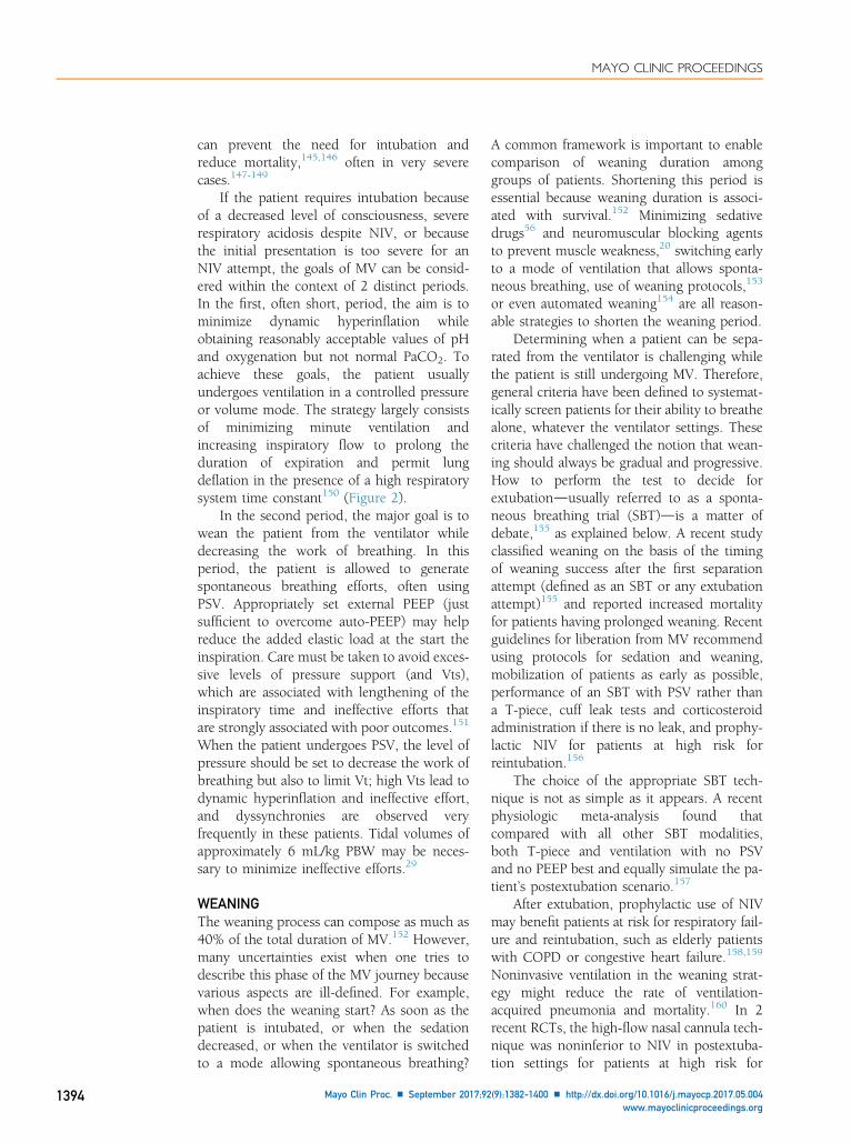
FIGURE 2. Ventilator waveform and values in a patient undergoing volume assist-control ventilation showing expiration flow limi-tation. Note typical sign of expiratory flow limitation on the flow tracing: during the expiration phase, the flow waveform reaches apeak higher than �1 L/s and abruptly returns to very low values and oscillates around this value until the next inspiration. The end ofexpiration is interrupted by the next insufflation before flow reaches zero (arrows), indicating dynamic hyperinflation and intrinsicpositive end-expiratory pressure (PEEP); total PEEP (PEEPtot) is 11 cm H2O (obtained during end-expiratory occlusion [EEO]). In anormal patient, the flow waveform would trace a quasi-exponential curve from the peak to 0. Plateau pressure (Pplat) is assessedduring the end-inspiratory occlusion (EIO: 19 cm H2O), resulting in a driving pressure of 8 cm H2O (Pplat e PEEPtot). The inspiratoryflow is 0.8 L/s, resulting in high airway resistance of 29 cm H2O per L/s ([peak airway pressure (Ppeak)� Pplat]/flow¼ [42� 19]/0.8 ¼28.75). In an intubated adult patient with normal lungs, resistances are usually less than 10 cm H2O per L/s. E ¼ passive expiration;I ¼ inspiration due to ventilator insufflation; Paw ¼ airway pressure; PEEPe ¼ external PEEP.
MECHANICAL VENTILATION: STATE OF THE ART
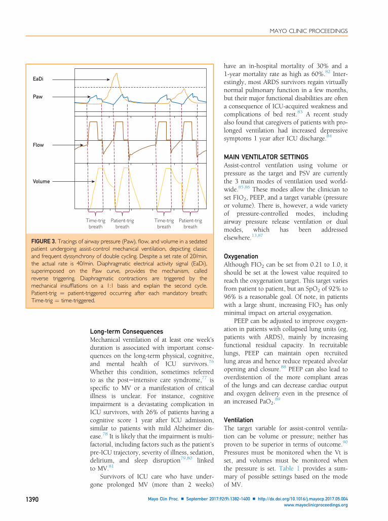
which are summarized in Table 2. Figure 3presents an example of a ventilator monitordisplaying reverse triggering with doublecycling. Often, these dyssynchronies indicatea mismatch between the ventilatory needs ofthe patient and the amount of ventilationdelivered. Although association does notimply causality, patients with greater numbersof dyssynchronies have poorer outcomesincluding longer durations of ventilation,longer ICU stays, and higher mortality.26,29,30
In some cases, this worse outcome may beexplained by increased Vts, breath stacking,intrinsic PEEP,31 or regional hyperinflation,32
but dyssynchronies may also be a marker of
Mayo Clin Proc. n September 2017;92(9):1382-1400 n http://dx.doiwww.mayoclinicproceedings.org
the severity of the underlying lung pathophys-iology. Although improving patient-ventilatory synchrony makes intuitive sense,we lack definitive data proving that itimproves patients’ outcomes.
COMPLICATIONS OF MVMechanical ventilation is often lifesaving but isassociated with serious complications, in partbecause it is delivered to patients at high riskof lung or cardiac compromise. These compli-cations may be related to the direct mechanicaleffects of the intrathoracic pressures generatedby the ventilator, to alveolar and systemicinflammation, or to neural stimulation. There
.org/10.1016/j.mayocp.2017.05.004 1385
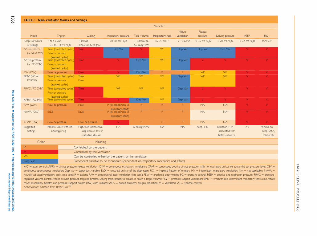
TABLE 1. Main Ventilator Modes and Settings
Mode
Variable
Trigger Cycling Inspiratory pressure Tidal volume Respiratory rateMinute
ventilationPlateaupressure Driving pressure PEEP FIO2
Ranges of values
or settings
1 to 5 L/min
�0.5 to �3 cm H2O
1 second
30%-70% peak flow
10-30 cm H2O w200-600 mL
4-8mL/kg PBW
10-35 min�1 w7-12 L/min 15-35 cm H2O 8-20 cm H2O 0-22 cm H2O 0.21-1.0
A/C in volume(or VC-CMV)
Time (controlled cycles)Flow or pressure
(assisted cycles)
Time Dep Var V V/P Dep Var Dep Var Dep Var V V
A/C in pressure(or PC-CMV)
Time (controlled cycles)Flow or pressure
(assisted cycles)
Time V Dep Var V/P Dep Var V V V V
PSV (CSV) Flow or pressure Flow V Dep Var P P V/P V/P V VSIMV (VC or
PC-IMV)Time (controlled cycles)Flow or pressure
(assisted cycles)
TimeFlow
V/P V/P V/P Dep Var V/P V/P V V
PRVC (PC-CMV) Time (controlled cycles)Flow or pressure
(assisted cycles)
Time V/P V/P V/P Dep Var V V V V
APRV (PC-IMV) Time (controlled cycles) Time V Dep Var V/P Dep Var V V V V
PAV (CSV) Flow or pressure Flow P (in proportion toinspiratory effort)
P P P NA NA V V
NAVA (CSV) EaDi EaDi P (in proportion toinspiratory effort)
P P P NA NA V V
CPAP (CSV) Flow or pressure Flow or pressure V P P P NA NA V V
Suggestedsettings
Minimal value with noautotriggering
High % in obstructivelung disease, low inrestrictive disease
NA 6 mL/kg PBW NA NA Keep <30 Less thanw14associated withbetter outcome
�5 Minimal tokeep SpO2
90%-94%
Color Meaning
P Controlled by the patientV Controlled by the ventilatorV/P Can be controlled either by the patient or the ventilatorDep Var Dependent variable to be monitored (dependent on respiratory mechanics and effort)
A/C ¼ assist-control; APRV ¼ airway pressure release ventilation; CMV ¼ continuous mandatory ventilation; CPAP ¼ continuous positive airway pressure, with no inspiratory assistance above the set pressure level; CSV ¼continuous spontaneous ventilation; Dep Var ¼ dependant variable; EaDi ¼ electrical activity of the diaphragm; FIO2 ¼ inspired fraction of oxygen; IMV ¼ intermittent mandatory ventilation; NA ¼ not applicable; NAVA ¼neurally adjusted ventilatory assist (see text); P ¼ patient; PAV ¼ proportional assist ventilation (see text); PBW ¼ predicted body weight; PC ¼ pressure control; PEEP ¼ positive end-expiration pressure; PRVC ¼ pressure-regulated volume control, which delivers pressure-targeted breaths, varying from breath to breath to reach a target volume; PSV ¼ pressure support ventilation; SIMV ¼ synchronized intermittent mandatory ventilation, whichmixes mandatory breaths and pressure support breath (PSV) each minute; SpO2 ¼ pulsed oximetry oxygen saturation; V ¼ ventilator; VC ¼ volume control.Abbreviations adapted from Respir Care.13
MAYO
CLIN
ICPRO
CEED
INGS
1386Mayo
ClinProc.
nSeptem
ber2017;92(9):1382-1400
nhttp://dx.doi.org/10.1016/j.m
ayocp.2017.05.004www.m
ayoclinicproceedings.org

MECHANICAL VENTILATION: STATE OF THE ART
is evidence of cross-talk between the lung andthe brain and between the lung and the kid-neys, all influenced by MV.33,34 Many of thecomplications of MV can potentially beavoided or minimized. This factor is importantfrom a clinical perspective and is a major areaof current research.
Initiation of MVEndotracheal intubation is a critical procedurein which patients are at risk of respiratory and/or circulatory compromise.35,36 Before intuba-tion, the patient should be assessed for factorsindicating a possible difficult intubation; thereare specific scoring systems for the ICU.37 Pre-oxygenation is essential, and different tech-niques such as noninvasive ventilation(NIV)38 or high flow delivered via nasal can-nula have been proposed for patients withthe most severe disease. To avoid gastric aspi-ration, rapid-sequence intubation using asedative drug and a neuromuscular blockingagent is often recommended.39 Recommenda-tions and algorithms have been developed forpatients with a “difficult airway”.40,41
Hemodynamic EffectsPositive-pressure ventilation has long beenknown to have hemodynamic effects throughheart-lung interactions. These effects havebeen better understood, managed, and oftenprevented over the past few decades by anincreased understanding of the followingmechanisms. First, high intrathoracic pres-sure, especially high plateau pressures cannegatively impact right ventricular afterloadand function.42 Our understanding of auto-PEEP and the use of protective lung ventila-tion have markedly reduced the incidence ofhemodynamic complications through the useof lower volumes and pressures.43,44 Echo-graphic studies in patients with ARDS have re-ported a prevalence of acute cor pulmonale ofabout 22%,45,46 which is still quite high, butmarkedly lower than previously reported.44,47
Second, hypotensive effects of sedative agentsacting via negative inotropy, vasodilation, orcentral mechanisms are managed by appro-priate use of vasoactive drugs or fluids. Third,the use of partial ventilatory assist reducesintrathoracic pressures and minimizes seda-tion needs, facilitating the hemodynamic toler-ance of MV. Finally, pulmonary hypertension
Mayo Clin Proc. n September 2017;92(9):1382-1400 n http://dx.doiwww.mayoclinicproceedings.org
and PEEP, especially in patients with ARDS,can result in a right-to-left shunting across apatent foramen ovale and worsen hypoxemiain up to 20% of patients with ARDS.48
Complications of SedationIn the early phase of MV, sedation with orwithout paralysis is often required, especiallyfor patients with shock or ARDS or for those“fighting the ventilator.”49 The slow meta-bolism of sedative agents may unduly prolongthe duration of MV and lead to detrimentalshort- and long-term outcomes.50,51 Eachsedative agent has specific effects, and theappropriate choice of the type and dose ofsedative drugs may impact outcome. Data sug-gest that benzodiazepines are particularly asso-ciated with poorer long-term outcomes.52
Propofol is frequently used because of a rela-tively short half-life, but there are concernsassociated with prolonged infusion.53 Dexme-detomidine has been proposed as a promisingalternative to usual sedation because it reducesthe rate of delirium,54,55 but results from clin-ical trials have not been consistent. If sedationcannot be avoided, it is important to carefullymonitor the depth of a patient’s sedation andto use a sedation protocol, including dailyinterruption of sedation to avoid a state ofdeep sedation.56,57
Oxygen ToxicityMechanical ventilation allows patients toreceive a fraction of inspired oxygen (FIO2)of up to 1.0, which may be necessary for pa-tients with severe hypoxemia. However, highlevels of oxygen have toxic effects, whichhave been a concern since the early days ofMV.58 In low ventilation-perfusion ratio lungunits, high FIO2 can lead to reabsorption atel-ectasis,59 which can be minimized usinghigher levels of PEEP.60 Oxygen also hasextrapulmonary effectsdit can decrease car-diac output by decreasing parasympathetictone61 and increasing vascular resistance, andit has vasoconstrictive effects on cerebral andcoronary perfusion.62,63 Several studies havesuggested an independent association betweenhyperoxemia and hospital mortality in somegroups of patients (eg, those with cardiacarrest or stroke).64 Clinicians, however, tendto be much more sensitive to hypoxemiathan to hyperoxemia. Recent preliminary
.org/10.1016/j.mayocp.2017.05.004 1387

TABLE 2. Main Patient-Ventilator Dyssynchronies and Interactions
Dyssynchrony orpatient-ventilator interaction Description Pathophysiology Risks Main modes of MV Suggestions
During inspirationFlow starvation Delivered flow does not
match patient’s demand
d Insufficient peak flowd High respiratory drive
d Dyspnead High levels of work
of breathing
d A/C ventilation (volume) d Increase peak flow >50 L/min(direct setting or shorteninspiratory time to obtainthe same volume faster)
Short cycles Continuation of inspiratoryeffort after the end ofinsufflation
d Insufficient inspiratory timed High respiratory drive
d Eccentric contractions ofrespiratory muscles
d Double triggering
d A/C ventilation(pressure or volume)
Increase inspiratory time
Prolonged insufflation Continuation of insufflationafter the end of inspiratoryeffort
d Inadequate cyclingmechanism
d Gas trapping
d Shorten neural expirationand promote gas trapping
d Dyspnea
d A/C ventilation (pressure)d PSVd NIV
Modify cycling to make theinspiration shorter
Reverse triggering Diaphragmatic contractiontriggered by mechanicalinsufflation
Reflex mechanism in highlysedated patient
d Loss of protective ventilationd Monitoring of plateau
pressure inoperatived Eccentric contractions of
respiratory muscles
d A/C ventilation(pressure or volume)
d Paralyze if VT too high ordouble cycle
d Decrease sedation
Double cycles (during inspiration or expiration)Double cycles after reverse
triggeringReverse triggering of a second
cycleReflex mechanism in highly
sedated patientDouble the mechanical stress
on the lung
d A/C ventilation(pressure or volume)
d Paralyze if VT too high ordouble cycle
d Decrease sedation
Double (or triple) triggeringafter short cycles (breathstacking)
Continuation of inspiratoryeffort after the end ofinsufflation
d Insufficient inspiratory timed High respiratory drive
Double or triple the mechanicalstress on the lung
d A/C ventilation(pressure or volume)
d PSV
d Increase inspiratory timed Increase VTd Modify cycling to make
the inspiration longer
During expirationAutotriggering Cycles not triggered by the
patient
d Leaksd Water in the circuitd Excessively sensitive triggerd Cardiac oscillations
d Dyspnead Misleading information
on breathing patternd Severe hyperventilation
(eg, arrhythmias, reducedcerebral blood flow)
d Increase rate of lung stress
d A/C ventilation(pressure or volume)
d PSVd NIV
d Inspect tubingd Decrease trigger sensitivity
Continued on next page
MAYO
CLIN
ICPRO
CEED
INGS
1388Mayo
ClinProc.
nSeptem
ber2017;92(9):1382-1400
nhttp://dx.doi.org/10.1016/j.m
ayocp.2017.05.004www.m
ayoclinicproceedings.org

TABLE
2.Co
ntinue
d
Dyssynchron
yor
patient-ventilator
interaction
Descriptio
nPathop
hysio
logy
Risks
Mainmod
esof
MV
Suggestio
ns
Duringexpiratio
n,continued
Gas
trapping
Nextinspiratio
nstartsbefore
endof
exhalatio
nHightim
econstant
dPo
ordiaphragm
functio
nd
Hem
odynam
iceffects
dIneffectiveefforts
dAny
assistedmod
ed
NIV
Decreasehyperdynam
icinflation:
dIncrease
expiratio
ntim
ed
Decrease
minute
ventilatio
n(decreased
VTand/or
RR)
dDecreasefre
quency
Ineffectiveeffort
Effortunableto
triggerthe
ventilator
dInadequate
cycling
dExcessivesupport
dLargetim
econstant
dLo
wrespiratory
drive
dRe
peated
pleiom
etric
work
dErroneou
sdisplayof
respiratory
rate
dProlon
gedduratio
nof
ventilatio
n
dAny
assistedmod
ed
NIV
dIncrease
triggersensitivity
dDecreasesedatio
nd
Increase
expiratio
ntim
ed
Increase
PEEP
(to
equal
intrinsic
PEEP)
A/C
¼assist-control;MV¼
mechanicalventilation;
NIV
¼no
ninvasiveventilatio
n;PEEP
¼po
stiveend-expiratory
pressure;P
SV¼
pressure
supportventilatio
n;RR
¼respiratory
rate;V
T¼
tidalvolume.
MECHANICAL VENTILATION: STATE OF THE ART
Mayo Clin Proc. n September 2017;92(9):1382-1400 n http://www.mayoclinicproceedings.org
dx.doi
data suggest that conservative oxygen therapytargeting a PaO2 of 70 to 100 mm Hg or apulse oximetry oxygen saturation (SpO2) of94% to 98% results in lower ICU mortalitythan a conventional, more “liberal” approachwith higher PaO2 and SpO2 targets.
65
Effects on Respiratory Muscles andRespiratory InfectionsMechanical ventilation has been associatedwith respiratory muscle dysfunction andweaning difficulties.20,66-68 Disuse atrophy ofthe diaphragm appears to be a key mechanismfor these detrimental effects, suggesting theneed to better monitor respiratory muscleactivity. Partial modes of ventilation do notalways prevent this atrophy. Several studiesexamining diaphragm biopsies have foundthat changes in structure occur early after intu-bation.66 More than 50% of patients experi-ence dysfunction related to an excessive levelof assistance (controlled or partialventilation) or to insufficient assistance.67
Limb muscle weakness, referred to asICU-acquired weakness, and diaphragmdysfunction have only minimal overlap. Respi-ratory muscle dysfunction is at least twice asprevalent as limb muscle weakness at thetime of separation from MV and has a strongimpact on weaning.20
Intubated and ventilated patients are atrisk for ventilation-acquired pneumonia dueto microaspiration from the oropharyngealcavity and diminished host defense due todecreased cough efficiency and impairedmucociliary clearance. Recent guidelinesrecommend limitation of sedation and short-ening the duration of MV in order to minimizethe risk of ventilation-acquired pneumonia.69
Ventilator-Induced Lung InjuryMechanical ventilation can induce or worsenlung injury, referred to as ventilator-inducedlung injury (VILI).70-75 This disorder hasbecome a major concern in the modern eraof MV, profoundly modifying the clinical tar-gets of MV. Ventilator-induced lung injurymay impact a large number of patients, mostspecifically those with or at risk for ARDS. Pre-vention is described in greater detail in the“Acute Respiratory Distress Syndrome”section.
.org/10.1016/j.mayocp.2017.05.004 1389
EaDi
Paw
Flow
Volume
Time-trigbreath
Time-trigbreath
Patient-trigbreath
Patient-trigbreath
FIGURE 3. Tracings of airway pressure (Paw), flow, and volume in a sedatedpatient undergoing assist-control mechanical ventilation, depicting classicand frequent dyssynchrony of double cycling. Despite a set rate of 20/min,the actual rate is 40/min. Diaphragmatic electrical activity signal (EaDi),superimposed on the Paw curve, provides the mechanism, calledreverse triggering. Diaphragmatic contractions are triggered by themechanical insufflations on a 1:1 basis and explain the second cycle.Patient-trig ¼ patient-triggered occurring after each mandatory breath;Time-trig ¼ time-triggered.
MAYO CLINIC PROCEEDINGS
1390
Long-term ConsequencesMechanical ventilation of at least one week’sduration is associated with important conse-quences on the long-term physical, cognitive,and mental health of ICU survivors.76
Whether this condition, sometimes referredto as the posteintensive care syndrome,77 isspecific to MV or a manifestation of criticalillness is unclear. For instance, cognitiveimpairment is a devastating complication inICU survivors, with 26% of patients having acognitive score 1 year after ICU admission,similar to patients with mild Alzheimer dis-ease.78 It is likely that the impairment is multi-factorial, including factors such as the patient’spre-ICU trajectory, severity of illness, sedation,delirium, and sleep disruption79,80 linkedto MV.81
Survivors of ICU care who have under-gone prolonged MV (more than 2 weeks)
Mayo Clin Proc. n September 2017;92
have an in-hospital mortality of 30% and a1-year mortality rate as high as 60%.82 Inter-estingly, most ARDS survivors regain virtuallynormal pulmonary function in a few months,but their major functional disabilities are oftena consequence of ICU-acquired weakness andcomplications of bed rest.83 A recent studyalso found that caregivers of patients with pro-longed ventilation had increased depressivesymptoms 1 year after ICU discharge.84
MAIN VENTILATOR SETTINGSAssist-control ventilation using volume orpressure as the target and PSV are currentlythe 3 main modes of ventilation used world-wide.85,86 These modes allow the clinician toset FIO2, PEEP, and a target variable (pressureor volume). There is, however, a wide varietyof pressure-controlled modes, includingairway pressure release ventilation or dualmodes, which has been addressedelsewhere.13,87
OxygenationAlthough FIO2 can be set from 0.21 to 1.0, itshould be set at the lowest value required toreach the oxygenation target. This target variesfrom patient to patient, but an SpO2 of 92% to96% is a reasonable goal. Of note, in patientswith a large shunt, increasing FIO2 has onlyminimal impact on arterial oxygenation.
PEEP can be adjusted to improve oxygen-ation in patients with collapsed lung units (eg,patients with ARDS), mainly by increasingfunctional residual capacity. In recruitablelungs, PEEP can maintain open recruitedlung areas and hence reduce repeated alveolaropening and closure.88 PEEP can also lead tooverdistention of the more compliant areasof the lungs and can decrease cardiac outputand oxygen delivery even in the presence ofan increased PaO2.
89
VentilationThe target variable for assist-control ventila-tion can be volume or pressure; neither hasproven to be superior in terms of outcome.90
Pressures must be monitored when the Vt isset, and volumes must be monitored whenthe pressure is set. Table 1 provides a sum-mary of possible settings based on the modeof MV.
(9):1382-1400 n http://dx.doi.org/10.1016/j.mayocp.2017.05.004www.mayoclinicproceedings.org
MECHANICAL VENTILATION: STATE OF THE ART
In the past, a major goal of MV was toensure that patients had normal arterial bloodgas levels, with little regard to the harmscaused by MV. Currently, a priority is toensure that VILI is minimized while maintain-ing adequate, but not necessarily normal, gasexchange. The best oxygenation is not alwaysthe most protective, and moderate levels ofhypercapnia are considered acceptable. In thepast, high Vts were recommended on the basisof studies in anesthetized patients that foundthat small Vts led to atelectasis and hypox-emia.91 Atelectasis was related to the com-bined effects of high FIO2, anesthesia, andlack of PEEP. It took years of research torealize that high Vts, despite having favorableeffects on oxygenation, were harmful for thelungs and increased mortality.92
Current recommendations for setting Vt arebased on predicted body weight (PBW) andnot actual body weight because (normal) lungsize scales with PBW. One formula is: PBW(kg) ¼ 50.0 þ 0.91 (height in cm � 152.4)for males and PBW (kg) ¼ 45.5 þ 0.91 (heightin cm � 152.4) for females. The recommendedrange is 6 to 8 mL/kg PBW.
Partial modes of assist are very popular,based on the delivery of a pressure supportlevel. They are frequently used and generallywell tolerated.93 There are 2 concerns withthese modes. One is that patients can be easilyoverassisted29,94; experimental68 and clinical67
data suggest that despite the use of partial sup-port, insufficient muscle use can lead to atro-phy and dysfunction. Second, because the Vtcannot be controlled, patients with high respi-ratory drive may generate excessive Vts, whichcan lead to a form of patient self-inflicted lunginjury.95,96
Proportional Modes of VentilationTwo modes of ventilation are based on adifferent principle and address some of theconcerns discussed previously. These 2modes, which require a relatively preservedneuroventilatory drive, deliver pressure in pro-portion to the patient’s demand and let thepatient regulate Vt. One mode called propor-tional assist ventilation requires real timecalculation of the equation of motion of therespiratory system based on automated mea-surements of respiratory system complianceand resistance. The second mode uses the
Mayo Clin Proc. n September 2017;92(9):1382-1400 n http://dx.doiwww.mayoclinicproceedings.org
electrical activity of the diaphragm and iscalled neurally adjusted ventilatory assist(NAVA). The only setting required from theclinician is the amount of assistance: duringproportional assist ventilation, it is set as a per-centage of assistance, and for neurally adjustedventilatory assist, it is set by the proportional-ity factor between electrical activity of the dia-phragm and pressure. For both modes, Vt,frequency, and pressure are not set by theclinician. Both modes are very effective inreducing dyssynchronies and in adapting tochanges in ventilatory demand, explainingimprovement in sleep quality observed withtheir use.97-99 However, few outcome dataare available.100,101 Some experimental orhuman data suggest that they may allow asafer control of ventilation than routine lungprotective ventilation.102,103
ACUTE RESPIRATORY DISTRESSSYNDROMENo other ICU syndrome has been studied asmuch as ARDS. Understanding the impact ofMV on patients with ARDS has resulted inmajor changes in ventilator management overthe past 25 years.
A consensus definition of ARDS wasreleased in 1994, more than 25 years after itsinitial description.104 The most recent Berlindefinition tried to overcome some of the limi-tations of previous definitions.11,105 ARDS iscurrently defined by a new onset or worseningof respiratory symptoms with bilateral opaci-ties on chest radiography and a PaO2:FIO2
ratio 300 mm Hg or less while receivingPEEP of 5 cm H2O or higher. Concomitantheart failure can be present, but if no knownrisk factor for ARDS has been identified,congestive heart failure must be objectivelyruled out.
There are many predisposing factors thatcan lead to the development of ARDS, butthe lungs of patients with ARDS share severalcommon biological, cellular, and mechanicalcharacteristics. The lungs are edematous andheavy, adding considerable superimposedpressure to the dependent lung regions.Normally aerated tissue is greatly reducedand has been described as a “babylung.”106,107 The baby lung concept explainsthe low respiratory system compliance, highpressures, and high risk for VILI. Minimizing
.org/10.1016/j.mayocp.2017.05.004 1391

MAYO CLINIC PROCEEDINGS
1392
the risk of VILI has improved survival.70,74,108
In contrast, pharmacological approaches fortreating ARDS have been disappointing.
Different techniques have been used to tryto prevent intubation in patients with acutehypoxemic respiratory failure, including NIV.A high-flow nasal cannula is used increasinglyin patients with acute hypoxemic respiratoryfailure and has improved comfort, decreaseddyspnea, and decreased mouth and airwaydryness sensation compared with conven-tional oxygen therapy.109,110 A recent studyfound a similar rate of intubation but areduced mortality rate in the group of patientstreated with high-flow nasal cannulacompared with NIV or standard oxygen.111
Intubation was reduced in those with aPaO2:FIO2 ratio lower than 200 mm Hg. Itmay work in part by reducing the oropha-ryngeal dead space by a washout effect andby increasing end-expiratory pressure.112
The Acute Respiratory Distress SyndromeNetwork Lower Tidal Volume (ARMA) trialwas the first large multicenter clinical trial todocument the benefit of a lung protectivestrategy using lower than traditional Vts (w6mL/kg PBW) and limiting Pplat to 30 cmH2O.
113 Since then, accumulating evidencehas demonstrated that low Vts, with orwithout a certain degree of acidosis (permis-sive hypercapnia), are efficient in limitingVILI.114 Reducing instrumental dead space(eg, filters) is necessary, and increasing the res-piratory rate to 35 breaths/min is recommen-ded to minimize hypercapnia. There is someevidence that decreasing Vt even further mayimprove outcomes.115 Clinical trials areexploring the impact of lower Vts using extra-corporeal circulation to remove carbondioxide.116
How to best set the PEEP level for anypatient has been a matter of debate for 5decades. The initial focus was to improveoxygenation with higher PEEP, but the currentthinking is that any improvement in outcomeswith higher PEEP levels is due to decreasedVILI. Individual trials have failed to documentdecreased mortality with a higher PEEP strat-egy,71,117,118 but an individual patient datameta-analysis found that higher PEEP wasassociated with a 5% lower mortality rate inpatients with moderate or severe ARDS(PaO2:FIO2 ratio <200 mm Hg) but not in
Mayo Clin Proc. n September 2017;92
patients with a PaO2:FIO2 ratio higher than200 mm Hg.73 The high PEEP strategyimproved several secondary end points suchas hypoxemia, use of rescue therapies, andduration of organ failure and MV.
Measurement of esophageal pressure toestimate transpulmonary pressure at end-expiration is a promising approach.119,120 Astrategy titrating PEEP on the basis of transpul-monary pressures revealed improved oxygena-tion and compliance compared with standardsettings,121 and a larger clinical trial of thisapproach is currently ongoing (NCT01681225).
Recently, a reanalysis of 9 of the main ran-domized controlled trials (RCTs) in ARDScompared the impact of Vt, PEEP, Pplat,and driving pressure (DP ¼ Pplat � PEEP)on outcomes. Driving pressure change wasthe variable that best predicted mortal-ity,122,123 perhaps because it is equal to (Vt/CRS)die, Vt normalized to respiratory systemcompliance, the latter being related to lungsize. Conversely, PBW is a good predictor oflung size in healthy individuals but not in pa-tients with ARDS, who can have markedlydecreased lung volumes. The recent interna-tional multicenter observational LUNG SAFE(Large Observational Study to Understandthe Global Impact of Severe Acute RespiratoryFailure) study also found an associationbetween both higher Pplat and DP withmortality.86 These studies suggest that a safeventilatory strategy should first use a Vt of6mL/kg PBW, while limiting plateau anddriving pressure. Keeping DP below a riskylevel (eg, <15 cm H2O) may help, althoughno prospective data are available. High PEEPlevels (>10-15 cm H2O) seem beneficialin moderate and especially severe ARDS(PaO2:FIO2 ratio <200 mm Hg).
In moderately severe to severe ARDS with aPaO2:FIO2 ratio of less than 150 mm Hg,adjunctive therapies such as neuromuscularblockade for the first 48 hours49 or prone posi-tioning also result in improved survival.124,125
Implementation of the prone position requirestraining by the clinical team, but the evidencestrongly suggests that it should be appliedwhen the PaO2:FIO2 ratio remains lower than120 mm Hg despite protective ventilation.
Extracorporeal membrane oxygenationmay be beneficial in patients with the mostsevere ARDS and is currently under
(9):1382-1400 n http://dx.doi.org/10.1016/j.mayocp.2017.05.004www.mayoclinicproceedings.org

MECHANICAL VENTILATION: STATE OF THE ART
investigation. The results of the EOLIA (Extra-corporeal Membrane Oxygenation for SevereAcute Respiratory Distress Syndrome) trial(clinicaltrials.gov Identifier: NCT01470703)will provide valuable information.126,127 Atpresent, it seems reasonable to apply extracor-poreal membrane oxygenation if pronepositioning is ineffective.
Alveolar recruitment techniques vary andmay have adverse effects. Recent data indicatethat most of the effect of a sustained inflation(35-40 cm H2O) is obtained after 10 seconds,suggesting that such maneuvers can be termi-nated relatively early before adverse eventsoccur.128 Even if recruitment maneuvers sub-stantially improve oxygenation, this effect istransient, and the benefit on patient outcomesis still controversial.129,130
Two RCTs using high-frequency oscilla-tion found no benefit for moderate and severeARDS, and one of them even found a highermortality rate for patients treated with thistechnique.131,132 Therefore, high-frequencyoscillation is not recommended as first-linetherapy for patients with ARDS. However, arecent meta-analysis suggests that it may bebeneficial in very severely hypoxemic pa-tients.133 Inhaled nitric oxide can lead to vaso-dilation of the well-ventilated alveoli withsubsequent improvement in oxygenation buthas been found in multiple studies to notimpact mortality and may have adverse effectssuch as increased risk of renaldysfunction.134,135
PROTECTIVE VENTILATION FOR PATIENTSWITH RELATIVELY NORMAL LUNGSThere is accumulating evidence for the benefi-cial effects of lung protective ventilation inpatients without ARDS,136 including those un-dergoing major surgical procedures, patientswithout ARDS at presentation, and in brain-dead patients who are potential lung donors.
For surgical patients with previouslyhealthy lungs, the conventional strategy haspreviously been to combine high Vts(w10-15 mL/kg) with high FIO2 using lowor no PEEP. The goal with this strategy wasto prevent atelectasis.137,138 In recent years,several studies have examined lung protectiveventilation strategies (low Vt, PEEP with orwithout recruitment maneuvers) in the oper-ating room. One study reported a 3-fold
Mayo Clin Proc. n September 2017;92(9):1382-1400 n http://dx.doiwww.mayoclinicproceedings.org
reduction in postoperative complications andin the requirement of postoperative MV withthis strategy compared with conventionalventilation in patients undergoing majorabdominal operations.139 Other studies inpatients undergoing thoracic and abdominalsurgical procedures have documented reducedpostoperative pulmonary and extrapulmonarycomplications with lower health care utiliza-tion when a protective ventilation strategywas used.139,140 Protective ventilation is notassociated with additional risk of intraopera-tive complications.
In intubated ICU patients not presentingwith ARDS on admission, a strategy usinglower Vts was associated with shorter durationof MV.141 A meta-analysis examining surgicaland ICU patients found that lower Vts werebeneficial for all important outcomesincluding evolution to ARDS, pneumonia,hospital length of stay, and mortality.136
Finally, in brain-dead potential organdonors, a lung protective ventilation strategymaintaining sufficient PEEP and avoidingderecruitment allowed optimization of lungtransplant leading to a 2-fold increase inharvested lungs compared with a conventionalstrategy with the same rate of success and6-month survival rate.142
VENTILATION IN PATIENTS WITH COPDExacerbations of COPD are characterized by amarked worsening of respiratory mechanicssecondary to increased airway resistance, expi-ratory collapse of small airways limiting expi-ratory flow, development of auto-PEEP andhyperinflation, and increased work of breath-ing. The development of auto-PEEP hasimportant consequences including increasedwork of breathing (inspiratory thresholdloading), decreased respiratory muscle effi-ciency (flattened diaphragms), and hemody-namic compromise. Patients are unable toachieve sufficient Vts despite strong respira-tory efforts and have markedly elevatedoxygen cost of breathing. In these patients,the physiologic rationale for NIV is verystrongdNIV improves ventilatory efficiency,decreases respiratory rate, decreases the workof breathing, and increases alveolar ventilationby increasing Vt.143 This approach often imp-roves the patient’s level of consciousness.144
Many studies have found that the use of NIV
.org/10.1016/j.mayocp.2017.05.004 1393
MAYO CLINIC PROCEEDINGS
1394
can prevent the need for intubation andreduce mortality,145,146 often in very severecases.147-149
If the patient requires intubation becauseof a decreased level of consciousness, severerespiratory acidosis despite NIV, or becausethe initial presentation is too severe for anNIV attempt, the goals of MV can be consid-ered within the context of 2 distinct periods.In the first, often short, period, the aim is tominimize dynamic hyperinflation whileobtaining reasonably acceptable values of pHand oxygenation but not normal PaCO2. Toachieve these goals, the patient usuallyundergoes ventilation in a controlled pressureor volume mode. The strategy largely consistsof minimizing minute ventilation andincreasing inspiratory flow to prolong theduration of expiration and permit lungdeflation in the presence of a high respiratorysystem time constant150 (Figure 2).
In the second period, the major goal is towean the patient from the ventilator whiledecreasing the work of breathing. In thisperiod, the patient is allowed to generatespontaneous breathing efforts, often usingPSV. Appropriately set external PEEP (justsufficient to overcome auto-PEEP) may helpreduce the added elastic load at the start theinspiration. Care must be taken to avoid exces-sive levels of pressure support (and Vts),which are associated with lengthening of theinspiratory time and ineffective efforts thatare strongly associated with poor outcomes.151
When the patient undergoes PSV, the level ofpressure should be set to decrease the work ofbreathing but also to limit Vt; high Vts lead todynamic hyperinflation and ineffective effort,and dyssynchronies are observed veryfrequently in these patients. Tidal volumes ofapproximately 6 mL/kg PBW may be neces-sary to minimize ineffective efforts.29
WEANINGThe weaning process can compose as much as40% of the total duration of MV.152 However,many uncertainties exist when one tries todescribe this phase of the MV journey becausevarious aspects are ill-defined. For example,when does the weaning start? As soon as thepatient is intubated, or when the sedationdecreased, or when the ventilator is switchedto a mode allowing spontaneous breathing?
Mayo Clin Proc. n September 2017;92
A common framework is important to enablecomparison of weaning duration amonggroups of patients. Shortening this period isessential because weaning duration is associ-ated with survival.152 Minimizing sedativedrugs56 and neuromuscular blocking agentsto prevent muscle weakness,20 switching earlyto a mode of ventilation that allows sponta-neous breathing, use of weaning protocols,153
or even automated weaning154 are all reason-able strategies to shorten the weaning period.
Determining when a patient can be sepa-rated from the ventilator is challenging whilethe patient is still undergoing MV. Therefore,general criteria have been defined to systemat-ically screen patients for their ability to breathealone, whatever the ventilator settings. Thesecriteria have challenged the notion that wean-ing should always be gradual and progressive.How to perform the test to decide forextubationdusually referred to as a sponta-neous breathing trial (SBT)dis a matter ofdebate,155 as explained below. A recent studyclassified weaning on the basis of the timingof weaning success after the first separationattempt (defined as an SBT or any extubationattempt)155 and reported increased mortalityfor patients having prolonged weaning. Recentguidelines for liberation from MV recommendusing protocols for sedation and weaning,mobilization of patients as early as possible,performance of an SBT with PSV rather thana T-piece, cuff leak tests and corticosteroidadministration if there is no leak, and prophy-lactic NIV for patients at high risk forreintubation.156
The choice of the appropriate SBT tech-nique is not as simple as it appears. A recentphysiologic meta-analysis found thatcompared with all other SBT modalities,both T-piece and ventilation with no PSVand no PEEP best and equally simulate the pa-tient’s postextubation scenario.157
After extubation, prophylactic use of NIVmay benefit patients at risk for respiratory fail-ure and reintubation, such as elderly patientswith COPD or congestive heart failure.158,159
Noninvasive ventilation in the weaning strat-egy might reduce the rate of ventilation-acquired pneumonia and mortality.160 In 2recent RCTs, the high-flow nasal cannula tech-nique was noninferior to NIV in postextuba-tion settings for patients at high risk for
(9):1382-1400 n http://dx.doi.org/10.1016/j.mayocp.2017.05.004www.mayoclinicproceedings.org

MECHANICAL VENTILATION: STATE OF THE ART
respiratory failure161 and even decreased therate of reintubation for patients at low risk.162
AVENUES FOR IMPROVEMENTOur understanding of the pathophysiology ofacute respiratory diseases, the impact of venti-lator settings on dyssynchronies, and the com-plications of MV have all markedly improvedduring the past few decades. Nevertheless,many unanswered questions remain. Giventhe potential iatrogenic consequences of inad-equate delivery of MV, one might assume thatavoiding invasive MV at any cost wouldbenefit the patient. However, recent data sug-gest that spontaneous ventilation can also leadto lung injury in patients with high respiratorydrive.163 Patients breathing spontaneously,whether intubated or not, can experienceself-inflicted lung injury due to high minuteventilation and increased Vts.96,164 Thus,spontaneous ventilation can also be harmful,and very high respiratory drive with the devel-opment of very large Vts may be an indicationfor intubation with heavy sedation or neuro-muscular blocking agents. Identifying whichspontaneously breathing patients are atincreased risk for this type of injury is animportant area of future research.
A promising approach to limiting compli-cations from MV in patients with ARDS orCOPD is the use of extracorporeal life support.These techniques range from extracorporealcarbon dioxide removal, which is the leastinvasive and can be delivered through a rela-tively small-bore cannula (dual-lumen13-17Fr diameter) at a blood flow of lessthan 500 mL/min, to full extracorporeal mem-brane oxygenation requiring a large venouscannula (with a minimum diameter of 23Fr)to allow flow rates of more than 4 L/min.The relative efficacy of each of these tech-niques is currently being examined in clinicaltrials.
CONCLUSIONDecades of research, progress, and clinicalmonitoring has led to an increased under-standing of the physiology of MV. A concep-tual revolution occurred when the goal ofMV moved from normalizing blood gas levelsto minimizing VILI while maintainingadequate (albeit not necessarily normal) gasexchange. We now know that management
Mayo Clin Proc. n September 2017;92(9):1382-1400 n http://dx.doiwww.mayoclinicproceedings.org
during the acute phase has a strong impacton long-term outcome and disabilities, andthis focus on long-term outcomes will be afocus for future research. The MV journey ismaking progress but is still far from its ulti-mate destination.
ACKNOWLEDGMENTSWe thank Dr Lu Chen for providing the trac-ings used in Figures 1 and 2.
Abbreviations and Acronyms: ARDS = acute respiratorydistress syndrome; COPD = chronic obstructive pulmonarydisease; CRS = compliance of the respiratory system; ERS =elastance of the respiratory system; FIO2 = fraction ofinspired oxygen; ICU = intensive care unit; MV = mechanicalventilation; NIV = noninvasive ventilation; DP = pressurechange; PBW = predicted body weight; PEEP = positiveend-expiratory pressure; P0 = initial alveolar pressure;Pplat = plateau pressure; PSV = pressure support venti-lation; RCT = randomized controlled trial; SBT = sponta-neous breathing trial; SpO2 = pulse oximetry oxygensaturation; VILI = ventilator-induced lung injury; Vt = tidalvolume
Potential Competing Interests: Dr Slutsky is a consultantfor Baxter, Novalung/XENIOS AG, and MAQUET HoldingB.V. & Co. Dr Brochard’s laboratory has received grants orequipment from Covidien (research on PAV), Maquet(NAVA), Fisher Paykel (high flow), Philips (sleep), AirLiquide (Helium, CPR), General Electric (lung volume,ultrasound).
Correspondence: Address to Arthur S. Slutsky, MD,Keenan Centre for Biomedical Research, Li Ka Shing Knowl-edge Institute, St. Michael’s Hospital, 209 Victoria St, Room305, Toronto, ON M5B 1W8, Canada ([email protected]).
REFERENCES1. Vesalius A. De humani corporis fabrica. Basel, Switzerland:
Johannes Oporinus; 1543.2. Slutsky AS. History of mechanical ventilation: from Vesalius to
ventilator-induced lung injury. Am J Respir Crit Care Med. 2015;191(10):1106-1115.
3. Drinker P, Shaw LA. An apparatus for the prolonged admin-istration of artificial respiration, I: A design for adults and chil-dren. J Clin Invest. 1929;7(2):229-247.
4. Ibsen B. The anæsthetist’s viewpoint on the treatment of res-piratory complications in poliomyelitis during the epidemic inCopenhagen, 1952. Proc R Soc Med. 1954;47(1):72-74.
5. Lassen HCA. A preliminary report on the 1952 epidemic ofpoliomyelitis in Copenhagen with special reference to thetreatment of acute respiratory insufficiency. Lancet. 1953;1(6749):37-41.
6. Engström C-G. Treatment of severe cases of respiratoryparalysis by the Engström universal respirator. Br Med J.1954;2(4889):666-669.
7. Ashbaugh DG, Bigelow DB, Petty TL, Levine BE. Acute respi-ratory distress in adults. Lancet. 1967;2(7511):319-323.
8. Ingelstedt S, Jonson B, Nordström L, Olsson SG. A servo-controlled ventilator measuring expired minute volume,
.org/10.1016/j.mayocp.2017.05.004 1395

MAYO CLINIC PROCEEDINGS
1396
airway flow and pressure. Acta Anaesthesiol Scand Suppl. 1972;47:7-27.
9. Kacmarek RM. The mechanical ventilator: past, present, andfuture. Respir Care. 2011;56(8):1170-1180.
10. Mehta AB, Syeda SN, Wiener RS, Walkey AJ. Epidemiolog-ical trends in invasive mechanical ventilation in the UnitedStates: a population-based study. J Crit Care. 2015;30(6):1217-1221.
11. ARDS Definition Task Force. Acute respiratory distresssyndrome: the Berlin Definition. JAMA. 2012;307(23):2526-2533.
12. Henderson WR, Chen L, Amato MBP, Brochard LJ. Fifty yearsof research in ARDS: respiratory mechanics in acute respira-tory distress syndrome [published online ahead of print March17, 2017]. Am J Respir Crit Care Med. http://dx.doi.org/10.1164/rccm.201612-2495CI.
13. Chatburn RL, El-Khatib M, Mireles-Cabodevila E. A taxonomyfor mechanical ventilation: 10 fundamental maxims. RespirCare. 2014;59(11):1747-1763.
14. Laghi F, Tobin MJ. Indications for mechanical ventilation. In:Tobin MJ, ed. Principles and Practice of Mechanical Ventilation. 3rded. New York, NY: McGraw-Hill Education; 2012:101-135.
15. Jubran A, Tobin MJ. Pathophysiologic basis of acute respiratorydistress in patients who fail a trial of weaning from mechanicalventilation. Am J Respir Crit Care Med. 1997;155(3):906-915.
16. Ward ME, Corbeil C, Gibbons W, Newman S, Macklem PT.Optimization of respiratory muscle relaxation during mechan-ical ventilation. Anesthesiology. 1988;69(1):29-35.
17. Cinnella G, Conti G, Lofaso F, et al. Effects of assisted ventila-tion on the work of breathing: volume-controlled versuspressure-controlled ventilation. Am J Respir Crit Care Med.1996;153(3):1025-1033.
18. Carteaux G, Mancebo J, Mercat A, et al. Bedside adjustment ofproportional assist ventilation to target a predefined range ofrespiratory effort. Crit Care Med. 2013;41(9):2125-2132.
19. Cecchini J, Schmidt M, Demoule A, Similowski T. Increaseddiaphragmatic contribution to inspiratory effort duringneurally adjusted ventilatory assistance versus pressure sup-port: an electromyographic study. Anesthesiology. 2014;121(5):1028-1036.
20. Dres M, Dubé B-P, Mayaux J, et al. Coexistence and impact oflimb muscle and diaphragm weakness at time of liberationfrom mechanical ventilation in medical intensive care unit pa-tients. Am J Respir Crit Care Med. 2017;195(1):57-66.
21. Chao DC, Scheinhorn DJ, Stearn-Hassenpflug M. Patient-ventilator trigger asynchrony in prolonged mechanical ventila-tion. Chest. 1997;112(6):1592-1599.
22. de Wit M, Pedram S, Best AM, Epstein SK. Observationalstudy of patient-ventilator asynchrony and relationship tosedation level. J Crit Care. 2009;24(1):74-80.
23. Colombo D, Cammarota G, Alemani M, et al. Efficacy ofventilator waveforms observation in detecting patient-ventilator asynchrony. Crit Care Med. 2011;39(11):2452-2457.
24. Akoumianaki E, Lyazidi A, Rey N, et al. Mechanicalventilation-induced reverse-triggered breaths: a frequentlyunrecognized form of neuromechanical coupling. Chest.2013;143(4):927-938.
25. Dres M, Rittayamai N, Brochard L. Monitoring patient-ventilator asynchrony. Curr Opin Crit Care. 2016;22(3):246-253.
26. Thille AW, Rodriguez P, Cabello B, Lellouche F, Brochard L.Patient-ventilator asynchrony during assisted mechanical venti-lation. Intensive Care Med. 2006;32(10):1515-1522.
27. Vignaux L, Vargas F, Roeseler J, et al. Patient-ventilator asyn-chrony during non-invasive ventilation for acute respiratoryfailure: a multicenter study. Intensive Care Med. 2009;35(5):840-846.
28. Carteaux G, Lyazidi A, Cordoba-Izquierdo A, et al. Patient-ventilator asynchrony during noninvasive ventilation: a benchand clinical study. Chest. 2012;142(2):367-376.
Mayo Clin Proc. n September 2017;92
29. Thille AW, Cabello B, Galia F, Lyazidi A, Brochard L. Reduc-tion of patient-ventilator asynchrony by reducing tidal volumeduring pressure-support ventilation. Intensive Care Med. 2008;34(8):1477-1486.
30. Blanch L, Villagra A, Sales B, et al. Asynchronies during me-chanical ventilation are associated with mortality. IntensiveCare Med. 2015;41(4):633-641.
31. Beitler JR, Sands SA, Loring SH, et al. Quantifying unintendedexposure to high tidal volumes from breath stacking dyssyn-chrony in ARDS: the BREATHE criteria. Intensive Care Med.2016;42(9):1427-1436.
32. Yoshida T, Torsani V, Gomes S, et al. Spontaneous effortcauses occult pendelluft during mechanical ventilation. Am JRespir Crit Care Med. 2013;188(12):1420-1427.
33. Husain-Syed F, Slutsky AS, Ronco C. Lung-kidney cross-talk inthe critically ill patient. Am J Respir Crit Care Med. 2016;194(4):402-414.
34. Blanch L, Quintel M. Lung-brain cross talk in the critically ill.Intensive Care Med. 2017;43(4):557-559.
35. Nolan JP, Kelly FE. Airway challenges in critical care. Anaes-thesia. 2011;66(suppl 2):81-92.
36. Jaber S, Jung B, Chanques G. Endotracheal intubation in theICU. In: Vincent J-L, ed. Yearbook of Intensive Care andEmergency Medicine. Berlin, Germany: Springer-Verlag;2009:313-321.
37. De Jong A, Molinari N, Terzi N, et al; AzuRéa Network for theFrida-Réa Study Group. Early identification of patients at riskfor difficult intubation in the intensive care unit: developmentand validation of the MACOCHA score in a multicentercohort study. Am J Respir Crit Care Med. 2013;187(8):832-839.
38. Baillard C, Fosse J-P, Sebbane M, et al. Noninvasive ventilationimproves preoxygenation before intubation of hypoxic pa-tients. Am J Respir Crit Care Med. 2006;174(2):171-177.
39. Brown CA III, Bair AE, Pallin DJ, Walls RM; NEAR III Investiga-tors. Techniques, success, and adverse events of emergencydepartment adult intubations. Ann Emerg Med. 2015;65(4):363-370.e1.
40. Lavery GG, McCloskey BV. The difficult airway in adult criticalcare. Crit Care Med. 2008;36(7):2163-2173.
41. Hagberg CA, Gabel JC, Connis RT. Difficult Airway Society2015 guidelines for the management of unanticipated difficultintubation in adults: not just another algorithm [publishedcorrection appears in Br J Anaesth. 2016;116(2):309]. Br JAnaesth. 2015;115(6):812-814.
42. Schmitt JM, Vieillard-Baron A, Augarde R, Prin S, Page B,Jardin F. Positive end-expiratory pressure titration in acute res-piratory distress syndrome patients: impact on right ventricularoutflow impedance evaluated by pulmonary artery Dopplerflow velocity measurements. Crit Care Med. 2001;29(6):1154-1158.
43. Guyton AC, Lindsey AW, Abernathy B, Richardson T. Venousreturn at various right atrial pressures and the normal venousreturn curve. Am J Physiol. 1957;189(3):609-615.
44. Vieillard-Baron A, Schmitt JM, Augarde R, et al. Acute cor pul-monale in acute respiratory distress syndrome submitted toprotective ventilation: incidence, clinical implications, andprognosis [published correction appears in Crit Care Med.2002;30(3):726]. Crit Care Med. 2001;29(8):1551-1555.
45. Mekontso Dessap A, Boissier F, Charron C, et al. Acute corpulmonale during protective ventilation for acute respiratorydistress syndrome: prevalence, predictors, and clinical impact.Intensive Care Med. 2016;42(5):862-870.
46. Lhéritier G, Legras A, Caille A, et al. Prevalence and prognosticvalue of acute cor pulmonale and patent foramen ovale inventilated patients with early acute respiratory distress syn-drome: a multicenter study. Intensive Care Med. 2013;39(10):1734-1742.
47. Repessé X, Charron C, Vieillard-Baron A. Acute cor pulmo-nale in ARDS: rationale for protecting the right ventricle.Chest. 2015;147(1):259-265.
(9):1382-1400 n http://dx.doi.org/10.1016/j.mayocp.2017.05.004www.mayoclinicproceedings.org

MECHANICAL VENTILATION: STATE OF THE ART
48. Mekontso Dessap A, Boissier F, Leon R, et al. Prevalence andprognosis of shunting across patent foramen ovale duringacute respiratory distress syndrome. Crit Care Med. 2010;38(9):1786-1792.
49. Papazian L, Forel J-M, Gacouin A, et al; ACURASYS Study In-vestigators. Neuromuscular blockers in early acute respiratorydistress syndrome. N Engl J Med. 2010;363(12):1107-1116.
50. Pisani MA, Murphy TE, Araujo KLB, Slattum P, Van Ness PH,Inouye SK. Benzodiazepine and opioid use and the duration ofintensive care unit delirium in an older population. Crit CareMed. 2009;37(1):177-183.
51. Mehta S, Cook D, Devlin JW, et al; SLEAP Investigators; Ca-nadian Critical Care Trials Group. Prevalence, risk factors,and outcomes of delirium in mechanically ventilated adults.Crit Care Med. 2015;43(3):557-566.
52. Porhomayon J, El-Solh AA, Adlparvar G, Jaoude P, Nader ND.Impact of sedation on cognitive function in mechanically venti-lated patients. Lung. 2016;194(1):43-52.
53. Kraj�cová A, Waldauf P, And�el M, Du�ska F. Propofol infusionsyndrome: a structured review of experimental studies and153 published case reports. Crit Care. 2015;19:398.
54. Djaiani G, Silverton N, Fedorko L, et al. Dexmedetomidineversus propofol sedation reduces delirium after cardiac sur-gery: a randomized controlled trial. Anesthesiology. 2016;124(2):362-368.
55. Reade MC, Eastwood GM, Bellomo R, et al; DahLIA Inves-tigators; Australian and New Zealand Intensive Care SocietyClinical Trials Group. Effect of dexmedetomidine added tostandard care on ventilator-free time in patients withagitated delirium: a randomized clinical trial [publishedcorrection appears in JAMA. 2016;316(7):775]. JAMA.2016;315(14):1460-1468.
56. Girard TD, Kress JP, Fuchs BD, et al. Efficacy and safety of apaired sedation and ventilator weaning protocol for mechan-ically ventilated patients in intensive care (Awakening andBreathing Controlled trial): a randomised controlled trial. Lan-cet. 2008;371(9607):126-134.
57. Minhas MA, Velasquez AG, Kaul A, Salinas PD, Celi LA. Effectof protocolized sedation on clinical outcomes in mechanicallyventilated intensive care unit patients: a systematic review andmeta-analysis of randomized controlled trials. Mayo Clin Proc.2015;90(5):613-623.
58. Nash G, Blennerhassett JB, Pontoppidan H. Pulmonary lesionsassociated with oxygen therapy and artifical ventilation. N EnglJ Med. 1967;276(7):368-374.
59. Santos C, Ferrer M, Roca J, Torres A, Hernández C, Rodri-guez-Roisin R. Pulmonary gas exchange response to oxygenbreathing in acute lung injury. Am J Respir Crit Care Med.2000;161(1):26-31.
60. Aboab J, Jonson B, Kouatchet A, Taille S, Niklason L,Brochard L. Effect of inspired oxygen fraction on alveolar der-ecruitment in acute respiratory distress syndrome. IntensiveCare Med. 2006;32(12):1979-1986.
61. Whalen RE, Saltzman HA, Holloway DH Jr, McIntosh HD,Sieker HO, Brown IW Jr. Cardiovascular and blood gas re-sponses to hyperbaric oxygenation. Am J Cardiol. 1965;15:638-646.
62. Floyd TF, Clark JM, Gelfand R, et al. Independent cerebralvasoconstrictive effects of hyperoxia and accompanying arte-rial hypocapnia at 1 ATA. J Appl Physiol (1985). 2003;95(6):2453-2461.
63. Mackenzie GJ, Flenley DC, Taylor SH, Mcdonald AH,Staunton HP, Donald KW. Circulatory and respiratory studiesin myocardial infarction and cardiogenic shock. Lancet. 1964;2(7364):825-832.
64. Kilgannon JH, Jones AE, Shapiro NI, et al; Emergency MedicineShock Research Network (EMShockNet) Investigators. Asso-ciation between arterial hyperoxia following resuscitationfrom cardiac arrest and in-hospital mortality. JAMA. 2010;303(21):2165-2171.
Mayo Clin Proc. n September 2017;92(9):1382-1400 n http://dx.doiwww.mayoclinicproceedings.org
65. Girardis M, Busani S, Damiani E, et al. Effect of conservative vsconventional oxygen therapy on mortality among patients inan intensive care unit: the Oxygen-ICU Randomized ClinicalTrial. JAMA. 2016;316(15):1583-1589.
66. Levine S, Nguyen T, Taylor N, et al. Rapid disuse atrophy ofdiaphragm fibers in mechanically ventilated humans. N Engl JMed. 2008;358(13):1327-1335.
67. Goligher EC, Fan E, Herridge MS, et al. Evolution of diaphragmthickness during mechanical ventilation: impact of inspiratoryeffort. Am J Respir Crit Care Med. 2015;192(9):1080-1088.
68. Hudson MB, Smuder AJ, Nelson WB, Bruells CS, Levine S,Powers SK. Both high level pressure support ventilation andcontrolled mechanical ventilation induce diaphragm dysfunc-tion and atrophy. Crit Care Med. 2012;40(4):1254-1260.
69. Kalil AC, Metersky ML, Klompas M, et al. Management ofadults with hospital-acquired and ventilator-associated pneu-monia: 2016 clinical practice guidelines by the Infectious Dis-eases Society of America and the American Thoracic Society.Clin Infect Dis. 2016;63(5):e61-e111.
70. Dreyfuss D, Saumon G. Ventilator-induced lung injury: lessonsfrom experimental studies. Am J Respir Crit Care Med. 1998;157(1):294-323.
71. Brower RG, Lanken PN, MacIntyre N, et al; National Heart,Lung, and Blood Institute ARDS Clinical Trials Network.Higher versus lower positive end-expiratory pressures in pa-tients with the acute respiratory distress syndrome. N Engl JMed. 2004;351(4):327-336.
72. Tremblay LN, Slutsky AS. Ventilator-induced lung injury:from the bench to the bedside. Intensive Care Med. 2006;32(1):24-33.
73. Briel M, Meade M, Mercat A, et al. Higher vs lower positiveend-expiratory pressure in patients with acute lung injuryand acute respiratory distress syndrome: systematic reviewand meta-analysis. JAMA. 2010;303(9):865-873.
74. Slutsky AS, Ranieri VM. Ventilator-induced lung injury [pub-lished correction appears in N Engl J Med. 2014;370(17):1668-1669]. N Engl J Med. 2013;369(22):2126-2136.
75. Goligher EC, Kavanagh BP, Rubenfeld GD, et al. Oxygenationresponse to positive end-expiratory pressure predicts mortal-ity in acute respiratory distress syndrome: a secondary analysisof the LOVS and ExPress trials. Am J Respir Crit Care Med.2014;190(1):70-76.
76. Herridge MS, Chu LM, Matte A, et al; RECOVER ProgramInvestigators (Phase 1: towards RECOVER); Canadian Crit-ical Care Trials Group. The RECOVER Program: disabilityrisk groups and 1-year outcome after 7 or more days ofmechanical ventilation. Am J Respir Crit Care Med. 2016;194(7):831-844.
77. Needham DM, Davidson J, Cohen H, et al. Improving long-term outcomes after discharge from intensive care unit: reportfrom a stakeholders’ conference. Crit Care Med. 2012;40(2):502-509.
78. Pandharipande PP, Girard TD, Jackson JC, et al; BRAIN-ICUStudy Investigators. Long-term cognitive impairment after crit-ical illness. N Engl J Med. 2013;369(14):1306-1316.
79. Parthasarathy S, Tobin MJ. Effect of ventilator mode on sleepquality in critically ill patients. Am J Respir Crit Care Med. 2002;166(11):1423-1429.
80. Córdoba-Izquierdo A, Drouot X, Thille AW, et al. Sleep in hy-percapnic critical care patients under noninvasive ventilation:conventional versus dedicated ventilators. Crit Care Med.2013;41(1):60-68.
81. Wilcox ME, Brummel NE, Archer K, Ely EW, Jackson JC,Hopkins RO. Cognitive dysfunction in ICU patients: risk fac-tors, predictors, and rehabilitation interventions. Crit CareMed. 2013;41(9, suppl 1):S81-S98.
82. Damuth E, Mitchell JA, Bartock JL, Roberts BW, Trzeciak S.Long-term survival of critically ill patients treated with pro-longed mechanical ventilation: a systematic review andmeta-analysis. Lancet Respir Med. 2015;3(7):544-553.
.org/10.1016/j.mayocp.2017.05.004 1397
MAYO CLINIC PROCEEDINGS
1398
83. Herridge MS, Tansey CM, Matté A, et al. Functional disability 5years after acute respiratory distress syndrome. N Engl J Med.2011;364(14):1293-1304.
84. Cameron JI, Chu LM, Matte A, et al; RECOVER Program In-vestigators (Phase 1: towards RECOVER); Canadian CriticalCare Trials Group. One-year outcomes in caregivers of criti-cally ill patients. N Engl J Med. 2016;374(19):1831-1841.
85. Esteban A, Frutos-Vivar F, Muriel A, et al. Evolution of mortal-ity over time in patients receiving mechanical ventilation. Am JRespir Crit Care Med. 2013;188(2):220-230.
86. Bellani G, Laffey JG, Pham T, et al; LUNG SAFE Investigators;ESICM Trials Group. Epidemiology, patterns of care, and mor-tality for patients with acute respiratory distress syndrome inintensive care units in 50 countries [published correction ap-pears in JAMA. 2016;316(3):350]. JAMA. 2016;315(8):788-800.
87. Richard JCM, Lyazidi A, Akoumianaki E, et al. Potentially harm-ful effects of inspiratory synchronization during pressure pre-set ventilation. Intensive Care Med. 2013;39(11):2003-2010.
88. Caironi P, Cressoni M, Chiumello D, et al. Lung opening andclosing during ventilation of acute respiratory distress syn-drome. Am J Respir Crit Care Med. 2010;181(6):578-586.
89. Suter PM, Fairley B, Isenberg MD. Optimum end-expiratoryairway pressure in patients with acute pulmonary failure.N Engl J Med. 1975;292(6):284-289.
90. Rittayamai N, Katsios CM, Beloncle F, Friedrich JO, Mancebo J,Brochard L. Pressure-controlled vs volume-controlled ventila-tion in acute respiratory failure: a physiology-based narrativeand systematic review. Chest. 2015;148(2):340-355.
91. Bendixen HH, Hedley-Whyte J, Laver MB. Impaired oxygena-tion in surgical patients during general anesthesia withcontrolled ventilation: a concept of atelectasis. N Engl J Med.1963;269:991-996.
92. Tobin MJ. Culmination of an era in research on the acute res-piratory distress syndrome [editorial]. N Engl J Med. 2000;342(18):1360-1361.
93. Brochard LJ, Lellouche F. Pressure-support ventilation. In:Tobin MJ, ed. Principles and Practice of Mechanical Ventilation.3rd ed. New York, NY: McGraw-Hill Education; 2012:199-225.
94. Beck J, Gottfried SB, Navalesi P, et al. Electrical activity of thediaphragm during pressure support ventilation in acute respi-ratory failure. Am J Respir Crit Care Med. 2001;164(3):419-424.
95. Mascheroni D, Kolobow T, Fumagalli R, Moretti MP, Chen V,Buckhold D. Acute respiratory failure following pharmacolog-ically induced hyperventilation: an experimental animal study.Intensive Care Med. 1988;15(1):8-14.
96. Brochard L, Slutsky A, Pesenti A. Mechanical ventilation tominimize progression of lung injury in acute respiratory failure.Am J Respir Crit Care Med. 2017;195(4):438-442.
97. Delisle S, Ouellet P, Bellemare P, Tétrault J-P, Arsenault P.Sleep quality in mechanically ventilated patients: comparisonbetween NAVA and PSV modes. Ann Intensive Care. 2011;1(1):42.
98. Schmidt M, Kindler F, Cecchini J, et al. Neurally adjusted venti-latory assist and proportional assist ventilation both improvepatient-ventilator interaction. Crit Care. 2015;19:56.
99. Alexopoulou C, Kondili E, Plataki M, Georgopoulos D. Pa-tient-ventilator synchrony and sleep quality with proportionalassist and pressure support ventilation. Intensive Care Med.2013;39(6):1040-1047.
100. Xirouchaki N, Kondili E, Vaporidi K, et al. Proportional assistventilation with load-adjustable gain factors in critically ill pa-tients: comparison with pressure support. Intensive CareMed. 2008;34(11):2026-2034.
101. Demoule A, Clavel M, Rolland-Debord C, et al. Neurallyadjusted ventilatory assist as an alternative to pressure supportventilation in adults: a French multicentre randomized trial.Intensive Care Med. 2016;42(11):1723-1732.
102. Brander L, Sinderby C, Lecomte F, et al. Neurally adjustedventilatory assist decreases ventilator-induced lung injury and
Mayo Clin Proc. n September 2017;92
non-pulmonary organ dysfunction in rabbits with acute lunginjury. Intensive Care Med. 2009;35(11):1979-1989.
103. Vaporidi K, Babalis D, Chytas A, et al. Clusters of ineffectiveefforts during mechanical ventilation: impact on outcome.Intensive Care Med. 2017;43(2):184-191.
104. Bernard GR, Artigas A, Brigham KL, et al. The American-European Consensus Conference on ARDS: definitions,mechanisms, relevant outcomes, and clinical trial coordination.Am J Respir Crit Care Med. 1994;149(3, pt 1):818-824.
105. Ferguson ND, Fan E, Camporota L, et al. The Berlin definitionof ARDS: an expanded rationale, justification, and supplemen-tary material [published correction appears in Intensive CareMed. 2012;38(10):1731-1732]. Intensive Care Med. 2012;38(10):1573-1582.
106. Gattinoni L, Pesenti A, Avalli L, Rossi F, Bombino M. Pressure-volume curve of total respiratory system in acute respiratoryfailure: computed tomographic scan study. Am Rev RespirDis. 1987;136(3):730-736.
107. Gattinoni L, Pesenti A. The concept of “baby lung”. IntensiveCare Med. 2005;31(6):776-784.
108. Kneyber MCJ, Zhang H, Slutsky AS. Ventilator-induced lunginjury: similarity and differences between children and adults.Am J Respir Crit Care Med. 2014;190(3):258-265.
109. Cuquemelle E, Pham T, Papon J-F, Louis B, Danin P-E,Brochard L. Heated and humidified high-flow oxygen therapyreduces discomfort during hypoxemic respiratory failure.Respir Care. 2012;57(10):1571-1577.
110. Rittayamai N, Tscheikuna J, Rujiwit P. High-flow nasal cannulaversus conventional oxygen therapy after endotracheal extu-bation: a randomized crossover physiologic study. RespirCare. 2014;59(4):485-490.
111. Frat J-P, Thille AW, Mercat A, et al; FLORALI Study Group;REVA Network. High-flow oxygen through nasal cannula inacute hypoxemic respiratory failure. N Engl J Med. 2015;372(23):2185-2196.
112. Goligher EC, Slutsky AS. Not just oxygen? mechanisms ofbenefit from high-flow nasal cannula in hypoxemic respi-ratory failure. Am J Respir Crit Care Med. 2017;195(9):1128-1131.
113. Acute Respiratory Distress Syndrome Network. Ventilationwith lower tidal volumes as compared with traditional tidalvolumes for acute lung injury and the acute respiratory distresssyndrome. N Engl J Med. 2000;342(18):1301-1308.
114. Needham DM, Yang T, Dinglas VD, et al. Timing of low tidalvolume ventilation and intensive care unit mortality in acuterespiratory distress syndrome: a prospective cohort study.Am J Respir Crit Care Med. 2015;191(2):177-185.
115. Hager DN, Krishnan JA, Hayden DL, Brower RG; ARDS Clin-ical Trials Network. Tidal volume reduction in patients withacute lung injury when plateau pressures are not high. Am JRespir Crit Care Med. 2005;172(10):1241-1245.
116. Bein T, Weber-Carstens S, Goldmann A, et al. Lower tidal vol-ume strategy (z3 ml/kg) combined with extracorporeal CO2
removal versus ‘conventional’ protective ventilation (6 ml/kg)in severe ARDS: the prospective randomized Xtravent-study.Intensive Care Med. 2013;39(5):847-856.
117. Mercat A, Richard J-CM, Vielle B, et al; Expiratory Pressure(Express) Study Group. Positive end-expiratory pressuresetting in adults with acute lung injury and acute respiratorydistress syndrome: a randomized controlled trial. JAMA.2008;299(6):646-655.
118. Meade MO, Cook DJ, Guyatt GH, et al; Lung Open Ventila-tion Study Investigators. Ventilation strategy using low tidalvolumes, recruitment maneuvers, and high positive end-expiratory pressure for acute lung injury and acute respiratorydistress syndrome: a randomized controlled trial. JAMA. 2008;299(6):637-645.
119. Akoumianaki E, Maggiore SM, Valenza F, et al; PLUG WorkingGroup (Acute Respiratory Failure Section of the EuropeanSociety of Intensive Care Medicine). The application of
(9):1382-1400 n http://dx.doi.org/10.1016/j.mayocp.2017.05.004www.mayoclinicproceedings.org

MECHANICAL VENTILATION: STATE OF THE ART
esophageal pressure measurement in patients with respiratoryfailure. Am J Respir Crit Care Med. 2014;189(5):520-531.
120. Mauri T, Yoshida T, Bellani G, et al; PLeUral pressure workingGroup (PLUGdAcute Respiratory Failure section of the Eu-ropean Society of Intensive Care Medicine). Esophageal andtranspulmonary pressure in the clinical setting: meaning, use-fulness and perspectives. Intensive Care Med. 2016;42(9):1360-1373.
121. Talmor D, Sarge T, Malhotra A, et al. Mechanical ventilationguided by esophageal pressure in acute lung injury. N Engl JMed. 2008;359(20):2095-2104.
122. Amato MBP, Meade MO, Slutsky AS, et al. Driving pressureand survival in the acute respiratory distress syndrome.N Engl J Med. 2015;372(8):747-755.
123. Laffey JG, Bellani G, Pham T, et al; LUNG SAFE Investigatorsand the ESICM Trials Group. Potentially modifiable factorscontributing to outcome from acute respiratory distress syn-drome: the LUNG SAFE study. Intensive Care Med. 2016;42(12):1865-1876.
124. Guérin C, Reignier J, Richard J-C, et al; PROSEVA StudyGroup. Prone positioning in severe acute respiratory distresssyndrome. N Engl J Med. 2013;368(23):2159-2168.
125. Bloomfield R, Noble DW, Sudlow A. Prone position for acuterespiratory failure in adults. Cochrane Database Syst Rev. 2015;(11):CD008095.
126. Brodie D, Bacchetta M. Extracorporeal membrane oxygen-ation for ARDS in adults. N Engl J Med. 2011;365(20):1905-1914.
127. Combes A, Brodie D, Bartlett R, et al; International ECMONetwork (ECMONet). Position paper for the organizationof extracorporeal membrane oxygenation programs for acuterespiratory failure in adult patients. Am J Respir Crit Care Med.2014;190(5):488-496.
128. Arnal J-M, Paquet J, Wysocki M, et al. Optimal duration of asustained inflation recruitment maneuver in ARDS patients.Intensive Care Med. 2011;37(10):1588-1594.
129. Fan E, Wilcox ME, Brower RG, et al. Recruitment maneuversfor acute lung injury: a systematic review. Am J Respir Crit CareMed. 2008;178(11):1156-1163.
130. Suzumura EA, Figueiró M, Normilio-Silva K, et al. Effects of alve-olar recruitment maneuvers on clinical outcomes in patientswith acute respiratory distress syndrome: a systematic reviewand meta-analysis. Intensive Care Med. 2014;40(9):1227-1240.
131. Young D, Lamb SE, Shah S, et al. High-frequency oscillationfor acute respiratory distress syndrome. N Engl J Med. 2013;368(9):806-813.
132. Ferguson ND, Cook DJ, Guyatt GH, et al; OSCILLATE TrialInvestigators; Canadian Critical Care Trials Group. High-fre-quency oscillation in early acute respiratory distress syndrome.N Engl J Med. 2013;368(9):795-805.
133. Meade MO, Young D, Hanna S, et al. Severity of hypoxemiaand effect of high frequency oscillatory ventilation in ARDS[published online ahead of print February 28, 2017]. Am JRespir Crit Care Med. http://dx.doi.org/10.1164/rccm.201609-1938OC.
134. Gebistorf F, Karam O, Wetterslev J, Afshari A. Inhaled nitricoxide for acute respiratory distress syndrome (ARDS) in chil-dren and adults. Cochrane Database Syst Rev. 2016;(6):CD002787.
135. Ruan S-Y, Huang T-M, Wu H-Y, Wu H-D, Yu C-J, Lai M-S.Inhaled nitric oxide therapy and risk of renal dysfunction: a sys-tematic review and meta-analysis of randomized trials. CritCare. 2015;19:137.
136. Serpa Neto A, Cardoso SO, Manetta JA, et al. Association be-tween use of lung-protective ventilation with lower tidal vol-umes and clinical outcomes among patients without acuterespiratory distress syndrome: a meta-analysis. JAMA. 2012;308(16):1651-1659.
137. Jaber S, Coisel Y, Chanques G, et al. A multicentre observa-tional study of intra-operative ventilatory management during
Mayo Clin Proc. n September 2017;92(9):1382-1400 n http://dx.doiwww.mayoclinicproceedings.org
general anaesthesia: tidal volumes and relation to body weight.Anaesthesia. 2012;67(9):999-1008.
138. Hess DR, Kondili D, Burns E, Bittner EA, Schmidt UH. A 5-year observational study of lung-protective ventilation in theoperating room: a single-center experience. J Crit Care. 2013;28(4):533.e9-e15.
139. Futier E, Constantin J-M, Paugam-Burtz C, et al; IMPROVEStudy Group. A trial of intraoperative low-tidal-volumeventilation in abdominal surgery. N Engl J Med. 2013;369(5):428-437.
140. Serpa Neto A, Hemmes SNT, Barbas CSV, et al; PROVENetwork Investigators. Protective versus conventional ventila-tion for surgery: a systematic review and individual patient datameta-analysis. Anesthesiology. 2015;123(1):66-78.
141. Serpa Neto A, Simonis FD, Barbas CSV, et al. Association be-tween tidal volume size, duration of ventilation, and sedationneeds in patients without acute respiratory distress syndrome:an individual patient data meta-analysis. Intensive Care Med.2014;40(7):950-957.
142. Mascia L, Pasero D, Slutsky AS, et al. Effect of a lung protectivestrategy for organ donors on eligibility and availability of lungsfor transplantation: a randomized controlled trial. JAMA. 2010;304(23):2620-2627.
143. Brochard L, Isabey D, Piquet J, et al. Reversal of acute exacerba-tions of chronic obstructive lung disease by inspiratory assis-tance with a face mask.N Engl J Med. 1990;323(22):1523-1530.
144. Scala R, Naldi M, Archinucci I, Coniglio G, Nava S. Noninva-sive positive pressure ventilation in patients with acute exacer-bations of COPD and varying levels of consciousness. Chest.2005;128(3):1657-1666.
145. Brochard L, Mancebo J, Wysocki M, et al. Noninvasive venti-lation for acute exacerbations of chronic obstructive pulmo-nary disease. N Engl J Med. 1995;333(13):817-822.
146. Plant PK, Owen JL, Elliott MW. Early use of non-invasiveventilation for acute exacerbations of chronic obstructivepulmonary disease on general respiratory wards: a multi-centre randomised controlled trial. Lancet. 2000;355(9219):1931-1935.
147. Conti G, Antonelli M, Navalesi P, et al. Noninvasive vs. con-ventional mechanical ventilation in patients with chronicobstructive pulmonary disease after failure of medical treat-ment in the ward: a randomized trial. Intensive Care Med.2002;28(12):1701-1707.
148. Squadrone E, Frigerio P, Fogliati C, et al. Noninvasive vs inva-sive ventilation in COPD patients with severe acute respira-tory failure deemed to require ventilatory assistance.Intensive Care Med. 2004;30(7):1303-1310.
149. Lindenauer PK, Stefan MS, Shieh M-S, Pekow PS,Rothberg MB, Hill NS. Outcomes associated with invasiveand noninvasive ventilation among patients hospitalized withexacerbations of chronic obstructive pulmonary disease.JAMA Intern Med. 2014;174(12):1982-1993.
150. Rossi A, Polese G, Brandi G, Conti G. Intrinsic positive end-expiratory pressure (PEEPi). Intensive Care Med. 1995;21(6):522-536.
151. Nava S, Bruschi C, Rubini F, Palo A, Iotti G, Braschi A. Respi-ratory response and inspiratory effort during pressure supportventilation in COPD patients. Intensive Care Med. 1995;21(11):871-879.
152. McConville JF, Kress JP. Weaning patients from the ventilator.N Engl J Med. 2012;367(23):2233-2239.
153. Blackwood B, Burns KEA, Cardwell CR, O’Halloran P. Proto-colized versus non-protocolized weaning for reducing theduration of mechanical ventilation in critically ill adult patients.Cochrane Database Syst Rev. 2014;(11):CD006904.
154. Rose L, Schultz MJ, Cardwell CR, Jouvet P, McAuley DF,Blackwood B. Automated versus non-automated weaningfor reducing the duration of mechanical ventilation for criti-cally ill adults and children: a cochrane systematic reviewand meta-analysis. Crit Care. 2015;19:48.
.org/10.1016/j.mayocp.2017.05.004 1399

MAYO CLINIC PROCEEDINGS
1400
155. Béduneau G, Pham T, Schortgen F, et al; WIND (Weaning ac-cording to a New Definition) Study Group and the REVA(Réseau Européen de Recherche en Ventilation Artificielle)Network. Epidemiology of weaning outcome according to anew definition: the WIND Study. Am J Respir Crit Care Med.2017;195(6):772-783.
156. Schmidt GA, Girard TD, Kress JP, et al. Official executive sum-mary of an American Thoracic Society/American College ofChest Physicians clinical practice guideline: liberation from me-chanical ventilation in critically ill adults. Am J Respir Crit CareMed. 2017;195(1):115-119.
157. Sklar MC, Burns K, Rittayamai N, et al. Effort to breathewith various spontaneous breathing trial techniques: a phys-iological meta-analysis. Am J Respir Crit Care Med. 2017;195(11):1477-1485.
158. Nava S, Gregoretti C, Fanfulla F, et al. Noninvasive ventilationto prevent respiratory failure after extubation in high-risk pa-tients. Crit Care Med. 2005;33(11):2465-2470.
159. Ferrer M, Sellarés J, Valencia M, et al. Non-invasive ventilationafter extubation in hypercapnic patients with chronic respira-tory disorders: randomised controlled trial. Lancet. 2009;374(9695):1082-1088.
Mayo Clin Proc. n September 2017;92
160. Burns KEA, Meade MO, Premji A, Adhikari NKJ. Noninvasivepositive-pressure ventilation as a weaning strategy for intu-bated adults with respiratory failure. Cochrane Database SystRev. 2013;(12):CD004127.
161. Hernández G, Vaquero C, Colinas L, et al. Effect of postextu-bation high-flow nasal cannula vs noninvasive ventilation onreintubation and postextubation respiratory failure in high-risk patients: a randomized clinical trial [published correctionsappear in JAMA. 2016;316(19):2047-2048 and JAMA. 2017;317(8):858]. JAMA. 2016;316(15):1565-1574.
162. Hernández G, Vaquero C, González P, et al. Effect of postex-tubation high-flow nasal cannula vs conventional oxygen ther-apy on reintubation in low-risk patients: a randomized clinicaltrial. JAMA. 2016;315(13):1354-1361.
163. Carteaux G, Millán-Guilarte T, De Prost N, et al. Failure ofnoninvasive ventilation for de novo acute hypoxemic respira-tory failure: role of tidal volume. Crit Care Med. 2016;44(2):282-290.
164. Yoshida T, Fujino Y, Amato MBP, Kavanagh BP. Sponta-neous breathing during mechanical ventilation: risks, mecha-nisms, and management. Am J Respir Crit Care Med. 2017;195(8):985-992.
(9):1382-1400 n http://dx.doi.org/10.1016/j.mayocp.2017.05.004www.mayoclinicproceedings.org