Mechanical Reinforcement of Wool Fiber through
12
Fibers 2013, 1, 47-58; doi:10.3390/fib1030047 fibers ISSN 2079-6439 www.mdpi.com/journal/fibers Article Mechanical Reinforcement of Wool Fiber through Polyelectrolyte Complexation with Chitosan and Gellan Gum Khairul Anuar Mat Amin 1,2, * and Marc in het Panhuis 1,3 1 Soft Materials Group, School of Chemistry, University of Wollongong, Wollongong, NSW 2522, Australia; E-Mails: [email protected] (K.A.M.A.); [email protected] (M.P.) 2 Department of Chemical Sciences, Faculty Science and Technology, Universiti Malaysia Terengganu, 21030 Terengganu, Malaysia 3 Intelligent Polymer Research Institute, ARC Centre of Excellence for Electromaterials Science, AIIM Facility, University of Wollongong, Wollongong, NSW 2522, Australia * Author to whom correspondence should be addressed; E-Mail: [email protected]; Tel.: +60-96-68-3189; Fax: +60-96-69-4660. Received: 13 August 2013; in revised form: 17 September 2013 / Accepted: 14 October 2013 / Published: 18 October 2013 Abstract: The formation of polyelectrolyte complex (PEC) wool fibers formed by dipping chitosan or gellan gum-treated wool fibers into biopolymer solutions of opposite charge is reported. Treating wool fibers with chitosan (CH) and gellan gum (GG) solutions containing food dyes resulted in improved mechanical characteristics compared to wool fibers. In contrast, pH modification of the solutions resulted in the opposite effect. The mechanical characteristics of PEC-treated fibers were affected by the order of addition, i.e., dipping GG-treated fibers into chitosan resulted in mechanical reinforcement, whereas the reverse-order process did not. Keywords: mechanical properties; polyelectrolyte complex; chitosan; gellan gum; wool 1. Introduction Polyelectrolyte complexes (PECs) have attracted the interest of scientists not only to solve the mechanism of polyelectrolyte complexation [1,2], but also to develop a wide range of applications, such as nanofiltration [3], coating [4], tissue engineering [5,6] and encapsulation [7]. Polyelectrolyte OPEN ACCESS
Transcript of Mechanical Reinforcement of Wool Fiber through
Fibers 2013, 1, 47-58; doi:10.3390/fib1030047
fibers ISSN 2079-6439
www.mdpi.com/journal/fibers
Article
Mechanical Reinforcement of Wool Fiber through
Polyelectrolyte Complexation with Chitosan and Gellan Gum
Khairul Anuar Mat Amin 1,2,
* and Marc in het Panhuis 1,3
1 Soft Materials Group, School of Chemistry, University of Wollongong, Wollongong, NSW 2522,
Australia; E-Mails: [email protected] (K.A.M.A.); [email protected] (M.P.) 2
Department of Chemical Sciences, Faculty Science and Technology, Universiti Malaysia
Terengganu, 21030 Terengganu, Malaysia 3 Intelligent Polymer Research Institute, ARC Centre of Excellence for Electromaterials Science,
AIIM Facility, University of Wollongong, Wollongong, NSW 2522, Australia
* Author to whom correspondence should be addressed; E-Mail: [email protected];
Tel.: +60-96-68-3189; Fax: +60-96-69-4660.
Received: 13 August 2013; in revised form: 17 September 2013 / Accepted: 14 October 2013 /
Published: 18 October 2013
Abstract: The formation of polyelectrolyte complex (PEC) wool fibers formed by dipping
chitosan or gellan gum-treated wool fibers into biopolymer solutions of opposite charge is
reported. Treating wool fibers with chitosan (CH) and gellan gum (GG) solutions
containing food dyes resulted in improved mechanical characteristics compared to wool
fibers. In contrast, pH modification of the solutions resulted in the opposite effect. The
mechanical characteristics of PEC-treated fibers were affected by the order of addition, i.e.,
dipping GG-treated fibers into chitosan resulted in mechanical reinforcement, whereas the
reverse-order process did not.
Keywords: mechanical properties; polyelectrolyte complex; chitosan; gellan gum; wool
1. Introduction
Polyelectrolyte complexes (PECs) have attracted the interest of scientists not only to solve the
mechanism of polyelectrolyte complexation [1,2], but also to develop a wide range of applications,
such as nanofiltration [3], coating [4], tissue engineering [5,6] and encapsulation [7]. Polyelectrolyte
OPEN ACCESS
Fibers 2013, 1 48
complexes have been successfully developed on a number of different substrates including glass,
quartz, mica, silicone, gold and silver [8].
To date, only a few papers have described the coating and dyeing of textile fabrics using the
polyelectrolyte complexation technique. For example, Polowinski’s team has used this technique to
modify the surface of textile fabrics to acidic or basic and then treated with the opposite charges [9–11].
This process changes the surface charge, hydrophilicity and electro-kinetic potential, as well as
improving dye absorbing capability. Dubas et al. also successfully coated nylon and silk fibers
containing silver nanoparticles using a polyelectrolyte complex [12]. The resulting fibers exhibited
antimicrobial activity against Staphylococcus aureus of up to 80%.
Natural polysaccharides such as chitosan (CH) and gellan gum (GG) are suitable materials for
polyelectrolyte complexation. Chitosan is a linear cationic polyelectrolyte derived from chitin, one of
the most abundant polysaccharides occurring in nature [13]. Gellan gum is a linear anionic
polysaccharide produced by Sphingomonas elodea bacteria [14]. Chitosan and gellan gum are
attractive polyelectrolytes due to their biocompatibility and biodegradability. For example, GG is
approved by the US Food and Drug Administration (FDA) and the European Union (E 415) for use in
the food industry and is an emerging scaffold material for tissue engineering applications [15]. To date,
a limited number of CH-GG polyelectrolyte complexes have been reported which focus on the
development of capsules and fibers [16–20]. Our recent work has shown that free-standing films can
be formed using polyelectrolyte complexaton of CH and GG [21] and that these dual-layer films can
be engineered to exhibit antibacterial activity while supporting growth of cells [22].
In this paper, we describe the formation of polyelectrolyte complexes of chitosan and gellan gum
using wool fibers as a substrate material. Pristine wool fibers were first coated with either chitosan or
gellan gum to modify the fiber surface. These so-called ―single-treated‖ wool fibers were then
immersed in solutions of opposite charge to form PEC-treated wool fibers. The effects of immersion
duration, dyeing, pH adjustment and order of addition of single-treated and PEC-treated wool fibers on
the mechanical properties are discussed.
2. Experimental Section
2.1. Materials
Chitosan (CH; medium molecular weight, Mw ≈ 1.9–3.1 × 10⁵ g/mol, 75% degree of deacetylation
(DD), viscosity ≈ 453 mPa s (1% (w/v) chitosan in 1% acetic acid), product number 448877−lot
number 07918TE), and glycerin were obtained from Sigma Aldrich, Australia. Gellan gum (GG; low
acyl, Mw ≈ 2–3 × 10⁵ Da, lot number 7K 1383A) was a gift from CP Kelco, USA. According to the
manufacturer’s specification, all acyl groups have been removed from the glucose residue for this form
of gellan gum. Pristine wool fiber (100% wool, in strand of 4 thinner wools, dreamtime, colour 0051
(light colour), lot number 760840) were purchased from Patons, Australia. Food dyes (blue and yellow)
were purchased from Queen Essence Food Colours, Australia. All materials were used as received.
Fibers 2013, 1 49
2.2. Preparation of Chitosan and Gellan Gum Solutions
CH (2% (w/v)) and glycerine (0.6% (w/v)) were dissolved in an acetic acid (0.08 M) solution under
continuous stirring for 2 h at 70 °C. GG (1% (w/v)) and glycerine (0.5% (w/v)) were dissolved in
Milli-Q water (resistivity, 18.2 MΩ cm) under continuous stirring for 2 h at 70 °C. The pH values of
these as-prepared CH and GG solutions were pH ~5.6 and pH ~5.4, respectively. CH and GG solutions
with pH ~1.8 (CH) and pH ~12 (GG) were prepared from the as-prepared solutions through addition of
concentrated acetic acid (glacial, 17 M) and sodium hydroxide (1 M), respectively. CH-dyed and
GG-dyed solutions were prepared by additions of 0.25% (v/v) of blue and yellow food dyes, respectively.
2.3. Single Treated Fiber
CH treated fiber (CH-fiber) and GG treated fiber (GG-fiber) were prepared by immersing pristine
wool fibers into CH solution (25 °C) and GG solution (70 °C) in a beaker for 2, 5, 10, 20 and 30 min.
CH-dyed and GG-dyed fibers were prepared by immersing wool fibers into CH-dyed and GG-dyed
solutions for 20 min and 5 min, respectively.
2.4. Polyelectrolyte Complex Treated Wool Fiber
Polyelectrolyte complex (PEC) wool fibers were prepared by immersing CH-dyed and GG-dyed
fibers into solutions of opposite charge (i.e., GG-dyed solution or CH-dyed solution) for 20 min and
5 min, respectively. The CH-dyed fiber immersed into the GG-dyed solution is hereafter referred to as
CHint
°GG, whereas GG-dyed fiber immersed into the CH-dyed solution is hereafter referred to as
GGint
°CH. The pH adjusted of dyed-fibers or dyed solutions indicated by the addition of ―pH‖ to the
name. For example, a PEC fiber prepared by immersing a pH adjusted CH-dyed fiber into GG-dyed
solution is hereafter referred to as CH(pH)int
°GG.
2.5. Drying the Treated Fiber
Single treated fiber and polyelectrolyte complex treated fiber were dried under controlled ambient
conditions (21 ± 0.5 °C, 60 ± 10% relative humidity) for 2 days using a temperature humidity chamber
(Thermoline Scientific), and pre-conditioned for another 2 days under the same controlled ambient
conditions (21 ± 0.5 °C, 60 ± 10% relative humidity) prior to testing.
2.6. Characterization
Stress-strain measurements were obtained using an Instron Universal Testing Machine model 5565
with ± 10 kN grips and cross-head speed 20 mm/min. The treated fibers were cut to 100 mm and the
diameters (containing 4 strands of twisted thinner wool) were measured with an optical microscope
(Leica Z16 APO) and analyzed with Leica Application Suite (version 3.1.0 R1). Young’s modulus (E),
tensile strength (TS), and work of extension (U) were calculated from the slope of the linear part of the
stress/strain curve, the maximum stress, and by integrating the area under the stress/strain curve,
respectively. Extension or elongation (ε) was also recorded.

Fibers 2013, 1 50
The pH of solutions and treated-wool fibers were measured using a Metrohm 826 pH mobile pH
meters. The cross-section of treated wool fibers were imaged using the optical microscope.
2.7. Kubelka-Munk Analysis
The images of dyed-wool fiber were captured by the optical microscope and saved in 8-bit colour
format (including red, green and blue channels). These images were analyzed using the Kubelka-Munk
(K-M) equation (1) for each of the three channels,
[K/S = (1 − R)2/2R] (1)
where K is the coefficient of absorption, S is scattering spectrum, and R is the fraction of light
reflected. The ratio (K/S) obtained is proportional to the ―concentration‖ of the colourant. A minimum
of 5 measurements were made across the dyed-wool fiber to calculate the mean value.
3. Results and Discussion
3.1. Single-Treated Fiber
The mechanical properties of pristine wool fibers immersed in CH (CH-fiber) and GG (GG-fiber)
solutions at 25 °C and 70 °C, respectively are shown in Figure 1 and Table 1. Gellan gum solutions
were kept at a temperature (~ 70 °C) above the gelation temperature (30 °C). The immersion time to
reach mechanical reinforcement was shorter for GG (5 min) than for the CH solution (20 min), see
Figure 1A,B and Table 1. The higher temperature for GG also affected the persistence length (chain
stiffness) which has been previously reported as ≤9.4 nm at 40 °C [23]. In contrast, the corresponding
value for CH is 81 nm at 25 °C [24]. In other words, it is assumed that GG chains are more flexible
(less stiff) compared to CH chains and thus diffuse easily into wool fiber.
Treated CH-fiber and GG-fiber at 20 min and 5 min, respectively showed that the tensile strength (TS),
Young’s modulus (E) and work of extension (U) values increased significantly compared to the
corresponding values for pristine wool fiber (Table 1). For example, the E of CH-fiber (20 min) and
GG-fiber (5 min) increased by 5-fold and 4-fold, respectively. However, the treatment of wool fiber
with CH and GG comes at a cost of reduce values for extension (ε). The loose structure of pristine
wool fiber (consisting strand of 4 thinner wools) yielded = 64 ± 2%. This reduced to 50 ± 3% and
54 ± 1% for the CH-fiber and GG-fiber, respectively.
Fibers 2013, 1 51
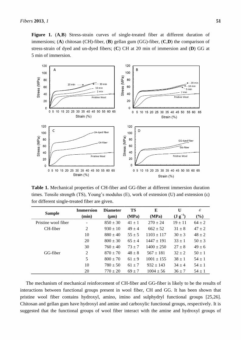
Figure 1. (A,B) Stress-strain curves of single-treated fiber at different duration of
immersions; (A) chitosan (CH)-fiber, (B) gellan gum (GG)-fiber, (C,D) the comparison of
stress-strain of dyed and un-dyed fibers; (C) CH at 20 min of immersion and (D) GG at
5 min of immersion.
Table 1. Mechanical properties of CH-fiber and GG-fiber at different immersion duration
times. Tensile strength (TS), Young’s modulus (E), work of extension (U) and extension (ε)
for different single-treated fiber are given.
Sample Immersion
(min)
Diameter
(µm)
TS
(MPa)
E
(MPa)
U
(J g−1
)
(%)
Pristine wool fiber - 850 ± 30 41 ± 1 270 ± 24 19 ± 11 64 ± 2
CH-fiber 2 930 ± 10 49 ± 4 662 ± 52 31 ± 8 47 ± 2
10 880 ± 40 55 ± 5 1103 ± 117 30 ± 3 48 ± 2
20 800 ± 30 65 ± 4 1447 ± 191 33 ± 1 50 ± 3
30 760 ± 40 73 ± 7 1400 ± 250 27 ± 8 49 ± 6
GG-fiber 2 870 ± 70 48 ± 8 567 ± 181 32 ± 2 50 ± 1
5 800 ± 70 61 ± 9 1001 ± 155 38 ± 1 54 ± 1
10 780 ± 50 61 ± 7 932 ± 143 34 ± 4 54 ± 1
20 770 ± 20 69 ± 7 1004 ± 56 36 ± 7 54 ± 1
The mechanism of mechanical reinforcement of CH-fiber and GG-fiber is likely to be the results of
interactions between functional groups present in wool fiber, CH and GG. It has been shown that
pristine wool fiber contains hydroxyl, amino, imino and sulphydryl functional groups [25,26].
Chitosan and gellan gum have hydroxyl and amine and carboxylic functional groups, respectively. It is
suggested that the functional groups of wool fiber interact with the amine and hydroxyl groups of
Fibers 2013, 1 52
chitosan, and with the hydroxyl and carboxyl groups of gellan gum [27]. Figure 1 and Table 1 show
that the reinforcement CH-fiber (20 min immersion, E =1447 ± 191 MPa) is larger than GG-fiber (5 min
immersion, E = 1001 ± 155 MPa). The reinforcement with chitosan has previously been linked to the
van der Waals interaction [28,29]. Optical analysis of the cross-section and fracture topography for the
fibers supports the difference in reinforcement (Figure 2).
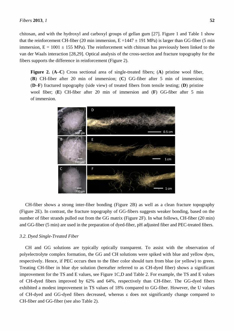
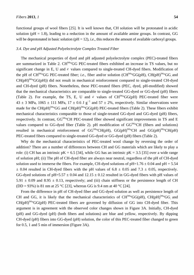
Figure 2. (A–C) Cross sectional area of single-treated fibers; (A) pristine wool fiber,
(B) CH-fiber after 20 min of immersion; (C) GG-fiber after 5 min of immersion;
(D–F) fractured topography (side view) of treated fibers from tensile testing; (D) pristine
wool fiber; (E) CH-fiber after 20 min of immersion and (F) GG-fiber after 5 min
of immersion.
CH-fiber shows a strong inter-fiber bonding (Figure 2B) as well as a clean fracture topography
(Figure 2E). In contrast, the fracture topography of GG-fibers suggests weaker bonding, based on the
number of fiber strands pulled out from the GG matrix (Figure 2F). In what follows, CH-fiber (20 min)
and GG-fiber (5 min) are used in the preparation of dyed-fiber, pH adjusted fiber and PEC-treated fibers.
3.2. Dyed Single-Treated Fiber
CH and GG solutions are typically optically transparent. To assist with the observation of
polyelectrolyte complex formation, the GG and CH solutions were spiked with blue and yellow dyes,
respectively. Hence, if PEC occurs then to the fiber color should turn from blue (or yellow) to green.
Treating CH-fiber in blue dye solution (hereafter referred to as CH-dyed fiber) shows a significant
improvement for the TS and E values, see Figure 1C,D and Table 2. For example, the TS and E values
of CH-dyed fibers improved by 62% and 64%, respectively than CH-fiber. The GG-dyed fibers
exhibited a modest improvement in TS values of 18% compared to GG-fiber. However, the U values
of CH-dyed and GG-dyed fibers decreased, whereas ε does not significantly change compared to
CH-fiber and GG-fiber (see also Table 2).
Fibers 2013, 1 53
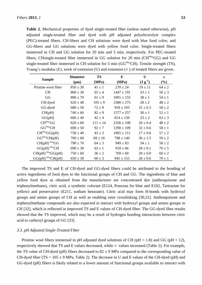
Table 2. Mechanical properties of dyed single-treated fiber (unless stated otherwise), pH
adjusted single-treated fiber and dyed with pH adjusted polyelectrolyte complex
(PEC)-treated fibers. CH-fibers and CH solutions were dyed with blue food color; and
GG-fibers and GG solutions were dyed with yellow food color. Single-treated fibers
immersed in CH and GG solution for 20 min and 5 min, respectively. For PEC-treated
fibers, CHsingle-treated fiber immersed in GG solution for 20 min (CHint
°GG) and GG
single-treated fiber immersed in CH solution for 5 min (GGint
°CH). Tensile strength (TS),
Young’s modulus (E), work of extension (U) and extension ( ) of treated fibers are given.
Sample Diameter
(µm)
TS
(MPa)
E
(MPa)
U
(J g−1
)
ε
(%)
Pristine wool fiber 850 ± 30 41 ± 1 270 ± 24 19 ± 11 64 ± 2
CH 800 ± 30 65 ± 4 1447 ± 191 33 ± 1 50 ± 3
GG 800 ± 70 61 ± 9 1001 ± 155 38 ± 1 54 ± 1
CH-dyed 620 ± 40 105 ± 9 2380 ± 275 28 ± 2 48 ± 2
GG-dyed 680 ± 50 72 ± 9 959 ± 103 31 ± 0.3 58 ± 2
CH(pH) 740 ± 60 82 ± 9 1577 ± 257 30 ± 1 52 ± 1
GG(pH) 660 ± 40 42 ± 4 814 ± 150 23 ± 2 63 ± 3
CHint°GG 620 ± 60 111 ± 14 2356 ± 108 26 ± 0.4 48 ± 2
GGint°CH 690 ± 50 92 ± 7 1290 ± 109 32 ± 0.6 58 ± 1
CHint°GG(pH) 730 ± 40 43 ± 3 1065 ± 111 17 ± 0.6 57 ± 2
GGint°CH(pH) 700 ± 60 69 ± 16 798 ± 140 36 ± 1.5 59 ± 2
CH(pH) int°GG 790 ± 70 64 ± 3 949 ± 83 34 ± 1 58 ± 2
GG(pH) int°CH 690 ± 30 63 ± 1 918 ± 66 28 ± 0.1 70 ± 5
CH(pH) int°GG(pH) 750 ± 60 36 ± 2 769 ± 60 20 ± 0.8 60 ± 2
GG(pH) int°CH(pH) 650 ± 30 60 ± 3 945 ± 155 26 ± 0.6 70 ± 1
The improved TS and E of CH-dyed and GG-dyed fibers could be attributed to the bonding of
active ingredients of food dyes to the functional groups of CH and GG. The ingredients of blue and
yellow food dyes as obtained from the manufacturer are concentrated dye (anthraquinone and
triphenylmethane), citric acid, a synthetic colorant (E124, Ponceau for blue and E102, Tartrazine for
yellow) and preservative (E211, sodium benzoate). Citric acid may form H-bonds with hydroxyl
groups and amino groups of CH as well as enabling ester crosslinking [30,31]. Anthraquinone and
triphenylmethane compounds are also expected to interact with hydroxyl groups and amino groups in
CH [32], which is reflected in improved TS and E values of CH-dyed fiber. The GG-dyed fiber results
showed that the TS improved, which may be a result of hydrogen bonding interactions between citric
acid to carboxyl groups of GG [33].
3.3. pH Adjusted Single-Treated Fiber
Pristine wool fibers immersed in pH adjusted dyed solutions of CH (pH = 1.8) and GG (pH = 12),
respectively showed that TS and E values decreased, while values increased (Table 2). For example,
the TS value of CH-dyed (pH) fibers decreased to 82 ± 9 MPa compared to the corresponding value of
CH-dyed fiber (TS = 105 ± 9 MPa, Table 2). The decrease in U and E values of the CH-dyed (pH) and
GG-dyed (pH) fibers is likely related to a lower amount of functional groups available to interact with
Fibers 2013, 1 54
functional groups of wool fibers [25]. It is well known that, CH solution will be protonated in acidic
solution (pH = 1.8), leading to a reduction in the amount of available amine groups. In contrast, GG
will be deprotonated in basic solution (pH = 12), i.e., this reduces the amount of available carboxyl groups.
3.4. Dye and pH Adjusted Polyelectrolyte Complex Treated Fiber
The mechanical properties of dyed and pH adjusted polyelectrolyte complex (PEC)-treated fibers
are summarized in Table 2. CHint
°GG PEC-treated fibers exhibited an increase in TS values, but no
significant change in E, U and values compared to single-treated CH-dyed fibers. Modification of
the pH of CHint
°GG PEC-treated fiber; i.e., fiber and/or solution (CHint
°GG(pH), CH(pH)int
°GG, and
CH(pH)int
°GG(pH)) did not result in mechanical reinforcement compared to single-treated CH-dyed
and CH-dyed (pH) fibers. Nonetheless, these PEC-treated fibers (PEC, dyed, pH-modified) showed
that the mechanical characteristics are comparable to single-treated GG-dyed or GG-dyed (pH) fibers
(Table 2). For example, the TS, E, U and values of CHint
°GG(pH) PEC-treated fiber were
43 ± 3 MPa, 1065 ± 111 MPa, 17 ± 0.6 J g−1
and 57 ± 2%, respectively. Similar observations were
made for the CH(pH)int
°GG and CH(pH)int
°GG(pH) PEC-treated fibers (Table 2). These fibers exhibit
mechanical characteristics comparable to those of single-treated GG-dyed and GG-dyed (pH) fibers,
respectively. In contrast, GGint
°CH PEC-treated fiber showed significant improvements in TS and E
values compared to GG-dyed fiber (Table 2). pH modification of GGint
°CH (fibers and solutions)
resulted in mechanical reinforcement of GGint
°CH(pH), GG(pH)int
°CH and GG(pH)int
°CH(pH)
PEC-treated fibers compared to single-treated GG-dyed or GG-dyed (pH) fibers (Table 2).
Why do the mechanical characteristics of PEC-treated wool change by reversing the order of
addition? There are a number of differences between CH and GG materials which are likely to play a
role: (i) CH has an intrinsic pK = 6.5 [34], while GG has an intrinsic pK = 3.5 [35] over a wide range
of solution pH; (ii) The pH of CH-dyed fiber are always near neutral, regardless of the pH of CH-dyed
solution used to immerse the fibers. For example, CH-dyed solutions of pH=1.76 ± 0.04 and pH = 5.54
± 0.04 resulted in CH-dyed fibers with the pH values of 6.8 ± 0.05 and 7.3 ± 0.05, respectively.
GG-dyed solutions of pH=5.57 ± 0.04 and 12.15 ± 0.12 resulted in GG-dyed fibers with pH values of
5.91 ± 0.09 and 8.95 ± 0.13, respectively; and (iii) chain stiffness or the persistence length of CH
(DD = 93%) is 81 nm at 25 °C [23], whereas GG is 9.4 nm at 40 °C [24].
From the difference in pH of CH-dyed fiber and GG-dyed solution as well as persistence length of
CH and GG, it is likely that the mechanical characteristics of CHint
°GG(pH), CH(pH)int
°GG, and
CH(pH)int
°GG(pH) PEC-treated fibers are governed by diffusion of GG into CH-dyed fiber. This
argument is in agreement with the observed color changes shown in Figure 3A. Initially, CH-dyed
(pH) and GG-dyed (pH) (both fibers and solutions) are blue and yellow, respectively. By dipping
CH-dyed (pH) fibers into GG-dyed (pH) solution, the color of this PEC-treated fiber changed to green
for 0.5, 1 and 5 min of immersion (Figure 3A).

Fibers 2013, 1 55
Figure 3. (A) Color transformation of CH(pH)int
°GG(pH) PEC-coated fiber at 0.5, 1, 5, 10,
15 and 20 min of immersions and (B) K/S values of red (◊) and green (□) channels as a
function of duration of immersions.
The green color partially changed to yellow after 10 min of immersion and completely changed to
yellow after 20 min immersion. The change in color from green back to yellow may indicate the
build-up of an outer GG layer on top of the PEC layer. These color changes were also followed using
Kubelka-munk analysis [36]. In contrast, CH is unable to penetrate into GG in GGint
°CH,
GGint
°CH(pH) and GG(pH)int
°CH(pH) PEC-treated fibers as shown in Figure 4.
Figure 4. (A) Single-coated fibers GG-dyed fibers before and after pH modification, (B-D)
PEC-coated fibers; (B) GGint
°CH, (C) GGint
°CH(pH) and (D) GG(pH)int
°CH(pH). Those
PEC-coated fibers are covered with a green colour on the outer layer and yellow colour at
core of each fiber.
4. Conclusions
PEC wool fibers were developed by polyelectrolyte complexation of chitosan and gellan gum.
Pristine wool fibers treated with GG and CH displayed their optimum mechanical reinforcement after
immersion of 5 min and 20 min, respectively. Incorporating a dye in the biopolymer solutions resulted

Fibers 2013, 1 56
in improved mechanical properties compared to fibers prepared without dyes. In contrast, preparing
fibers with pH modified dye solutions resulted in a decrease in the mechanical characteristics. PEC
fibers were prepared by dipping biopolymer treated fibers into biopolymer solutions of opposite
charge. The order of addition was found to affect the mechanical characteristics of the PEC fibers. It is
suggested that the difference in observed mechanical characteristics is due to the ability of GG to
diffuse into CH-dyed fiber, and the inability of CH to diffusion in GG-dyed fibers. This paper
contributes to the development of biopolymer reinforced textile materials.
Acknowledgments
This work was supported by the Government of Malaysia (K. A. Mat Amin), University of
Wollongong (URC Small Grant) and Australian Research Council Centre of Excellence and Future
Fellowship programs.
Conflicts of Interest
The authors declare no conflict of interest.
References
1. Hoogeveen, N.G.; Cohen Stuart, M.A.; Fleer, G.J.; Böhmer, M.R. Formation and stability of
multilayers of polyelectrolytes. Langmuir 1996, 12, 3675–3681.
2. Woelki, S.; Kohler, H.-H. Effect of dispersion forces on the potential of charged interfaces. Chem.
Phys. 2004, 306, 209–217.
3. Miller, M.D.; Bruening, M.L. Controlling the nanofiltration properties of multilayer
polyelectrolyte membranes through variation of film composition. Langmuir 2004, 20, 11545–11551.
4. Van Den Beucken, J.J.J.P.; Vos, M.R.J.; Thüne, P.C.; Hayakawa, T.; Fukushima, T.; Okahata, Y.;
et al. Fabrication, characterization, and biological assessment of multilayered DNA-coatings for
biomaterial purposes. Biomaterials 2006, 27, 691–701.
5. Tang, Z.; Wang, Y.; Podsiadlo, P.; Kotov, N.A. Biomedical applications of layer-by-layer
assembly: from biomimetics to tissue engineering. Adv. Mater. 2006, 18, 3203–3224.
6. Jiao, Y.-P.; Cui, F.-Z. Surface modification of polyester biomaterials for tissue engineering.
Biomed. Mater. 2007, 2, doi:10.1088/1748-6041/2/4/R02.
7. Köhler, K.; Sukhorukov, G.B. Heat Treatment of polyelectrolyte multilayer capsules: a versatile
method for encapsulation. Adv. Funct. Mater. 2007, 17, 2053–2061.
8. Bertrand, P.; Jonas, A.; Laschewsky, A.; Legras, R. Ultrathin polymer coatings by complexation
of polyelectrolytes at interfaces: suitable materials, structure and properties. Macromol. Rapid
Comm. 2000, 21, 319–348.
9. Połowiński, S. Deposition of polymer complex layers onto nonwoven textiles. J. Appl. Polym. Sci.
2007, 103, 1700–1705.
10. Stefan, P. Nonwoven fabrics modified with deposited nanolayers. Polimery 2007, 52, 357–361.
Fibers 2013, 1 57
11. Stefan, Połowiński; Stawski, D. Thermogravimetric measurements of poly(propylene) nonwovens
containing deposited layers of polyelectrolytes and colloidal particles of noble metals. Fibres Text.
East. Eur. 2007, 15, 82–85.
12. Dubas, S.T.; Kumlangdudsana, P.; Potiyaraj, P. Layer-by-layer deposition of antimicrobial silver
nanoparticles on textile fibers. Colloids and Surfaces A: Physicochem. Eng. Asp. 2006, 289, 105–109.
13. Agullo, E.; Rodriguez, M.S.; Ramos, V.; Albertengo, L. Present and future role of chitin and
chitosan in food. Macromol. Biosci. 2003, 3, 521–530.
14. Giavasis, I.; Harvey, L.M.; McNeil, B. Gellan Gum. Cr. Rev. Biotechn. 2000, 20, 177–211.
15. Smith, A.M.; Shelton, R.M.; Perrie, Y.; Harris, J.J. An initial evaluation of gellan gum as a
material for tissue engineering applications. J. Biomater. Appl. 2007, 22, 241–254.
16. Amaike, M.; Senoo, Y.; Yamamoto, H. Sphere, honeycomb, regularly spaced droplet and fiber
structures of polyion complexes of chitosan and gellan. Macromol. Rapid Comm. 1998, 19, 287–289.
17. Yamamoto, H.; Horita, C.; Senoo, Y.; Nishida, A.; Ohkawa, K. Polyion complex fiber and
capsule formed by self-assembly of poly-L-Lysine and gellan at solution interfaces. J. Appl.
Polym. Sci. 2001, 79, 437–446.
18. Yamamoto, H.; Ohkawa, K.; Nakamura, E.; Miyamoto, K.; Komai, T. Preparation of polyion
complex capsule and fiber of chitosan and gellan-sulfate at aqueous interface. Bull. Chem. Soc.
Jpn. 2003, 76, 2053–2057.
19. Ohkawa, K.; Kitagawa, T.; Yamamoto, H. Preparation and characterization of chitosan-gellan
hybrid capsules formed by self-assembly at an aqueous solution interface. Macromol. Mater Eng.
2004, 289, 33–40.
20. Meier, C.; Welland, M.E. Wet-Spinning of amyloid protein nanofibers into multifunctional
high-performance biofibers. Biomacromolecules 2011, 12, 3453–3459.
21. Amin, K.A.M.; Panhuis, Mih. Polyelectrolyte complex materials from chitosan and gellan gum.
Carbohyd. Polym. 2011, 86, 352–358.
22. Mat Amin, K.A.; Gilmore, K.J.; Matic, J.; Poon, S.; Walker, M.J.; Wilson, M.R.; et al.
Polyelectrolyte complex materials consisting of antibacterial and cell-supporting layers.
Macromol. Biosci. 2012, 12, 374–382.
23. Zhang, X.; Yang, D.; Nie, J. Chitosan/polyethylene glycol diacrylate films as potential wound
dressing material. Int. J. Biol. Macromol. 2008, 43, 456–462.
24. Baxter, S.; Zivanovic, S.; Weiss, J. Molecular weight and degree of acetylation of high-intensity
ultrasonicated chitosan. Food Hydrocolloid. 2005, 19, 821–830.
25. Kantouch, A.; Heheish, A.; Bendak, A. Ceiv initiated graft polymerization of methyl methacrylate
on wool fibres. Eur. Polym. J. 1971, 7, 153–163.
26. Sun, D.; Stylios, G.K. Fabric surface properties affected by low temperature plasma treatment. J.
Mater. Process. Tech. 2006, 173, 172–177.
27. Cardamone, J.M.; Yao, J.; Nuńez, A. Controlling shrinkage in wool fabrics: effective hydrogen
peroxide systems. Text. Res. J. 2004, 74, 887–898.
28. Strnad, S.; Šauper, O.; Jazbec, A.; Stana-Kleinschek, K. Influence of chemical modification on
sorption and mechanical properties of cotton fibers treated with chitosan. Text. Res. J. 2008, 78,
390–398.

Fibers 2013, 1 58
29. Lim, S.-H.; Hudson, S.H. Application of a fibre-reactive chitosan derivative to cotton fabric as a
zero-salt dyeing auxiliary. Color. Technol. 2004, 120, 108–113.
30. Chung, Y.-S.; Lee, K.-K.; Kim, J.-W. Durable Press and Antimicrobial Finishing of Cotton
Fabrics with a Citric Acid and Chitosan Treatment. Text. Res. J. 1998, 68, 772–775.
31. Shu, X.Z.; Zhu, K.J.; Song, W. Novel pH-sensitive citrate cross-linked chitosan film for drug
controlled release. Int. J. Pharm. 2001, 212, 19–28.
32. Verma, P.; Baldrian, P.; Nerud, F. Decolorization of structurally different synthetic dyes using
cobalt(II)/ascorbic acid/hydrogen peroxide system. Chemosphere 2003, 50, 975–979.
33. Marshall, W.E.; Wartelle, L.H.; Boler, D.E.; Johns, M.M.; Toles, C.A. Enhanced metal adsorption
by soybean hulls modified with citric acid. Bioresource Technol. 1999, 69, 263–268.
34. Liu, W.; Sun, S.; Cao, Z.; Zhang, X.; Yao, K.; Lu, W.W.; et al. An investigation on the
physicochemical properties of chitosan/DNA polyelectrolyte complexes. Biomaterials 2005, 26,
2705–2711.
35. Sworn, G.; Sanderson, G.R.; Gibson, W. Gellan gum fluid gels. Food Hydrocolloid. 1995, 9,
265–271.
36. Barron, V.; Torrent, J. Use of the Kubelka—Munk theory to study the influence of iron oxides on
soil colour. J. Soil Sci. 1986, 37, 499–510.
© 2013 by the authors; licensee MDPI, Basel, Switzerland. This article is an open access article
distributed under the terms and conditions of the Creative Commons Attribution license
(http://creativecommons.org/licenses/by/3.0/).